Robert E. Bristow
In the United States, cancer of the uterine cervix is the sixth most common solid cancer in women after carcinoma of the breast, lung, colorectum, endometrium, and ovary. The American Cancer Society has estimated that in 2002 there will be 13,000 new cases of invasive carcinoma of the cervix in the United States, over 50,000 cases of carcinoma in situ, and 4,100 deaths from the disease. The average age of diagnosis for cervical cancer is 52 years, and the distribution of cases is bimodal with peaks at 35 to 39 years and 60 to 64 years. Worldwide, cervical cancer continues to be a leading cause of death due to cancer among women, with approximately 500,000 cases occurring annually. Lifetime risks for cervical cancer show significant geographic variation, ranging from 0.4% in Israel to 5.3% in Cali, Colombia, where cervical cancer is the most common malignancy in women.
RISK FACTORS
Screening
Cytologic evaluation of cells obtained from the cervix and vagina was first proposed by Papanicolaou and Traut in the 1940s as a method for detecting cervical cancer and its precursors. Since that time, cervical cytology has proved to be the most efficacious and cost-effective method for cancer screening. By increasing detection of preinvasive and early invasive disease, cervical cancer screening with the Pap smear has decreased both the incidence and mortality from cervical cancer in communities with active screening programs. A single negative Pap smear may decrease the risk for cervical cancer by 45%, and nine negative smears during a lifetime decreases the risk by as much as 99%. Eddy, using a mathematical model, indicated that in women 35 to 64 years of age, screening intervals of 10, 5, and 3 years would reduce the incidence of invasive cervical cancer by 64%, 84%, and 91%, respectively. Despite the recognized benefits of cytologic screening, substantial subgroups of women in the United States have not been screened or are not screened at regular intervals. One-half of women with newly diagnosed invasive cervical carcinoma have never had a Pap smear, and another 10% have not had a Pap smear in the 5 years preceding diagnosis. The absence of prior regular Pap smear screening is associated with a two- to six-fold increase in the risk of developing cervical cancer. Unscreened populations include older women, the uninsured, ethnic minorities, and women of lower socioeconomic status, particularly those in rural areas. Women over age 65 years should continue to be screened, as 25% of all cases of cervical cancer and 41% of deaths from the disease occur in women in this age group.
Race
Although the incidence of cervical cancer in the United States has declined significantly over the past 50 years, the rates among African Americans remain about twice as high as those among whites. The incidence is also approximately two times higher for Hispanic Americans and even higher for Native Americans, while most Asian-American groups experience rates similar to whites. These differences are at least partially accounted for by the strong inverse association between cervical cancer incidence and socioeconomic factors. When socioeconomic differences are controlled for, the excess risk of cervical cancer among African Americans is substantially reduced, from over 70% to less than 30%. Racial differences are also apparent in survival, with 59% of African Americans with cervical cancer surviving 5 years, compared to 67% of whites with the disease.
Sexual and Reproductive Factors
First intercourse before 16 years of age is associated with a two-fold increased risk of cervical cancer compared with that for women whose first intercourse occurred after age 20 years. Cervical cancer risk is also directly proportional to the number of lifetime sexual partners. Although difficult to separate epidemiologically, there is evidence to indicate that both early age at first coitus and the number of lifetime sexual partners have independent effects on cervical cancer risk. Increasing parity also appears to be a separate risk factor for cervical cancer, even after controlling for socioeconomic and reproductive characteristics. There is little evidence to support an association between age of menarche, age at menopause, or character of menses with carcinoma of the cervix.
Smoking
Cigarette smoking has emerged as an important etiologic factor in squamous cell carcinoma (SCC) of the cervix. The increased risk for smokers is approximately two-fold, with the highest risk observed for long-term or high-intensity smokers. Proposed mechanisms include genotoxic or immunosuppressive effects of smoke-derived nicotine and cotinine, which can be detected in high levels in the cervical mucus of smokers.
Contraceptive Use
Numerous confounding factors—time interval since last cervical smear, sexual behavior, and role of the male partner, among others—complicate the interpretation of the data on oral contraceptive use and cervical cancer. After controlling for these and other variables, it appears that long-term oral contraceptive users (5 years or more) have about a two-fold increased risk of cervical cancer, compared to nonusers. Use of barrier methods of contraception, especially those that combine both mechanical and chemical protection, have been shown to lower the risk of cervical cancer, presumably because of reduced exposure to infectious agents.
Immunosuppression
Cell-mediated immunity appears to be a factor in the development of cervical cancer. Immunocompromised women (e.g., from renal transplantation or human immunodeficiency virus [HIV] infection) may not only be at higher risk for the disease but also demonstrate more rapid progression from preinvasive to invasive lesions and an accelerated course once invasive disease has been diagnosed. Maiman and colleagues report that HIV-positive women with cervical cancer may have a higher recurrence risk and cancer-related death rate compared to HIV-negative control subjects.
Human Papillomavirus Infection
During the past decade, epidemiologic evidence has accumulated, implicating infection with human papillomavirus (HPV) as a likely etiologic agent in cervical SCC. All known types of HPV have a similar structural and genomic organization. They are non-enveloped virions with a double-stranded circular DNA genome of 7,800 to 7,900 base pairs and an icosahedral capsid. HPV DNA is present in virtually all cases (93%) of cervical cancer and its precursor lesions. While HPV infection is thought to be a component of neoplastic transformation, it is unlikely to be entirely sufficient in and of itself.
HPV colonizes mucosal or cutaneous epithelium and may induce hyperproliferation, resulting in the formation of warts at the site of infection. Based on differences in DNA sequencing, over 70 different types of HPV have been identified, 23 of which are known to infect the anogenital tract. Low oncogenic risk–type viruses include types 6, 11, 42, 43, and 44 and are associated with condyloma acuminatum and some cases of low-grade squamous intraepithelial lesions, but rarely with invasive cancer. High oncogenic risk–type viruses include types 16, 18, 31, 45, and 56 and are commonly detected in women with high-grade squamous intraepithelial lesions (HGSIL) and invasive cancer. HPV types 33, 35, 39, 51, and 52 can be considered as being of intermediate oncogenic risk, as they are associated with HGSIL but are uncommonly detected in invasive carcinomas.
Following acute HPV infection, three clinical sequelae are possible:
1. Latent viral infection occurs when the HPV genome becomes stabilized as a nonintegrated episome and remains in the host cell without clinical or morphologic changes in the squamous epithelium. Clinically, patients may display no gross or morphologic evidence of infection but still harbor virus as demonstrated by DNA detection techniques.
2. Active infection, manifested by proliferation of squamous epithelium into benign tumors (warts), is present when HPV undergoes vegetative replication.
3. Highly oncogenic HPV viruses associated with high-grade lesions can become integrated into the host genome, interrupting control of proliferation by certain oncoproteins.
Initiation of cervical dysplasia and carcinoma may involve interactions between HPV and specific genes that regulate cell growth. The E6 and E7 open reading frames of the HPV genome are particularly important in the immortalization and transformation of infected cells. In HPV types 16 and 18, the protein products synthesized from the E6 and E7 open reading frames can bind to the gene products of the p53 and retinoblastoma (Rb) tumor suppressor genes, respectively. Viral integration results in the overexpression of the E6 and E7 viral protein products with increased binding and inactivation of their respective tumor suppressor proteins. Removal of these two inhibitory influences on cellular proliferation is thought to provide HPV-infected cells with a growth advantage, ultimately leading to neoplastic transformation.
CLINICAL FEATURES
Presenting Symptoms
The most common symptom of cervical cancer is abnormal vaginal bleeding or discharge. Abnormal bleeding may take the form of postcoital spotting, intermenstrual bleeding, menorrhagia, or postmenopausal spotting. If bleeding has been chronic, a patient may complain of fatigue or other symptoms related to anemia. Serosanguineous or yellowish vaginal discharge, frequently associated with a foul odor, may accompany an advanced or necrotic carcinoma. Pelvic pain may result from locally advanced disease or tumor necrosis. Tumor extension to the pelvic side wall may cause sciatic pain or back pain associated with urinary tract obstruction and hydronephrosis. Metastatic tumor to the iliac and paraaortic lymph nodes can extend into the lumbosacral nerve roots and present as lumbosacral back pain. Urinary or rectal symptoms (e.g., hematuria, hematochezia, fistulae) can be associated with bladder or rectal invasion by advanced-stage cervical carcinoma.
Physical Findings
Invasive carcinoma of the cervix displays a wide range of gross appearances. Early lesions may be focally indurated or ulcerated, or present as a slightly elevated and granular area that bleeds readily on contact (Fig. 52.1). More advanced tumors have several types of gross appearance: exophytic, endophytic, or infiltrative. Exophytic tumors characteristically have a polypoid or papillary appearance and will often demonstrate contact bleeding. Endophytic tumors are usually ulcerated or nodular but may be clinically inapparent if located high within the endocervical canal. Such tumors frequently invade deep into the cervical stroma to produce an enlarged, hard, barrel-shaped cervix that may only be appreciated on rectovaginal pelvic examination. The infiltrative pattern of tumor growth may produce surrounding tissue necrosis and erosion of normal anatomic landmarks.
|
|
|
FIG. 52.1. Gross appearance of squamous cell carcinoma of the cervix. This is an ulcerative type of lesion. |
Spread of Disease
Parametrial Extension
Cervical cancer generally follows an orderly pattern of disease progression. Initially, tumor cells usually spread through parametrial lymphatic vessels, expanding and replacing parametrial lymph nodes (Fig. 52.2). These individual tumor masses enlarge and become confluent, eventually replacing the normal parametrial tissue. Less commonly, the central tumor mass reaches the pelvic sidewall by direct contiguous extension through the cardinal (Mackenrodt) ligament. Significant involvement of the medial portion of this ligament may result in ureteral obstruction and hydronephrosis.
|
|
|
FIG. 52.2. Anatomic pathways of spread in invasive cervical carcinoma. |
Lymph Node Involvement
The pelvic lymph nodes will usually be the first site of embolic lymphatic spread of cervical cancer. The lymph node groups most commonly involved are the obturator, external iliac, and hypogastric (Fig. 52.2). The inferior gluteal and presacral lymph nodes are less frequently involved. Secondary nodal involvement (i.e., common iliac, paraaortic) rarely occurs in the absence of pelvic nodal disease. The percentage of involved lymph nodes increases directly with primary tumor volume. Rarely, retrograde lymphatic embolization may occur to the inguinal lymph nodes. Patients with locally advanced pelvic disease may have detectable metastatic spread to the scalene nodes. Consequently, careful clinical assessment of the groin and supraclavicular fossa areas should be included as part of the physical examination.
Vaginal Extension
When the primary tumor has extended beyond the confines of the cervix, the upper vagina is frequently involved (50% of cases). Anterior extension through the vesicovaginal septum is most common, often obliterating the dissection plane between the bladder and underlying cervical tumor, making surgical therapy difficult or impossible. Posteriorly, a deep peritoneal cul-de-sac (pouch of Douglas) can represent an anatomic barrier to direct tumor spread from the cervix and vagina to the rectum.
Bladder and Rectal Involvement
In the absence of lateral parametrial disease, anterior and posterior spread of cervical cancer to the bladder and rectum is uncommon. Approximately 20% of patients with tumor extending to the pelvic sidewall will have biopsy-proven bladder invasion.
Endometrial Involvement
The endometrium is involved in 2% to 10% of cervical cancer cases treated with surgery, although the overall incidence (including nonsurgical cases) is unknown. Although endometrial extension does not alter a patient's stage of disease, it has been associated with decreased survival and a higher incidence of distant metastases.
Ovarian Metastasis
Ovarian involvement with cervical cancer is rare but, when present, most likely occurs through the lymphatic connections between the uterus and adnexal structures. In patients undergoing surgical treatment for early-stage disease, ovarian metastasis is present in less than 1% of SCCs and slightly more than 1% of adenocarcinomas. The true incidence of ovarian involvement with advanced-stage tumors is unknown, as pathologic evaluation of the adnexae is not commonly performed in such cases.
Hematogenous Spread
Hematogenous spread of cervical cancer is uncommon, particularly at the time of initial diagnosis. However, when blood-borne metastases do occur, the lung, liver, and bone are the sites most frequently involved. Metastasis to the bowel, adrenal gland, spleen, and brain are extremely rare.
DIAGNOSIS AND STAGING
Diagnosis and Evaluation of Disease Extent
Pathologic documentation of invasive disease should always be obtained prior to initiating therapy for cervical cancer. The International Federation of Gynecology and Obstetrics (FIGO) staging system used for the diagnosis and evaluation of cervical carcinoma is based on clinical evaluation (visual inspection, palpation, colposcopy, endocervical curettage, cervical biopsy, or conization), radiographic examination of the chest, kidneys, and skeleton, and ECC and biopsies. Cervical cancer is one of the few gynecologic malignancies (vaginal cancer being the other) that still uses a clinically based staging system by international convention, which facilitates comparison of results between institutions irrespective of the availability of diagnostic technological resources. Staging procedures allowed by FIGO convention are shown in Table 52.1. Lymphangiograms, arteriograms, computed tomographic (CT) scans, magnetic resonance imaging (MRI), and laparoscopy or laparotomy should not be used for clinical staging. However, information from such additional studies and procedures may be used to modify the treatment approach.
|
|
|
TABLE 52.1. Staging procedures for cervical cancer |
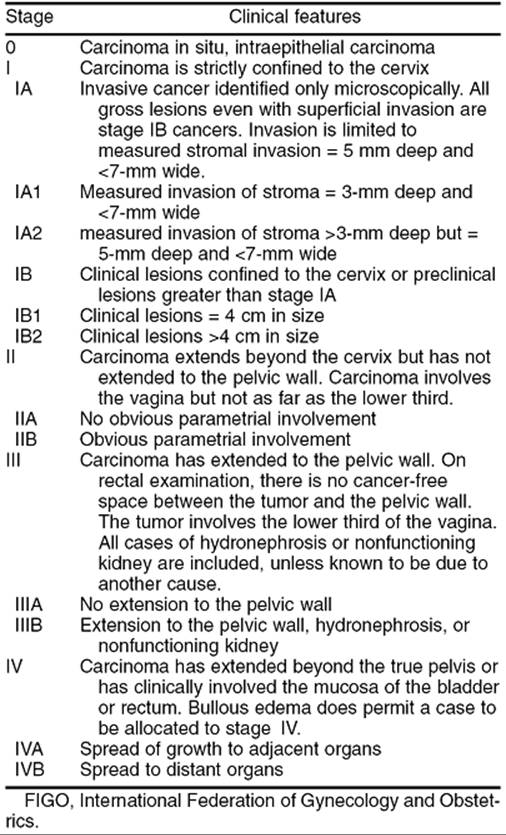
In 1995, FIGO revised the clinical staging system of cervical carcinoma (Table 52.2). Stage I disease includes those neoplasms that are clinically confined to the cervix. In the current staging classification, stage IA tumors are those that are diagnosed by microscopic evaluation of a conization specimen (microinvasive carcinoma). Stage IA1 is defined as a tumor with stromal invasion no greater than 3 mm in depth beneath the basement membrane and no wider than 7 mm. This definition reflects data indicating that patients with less than 3 mm of invasion are at very low risk of metastatic disease and may be treated more conservatively. All grossly visible lesions should technically be classified as stage IB. The 1995 staging system divides stage IB lesions into stage IB1 (no greater than 4 cm in maximal diameter) and stage IB2 (greater than 4 cm in maximal diameter), reflecting the prognostic importance of tumor volume for macroscopic lesions limited to the cervix.
|
|
|
TABLE 52.2. FIGO staging of cervical carcinoma |
Determination of clinical stage depends on careful inspection and palpation of the cervix, vagina, and pelvis. Examination under anesthesia is recommended. It is important to palpate the entire vagina to determine whether disease is limited to the cervix (IB), extends to the upper two-thirds of the vagina (IIA), or also involves the lower third of the vagina (IIIA) (Fig. 52.3). Tumor extension into the parametrial tissue (IIB) or to the pelvic sidewall (IIIB) is best appreciated on rectovaginal examination. It is impossible at clinical examination to decide whether a smooth and indurated parametrium is truly cancerous or only inflammatory, and the case should be considered stage III only if the parametrium is nodular on the pelvic wall or if the growth itself extends to the pelvic wall. Biopsy-proven invasion of bladder or rectal mucosa by cystoscopy or proctoscopy is required for a diagnosis of stage IVA disease. In the United States, the distribution of patients by clinical stage is stage I, 38%; stage II, 32%; stage III, 26%; and stage IV, 4%.
|
|
|
FIG. 52.3. Clinical stages of carcinoma of the cervix. In stage I, only the cervix is involved. In stage II, the parametrium or upper two-thirds of the vagina is involved. In stage III, the malignancy extends to the pelvic sidewall or involves the lower third of the vagina. Stage IV reflects involvement of the bladder or rectal mucosa (stage IVA) or distant metastasis (stage IVB). |
When there is doubt concerning which stage a tumor should be assigned, the earlier stage is mandatory. Once a clinical stage has been determined and treatment initiated, subsequent findings on either extended clinical staging (CT, etc.) or surgical exploration should not alter the assigned stage. Assignment of a more advanced stage during treatment will result in an apparent but deceptive improvement in the results of treatment for earlier stage disease.
Surgical Staging
The current FIGO staging classification for cervical cancer is based on pretreatment clinical findings. Only the subclassification of stage I (IA1, IA2) requires pathologic assessment. Discrepancies between clinical staging and surgicopathologic findings range from 17.3% to 38.5% in patients with clinical stage I disease, to 42.9% to 89.5% in patients with stage III disease. This has led some authors to emphasize surgical staging of cervical carcinoma to identify occult tumor spread and determine the presence of extrapelvic disease so that adjunctive or extended-field radiation therapy may be offered. Transperitoneal surgical staging procedures (e.g. laparotomy), when followed by abdominopelvic irradiation, are associated with appreciable complications, particularly enteric morbidity. The extraperitoneal surgical approach can be performed through a paraumbilical or paramedian incision and allows accurate assessment of pelvic and paraaortic nodal status. Using this approach, an incision in the peritoneum is avoided and adhesion formation is minimized. It is associated with few complications and does not delay institution of radiation therapy. Laparoscopic surgical staging for cervical cancer has also been advocated; however, the safety and efficacy of this approach has yet to be determined.
PROGNOSTIC VARIABLES
Tumor Characteristics
Clinical stage of disease at the time of presentation is the most important determinant of subsequent survival, regardless of treatment modality. Five-year survival declines as FIGO stage at diagnosis increases: stage IA, 97%; stage IB, 70% to 85%; stage II, 60% to 70%; stage III, 30% to 45%; stage IV, 12% to 18%. Prognostic variables directly related to surgicopathologic tumor characteristics and their effect on survival were compiled by Kosary for the National Cancer Institutes Surveillance, Epidemiology, and End Results (SEER) program for the period of 1973 through 1987. This study included 17,119 cases of invasive cervical cancer and found that FIGO stage, tumor histology, histologic grade, and lymph node status are all independent prognostic variables relating to survival.
SCCs accounted for 74.9% of cases in the SEER database and can be categorized according to the degree of histologic tumor differentiation. Well-differentiated tumors account for about 5% of cervical SCCs and are composed of sheets and cords of cells with abundant acidophilic cytoplasm, clearly visible intercellular bridges, and often production of variable amounts of keratin. Moderately differentiated tumors are the most common variety, with 85% of SCCs falling in this category (Fig. 52.4). These tumors are characterized microscopically by masses and cords of spindle-shaped squamous cells with elongated nuclei and scant cytoplasm and more frequent mitoses. Poorly differentiated tumors have a rapid growth rate, with numerous mitoses and cells with closely crowded nuclei and scant cytoplasm. These tumors are difficult to recognize as having originated in squamous cells and constitute approximately 10% of cervical SCCs. The degree of histologic tumor differentiation does correlate with overall survival. In the SEER database, 5-year survival for patients with well-differentiated tumors was 74.5%; for those with moderately differentiated tumors it was 63.7%, and it fell to 51.4% for those patients with poorly differentiated carcinomas. Approximately 15% to 20% of cervical cancer cases are adenocarcinomas. After controlling for known prognostic variables, the SEER study found no difference in overall survival between patients with cervical SCC and adenocarcinoma. However, adenosquamous histology was associated with decreased survival. Survival is also correlated with depth of tumor invasion into the cervical stroma and overall tumor volume.
|
|
|
FIG. 52.4. Moderately differentiated squamous cell carcinoma of the cervix. |
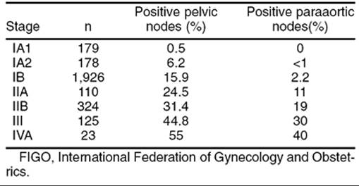
Among surgically treated patients, survival is directly related to the number and location of lymph node metastasis. The frequency of positive lymph nodes increases with the stage of disease (Table 52.3). For all stages of disease, when both pelvic and paraaortic lymph nodes are negative, the 5-year survival rate is 75.2%. Survival decreases to 45.6% with positive pelvic nodes, and the risk of recurrence is related to the number of nodes involved. The recurrence rate is 35% with one positive pelvic lymph node, 59% with two or three positive nodes, and 69% with metastases to more than three pelvic lymph nodes. When paraaortic nodes are involved, the 5-year survival rate ranges from 15% to 45%.
|
|
|
TABLE 52.3. Incidence of pelvic and paraaortic lymph node metastasis by FIGO stage of cervicalcarcinoma |
Host Factors
Whether or not patient age at diagnosis of cervical cancer is a significant and independent predictor of clinical outcome remains controversial. Some investigators have observed decreased survival in women younger than 35 to 40 years, who have a greater frequency of poorly differentiated tumors. Others have found no significant difference in survival between younger and older patients.
Several hematologic parameters have been associated with cervical cancer survival outcome. The incidence of pretreatment anemia (hemoglobin of 12 g/dL or less) increases with advancing stage of disease, occurring in 25%, 33%, and 45% of patients with stages I, II, and III disease, respectively. Anemia is associated with a higher incidence of pelvic recurrences and decreased survival, primarily due to more frequent radiation therapy failures. Tumor hypoxia is the proposed mechanism of radio resistance in the presence of anemia. Another prognostic hematologic parameter is thrombocytosis (>400,000/mm3), which has been associated with decreased survival after controlling for cell type, stage, and age.
Coexistent medical conditions may also affect the success of treatment. Diabetes and hypertension are frequently associated with significant vascular disease and potentially contribute to both tumor hypoxia and decreased blood supply to normal pelvic tissues. Patients with these conditions are subject to a higher incidence of treatment complications and pelvic tumor recurrence, as well as decreased survival.
TREATMENT MODALITIES
Surgery and radiation therapy are the two primary therapeutic modalities most commonly used to treat invasive cervical carcinoma. In general, primary surgical management is limited to patients with stage I and IIA disease, while radiation therapy can be applied to patients with all stages of disease. For patients with early-stage disease, multiple factors should be considered in selecting the most appropriate treatment program. Age is not a contraindication to surgical management, provided the patient does not have significant medical comorbidity. Young patients desiring ovarian preservation and sexually active patients are preferentially managed surgically. Other reasons for the selection of radical surgery over radiation include concomitant inflammatory bowel disease, previous radiation for other disease, and the presence of a coexistent adnexal neoplasm.
Primary Surgery
Surgery provides the opportunity to perform a thorough pelvic and abdominal exploration, which can identify patients with a disparity between the clinical and surgicopathologic stages. Such patients can then be offered an individualized treatment plan based on their precise disease status. The primary surgical management of cervical cancer generally consists of hysterectomy. For patients with stage IA1 lesions who desire fertility preservation, cervical conization with clear surgical margins is acceptable treatment. The safety and efficacy of surgery to preserve fertility (e.g., radical trachelectomy) for patients with larger stage I lesions however has yet to be fully evaluated. There are five distinct variations or types of hysterectomy used in the treatment of cervical cancer.
Types of Hysterectomy
Type I
A type I hysterectomy refers to the standard extrafascial total abdominal hysterectomy. This procedure ensures complete removal of the cervix with minimal disruption to surrounding structures and is appropriate treatment for stage IA1 disease.
Type II
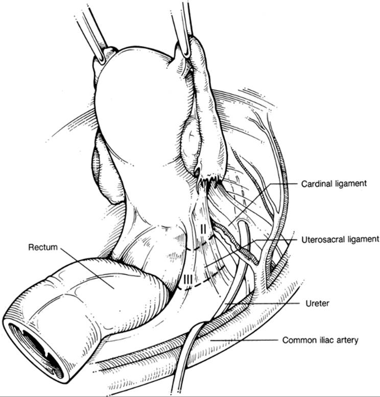
A type II hysterectomy is also referred to as a modified radical hysterectomy (Fig. 52.5, Fig. 52.6 and Fig. 52.7). This procedure involves dissection of the ureters from the parametrial and paracervical tissues down to the ureterovesical junction. This permits removal of all parametrial tissue medial to the ureters, as well as the medial half of the uterosacral ligament and proximal 1 to 2 cm of vagina. This operation is usually performed in conjunction with pelvic lymphadenectomy.
|
|
|
FIG. 52.5. Anatomic dissection of radical hysterectomy. The cardinal ligaments are transected (dashed line) at the level of the ureter (type II) or at the pelvic side wall (type III). |
|
|
|
FIG. 52.6. Anatomic dissection of radical hysterectomy. The uterosacral ligaments are divided at the sacrum (type III) or midway between the sacrum and the uterus (type II). |
|
|
|
FIG. 52.7. Anatomic dissection of radical hysterectomy. In a type II radical hysterectomy, the upper 1 to 2 cm of vagina is excised. In type III radical hysterectomy, the proximal one-third to one-half of the vagina is removed. |
Type III
In a type III or radical abdominal hysterectomy, the ureters are completely dissected from within the paracervical tunnel, and the bladder and rectum are extensively mobilized (Fig. 52.5, Fig. 52.6 and Fig. 52.7). Establishing the paravesical and pararectal spaces facilitates removal of all the parametrial tissue out to the pelvic sidewall, complete resection of the uterosacral ligaments, and excision of the upper one-third to one-half of the vagina. Bilateral pelvic lymphadenectomy is performed with this procedure, removing all lymph-bearing tissue between the mid-common iliac vessels distally to the circumflex iliac vessels and from within the obturator fossa ventral to the obturator nerve.
Type IV/Type V
A type IV or extended radical hysterectomy includes removal of the superior vesical artery, periureteral tissue, and up to three fourths of the vagina. In a type V or partial exenteration operation, the distal ureters and a portion of the bladder are resected. Type IV and type V procedures are rarely performed today because most patients with disease extensive enough to require these operations can be more adequately treated with primary radiation therapy.
Complications of Radical Abdominal Hysterectomy
Modern surgical techniques and anesthesia have reduced the operative mortality associated with radical hysterectomy to 0.6%. Potentially fatal pulmonary embolism occurs in 1% to 2% of patients. Urinary and bowel fistula formation and incisional complications related to surgical treatment tend to occur early in the postoperative period and are usually amenable to surgical repair. Ureterovaginal and vesicovaginal fistulae occur in 2% and 0.9% of patients, respectively. The most commonly observed complication after radical hysterectomy is urinary dysfunction resulting from partial denervation of the detrusor muscle during excision of the paracervical and paravaginal tissue. Radical hysterectomy results in vaginal shortening; however, with sexual activity gradual lengthening will occur. Pelvic lymphocyst formation occurs in 2% to 6.7% of patients following radical hysterectomy and pelvic lymphadenectomy. The incidence is somewhat lower when the retroperitoneal spaces are left open. Most lymphocysts are asymptomatic and do not require intervention; however, lymphocysts may occasionally produce pelvic pain, ureteral obstruction, or partial venous obstruction with thrombosis.
Primary Radiation Therapy
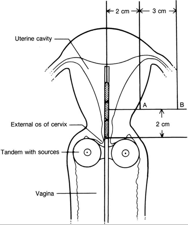
Radiation therapy can be used for all stages of disease and for most patients regardless of age, body habitus, or coexistent medical conditions. The recommended nomenclature for measurement of absorbed radiation dose is the gray (Gy); 1 Gy is equal to one joule (J) of energy absorbed per kilogram of substance. Radiation dose is also commonly expressed as centi-gray (cGy), with 100 cGy equal to 1Gy. By convention, the irradiation dose used in the treatment of cervical cancer is described relative to two anatomic landmarks within the pelvis. Point A is defined as a point 2 cm above the lateral vaginal fornix and 2 cm lateral to the uterine canal corresponding to the paracervical triangle. Point B reflects the dose delivered to the pelvic sidewall and is located 3 cm lateral to point A.
The technical treatment modalities used in modern radiation therapy for cervical cancer consist of a combination of external irradiation and local irradiation. External irradiation is delivered from a source remote from the body (e.g., linear accelerator, cobalt-60) and is used to treat the regional lymph nodes, decrease tumor volume, and reduce the anatomic distortion produced by larger tumor masses. External beam irradiation treatment for cervical cancer is usually delivered using a four-field technique (anterior, posterior, and lateral fields). The precise treatment volume is determined according to individual patient anatomy, but usually measures 15 cm × 15 cm to 18 cm × 18 cm. The pelvic radiation field extends 1 to 2 cm beyond the lateral borders of the bony pelvis and inferiorly beyond the border of the obturator foramen. The cephalad margin may be extended to 18 cm in length to treat the common iliac lymph nodes or even higher if paraaortic lymph node coverage is necessary. A typical external beam radiation treatment portal for cervical cancer is shown in Fig. 52.8.
|
|
|
FIG. 52.8. Whole pelvis radiation treatment field for cervical cancer. In this case, the lower margin of the treatment field extends below the pubic symphysis to provide coverage of the proximal vagina. Lead tapes (white stripes) are used for excluding the corners of a square field, reducing the total irradiated volume by approximately 10%. |
Brachytherapy, refers to a radiation source in direct proximity to the target tissue and may be delivered using a variety of intracavitary applicator devices, but the intrauterine tandem and vaginal colpostats are used most frequently for primary treatment (Fig. 52.9). Once adequate placement is assured, the device is after-loaded with radioactive isotope (e.g., radium-226, cesium-137, iridium-192). Alternatively, vaginal cylinders or interstitial needle implants may be used to deliver local radiation therapy, depending on patient anatomy and tumor distribution.
|
|
|
FIG. 52.9. Diagram of a typical intrauterine tandem and vaginal colpostat brachytherapy placement for cervical cancer showing the anatomic landmarks point A and point B. |
The total dose delivered to point A is determined by the volume of disease to be treated and ranges from 6,500 to 7,000 cGy for small stage IB lesions to 8,500 to 9,000 cGy for bulky stage IIB and stage III lesions. For patients with documented paraaortic node metastasis, or those at high risk, 4,500 cGy of extended field irradiation is delivered to the paraaortic region. Cure rates with irradiation therapy are stage-dependent, with 5-year survival rates averaging 70% to 85% for stage I, 60% for stage II, 45% for stage III, and 18% to 20% for stage IV disease.
Concurrent Chemotherapy and Radiation Therapy
In 1999, five multi-institutional, randomized controlled trials reported a survival advantage associated with the concurrent administration of chemotherapy and radiation therapy in the management of cervical cancer. Although these trials differed in their inclusion criteria, chemotherapy schedules, and prescribed radiation treatment, all demonstrated a similar improvement in progression-free survival (10%–27%) and overall survival (10%–17%). Many centers now administer concurrent treatment with chemotherapy and radiation therapy as standard practice for patients with locally advanced cervical cancer. Cisplatin (Platinol), 5-fluorouracil (Adrucil), and hydroxyurea (Hydrea) have been the chemotherapeutic agents most extensively studied as part of a combined modality treatment program. A commonly used contemporary regimen is cisplatin 40 mg per m2 administered intravenously on a weekly basis during the radiation treatment interval.
Complications of Radiation Therapy
Radiation therapy is associated with both acute and chronic complications. Perforation of the uterus may occur at the time of intracavitary insertion and, if unrecognized, may result in significant blood loss, radiation damage, and peritonitis. Appropriate management consists of removal of the implant and broad-spectrum antibiotic coverage if signs of infection are present. Vaginal fibrosis and stenosis is the most common chronic complication of radiation therapy for cervical cancer and is seen in up to 70% of cases. Ovarian function is lost in virtually all patients undergoing radiation therapy to the pelvis. Proctosigmoiditis occurs in up to 8% of patients undergoing radiation therapy for cervical cancer. Symptoms include abdominal pain, diarrhea, and nausea. An antispasmodic agent, a low-gluten and low-lactose diet, and steroid enemas may be useful; however, severe cases may require hyperalimentation and a diverting colostomy. Hemorrhagic cystitis is seen in approximately 3% of patients undergoing radiation therapy for cervical cancer. In contrast to surgical therapy, fistulous complications associated with radiation therapy tend to occur late and are more difficult to repair secondary to poorly vascularized tissues from radiation fibrosis and vasculitis. Rectovaginal and vesicovaginal fistulae each occur in approximately 1% of cervical cancer patients treated with irradiation. In such cases, biopsy specimens should be obtained from the edge of the fistula to rule out recurrent cancer. Diversion of the fecal (colostomy) or urinary (percutaneous nephrostomy) stream is usually required to allow adequate healing (3–6 months) prior to surgical repair. Two percent of patients experience small bowel obstruction as a consequence of radiation therapy; it is more common in those patients with vascular disease or a history of previous abdominal surgery. The most common site of small bowel obstruction is the terminal ileum, which is relatively fixed within the radiation field by the cecum. Complete small bowel obstruction or cases recalcitrant to conservative management require surgical intervention.
Chemotherapy
Chemotherapy as the sole mode of treating cervical cancer is indicated for patients with extrapelvic metastases (stage IVB) or those with recurrent disease who are not candidates for radiation therapy or exenterative surgery. Cisplatin has been the most extensively studied agent and has demonstrated the most consistent clinical response rates. Complete clinical responses have been observed in 24% of patients, with an additional 16% demonstrating a partial response. Unfortunately, in most series, responses to cisplatin are short-lived (3–6 months). Other agents demonstrating at least partial activity against cervical cancer include carboplatin (Paraplatin), ifosfamide (Ifex), doxorubicin hydrochloride (Adriamycin), vinblastine sulfate (Velban), vincristine sulfate (Oncovin), 5-fluorouracil, methotrexate, and altretamine (Hexalen). There is little objective evidence to suggest that combination chemotherapy is superior to single-agent cisplatin treatment in improving the overall survival of patients with advanced or recurrent cervical cancer.
GENERAL MANAGEMENT BY STAGE
Stage IA1
The 5-year survival rate of these patients approaches 100% with primary surgical therapy. Extrafascial hysterectomy is adequate treatment for this group of patients. Conization may be used selectively if preservation of fertility is desired, provided the surgical margins are free of disease. In the absence of lymph–vascular invasion, the incidence of pelvic lymph node metastasis is 0.3%, and lymphadenectomy is not indicated. In the presence of lymph–vascular involvement, the risk of pelvic node metastasis increases to 2.6%. Pelvic lymphadenectomy and extrafascial hysterectomy should be performed in these cases. In patients who are medically inoperable, stage IA1 carcinoma can be effectively treated with intracavitary radiation.
Stage IA2
Microinvasive carcinoma with stromal invasion of 3.1 to 5.0 mm is associated with positive pelvic lymph nodes in 6.2% of patients. The preferred treatment for these lesions is modified radical (type II) hysterectomy with pelvic lymphadenectomy. Radiation therapy is equally effective from a survival standpoint but may carry a greater risk of post-treatment morbidity compared to modified radical hysterectomy.
Stages IB1, IB2, IIA
Both radical surgery and radiation therapy are equally effective in treating stages IB and IIA carcinoma of the cervix. Numerous uncontrolled studies support the merits of each modality, with no significant differences in pelvic tumor control or overall survival. Zander and colleagues reported on 1,092 patients with stages IB and II cervical cancer treated with radical (type III) hysterectomy and pelvic lymphadenectomy. Five-year survival rates were 84.5% for stage IB and 71.1% for stage II disease. Similar survival rates are obtained with primary radiation therapy. In one series, Perez and associates reported 5-year survival rates of 85% for 312 patients with stage IB disease and 70% for 98 patients with stage IIA disease treated with primary radiation therapy.
Treatment should be individualized for patients with bulky stage I (IB2) tumors. Tumor expansion of the upper endocervix and lower uterine segment can distort cervical anatomy and lead to suboptimal placement of intracavitary radiation sources. Consequently, the central failure rate has been reported as high as 17.5% in patients with cervical lesions greater than 6 cm treated with radiation alone. In such situations, a “completion” extrafascial hysterectomy is usually performed following radiation therapy. While many clinicians limit the use of radical hysterectomy to patients with small stage IB (<3–4 cm) or stage IIA lesions, there is evidence that acceptable survival rates can be obtained with primary surgical treatment in patients with bulky disease confined to the cervix. Five-year survival rates range from 73.6% to 82% after radical hysterectomy and pelvic lymphadenectomy for cervical lesions greater than 4 cm. Survival decreases to 66% at 5 years for lesions greater than 6 cm.
An alternative management plan for patients with bulky local disease is to administer chemotherapy in order to reduce the primary tumor volume prior to attempting radical hysterectomy. This approach has been termed neoadjuvant chemotherapy. Cisplatin, bleomycin sulfate (Blenoxane), and vinblastine has been the most extensively used drug combination. When chemotherapy is administered prior to surgery, complete clinical response rates range from 17% to 44%, with overall response rates of 80% to 90%. In addition to increasing surgical resectability, preoperative chemotherapy also decreases the number of positive pelvic lymph nodes and, in some studies, has seemingly improved 2- and 3-year survival rates.
Adjuvant Therapy Following Surgery
Data are limited concerning the efficacy of postoperative pelvic irradiation in patients at high risk of recurrence after radical hysterectomy and pelvic lymphadenectomy.
High-risk prognostic factors include positive pelvic lymph nodes, microscopic parametrial invasion, pelvic lymph node metastases, deep cervical invasion, and positive or close surgical margins. Sedlis and co-workers report results of a randomized, prospective trial of the Gynecologic Oncology Group comparing postoperative pelvic radiation therapy versus no further therapy for patients with high-risk stage IB cervical cancer following radical hysterectomy and pelvic lymphadenectomy. High- risk factors included large tumor diameter, deep stromal invasion, and the presence of tumor in capillary lymphatic spaces. For patients with these risk factors and negative pelvic lymph nodes, adjuvant pelvic radiation was associated with a statistically significant 47% reduction in the risk of disease recurrence. Survival analysis for this study however awaits additional data maturation.
Grossly Positive Lymph Nodes Encountered at Radical Hysterectomy
The management of patients with grossly positive lymph nodes encountered at the time of radical hysterectomy has been controversial. Although there are no controlled studies, radiation therapy is commonly administered in such circumstances. In order to minimize postoperative radiation-related complications and preserve cervical anatomy for brachytherapy radiation placement, some clinicians advocate abandoning the surgical procedure once metastatic disease is confirmed by intraoperative histologic frozen section. Conversely, retrospective data suggest that postoperative irradiation after resection of all gross nodal disease and radical hysterectomy is associated with a lower rate of local recurrence and may provide a modest gain in survival, particularly for patients with three or more positive pelvic lymph nodes. Hacker and colleagues report their experience with patients undergoing complete resection of grossly positive lymph nodes in conjunction with radical hysterectomy and postoperative radiation therapy. The 5-year survival rates were 80% for patients with positive pelvic nodes and 48% for patients with positive paraaortic nodes; however, serious morbidity occurred in 18% of patients in this series. In addition, lower-extremity lymphedema has been reported in up to 23.4% of patients receiving combined modality therapy.
Stages IIB, III, IVA, and IVB
Radiation therapy is the treatment of choice for patients with stage IIB and more advanced disease. Radiation therapy for invasive cervical cancer is given as a combination of external and intracavitary treatments as described earlier. Long-term survival rates are approximately 60% for stage II, 45% for stage III, and 18% for stage IV disease. Patients with stage IVB disease are usually treated with chemotherapy alone or chemotherapy in combination with local irradiation for palliation of symptoms. These patients have a uniformly poor prognosis regardless of treatment modality.
Post-treatment Surveillance
Among patients with recurrent cervical cancer, recurrence is detected within 1 year in 50% of patients and within 2 years in more than 80%. Pelvic examination and lymph node evaluation, including supraclavicular nodes, should be performed every 3 months for 2 years and then every 6 months for an additional 3 years. As many as 70% of patients with recurrent cervical cancer in the pelvis will have abnormal cervical or vaginal cytology; therefore, appropriate cytologic smears should be obtained at the time of each routine examination. Any palpable pelvic mass should be evaluated by CT with fine-needle aspiration cytology if possible. A chest x-ray should be obtained annually to detect pulmonary metastases.
TREATMENT OF RECURRENT CERVICAL CANCER
General Considerations
Cervical cancer detected within the first 6 months after primary therapy is often termed persistent cancer, while that diagnosed later is referred to as recurrent disease. Appropriate treatment of recurrent cervical cancer is dictated by both the site of recurrence and the modality of primary therapy. In general, patients in whom locally recurrent disease develops following primary surgery should be considered for salvage radiation therapy. Conversely, surgical treatment should be considered for those patients with recurrent central disease who initially received irradiation. Distantly metastatic recurrent tumor is not amenable to either modality alone and is an indication for palliative chemotherapy and possibly radiation therapy for local control.
Surgical Treatment of Recurrent Cervical Cancer
Only patients with recurrent tumor confined to the central pelvis are candidates for surgical intervention. Total hysterectomy is inadequate treatment for centrally recurrent cervical cancer. Additionally, when radical hysterectomy is performed following maximum-dose radiation therapy, 20% to 50% of patients will experience ureteral strictures, urinary fistulae, or other serious complications. Therefore, pelvic exenteration is usually the procedure of choice for centrally recurrent cervical cancer.
Prior to exenterative surgery, a thorough investigation should be undertaken to rule out extrapelvic metastases. The clinical triad of unilateral leg edema, sciatic pain, and ureteral obstruction heralds tumor extension to the pelvic sidewall and is a contraindication to surgery. In most series, approximately 25% of patients with recurrent cervical cancer are deemed satisfactory candidates for exenterative surgery.
Anterior exenteration is indicated for treatment of recurrent cervical cancer limited to the cervix, anterior vagina, or bladder. The procedure combines radical cystectomy with radical hysterectomy and vaginectomy. Posterior exenteration combines abdominal perineal resection of the rectum with radical hysterectomy and vaginectomy and is indicated for lesions confined to the posterior fornix and rectovaginal septum. Total pelvic exenteration is most often required for recurrent cervical cancer. The procedure involves the en-bloc excision of the bladder, uterus, rectum, and vagina (Fig. 52.10).
|
|
|
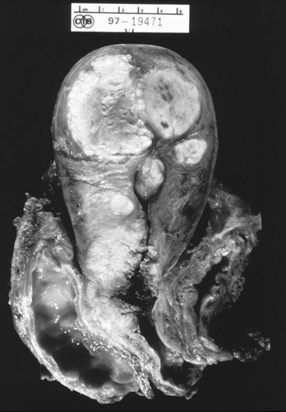
FIG. 52.10. Total pelvic exenteration performed for recurrent cervical cancer involves en-bloc resection of the bladder, vagina, uterus, and rectum. In this specimen, recurrent cervical cancer has replaced the cervix (uterine leiomyomata occupy the uterine fundus). |
Using current surgical stapling devices, low-rectal reanastomosis can be performed in approximately 70% of cases. Reconstruction of the urinary system is accomplished using either an intestinal urinary conduit or one of the many techniques of continent urinary diversion (Miami pouch, Indiana pouch). A neovagina can be created by a variety of techniques using myocutaneous flaps (e.g., bulbocavernosus, gracilis, transverse rectus abdominus) or an omental flap with split-thickness skin graft. Modern surgical techniques and intensive care unit support have reduced the perioperative mortality to less than 7% in recent series. With proper patient selection and sound surgical judgment, 5-year survival rates after pelvic exenteration range from 45% to 61%.
GLANDULAR LESIONS OF THE CERVIX
Adenocarcinoma In Situ
Adenocarcinoma in situ (AIS) is characterized by replacement of the endocervical glandular cells by tall columnar cells with nuclear stratification, hyperchromatism, irregularity, and increased mitotic activity (Fig. 52.11).
About 50% of women with AIS also have coexistent squamous cervical intraepithelial neoplasia (CIN). A diagnosis of AIS may be detected incidentally at the time of conization performed for CIN. Historically, a point of major concern regarding AIS is that these lesions were multifocal, so that conization margins were thought to be unreliable in predicting the presence of residual disease. In one study, Poyner and associates report on 28 patients with AIS in which 4 of 10 patients with negative conization margins had residual AIS in hysterectomy or repeat conization specimens. In contrast, studies using careful histologic sectioning indicate that cervical AIS lesions are usually located within the transformation zone and that true multifocality occurs in fewer than 15% of cases. A retrospective study from Shin and co-workers found only one case of residual or recurrent AIS among 98 patients undergoing conization with negative surgical margins and followed with conservative surveillance. As additional data accumulate, it appears that negative conization margins may be a more reliable indicator of disease clearance than previously thought. For young patients desiring to maintain reproductive capacity, AIS appears to be safely managed by cold knife conization and diligent surveillance. Clear surgical margins however are an absolute prerequisite to conservative management. For patients who have completed their childbearing, a simple hysterectomy should be performed because of the risk of recurrence, even in the presence of negative margins. Patients with unreliable follow-up and those with persistently positive conization margins should be offered simple hysterectomy as definitive therapy.
|
|
|
FIG. 52.11. Cervical adenocarcinoma in situ showing tall columnar cells with nuclear stratification, hyperchromatism, and irregularity. |
Cervical Adenocarcinoma
Adenocarcinoma of the cervix accounts for approximately 10% to 15% of all invasive cervical neoplasms. As with SCC of the cervix, tumor size, depth of invasion, and histologic tumor grade have been identified as predictors of pelvic lymph node metastasis and overall survival. Although cervical adenocarcinoma has been reported to have a worse prognosis than similar stage SCC, this difference is due, at least in part, to the tendency of adenocarcinoma to grow endophytically and establish a large tumor volume prior to clinical detection. When cervical adenocarcinoma and SCC are comparatively matched by patient age, clinical stage, tumor volume, and treatment method, survival outcomes are not significantly different. In general, the same treatment algorithms can be applied to patients with adenocarcinoma of the cervix as to those with cervical SCC. Some authors have advocated performing a “completion” simple hysterectomy for patients with bulky, barrel-shaped adenocarcinomas of the cervix following primary radiation therapy. While this approach may decrease the risk of local recurrence, an associated survival benefit has yet to be conclusively demonstrated.
SMALL CELL CARCINOMA
Small cell carcinoma of the uterine cervix is similar to small cell “neuroendocrine” tumor of the lung and other anatomic locations. These tumors are clinically aggressive, demonstrating a marked propensity to metastasize to local and distant sites. At the time of presentation, disease is often widely disseminated, with bone, brain, and liver being the most common sites. Because of the high metastatic potential of small cell carcinoma, local therapy alone (surgery or radiation) rarely results in long-term survival. Multi-agent chemotherapy, in combination with external-beam and intracavitary radiation therapy, is a therapeutic approach currently under study. The two most commonly used chemotherapeutic regimens are vincristine, doxorubicin, and cyclophosphamide (VAC) and etoposide and platinum (i.e., cisplatin) (EP).
CARCINOMA OF THE CERVICAL STUMP
The natural history and patterns of spread of carcinoma of the cervical stump are similar to those of carcinoma of the intact uterus. The diagnostic evaluation, clinical staging, and principles of staging are also unchanged. In appropriate surgical candidates, early-stage disease can be treated with simple or radical trachelectomy with or without lymphadenectomy, depending on the volume of disease. Advanced-stage disease is treated with radiation therapy. However, the lack of a uterine cavity can make placement of intracavitary radiation sources difficult or impossible. In this case, vaginal colpostats alone or an interstitial needle implant technique can be used in combination with external-beam therapy.
INCIDENTAL CERVICAL CANCER FOUND AT SIMPLE HYSTERECTOMY
Invasive cervical cancer may be incidentally discovered in the surgical specimen after hysterectomy has been performed. For disease more advanced than stage IA1 (without lymph-vascular involvement), simple hysterectomy is inadequate treatment, as the parametria, vaginal cuff, and pelvic lymph nodes may harbor residual tumor. Additional treatment is dictated by the volume of disease and the status of the surgical margins of resection.
Radical surgery following simple hysterectomy for invasive cervical cancer generally includes radical parametrectomy, resection of the cardinal ligaments, excision of the vaginal stump, and pelvic lymphadenectomy. Although it may be technically difficult to perform an adequate radical resection, reoperation should be considered in selected clinical situations, particularly for young patients in whom ovarian preservation is desired. Use of postoperative adjuvant radiation therapy is dictated by surgical and pathologic findings.
Cervical carcinoma at the margins of resection after simple hysterectomy or the presence of gross residual tumor are both absolute indications for radiation therapy. Patients with such findings have a much less favorable prognosis than those without residual tumor and those with comparable disease who have been appropriately staged and treated with radiation alone. Radiation therapy is also well suited for older patients or those who are poor surgical candidates. Five-year survival is 95% to 100% for patients with microscopic disease, while 82% to 84% of those with macroscopic disease and negative surgical margins survive 5 years. If the surgical margins of resection are microscopically involved with carcinoma, 5-year survival ranges from 38% to 87%; survival drops to 20% to 47% for patients with gross residual tumor.
CERVICAL CANCER IN PREGNANCY
Cervical cancer is one of the most common malignancies in pregnancy, with an estimated incidence ranging from 1 in 1,200 to 1 in 2,200 pregnancies (1.6–10.6 cases per 10,000 pregnancies). Conversely, 1 of every 34 women diagnosed with cervical cancer is pregnant at the time of diagnosis. The most common complaint of pregnant patients with cervical cancer is abnormal bleeding. However, confusion between the symptoms of early cervical cancer and those of normal pregnancy frequently leads to a delay in diagnosis. Pathologic confirmation of the presence of invasive cervical cancer should be obtained by directed biopsy in the presence of a grossly visible lesion. Conization is only indicated for those patients with apparent microinvasive disease on directed biopsy or for patients with persistent cytologic evidence of invasive cancer in the absence of a colposcopically visible lesion. Diagnostic conization should only be considered when a diagnosis of invasive cancer will result in a modification of treatment recommendations, timing, or mode of delivery. From an obstetric standpoint, the optimal time to perform conization is between 14 and 20 weeks gestational age or after the time of fetal viability has been reached.
The same clinical staging system of cervical cancer is employed for both nonpregnant and pregnant patients alike (see Table 52.2). For pregnant patients with cervical cancer, the use of MRI may be an appropriate substitute for intravenous pyelogram (IVP) or CT if minimizing fetal exposure to ionizing radiation is desirable. Once the diagnosis of cervical cancer has been established, treatment recommendations are individualized and are dependent on the stage of disease, gestational age at the time of diagnosis, and the desires of the patient regarding continuation of the pregnancy. Patients with well-documented stage IA1 disease (conization with negative surgical margins) may be managed with vaginal delivery and reevaluation postpartum. If the patient has completed childbearing, a simple extrafascial hysterectomy would be appropriate, otherwise close clinical follow-up is required. For patients with stage IA2 disease, a modified radical cesarean hysterectomy with pelvic lymph node dissection is the preferred treatment. Patients with stage IB or IIA disease may be treated with surgery in the form of radical hysterectomy and pelvic lymph node dissection, either in conjunction with cesarean section or with the fetus in situ, depending on the gestational age. Radiation therapy is the treatment of choice for patients with stage IIB to IVA disease or those with stage IB and IIA disease who are not favorable candidates for radical hysterectomy. Radiation therapy may be initiated with the fetus in situ for nonviable pregnancies, with spontaneous abortion occurring 4 to 5 weeks after starting treatment. For more advanced gestations, classic cesarean delivery is performed initially, with radiation therapy commencing 2 to 3 weeks following delivery. The issue of delaying treatment in order to reach a gestational age consistent with fetal viability has been controversial. Although the data are limited and retrospective in nature, it appears that a treatment delay of 6 to 12 weeks is not detrimental for patients with localized (stage I) disease. Treatment delays are not recommended for patients with more advanced disease.
SUMMARY POINTS
· Regular cervical cytologic screening with the Pap smear is the single most effective means of reducing the incidence and mortality of invasive cervical cancer.
· By convention, cervical cancer is a clinically staged disease; however, ancillary tests and surgical staging may provide useful information and facilitate a treatment approach tailored to the true extent of disease.
· Surgery (radical hysterectomy with pelvic lymphadenectomy) and radiation therapy are associated with equivalent survival outcomes for patients with early-stage (I–IIA) cervical cancer.
· Patients with advanced-stage disease (IIB–IVA) should receive radiation therapy with curative intent, preferably in combination with concurrent chemotherapy.
SUGGESTED READINGS
Risk Factors
Eddy GL. Screening for cervical cancer. Ann Intern Med 1990;113:214.
Jemal A, Thomas A, Murray T, et al. Cancer statistics 2002. CA Cancer J Clin 2002;52:23.
Maiman M, Fruchter RG, Guy L, et al. Human immunodeficiency virus infection and invasive cervical carcinoma. Cancer 1993;71:402.
Clinical Features
Delgado G, Bundy BN, Fowler WC, et al. A prospective surgical pathological study of stage I squamous carcinoma of the cervix: a Gynecologic Oncology Group Study. Gynecol Oncol 1989;35:314.
Diagnosis and Staging
Cosin JA, Fowler JM, Chen MD, et al. Pretreatment surgical staging of patients with cervical carcinoma: the case for lymph node debulking. Cancer1998;82:2241.
Lagasse LD, Creasman WT, Shingleton HM, et al. Results and complications of operative staging in cervical cancer: experience of the Gynecologic Oncology Group. Gynecol Oncol 1980;9:90.
Prognostic Variables
Kosary CL. FIGO stage, histology, histologic grade, age and race as prognostic factors in determining survival for cancers of the female gynecologic system: an analysis of 1973–87 SEER cases of cancers of the endometrium, cervix, ovary, vulva, and vagina. Semin Surg Oncol 1994;10:31.
Treatment Modalities
Abu-Rustum NR, Hoskins WJ. Radical abdominal hysterectomy. Surg Clin North Am 2001;81:815.
Keys HM, Bundy BM, Stehman FB, et al. Cisplatin, radiation and adjuvant hysterectomy compared with radiation and adjuvant hysterectomy for bulky stage IB cervical carcinoma. N Engl J Med 1999;340:1154.
Morris M, Eifel PJ, Lu J, et al. Pelvic radiation with concurrent chemotherapy versus pelvic and para-aortic radiation for high-risk cervical cancer. A randomized Radiation Therapy Oncology Group clinical trial. N Engl J Med 1999;340:1137.
Omura GA. Chemotherapy for stage IVB or recurrent cancer of the uterine cervix. J Natl Cancer Inst Monogr 1996;21:123.
Peters WA, Liu PY, Barrett RJ, et al. Cisplatin and 5-fluorouracil plus radiation therapy are superior to radiation therapy as adjunctive in high-risk early-stage carcinoma of the cervix after radical hysterectomy and pelvic lymphadenectomy: report of a phase III intergroup study. Gynecol Oncol 1999;72:443.
Piver MS, Rutledge F, Smith JP. Five classes of extended hysterectomy for women with cervical cancer. Obstet Gynecol 1974;44:265.
Rose PG, Bundy BN, Watkins EB, et al. Concurrent cisplatin-based chemoradiation in locally advanced cervical cancer. N Engl J Med 1999;340:1144.
Whitney CW, Sause W, Bundy BN, et al. A randomized comparison of fluorouracil plus cisplatin versus hydroxyurea as an adjunct to radiation therapy in stages IIB-IVA carcinoma of the cervix with negative para-aortic lymph nodes. A Gynecologic Oncology Group and Southwest Oncology Group Study. J Clin Oncol 1999;17:1339.
General Management by Stage
Alvarez RD, Gelder MS, Gore H, et al. Radical hysterectomy in the treatment of patients with bulky early stage carcinoma of the cervix uteri. Surg Gynecol Obstet 1993;176:539.
Hacker NF, Wain GV, Nicklin JL. Resection of bulky positive lymph nodes in patients with cervical carcinoma. Int J Gynecol Cancer 1995;5:250–256.
Kenter GG, Hellebrekers BWJ, Zwinderman KH, et al. The case for completing the lymphadenectomy when positive lymph nodes are found during radical hysterectomy for cervical carcinoma. Acta Obstet Gynecol Scand 2000;79:72.
Morrow CP, Shigleton HM, Averette HE, et al. Is pelvic irradiation beneficial in the postoperative management of stage IB squamous cell carcinoma of the cervix with pelvic lymph node metastases treated by radical hysterectomy and pelvic lymphadenectomy? A report from the Presidential Panel at the 1979 Annual Meeting of the Society of Gynecologic Oncologists. Gynecol Oncol 1980;10:105.
Perez CA, Camel HM, Kuske RR, et al. Radiation therapy alone in the treatment of carcinoma of the uterine cervix: a 20-year experience. Gynecol Oncol1986;23:127.
Sedlis A, Bundy BN, Rotman MZ, et al. A randomized trial of pelvic radiation therapy versus no further therapy in selected patients with stage IB carcinoma of the cervix after radical hysterectomy and pelvic lymphadenectomy: a Gynecologic Oncology Group Study. Gynecol Oncol 1999;73:177.
Zander J, Baltzer J, Lohe KJ, et al. Carcinoma of the cervix: an attempt to individualize treatment. Results of a 20-year cooperative study. Am J Obstet Gynecol 1981;139:752.
Glandular Lesions of the Cervix
Anton-Culver H, Bloss JD, Bringman D, et al. Comparison of adenocarcinoma and squamous cell carcinoma of the uterine cervix: a population-based epidemiologic study. Am J Obstet Gynecol 1992;166:1507.
Ostor AG, Duncan A, Quinn M, et al. Adenocarcinoma in situ of the uterine cervix: an experience with 100 cases. Gynecol Oncol 2000;79:207.
Poyner EA, Barakat RR, Hoskins WJ. Management and follow-up of patients with adenocarcinoma in situ of the uterine cervix. Gynecol Oncol 1995;57:158.
Shin CH, Schorge JO, Lee KR, et al. Conservative management of adenocarcinoma in situ of the cervix. Gynecol Oncol 2000;79:6.
Cervical Cancer in Pregnancy
van der Vange N, Weverling GJ, Ketting BW, et al. The prognosis of cervical cancer associated with pregnancy: a matched cohort study. Obstet Gynecol1995;85:1022.