David G. Mutch
The uterus and endometrium are unique organs. Almost any type of tissue histology can and does arise from these pluripotent tissues. This chapter will present premalignant and malignant conditions of the uterus. This includes the premalignant condition endometrial hyperplasia, as well as the frankly malignant conditions of the uterus that comprise the following two broad categories: (a) epithelial cancer and (b) mesenchymal tumors. Both of these types of malignancies develop within the lining or the body of the uterus, and this chapter will describe the epidemiology, staging, and treatment of these cancers. The epithelial cancers are primarily comprised of endometrioid, clear cell, and papillary serous type tumors. The mesenchymal tumors are less common and are comprised of three broad categories, (a) mixed müllerian tumors, (b) stromal tumors, and (c) leiomyosarcomas.
ENDOMETRIAL HYPERPLASIA
Classification
Endometrial hyperplasia is an abnormal condition that usually represents an overgrowth of the endometrium. This terminology encompasses a wide variety of conditions. Some of these are clearly benign and some potentially malignant. Unfortunately, there have been so many different classifications over the years that there is significant confusion about the meaning of the term endometrial hyperplasia. For many years, it has been suggested that endometrial hyperplasia reflects the histologic representation of the continuum between normal proliferating endometrium and adenocarcinoma in situ. This theory was based on studies first reported by Gusberg and Kaplan in 1963. In that study, the authors reported that 20% of patients who had a hysterectomy were found to have a coexisting adenocarcinoma and that endometrial cancer developed in almost 12% of the remaining patients, with an average follow-up of 5.3 years. They concluded that the risk of cancer was significantly higher in women with endometrial hyperplasia than those without and that 10 years later the cumulative risk for cancer was approximately 30%.
During the ensuing years, several different classification schemes were developed, which resulted in increased confusion regarding both diagnostic criteria and prognosis of the various subtypes of hyperplasia. In the early 1990s the International Society of Gynecological Pathologists endorsed a classification that used both architectural features (the degree of glandular crowding and complexity) and cytologic features, especially cellular atypia. Simple hyperplasia is defined as abnormally thickened endometrium with histologic evidence of an increased ratio of glands to stroma; the glands are cystically dilated and somewhat irregular with some infolding and budding. Complex hyperplasia represents glandular crowding with even less intervening stroma, and the glands show significant infolding and budding (Fig. 54.1). Atypical hyperplasia refers to either simple or complex architectural patterns, in which the cells lining the glands show loss of polarity, nuclear enlargement with increased nucleus-to-cytoplasm ratio and prominent nucleoli, and irregularly condensed chromatin (Fig. 54.2). Cystic hyperplasia is a benign condition arising from inactive endometrium and is not premalignant.
|
|
|
FIG. 54.1. Complex hyperplasia without atypia. Closely packed glands are connected by morular squamous metaplasia. No cytologic atypia is present. |
|
|
|
FIG. 54.2. Complex hyperplasia with atypia. Closely packed, irregular glands demonstrate cytologic atypia in the form of loss of polarity and rounding up of the nuclei and nucleoli. |
Since the study by Gusberg and Kaplan, additional studies have clarified some of the questions regarding this disorder. Using the new criteria, Kurman and colleagues reported that the risk of progression of endometrial hyperplasia to cancer varied according to the subtype. In this study of 170 patients, untreated endometrial hyperplasia regressed spontaneously in 74% and remained stable for more than 10 years in 18%. The risk of progression to cancer was 1% for patients with simple hyperplasia without atypia, 3% for those with complex hyperplasia without atypia, 8% for those with atypical simple hyperplasia, and 29% for patients with complex atypical adenomatous hyperplasia (Table 54.1).
|
|
|
TABLE 54.1. Natural history of premalignant endometrial conditions |
Clinical Picture and Diagnosis
Endometrial hyperplasia usually becomes apparent with irregular vaginal bleeding. For this reason, this symptom should be evaluated with liberal use of the endometrial biopsy. Endometrial hyperplasia often is associated with a history of unopposed estrogen exposure, either exogenous or endogenous. In premenopausal women it is associated with obesity and anovulation. Therefore, women with known polycystic ovary syndrome or known anovulation are at increased risk for developing this disease, as are obese women. Any abnormal bleeding should be investigated in the postmenopausal woman, but features that would increase the suspicion of an abnormal endometrium are unopposed estrogen exposure from obesity or oral intake.
Cervical cytologic screening techniques are not a reliable means to diagnose endometrial abnormalities and should not be used to screen for endometrial abnormalities. Nonetheless the Pap smear can reveal endometrial abnormalities. Atypical glandular cells of undetermined significance (AGUS) should be evaluated aggressively, because the risk of having underlying endometrial pathology is high. Endometrial biopsy clearly should be included in the evaluation of this abnormal cytology. An endometrial biopsy is reported to be 97% as sensitive as a formal dilation and curettage (D&C). Therefore a D&C to evaluate these abnormalities is usually not necessary and should be used only when an endometrial biopsy cannot be performed, when persistent bleeding is unexplained by the biopsy, or when there is some uterine structural abnormality suspected, such as a polyp.
Management
Therapy for endometrial hyperplasia must be individualized and depends on histologic criteria, predisposing factors, patient age, and desire to maintain fertility. In addition to differences in overall prognosis, the various subtypes of endometrial hyperplasia also respond differently to progestin therapy. Ferenczy and Gelfand reported on the results of progestin therapy in 85 postmenopausal women with endometrial hyperplasia. Patients who had hyperplasia without atypia enjoyed complete reversal of the abnormality following treatment with medroxyprogesterone acetate, 10 to 20 mg daily. Patients with cellular atypia noted only a 50% response rate to the same treatment regimen. In patients with atypical hyperplasia, there was also an increased risk of recurrent hyperplasia or cancer after completion of the progestin therapy when compared with those who had hyperplasia without atypia (50% vs. 6%).
Premenopausal women can be treated with oral contraceptives for 3 months if they have no significant contraindications to their use. Most studies suggest that cancer will develop eventually in about 20% to 30% of patients with complex atypical adenomatous hyperplasia, but some have suggested a risk as high as 82% for those with untreated atypical hyperplasia. The risk of progression appears to be higher for postmenopausal than premenopausal women. The mean duration of progression from endometrial hyperplasia to carcinoma is about 10 years for lesions without atypia and 4 years for atypical lesions. In patients with atypical hyperplasia diagnosed by an endometrial biopsy specimen, a formal D&C should be considered to rule out a coexisting adenocarcinoma. It has been suggested that if gland epithelium is found within the stroma (i.e., stromal invasion) of the curettage specimen, even if the diagnosis is hyperplasia, there is a significant chance that the uterus still contains an endometrial cancer. If a diagnosis of carcinoma in situ is made by endometrial biopsy or D&C, in almost all circumstances a hysterectomy should be performed, because this is not a clearly reproducible diagnosis and actually may represent early invasive cancer or sampling error.
Progestational therapy is very effective in reversing endometrial hyperplasia without atypia. For these patients, either cyclic or continuous therapy is appropriate, using medroxyprogesterone acetate, 10 to 20 mg per day, or megestrol acetate (Megace), 20 to 40 mg per day, for either 14 days each month or daily. Therapy should be continued for 3 months, and then the endometrium should again be sampled to document response. The hyperplasia will revert to normal in 75% to 90% of patients treated with progestins. In patients who desire pregnancy, ovulation induction can be considered using either clomiphene or menotropins (Pergonal). If a patient does not desire pregnancy, oral contraceptive therapy should be considered. In those patients with persistent hyperplasia, definitive surgery should strongly be considered.
Patients with atypical hyperplasia often will opt for a hysterectomy and bilateral salpingo-oophorectomy when made aware of the risk of coexisting adenocarcinoma (approximately 20%) and the malignant potential of these lesions. A truly postmenopausal woman (last menses 2 or more years ago) should be encouraged strongly to undergo hysterectomy, with progestin therapy reserved for patients with severe medical problems that would make them very poor surgical candidates. If the decision is made to treat a patient medically, daily progestin therapy for 3 months is recommended, followed by repeat endometrial sampling. If hyperplasia persists in these patients, a D&C should be performed to rule out a coexisting malignancy, or a hysterectomy should be performed. At the time of hysterectomy, the uterus should be opened intraoperatively, with frozen section if indicated to document the presence and extent of any malignancy so that surgical staging can be performed at the same time, if appropriate.
An interesting, but still experimental, approach is insertion of an intrauterine contraceptive progesterone system (ICPS), which releases continuous therapeutic doses of progesterone or levonorgestrel. Until more experience is gained with the ICPS, it should not be employed outside a research setting. Two preliminary studies reported on the use of danazol (Danocrine) to treat women with adenomatous hyperplasia, and in both studies all women treated were noted to have reversal of the hyperplasia on repeat endometrial sampling. In all premenopausal women, menses resumed within 1 to 2 months following therapy. In the first study, atrophic changes were noted on repeat endometrial sampling even though the patients had normal serum estradiol levels, suggesting a direct effect on the endometrium.
A relatively recently described risk factor for endometrial hyperplasia and carcinoma is the use of tamoxifen citrate (Nolvadex) as an adjuvant treatment for breast cancer. Tamoxifen has been associated with a six-fold to seven-fold risk of developing endometrial cancer and an increased risk of endometrial hyperplasia, polyps, and growth of fibroids. The best method for monitoring women taking tamoxifen is unknown. Obviously, patients should have an annual pelvic examination and Pap smear. Endometrial sampling should be performed in all patients with abnormal uterine bleeding. Because tamoxifen acts as a weak estrogen, it is not unreasonable to consider annual endometrial biopsy or sonographic evaluation of endometrial stripe thickness. This recommendation, however, is not based on prospective studies but rather on an intuitive approach. According to current information, women taking tamoxifen have a greater than 84% chance of developing a thickened endometrium, making this technique relatively insensitive in evaluating the endometrium for pathology. Based on published studies, sonography is useful only for ruling out significant pathology if the endometrial stripe is less than 5 mm in thickness. Therefore, this may not be useful in screening women on tamoxifen.
In summary, the management of endometrial hyperplasia should be individualized, depending on the histologic findings and the patient's age health and reproductive desires. Treatment options include hormone therapy and surgery. Due to the increasing use of tamoxifen, gynecologists should expect to see more women using this medication and should be aware of the risk of both benign and malignant changes of the endometrium and be prepared to evaluate them for these abnormalities.
ENDOMETRIAL CARCINOMA
Incidence and Epidemiology
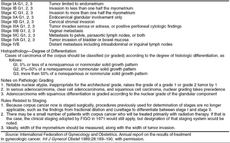
The American Cancer Society estimated that there were approximately 38,000 cases of epithelial endometrial carcinoma in the United States during 2001, accounting for about 7% of all malignancies in women. It ranks seventh in cause of death from cancer in women and accounts for about 6,000–6,500 deaths each year. At this time, endometrial cancer is the fourth most common cancer in women and is the most common gynecologic cancer. Occurring more frequently than endometrial cancer are lung, breast, and colon cancers. Endometrial cancer generally is thought to be a disease of postmenopausal women. However, one fourth of the cases may occur in women who are premenopausal and about 5% occur in women under the age of 40. Generally, the prognosis is good, with an overall survival rate for all cases of about 75%. Management for this disease has evolved over the past half-century. Preoperative radiation therapy followed by hysterectomy was the standard before the early 1980s, but now almost all patients are staged surgically, and postoperative management is tailored to the risk factors identified at the time of surgical staging. In 1988, this primary operative approach was formalized by FIGO (Federation of International Gynecology and Obstetrics) and now requires surgical staging to accurately assign stages and determine appropriate treatment and progress of this disease (Table 54.2).
|
|
|
TABLE 54.2. FIGO staging for carcinoma of the corpus uteri |
According to data from the American Cancer Society, the number of deaths from endometrial cancer was about 4,000 in 1990. In 2000, the mortality from endometrial cancer increased to 6,500. The exact reason for this is increase is unclear. Many suggest that this increased incidence is caused by the increased use of estrogen replacement therapy. Several known pieces of information can be used to argue against this. First, death from estrogen-induced endometrial cancer is rare. These cancers usually are well differentiated and carry a low mortality rate. Also, the rate of endometrial cancer is increasing in countries such as Norway and Czechoslovakia, yet estrogens rarely are prescribed in these countries. The incidence of endometrial carcinoma has increased over the last 50 years, most likely because of the aging population, the increased frequency of certain predisposing conditions such as obesity, and improved methods of diagnosis.
Adenocarcinoma of the endometrium is mainly a malignancy of postmenopausal women and is increasingly virulent with advancing age. Peak age at diagnosis is between 50 and 65 years, and approximately 25% of all cases of endometrial carcinoma are diagnosed in premenopausal women and 5% in women younger than 40 years. Usually, but not always, these young women are either obese, chronically anovulatory, or both. Women with endometrial cancer diagnosed at an early age should be queried about family history. Endometrial cancer is the most commonly inherited gynecologic malignancy. It is the second most common cancer described in hereditary nonpolyposis colon cancer syndrome (HNPCC), also known as the Lynch syndrome.
Bohkman proposed that there may be two types of endometrial cancers, designated type I and type II. Type I endometrial cancer is estrogen dependent and is thought to progress typically from hyperplasia to frank cancer in a stepwise fashion. This type of malignancy typically occurs in younger, perimenopausal women with a history of exposure to unopposed estrogen. These tumors tend to arise in areas of hyperplasia, to be well differentiated, and to be associated with a more favorable prognosis. The latter type of cancer occurs in older women without estrogen stimulation of the endometrium, is not often associated with endometrial hyperplasia, and tends to be more commonly associated with poorly differentiated cancer or those of unusual histologic type. It generally carries a worse prognosis. Because cancer is a genetic disease and we now realize that it develops as the result of an accumulation of mutations in genes necessary for normal cellular function, the pathways necessary for the development of these two types of cancers may be different.
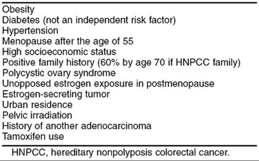
The main risk factor for endometrial adenocarcinoma is long-term unopposed estrogen exposure of either endogenous or exogenous origin. Obesity, nulliparity, and late menopause appear to be associated with high endogenous levels of unopposed estrogen. In obese women, there is an increased peripheral conversion of androstenedione to estrone by fat cells. Nulliparity seems to be associated with endometrial cancer, because ovarian dysfunction (chronic anovulatory cycles and polycystic ovaries) contributes to both the infertility and the unopposed estrogen levels. Estrogen-secreting tumors, such as granulosa cell tumors, are associated with endometrial cancer up to 25% of the time. Other risk factors include a history of pelvic irradiation, a history of breast or ovarian cancer, and use of tamoxifen (Table 54.3).
|
|
|
TABLE 54.3. Factors associated with an increased incidence of endometrial cancer |
Oral contraceptives appear to provide protection from endometrial cancer. At least eight population-based studies suggest that this is so. The use of oral contraceptives appears to decrease the risk of developing endometrial cancer in women 20 to 54 years of age by 50% over those women who never have used oral contraceptives. This protective effect appears to last for at least 10 years in women who used the pill for at least 1 year. Cigarette smoking also decreases the risk of endometrial cancer. There appears to be a dosage relationship to this benefit, such that the greater the number of cigarettes smoked the less the risk of developing cancer. In one study this decrease was quite profound, 30% when one pack was smoked and another 30% if more than one pack was smoked. Furthermore, the greatest risk reduction was in the heaviest women. This is not too surprising, because it is these women who are at the highest risk of developing endometrial cancer, so they might be expected to enjoy the greatest benefit from a factor which decreased their risk. This benefit of risk reduction is strongly outweighed by the increased risk of other health problems of smoking such as heart and lung disease and, therefore, appears to be small consolation.
The relationship between unopposed estrogen exposure and endometrial cancer is well described. Although the risk of endometrial cancer is increased in these women, their prognosis tends to be better. Women with unopposed estrogen use tend to have lower stage, lower grade lesions with better prognosis.
Race is another predictor of survival and type of endometrial cancer. White women have a higher incidence but also have a higher survival rate than black women. This originally was thought to be secondary to socioeconomic status, in that the diagnosis was made later because of less access to health care and, therefore, the cancer was of a higher stage. However, when these variables are controlled for, the survival appears to be less in black women than white. The exact reason for this is unclear. Black women have similar survival to white women when matched for poor prognostic factors. It appears that black women tend to have more poor prognostic variables. Many authors have evaluated this disparity and the etiology is not clear. With the advent of the human genome project, a better understanding of the genetic differences among cancers can be evaluated and understood. This may give us some insight into how they are expressed in different patient populations.
Breast cancer is the most common cancer diagnosed in women in the United States, thereby making tamoxifen a widely used drug in this country. It is estimated that about 80,000 women will start taking tamoxifen each year. Tamoxifen is a selective estrogen receptor modulator, and it has varying effects on estrogen-responsive tissue. The endometrium responds to tamoxifen stimulation much like it does to estrogen. Therefore, the effects of tamoxifen on the endometrium are like those of unopposed estrogen, so the risk of endometrial cancer in women on tamoxifen therapy is increased. In 1985, Killackey reported on three patients with breast cancer who were receiving tamoxifen and who developed endometrial cancer. This and many other reports suggest that there is a significant increase in the risk of developing endometrial cancer while on tamoxifen. The National Surgical Adjuvant Breast and Bowel Project (NSABP) is one of the best studies published on this subject. This study analyzed 2,843 patients with node-negative estrogen receptor–positive breast cancer who were randomized to placebo or 20 mg of tamoxifen a day. There was a significant increase in the risk of endometrial cancer in the tamoxifen arm such that the relative risk was almost 3 times that of the control arm. These data combined with others suggest that the increased risk of endometrial cancer in women taking tamoxifen is between 2 and 3 times that of the population not taking tamoxifen. Women who have breast cancer are also at increased risk to develop endometrial cancer. These studies often do not take this increased susceptibility into account and, therefore, may overstate the risk of endometrial cancer in women taking tamoxifen. The benefits of tamoxifen use for prevention of breast cancer recurrence, more than 120 per 1,000 women, far outweigh the risks of 6 endometrial cancers per 1,000 women.
Genetics
Cancer is the result of accumulation of mutations within the human genome. These mutations can be either acquired or inherited. As we come to understand more through the Human Genome Project and other research, we will come to understand even better the mechanism by which these cancers develop. It is, therefore, essential that clinicians be aware of the information provided by the human genome and use it to better help their patients
Endometrial cancer is the most commonly inherited gynecologic cancer. The clearest hereditary link to this disease is seen in HNPCC, also known as the Lynch syndrome after Henry Lynch, who first described this familial cancer syndrome. Clinical features of HNPCC were described first in the medical literature in 1913 by Aldred Warthin, who identified a clustering of predominantly stomach and endometrial cancers in the family of his seamstress. This inherited pattern of cancers gained little attention over the subsequent 50 years or more until characterized by Henry Lynch as the cancer family syndrome. Subsequently, the pattern of autosomal dominant risk of gastrointestinal cancers and gynecologic cancers became known as HNPCC, although some experts continue to use Lynch syndrome because they think that it more accurately encompasses both the colorectal tumors and extracolonic cancers.
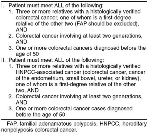
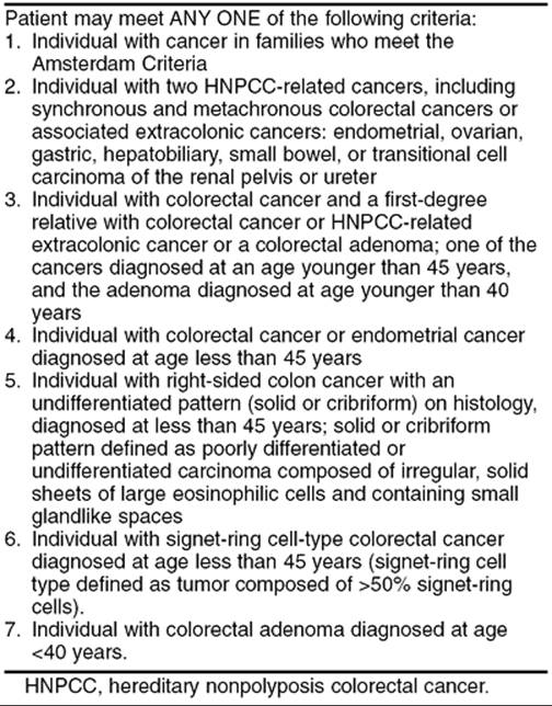
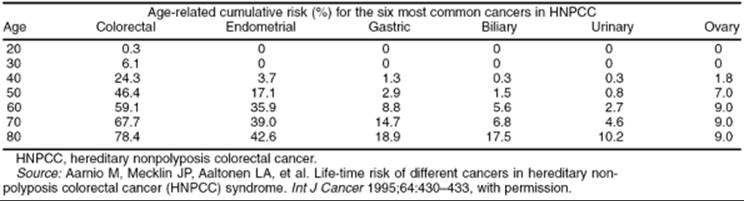
HNPCC is a clinically defined disease in which the primary genetic defect has been identified as mismatch repair genes. It is an autosomal dominant disease with a risk of penetrance of an inherited mutation of approximately 80%. That means that 80% of the patients who inherit one of these mutations will develop a cancer. During the 1990s, specific genes responsible for HNPCC were identified and are known as the mismatch repair genes, but the syndrome is based on family history. The criteria for defining this disease can be seen in Table 54.4 and Table 54.5. These criteria have evolved gradually from those factors focused on colorectal cancers to those that may encompass all cancers associated with the syndrome. Not all patients who fit the criteria of having clinical HNPCC have a mutation in these mismatch repair genes, indicating that we still have more to learn about the development of this disease. We believe that as many as 10% to 30% of cancers ultimately may be categorized as familial. Most of the inherited abnormalities in this group of patients are unknown. Only about 5% can be characterized as HNPCC or Lynch. Thus, the disease of interest, HNPCC, accounts for an important, but minor, percentage of all colorectal or endometrial cancers. We are just coming to understand that women with relatives with this disease can be at increased risk for other malignancies. This brings home the point that all women with cancers should have a detailed family history taken. This will assess their risk of developing cancers and determine any relative risk for their family members. For instance, the normal population has about a 2% chance of developing a colon cancer, versus 80% cumulative risk in a patient with clear HNPCC. When counseling patients, the cumulative risk of developing a cancer over the course of one's life is the most informative means of conveying this information. An example of cumulative risk assessment for various HNPCC cancers in a patient with HNPCC can be seen in Table 54.6.
|
|
|
TABLE 54.4. Amsterdam criteria |
|
|
|
TABLE 54.5. Bethesda criteria |
|
|
|
TABLE 54.6. Cumulative risk |
Interestingly, the risk of a woman with HNPCC developing colorectal cancer may be less than that of a male with HNPCC (60% vs. 80%). Estrogen may play a protective role in this decreased risk. As stated previously, endometrial cancer is the second most common disease in women with HNPCC. An HNPCC mutation confers a significant cumulative risk of developing an endometrial cancer. In the normal population, a lifetime risk is about 1.5%, versus 60% if one has inherited a mismatch repair defect or has clinically defined HNPCC.
Genes responsible for the development of a cancer are oncogenes, which are dominantly expressed cellular control genes, and tumor suppressor genes, which are nondominant cellular control genes. A more recently described set of genes is called mismatch repair genes. These genes are responsible for ensuring fidelity in the DNA replicative process. The job of these genes is to search for and identify mismatches that occur during replication and to excise and repair the errors. In humans, they consist of the genes MLH1, MSH2, MSH6, PMS1, and PMS2. It is these genes that are responsible for the development of HNPCC cancers. When they are mutated and do not repair mismatches in the DNA, errors accumulate through normal division, and eventually an error occurs in a gene(s) responsible for control of cell growth. The importance of these mismatch repair genes is made clear when we see how highly conserved they are across species. The mismatch repair genes of Escherichia coli are not that different from the mismatch repair genes we find in humans. We now have the ability to test for mutations in these genes and are, therefore, able to assess the risk of an individual getting a cancer and passing on that risk to offspring. Any patient with endometrial cancer should have a thorough family history taken to detect the possibility of an inherited problem. If genetic testing is deemed appropriate, this may be offered to the patient and family. However, before genetic testing is recommended, the patient should undergo counseling by a trained genetic counselor.
In summary, HNPCC is a clinically defined disease described by the Amsterdam and Bethesda criteria. Over time, the focus on these syndromes has shifted to extracolonic cancers such as endometrial cancer. The Amsterdam Criteria II begin to take this into account, and the Bethesda criteria continued to expand on this concept. Importantly, as gynecologists we see a fair number of synchronous or metachronous ovarian and endometrial cancers. These often occur in younger women and should raise the suspicion of an HNPCC association. Any patient with cancer should have a detailed family history taken to assess her risk of developing another cancer and to help assess her family members' risk of developing a cancer. If appropriate, family members should be offered genetic counseling and possible testing for mutation in the mismatch repair system.
Diagnosis
There are no accepted routine screening methods for detecting endometrial cancer in asymptomatic women or in women at increased risk. Given the low incidence of endometrial cancer, this would be prohibitive from a cost standpoint. Furthermore, the complications of screening may be higher than the benefits. Clinical studies have evaluated routine endometrial biopsy, transvaginal ultrasonography, and Pap smears, but none of these techniques is sensitive or specific enough to be applied to the general population. Even though a routine Pap smear cannot be relied on as a screen for endometrial cancer, this malignancy should be suspected in any nonpregnant woman with atypical endometrial cells or in any postmenopausal woman with normal endometrial cells on a Pap smear. A Pap smear which reveals AGUS in a woman over the age of 35 should be evaluated by colposcopy, endocervical curettage, and endometrial biopsy to rule out endometrial cancer, in addition to a possible cervical lesion.
Abnormal uterine bleeding is the most common initial symptom of endometrial cancer. Any bleeding in a postmenopausal woman must be evaluated promptly, although overall only about 20% of these patients have a genital malignancy. The likelihood that postmenopausal bleeding is indicative of a uterine cancer clearly increases with increasing age. Perimenopausal women with abnormal bleeding must also undergo thorough investigation, with the most suspicious patterns being increased menstrual flow, a decreased menstrual interval, and intermenstrual bleeding. Whenever possible, the diagnostic procedure for evaluating the endometrium should be an office endometrial biopsy which, under optimal conditions, approaches the accuracy of a formal D&C (greater than 90%). An endocervical curettage (ECC) usually should be performed in the evaluation of postmenopausal bleeding to rule out an endocervical carcinoma as the etiology. If the endometrial biopsy and ECC are satisfactory (with adequate tissue for a diagnosis) and demonstrate no significant abnormality, no further evaluation is necessary. If postmenopausal bleeding is persistent or recurrent or other high-risk factors exist, a fractional D&C should be considered. High-risk factors include atypical hyperplasia or endometrial polyps. Hysteroscopy should be considered if bleeding is recurrent or if either polyps or submucous fibroids are suspected. Many clinical studies now support the usefulness of transvaginal ultrasonography for evaluating the thickness of the endometrium in patients with postmenopausal bleeding. Various investigators have suggested that when the endometrial stripe is thinner than 5 mm, the cause of bleeding usually is related to atrophy, but there have been occasional reports of biopsy-proven malignancy even in patients with thin endometrial stripes. Additionally, almost all women on tamoxifen will have a thickened endometrium, making this test less useful in this patient population.
If endometrial adenocarcinoma is diagnosed, further evaluation of the patient is necessary prior to deciding on the therapeutic approach. A careful physical examination should be performed, with particular attention to supraclavicular and inguinal lymph nodes. A thorough evaluation of the abdomen should be performed, and a bimanual rectovaginal examination to evaluate the size and mobility of the uterus is important. The size and consistency of the cervix, the adnexal structures and parametrium, and the entire vagina, vulva, and rectum are important, and these structures should be evaluated for nodules, masses, induration, and plaquelike lesions that might signify metastatic disease. Any suspicious genital lesions should be sampled for biopsy, and the stool should be tested for occult blood. The histologic subtype and grade of tumor can be determined from the endometrial biopsy or D&C specimen, but it is important to remember that approximately one third of the time the final grade of tumor as determined on the hysterectomy specimen will differ from the original. Additionally, a chest radiograph and routine laboratory studies should be obtained. Based on other risk factors and symptoms, consideration should be given to performing a barium enema examination or a colonoscopy, and preoperative computed tomography or magnetic resonance imaging to evaluate the uterus for depth of invasion and to document occult metastatic disease. An additional test that successfully predicts either deep myometrial invasion or distant disease is the serum tumor marker CA-125. Several studies have documented that 53% to 87% of patients with clinical stage I disease who later were found to have had extrauterine disease at the time of surgery, had elevated CA-125, compared with only 2% to 12% of patients with surgically validated stage I disease. These studies may not be routinely necessary if the decision for full surgical staging can be made intraoperatively (i.e., if the necessary surgical expertise is available to perform pelvic and periaortic lymphadenectomy).
Because endometrial cancer is now staged surgically instead of using the old clinical schema, several practical considerations should be noted. In the past, preoperative irradiation was used in many patients, especially those with grade 2 or 3 lesions, positive ECC findings, or a large uterus. Depending on where the patient was treated, preoperative irradiation could consist of intracavitary radium or cesium for approximately 72 hours or external-beam irradiation of 4,000 to 4,500 cGy to the pelvis. Using these types of treatment plans, many patients received either unnecessary radiation or radiation that did not encompass all of their disease (e.g., paraaortic node metastasis). A second consideration is that all patients should at least be evaluated for full surgical staging, including selective pelvic and paraaortic node dissection. With the adoption of the new surgical staging scheme (see Table 54.2), most patients should undergo primary surgical therapy, including a hysterectomy, bilateral salpingo-oophorectomy, peritoneal cytologic analysis, and selective pelvic and paraaortic node dissection, as indicated. Because endometrial cancer can spread via both the lymphatics and the bloodstream, as well as transmurally or transtubally into the peritoneal cavity, a thorough exploration of the entire abdominal cavity is necessary to document any extrauterine metastasis. Only patients with technically inoperable tumors or medical conditions that make them poor operative candidates should be considered for primary radiation therapy. In these cases, patients should be staged using the older clinical staging schema.
Prognostic Factors
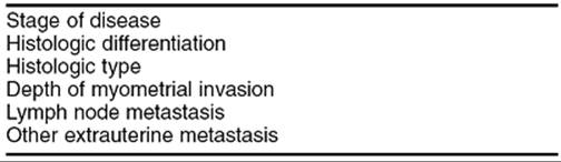
Several well-recognized factors can be used to predict the prognosis of a patient with adenocarcinoma of the endometrium. Preoperative evaluation, coupled with the findings at the time of surgical staging and final pathology, allows individualization of postoperative therapy. These factors are exceedingly important in selecting appropriate postoperative therapy for patients (Table 54.7).
|
|
|
TABLE 54.7. Prognostic factors |
Staging
Until the late 1980s, endometrial cancer was clinically staged using clinical information obtained from examination of the patient and the fractional D&C (Table 54.8). This staging system, however, failed to recognize important prognostic factors, such as deep myometrial invasion and clinically occult extrauterine disease, and often underestimated the extent of disease. Because of these inadequacies, FIGO modified this staging schema in 1988 to reflect the information obtained from surgical exploration and pathologic evaluation of removed tissue. Endometrial cancer is now staged according to the FIGO staging system (see Table 54.2). The recommendation is for all medically operable patients with clinical stage I disease, regardless of tumor grade, to undergo an extrafascial total abdominal hysterectomy and bilateral salpingo-oophorectomy for both staging and therapeutic purposes. A radical hysterectomy may be appropriate in certain circumstances in which the disease is known to involve the cervix or parametrium. The abdominal wall incision should be adequate to perform both pelvic and periaortic lymph node sampling, if indicated by tumor grade and depth of invasion into the myometrium. Immediately after the peritoneal cavity is entered, fluid (either ascites or washing) is obtained for cytologic analysis.
|
|
|
TABLE 54.8. Clinical staging |
Although the FIGO surgical staging schema has been in effect for more than 10 years, there is still considerable controversy as to which patients should undergo the pelvic and periaortic lymph node sampling as required by this schema. It is also unclear how to select these patients and which preoperative and intraoperative findings can be used to define the patient population most likely to benefit from this procedure. Many institutions send the uterus for frozen section analysis intraoperatively and use the pathologic determination of tumor grade and depth of invasion to determine the need for full surgical staging. Others use preoperative information obtained from the endometrial biopsy specimen and radiographic information, especially when patients need to be referred to another center for surgical staging. Because the error rate for poor prognostic features is high with intraoperative evaluation, more gynecologic oncologists are recommending that all patients undergo staging so that all necessary information is available for postoperative treatment planning.
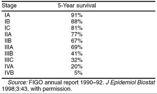
In a large study by the Gynecologic Oncology Group (GOG) of 621 women with clinical stage I disease, 22% of patients were found to have disease outside the uterus. The sites of distant disease were evenly divided between periaortic and pelvic lymph nodes and positive peritoneal cytology. Adnexal involvement was less likely than nodal involvement or positive peritoneal cytologic findings. Equally important, an additional study documented that only 24% of patients with clinical stage II disease actually had pathologic confirmation of cervical involvement at the time of hysterectomy. On occasion, patients are found to have simultaneous endometrial and ovarian cancers, with a reported incidence rate of 1.4% to 3.8%. Although the difference between metastatic and synchronous primary tumors usually can be determined by routine pathologic examination, this determination occasionally cannot be made with any degree of certainty. The survival of patients with endometrial cancer by stage can be seen in Table 54.9.
|
|
|
TABLE 54.9. Survival by stage |
Tumor Grade
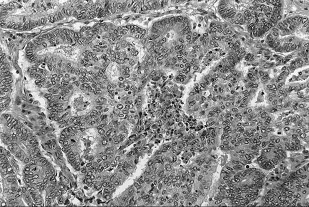
Grading provides a measure of tumor aggressiveness, and it is now an essential part of the FIGO staging for endometrial cancer. Tumor grade is one of the most important prognostic factors for predicting overall survival. Assignment of tumor grade is based on both architectural patterns, such as gland formation, and nuclear atypia and is affected significantly by interobserver reproducibility. It is also not unusual for the tumor grade from the endometrial biopsy to vary from the final tumor grade from the hysterectomy specimen. The higher the tumor grade, the more likely the patient will have deep myometrial invasion, as well as lymph node and other metastases. Grade is clearly an independent predictor of long-term survival, as well as a predictor of other poor prognostic features. Survival based on grade of the tumor alone can be seen in Table 54.10 and the relationship between grade and the depth of myometrial invasion can be seen in Table 54.11. Examples of the various degrees of differentiation can be seen in Fig. 54.3, Fig. 54.4 and Fig. 54.5).
|
|
|
TABLE 54.10. Endometrial cancer differentiation and survival rate |
|
|
|
TABLE 54.11. Differentiation and degree of myometrial invasion |
|
|
|
FIG. 54.3. Well-differentiated adenocarcinoma. Back-to-back glands with no intervening stroma. |
|
|
|
FIG. 54.4. Moderately differentiated adenocarcinoma The left half of this photograph shows back-to-back glands, while the central portion shows solid carcinoma. |
|
|
|
FIG. 54.5. Poorly differentiated adenocarcinoma. Predominantly solid carcinoma with very focal glandular differentiated |
Depth of Myometrial Invasion
Depth of myometrial invasion is another important prognostic factor that directly correlates with the likelihood of extrauterine disease. In the new surgical staging criteria, maximum depth of myometrial invasion determines the subcategories within stage I disease. It is believed that increasing depth of invasion is associated with a higher likelihood of access to the lymphatic system, thereby increasing the incidence of both pelvic and periaortic lymph node metastasis. In a study by Boronow and others, only 1% of patients without myometrial invasion had pelvic lymph node involvement, compared with 25% having positive pelvic lymph nodes with outer-third involvement. Survival was also shown to be affected by depth of invasion, with an 80% to 90% 5-year survival with minimal invasion compared with 60% survival with deep invasion. For early-grade lesions, gross evaluation of depth of invasion is quite accurate at about 85% to 90%. As the cancer becomes less well differentiated, the gross evaluation is less accurate because the margins of the cancer are more infiltrative and less pushing. As the cancer becomes less well differentiated, there is a greater likelihood of deep myometrial invasion and an associated decrease in survival (see Table 54.11).
Lymph Node Metastasis
For patients with clinical stage I endometrial cancer, lymph node involvement is the most important independent prognostic factor, being associated with a six-fold increase in the risk of recurrent disease. In these patients, approximately 10% will have positive pelvic lymph nodes, and 6% will have positive periaortic lymph nodes, with periaortic node involvement being the most important predictor of survival. The incidence of lymph node involvement correlates with stage and grade of tumor, depth of myometrial involvement, and location of the cancer in the uterus (i.e., fundal versus lower uterine segment). In patients with clinical stage II disease, lymph node metastasis occurs in approximately 35%.
Adnexal and Intraperitoneal Involvement
Clinically occult adnexal metastasis occurs in approximately 10% of patients with clinical stage I disease, with most having other high-risk factors such as deep myometrial invasion or lymph node involvement. In patients with adnexal metastasis as their only high-risk factor, overall survival seems to be minimally affected. Other extrauterine intraperitoneal disease is associated with a significant decrease in survival.
Tumor Size
Schink and colleagues reported that the size of the endometrial tumor was of prognostic significance. They reviewed tumor size in 91 patients with disease apparently confined to the uterus and found that if the tumor was smaller than 2 cm, the chance of nodal metastasis was about 6%. However, if the tumor was larger than 2 cm, the chance of nodal metastasis was over 20%. In this study, tumor size was an independent prognostic variable.
Histologic Subtype
The histologic subtype of adenocarcinoma may have an independent impact on patient outcome. Approximately 80% of endometrial cancers are of endometrioid histology. These cancers can be of varying degrees of differentiation. There are thought to be of two subtypes as follows: (a) type 1 arises from endometrial hyperplasia, is estrogen dependent, and usually confers a better prognosis; (b) type 2 endometrial cancer tends to be more poorly differentiated, is not dependent on estrogen stimulation, and generally carries a worse prognosis. Squamous differentiation is seen in 15% to 25% of endometrioid tumors. The significance of squamous differentiation is unclear, and its impact has been debated for many years. In the past, the terms adenoacanthoma and adenosquamous carcinoma were used to identify adenocarcinomas with either benign or malignant squamous differentiation. More recently, Zaino and colleagues recommended that these terms be replaced with the more descriptive term adenocarcinoma with squamous differentiation. Although the squamous elements can have varying degrees of differentiation, they usually correlate with the glandular components. Furthermore, the differentiation of the glandular component appears to be the important prognostic feature.
Papillary serous and clear cell cancers of the endometrium are both quite rare (approximately 5% each) but have a significantly worse prognosis compared with that of endometrioid adenocarcinoma. Both of these histologic types are more common in older women, who tend to have distant disease, even when clinically it appears to be stage I (Fig. 54.6 and Fig. 54.7).
|
|
|
FIG. 54.6. Clear cell carcinoma. The tumor forms sheets of cells with cleared cytoplasm and pleomorphic nuclei. |
|
|
|
FIG. 54.7. Serous carcinoma. The tumor shows marked intraluminal complexity and high-grade nuclear atypia. |
Age
Although it is recognized clearly that advancing patient age at diagnosis is associated with a poorer outcome, there is no agreement as to why this is true. In a study by Lurain and colleagues, increasing patient age was an independent prognostic factor associated with recurrent disease. No patient under age 50 years developed recurrent cancer, compared with 12% of women between 50 and 75 years and 33% of women older than 75 years. For every 1-year increase in age over 50 years, there was a 7% increase in the rate of recurrence. Younger patients tend to have early, well-differentiated lesions with no or minimal myometrial invasion. Younger patients also tend to be in better general health, thereby allowing more aggressive definitive surgery. It has been suggested that older women are more likely to ignore signs of vaginal bleeding, thereby delaying diagnosis until the stage is more advanced. Interestingly, however, the length of time between onset of bleeding and presentation to a clinician does not correlate with tumor stage, and patients with advanced stage disease have not necessarily delayed seeking medical care.
Peritoneal Cytology
The significance of malignant peritoneal cytologic findings obtained during surgical staging for endometrial cancer is controversial. In essentially all published studies, a positive peritoneal cytology result is associated with other high-risk features such as deep myometrial invasion, adnexal spread, positive lymph node metastasis, and cervical involvement. Several studies showed a two-fold to three-fold increase in the risk of recurrent disease in patients with positive peritoneal cytology, but often these patients have one or more other high-risk factors, and many times the recurrent disease is outside the peritoneal cavity. In patients found to have positive cytologic findings, it is unclear whether any currently available therapy would have any impact on eventual outcome.
Race
Even with correction for age and tumor stage, black women with endometrial cancer have a much poorer prognosis than white women. For all stages of endometrial cancer, white women have an 86% 5-year survival compared with a 55% 5-year survival for black women, and within each stage the relative survival is worse for blacks.
Other Factors
Many other factors have been shown to affect prognosis, including hormone receptor status of the tumor, DNA content or ploidy, proliferative index, and oncogene expression. Both estrogen receptor and progesterone receptor levels have been shown to be prognostic factors independent of tumor grade, with the progesterone receptor level being the more important for predicting overall survival. Progesterone receptor levels also can be used to predict which patients with advanced or recurrent disease are more likely to respond to hormone therapy.
Management
The primary management for endometrial cancer is surgery (Table 54.12), consisting of removal of the uterus, cervix, and adnexal structures. Surgical staging procedures, including careful exploration of the abdomen and pelvis and lymphadenectomy, often are done at the same time, as described above. As soon as the uterus has been removed, it should be sent to the pathology laboratory for frozen section to determine tumor grade and depth of invasion, as well as to obtain tissue for estrogen and progesterone receptor levels and other studies, as indicated. For patients in whom lymph node sampling is indicated (see Table 54.12), representative samples of lymph node–bearing tissue from the lower aorta and vena cava are removed. Pelvic lymph node sampling should remove nodes from common and external iliac and obturator regions. An omental biopsy may be performed and is indicated for patients with papillary serous and clear cell cancers. In patients who are very poor candidates for exploratory laparotomy (e.g., morbidly obese), vaginal hysterectomy or laparoscopically assisted vaginal hysterectomy should be considered. Every effort should be made to remove the adnexal structures; therefore, laparoscopically assisted vaginal hysterectomy may be preferable. If appropriate surgical expertise is available, the full surgical staging, including pelvic and periaortic lymphadenectomy, can be performed through the laparoscope.
Until additional information is obtained, vaginal hysterectomy should be limited to very select patients who otherwise might be given radiation therapy only. In patients with clinically apparent cervical involvement, radical hysterectomy with bilateral salpingo-oophorectomy and lymph node dissection may be considered.
|
|
|
TABLE 54.12. Surgical therapy of endometrial cancer |
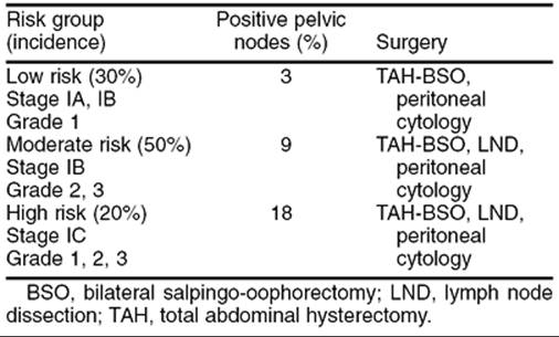
Following surgery and review of the final pathology report, patients may be divided into risk groups based on the many prognostic factors discussed above. This classification can then be used to individualize the recommended postoperative therapy, if needed. Because formal surgical staging has been utilized widely for less than 10 years, most of the information on recommendations for postoperative radiation therapy are based on clinically staged patients. The risk of pelvic recurrence in patients with high-risk factors but negative surgical staging is largely unknown. The GOG study based on surgicopathologic correlation found that even when sampled lymph nodes were negative, patients with deep myometrial invasion and poorly differentiated cancers were still at a higher risk of recurrence. Even though the surgical staging procedures failed to document extrauterine pelvic disease, postoperative pelvic irradiation was associated with a decreased risk of local recurrence. It is not yet clear whether surgical staging can define a subgroup of stage I patients, other than the already identified low-risk patients, who can be treated without adjuvant radiation therapy.
Treatment for patients with stage II disease remains somewhat controversial. Most studies suggest that patients with cervical involvement are at a higher risk for vaginal vault recurrence, and often recommend adjuvant vaginal cuff irradiation. Whether or not those patients with otherwise negative surgical staging also need pelvic irradiation remains to be confirmed. With clinically apparent cervical involvement, treatment recommendations include either radical hysterectomy or preoperative irradiation followed by extrafascial hysterectomy.
Treatment for patients with documented extrauterine disease (stages III and IV) should be individualized to encompass the true extent of the disease. Therapeutic options include pelvic irradiation, extended-field irradiation to cover the periaortic lymph nodes, whole-abdomen irradiation, and systemic hormone therapy or chemotherapy. In most cases, management of distant metastasis with either high-dose progestins or chemotherapy is palliative only, with little expectation for long-term control of disease. The most widely used chemotherapy agents include doxorubicin hydrochloride (Adriamycin) and cisplatin (Platinol) or carboplatin (Paraplatin).
Progestins have been used for many years in the management of recurrent endometrial carcinoma, with approximately one third of patients having a favorable response. Patients with well-differentiated tumors have a higher response rate than those with moderately or poorly differentiated cancers. Although the role of estrogen and progesterone receptors in endometrial cancer therapy has not yet been accepted widely, it does appear that the responsiveness of recurrent tumor to progestin therapy is related to the content of both estrogen and progesterone receptors. If both receptors are present, the likelihood of a favorable response is good, regardless of tumor grade or other high-risk factors. If the concentration of receptors is low, it is unlikely that the recurrent tumor will respond to progestins, and other chemotherapy should be considered.
Endometrial cancer in approximately 10% to 15% of patients is inoperable, usually because of morbid obesity or severe intercurrent disease. In these patients, primary radiation therapy should be considered. In most cases, a combination of external beam and intracavitary irradiation should be used, and with definitive therapy approximately 85% to 90% of patients with early-stage disease will have control of uterine disease. The overall risk of recurrence in these patients with stage I disease correlates with tumor grade, with a 5-year survival of 94% for grade 1, 92% for grade 2, and 78% for grade 3 tumors.
Posttreatment Surveillance
Many studies have confirmed that after treatment of endometrial cancer, most recurrences will occur within 3 years. Approximately one half of these recurrences will be asymptomatic and, thus, the traditional recommendation has been to follow patients with physical examination and vaginal cytology every 3 to 4 months for the first 2 to 3 years, and then at 6-month intervals for at least 5 years. Serial serum CA-125 measurements also have been suggested for surveillance of patients who have been treated for endometrial cancer, although the level may be normal in patients with early recurrent disease. Because treated endometrial cancer will not recur in the great majority of patients, however, several studies have addressed the cost effectiveness of this traditional recommendation. In a study from the M.D. Anderson Cancer Center, 59% of patients with recurrence were asymptomatic, with over one half being picked up on physical examination, 26% by an elevated CA-125, and only 4% by vaginal cytologic examination. Because of these findings, these authors recommended that physical examination, serum CA-125 assay, and vaginal cytology be performed only every 6 to 12 months on asymptomatic patients. A Canadian study reported that only one recurrence was documented during more than 200 routine follow-up visits after treatment for endometrial cancer, using a schedule of examinations of every 3 months for the first year, every 4 months for the second year, and every 6 months thereafter. There was no difference in the salvage rates of recurrent disease picked up during routine surveillance compared with recurrent disease documented in symptomatic patients. In this study, no recurrence was picked up by vaginal cytology alone. In a third study from Duke University, an analysis of various follow-up techniques demonstrated that routine vaginal cytology and chest radiograph were not cost effective. The recommendation of these investigators is to follow patients every 6 months with examination alone, using additional testing to evaluate any symptoms.
Estrogen Replacement Therapy After Treatment
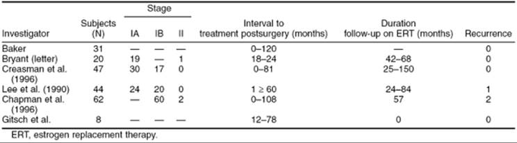
For many years, it has been thought that a history of endometrial cancer, even successfully treated, was an absolute contraindication to estrogen replacement therapy (ERT), because adenocarcinoma of the endometrium is considered an estrogen-dependent neoplasm. Because no scientific data support the contention that ERT is dangerous for patients who have had a hysterectomy for endometrial cancer and because the body of evidence is increasing in supporting the value of ERT in decreasing morbidity and mortality from heart disease, strokes, and osteoporosis, many physicians and patients are questioning the earlier proscription. During the last decade, there have been several small retrospective studies of patients given ERT following treatment for early-stage endometrial cancer (Table 54.13). In 1986, Creasman and coworkers reported on 221 patients with stage I endometrial cancer, of whom 47 (21%) were given postoperative estrogen replacement for a median of 26 months. Statistical analysis revealed no increased risk of recurrence or death between those who received ERT and those who did not when adjustments were made for tumor grade, myometrial invasion, nodal metastasis, peritoneal cytology, and age. In fact, the risk of recurrence was significantly higher in the untreated group (15% vs. 2%), as was the risk of dying of intercurrent disease. Similar study design and conclusions were reported 3 years later by Lee and others. In both of these studies, selection bias may have contributed to the results, but it does appear that a low-risk group of patients can be selected who can safely take estrogen replacement. Chapman and colleagues retrospectively reviewed information on 123 patients with stages I and II endometrial cancer, of whom 62 received ERT, and again documented no increase in recurrences or deaths from this malignancy. In a Committee Opinion in August 1993, the American College of Obstetricians and Gynecologists concluded that there are no definitive data to support specific recommendations regarding ERT for women previously treated for endometrial cancer. The opinion states that estrogens could be used for the same indications as for any other woman, except that the selection of appropriate candidates should be based on prognostic indicators and the risk that a patient is willing to assume. The gynecologic practice committee, due to the paucity of data, could not evaluate the need for progestational agents in addition to estrogens. A large GOG trial is underway to assess the risk of hormonal replacement in these patients.
|
|
|
TABLE 54.13. Effect of estrogen replacement therapy on endometrial cancer recurrence |
UTERINE SARCOMAS
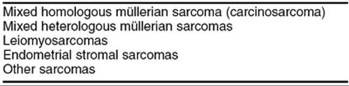
Sarcomas of the uterus carry a poor prognosis but, fortunately, they are rare and represent less than 5% of all uterine tumors. The incidence of these tumors is about 1.7 per 100,000 women per year in the United States. The classification of the various histologic types, in order of frequency, can be seen in Table 54.14. There is some retrospective evidence that sarcomas tend to arise more commonly in patients who have undergone pelvic irradiation in the past. A clear cause and effect relationship between irradiation and sarcomas has not been established, and new data have failed to corroborate the older literature.
|
|
|
TABLE 54.14. Classification of uterine sarcomas by frequency |
These lesions arise primarily from two tissues: endometrial sarcomas arise from endometrial glands and stroma, and leiomyosarcomas from the myometrium itself. The malignant mixed müllerian tumors arise from the pluripotent endometrial stroma. Other sarcomas, such as angiosarcoma and fibrosarcoma, arise in supporting tissues. In general, uterine sarcomas are the most malignant type of uterine tumor and tend to differ significantly from endometrial adenocarcinomas with regard to patterns of spread and prognosis.
The relative incidence of the various subtypes of uterine sarcomas differs significantly in the literature, with mixed mesodermal sarcomas being the most common in recent studies. The staging criteria for uterine sarcomas are based on the FIGO classification for endometrial cancers (see Table 54.2).
Classification
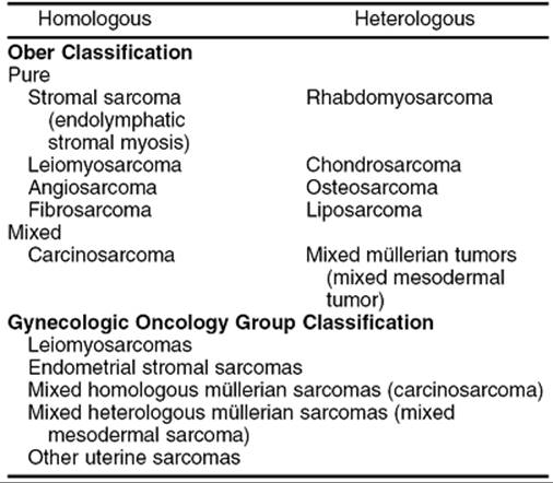
Numerous classification schemes for uterine sarcomas have been proposed, depending on both cell type and site of origin. In 1959, Ober suggested a classification of the endometrial sarcomas. This does not include the pure sarcomas, which are self-explanatory. This classification and can be seen in Table 54.15. Generally, homologous tumors are comprised of tumors that should belong in the normal uterus, such as smooth muscle or endometrial stroma. Heterologous elements are those tissues that normally would not be found in the uterus, such as bone (osteosarcoma) or cartilage (chondrosarcoma). Most pathologists have adopted this classification, which has the advantage of being able to classify these malignancies for study, because most tumors fall into four major groups. Pure tumors are composed of only one cell type; whereas mixed tumors have more than one cell type. Homologous tumors contain tissue elements that are indigenous to the uterus, whereas heterologous tumors are defined as those that contain tissue elements foreign to the uterus (Table 54.15). Because the great majority of uterine sarcomas fall into one of four categories, the GOG has accepted a more simplified classification:
|
|
|
TABLE 54.15. Classifications of uterine sarcomas |
· Leiomyosarcoma
· Endometrial stromal sarcoma
· Mixed homologous müllerian sarcoma (carcinosarcoma)
· Mixed heterologous müllerian sarcoma (mixed mesodermal sarcoma)
Leiomyosarcoma
Leiomyosarcoma is a malignancy of smooth muscle. This sarcoma was thought to be the most common of the malignant mesenchymal tumors, a concept that is supported by literature before the 1970s. However, more recent data from the GOG and other sources suggest that leiomyosarcoma is the second most common sarcoma of the uterus and accounts for about 1 out of 7 of all uterine sarcomas, or about 16%. This information is detailed in GOG data published on 447 uterine sarcomas. This malignancy is found in younger women, with a median age at diagnosis between 43 and 53 years. Leiomyosarcoma is more common in black women and is associated with a poorer prognosis in these patients. The patterns of spread for leiomyosarcoma appears to be via both lymphatics and the vasculature, because distant metastases will develop in many patients even if lymph node biopsy results are negative at the time of surgery.
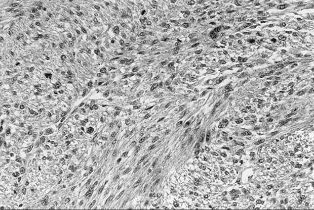
Although there is some disagreement about the exact histologic criteria required for a diagnosis of leiomyosarcoma, the most important factor appears to be the mitotic count of the tumor. Cellular myomas and bizarre leiomyomas may appear to be malignant, but if there are fewer than 5 mitoses per 10 high-power fields (HPF), the lesion is considered benign. Tumors with more than 10 mitoses per 10 HPF are malignant, and those with 5 to 10 mitoses per 10 HPF are thought to have an uncertain potential for malignancy. Many investigators agree that 5-year survival is related to the number of mitoses per 10 HPF, with a 95% to 98% survival with fewer than 5 mitoses per 10 HPF, approximately 40% survival with 5 to 10 mitoses per 10 HPF, and very poor survival (15%–20%) with more than 10 mitoses per 10 HPF. An example of a leiomyosarcoma can be seen in Figure 54.8. The etiology of leiomyosarcoma is unclear. As its name suggests, this tumor was thought to arise within benign leiomyoma. However, in most cases it appears to arise independently of these tumors. Hysterectomy was felt to be indicated for a rapidly enlarging uterus or rapidly growing fibroid tumors for fear that this represented degeneration of leiomyomas into a sarcoma. Investigators from University of Southern California addressed this question best. These authors retrospectively reviewed a large series of patients with “rapidly enlarging” leiomyomas and found the incidence of sarcoma to be very low. They recommended that surgery for a concern of leiomyosarcoma was not warranted for fibroid tumors that were enlarging rapidly, and reasons for surgery should focus on other issues such as symptoms of pressure, pain, or bleeding.
|
|
|
FIG. 54.8. Leiomyosarcoma. Interlacing fascicles of smooth muscle cells exhibit high-grade cytologic atypia and increased mitotic activity. Other areas of this tumor showed coagulative necrosis. |
This sarcoma should be treated as any other uterine malignancy. This should include comprehensive staging with total abdominal hysterectomy, bilateral salpingo-oophorectomy, and pelvic and paraaortic lymph node dissection. The staging system for leiomyosarcomas is the same as for an endometrial cancer (see Table 54.2).
Endometrial Stromal Sarcomas
Endometrial stromal sarcomas are the least common of the three most common uterine sarcomas. The most-often-seen initial symptom of an endometrial stromal sarcoma is vaginal bleeding. These sarcomas are generally yellow in color, fleshy in texture, and often polypoid. At the time of diagnosis and treatment, usually by hysterectomy, 40% of patients have disease outside of the uterus. Endometrial stromal sarcomas are just as aggressive as the other uterine sarcomas and the prognosis is very similar.
Endometrial stromal tumors usually are divided into three groups: benign stromal nodules, endolymphatic stromal myosis and endometrial stromal sarcoma. Benign stromal nodules usually are well-circumscribed tumors with clearly defined pushing margins. These tumors may grow to many centimeters but are always benign. No cases of metastasis or recurrence have been reported. The second histologic type, endolymphatic stromal myosis, is an infiltrative tumor that generally has an indolent course. Another name for endolymphatic stromal myosis is low-grade stromal sarcoma. Gross inspection of the surgical specimen often reveals an infiltrative growth pattern, and it can project from the cut surface in a worm-like manner that may also extend into blood vessels in the broad ligament. Microscopically, there are little cellular atypia and few, if any, mitoses. The clinical course is slowly progressive and management is usually by surgery alone. However, the tumor can recur many years after initial incidence. It is a hormonally responsive tumor and often grows in the presence of estrogen and can regress or stabilize when exposed to progestational agents. There are reports of low-grade endometrial stromal sarcomas being stabilized for many years with progestational therapy.
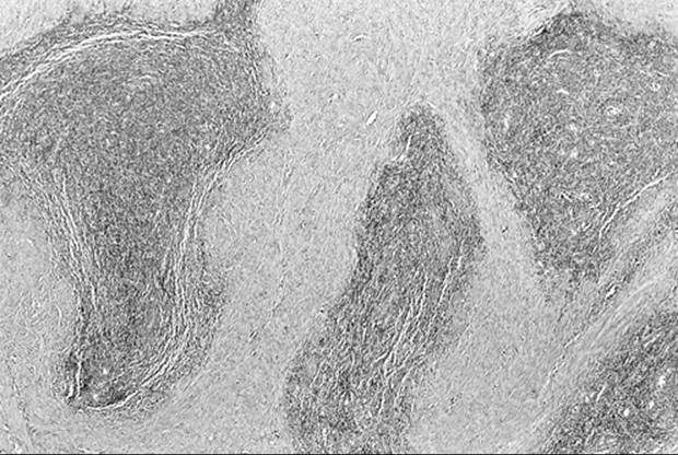
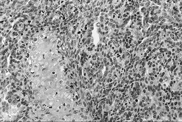
Endometrial stromal sarcoma, on the other hand, has a much more aggressive course, with frequent and widespread metastases and a very poor prognosis. As with leiomyosarcoma, the diagnosis of stromal sarcoma versus low-grade stromal sarcoma depends on the number of mitoses per 10 HPF, with 10 mitoses being the cutoff to categorize the tumor as an endometrial stromal sarcoma (Fig. 54.9). Despite this distinction, controversy still exists as to the prognostic significance of the number of mitoses seen. A study from the Mayo Clinic failed to identify this feature as an independent prognostic variable. As with most sarcomas, irradiation may control local disease but has little effect on overall survival. These tumors often exhibit high concentrations of both estrogen and progesterone receptors, and occasional responses have been seen when high-dose progesterone therapy is administered.
|
|
|
FIG. 54.9. Endometrial stromal sarcoma. Nests of bland endometrial stromal cells diffusely infiltrate the myometrium. |
Mixed Müllerian Tumor
This malignancy is the most common uterine sarcoma, accounting for more than 50% of all cases in recent series. This malignancy is aggressive, and more than 60% of patients will have disease outside of the uterus at the time of diagnosis. It tends to metastasize early via lymphatic vessels, blood vessels, and local spread. It is more common in blacks than whites and is associated with prior pelvic irradiation in up to one third of patients. The main initial symptom is vaginal bleeding and, on examination, most patients are found to have an enlarged uterus, often with a polypoid mass protruding through the cervix. This tumor tends to be aggressive, with early metastasis to pelvic and periaortic lymph nodes and adjacent tissue. Hematogenous spread, especially to the liver and lungs, is common.
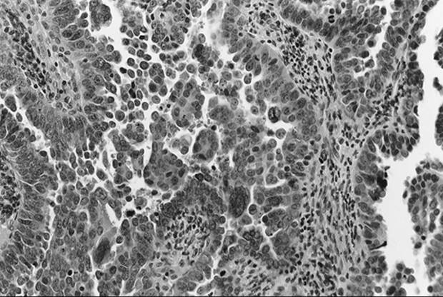
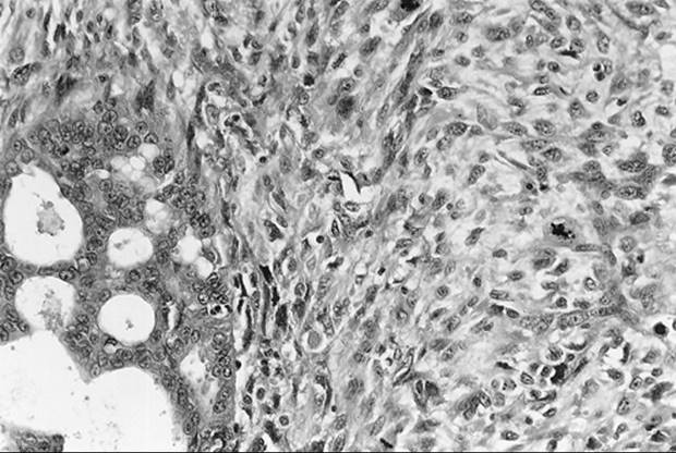
Histologically, it is composed of both sarcomatous and carcinomatous elements, with the sarcomatous element being divided into homologous types (tissue normally found in the uterus; Fig. 54.10) and heterologous types (tissue such as cartilage or bone not usually found in the uterus; Fig. 54.11). In some series, heterologous elements were associated with a worse prognosis. Rhabdomyosarcoma was the most common heterologous element in these cancers. In the most recent GOG series, the median progression-free interval of homologous tumors was 22.7 months, with a median progression-free interval of 62.6 months in the homologous tumors. Staging is very important in this tumor, because extrauterine disease correlates very strongly with survival. Almost no one with disease outside the uterus is cured of her disease. The single most important factor correlating with survival other than extrauterine disease is depth of myometrial invasion. Recurrences usually occur within the first 2 years and often are comprised of only the carcinomatous elements.
|
|
|
FIG. 54.10. Malignant mixed müllerian tumor, homologous type. Well differentiated. |
|
|
|
FIG. 54.11. Malignant mixed müllerian tumor, heterologous type. |
Other Sarcomas
Pure heterologous uterine sarcomas are rare. Rhabdomyosarcoma is the most common and usually occurs in children. This used to be called sarcoma botryoides in children. Management of this rare group of tumors has changed over the last 50 years from primary surgery to chemotherapy. Müllerian adenosarcoma is a rare sarcoma originally described by Skully. This tumor usually causes vaginal bleeding and is of low malignant potential. Its course is indolent and prolonged, with an overall long-term survival rate of about 90%.
Treatment
Total abdominal hysterectomy with bilateral salpingo-oophorectomy is the treatment of choice for almost all uterine sarcomas. Because there is no formal staging system for sarcomas, most authorities feel that the endometrial staging system should be used. Local spread of a sarcoma can be treated with radiation therapy. This may not improve overall survival but will control local disease and diminish the likelihood of the morbidity of a pelvic recurrence. Unfortunately, there are no good prospective randomized trials evaluating the efficacy of postoperative radiation therapy on overall survival. The retrospective studies that do exist show only a benefit for local control. The largest study to address this issue by Salazar comprised more than 900 patients and failed to show a statistically significant survival benefit with postoperative radiation therapy, only a benefit of local control. One can argue that one must obtain control of microscopic local disease before one can cure the cancer. Typically 5,000 to 6,000 cGy is given with or without adjuvant brachytherapy. Investigators from the Mallinckrodt Institute of Radiology noted fewer pelvic recurrences when more than 5,000 cGy were used.
Some patients with leiomyosarcoma may experience an isolated pulmonary metastasis. These sometimes can be resected for cure. Significant 5-year survivals have been recorded in these rare situations. Recurrences of low-grade tumors should be considered for resection no matter where they are.
Recurrences also can be treated with chemotherapy. Many agents have shown activity (Table 54.16). Unfortunately, no therapy has shown a significant increase in overall survival.
|
|
|
TABLE 54.16. Active agents in uterine sarcomas in order of response rate |
SUMMARY POINTS
· Uterine cancers comprise a wide variety of histologic types that carry a wide range of metastatic potential and survival.
· Endometrial hyperplasia is a premalignant precursor to endometrioid adenocarcinoma. Simple hyperplasia has a low propensity to progress to a cancer (less than 1%), whereas complex hyperplasia with atypia has a 30% chance of developing into a cancer.
· Endometrial cancer is the most common gynecologic cancer. Type I endometrial cancers are associated with relative estrogen excess. This can be endogenous, as in obese women who have significant conversion of steroids to estrone, or it can be exogenous, as in women who take unopposed estrogen replacement therapy.
· Type II endometrial cancers are not associated with estrogen and are comprised of more aggressive histologic types, such as poorly differentiated endometrioid, papillary serous, and clear cell cancers. These later types have a survival rate as poor as 60%, compared with a greater than 80% survival in the other histologic types.
· Management of endometrial cancers consists of comprehensive staging, including a total abdominal hysterectomy, bilateral salpingo-oophorectomy, pelvic washings, pelvic and paraaortic lymph node dissection, and careful abdominal exploration. Management may also include postoperative radiation therapy.
· Postoperative surveillance should include regular examination with Pap smear. The most common site of recurrence is at the vaginal cuff. Radiation treatment of an isolated cuff recurrence has a cure rate of 50% with external radiation therapy.
· Endometrial cancer is the most common inherited form of gynecologic cancer and is associated with HNPCC or Lynch II syndrome. It is important to take a careful family history to assess if the risk is high, and formal genetic counseling and genetic testing may be appropriate.
· Sarcomas are the most lethal of the uterine malignancies, with an overall survival of around 50%. There are several histologic types of sarcomas. The most common is the malignant mixed müllerian tumor, followed by the leiomyosarcoma, and finally by the endometrial stromal sarcomas. Therapy consists of exploratory laparotomy and staging procedure as outlined for endometrial cancers. Postoperative radiation therapy may be recommended, but this is associated only with decreased pelvic recurrence and not improved survival.
SUGGESTED READINGS
Endometrial Hyperplasia
Gusberg SB, Kaplan AL. Precursors of corpus cancer. IV. Adenomatous hyperplasia as stage 0 carcinoma of the endometrium. Am J Obstet Gynecol1963;87:662–678.
Kurman RJ, ed. Blaustein's pathology of the female genital tract, fourth ed. New York: Springer-Verlag, 1994.
Kurman RJ, Kaminski PF, Norris HJ. The behavior of endometrial hyperplasia: a long-term study of untreated hyperplasia in 170 patients. Cancer1985;56:403–412.
Mencaglia L, Valle RF, Perino A, et al. Early detection of endometrial carcinoma and its precursors. Curr Probl Obstet Gynecol Fertil 1988;11:173–202.
Endometrial Carcinoma
Bokhman JV. Two pathogenetic types of endometrial carcinoma. Gynecol Oncol 1983;15:10–17.
Boronow RC, Morrow CP, Creasman WT, et al. Surgical staging in endometrial cancer: clinical-pathologic findings of a prospective study. Obstet Gynecol1984;63:825–832.
Chapman JA, Di Saia PJ, Osann K, et al. Estrogen replacement in surgical stage I and II endometrial cancer survivors. Am J Obstet Gynecol1996;175:1195–1200.
Creasman WT. Limited disease: role of surgery. Semin Oncol 1994;21:79–83.
Creasman WT, Henderson D, Hinshaw W, et al. Estrogen replacement therapy in the patient treated for endometrial cancer. Obstet Gynecol 1986;67:326–330.
Creasman WT, Morrow CP, Bundy BN, et al. Surgical pathological spread patterns of endometrial cancer: a Gynecologic Oncology Group study. Cancer1987;60:2035–2041.
Ferenczy A, Gelfand M. The biologic significance of cytologic atypia in progestin-treated endometrial hyperplasia. Am J Obstet Gynecol 1989;160:126–131.
Gordon MD, Ireland K. Pathology of hyperplasia and carcinoma of the endometrium. Semin Oncol 1994;21:64–70.
Grigsby P, Kuske R, Perez C, et al. Medically inoperable stage I adenocarcinoma of the endometrium treated with radiotherapy alone. Int J Radiat Oncol Biol Phys 1987;13:483–488.
Karasek K, Faul C. Changing concepts in the management of endometrial cancer. Oncology 1996;10:1099–1110.
Killacky MA, Hakes TB, Pierce VK. Endometrial adenocarcinoma in breast cancer patients receiving antiestrogens. Cancer Treat Rep 1985;69:273–276.
Lee RB, Burke TW, Park RC. Estrogen replacement therapy following treatment for stage I endometrial cancer. Gynecol Oncol 1990;36:189–191.
Lurain JR, Rice BL, Rademaker AW, et al. Prognostic factors associated with recurrence in clinical stage I adenocarcinoma of the endometrium. Obstet Gynecol 1991;78:63–69.
Morrow CP, Bundy BN, Kurman RJ, et al. Relationship between surgical-pathological risk factors and outcome in clinical stages I and II carcinoma of the endometrium: a Gynecologic Oncology Group study. Gynecol Oncol 1991;40:55–65.
Muss HB. Chemotherapy of metastatic endometrial cancer. Semin Oncol 1994;21:107–113.
Prat J, Matias-Guiu X, Barreto J. Simultaneous carcinoma involving the endometrium and the ovary. Cancer 1991;68:2455–2459.
Schink JC. Tumor size in endometrial cancer: a prospective factor for lymphnode metastases. Obstet Gynecol 1987;70:216–222.
Terakawa N, Inoue M, Shimizu I, et al. Preliminary report on the use of danazol in the treatment of endometrial hyperplasia. Cancer 1988;62:2618–2621.
Uziely B, Lewin A, Brufman G, et al. The effect of tamoxifen on the endometrium. Breast Cancer Res Treat 1993;26:101–105.
Zaino RJ, Kurman R, Herbold D, et al. The significance of squamous differentiation in endometrial carcinoma. Cancer 1991;68:2293–2302.
Uterine Sarcomas
Clements PB, Skully RE. Mullerian adenosarcoma of the uterus. Clinical and pathological anatomy of 10 cases of a distinctive type of mullerian mixed tumor. Cancer 1974;34:1138–1149.
Genest P, et al. Prognostic factors in early carcinoma of the endometrium. Am J Clin Oncol 1987;10:71–78.
Ober WB. Uterine sarcomas: Histogenesis and taxonomy. Ann N Y Acad Sci 1959;75:568–576.
Piver MS, Rutledge FN, Copeland L, et. al. Uterine endolymphatic stromal myosis: a collaborative study. Obstet Gynecol 1984;64:173.
Salazar OM, Bonfiglio TA, Patten SF, et al. Uterine sarcomas: natural history, treatment and prognosis. Cancer 1978;42:1152–1160.
Wolfson, AH, Wolfson DJ, Sittler SY, et al. A multivariate analysis of clinicopathologic factors for predicting outcome in uterine sarcomas. Gynecol Oncol1994;52:56–63.