
Boback M. Berookhim1, Alexander W. Pastuszak2, Natan Bar-Chama3 and Mohit Khera4
(1)
Department of Urology, Lenox Hill Hospital, New York, NY, USA
(2)
Division of Male Reproductive Medicine and Surgery, Center for Reproductive Medicine, Scott Department of Urology, Baylor College of Medicine, Houston, TX, USA
(3)
Department of Urology, Mount Sinai School of Medicine, New York, NY, USA
(4)
Scott Department of Urology, Baylor College of Medicine, Houston, TX, USA
Mohit Khera
Email: mkhera@bcm.edu
Keywords
Erectile dysfunctionEpidemiologyCardiovascular diseasePenile Doppler ultrasonographyBiothesiometryIntracavernosal injection testingCavernosometryEndothelial reactivity
6.1 Introduction
Among reported sexual dysfunctions in men, erectile dysfunction (ED) is the most frequently treated worldwide [1]. It is defined as the recurrent or consistent inability to obtain and/or maintain a penile erection sufficient for satisfactory sexual performance [2]. Erectile dysfunction has significant negative effects on the quality of life of both the patient and his partner, affecting the patient’s emotional and psychological well-being, and is associated with poor relationship satisfaction, negative general health perceptions, and role limitations [3, 4]. Temporary ED is a fairly common phenomenon and is often related to transient life circumstances, disappearing after resolution of the underlying circumstances. However, it is generally accepted that men with ED symptoms present for at least 3 months should undergo a medical evaluation [1, 5].
6.2 Epidemiology
Estimates on the prevalence of ED on a worldwide basis are highly variable, as a result of varying study methodologies, questionnaires, and tools used for surveys, as well as differences in the definition of ED. It is estimated that at least 20 million men in the United States suffer from ED [6]. The landmark Massachusetts Male Aging Study (MMAS) was among the first studies to report on ED in a standardized fashion and has described a combined prevalence of any degree of ED in 52 % of men over the age of 40 [7]. The study further reported an increase in the rate of moderate to severe/complete ED from 22 to 49 % between the ages of 40 and 70 years. More recently, the National Social Life, Health, and Aging Project (NSHAP) performed a survey evaluation of over 1450 American men to evaluate sexual issues in older adults and reported ED in 31 % of men 57–64 years old and 45 % of men 65–74 years old, with an adjusted odds ratio of 1.83 comparing these two age groups [8]. The prevalence of ED appears to be similar among different ethnic groups, with one study reporting a 22 % rate of ED in white men over 40 years, a 24 % rate among black men, and a 20 % rate among Hispanic men [9].
6.3 Classifications of Erectile Dysfunction
Erection is the result of a complex physiologic interaction of psychological, neuronal, hormonal, vascular, and cavernous smooth muscle systems, with derangements in any one system predisposing to ED. Although often multifactorial in nature, ED is further classified according to its origin: psychogenic, vasculogenic, endocrinologic, neurogenic, and drug induced (Table 6.1).
Table 6.1
Classification of erectile dysfunction and selected causes
|
Classifications |
Specific causes |
|
Psychogenic |
Interpersonal factors Anxiety/anxiety disorders Disorders of sexual intimacy |
|
Vasculogenic |
Arteriogenic Cardiovascular disease Endothelial dysfunction Venogenic |
|
Endocrinologic |
Hypogonadism Hyperprolactinemia Hypothyroidism Hyperthyroidism |
|
Neurogenic |
Supraspinal Brain tumor Cerebrovascular accident Parkinson’s disease Dementia Temporal epilepsy Spinal Multiple sclerosis Spinal cord injury Transverse myelitis Myelodysplasia Lumbar disc disease Iatrogenic (secondary to spinal surgery) Peripheral Lower motor neuron lesions Diabetes mellitus Trauma Radical pelvic surgery |
|
Drug induced |
Antihypertensives Psychotropics Antiandrogens 5-α reductase inhibitors Digoxin |
Psychogenic (nonorganic) ED is predominantly or exclusively related to psychological or interpersonal factors [10]. It is an adrenergic-mediated phenomenon and is frequently partner related, performance related, or associated with psychological distress [11]. Psychogenic ED is a diagnosis of exclusion, once physical (organic) factors have been ruled out, and the clinical features include sudden onset ED, with intermittency of function or a situational nature to the erectile problems, as well as reports of good nocturnal erections, as well as difficulty with achieving orgasm [12].
ED and cardiovascular disease (CVD) share common risk factors, leading to the concept that vasculogenic ED is another manifestation of vascular disease [13]. One survey of over 7500 patients with hypertension and diabetes demonstrated ED in nearly 70 % of patients with either hypertension or diabetes alone and in 78 % of patients with both conditions [14]. Furthermore, a report of over 2400 patients has demonstrated each of these comorbidities to be independently associated with ED [15]. Age-adjusted odds ratios of having ED were 4.0 in diabetics, 1.58 in patients with hypertension, 1.63 in men with high cholesterol, 2.63 in men with peripheral vascular disease, and 2.5 among smokers.
ED is also a strong predictor of subsequent cardiovascular events. Data from a meta-analysis of over 45,000 participants from seven cohort studies reported a relative risk (RR) of coronary events in men with ED of 1.47 [16]. Among patients participating in the Prostate Cancer Prevention Trial (PCPT), incident ED was associated with a 25 % increased likelihood of subsequent cardiovascular events during the 5-year study follow-up. Men with either incident or prevalent ED during the study period were at a 45 % increased risk of cardiovascular events [17]. Additionally, ED may be an independent marker of cardiovascular events and all-cause mortality after adjusting for age, weight, hypertension, diabetes, hyperlipidemia, and cigarette smoking [18].
With respect to endocrinologic ED, the effect of androgens, specifically testosterone, on sexual desire, interest, and orgasmic function has been well established. Additionally, androgens play a role in overall erectile function and may maintain the fibroelastic properties of penile tissue [19, 20]. However, low serum testosterone has not been clearly linked to the presence or severity of ED [21–23]. Similarly, hyperprolactinemia has been shown to be associated with low libido, while clear association with erectile function remains the subject of debate [5, 24].
Neurogenic ED occurs secondary to neurologic impairment in either the central nervous system or peripheral nerves and is relatively uncommon in the overall population. It results from strokes, dementia, Parkinson’s disease, central nervous system tumors, spinal cord injury, multiple sclerosis, as well as lower motor neuron lesions caused by trauma, major pelvic pathologies, and pelvic surgery, including radical prostatectomy and cystoprostatectomy [25].
Drug-induced ED has been reported with a number of different drug classes, including antihypertensives, psychotropics, antiandrogens, 5-α reductase inhibitors, and digoxin.
6.4 Additional Risk Factors for Erectile Dysfunction
In addition to the risk factors reported in the classification of ED above, benign prostatic hypertrophy (BPH) and lower urinary tract symptoms (LUTS) have also been linked to ED [26]. LUTS are an independent predictor of ED, with increasing prevalence of ED with increases in LUTS severity [27, 28]. The pathophysiology of these comorbid conditions is not entirely understood, but may be the same [29].
6.5 Clinical Evaluation of the Male with Erectile Dysfunction
6.5.1 History and Physical Examination
The initial evaluation of ED must include a complete medical, sexual, and psychosocial history [30]. The medical history must evaluate for the potential roles of the associated medical conditions described above with respect to ED and will help to differentiate between organic and psychogenic causes of ED, although the clinician must consider a psychogenic component in all forms of ED. A detailed sexual history is critically important, aimed at determining the severity, onset, and duration of the ED, and serves to provide additional information as to the potential etiology of ED. Erectile rigidity and sustainability should also be evaluated, and the patient should be queried as to the presence of nocturnal erections, which can further distinguish between organic and psychogenic causes. A psychosocial assessment is essential, given the association between ED and overall quality of life and relationship quality, confidence, self-esteem, and depression [31]. Identification of personal barriers to treatment and resistance to therapy in the ED patient may also facilitate effective treatment [32].
Given the often sensitive subject matter, clinicians should consider using validated questionnaires to ease into the conversation with an ED patient and to provide objective assessment of erective function. Such questionnaires include the gold standard International Index of Erectile Function (IIEF) and the abridged, five-item version of the IIEF, the Sexual Health Inventory for Men (SHIM) [33, 34]. The SHIM is one of the most common validated instruments for evaluation of ED severity and is specifically intended as an office screening tool for ED [35]. In scoring the SHIM, lower scores portend worse ED with classification into five severity grades: no ED (SHIM score 22–25), mild [17–21], mild to moderate [12–16], moderate [8–11], and severe [1–7].
In addition to the history of the patient presenting with ED, evaluation of the partner can serve to improve satisfaction for both partners, may improve compliance with ED therapy, and can potentially improve erectile function outcomes [36–39]. In the case of heterosexual relationships, male erectile function and female sexual function are interdependent, and one can assume that the same relationship could be demonstrated in same-sex couples. Sexual dysfunction in the partner may cause distress in the patient and can generate partner avoidance and antipathy, leading to overt sexual dysfunction [40].
Physical examination in men with ED is recommended, but not always necessary, as the examination infrequently reveals a specific etiology of ED, according to the International Consultation on Sexual Medicine (ICSM) committee [30]. Nevertheless, a standard general physical exam, with particular focus on the cardiovascular examination (including evaluation of blood pressure, heart rate, and peripheral pulses), should be performed. Furthermore, a focused genital examination, evaluating the penis for lesions, scars, tunical plaques, and meatal position, as well as examination of the testes for size, consistency, and presence of masses, is warranted. Digital rectal examination should be performed in the appropriately aged man.
6.5.2 Laboratory Testing
As noted from the physical examination, lab testing should be focused and is aimed at diagnosing medical conditions associated with ED. The ICSM committee recommends testing which includes fasting blood glucose, a lipid profile, and serum testosterone levels, with optional examinations such as thyroid function testing based on the clinical scenario [30, 41]. Glycosylated hemoglobin (hemoglobin A1c) is strongly associated with ED (OR 3.19) and can be considered in lieu of fasting blood glucose to screen for diabetes mellitus or in men with known diabetes mellitus [42]. The Princeton III Consensus, a multispecialty conference aimed at optimizing sexual function and promoting cardiovascular health, issued a statement that all men over the age of 30 with organic ED should be considered at increased CVD risk unless further evaluation suggests otherwise [43]. The Princeton Consensus recommends a resting electrocardiogram and serum creatinine level in addition to the previously described testing in men without known CVD. Abnormalities in these and the laboratory studies described above should prompt referral to either a primary care physician or cardiologist for further evaluation.
6.5.3 Adjunctive Testing
The various diagnostic tests used in the evaluation of the patient with erectile dysfunction are described below. A brief summary is provided in Table 6.2.
Table 6.2
Adjunctive testing in the evaluation of erectile dysfunction
|
Diagnostic modality |
Role in evaluation |
|
Nocturnal penile tumescence monitoring |
Presence of nocturnal erections Primarily of historical use |
|
Biothesiometry |
Penile vibratory sensation Not specific to erectile dysfunction |
|
Penile Doppler ultrasonography |
Assessment of vasculogenic erectile dysfunction Noninvasive, office-based procedure |
|
Dynamic infusion cavernosometry and cavernosography |
Definitive study to confirm arteriogenic or venogenic erectile dysfunction Primarily of historical use |
|
Selective internal pudendal angiography |
Anatomic evaluation of arterial inflow to the penis No data on functional outcomes Invasive |
|
Endothelial reactivity testing |
Presence of endothelial dysfunction May guide overall cardiovascular assessment |
6.5.3.1 Nocturnal Penile Tumescence Monitoring
Nocturnal penile tumescence (NPT) monitoring, used to study nocturnal erectile quality and to aid in the distinction between psychogenic ED and organic ED, may be used in a sleep laboratory setting [44]. Nocturnal monitoring evaluates the rigidity, number, and duration of erectile events during sleep [45]. While potentially relevant from a research standpoint, nocturnal testing is generally not helpful in predicting treatment response in these patients. Therefore, its routine use for diagnostic purposes is limited.
6.5.3.2 Biothesiometry
Although it is not directly associated with erectile function and rigidity, a subset of patients presenting with ED will report decreased penile sensitivity. Biothesiometry can be used for assessment of somatic nerve sensitivity in these patients and may be of use in the evaluation of ED [46]. Vibratory stimuli are administered on the fingertips as a control site and at various locations throughout the penis, with stepwise increases in amplitude until the patient reports sensation of the stimulus. Biothesiometry may be useful in the diagnosis of neurogenic ED, particularly in patients with distal/peripheral neuropathies as seen in diabetes mellitus, although frequently patients with neurogenic ED, such as those after radical pelvic surgery, will report no sensory changes due to the absence of a sensory component to the periprostatic cavernous nerves. Patients presenting with abnormal biothesiometry results will frequently be referred for more invasive and in-depth neurological studies, including measurement of somatosensory evoked potentials.
6.5.4 Vascular Evaluation
Vascular evaluation is used in the patient with ED in order to help define whether the cause is due to arterial insufficiency or veno-occlusive dysfunction.
6.5.4.1 Historical Testing
The penile brachial pressure index (PBI) refers to the ratio of penile systolic to brachial systolic pressure value and has been used to evaluate for the presence of significant hemodynamic occlusion proximal to the penile arteries contributing to ED. There are, however, concerns about the validity of this technique secondary to significant interobserver variability and the false-positive diagnosis of arterial insufficiency. As such, the use of PBI in the evaluation of ED is primarily of historical significance [47, 48].
Intracavernosal injection (ICI) testing , performed by injection of vasodilatory drugs combined with genital or audiovisual stimulation, is the simplest way to assess organic erectile function. After injection, the erectile response is evaluated by a clinician, who can rate both erectile rigidity and response duration. Failure to obtain a rigid erection may indicate vascular disease, but may also be the result of excessive anxiety during ICI testing [49]. The primary value of ICI testing is to define a functional veno-occlusive mechanism in men who develop a rigid and sustained erection and historically was performed without ultrasonography [50].
6.5.4.2 Penile Doppler Ultrasonography
Modern approaches to evaluation of erectile function frequently utilize penile Doppler ultrasound (PDU) , a reliable and noninvasive method for evaluating ED. It provides a physiologic diagnosis and can help guide therapy in patients with a poor response to oral ED therapy, can help differentiate between psychogenic ED and vascular ED, and suggests the need for cardiovascular evaluation in the man with vasculogenic ED without overt CVD risk factors [51]. An erection is induced using ICI with vasoactive medications, and rigidity and sustainability of the erection are noted. With the patient in supine position, the penis is scanned and the location of the left and right cavernosal arteries is identified (Fig. 6.1). Peak systolic velocity (PSV), end diastolic velocity (EDV), and resistive indices (RI) (RI = PSV − EDV/PSV) are measured. The penis is further evaluated to observe for the presence of tunical plaque (Peyronie’s disease), fibrosis, and calcification.

Fig. 6.1
Penile duplex ultrasound evaluation of erectile dysfunction. (a) Normal penile duplex US, with peak systolic velocity (PSV) of 47.7 cm/s (V2). The left panel represents the US image with the Doppler measure set over an artery. The right panel demonstrates the vascular flow trace. (b) Arterial insufficiency, with a PSV of 19.9 cm/s (V1). (c) Venous leak, with an end diastolic velocity (EDV) of 9.6 cm/s (V2). No evidence of arterial insufficiency is noted with a PSV of 61.3 cm/s. (d) Both arterial insufficiency (PSV 22.8 cm/s) and venous leak (EDV 6.3 cm/s).
Various cutoffs have been suggested for normal PSV values, but it is generally accepted that the patients with PSV below 25 cm/s have evidence of arteriogenic ED, with a sensitivity of 100 % and specificity of 86 % among patients with abnormalities on pudendal angiography [52, 53]. Veno-occlusive dysfunction is evaluated using EDV, in the presence of normal arterial inflow. Generally, EDV of greater than 5 cm/s is accepted as the measurement at which corporal veno-occlusive dysfunction is present [51, 54]. Given concerns for the specificity of EDV alone for the diagnosis of veno-occlusive ED in patients with arterial insufficiency, RI has been used with a threshold of less than 0.75 considered abnormal [51, 55]. Ultimately, however, the quality of the erection attained with ICI must be taken into account in analyzing the EDV values during PDU. Men with significant anxiety associated with testing may not achieve an erection during the study, but may present with a delayed, rigid erection following testing. These patients are unlikely to have true veno-occlusive dysfunction and likely have a significant psychogenic/anxiety component to their ED.
6.5.4.3 Dynamic Infusion Cavernosometry and Cavernosography
Dynamic infusion cavernosometry and cavernosography (DICC) is the most accurate assessment of erectile hemodynamics. Given the availability and relative ease of PDU, and the specialized equipment and training needed to perform DICC, its clinical use is limited primarily to young, healthy men with a history of perineal or pelvic trauma being considered for penile revascularization. Cavernosometry involves placement of a butterfly needle in each corporal body, with one connected to a pressure transducer and the other to a server-controlled pump for heparinized saline infusion [56, 57]. After induction of erection, the parameters recorded include the equilibrium pressure (mmHg) within the corpus cavernosum; the cavernosal artery inflow gradient (mmHg), which is the difference between brachial artery systolic pressure and the cavernosal artery occlusion pressure and measured on both sides; flow to maintain, defined as the flow of saline required to maintain a given intracorporal pressure; and intracorporal pressure decay. If the cavernosometry demonstrates veno-occlusive dysfunction, cavernosography may be performed, where radiopaque dye is injected intracavernosally and a radiograph is obtained in order to demonstrate the site of venous drainage (Fig. 6.2).
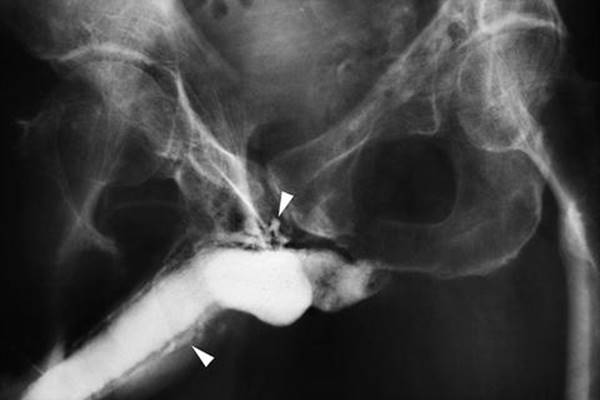
Fig. 6.2
Penile cavernosography . Penile cavernosogram demonstrating venous leak (white arrows). Courtesy of Irving J. Fishman, MD
Standardized data for DICC results are not available. However, generally used normal values are cavernosal artery occlusion pressure less than 30 mmHg, flow to maintain of less than 5 ml/min, and a pressure decay of less than 45 mmHg over 30 s [57].
6.5.4.4 Selective Internal Pudendal Arteriography
Penile arteriography is of limited use as a diagnostic modality for ED. It is an anatomic study that does not assess erectile function and is required in patients under consideration for penile revascularization surgery. A technically challenging and invasive procedure, pudendal/penile angiography requires an interventional radiologist with skill at cannulating both the internal pudendal arteries and the inferior epigastric arteries, which are used for revascularization [51]. Arterial inflow to the penis should be maximized using an intracavernosally delivered vasoactive agent, usually administered prior to contrast injection.
6.5.4.5 Endothelial Reactivity Testing
Recent studies have sought to analyze the presence of endothelial dysfunction as a precursor to overt CVD/atherosclerosis in patients presenting with ED. Increased flow and the resultant vasodilation in the penile arteries necessary for erection are mediated largely through nitric oxide (NO) produced by the endothelium. Endothelial dysfunction in patients with ED, but without evidence of other significant CVD, has been found within the penile vasculature but not within the small arteries of the forearm, suggesting that dysfunction occurs earlier within the penile endothelium than in other vascular beds [58]. In addition, increasing arterial stiffness, as evaluated with increasing pulse pressures, is associated with arteriogenic ED [59]. As a result, a number of diagnostic modalities have been used to assess endothelial function, including nonspecific serum markers (e.g., endothelin-1, interleukin 6, tumor necrosis factor-α, and C-reactive protein), analysis of flow-mediated dilation of the brachial artery (requiring ultrasound measurement of dilation in the brachial artery after arterial occlusion and administration of nitroglycerine—primarily a research modality), and office-based reactive hyperemia peripheral arterial tonometry (RH-PAT). The published literature provides conflicting information as to the utility of RH-PAT testing in the evaluation of men with possible vascular ED as part of their overall cardiovascular assessment [60, 61].
6.6 Conclusions
ED is a highly prevalent condition noted in the aging male and can be associated with a variety of different etiologies. Patients with vasculogenic ED are at risk for CVD and warrant further evaluation as deemed necessary on history, on physical examination, and with vascular testing. Diagnostic testing can be useful in clarifying the etiology of ED and should be used with a clear clinical indication and question which can help to clarify treatment options and long-term prognosis.
References
1.
Porst H, Burnett A, Brock G, Ghanem H, Giuliano F, Glina S, et al. SOP conservative (medical and mechanical) treatment of erectile dysfunction. J Sex Med. 2013;10(1):130–71.CrossRefPubMed
2.
Consensus NIH. Conference. Impotence. NIH consensus development panel on impotence. JAMA. 1993;270(1):83–90.CrossRef
3.
Litwin MS, Nied RJ, Dhanani N. Health-related quality of life in men with erectile dysfunction. J Gen Intern Med. 1998;13(3):159–66.CrossRefPubMedPubMedCentral
4.
Shabsigh R, Klein LT, Seidman S, Kaplan SA, Lehrhoff BJ, Ritter JS. Increased incidence of depressive symptoms in men with erectile dysfunction. Urology. 1998;52(5):848–52.CrossRefPubMed
5.
Lewis RW, Fugl-Meyer KS, Corona G, Hayes RD, Laumann EO, Moreira Jr ED, et al. Definitions/epidemiology/risk factors for sexual dysfunction. J Sex Med. 2010;7(4 Pt 2):1598–607.CrossRefPubMed
6.
Lue TF. Erectile dysfunction. N Engl J Med. 2000;342(24):1802–13.CrossRefPubMed
7.
Feldman HA, Goldstein I, Hatzichristou DG, Krane RJ, McKinlay JB. Impotence and its medical and psychosocial correlates: results of the Massachusetts male aging study. J Urol. 1994;151(1):54–61.PubMed
8.
Lindau ST, Schumm LP, Laumann EO, Levinson W, O’Muircheartaigh CA, Waite LJ. A study of sexuality and health among older adults in the United States. N Engl J Med. 2007;357(8):762–74.CrossRefPubMedPubMedCentral
9.
Laumann EO, West S, Glasser D, Carson C, Rosen R, Kang JH. Prevalence and correlates of erectile dysfunction by race and ethnicity among men aged 40 or older in the United States: from the male attitudes regarding sexual health survey. J Sex Med. 2007;4(1):57–65.CrossRefPubMed
10.
Rosen RC. Psychogenic erectile dysfunction. Classification and management. Urol Clin North Am. 2001;28(2):269–78.CrossRefPubMed
11.
Lizza EF, Rosen RC. Definition and classification of erectile dysfunction: report of the nomenclature committee of the international society of impotence research. Int J Impot Res. 1999;11(3):141–3.CrossRefPubMed
12.
Deveci S, O’Brien K, Ahmed A, Parker M, Guhring P, Mulhall JP. Can the international index of erectile function distinguish between organic and psychogenic erectile function? BJU Int. 2008;102(3):354–6.CrossRefPubMed
13.
Sullivan ME, Thompson CS, Dashwood MR, Khan MA, Jeremy JY, Morgan RJ, et al. Nitric oxide and penile erection: is erectile dysfunction another manifestation of vascular disease? Cardiovasc Res. 1999;43(3):658–65.CrossRefPubMed
14.
Giuliano FA, Leriche A, Jaudinot EO, de Gendre AS. Prevalence of erectile dysfunction among 7689 patients with diabetes or hypertension, or both. Urology. 2004;64(6):1196–201.CrossRefPubMed
15.
Martin-Morales A, Saenz de Tejada I, Rodriguez-Vela L, Jimenez-Cruz JF, Burgos-Rodriguez R. Prevalence and independent risk factors for erectile dysfunction in Spain: results of the epidemiologia de la disfuncion erectil masculina study. J Urol. 2001;166(2):569–74 (Discussion 74–5).CrossRefPubMed
16.
Guo W, Liao C, Zou Y, Li F, Li T, Zhou Q, et al. Erectile dysfunction and risk of clinical cardiovascular events: a meta-analysis of seven cohort studies. J Sex Med. 2010;7(8):2805–16.CrossRefPubMed
17.
Thompson IM, Tangen CM, Goodman PJ, Probstfield JL, Moinpour CM, Coltman CA. Erectile dysfunction and subsequent cardiovascular disease. JAMA. 2005;294(23):2996–3002.CrossRefPubMed
18.
Gupta BP, Murad MH, Clifton MM, Prokop L, Nehra A, Kopecky SL. The effect of lifestyle modification and cardiovascular risk factor reduction on erectile dysfunction: a systematic review and meta-analysis. Arch Intern Med. 2011;171(20):1797–803.CrossRefPubMed
19.
Traish AM, Park K, Dhir V, Kim NN, Moreland RB, Goldstein I. Effects of castration and androgen replacement on erectile function in a rabbit model. Endocrinology. 1999;140(4):1861–8.PubMed
20.
Traish AM, Toselli P, Jeong SJ, Kim NN. Adipocyte accumulation in penile corpus cavernosum of the orchiectomized rabbit: a potential mechanism for veno-occlusive dysfunction in androgen deficiency. J Androl. 2005;26(2):242–8.CrossRefPubMed
21.
Rhoden EL, Teloken C, Sogari PR, Souto CA. The relationship of serum testosterone to erectile function in normal aging men. J Urol. 2002;167(4):1745–8.CrossRefPubMed
22.
Corona G, Mannucci E, Mansani R, Petrone L, Bartolini M, Giommi R, et al. Aging and pathogenesis of erectile dysfunction. Int J Impot Res. 2004;16(5):395–402.CrossRefPubMed
23.
Isidori AM, Buvat J, Corona G, Goldstein I, Jannini EA, Lenzi A, et al. A critical analysis of the role of testosterone in erectile function: from pathophysiology to treatment-a systematic review. Eur Urol. 2014;65(1):99–112.CrossRefPubMed
24.
Corona G, Mannucci E, Fisher AD, Lotti F, Ricca V, Balercia G, et al. Effect of hyperprolactinemia in male patients consulting for sexual dysfunction. J Sex Med. 2007;4(5):1485–93.CrossRefPubMed
25.
Saenz de Tejada I, Angulo J, Cellek S, Gonzalez-Cadavid N, Heaton J, Pickard R, et al. Pathophysiology of erectile dysfunction. J Sex Med. 2005;2(1):26–39.CrossRefPubMed
26.
Berookhim B, Steinway M, Bar-Chama N, Seftel A. Benign prostatic hyperplasia evaluation, association with sexual dysfunction, treatment, and practice patterns according to physician specialty. Curr Prostate Rep. 2009;7(1):25–31.CrossRef
27.
Braun MH, Sommer F, Haupt G, Mathers MJ, Reifenrath B, Engelmann UH. Lower urinary tract symptoms and erectile dysfunction: co-morbidity or typical “aging male” symptoms? Results of the “cologne male survey”. Eur Urol. 2003;44(5):588–94.CrossRefPubMed
28.
Blanker MH, Bohnen AM, Groeneveld FP, Bernsen RM, Prins A, Thomas S, et al. Correlates for erectile and ejaculatory dysfunction in older Dutch men: a community-based study. J Am Geriatr Soc. 2001;49(4):436–42.CrossRefPubMed
29.
Glina S, Glina FP. Pathogenic mechanisms linking benign prostatic hyperplasia, lower urinary tract symptoms and erectile dysfunction. Ther Adv Urol. 2013;5(4):211–8.CrossRefPubMedPubMedCentral
30.
Hatzichristou D, Rosen RC, Derogatis LR, Low WY, Meuleman EJ, Sadovsky R, et al. Recommendations for the clinical evaluation of men and women with sexual dysfunction. J Sex Med. 2010;7(1 Pt 2):337–48.CrossRefPubMed
31.
McCabe MP, Althof SE. A systematic review of the psychosocial outcomes associated with erectile dysfunction: does the impact of erectile dysfunction extend beyond a man’s inability to have sex? J Sex Med. 2013;11:347–63.CrossRefPubMed
32.
Perelman MA. Psychosocial evaluation and combination treatment of men with erectile dysfunction. Urol Clin North Am. 2005;32(4):431–45, vi.
33.
Rosen RC, Riley A, Wagner G, Osterloh IH, Kirkpatrick J, Mishra A. The international index of erectile function (IIEF): a multidimensional scale for assessment of erectile dysfunction. Urology. 1997;49(6):822–30.CrossRefPubMed
34.
Rosen RC, Cappelleri JC, Smith MD, Lipsky J, Pena BM. Development and evaluation of an abridged, 5-item version of the international index of erectile function (IIEF-5) as a diagnostic tool for erectile dysfunction. Int J Impot Res. 1999;11(6):319–26.CrossRefPubMed
35.
Cappelleri JC, Rosen RC. The sexual health inventory for men (SHIM): a 5-year review of research and clinical experience. Int J Impot Res. 2005;17(4):307–19.CrossRefPubMed
36.
Fisher WA, Eardley I, McCabe M, Sand M. Erectile dysfunction (ED) is a shared sexual concern of couples II: association of female partner characteristics with male partner ED treatment seeking and phosphodiesterase type 5 inhibitor utilization. J Sex Med. 2009;6(11):3111–24.CrossRefPubMed
37.
Jiann BP, Su CC, Tsai JY. Is female sexual function related to the male partners’ erectile function? J Sex Med. 2013;10(2):420–9.CrossRefPubMed
38.
Moskovic DJ, Mohamed O, Sathyamoorthy K, Miles BJ, Link RE, Lipshultz LI, et al. The female factor: predicting compliance with a post-prostatectomy erectile preservation program. J Sex Med. 2010;7(11):3659–65.CrossRefPubMed
39.
Corona G, Petrone L, Mannucci E, Magini A, Lotti F, Ricca V, et al. Assessment of the relational factor in male patients consulting for sexual dysfunction: the concept of couple sexual dysfunction. J Androl. 2006;27(6):795–801.CrossRefPubMed
40.
Althof SE, Leiblum SR, Chevret-Measson M, Hartmann U, Levine SB, McCabe M, et al. Psychological and interpersonal dimensions of sexual function and dysfunction. J Sex Med. 2005;2(6):793–800.CrossRefPubMed
41.
Ghanem HM, Salonia A, Martin-Morales A. SOP: physical examination and laboratory testing for men with erectile dysfunction. J Sex Med. 2012;10:108–10.CrossRefPubMed
42.
Weinberg AE, Eisenberg M, Patel CJ, Chertow GM, Leppert JT. Diabetes severity, metabolic syndrome, and the risk of erectile dysfunction. J Sex Med. 2013;10:3102–9.CrossRefPubMedPubMedCentral
43.
Nehra A, Jackson G, Miner M, Billups KL, Burnett AL, Buvat J, et al. The Princeton III consensus recommendations for the management of erectile dysfunction and cardiovascular disease. Mayo Clin Proc. 2012;87(8):766–78.CrossRefPubMedPubMedCentral
44.
Wasserman MD, Pollak CP, Spielman AJ, Weitzman ED. The differential diagnosis of impotence. The measurement of nocturnal penile tumescence. JAMA. 1980;243(20):2038–42.CrossRefPubMed
45.
Bradley WE, Timm GW, Gallagher JM, Johnson BK. New method for continuous measurement of nocturnal penile tumescence and rigidity. Urology. 1985;26(1):4–9.CrossRefPubMed
46.
Bemelmans BL, Hendrikx LB, Koldewijn EL, Lemmens WA, Debruyne FM, Meuleman EJ. Comparison of biothesiometry and neuro-urophysiological investigations for the clinical evaluation of patients with erectile dysfunction. J Urol. 1995;153(5):1483–6.CrossRefPubMed
47.
Aitchison M, Aitchison J, Carter R. Is the penile brachial index a reproducible and useful measurement? Br J Urol. 1990;66(2):202–4.CrossRefPubMed
48.
Mahe G, Leftheriotis G, Picquet J, Jaquinandi V, Saumet JL, Abraham P. A normal penile pressure cannot rule out the presence of lesions on the arteries supplying the hypogastric circulation in patients with arterial claudication. Vasc Med. 2009;14(4):331–8.CrossRefPubMed
49.
Aversa A, Rocchietti-March M, Caprio M, Giannini D, Isidori A, Fabbri A. Anxiety-induced failure in erectile response to intracorporeal prostaglandin-E1 in non-organic male impotence: a new diagnostic approach. Int J Androl. 1996;19(5):307–13.CrossRefPubMed
50.
Virag R, Frydman D, Legman M, Virag H. Intracavernous injection of papaverine as a diagnostic and therapeutic method in erectile failure. Angiology. 1984;35(2):79–87.CrossRefPubMed
51.
Sikka SC, Hellstrom WJ, Brock G, Morales AM. Standardization of vascular assessment of erectile dysfunction: standard operating procedures for duplex ultrasound. J Sex Med. 2012;10:120–9.CrossRefPubMed
52.
Lue TF, Hricak H, Marich KW, Tanagho EA. Vasculogenic impotence evaluated by high-resolution ultrasonography and pulsed Doppler spectrum analysis. Radiology. 1985;155(3):777–81.CrossRefPubMed
53.
Quam JP, King BF, James EM, Lewis RW, Brakke DM, Ilstrup DM, et al. Duplex and color Doppler sonographic evaluation of vasculogenic impotence. AJR Am J Roentgenol. 1989;153(6):1141–7.CrossRefPubMed
54.
Benson CB, Aruny JE, Vickers Jr MA. Correlation of duplex sonography with arteriography in patients with erectile dysfunction. AJR Am J Roentgenol. 1993;160(1):71–3.CrossRefPubMed
55.
Wilkins CJ, Sriprasad S, Sidhu PS. Colour Doppler ultrasound of the penis. Clin Radiol. 2003;58(7):514–23.CrossRefPubMed
56.
Vardi Y, Glina S, Mulhall JP, Menchini F, Munarriz R. Cavernosometry: is it a dinosaur? J Sex Med. 2008;5(4):760–4.CrossRefPubMed
57.
Glina S, Ghanem H. SOP: corpus cavernosum assessment (cavernosography/cavernosometry). J Sex Med. 2013;10(1):111–4.
58.
Kaiser DR, Billups K, Mason C, Wetterling R, Lundberg JL, Bank AJ. Impaired brachial artery endothelium-dependent and -independent vasodilation in men with erectile dysfunction and no other clinical cardiovascular disease. J Am Coll Cardiol. 2004;43(2):179–84.CrossRefPubMed
59.
Corona G, Mannucci E, Lotti F, Fisher AD, Bandini E, Balercia G, et al. Pulse pressure, an index of arterial stiffness, is associated with androgen deficiency and impaired penile blood flow in men with ED. J Sex Med. 2009;6(1):285–93.CrossRefPubMed
60.
Aversa A, Francomano D, Bruzziches R, Pili M, Natali M, Spera G, et al. The application of digital pulse amplitude tonometry to the diagnostic investigation of endothelial dysfunction in men with erectile dysfunction. Andrologia. 2011;43(1):9–15.CrossRefPubMed
61.
Mehta A, Miner M, Sigman M. Assessment of EndoPAT scores in men with vasculogenic and non-vasculogenic erectile dysfunction. Int J Clin Pract. 2013;67(1):46–51.CrossRefPubMed