
ANTIGEN RECEPTORS
The immune system has two main ways of specifically recognizing antigen. B cells recognize intact antigens by using immunoglobulin (antibody) as their receptor. In contrast, T cells recognize antigen originating from within other cells, using their T-cell receptor (TCR). Many cells also have receptors for components of microbial pathogens that antedate the evolution of lymphocytes.
Antigen is the term used to describe any molecule that can be recognized by B cells or T cells. In general, immunoglobulins recognize and bind to intact antigens or large fragments that have retained some tertiary structure. Most T cells only recognize polypeptide fragments of antigens that have become associated with molecules encoded by the major histocompatibility complex (MHC) and that are expressed on the surface of other cells of the body.
Antigenic determinants, or epitopes, are the parts of an antigen to which an immunoglobulin binds. Antigens usually have many determinants, which may be different from each other or may be repeated molecular structures. Virtually the entire surface of a protein is potentially antigenic. Figure 2.1 illustrates epitopes on lysozyme recognized by three different monoclonal antibodies.
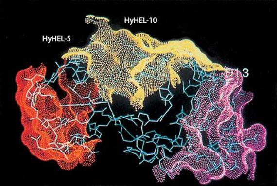
Fig. 2.1 Three epitopes of lysozyme. Courtesy of D. R. Davis.
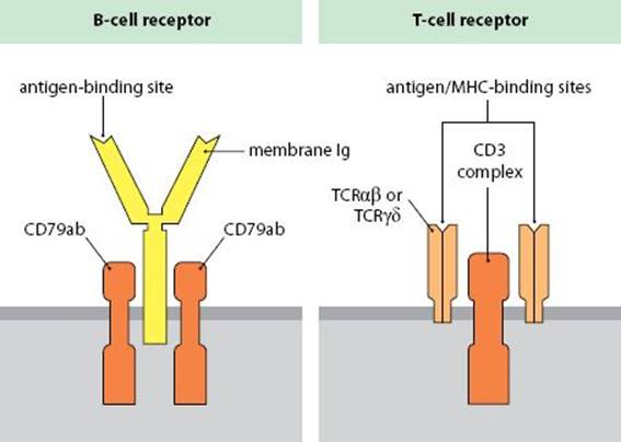
Fig. 2.2 Antigen receptors on B cells and T cells.
Antibodies (Ab)/Immunoglobulins (Ig) were originally identified as a class of serum proteins induced after contact with antigen, which bind specifically to the antigen that induced their formation. Subsequently it was found that B cells use a membrane-bound form of their secreted antibody as their antigen receptor (BCR). Surface immunoglobulins on B cells are associated with two polypeptides, Igα and Igβ (CD79a and CD79b).
Igα and Igβ (CD79) are transmembrane molecules that transduce activation signals to the B cell and are required for the expression of membrane Ig. Hence CD79 is a marker of mature B cells.
T-cell antigen receptors (TCRs) are integral membrane proteins on all mature T cells that specifically recognize antigenic peptides associated with MHC-encoded molecules. The receptor consists of a heterodimer responsible for MHC/antigen binding and a cluster of associated membrane-bound polypeptides, the CD3 complex, which triggers cell activation. The MHC/antigen-binding portion of the TCR varies between different clones of T cells, but the peptides of the CD3 complex (γ, δ, ε2, ζ2) are invariant.
Immunoreceptor tyrosine activation motifs (ITAMs) are segments found in the intracytoplasmic portion of many immune receptors, including CD79 and CD3, which are targets for phosphorylation by tyrosine kinases. ITAM phosphorylation promotes cell activation. Analogous inhibitory motifs (ITIMs) block cell activation if they become phosphorylated.
ANTIBODY STRUCTURE
Heavy chains and Light chains Antibody molecules all have a basic four polypeptide chain structure consisting of two identical light (L) chains and two identical heavy (H) chains, stabilized and cross-linked by intra- and inter-chain disulfide bonds (red) (Fig. 2.3). The heavy chains are glycosylated (between the CH2 domains of IgG). There are five major types of Ig heavy chain (μ, γ, α, ε, δ), consisting of 450–600 amino acid residues, and the type determines the class of antibody. Light chains are of two main types (κ, λ), consisting of about 230 residues. Either type of light chain may associate with any of the heavy chains. Both heavy and light chains are folded into domains.
Pre-B cell receptor is a receptor found on developing B cells consisting of one μ heavy chain and surrogate light chains.
Membrane and Secreted immunoglobulins (Ig) Antibodies can be produced either as integral membrane proteins of mature B cells, which act as their antigen receptor, or in a secreted form. Secreted Igs are structurally identical to their membrane counterparts, except that they lack the transmembrane segment and short intracytoplasmic segment at the C-terminus. Secreted Igs are present in extracellular fluids and secretions. Virgin B cells produce membrane Ig, but after activation by antigen and differentiation into plasma cells they switch to the production of secreted immunoglobulins.

Fig. 2.3 Polypeptide chain structure of IgG1.
Variable (V) and Constant (C) regions Examination of the degree of amino acid variability between different antibody molecules of the same class shows that the largest amount of sequence variation is concentrated in the N-terminal domains of the light and heavy chains; this is therefore called the V region. The V regions of one light and one heavy chain form an antigen-binding site. The remaining domains are relatively invariant within any particular class of antibody, and so are called the constant (C) region. The domains of antibody molecules are named according to whether they are variable or constant and according to whether they are in the light or heavy chain. For example:
VH and VL are variable domains of heavy and light chains.
CL and CH1 are the constant domains of the light chain and the first constant domain of the heavy chain, respectively.
Cγ, Cμ etc. are domains of the heavy chain, which indicate the class of antibody. For example, Cμ1 is the first constant domain of the μ heavy chain of IgM antibody.

Fig. 2.4 The folding pattern of an immunoglobulin light chain.
Fab and Fc regions refer to two regions of the antibody, corresponding to antibody-binding (Fab) and constant (Fc) regions. The nomenclature originally refers to fragments produced by digestion with the enzyme papain.
Hinge region is a section of the heavy chain that contains inter-heavy chain disulfide bonds and confers segmental flexibility on the antibody molecule, so that both antigen-binding sites can independently engage the surface of a pathogen.
ANTIBODY–STRUCTURAL VARIATIONS
Classes and Subclasses (Isotypes) Antibodies may be grouped on the basis of structural similarities into different classes and subclasses, depending on their heavy chains. Each class serves different functions. In mammals there are five antibody classes: IgG, IgM, IgA, IgD, and IgE. IgG and IgA are further divided into subclasses. The number of subclasses varies between species. For example in humans there are four IgG subclasses, IgG1 to IgG4. As there is a gene in every individual for every one of the classes and subclasses, these are isotypic variants or antibody isotypes.
Kappa and Lambda chains Antibody light chains may also be divided into two types, namely κ and λ, which are encoded by separate gene loci. They, too, are isotypic variants. Either type of light chain can combine with one of the heavy chains.
Allelic exclusion is the process by which a cell uses either the gene from its maternal chromosome or the one from the paternal chromosome, but not both. Individual B cells display allelic exclusion of their heavy and light chain genes. T cells also display allelic exclusion of their TCR αβ or γδ heterodimers.
Single-chain antibodies Species of Camelidae (camels and llamas) produce antibodies consisting of paired heavy chains (no light chains). These antibodies demonstrate that a heavy chain alone can form an effective antigen-binding site and they have acted as models for the development of genetically engineered antibodies.
Single-domain antibodies (nanobodies) are single antibody V domains produced by genetic engineering, which can be used for applications that require immune recognition or targeting.
Idiotypes (Ids) are variants caused by the large amount of structural heterogeneity in the immunoglobulin V regions, which is related to the diversity required to bind different antigens. Some idiotypes are only made by animals that have a particular set of Ig genes (haplotypes) and these are ‘germline idiotypes.’
Recurrent and dominant idiotypes Sometimes a particular idiotype is frequently seen in the immune response of different individuals to a particular antigen. This is a recurrent idiotype. If an idiotype constitutes a major part of an antibody response to that antigen, then it is a dominant idiotype.
Idiotopes are antigenic determinants on the V regions of antibodies that can be recognized by anti-idiotypic antibodies. An idiotype is identified by the collection of idiotopes it expresses. Individual idiotopes may be present on more than one antibody.
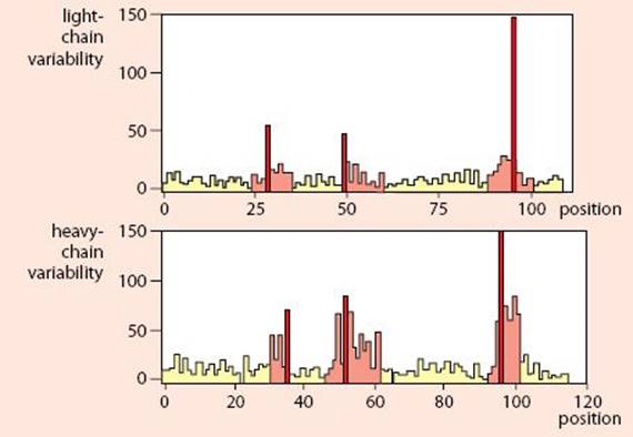
Fig. 2.5 Kabat and Wu plot of antibody heavy and light chains.
Kabat and Wu plot shows the amino acid sequence variability in immunoglobulin, determined by comparing the amino acid sequences of many different antibodies. It plots variability against amino acid position, thereby highlighting the most variable regions of the heavy and light chains.
Hypervariable regions and Framework segments. Within the V domains of the heavy and light chains there are three regions of greatest variability, which are clustered at the antigen-binding site. (red in the Kabat and Wu plot above). These hypervariable segments are separated by relatively invariant framework segments.
Complementarity-determining regions (CDRs) are the parts of the V domains that form the antigen-binding site. V-domain folding brings the CDRs together at the distal tip of the molecule.
Allotypes are variants due to intraspecies genetic differences. Each individual has a particular variant at each Ig gene locus, which will often differ from those in other individuals. In humans the Gm series of allotypes is found on IgG heavy chains.
Immunoglobulin supergene family (IgSF) The domain structure seen in antibodies consisting of three or four polypeptide loops stabilized by β-pleated sheet and a disulfide bond (sometimes called a β-barrel) is found in many molecules, which all belong to the immunoglobulin supergene family (see Fig. 2.4). The domain is seen in cell-surface receptors, including CD2, CD4, CD8, the T-cell receptor (TCR), MHC molecules, and Fc receptors (CD16, CD32, CD64).
ANTIBODY FUNCTIONS
Antibodies are bifunctional molecules. Their first function is to bind antigen and their second is to interact with host tissues and effector systems to facilitate removal of the antigen. Some antibody functions can be mediated just by binding to the antigen. For example, antibodies against surface molecules of viruses can prevent them from binding to and infecting host cells. However, most antibody functions require the antigen:antibody complex to bind to Fc receptors on cells. The antigen-binding sites are formed from the V domains of a heavy and light chain, whereas the C domains of the Fc region interact with cells of the immune system and C1q of the complement system. The different antibody classes and subclasses interact with different cells and so have different functions.
IgG is the major serum Ig and constitutes the main antibody in secondary immune responses to most antigens. In humans it is transferred across the placenta to provide protection in neonatal life. All IgG subclasses, except IgG4, can bind to C1q by sites in Cγ2 to activate the complement classical pathway. IgG can act as an opsonin by cross-linking immune complexes to Fc receptors on neutrophils and macrophages. It can also sensitize target cells for destruction by large granular lymphocytes with Fc receptors.
FcRn is the placental Fc receptor that binds all subclasses of IgG and transports them to the fetal circulation.
IgM is a pentamer of the basic four-chain structure. It is the first class to be produced during the development of the immune system and in the primary immune response. It fixes complement very efficiently and is the main antibody component of the response to T-independent antigens.
IgD is a trace antibody in serum but acts as a cell-surface receptor on many B cells, where it is co-expressed with IgM. IgD appears on differentiating B cells after activation, but is absent from mature antibody-forming cells.
IgA occurs as monomers, dimers, and polymers of the basic four-chain unit, existing in humans mostly as monomers and in other species as dimers. IgA is the most abundant Ig class in secretions, where it protects mucous membranes. It is also found in colostrum and is particularly important in protecting the neonates of species that do not transfer IgG across the placenta.
J chain is a polypeptide present in polymeric Igs (IgM and IgA), which facilitates their polymerization. It is synthesized by B cells but is not encoded by the Ig genes.
Poly-Ig receptor is present on the serosal surface of epithelial cells that transport and secrete IgA. It is a member of the Ig supergene family, with five domains. IgA dimers bind to the receptor and are transported across the epithelium. The receptor is then cleaved, forming the secretory piece and releasing secreted IgA by exocytosis.
Secretory piece is the released form of the poly-Ig receptor, which attaches to IgA by disulfide bonds and is wound around the C domains of IgA to protect it from degradation by enzymes.
IgE binds to high-affinity Fc receptors (FcεRI) on mast cells and basophils, where it sensitizes them to release inflammatory mediators such as histamine after contact with antigen. IgE is particularly important in protection against helminthic infections, but it also mediates type I hypersensitivity reactions, such as asthma and hayfever.
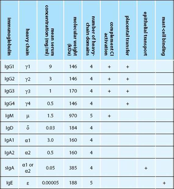
Fig. 2.6 Properties of human immunoglobulin isotypes.
ANTIBODY GENES
The genes for antibodies lie at three gene loci on separate chromosomes; these are the K, L (κ, λ), and H (heavy-chain) loci. At each of these loci there are large numbers of different gene segments encoding polypeptides (exons), separated by segments that do not encode protein (introns) but contain sequences important in gene control and the process of recombination. The antibody genes undergo a number of recombinational events during B-cell development and maturation. The first events are DNA rearrangements of H and L chain genes, to form gene segments encoding their V domains.
Generation of diversity is the process by which large numbers of antibody V regions are generated. This is achieved by:
•Many different germline V genes in the K, L, and H loci
•Recombination between V, D, and J gene segments
•Insertion of nongermline (N) nucleotides into the joints
•Varied combinations of light and heavy chains
•Somatic mutation of V genes in individual B cells
T-cell receptors are diversified by similar mechanisms, although TCR genes are not subject to somatic mutation.
V genes encode the N-terminal 95 (approximately) amino acids of the antibody V domains. The number of V genes at each locus varies between loci and species. Analogous V genes are present in the four gene loci encoding TCR chains.
J genes and D genes To produce a gene encoding a heavy-chain V domain, any one of the H-chain V genes is recombined with any of a small number of D (Diversity) and J (Joining) genes to produce a VDJ gene. Recombination of light-chain genes is similar, except that they have no D-gene segments, and a V gene is recombined directly to a J gene. The T-cell receptor B and D loci have analogous D and J genes, while the TCR A and G loci have only J genes. (Note that J genes should not be confused with J chains.)
Recombination signal sequences (RSSs) and the 12/23 rule Somatic recombination is the process by which the various gene segments for antigen receptors are brought together and joined. This process depends on specific recombination sequences flanking each V, D, and J gene that appose the segments, which are then enzymatically cut and rejoined to remove the intervening introns. The sequences consist of a heptamer, 12 or 23 bases, and a nonamer. The 12/23 rule states that a flanking sequence with 12 bases can only recombine with one of 23 bases. This ensures that heavy chains only make VDJ recombinations and light chains only make VJ recombinations.
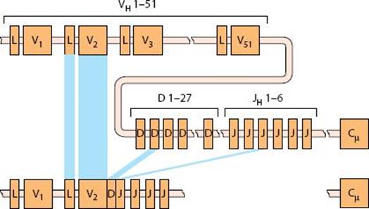
Fig. 2.7 VDJ recombination in the human IGH locus.
Junctional diversity is created when the ligation point in antibody gene recombination (VJ, VD, DJ) differs between B cells using the same gene segments. The shift produces different codons—see the sequence of the antibody S107 in Fig. 2.8.
N-region diversity is created when additional nucleotides are inserted into the gap between recombining gene segments with no corresponding germline DNA sequence—see the sequence of antibody M167 in Fig. 2.8. The reading frame must be restored to produce a functional antibody.

Fig. 2.8 Junctional and N-region diversity at a VD junction.
RAG-1, RAG2 (Recombination-activating genes) control the recombination of the TCR gene in T cells or the Ig genes in B cells. The enzymes recognize the recombination signal sequences and bring them together to initiate recombination by producing a double-strand break.
Terminal deoxynucleotidyl transferase (Tdt) is an enzyme that can add nucleotides to the exposed ends of the DNA during the recombination; these nucleotides become incorporated into the junctions between the V, D, and J gene segments.
Somatic hypermutation is the process by which DNA base changes occur during the lifetime of a B cell, producing point mutations in the Ig polypeptides. The high rate of mutation is centered on the recombined VJ and VDJ genes. The mechanism is activated in centroblasts and associated with class switching—IgG molecules usually vary more from germline sequences than IgM.
Antibody synthesis The segment of DNA encoding the recombined VDJ (heavy chain) or VJ (light chain) region and the C domains is transcribed into a primary RNA transcript that still contains introns. The transcript is then spliced to remove the introns, a process that involves recognition of sequences (donor and acceptor junctions) flanking the exons. This leaves mRNA, which is translated across the membrane of the endoplasmic reticulum (ER). Each mRNA has a leader or signal sequence, by which it is directed to the ER. The process is illustrated below for a membrane IgM μ polypeptide. Complete Igs are assembled and glycosylated in the ER and stored in the Golgi apparatus. Secreted Ig is released by exocytosis, whereas membrane Ig, associated with CD79-signaling peptides, is moved to the cell surface.
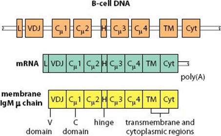
Fig. 2.9 Production of an IgM µ polypeptide.
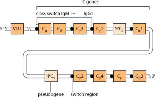
Fig. 2.10 Class switching in the human IGH C-gene locus.
C genes The heavy-chain constant-region genes are arranged downstream (3′) of the recombined VDJ gene. Each gene consists of a series of exons encoding the individual C domains, as well as separate exons for the hinge (except IgA) and for the transmembrane and cytoplasmic regions. The primary transcript of the heavy chains can be processed, to produce mRNA either for membrane or for secreted Ig. To produce membrane Ig, the exons for the transmembrane segments are spliced to a point just within the final C domain. If this does not occur, the stop signal is retained and mRNA for secreted Ig is produced. The point of polyadenylation controls how the primary transcript will be spliced. Initially a B cell joins a μ gene to its VDJ gene, but other C genes may displace the μ gene in a process called class switching.
Class switching is a process by which a B cell can switch the class of Ig it produces while retaining the same antigen specificity. All the heavy-chain constant-region genes except Cδ are preceded by a switching sequence. Switching is effected by bringing a new C gene up to the position occupied by the μ C gene, with the loss of the intervening C genes. This process is illustrated above for the switch from IgM to IgG1. It is also possible for a cell to switch classes by producing very long primary transcripts, which are then spliced to connect the new C gene to VDJ. Indeed, this is the only way in which IgD (which lacks a switch sequence) can be produced. The process is controlled by T cells and modulated by cytokines. For example, in humans IL-4 promotes switching to IgG4 and IgE, whereas IL-5 promotes a switch to IgA.
ANTIBODY BIOTECHNOLOGY
Much of the early work on the elucidation of antibody structure was performed using fragments of antibodies prepared by enzyme digestion. For example, the Fab and F(ab′)2 fragments of IgG are produced by digestion with papain or pepsin, respectively. Lacking the Fc region, they are both useful in determining which antibody functions are Fc-dependent. The development of monoclonal antibody technology was a major step forward in allowing researchers to produce large quantities of well-defined antibodies. More recently, genetic engineering has been used to generate antibodies and antibody fragments for specific applications.
Polyclonal and monoclonal antibodies Immunization of an animal stimulates antibody production from a large number of different clones of B cells. These antibodies will differ in their epitope specificity and affinity for the antigen. Such antibodies are referred to as ‘polyclonal.’ In contrast, the antibodies produced by a single clone of B cells (monoclonal antibodies) have a defined specificity and affinity. Note that a monoclonal antibody is not necessarily of higher affinity than a polyclonal antibody, and whether it is more effective than polyclonal antibody depends on the assay or purpose it is used for.
Phage display antibodies is a method for producing antibody fragments. Mixed mRNAs for antibody VH and VL domains are cross-linked with a spacer to give a gene for an FV fragment. The gene is inserted into a vector (phage), which expresses the FV on its tips. Phages are selected according to their binding specificity and transfected into bacteria to synthesize the FV fragments.
Humanized antibodies are required where the antibody itself must not be antigenic, for example, for long-term therapy of patients. The genes for the antigen-binding hypervariable regions of the required antibody are spliced into the genes encoding the framework regions of a human heavy- or light-chain V-domain.
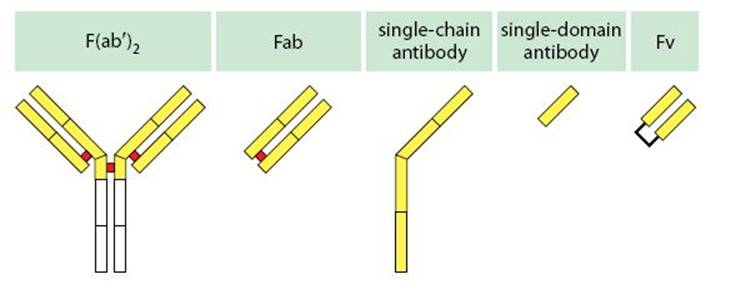
Fig. 2.11 Antibody fragments.
ANTIGENS
Immunogens An antigen is any molecule recognized by the immune system, but the term immunogen is reserved for those antigens that elicit a strong immune response, particularly in the context of protective immunity to pathogenic organisms.
Haptens and Carriers Artificial antigens have been used to examine the immune response. In particular, small antigenic determinants (haptens) are covalently coupled to larger molecules (carriers). Haptens bind to antibodies but cannot by themselves elicit an antibody response. Haptens are recognized by B cells, which present fragments of the carriers to T cells.
T-dependent antigens need to be recognized by both T cells and B cells to elicit an antibody response. Most protein antigens fall into this category. T-dependent antigens induce class switching to IgG and IgA with an increase in antibody affinity.
T-independent antigens can stimulate B cells to produce antibody without T-cell help. Most such antigens are large polymeric molecules, with repeated epitopes, capable of cross-linking surface Ig on B cells, and they are only slowly degraded.
Type I and Type II T-independent antigens are differentiated according to their ability to activate different B-cell subsets. Type I antigens stimulate both Lyb5+ and Lyb5− cells in mouse, whereas type II antigens can only act on Lyb5+ cells.

Fig. 2.12 Properties of commonly used T-independent antigens.
ANTIGEN–ANTIBODY INTERACTIONS
Epitopes and Paratopes are part of a nomenclature used to describe the interaction between antigen and antigen-receptor molecules, including antibodies. An epitope is an antigenic determinant; a paratope, formed by hypervariable loops of V domains, is the part of the antibody that binds to the epitope.
Contact residues are the amino acids of the epitope and paratope that contribute to the antigen–antibody bond.
Continuous and Discontinuous epitopes Study of the molecular interaction between antigen and antibody shows that some of the epitopes are formed by one linear stretch of amino acids (continuous epitope). In most cases, however, an epitope has contact residues from different sections of an antigen brought together by folding of the polypeptide (discontinuous epitope).
Antigen–antibody bond Antibodies bind specifically to the antigen that induced their formation by multiple noncovalent bonds, including van der Waals forces, salt bridges, hydrogen bonds, and hydrophobic interactions. Crystallographic studies of immune complexes between antibodies and protein antigens indicate that they interact by complementary surfaces of up to 1000 Å2 with the third hypervariable region (VJ, VDJ) lying near the centre of the binding site. Hypervariable regions of both L and H chains contribute contact residues. Figure 2.13 (top) shows lysozyme antigen (green) and the light (yellow) and heavy (blue) chains of complexed anti-lysozyme Fab. The lower diagram shows the molecules rotated forward through 90°, with contact residues (red) numbered on the interacting faces.
Charge neutralization refers to the observation that charged contact residues on an epitope are often neutralized by residues of an opposite charge on the paratope. This is particularly important at the center of the binding site.
Induced fit refers to the flexing of residues in the hypervariable loops in contact with the epitope, which may occur to allow optimum fit between the interacting molecules.
Antibody affinity is a measure of the bond strength between a single epitope and a paratope. It depends on the sum of the bond energies of the noncovalent interactions, set against the natural repulsion between molecules and the energy required to make any necessary distortions to allow binding (induced fit).
Antibody valency describes the number of binding sites on a molecule. For example, IgG has two sites and IgM has ten, although the actual number of bonds that can be formed depends on the configuration of the antigen.
Antibody avidity is the total strength of an antigen–antibody bond, which is related to the affinity of the paratope–epitope bonds and antibody valency. Binding energy is much enhanced when several bonds form, so avidity usually exceeds affinity.
Cross-reaction Some antisera are not totally specific for their inducing antigen, but bind related (cross-reacting) antigens, either because they share a common epitope or because the molecular shapes of the cross-reacting antigens are similar.
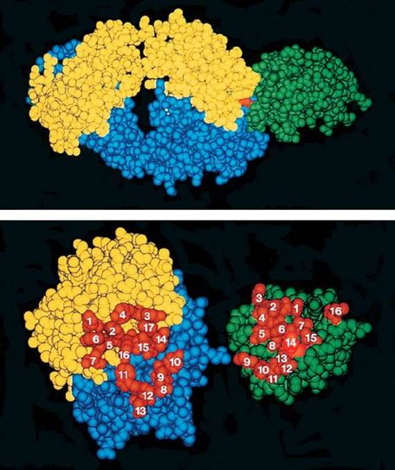
Fig. 2.13 The Fab:lysozyme complex. Courtesy of R.J. Poljak from Science 1986, 233:747. Copyright 1986 by the AAAS.
T-CELL ANTIGEN RECEPTOR (TCR)
The TH cell antigen receptor consists of a heterodimer (Ti) and a number of associated polypeptides that form the CD3 complex. The dimer recognizes processed antigen associated with an MHC molecule. The CD3 complex is required for receptor expression and is involved in signal transduction.
TCRαβ (TCR2) and TCRγδ (TCR1) The polypeptide chains for the antigen-binding portion of the receptor are encoded by four different gene loci: TCRA, B, G, and D. Any T cell will express either an αβ or a γδ receptor. The great majority of thymocytes and peripheral T cells have a TCRαβ.
Ti is a term used to distinguish the antigen:MHC-binding portion (which differs between cells) from the monomorphic CD3 complex. The N-terminal domains of αβ or γδ resemble a membrane-bound Fab, with variable (V) domains forming the antigen:MHC receptor and membrane-proximal constant (C) domains.
CD3 complex in humans consists of four polypeptide chains, each of which spans the cell membrane. These are the γ, δ, ε, and ζ chains. The first three are structurally related single-domain members of the Ig supergene family; the ζ chains are unrelated and form ζ–ζ dimers. In mice, a fifth chain, η, is also present as a minority alternative partner for ζ chains, making a η–ζ dimer. The CD3 ζ–ζ dimer has intracellular ITAM motifs, which become phosphorylated after the receptor binds to antigen:MHC, allowing it to bind to kinases, which initiate T-cell activation.

Fig. 2.14 A model of a T-cell receptor complex (TCR2).
T-CELL RECEPTOR GENES
The genes for the antigen:MHC-binding portion of the TCR are similar to those of antibody, in that they consist of multiple V, D, and J segments which become recombined during T-cell development to produce function VDJ or VJ genes (see p. 34). These encode the N-terminal V domains of the TCR. The α and γ loci have V and J segments only, whereas β and δ have V, D, and J segments. The recombined V gene is linked to the exons for the C domains, the short hingelike section (containing the interchain disulfide bond), the transmembrane and cytoplasmic segments. The layouts of the human α and β loci are shown below, and those of the mouse α, β, and δ loci are very similar. Note that there are tandem sets of genes for the β-chain D, J, and C regions. Each locus is distinct, although the δ-chain D, J, and C genes lie between the Vα and Jα genes. The process of recombination can permit variability in the precise linking position of V to J, the possibility of linking D segments in all three reading frames and the addition of N-region diversity—that is, insertion of bases not encoded in the germline. Theoretically, the arrangement of recombination sequences flanking the Dβ and Dδ genes permits the assembly of genes with more than one D region (that is, VDDJ). In contrast to antibody genes, the TCR genes do not undergo somatic hypermutation. Nevertheless the amount of diversity that can be generated is at least as great as for antibodies. The genes for the γ, δ, and ε polypeptides of the CD3 complex do not rearrange and are closely linked on chromosome 11 in humans. All CD3 genes are required for TCR expression, and charged residues in the CD3 transmembrane segments are thought to be involved in association with the antigen-binding αβ or γδ dimers.
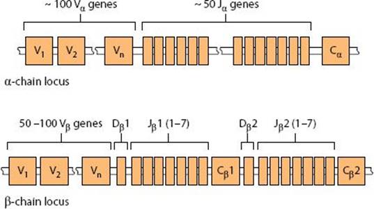
Fig. 2.15 Genes of the human TCRA (α) and TCRB (β) loci.
MHC MOLECULES
Major Histocompatibility Complex (MHC) is a large group of genes, including those encoding the class I and II MHC molecules, involved in the presentation of antigen to T cells. The complex was originally identified as a locus encoding allogeneic cell-surface molecules involved in graft rejection. A variety of other proteins are also encoded in the MHC, including complement components (C4, C2, FB), heat shock proteins, and cytokines (TNF-α, TNF-β).
MHC class I molecules are integral membrane proteins found on all nucleated cells and platelets. They are the classical transplantation antigens, each having one polypeptide chain encoded within the MHC that traverses the plasma membrane. The extracellular portion has three domains (α1–α3). The membrane-proximal α3 domain is associated with β2-microglobulin, whereas the two N-terminal domains form an antigen-binding pocket, consisting of a base of β-pleated sheet derived from both α1 and α2 domains, surrounded by two loops of α helix. Residues facing into the binding pocket vary between different molecules and haplotypes, to allow different antigenic peptides to bind. The α3domain has a binding site for CD8.
β2-Microglobulin (β2m) is a polypeptide encoded by a gene outside the MHC, which forms a single domain related to Ig domains. It is necessary for loading and transport of class I to the cell surface.
Class I-like (nonclassical, Ib) MHC molecules have the same basic structure as MHC class I molecules, and a variety of functions. Some are encoded within the MHC, but many are not.

Fig. 2.16 Structures of MHC class I and class II molecules.
CD1 is a group of four MHC class I-like molecules with deep antigen-binding pockets that can accommodate acyl groups of glycolipid and lipoprotein antigens which they present to T cells, such as lipoarabinomannan from mycobacteria.
MHC class II molecules (Ia antigens) are expressed on B cells, macrophages, monocytes, APCs, and some T cells. They consist of two noncovalently linked polypeptides (α and β), both encoded within the MHC, which both traverse the plasma membrane, each having two extracellular domains. Class II molecules resemble class I molecules with the N-terminal α1 and β1 domains forming the peptide-binding site. Another site in the β2 domain binds to CD4. Several class II-like genes (DM) are also encoded in the MHC. They facilitate the loading of antigenic peptides onto the class II molecules.

Fig. 2.17 Structure of a class I MHC molecule.
MHC GENES
A major histocompatibility complex is found in all mammal species. In humans the locus is called HLA; in mice it is the H-2 complex and in rats it is RT-1.
HLA (Human Leukocyte Antigen) locus is the human major histocompatibility complex, so called because the MHC molecules were originally identified as antigens on the surface of leukocytes and genetic variability in the MHC molecules was identified serologically. Nowadays variations are identified by genotyping. The HLA complex contains more than 220 individual gene loci, of which 21 have an immunological function. The class I and class II genes are highly polymorphic, with more than 6000 class I sequence variants and 1500 class II variants identified. There is also some variation in copy number in individual loci between haplotypes. The gene complex is located on chromosome 6, and it includes three principal class I and three class II loci.
HLA-A, -B and –C loci encode the α chains of the classical MHC class I molecules, expressed by all nucleated cells, which present antigens to CD8+ cytotoxic T cells.
HLA-E encodes a class I-like molecule that presents the signal sequence (leader) peptides of the classical MHC class I molecules to NK cells. The complex is recognized by a receptor consisting of CD94 and NKG2. HLA-E genes have limited polymorphism.
HLA-G is a class I-like molecule expressed on the placental syncytiotrophoblasts (which do not express HLA-A, -B, and –C) and is thought to prevent allograft rejection of the fetus mediated by NK cells. It can be produced in membrane-bound and soluble forms.

Fig. 2.18 HLA—the human major histocompatibility complex.
HLA-DP, -DQ, and –DR loci encode class II MHC molecules expressed on APCs, which present peptides to CD4+ T cells. Originally these were described as HLA-D specificities, detected by their ability to stimulate allogeneic cells in mixed lymphocyte cultures. Later they were defined serologically and most recently by gene sequence. DP and DQ each encode one pair of class II α and β chains, plus pseudogenes. The DR locus encodes one nonpolymorphic α chain and one to four β chains depending on the individual haplotype. Since α chains encoded on one chromosome can combine with β chains encoded on the other, this is a source of additional structural diversity in class II molecules.
HLA-DM encodes the class II molecule DM, which is involved in loading peptides onto class II molecules.
LMP-2 and LMP-7 encode components of proteasomes which are induced by interferon-γ and modify the proteasome function.
TAP-1 and TAP-2 encode transporters that take antigenic peptides from the cytoplasm into the endoplasmic reticulum.
HLA-class III genes is a catch-all term for other genes encoded within the MHC, including complement components C2 and FB, the pseudoalleles for C4 (C4F and C4S), which determine the Rogers and Chido blood groups, respectively. Genes for TNF, some heat shock proteins (such as HSP7), and enzymes (such as adrenal steroid 21-hydroxylase, CYP21) lie in this region.
H-2 is the mouse major histocompatibility complex, which lies on chromosome 17. There are six main regions: K,M,A,E, S, and D.
H-2K and H-2D encode class I MHC molecules. The K locus has one gene, whereas the number of genes in the D locus varies between strains.
H-2A and H-2E encode the α and β chains of the class II molecules. This was previously designated as the H-2I region and subdivided into I-A and I-E.
H-2S includes the genes for complement components and is analogous to the ‘class III’ region in humans.
H-2T region (Qa and Tla loci) lies downstream of the main H-2 complex and contains genes for more than 25 class-I like molecules. Some function as hemopoietic differentiation molecules; others present antigens or interact with NK cells. Some of them may be pseudogenes that act as a source of DNA for gene conversion with conventional class I molecules, to promote gene diversity. Some of the genes were originally identified on thymocytes or as thymic leukemia antigens (Tla).
INNATE IMMUNE RECOGNITION
Pathogen-associated molecular patterns (PAMPs) are common molecular motifs found on a number of pathogens. Examples are bacterial flagellin and double-stranded RNA. These motifs can be used to recognize pathogen infection.
Pattern recognition receptors (PRRs) is the generic term for cell-surface receptors and soluble molecules that recognize PAMPs. Many of these receptors are evolutionarily ancient (such as the Toll-like receptors TLR), and they are expressed on many different cell types. Mononuclear phagocytes have a particularly wide range of pattern recognition receptors.
Mannose receptor (CD206) is present on macrophages, monocytes and a subset of dendritic cells. The receptor contains eight C-type lectin domains that can bind carbohydrate groups containing mannosyl or fucosyl residues, and a terminal lectin domain that binds sulfated carbohydrate groups (Fig. 2.19). The receptor can recognize a number of microbial proteoglycans, but also binds endogenous proteins, including myeloperoxidase, lysosomal hydrolases, and some hormones.
Scavenger receptors are a structurally diverse group of receptors present on macrophages, dendritic cells, and some endothelial cells. Three receptors belonging to the SR-A family (SR-AI (CD204), SR-AII, and MARCO), bind to components of Gram-positive and Gram-negative bacteria, including lipopolysaccharide and lipoteichoic acids. They contribute to the ability of macrophages to phagocytose bacteria and promote clearance of apoptotic cells.

Fig. 2.19 Mannose receptor and scavenger receptor SR-AI.
Siglecs are a family of 12 proteins that bind sialic acid (sialic acid-binding, Ig-like lectins). Siglec-1 (sialoadhesin, CD169) is strongly expressed on macrophages in lymphoid tissues and less strongly on other tissue macrophages. It is thought to mediate intercellular adhesion by binding to extracellular matrix and other cell-surface molecules, including leukosialin (CD43) and the mannose receptor. Because sialic acid is expressed on eukaryotic cells but not on most microbes, it can distinguish them, and some of the siglecs inhibit immune activation. Siglec-2 (CD22) expressed on B cells is associated with the receptor complex (BCR) and promotes endocytosis. Siglec-3 (CD33) expressed on macrophages and myeloid stem cells also belongs to this family.
Dectins are receptors on macrophages and dendritic cells with a single lectin-like domain. Dectin-1 binds β-glucan from fungi and promotes their phagocytosis. Individuals lacking Dectin-1 are susceptible to mucosal candidiasis.
DC-SIGN (Dendritic cell-specific ICAM3-grabbing non-integrin) is a mannose-binding C-type lectin found on dendritic cells and some macrophages. It interacts with Toll-like receptors and is thought to promote signaling between APCs and T cells.
MINCLE (Macrophage-inducible C-type lectin) recognizes fungal pathogens, as well as components of necrotic cells. It signals via the ITAM-containing γ chain of Fc receptors (FcRγ).

Fig. 2.20 Lectin-like receptors.
Toll-like receptors (TLRs) are a family of receptors involved in the recognition of a wide range of microbial molecules (Fig. 2.21). The prototypic receptor Toll was first identified in the fruitfly Drosophila, but several TLRs are found in mammals, particularly on mononuclear phagocytes. Each receptor recognizes a small range of conserved molecules from a group of pathogens. Most of them are located at the cell surface, but TLR3, 7, 8, and 9, which recognize viral components, are on endosomes. The TLRs have an intracellular domain, similar to that on the IL-1 receptor. Ligation of TLRs activates cells, leading to the production of inflammatory cytokines, including TNF-α and IL-12. It also enhances the cells’ antimicrobial killing mechanisms and antigen-presenting capacity. Signals from TLRs potentiate macrophage activation by IFN-γ.
TLR2 can form heterodimers with TLR1 or TLR6, generating receptors that recognize a variety of microbial components.
TLR4 is the best-characterized of this family of receptors. It binds to LPS as well as a number of host protein molecules that are released at sites of damage or infection, such as heat shock protein-60 (HSP60), and a variant of fibronectin produced at sites of inflammation.
CD14 and LPS-binding protein The binding of LPS to TLR4 depends on two additional proteins: CD14, a cell-surface molecule of macrophages that acts as a co-receptor for LPS, and LPS-binding protein, a serum molecule that captures LPS and transfers it to CD14 (Fig. 2.22).

Fig. 2.21 Properties of the Toll-like receptors (TLRs).
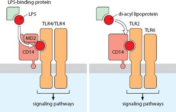
Fig. 2.22 Activation of macrophages by LPS.
Pentraxins are a group of soluble pentameric molecules that exhibit calcium-dependent binding to carbohydrates. The group includes C-reactive protein (CRP), serum amyloid-P (SAP) and pentraxin-3 (PTX3). Both CRP and SAP are primarily produced and broken down by the liver.
C-reactive protein (CRP) is an acute-phase protein that increases rapidly in the serum during inflammation and is used as a clinical marker of inflammation. It binds to phosphocholine groups on pneumococci, which opsonizes them and promotes their phagocytosis by macrophages, both directly and by activating complement.
Serum amyloid-P (SAP) recognizes a number of products of tissue breakdown, including amyloid fibers.
Ficolins are a group of three soluble lectins. Ficolin-1 (FCN1), secreted by mononuclear phagocytes, recognizes components of the cell wall of Gram-positive bacteria and activates the lectin pathway of complement, to opsonize them. Ficolin-2 (FCN2) also recognizes components of the bacterial cell wall and apoptotic cells.
Collectins is the name for the complement components mannan-binding lectin and conglutinin, soluble pattern recognition receptors that can activate complement.