
ADAPTIVE AND INNATE IMMUNITY
The immune response is mediated by a variety of cells and soluble factors, broadly classified according to whether they mediate adaptive (acquired) or innate (natural) immunity.
Adaptive (acquired) immunity is specific for the inducing agent and is marked by an enhanced response on repeated encounters with that agent. Thus the key features of the adaptive immune response are memory and specificity.
Innate (natural) immunity depends on a variety of immunological effector mechanisms, which are neither specific for particular infectious agents nor improved by repeated encounters with the same agent. In practice, there is considerable overlap between these two types of immunity: antibodies can direct or activate elements of the innate system, such as phagocytes and complement. Receptors of the innate immune system, including phagocyte receptors, are described on pages 48–51. Other elements of innate immunity are outlined below.
Complement system is a group of serum molecules involved in the control of inflammation, the removal of immune complexes and lysis of pathogens or cells sensitized by antibody, or mediators of the collectin, ficolin, and pentraxin families.
Acute-phase proteins are serum molecules that increase rapidly at the onset of infection, such as C-reactive protein, serum amyloid-P, serum amyloid-A, and mannan-binding lectin (MBL).
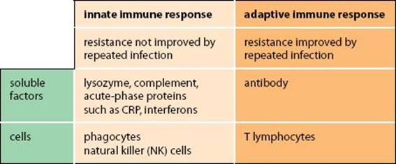
Fig. 3.1 Elements of the innate and adaptive immune systems.
Interferons (IFNs) are a group of molecules that limit the spread of viral infections. There are three types: IFN-α and IFN-β, produced by leukocytes and fibroblasts, and IFN-γ, produced by activated T cells and NK cells. Interferons from activated or virally infected cells bind to receptors on nearby cells, inducing them to make antiviral proteins. IFN-α and IFN-β bind to a type I IFN receptor, whereas IFN-γ binds to a type II receptor. IFN-γ also has many other immunomodulatory functions (see p.69).
Antiviral proteins are molecules that are induced by IFN, which limit viral replication. Many of them are produced in an inactive form and are activated in infected cells by contact with viral products such as dsRNA. Activated antiviral proteins include some that block the initiation of protein synthesis and others that cause mRNA degradation, thus reducing viral protein synthesis.
Cell-mediated immunity and Humoral immunity are traditional ways of describing the different arms of the immune system. Antibody, complement and other soluble molecules constitute the humoral effector systems, whereas T cells, NK cells, and phagocytes constitute the cellular effectors. It is now more useful to think of the systems that recognize free antigens and those that recognize cell-associated antigens. For example, cytotoxic T cells can recognize antigens presented on cell membranes, which have originated from within that cell, whereas antibody is important in the recognition of free, extracellular antigens.
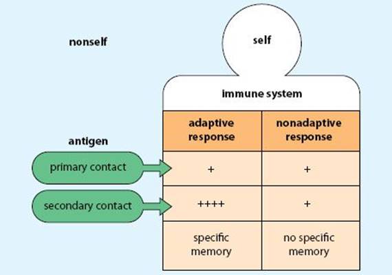
Fig. 3.2 Adaptive and nonadaptive (innate) immune responses.
ANTIBODY RESPONSE
After injection of an antigen, an antibody response develops, which may be divided into four phases: a lag phase in which no antibody is detected, followed by a phase in which the antibody titers rise logarithmically and then reach a plateau, and decline, as the antibodies are catabolized or cleared as immune complexes.
Primary and Secondary antibody responses The quality of the antibody response after the second (secondary) encounter with antigen differs from that after the first (primary) contact. The primary response has a longer lag phase, reaches a lower plateau, and declines more quickly than the secondary response. IgM is a major component of the primary response and is produced before IgG, whereas IgG is the main class represented in the secondary response. During their development, some B cells switch from IgM production to other classes, and this is the basis of the change in antibody isotype seen in the secondary response. Differences between the primary and secondary response are most noticeable when T-dependent antigens are used, but the route of entry and the way it is presented to T and B cells also affect the development of the response and the classes of antibody produced.
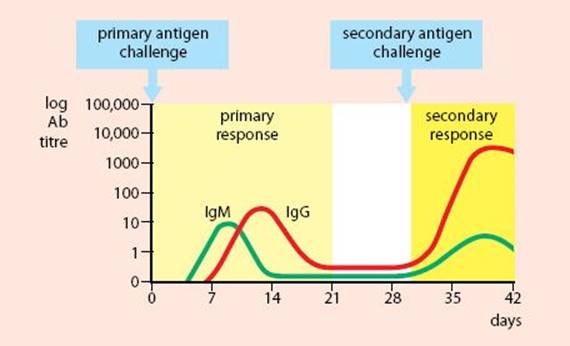
Fig. 3.3 Primary and secondary antibody responses.
Affinity maturation is the finding that the average affinity of the induced antibodies increases in the secondary response. The effect is largely confined to IgG and IgA, and is most marked when a low antigen dose is given in the secondary injection. Low levels of antigen bind preferentially to high-affinity B-cell clones and activate them—there is insufficient to activate low-affinity clones.
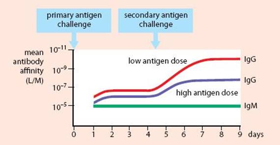
Fig. 3.4 Affinity maturation.
The underlying cellular basis of affinity maturation is the change in the affinity of B-cell clones, caused by somatic hypermutation of the antibody genes that occurs in germinal centers, where B cells compete for antigen on follicular dendritic cells. The process is accompanied by, but is not dependent on, class switching. It does not occur in the response to T-independent antigens, which is predominantly IgM antibody. Therefore the survival and development of high-affinity B cells depends on T cells.
Active immunization/Vaccination are the terms used for the active induction of protective immunity against a pathogen. This depends on the greater effectiveness of the secondary immune response. Vaccines may be live attenuated organisms, killed organisms, individual antigens of a pathogen, or modified antigens. In general, living organisms are more effective than killed ones or individual antigens, except in cases where the pathology is caused by a toxin (such as diphtheria). In this case a modified toxin or toxoid, which retains antigenicity but lacks pathogenicity, is preferred. Newer vaccines may be produced by genetic engineering. For example, genes for antigens of pathogenic viruses such as hepatitis can be inserted into nonpathogenic viruses such as vaccinia. It is also possible to insert antigen fragments that can stimulate T cells into such carrier viruses. For antigens that are only weakly immunogenic (such as some bacterial carbohydrates), coupling the antigen to an immunogenic carrier has often been successful. These preparations are called conjugate vaccines.
Passive immunization is the administration of antibodies preformed in another individual to contribute to protective immunity against a toxin. It is used when an individual’s own active immune response would be too slow, for example in producing a response to a snake venom or tetanus toxin.
CELL COOPERATION
Cooperation between cells involved in immune responses occurs at many levels. Dendritic cells can take up antigen in the periphery and transport it to secondary lymphoid tissues (spleen, lymph nodes, etc.) for presentation to T cells. B cells and macrophages can also internalize antigen, process it and present it in association with MHC class II molecules to CD4+ TH cells. Cytokines produced by activated TH2 cells stimulate B-cell growth and differentiation into plasma cells. Other cytokines can also activate TC cells, APCs, and mononuclear phagocytes, thereby facilitating uptake of antigen. IgG antibodies can sensitize target cells for attack by NK cells. IgE antibodies can sensitize mast cells and basophils to release their inflammatory mediators when they bind specific antigen. Cytokines and antibodies are soluble mediators of cell cooperation, but leukocytes also signal directly via cell-surface receptors. The most important direct interaction involves MHC molecules/antigen peptides contacting the T-cell receptor, but other interactions are essential for cellular cooperation, including adhesion and co-stimulation.
Antigen presentation is the process by which antigen is presented to lymphocytes in a form they can recognize. Most CD4+ T cells must be presented with antigen on MHC class II molecules, whereas CD8+TC cells recognize antigen on class I MHC molecules. Antigen must be processed into peptide fragments before it can associate with MHC molecules. The way in which an antigen is processed and the type of MHC molecule it associates with determine which T cells will recognize it and whether the antigen is immunogenic or tolerogenic. It also affects the type of immune response generated.
Adhesion is an essential component of the interactions between leukocytes and other cells. It controls the position of the cell in lymphoid tissue, controls migration into tissues, and is a prerequisite for antigen presentation and many immune effector functions.
Co-stimulation Most immune responses are initiated by antigen triggering B or T cells. However, cellular activation also requires other signals. These may be delivered via co-stimulatory molecules (such as CD40 for B cells, CD28 for T cells) or by cytokines. This is sometimes called the two-signal hypothesis, in which antigen provides the first signal and the other co-stimulatory interactions provide the second signal. Cells that only receive a first signal may become anergic (tolerant) to their particular antigen.
Cytokines are signaling proteins, many of which are involved in signaling between cells of the immune system. The group includes the interleukins (IL-1 to IL-35), interferons (IFNs), tumor necrosis factors (TNFs), transforming growth factors (TGFs), and colony-stimulating factors (CSFs). The term lymphokines was originally used for those cytokines produced by lymphocytes.
T-cell help describes the cooperative interactions between TH2 cells and B cells in the production of the antibody response to T-dependent antigens, or between TH1 and TH17 cells and phagocytic cells in cell-mediated responses. In either case the antigen-presenting cell presents processed antigen to the T cell, receives co-stimulatory signals and is then triggered by specific cytokines. For example, a B cell internalizes its own specific antigen and presents it to the T cell. It transduces co-stimulatory signals via CD40 and is further activated by IL-4, IL-2, and IL-13.
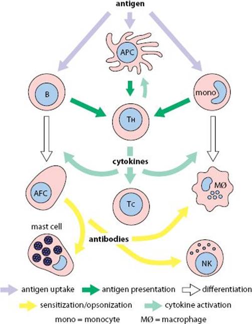
Fig. 3.5 Cooperation between cells in the immune response.
ANTIGEN PRESENTATION
Antigens are taken up by antigen-presenting cells in a variety of ways. B cells use surface antibody to bind and internalize their specific antigen. This is partly degraded (processed) and returned to the cell surface associated with MHC class II molecules, for recognition by TH2 cells. Theoretically, B cells can endocytose and present any antigen, but in practice they selectively concentrate only their own specific antigen in sufficient quantities. Mononuclear phagocytes phagocytose opsonized particles via their Fc and C3 receptors, which are then processed before presentation to TH1 cells. Immature dendritic cells take up antigen by phagocytosis using Fc, C3, scavenger, and lectin-family receptors. They lose these receptors and degrade antigen before migrating to lymph nodes, where they present it to T cells.
Antigen processing is the process of antigen breakdown and its association with MHC molecules. Blocking degradative pathways leaves cells unable to process and present most antigens. Different cell types have different capacities to degrade antigens and hence different abilities to stimulate T cells. There are two distinct pathways for antigen processing, used by MHC class I and II molecules. They are referred to as the internal and external pathways; MHC class I presents antigen from inside the cell, whereas MHC class II presents antigens that the cell has endocytosed.
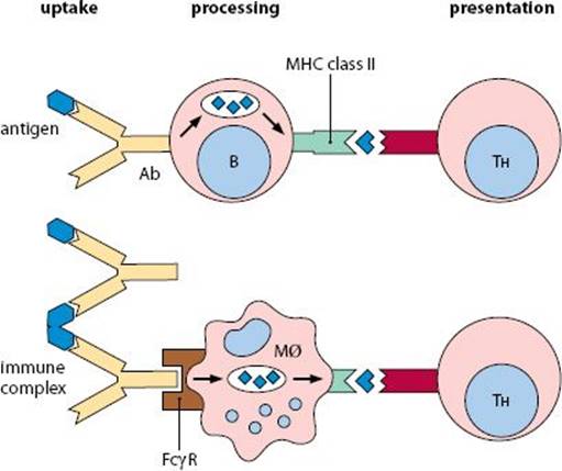
Fig. 3.6 Antigen processing and presentation by APCs.
Class II (external) pathway Antigens such as immune complexes that have been endocytosed by a cell associate preferentially with MHC class II molecules. They are partly degraded, and the endocytic vesicles containing peptide fragments then fuse with vesicles containing MHC class II molecules.
Invariant chain (Ii, CD74) MHC class II molecules are initially produced in association with an invariant chain, Ii, which is required for folding of the class II molecule and prevents peptides from binding to it in the endoplasmic reticulum. The invariant chain targets the class II molecules to the MIIC compartment.
MIIC compartment is an acidic endosomal compartment where antigenic peptide fragments and MHC class II molecules combine. The invariant chain is degraded, leaving a small peptide, CLIP, bound to the class II molecule. Once this has been replaced by an antigenic peptide, the class II:peptide complex can be finally processed (trimmed) before moving to the cell surface.
Antigenic peptides are the protein fragments that bind to MHC molecules. Class I molecules accommodate 8–9 amino acids in the peptide-binding groove, class II molecules 12–15 amino acids.
DM molecules are class II-like molecules that are required to facilitate loading of peptides onto the class II molecules.
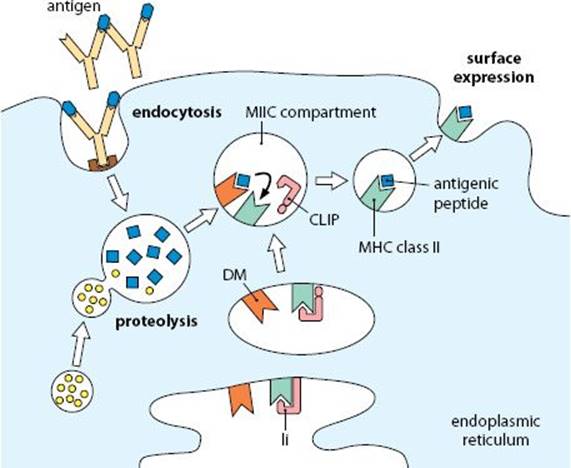
Fig. 3.7 Antigen presentation: MHC class II pathway.
Class I (Internal) pathway Antigens synthesized within a cell, such as viral polypeptides and the cell’s own proteins, associate preferentially with MHC class I molecules. Peptide fragments from the cytosol are sampled and presented, for review by CD8+ T cells.
Proteasomes are multicatalytic protease complexes that break down cytosolic proteins into fragments that may be loaded onto MHC class I molecules. Two components of the proteasome (LMP-2 and LMP-7) are encoded within the MHC.
TAP-1 and TAP-2 are MHC-encoded members of the ABC family of transporters. They transfer peptides across the membrane of the endoplasmic reticulum to be loaded onto MHC class I molecules.
Calnexin is a molecular chaperone that stabilizes the class I α chain, until it associates with β2-microglobulin and the peptide fragments. Once released from calnexin, assembly of the MHC:peptide complex takes place in a peptide loading complex and the peptide may then be trimmed by an ER-associated aminopeptidase. MHC:peptide complexes are transported to the cell surface, while incorrectly assembled complexes are degraded.
Anchor residues are critical amino acids that are required for an antigenic peptide to bind to an MHC molecule. The requirement for particular amino acids at each anchor position depends on the haplotype of the MHC molecule.
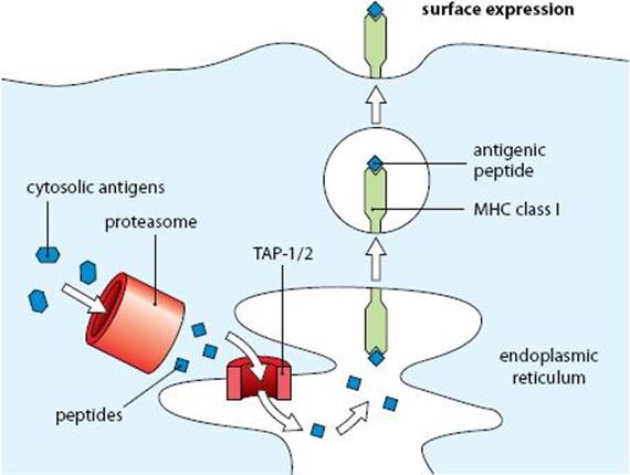
Fig. 3.8 Antigen presentation: MHC class I pathway.
MHC restriction describes the observation that T cells recognize antigen associated with particular MHC molecules, and usually do not recognize the same antigenic peptide if it associates with an MHC molecule of another haplotype. During development in the thymus, T cells that can interact with self MHC molecules are produced; these cells do not interact effectively with antigen-presenting cells of another MHC haplotype.
Cross-presentation may occur when an external antigen (normally presented by the class II pathway) is presented on MHC class I molecules. This mechanism can allow APCs to present viral antigens to CD8+ cytotoxic T cells, even if they have not themselves become infected.
CD4 and CD8 are functionally analogous molecules expressed on mature T cells; cells have either CD4 or CD8, but not both. CD8 consists of two disulfide-linked transmembrane polypeptides that can interact with the TCR on T cells and bind to a site in the α3 domain of class I molecules on the target cell (Figs 3.9 and 2.16). This interaction contributes to the stabilization of the immune recognition complex. CD4 has a single transmembrane polypeptide and binds MHC class II molecules on APCs.
lck is a kinase associated with CD4 and CD8. Binding of the T cell to MHC:antigen brings lck into proximity with the T cell receptor so that it phosphorylates CD3ζ to initiate T-cell activation.
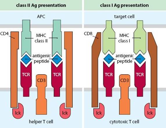
Fig. 3.9 Immune recognition by T cells.
T-CELL ACTIVATION
T cells require three types of signal for full activation:
•Antigenic peptide presented on an MHC molecule
•Co-stimulatory signals
•Signals from specific cytokines
If a cell does not receive a full set of signals, it will not divide and may even become anergic. Molecules such as CD2 and LFA-1 contribute to the adhesion between a T cell and an APC, and enhance activation signals, but co-stimulation transduced via CD28 is essential for activation.
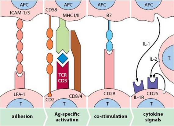
Fig. 3.10 Steps in T-cell activation.
Lymphocyte functional antigen-1 (LFA-1, CD11a/CD18) is a member of the β2-integrin family present on most leukocytes. It consists of two polypeptides (CD11a and CD18) that interact with intercellular adhesion molecules, ICAM-1, ICAM-2, and ICAM-3. Transient adhesion between lymphocytes and APCs is mediated by LFA-1 binding to ICAM-1 and ICAM-3. Lymphocyte activation enhances the affinity of LFA-1, thereby extending the interaction time between the T cell and APC. Binding of LFA-1 with ICAM-1 and ICAM-2 is also important in the attachment of leukocytes to endothelium and in the migration of cells across endothelium in normal tissues and at sites of inflammation.
ICAM-3 (CD50) is an adhesion molecule present on many leukocytes, which increases after lymphocyte activation and contributes to T-cell interactions with APCs.
CD2 (LFA-2) and CD58 (LFA-3) are a pair of molecules involved in T-cell activation. CD2 is expressed on all T cells. It has a single transmembrane polypeptide that acts as a receptor for CD58, a molecule that is widely distributed on many cell types. Interaction of CD2 with CD58 enhances the binding of the T cell to its target, amplifying the activation signal initiated by the TCR:CD3 complex.
CD28 and B7 (CD80, CD86) are molecules that critically regulate T-cell activation. CD28 is a co-stimulatory receptor present on 80% of CD4+ T cells and ~50% of CD8+ cells. The molecules B7-1 (CD80) and B7-2 (CD86), expressed on many APCs, are the principal ligands for CD28. As an immunological synapse forms, CD28 is released from intracellular stores, where it enhances the initial weak signal from the TCR. The cytoplasmic portion of CD28 associates with phosphatidylinositol 3-kinase which, in association with signals from the TCR, activates the MAP-kinase signaling pathway.
B7-1 (CD80) and B7-2 (CD86) are constitutively expressed on dendritic cells and most mononuclear phagocytes; expression is enhanced by GM-CSF, IFN-γ, and ligation of TLRs (for example, by LPS). B7 is induced on B cells by antigen binding, LPS stimulation, and ligation of CD40.
Immunological synapse is the complex of interacting molecules that link the APC and the T cell. Initially, adhesion molecules (LFA-1/ICAM-1, etc.) allow the cells to adhere to each other. As MHC molecules on the APC start to interact with the TCR complex, the adhesion molecules are relegated to the outside of the synapse, the pSMAC (peripheral supramolecular activation complex), while the TCR, CD2/CD58, CD28/B7, and MHC molecules localize at the center of the synapse—the cSMAC (Fig. 3.11).
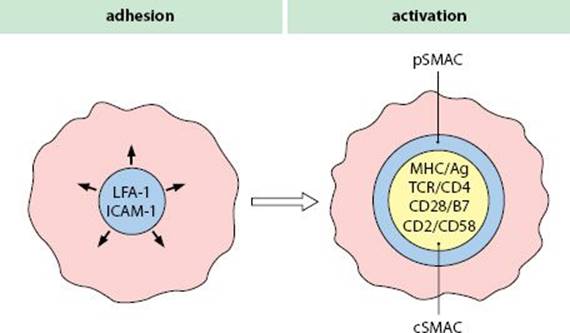
Fig. 3.11 Formation of an immunological synapse.
IL-2 receptor (IL-2R, CD25) is induced on activated T cells. The high-affinity receptor is formed when the induced α chain (CD25) associates with β and γ chains (CD122, CD132), which together form the low-affinity receptor. IL-2 is essential for T-cell division, and the high-affinity receptor persists for several days after T-cell activation. CD25 is also a characteristic marker of naturally occurring regulatory T cells (TREG cells), which may act by mopping up excessive IL-2, limiting activation of antigen-stimulated T cells.
CTLA-4 (CD152) is an alternative ligand for B7, which is not expressed on resting T cells but is induced after T-cell activation as CD28 declines. CTLA-4 has a higher affinity for B7 than does CD28, and by competing with CD28 for B7, CTLA-4 counters the co-stimulatory action of CD28. CTLA-4 is also constitutionally expressed in TREG cells. Mice deficient in CTLA-4 are more susceptible to autoimmune diseases; this is thought to be due both to excessive T-cell activation and reduced control by TREG cells.
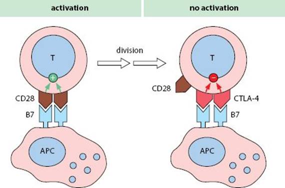
Fig. 3.12 The role of CTLA-4 in controlling T-cell activation.
PD1 (CD279) (Programmed Death-1) is another inhibitory receptor, belonging to the same family as CD28 and CTLA-4. It is expressed late after T-cell activation, and can bind to its ligands PD-L1 (CD273) or PD-L2 (CD274), which belong to the B7 family; the ligands are expressed on antigen-presenting cells. PD1 is also present on B cells, dendritic cells, and monocytes. It is thought to limit T-cell activation and to help prevent autoimmunity. In humans, polymorphisms in PD1 are linked to rheumatoid arthritis, Graves’ disease, type I diabetes, and multiple sclerosis.
CYTOKINE RECEPTORS
Cytokine receptors determine the responsiveness of a cell to particular cytokines. Receptors for IL-1, TNF, and the interferons are widely distributed. Others are induced on particular lineages for limited periods. For example, the high-affinity IL-2 receptor is present on antigen-activated cells for a limited period, but expression wanes if the T cell is not re-stimulated with antigen. Expression of IL-4 receptors occurs on activated B cells in an analogous fashion. Receptors for colony-stimulating factors appear during hemopoietic cell differentiation on the appropriate developing cells (see Fig. 1.11). The cytokine receptors fall into families on the basis of structural motifs and shared chains. For example, the receptors for IL-2, IL-4, IL-7, IL-9, and IL-15 have a common signaling polypeptide (CD122), but individual cytokine-binding chains. IL-3 and IL-5 share a different chain.
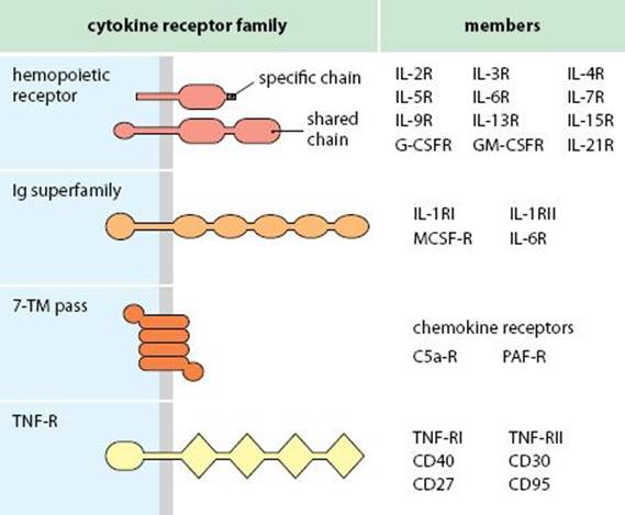
Fig. 3.13 Families of cytokine receptors.
Soluble cytokine receptors and cytokine inhibitors Several cytokine receptors are produced in a soluble, truncated form, lacking the membrane-spanning domains. Examples are the soluble TNF-R, IFN-γR, and IL-1R. They are thought to limit the effects and zone of action of cytokines in vivo. Cytokine inhibitors have also been identified. For example, IL-1RA (IL-1 receptor antagonist) binds to the IL-1 receptor but does not activate the cell.
B-CELL ACTIVATION
B cells responding to T-dependent antigens require three types of signal for their activation. The first signal is mediated by the binding of antigen, which is internalized, processed, and presented to T cells. Then a co-stimulatory signal is transduced via CD40, which is bound by CD40L on the T cells. Thereafter B cell division, differentiation, and Ig class switching are driven by a large number of different cytokines. Type 2 T-independent antigens, such as polysaccharides that cross-link B-cell-surface antibody, can activate B cells directly, although such cells still need cytokine signals.
Intermolecular help refers to the way in which B cells taking up particles with several different antigens (such as a virus) can then present all of those antigens to T cells. They thus get help from T cells recognizing antigens that they themselves do not recognize.
CD40 is a surface receptor on B cells, follicular dendritic cells, dendritic cells, macrophages, endothelium, and hemopoietic progenitors. It belongs to the TNF receptor family. It provides a critical co-stimulatory signal to B cells that is also needed for the development of germinal centers and B-cell memory.
CD40L (CD154) is the ligand for CD40, induced transiently on CD4+ T cells and some CD8+ cells, after activation. It is also present on eosinophils and basophils. CD40L is essential for the delivery of T-cell help to B cells. A defect in CD40L causes impaired class switching and results in hyper-IgM syndrome.
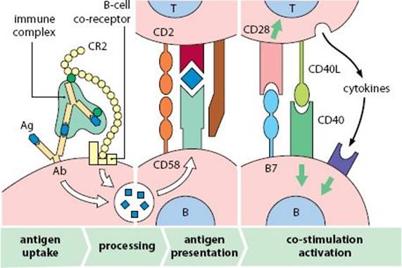
Fig. 3.14 Steps in B-cell activation.
CD72 and CD100 are co-stimulatory molecules for B-cell activation. CD72 on B cells binds to CD100, a widely distributed member of the semaphorin family, enhancing activation mediated by CD40.
CD45 (Leukocyte common antigen) is a phosphatase present on all leukocytes, and is produced in six different forms, using combinations of exons. B cells express the largest variant of CD45. It controls lymphocyte activation by acting on lck, which can phosphorylate the signaling portion of the TCR (CD3) and BCR (CD79).
B-cell co-receptor complex (CD19, CD21/CR2, CD81/TAPA-1) amplifies signaling from the B cell antigen receptor. Cross-linking of CD19 to surface Ig makes a B cell 100 times more sensitive to antigen. This is important in the initial development of an antibody response when B-cell antibody affinity is low. Immune complexes formed in the primary immune response may fix complement C3 and then bind to CD21 on the B cell, which is complement receptor type 2 (CR2). If the complexed antigen is recognized by the B-cell receptor, the complex cross-links the co-receptor complex and surface Ig, thereby activating the B cell very efficiently. This may explain the observation that complement is required for the development of secondary antibody responses and B-cell memory.
CD23 (FcεRII) is a low-affinity IgE receptor with a lectin domain that also binds CR2. It is expressed on B cells, activated macrophages, and follicular dendritic cells, but may also be released in a soluble form to act as a B-cell co-stimulatory factor.
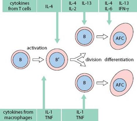
Fig. 3.15 Role of cytokines in B-cell development.
CYTOKINES
Cytokines, released by leukocytes and sometimes other cells, are very important in controlling the development of immune responses. They modulate the differentiation and division of hemopoietic stem cells and the activation of lymphocytes and phagocytes. They control the balance between cell-mediated responses and antibody production. Others can act as mediators of inflammation or as cytotoxins. Many cytokines have more than one action (pleiotropy), and different cells produce distinct blends of cytokines. The ability to respond to a cytokine depends on the expression of a specific receptor. Often more than one cytokine signal is required for a response, and in this case the different cytokines act synergistically. Helper T cells are particularly important sources of cytokines. Most cytokines act on cells other than those that produced them (paracrine action), but some can also stimulate the cell that produced them (autocrine action).
JAKS and STATs Cytokines signal cell activation by binding to specific receptors (see p. 65) that activate intracellular signaling pathways. Receptors that belong to the hemopoietic cytokine receptor family are associated with Janus kinases (JAKs). When the receptors become clustered after cytokine binding, the JAKs phosphorylate STATs (signal transducers and activators of transcription). The activated STATs, in association with other proteins, form transcription factors that migrate to the nucleus, bind to gene promoters, and induce the sets of genes that are associated with the response to each of the cytokines. Different JAKs and STATs are used by different cytokines and their receptors. In the example below, the interferon-α receptor is associated with JAKs Tyk2 and Jak1. These phosphorylate STAT1 and STAT2, which associate with p48 to form a transcription factor.
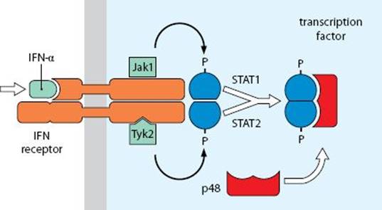
Fig. 3.16 JAKs and STATs in cytokine receptor signaling.
Interferon-γ (IFN-γ) is released by antigen-activated TH1 cells. In addition to its antiviral effects, IFN-γ enhances MHC class I on many cells and increases MHC class II and B7 expression on B cells and macrophages, thereby enhancing antigen presentation. At high levels it can induce class II on some tissue cells. It increases IL-2 receptors on TC cells, enhances the cytotoxic activity of NK cells and promotes B-cell differentiation. IFN-γ also acts as a macrophage arming factor, increasing Fc receptor expression, the respiratory burst, and nitric oxide production, so enhancing the ability of macrophages to destroy pathogens. It also inhibits TH2 and TH17 cells, and so reinforces TH1-type immune responses.
Migration inhibition factor (MIF) is released by activated T cells and inhibits macrophage migration. It causes the accumulation and activation of macrophages at the site of inflammation and is elevated in many chronic inflammatory conditions. MIF binds to CD74, acting on various transcription factors and modulating the cell damage sensor/tumor suppressor p53.
Tumor necrosis factor (TNF) and Lymphotoxin (LT) are structurally related cytokines encoded within the MHC. Lymphotoxin, released by TC cells, is also called TNF-β, whereas the original TNF, released by macrophages and several other cells, is TNF-α. A transmembrane form of lymphotoxin (LT-β), also produced by T cells, trimerizes with LT-α. TNF enhances the adhesiveness of vascular endothelium for leukocytes by inducing E-selectin, VCAM-1 and ICAM-1, thus promoting transendothelial migration. TNF also causes the mobilization of fat, which is partly responsible for the wasting (cachexia) seen in some chronic diseases. It also synergizes with IFN-γ in many of its actions such as MHC induction and macrophage activation. TNF and lymphotoxin can also induce cell death by apoptosis. Of the three receptors for this group of cytokines, one (TNFR1) has an intracytoplasmic death domain, which can recruit proteins that activate caspases, the principal mediators of apoptosis.
Transforming growth factor-β (TGF-β) is a group of five cytokines released by many cell types, including macrophages and platelets. They are mitogenic for fibroblasts and some other mesenchymal cells, and they enhance the production of extracellular matrix proteins. In general, TGF-β is strongly inhibitory of immune responses as it prevents the proliferation of both T and B cells, and it seems to be essential in controlling immune reactivity—TGF knockout mice develop severe chronic inflammatory reactions.
Interleukins (IL-1 to IL-35) are a diverse group of cytokines; most newly discovered cytokines are placed into this series. The functions are outlined in Fig. 3.17. Many of the interleukins fall into structurally related families:
IL-1 family—IL-1, IL-18, IL-33
IL-2 family—IL-2, IL-12, IL-15
IL-10 family—IL-10,IL-19, IL-20, IL-22, IL-24, IL-26
IL-12 family—IL-12, IL-23, IL-27, IL-35
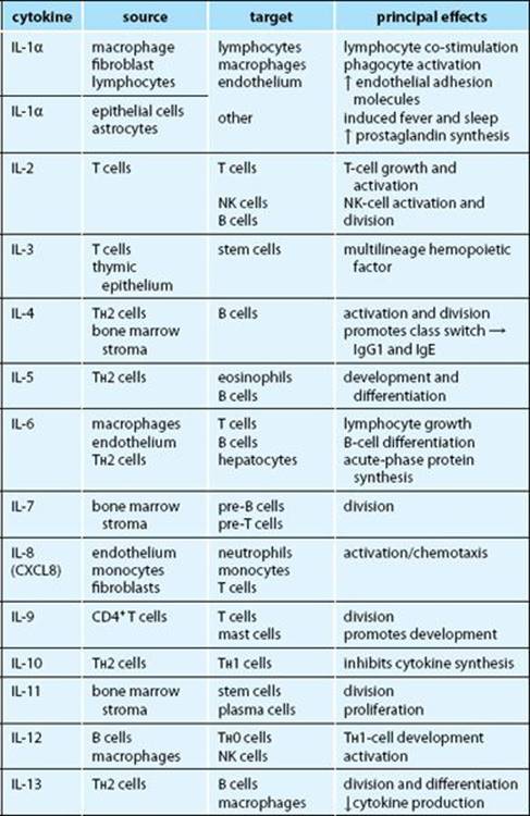
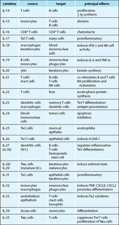
Fig. 3.17 The interleukins.
PHAGOCYTOSIS
Phagocytosis/Endocytosis is the process by which cells engulf particles and microorganisms. The particles first attach to the cell membrane of the phagocytic cell, either by general receptors, such as the mannose receptor, which binds bacterial carbohydrates, or by receptors for opsonins, such as IgG or C3b. Then the cell extends pseudopodia around the particle and internalizes it. Antibacterial, oxygen-dependent killing mechanisms are activated and lysosomes fuse with the phagosome. The lysosomal enzymes damage and digest the phagocytosed material, and digestion products are finally released. Endocytosis is a term that includes phagocytosis and pinocytosis (internalization of fluid).
Opsonization occurs when particles, microorganisms, or immune complexes become coated with molecules that allow them to bind to receptors on phagocytes, thereby enhancing their uptake.
Opsonins are molecules that bind to particles to be phagocytosed and to receptors on phagocytes, so acting as an adaptor between the two, such as IgG, C3b, C-reactive protein.
Immune adherence, effected by IgG and C3 products, refers to the attachment of opsonized particles to phagocytes, by binding to Fc and complement receptors (see pp. 74 and 75).
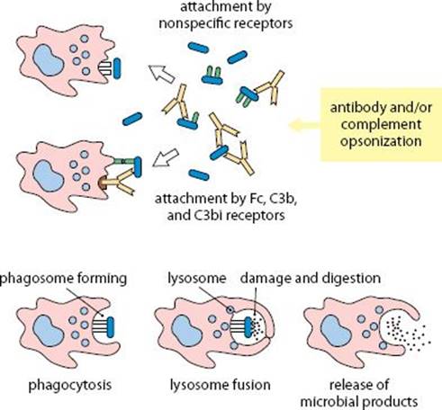
Fig. 3.18 Stages of phagocytosis.
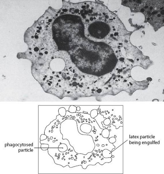
Fig. 3.19 Phagocytosis of latex by macrophages.
Phagosomes are membrane-enclosed intracellular vesicles that contain phagocytosed materials.
Lysosomes are organelles present in all cells. They contain enzymes that, in macrophages, damage and digest the phagocytosed material. Newly formed lysosomes are called ‘primary’, and mature lysosomes are ‘secondary.’
Phagolysosomes are formed by the fusion of phagosomes and lysosomes. Immediately after fusion, there is a brief rise in the pH of the phagolysosome, when neutral proteases (such as collagenase, elastase) and cationic proteins are active. Subsequently the pH falls and acid proteases (such as glycosidase, lipase) become active.
Frustrated phagocytosis occurs when phagocytes attach to material that cannot be phagocytosed (such as a basement membrane). The cells may release their lysosomal enzymes to the exterior (exocytosis). This process is thought to cause some of the damage in immune complex disease.
COMPLEMENT RECEPTORS
There are four different kinds of receptor for C3b or iC3b (CR1 to CR4), and three of them act as opsonic receptors for immune complexes on cells of the mononuclear phagocyte lineage.
CR1 (CD35) is a transmembrane protein consisting of a single polypeptide that is expressed on phagocytic cells, where it acts as a receptor for immune complexes. On human erythrocytes it facilitates transport of complexes to phagocytic cells in the spleen and liver. On other cells its principal function is to act as a cofactor for factor I.
CR2 (CD21) is structurally similar to CR1. It forms part of the B-cell co-receptor complex (Fig. 3.14) and is also present on follicular dendritic cells. It is involved in the uptake of immune complexes to germinal centers and in the development of B-cell memory.
CR3 (CD11b/CD18) is an integrin expressed on mononuclear phagocytes, neutrophils, and NK cells, where it facilitates the uptake of immune complexes with bound C3d. It is also involved in monocyte migration into tissues, by binding to ICAM-1.
CR4 (CD11c/CD18, p150/95) is an integrin that shares a β chain with CR3 and LFA-1. It has similar functions to CR3 and is highly expressed on tissue macrophages and dendritic cells.
CD93, present on monocytes, neutrophils, endothelium, and activated macrophages, was originally identified as a C1q receptor (C1qRp), but is now thought to be an adhesion molecule involved in clearance of apoptotic cells and in antimicrobial defense.
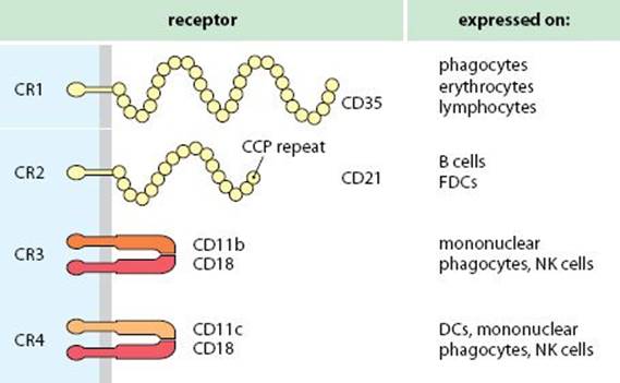
Fig. 3.20 Complement receptors.
Fc RECEPTORS
There are three well-defined receptors for IgG on phagocytes, which facilitate the uptake of immune complexes and allow cytotoxic cells to interact with targets. Two receptors for IgE have been described, FcεR1 and FcεRII; the first has a role in the control of inflammation, and the second has a role in immunoregulation and defense against parasitic worms.
FcγRI (CD64) is a high-affinity IgG receptor, capable of binding monomeric antibody. It is a characteristic marker of mononuclear phagocytes, but may also be expressed on neutrophils. It is involved in the uptake of immune complexes.
FcγRII (CD32) is a low-affinity receptor present on mononuclear phagocytes, neutrophils, eosinophils, platelets, and B cells. On phagocytes it facilitates uptake of large immune complexes, but on B cells it is thought to be involved in the control of antibody production. Cross-linking of the surface antibody (BCR) and FcγRII receptors on B cells leads to suppression of the B cell. Activation of platelets by immune complexes bound to their Fc receptors can cause degranulation with release of inflammatory mediators.
FcγRIII (CD16) is a low-affinity IgG receptor that occurs in two forms. On NK cells it is a transmembrane glycoprotein (FcγRIIIa) that can cross-link them to target cells sensitized with antibody. Engagement of this receptor on NK cells leads to cell activation. On macrophages and neutrophils, FcγRIII is a GPI-linked receptor (FcγRIIIb) attached to the outer leaflet of the plasma membrane, where it can bind immune complexes but cannot signal.
FcεRI is a high-affinity IgE receptor found on mast cells and basophils. These cells are sensitized by monomeric IgE bound to the receptor. When the specific antigen cross-links IgE bound to these receptors, it causes degranulation with release of histamine and other inflammatory mediators.
FcεRII (CD23) is a low-affinity IgE receptor with an immuno-regulatory function present on some B cells. A soluble form of the receptor acts as a signaling molecule between lymphocytes (see p. 67). It is also present on eosinophils, where it may allow them to engage parasites (such as schistosomes) coated with IgE.
FcαR (CD89) is expressed on phagocytic cells, and on some B and T cells, particularly in Peyer’s patches and the lamina propria. Hence it seems to be involved in the regulation of IgA synthesis.
PHAGOCYTE MICROBICIDAL SYSTEMS
Respiratory burst Shortly after phagocytosing material, neutrophils and macrophages undergo a burst of activity, during which they increase their oxygen consumption. This is associated with increased activity of the hexose monophosphate shunt and the production of H2O2 and O2•.
Oxygen-dependent killing occurs within phagosomes and is activated via cross-linking of the phagocytes’ C3 and Fc receptors. Initially an enzyme, NADPH oxidase, is assembled in the phagosome membrane; it reduces oxygen to superoxide (•O2−), which can then give rise to hydroxyl radicals (•HO), singlet oxygen (Δg′O2) and hydrogen peroxide (H2O2).
Reactive oxygen intermediates (ROIs) are the labile products of the oxygen-dependent killing pathway (Fig. 3.21) and can damage endocytosed bacteria. Cells prevent damage to themselves, by redox pathways involving the tripeptide glutathione, but some bacteria deploy similar defenses against ROIs.
Myeloperoxidase present in lysosomes can enter the phagosome where, in the presence of H2O2, it converts halide ions into toxic halogen compounds (such as hypohalite). Endocytosed peroxidase or catalase can also perform this reaction.
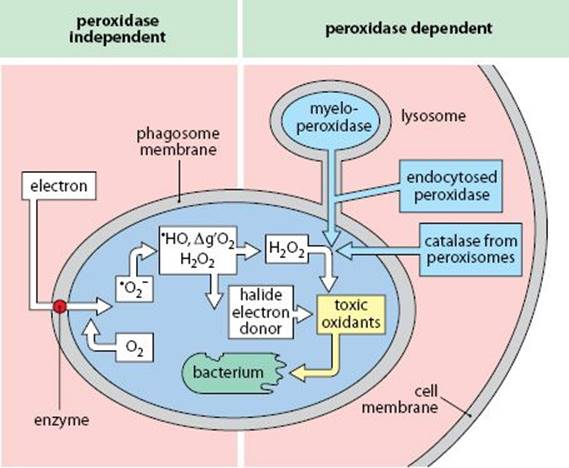
Fig. 3.21 Oxygen-dependent microbicidal activity.
Δg′O2 = singlet oxygen •HO = hydroxyl radical
Reactive nitrogen intermediates (RNIs) Murine macrophages that have been activated by IFN-γ and triggered with TNF express inducible nitric oxide synthetase (iNOS), which catalyses the production of nitric oxide, NO, which is toxic for some bacterial and fungal pathogens. Although human macrophages do not produce much NO, other cells, such as neutrophils, can do so. NO combines with ROIs to produce cytotoxic peroxynitrites.
Granules are specialized lysosomes of granulocytes that contain various bactericidal proteins. For example, neutrophil myeloperoxidase is in the primary (azurophilic) granules, whereas lactoferrin is in the secondary (neutrophil-specific) granules. Granule and lysosome contents are listed below.
Cationic proteins, found in neutrophil granules and in some macrophages, damage the outer phospholipid bilayer of some Gram-negative bacteria under alkaline conditions. This activity is produced by a number of molecules, including defensins and cathelicidins; some (such as cathepsin G) are enzymatically active.
Defensins are a group of small antimicrobial cytotoxic peptides that can be subdivided into three families, α, β, and θ. α-Defensins are found in the granules of neutrophils and macrophages in several species including humans. β-Defensins are present in the neutrophil granules of all mammals and in some epithelial cells. θ–Defensins are confined to primate granulocytes. Defensins are cationic proteins with a wide spectrum of antibacterial and antifungal actions that act by selectively damaging membranes with low levels of cholesterol and a high proportion of negatively charged phospholipids. They have some structural similarities to chemokines and have additional roles in opsonization and chemotaxis, by acting on chemokine receptors. For example, the defensin HBD-2 resembles CCL20, and both bind to the chemokine receptor CCR6.
Cathelicidins are a diverse family of small polypeptides with a common cathelin domain that are stored within granules of myeloid cells. On activation the cathelin domain is enzymatically removed and the peptides are released. In addition to antimicrobial properties, some cathelicidins have chemotactic and angiogenic activity and promote wound healing.
Lactoferrin is found in neutrophil granules. It binds tightly to iron, and deprives bacteria of this essential nutrient. Neutrophils loaded with iron are inefficient at destroying bacteria.
Lysozyme (muramidase) is an enzyme that digests a bond in the cell-wall proteoglycan of some Gram-positive bacteria. It is secreted constitutively by neutrophils and some macrophages and is present in many of the body’s secretions.
Macrophage activation refers to the enhanced microbicidal (or anti-tumor) activity seen in response to stimulation by inflammatory cytokines (TNF-α, IL-1, IFN-γ), complement fragments, and bacterial products that activate the Toll-like receptors. Activated cells secrete more enzymes and produce more superoxide and RNIs due to inducible nitric oxide synthetase. Figure 3.22 shows that macrophages treated with IFN-γ (left) have a greater capacity to destroy the parasite Leishmania donovani than do untreated macrophages (right).
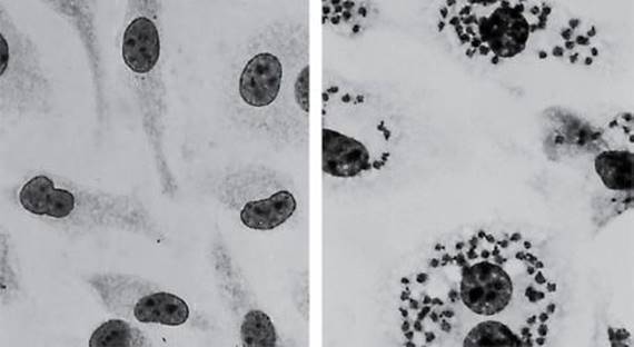
Fig. 3.22 Microbicidal activity of IFN-γ-activated macrophages.
Macrophage activation also induces expression of MHC class II and B7 to enhance antigen presentation. Enhanced phagocytosis of activated macrophages is related to increased expression of Fc and C3 receptors. Some receptors for chemotactic molecules (such as C5aR) are reduced; others are increased (such as CXCR3).
Macrophages can also be activated by cytokines released by TH2 cells, including IL-4 and IL-13. Such ‘alternatively activated’ macrophages increase expression of the mannose receptor and MHC class II, but do not show increased microbicidal activity.
nRAMP (natural resistance associated macrophage protein) is an ion pump that removes divalent cations from the phagosome, increasing macrophage resistance to mycobacterial infection.
Metalloproteases (MMP and ADAM) are zinc-containing enzymes involved in the degradation of extracellular matrix (matrix metalloproteases, MMP). ADAMs are transmembrane proteins containing a disintegrin and metalloprotease domain that modulate cell adhesion. Macrophage activation causes the synthesis of a number of new MMPs that are involved in tissue remodeling.
INTRACELLULAR RECEPTORS FOR PATHOGENS
Macrophages have a number of cytosolic molecules that can recognize intracellular bacterial and viral infections:
NOD-like receptors (NLRs) including NOD1 (nucleotide-binding oligomerization domain-containing protein-1) and NOD2 recognize bacterial peptidoglycans, for example from Salmonella and Shigella. Binding of peptidoglycans causes activation of NFκB and MAP-kinase pathways to induce transcription of cytokines that control inflammation.
RIG-like receptors (RLRS) include RIG-1 (retinoic acid inducible gene-1), which recognizes short dsRNA, and MDA5, which recognizes long dsRNA; dsRNA may be produced during viral replication. Binding of these receptors induces activation of NFκB. Both NLRs and RLRs are components of inflammasomes.
Inflammasomes are multicomponent complexes produced in myeloid cells, that include caspase-1 ( = interleukin-1 converting enzyme, ICE). ICE cleaves pro-IL-1β and pro-IL-18 into their active forms, which promotes inflammation. The precise composition of the inflammasome depends on the inducing agent (NLR, RLR, etc.) Assembly of the inflammasome also activates caspases to cause cell death by pyropoptosis.
Pyropoptosis describes programmed cell death after activation of inflammosomes, with the release of the pro-inflammatory cytokines IL-1 and IL-18.

Fig. 3.23 Intracellular pattern recognition receptors.
CYTOTOXICITY
Cytotoxicity is a general term for the ways in which lymphocytes, mononuclear phagocytes, and granulocytes can kill target cells. This kind of interaction is important in the destruction of cells that have become infected with viruses or intracellular microorganisms, which they are unable to eliminate.
T cell-mediated cytotoxicity involves the recognition of antigenic peptides associated with MHC class I molecules (usually) on the surface of the target cell and is effected by CD8+ TC cells. The attacking cell orientates its granules toward the target and releases the contents, including perforin and granzymes, at the junction between the cells. Cytokines such as lymphotoxin, or the engagement of CD95 on the target, may also signal cell death. The relative contribution of each component depends on the cytotoxic cell involved. Target cell death occurs by apoptosis.
Fas (CD95) and CD178 (CD95L) Fas is a receptor belonging to the TNF-R family expressed on many cell types. Ligation of CD95 by CD95L (CD178) induces target cell death. Fas has an intracytoplasmic ‘death’ domain which occurs on other receptors involved in cell survival or death.
Perforin is a pore-forming molecule related to complement C9, which polymerizes on the target cell membrane to form channels.
Granzymes are serine proteases found in the granules of cytotoxic T cells, which may enter the target cell via perforin pores. Granzyme-A nicks DNA and prevents DNA repair, while granzyme-B activates caspases 3, 7, and 8, which induce apoptosis.
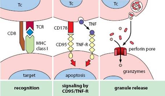
Fig. 3.24 Mechanisms of T cell-mediated cytotoxicity.
Caspases (cysteine aspartic acid proteases) are a group of proenzymes that become activated by cleavage into two or three subunits. They have a wide range of effects within the cell, affecting cell cycle control, DNA integrity and repair, and apoptosis. Ligation of Fas (CD95) by CD178, or the type I TNF receptor (TNFR-I, CD120a) by TNF-α or lymphotoxin, causes adaptor proteins to bind to the intracellular portion of the receptors and leads to activation of caspases 8 and 10. Activation of downstream effector caspases 3, 6, and 7 causes apoptosis.
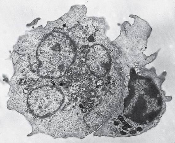
Fig. 3.25 Large granular lymphocyte (right) engaging a target cell (left). Courtesy of P. Penfold.
Antibody-dependent cell-mediated cytotoxicity (ADCC) involves the recognition of target cells coated with antibody. It may be effected by large granular lymphocytes, macrophages, or granulocytes, using their Fcγ receptors. The mechanism of cytotoxic damage depends on the effector cell; macrophages can release enzymes and ROIs, whereas LGLs use perforin and cytokines.
NK cell-mediated cytotoxicity is mediated by LGLs. They can kill some target cells that fail to express MHC class I or express allogeneic MHC class I. Thus they provide a line of defense against viruses that attempt to evade immune recognition by downregulating MHC expression. The mechanisms of cytotoxicity are similar to those used by TC cells, with granule components (perforin and granzymes) being particularly important.
Eosinophil-mediated cytotoxicity Eosinophils are only weakly phagocytic and are less efficient than neutrophils and macrophages at destroying endocytosed pathogens. However, they can exocytose their granule contents, releasing factors that are very effective at damaging certain large parasites. Eosinophils recognize targets via bound antibody, including IgE, which they bind via FcεRII. Eosinophil degranulation is triggered by ligation of FcεRII or FcγRII. It is also induced in vitro by cytokines including IL-5, TNF, IFN-β, and PAF. Eosinophil granules include phosphatases, aryl sulfatase, and histaminase, in addition to those listed below.
Major basic protein (MBP) is a highly cationic protein that forms a major component of the crystalloid core of eosinophil granules. It is solubilized before secretion and can damage parasites. Figure 3.26 illustrates progressive damage to a schistosomule larva incubated in MBP. MBP also causes damage and loss of bronchial epithelium in allergic asthma.
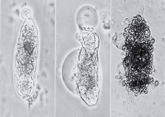
Fig. 3.26 Effect of major basic protein on a schistosomule larva. Courtesy of D. McLaren and Janice Taverne.
Eosinophil cationic protein (ECP) is a highly basic zinc-containing ribonuclease that binds avidly to negatively charged surfaces. It is particularly effective at damaging the tegument of schistosomes.
Eosinophil peroxidase is distinct from the myeloperoxidase produced by neutrophils and macrophages, but it serves a similar function in the generation of toxic hypohalites.
INFLAMMATION
Inflammation is the response of tissue to injury, with the function of bringing serum molecules and cells of the immune system to the site of damage. The reaction consists of three components:
•Increased blood supply to the region
•Increased capillary permeability
•Emigration of leukocytes from blood vessels into the tissues
Inflammation is an ordered process mediated by the appearance of intercellular adhesion molecules on endothelia and the release of various inflammatory mediators from tissue cells and leukocytes. Plasma enzyme systems are particularly important sources of inflammatory mediators. These include the complement, clotting, fibrinolytic (plasmin), and kinin systems. Also active are the mediators released by mast cells, basophils, and platelets, as well as the eicosanoids generated by many cells at sites of inflammation. Generally, neutrophils are the first cells to appear at acute inflammatory sites, followed by macrophages and lymphocytes, if there is an immunological challenge.
Vasodilation is the dilation of the local arterioles caused by the actions of mediators such as histamine on the smooth muscle in the vessel wall, allowing increased blood flow.
Transudate/exudate Normally, only small molecules pass freely through the capillary wall. The fluid that passes through is a transudate. If inflammation occurs, the endothelial cells are caused to retract, permitting larger molecules to pass out too. This fluid, which is also rich in cells, is an inflammatory exudate.
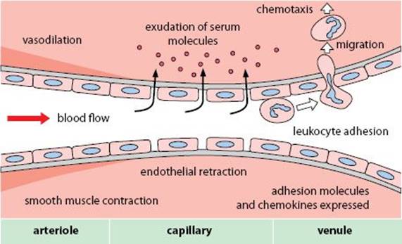
Fig. 3.27 Elements of inflammation.
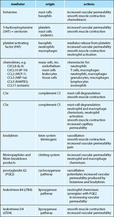
Fig. 3.28 Mediators of acute inflammation.
Mediators of inflammation include the plasma enzyme systems, cells of the immune system, and products of pathogens themselves. The principal mediators are listed in Fig. 3.28.
Kinins are generated after tissue injury. Bradykinin is a nonapeptide produced by the action of kallikrein on high-molecular-weight kininogen. Lysyl bradykinin (kallidin) is generated by the action of tissue kallikrein on low-molecular-weight kininogen. The kinins are exceptionally powerful vasoactive mediators, causing vasodilation and increased capillary permeability.
Eicosanoids are mediators produced from arachidonic acid, which is released from membranes by the action of phospholipase A2. Arachidonic acid is converted into eicosanoids by mast cells and macrophages, via two major pathways.
Prostaglandins (PG) and Thromboxanes (TX) are produced by the action of cyclooxygenase on arachidonic acid. They have diverse proinflammatory effects, often synergizing with other mediators.
Leukotrienes (LT) are produced by the lipoxygenase pathway, which generates mediators of acute inflammation and factors important in the later phase of type I hypersensitivity.
Formyl-methionyl (f-Met) peptides (such as fMLP) are bacterial products that are highly chemotactic for neutrophils—bacteria initiate protein translation with f-Met, but eukaryotes do not.
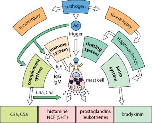
Fig. 3.29 Plasma enzyme systems.
MECHANISMS OF CELL MIGRATION
Leukocyte migration is controlled by molecules expressed on the surface of vascular endothelium that interact with complementary adhesion molecules on different populations of leukocytes. Most leukocyte migration occurs across venules. Several patterns of cell migration can be distinguished, including
•Movement of lymphocytes into secondary lymphoid tissues
•Migration of activated lymphocytes to sites of inflammation
•Migration of neutrophils into tissues during an acute immune response and migration of mononuclear cells into sites of chronic inflammation
Each pattern of migration is determined by particular sets of chemokines and adhesion molecules. There are three stages in the adhesion that precedes migration across the endothelium:
1.Slowing and rolling: most leukocyte migration occurs across venules as the shear force acting on circulating cells is lower and adhesion molecules are selectively expressed in venules. Initial slowing is mediated principally by selectins (such as E-selectin) on the endothelium, interacting with carbohydrate on the leukocytes.
2.Triggering: leukocytes that have been slowed may be triggered by chemokines released in the tissue or synthesized by the endothelium and bound to the endothelial cell surface. The chemokine signal is integrated over time, allowing cells to receive a sufficient signal to initiate migration. Triggering activates the integrins required for firm attachment to the endothelium.
3.Adhesion: the affinity of leukocyte integrins (such as LFA-1) on activated cells is increased, which allows them to bind to cell adhesion molecules (such as ICAM-1) induced on the endothelium by inflammatory cytokines. The integrins and CAMs are attached to the cytoskeleton of each cell, which allows the leukocyte to pull itself across the endothelium. Figure 3.30 shows a lymphocyte adhering to brain endothelium in encephalomyelitis.
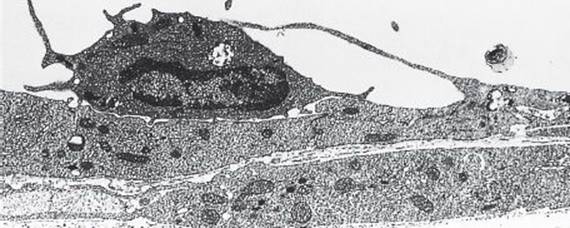
Fig. 3.30 Lymphocyte adhering to endothelium of the central nervous system. Courtesy of Clive Hawkins.
Diapedesis is the process by which adherent cells migrate across the endothelium and into tissues. Adherent cells extend pseudopodia into the junctions between endothelial cells, before squeezing through the gap. In tissues where endothelia have continuous tight junctions (for example, in the central nervous system (CNS)), migration occurs close to the junctions, but not through them. Enzymes released by the migrating cells dissolve the basal lamina. New adhesion molecules may now be mobilized to allow the cells to bind to cells of the tissue and extracellular matrix components.
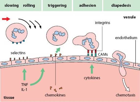
Fig. 3.31 Steps in leukocyte migration into tissues.
Chemotaxis is directional movement of cells in response to an inflammatory mediator. Cells are highly sensitive to, and migrate up, concentration gradients of molecules such as C5a, fMLP, and chemokines, if they have appropriate receptors.
Chemokinesis is increased random (nondirectional) movement of cells caused by inflammatory mediators, such as histamine.
Adhesion molecules belong to several different families. Some are constitutively expressed by cells (such as the integrin CR3 on mononuclear cells), whereas others may be induced by cytokines or cellular activation. Some adhesion molecules are retained in stores within the cells and may be quickly mobilized to the cell surface (such as LFA-1, stored in neutrophil ‘adhesomes’); others (such as ICAM-1 in endothelium) must be synthesized. The major families of adhesion molecules are listed overleaf.
Selectins (CD62) are a group of three adhesion molecules with lectin domains, that can bind to carbohydrate. P-selectin and E-selectin, induced on endothelium, help to slow migrating leukocytes before adhesion. L-selectin is expressed on lymphocytes and neutrophils; on lymphocytes it contributes to their binding to high endothelial venules in mucosal tissues.
PECAM (CD31), expressed on endothelium, platelets, and some leukocytes, can undergo homotypic adhesion, which contributes to tissue integrity and may act as a guide during migration.
Integrins consist of an α and a β chain, both of which traverse the cell membrane. Usually, the α chain is unique to each molecule, but the β chain may be shared with other molecules. Adhesion is dependent on divalent cations; when Mg2+ is bound they adopt a high-affinity form. Integrins often have more than one ligand-binding site, recognizing different molecules. Several integrins bind target sequences, related to Arg-Gly-Asp (RGD), in the ligand molecule.
Leukocyte integrins are a family of three molecules that share the β2 chain (CD18). They include LFA-1 (CD11a/CD18), important in migration of leukocytes across endothelium; CR3 (CD11b/CD18), expressed on all mononuclear phagocytes, which binds to ICAM-1 on endothelium at sites of inflammation; CR4 (CD11c/CD18), strongly expressed on tissue macrophages.
VLA (very late antigens) is the designation of the β1 integrin family, which includes two molecules that appear late on activated T cells and may be involved in binding to extracellular matrix. VLA-4, which binds to VCAM-1, is used by lymphocytes migrating to sites of inflammation, particularly in skin and CNS.
CAMs (ICAM-1, ICAM-2, VCAM-1, and MadCAM) belong to the Ig supergene family. ICAM-1 and VCAM-1 are induced on endothelium by TNF, IL-1, and IFN-γ at sites of inflammation. ICAM-2 is constitutively expressed on endothelium and may control the base level of leukocyte traffic through a tissue. MadCAM-1, the mucosal addressin, binds to both L-selectin and integrins, to control migration into mucosal lymphoid tissues.
CD44 is a widely distributed adhesion molecule that can be produced in different splice variants, which determine its ligand-binding functions. During transendothelial migration it localizes to the leading pseudopod and can bind extracellular matrix.
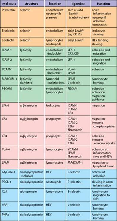
Fig. 3.32 Adhesion molecules for leukocyte migration.
CHEMOKINES AND CHEMOKINE RECEPTORS
Chemokines are a large group of cytokines that promote the chemotaxis and activation of a wide range of cells, including leukocytes. They are classified into four groups on the basis of their structure as α (CXC), β (CC), γ (C), and δ (CX3C)—the designation relates to the number and arrangement of conserved cysteine residues (C). Originally, they were given descriptive names such as macrophage chemotactic protein (MCP). However, these have been superseded by a system in which α chemokines are called CXCL1, CXCL2, etc., β chemokines are CCL1, CCL2, etc. For example, MCP-1 is now CCL2. Some chemokines are synthesized at sites of inflammation and control the migration of leukocytes across endothelium into inflamed tissues. Other chemokines are produced constitutively and control the normal movement of cells between lymphoid tissues and regions of these tissues, for example, between the cortex and germinal centers of lymph nodes. Figure 3.33 shows how chemokines can control the migration of different leukocytes into a site of inflammation. Inflammatory cytokines released in the tissue, such as TNF-α and IFN-γ, induce chemokine synthesis by the local endothelium, including CXCL8 (IL-8), acting on CXCR1, CCL2 (MCP-1) acting on CCR2, and CXCL10 (inflammatory protein-10, IP-10) acting on CXCR3. Which chemokines are produced depends on the tissue and the type of inflammation or immune response taking place. Chemokines can also be synthesized by tissue cells and transported to the endothelial surface. Each population of leukocytes has a different set of chemokine receptors, so the cells that enter a tissue differ, depending on the chemokines expressed on the endothelium.
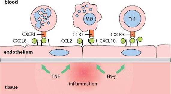
Fig. 3.33 Chemokine actions at sites of inflammation.
Chemokine receptors are designated according to which family of chemokines they recognize. For example α chemokines bind to CXCR1, CXCR2, etc. Most chemokines bind to several different chemokine receptors, and most receptors recognize several different chemokines. In addition, cells generally express several chemokine receptors so they can respond to a range of chemokines. Most cells of the body express some chemokine receptors at stages during their development, which control their position in the developing organism. Leukocytes change their receptors according to their state of differentiation and activation, which allows them to respond to inflammatory signals or position themselves in lymphoid tissues. For example, CCR7 is present on T cells, dendritic cells (DCs), and B cells. The chemokines that bind this receptor (CCL19 and CCL21) are produced in the T-cell areas of lymph nodes. Consequently T cells and DCs are attracted to these areas when they enter a lymph node. B cells can also be attracted to the T-cell areas when they express CCR7 after antigen stimulation. Figure 3.34 shows the complex pattern of chemokine receptor expression on leukocytes, but even this is simplified, because the relative expression is also important. For example, CXCR3 is found on T cells, but is highest on TH1 cells.
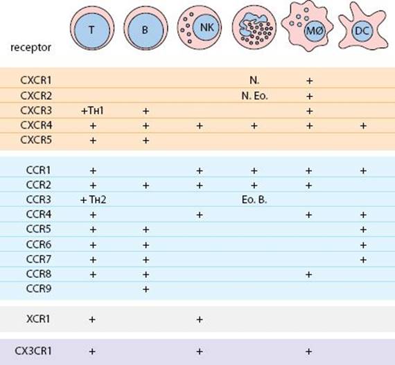
Fig. 3.34 Chemokine receptors on leukocytes.
N = neutrophil Eo = eosinophil b = basophil
COMPLEMENT
Complement is one of the serum enzyme systems. Its functions include mediating inflammation, opsonization of antigenic particles and microbes, and causing membrane damage to pathogens. The system consists of serum molecules, which may be activated via the classical, alternative, or lectin pathways. Molecules of the classical pathway are designated C1, C2, etc. Alternative-pathway molecules have letter designations, for example factor B (FB or just ‘B’). The properties of the components are given overleaf, and their receptors on p. 74. The complement components interact with each other so that the products of one reaction form the enzyme for the next. Thus, a small initial stimulus can trigger a cascade of activity. Small fragments of complement molecules produced by cleavage are lower-case (C3a, C5b). Inactivated enzymes are prefixed ‘i’ (for example, iC3b) and active enzymes are indicated with a bar (for example, C3b,Bb).
Classical pathway (yellow background) is activated by immune complexes binding to the C1q subcomponent of C1, which has six Fc-binding sites. C1q cleaves C1r and C1s. C1s then splits C4a from C4, and C2b from C2, leaving C4b,2a, which can cleave C3.
Alternative pathway (Properdin pathway or Amplification loop) (purple background) is activated in the presence of suitable surfaces or molecules, including microbial products. C3b can bind either H or B. Normally H is bound and C3b is inactivated by I, but in the presence of activators B is bound and then enzymatically split by D, releasing Ba and leaving C3b,Bb, which can cleave C3. This gives a feedback amplification loop to generate more C3b.
Lectin pathway (blue background) is activated by MBL or ficolins binding to bacterial carbohydrates.
C3 convertases, including C3b,Bb and C4b,2a, clip C3a from C3 to leave C3b. C3b has a labile binding site that allows it to bind covalently to nearby molecules with −OH or −NH2 groups. C3b together with a C3 convertase (such as C3b,Bb,3b) can cleave C5.
Lytic pathway (orange background) is activated when C5b is deposited on membranes and associates with C6, C7, C8, and C9 to form the membrane-attack complex.
Membrane-attack complex (MAC) is a structure of C5b678 and polymeric C9, which traverses the target cell membrane and allows osmotic leakage from the cell.
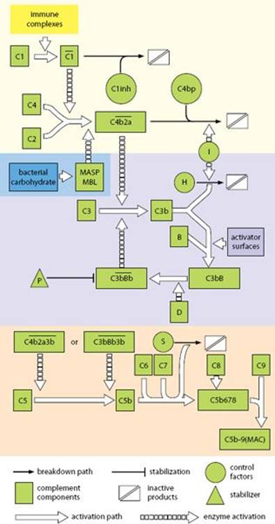
Fig. 3.35 Complement reaction pathways.
Complement fixation is the activation of complement, followed by deposition of the activated components on immune complexes or cell membranes. C3b and C4b can bind covalently to nearby molecules after cleavage of an internal thioester bond, which exposes a highly reactive group that can bind to −OH or −NH2. The reactive group decays quickly by hydrolysis if a link is not formed. Hence complement is only deposited close to sites of activation.
Bystander lysis is the phenomenon whereby cells in close proximity to a site of complement activation have active components deposited on them and may then be lysed.
Anaphylatoxins C3a and C5a cleaved from the N-termini of the α chains of C3 and C5 mediate inflammation by causing mast-cell degranulation, smooth muscle contraction and increased capillary permeability. C5a is also chemotactic for neutrophils and monocytes. In this way, these peptides mimic some of the reactions of anaphylaxis. They are substantially inactivated by removal of their C-terminal arginine by serum carboxypeptidases.
Mannan-binding lectin (MBL) is a polymeric, pattern recognition molecule of the collectin family related to C1q. It binds bacterial and fungal carbohydrates and can activate the lectin pathway. MBL deficiency is associated in infants with respiratory infections.
Control of complement activation is effected by the natural decay of enzymatically active convertases and the actions of the various inhibitors and inactivators listed opposite. Membrane-associated molecules also alter the rate of complement breakdown; CR1 and DAF promote the decay of C3b,Bb.
Decay-accelerating factor (DAF, CD55) and Membrane cofactor protein (MCP, CD46) are proteins normally present on many mammalian cell membranes that limit the activity of the alternative pathway and the assembly of C5 convertases.
Protectin (CD59) is a membrane protein that protects host cells from lysis by binding to C5b678, to prevent polymerization of C9.
Paroxysmal nocturnal hemoglobinuria (PNH) is a condition in which red cell breakdown occurs via the alternative pathway. Patients’ red cells are deficient in control proteins, particularly DAF.
Hereditary angioedema is due to a genetic deficiency of C1inh. There is uncontrolled local activation of C2, which undergoes conversion into a kinin that induces pathological local edema.
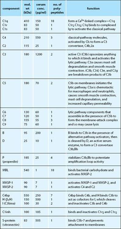
Fig. 3.36 The complement components.
IMMUNOREGULATION
The immune response is regulated primarily by antigen and co-stimulatory signals and secondarily by interactions between lymphocytes, APCs, and cells of the tissue. Antigen is the primary initiator of immune responses; the first signal required to trigger lymphocytes is antigen or antigen:MHC. Indeed, the immune system may be viewed as a homeostatic unit for the elimination of antigen. The essential role of antigen is seen at the cellular level. For example, antigen:MHC triggers T-cell activation and the expression of receptors for cytokines. Elimination of the antigen, by antibody or effector T cells, results in loss of the primary initiating stimulus, and the immune response is curtailed.
Danger signal is the idea that lymphocytes require both antigen stimulation and a ‘danger signal’ (co-stimulation) to become activated. The requirement for a dual signal acts as a fail-safe, to prevent unwanted immune reactions such as autoimmunity. In practice, danger signals are transduced by pattern recognition receptors (such as TLRs) that recognize microbial molecules.
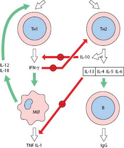
Fig. 3.37 Immunoregulation of TH1- and TH2-type responses.
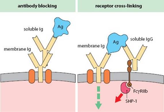
Fig. 3.38 Regulation of antibody production by antibody.
Antibody-mediated immunoregulation Antibody regulates its own production in several ways. Typically, IgM antibodies enhance the production of specific antibody, whereas specific IgG antibodies suppress further synthesis. The mechanisms include (1) binding to antigen, so preventing it from activating lymphocytes (Fig. 3.38, left); (2) binding to Fc receptors (FcγRIIb) on B cells, which in the presence of antigen cross-links the Fc receptors and surface Ig, delivering an inhibitory signal to the cells, mediated by the phosphatase SHP-1 (Fig. 3.38, right); (3) promoting immune complex formation and localization of antigen in germinal centers, so promoting Ig class switching and induction of B-cell memory.
Immune complex-mediated immunoregulation Complexes containing IgG generally suppress B-cell activation by the mechanisms shown in Fig. 3.38, whereas complexes containing IgM enhance it. In effect, IgM-containing complexes, produced early in an immune response, enhance the response, whereas IgG-containing complexes, produced after class switching, suppress it.
TH1- and TH2-type responses The TH1 subset promotes cellmediated immunity, whereas TH2 cells promote antibody production, including IgE (Fig. 3.37). Moreover, each mode of response suppresses the other. IFN-γ produced by TH1 cells limits proliferation of TH2 and TH17 cells, whereas IL-12 and IL-18 from mononuclear phagocytes promote TH1 development. Conversely, IL-10 from TH2 cells prevents cytokine production by TH1 cells, and IL-13 inhibits cytokine production by macrophages.
Regulation/Suppression A group of functionally defined, regulatory T cells (TREG cells) control the activity of other lymphocytes. (Originally, a population of CD8+ T suppressor cells was thought to mediate this activity, but most TREG cells are CD4+.) TREG cells can develop naturally in the thymus or may be induced in the periphery during immune responses (induced TREG cells). They constitute 5–10% of peripheral T cells. Regulation is an active process and can be distinguished from tolerance by transferring suppression with T cells. Animals lacking TREG cells are susceptible to aggressive inflammation and autoimmunity in the gut and endocrine organs. The cellular basis for TREGaction involves some or all of the following mechanisms: (1) reducing the co-stimulatory activity of dendritic cells, (2) release of anti-inflammatory cytokines (IL-10, TGF-β, IL-35), (3) modulating the mode of immune response (see Fig. 3.37), (4) consumption of IL-2, (5) a direct cytotoxic action on helper T and cytotoxic T cells.
Tissue-dependent regulation Immune reactions in tissues are controlled by regulatory cytokines (IL-10, TGF-β, etc.), eicosanoids and direct cell–cell interactions. Regulatory molecules include:
CD47, a widely distributed molecule that interacts with signal-inhibitory regulatory protein-α (SIRPα); it recruits a phosphatase SHP-2 to the membrane, which inhibits lymphocyte activation.
Fractalkine (CX3CL1), a chemokine that can be produced in a membrane or secreted form, acting on the receptor CX3CR1. The soluble form is chemotactic, but the membrane form, present on neurons, contributes to suppression of microglia in the CNS.
CD200, a 2 domain member of the Ig supergene family, expressed on keratinocytes and Langerhans cells. It binds to a receptor, CD200R1, found on myeloid cells and inhibits activation.
Network hypothesis is a theory that lymphocytes may be regulated by recognition of idiotypes on the antigen receptors of other cells or by idiotype-bearing antibodies. Such regulation is secondary to that mediated by antigen and cytokines, because of the redundancy in the immune system; if clones of lymphocytes are suppressed, their functions can be taken by other clones.
Psychoimmunology is a branch of immunology concerned with the interactions of the nervous, endocrine, and immune systems.
Sickness behavior describes the behavioral changes that occur in a person suffering from infection, including loss of appetite, reduced mobility, and extended sleep. Many of these changes have been related to the actions of IL-1 on the brain. IL-1 acts on temperature regulation centers in the hypothalamus to induce fever. It also suppresses appetite and induces slow-wave sleep.
Neuroendocrine regulation of immune responses seems to be important in damping immune and inflammatory responses, particularly via the production of corticosteroids.
Innervation of lymphoid tissues Thymus, spleen, and lymph nodes all receive sympathetic noradrenergic innervation, which controls blood flow through the lymphoid tissues, thus affecting lymphocyte traffic. However, fibers also run between the lymphocytes and seem to form junctions with individual cells. Denervation of lymphoid tissues can modulate immune responses.
Pituitary/adrenal axis Stress can induce release of adrenocortico-tropic hormone (ACTH) from the pituitary. This induces the release of glucocorticoids, which are immunosuppressive. Lymphocytes also produce ACTH in response to corticotropin-releasing factor. In addition the adrenal medulla releases catecholamines, which can alter leukocyte migration patterns and lymphocyte responsiveness.
Endocrine and neuropeptide regulation Lymphocytes carry receptors for many hormones, including insulin, thyroxine, growth hormone, and somatostatin. These hormones, as well as encephalins and endorphins, released during stress, modulate T- and B-cell functions in complex, dose-dependent ways.
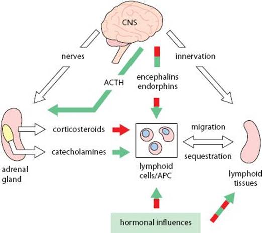
Fig. 3.39 Neuroendocrine regulation of immune responses.
TOLERANCE
Tolerance is the acquisition of nonresponsiveness to a molecule recognized by the immune system. Animals generally tolerate their own tissues: if they do not, autoimmune disease may result. Self-tolerance is thought to be due primarily to clonal deletion of cells in the neonatal period. As new mature lymphocytes develop they too are aborted, just when they are most susceptible to tolerization.
Neonatal tolerance Newborn animals are very susceptible to the induction of tolerance because of the general immaturity of their immune systems. Consequently, tolerance induced at this stage of life is very persistent.
Central tolerance refers to the induction of tolerance during lymphocyte development. Self-reactive T cells are deleted in the thymus, and self-reactive B cells in the bone marrow.
Peripheral tolerance is a necessary mechanism for maintaining tolerance to antigens that are not present in the primary lymphoid organs, or where the receptor is of low affinity.
B-cell tolerance In general, immature cells are more susceptible to tolerance induction than mature cells and can be tolerized by smaller doses of tolerogens. The dose of antigen and the way it is presented are critical. Self-reactive B cells fail to express Bcl-2 during development in the bone marrow or secondary lymphoid tissues and thus die by apoptosis. In the bone marrow, autoreactive B cells may escape deletion by editing their receptor specificity, done by making a new light-chain gene rearrangement. B cells may also become anergic to their antigen if they receive incomplete activation signals. Such cells downregulate surface IgM, while retaining IgD.
T-cell tolerance T cells are more easily tolerized than B cells. Once established, the duration of T-cell tolerance in an animal usually persists longer than for B cells. Immature T cells may be deleted during thymic development, although cells with low-avidity receptors remain. Mature T cells can be made anergic, depending on how antigen is presented to them (for example, lack of co-stimulation). Because B cells require help from TH2 cells, B-cell tolerance may be a consequence of T-cell tolerance.
Superantigens are antigens that bind strongly to MHC molecules and can induce clonal deletion of T cells. Potentially they can modulate the T-cell repertoire.
High-zone and Low-zone tolerance Tolerance is best induced by high levels of antigen (high zone), which tolerizes B cells. However, some antigens in subimmunogenic doses (low zone) can also tolerize the T-cell population.
Mucosal tolerance and Oral tolerance Many antigens fail to induce an immune response when presented across the nasal mucosa as an aerosol, or across the gut mucosa in food (oral tolerance). The effect is dependent on the dose and frequency of the antigenic challenge. The effect may be due to the deviation of the immune response to TH2-type, with the production of suppressive cytokines and/or due to TREGactivity.
Immune deviation refers to treatments aimed at switching the immune response from one mode to another (such as TH1 to TH2).
Tolerance mechanisms Several mechanisms maintain tolerance to self tissues (Fig. 3.40):
•Sequestration of antigen away from the immune system
•Central or peripheral tolerance induction of B and T cells
•Failure to process and present autoantigens by APCs
•Absence of co-stimulatory molecules on APCs
•Suppressive cytokines including IL-10 and TGF-β
•Direct and indirect actions of regulatory T cells
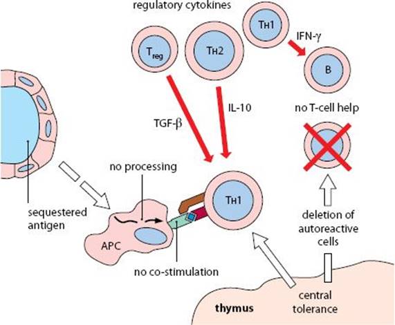
Fig. 3.40 Mechanisms for maintenance of self-tolerance.
GENETIC POLYMORPHISM IN THE IMMUNE RESPONSE
The genetic make-up of an individual affects their ability to mount an immune response. We can distinguish between mutations that clearly prevent the function of a particular component of the immune system and variants (polymorphisms) that affect the quality of an immune response. Polymorphisms are often stable in a population and many individuals have the variants, whereas mutations are deleterious and are usually lost over evolutionary timescales. The MHC is the most polymorphic of all gene loci, and variant MHC molecules differ in their ability to present antigen, leading to variations in immune response and disease susceptibility (Fig. 3.41). Polymorphism within the MHC is thought to reflect selection for variants that protect against particular infections, either extant pathogens or those that have occurred in historic times. One might therefore expect reports of particular MHC genes being associated with resistance to infectious disease. In practice there are many more reports of MHC haplotypes that are associated with susceptibility to infectious or autoimmune disease. This bias is seen because it is easier to identify positive disease associations and to get such results published. It is important to note that the relative risk associated with individual MHC genes (or haplotypes) depends on the population under investigation and the disease pattern. Susceptibility may be modified by other genes in the population, and in some cases the strain of pathogen can also affect whether an MHC gene is protective. Other polymorphisms affect the ability to respond to a wide range of antigens. For example, variants of TNF-α affect susceptibility to leprosy and severe cerebral malaria.
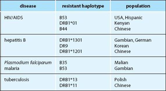
Fig. 3.41 HLA polymorphism and disease resistance.
Immune response (Ir) genes were first identified in inbred strains of mice, according to their effect on the antibody response to defined antigens. The most important Ir genes encode MHC class II molecules, which determine antigen presentation to helper T cells. MHC class I molecules affect presentation to TC cells and hence the ability to resist viral infections. There is also limited variation in MHC genes controlling antigen processing and presentation (DM, TAP, etc.). Genes encoding specific haplotypes of antigen receptors (IGH and TCR) have been linked to autoimmune conditions as well as restricting the responses to exogenous antigens (see clonal restriction below). Significant polymorphism is not confined to exons. For example, the promoter of the TNF-α gene is linked to autoimmunity in NZW mice. In addition, the promoters of MHC class II genes vary between strains, resulting in different responses to IFN-γ.
Repertoire is the sum total of antigen receptors produced by the immune system. The initial repertoire is partly determined by the genes of the TCR and antibody heavy and light chains.
Clonal restriction refers to an immune response produced by a limited number of clones. For example, the primary immune response to phosphocholine in Iga haplotype mice is dominated by the T15 idiotype. T-cell responses can also be clonally restricted, as a result of selective antigen presentation by particular MHC molecules expressed in a strain.
Biozzi mice are strains bred to give high or low antibody responses to an antigen (originally sheep erythrocytes). At least 10 non-MHC genes control responsiveness. The high and low responders differ in how their macrophages handle antigen; low responders degrade antigen quickly and do not present it well.
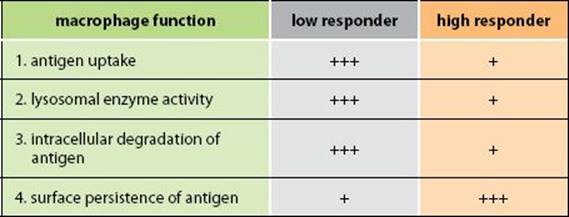
Fig. 3.42 Macrophage functions in Biozzi mice.
IMMUNOSUPPRESSION
Immunosuppression describes measures used to reduce immune responses, particularly in transplantation surgery to prevent graft rejection, and in the control of autoimmune diseases. Most drug treatments are not antigen-specific, although some have greater effects on the immune system than other tissues.
Steroids including glucocorticosteroids, corticosteroids and synthetic steroids (such as dexamethasone) have numerous immune-suppressive and anti-inflammatory effects, macrophages being particularly sensitive. Steroids inhibit arachidonic acid release and hence reduce eicosanoid production. They also reduce secretion of neutral proteases and IL-1. Steroids interfere with antigen presentation, inhibit the primary antibody response and reduce the number of circulating T cells.
Azathioprine and 6-mercaptopurine are purine analogues that act on small lymphocytes and dividing cells, thereby blocking the development of effector cells. Monocytes are reduced in number and NK cell activity is also inhibited.
Cyclophosphamide and Chlorambucil are alkylating agents that damage DNA and prevent its replication. They act primarily on lymphocytes and strongly inhibit antibody responses, but have little effect on phagocytes. Experimentally, cyclophosphamide prevents B cells from regenerating their receptors.
Methotrexate is an analogue of folic acid that inhibits DNA synthesis and repair, and hence lymphocyte proliferation.
Mycophenolate inhibits the synthesis of guanosine. Lymphocytes are particularly susceptible to inhibition by this drug.
Cyclosporin-A is a fungal metabolite that interferes with cytokine production by T cells, particularly IL-2, and it inhibits IL-2R expression; both are early events in lymphocyte activation. It does not affect lymphoblasts, nor is it antimitotic. It is used to treat acute graft rejection, but has increasingly been replaced by less toxic drugs, listed below.
Tacrolimus (FK506) is a bacterial macrolide that prevents T-cell activation and IL-2 transcription by acting on calcineurin, an enzyme required for signal transduction from the T-cell receptor.
Rapamycin inhibits the ability of T-cell growth factors to put T cells into cell cycle. Rapamycin and tacrolimus bind to the same receptor, although their modes of action are different.
Antagonist peptides are analogues of peptides that bind to MHC molecules of particular haplotypes. They have been used as an experimental treatment for autoimmune conditions. By occupying the MHC-binding site, they block access for autoantigen peptides.
Antibody therapy is the use of monoclonal antibodies to treat disease, usually tumors or autoimmune diseases. Some are used to prevent graft rejection (Fig. 3.43). The first monoclonal antibodies were produced in mice, but these were potentially immunogenic in humans and less suitable for long-term therapy. Immunogenicity was reduced by genetic engineering to produce chimeric antibodies containing the antigen-binding V domains of the original antibody and human C domains. Alternatively, humanized antibodies, with antigen-binding hypervariable regions inserted into a human antibody gene framework, are even less immunogenic. Fully human antibodies can be produced in mice transgenic for human immunoglobulin genes.
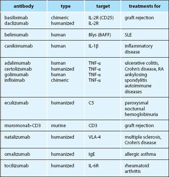
Fig. 3.43 Monoclonal therapeutic antibodies.
SLE = systemic lupus erythematosus; RA = rheumatoid arthritis; BAFF = B-cell activating factor.
IMMUNOPOTENTIATION
Biological response modifiers (BRM) are compounds that modify an immune response, usually enhancing it. They include immunopotentiating bacterial and viral products (see p. 50 Toll-like receptors), and physiologically active molecules, including cytokines, as well as the true adjuvants, which are administered together with antigen. A number of these substances have been used in an attempt to potentiate immune reactions in cancer, by inducing cytokine production or the expression of co-stimulatory molecules on APCs. Bacterial products include:
BCG (Bacillus Calmette–Guérin), a live nonvirulent strain of Mycobacterium bovis, which is used in vaccines for immunization against tuberculosis.
Muramyl dipeptide (MDP), the smallest adjuvant active part of BCG, extractable from the cell wall.
Endotoxin/Lipopolysaccharide (LPS), a component of Gram-negative bacterial cell walls that is mitogenic for B cells and activates macrophages after binding to TLRs (see p.51, Fig. 2.22).
Bordetella pertussis toxin/toxoid (PTx), a lymphocytosis-promoting factor, which binds glycans on many cell types, particularly T cells, on which it acts as a mitogen.
Thymic hormones are factors produced by the thymus that assist T-cell development in the thymus and their maintenance in the periphery. They include thymosin, thymopoietin, thymostimulin, and thymulin (facteur thymique sérique).
Adjuvants are compounds that enhance the immune response, when administered with antigen, to produce higher antibody titers and prolonged production. The distinction between primary and secondary immune responses becomes blurred when adjuvants are used. Adjuvants typically consist of a depot of antigen, which may include bacterial components.
Freund’s adjuvant is a water in oil emulsion containing the antigen (= Incomplete Freund’s adjuvant). Complete Freund’s adjuvant also includes dried, heat killed mycobacteria which induces very strong immune reactions and local necrosis. It is not used in any human vaccines.
Aluminum-adjuvanted vaccines use either aluminum hydroxide (alum) or aluminum phosphate mixed with the antigen, which becomes adsorbed on the surface of the gel. These adjuvants are used in many human vaccines, acting as antigen depots.
VACCINES
Vaccines are antigen preparations produced in a number of different ways, depending on the pathogen, its route of infection, and how it produces disease pathology. In addition to the antigen preparation and adjuvants (see opposite), vaccines often contain stabilizers and preservatives. Most vaccines are given by subcutaneous or intradermal injection, but some are given orally (such as rotavirus, polio (Sabin)) or nasally (for example, some flu vaccines).
Toxoids are chemically modified toxins, which retain antigenicity while destroying pathogenicity. They are used where the toxin produces the majority of the pathology (such as tetanus, diphtheria).
Attenuated live vaccines are live bacteria or viable viruses that have been modified to remove pathogenicity. They generally produce better immunity than killed organisms, but are more likely to produce adverse reactions. Also, because they may divide, they can be unsuitable for immunocompromised people.
Subunit vaccines consist of an antigenic subcomponent of the pathogen, produced either by fractionation or by biotechnology. For example, a subunit of hepatitis B isolated from blood was later superseded by the same antigen expressed in yeast.
Vector vaccines are produced by inserting genes for antigens of a pathogen into a nonpathogenic viral vector, such as vaccinia or adenovirus. Possible use in humans is under investigation.
Conjugate vaccines are used where the key antigenic component of the vaccine is only weakly immunogenic; for example, three polysaccharide antigens are coupled to diphtheria toxoid (carrier) in a conjugate vaccine for meningitis C. The toxoid component is presented to T cells, which help B cells make antibody against the polysaccharide antigens of the meningococcus.
DNA vaccines are experimental preparations, in which the DNA for an antigen, rather than the antigen itself, is used. The DNA is injected using a gene gun, and the technique relies on the DNA being taken up and expressed by cells of the recipient.
Combined vaccines Many vaccines are given in combinations, during early infancy. This is for convenience, to reduce the number of visits needed to an immunization clinic. Examples are the trivalent DPT vaccine—diphtheria, pertussis, tetanus. The UNICEF-recommended pentavalent vaccine also includes hepatitis B and Haemophilus influenzae B.