
J. DAVID BECKHAM AND KENNETH L. TYLER
Viral infections of the spinal cord occur as part of more extensive infection of the central nervous system (CNS) or peripheral nervous system. When myelitis occurs in association with encephalitis or meningitis, the resulting syndromes are referred to as encephalomyelitis or meningoencephalomyelitis. Myelitis associated with involvement of spinal nerve roots or peripheral nerves is referred to as myeloradiculitis or myeloradiculoneuritis. This chapter focuses on acute viral infections in which spinal cord involvement is the dominant feature. Acute myelitis associated with rabies virus and HIV infection is discussed elsewhere (see Chapters 17 and 19), as are cases of chronic viral myelitis due to infection with retroviruses including HIV and human T lymphotropic viruses (HTLVs). In addition to directly infecting and injuring the spinal cord, viruses can trigger postinfectious immune-mediated tissue injury. Spinal cord involvement is a common but rarely dominant feature of acute disseminated encephalomyelitis (ADEM) (see Chapter 22). Transverse myelitis (TVM) is an acute syndrome defined by the nature and extent of the anatomic injury to the spinal cord, often associated with antecedent viral infections. Specific causes of TVM are discussed under the individual viruses involved, and the idiopathic syndrome is briefly reviewed at the end of this chapter.
The term myelitis means “inflammation of the spinal cord” and refers to disease of the spinal cord caused by a direct infectious process, a postinfectious process, or another indirect mechanism of injury. The clinical features are determined to a large degree by the location and extent of the process both in the craniocaudal and the transverse axes of the spinal cord rather than by the inciting agent.
The clinical features of myelitis provide important clues to the anatomic location of the lesion but do not enable myelitis to be separated from other causes of intramedullary spinal cord injury. The clinical features of myelitis caused by different viruses overlap substantially, and identification of a specific viral etiology typically depends on the results of laboratory tests. The characteristic features of myelitis include variable combinations of weakness; sensory loss; and bowel, bladder, and sexual dysfunction, typically evolving over days. Apoplectic or hyperacute (hours) evolution of symptoms is occasionally seen in viral myelitis but is more typical of vascular spinal cord disease (1) (e.g., infarction resulting from atherosclerosis, arteritis, emboli or hemorrhage, or even venous thrombosis [Foix-Alajouanine syndrome]). Viral causes of chronic myelitis or myelopathy in which symptoms evolving over weeks or months are largely limited to HIV and HTLV.
Weakness in viral myelitis may be either of the upper motor neuron type with associated spasticity, hyperreflexia, and extensor-plantar reflexes or of the lower motor neuron type with flaccid weakness and decreased or absent deep tendon reflexes. Lower motor neuron involvement in the absence of significant sensory signs or symptoms is often referred to as acute flaccid paralysis or poliomyelitis-like illness, although the latter term is best reserved for cases in which pathology is limited to the anterior horns of the spinal cord. Involvement of motor neurons in the anterior horns or involvement of the anterior roots can result in prominent clinical and electrophysiologic evidence of denervation, including the presence of fasciculations and fibrillations. Sensory loss in myelitis can be radicular, dermatomal, or both. Depending on the transverse localization of the lesion(s), either loss of position and vibration sense or loss of pain and temperature may occur. Finding a “sensory level” below which sensory functions are lost is a classic hallmark of spinal cord disease. Either relative or absolute sparing of sensation in sacral dermatomes (“sacral sparing”) may occur when an intramedullary process such as viral myelitis leaves the most peripheral fibers in the spinothalamic tract relatively unharmed.
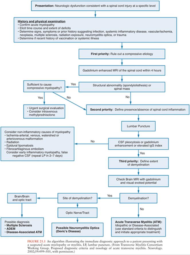
Patients with acute onset of signs and symptoms suggestive of spinal cord dysfunction are a medical emergency. Initial clinical and laboratory studies should be directed at trying to identify whether a compressive lesion is present and whether it is intramedullary or extramedullary in location. An algorithm for the immediate diagnostic approach to patients with acute myelopathy is shown in Figure 21.1(2). Table 21.1 summarizes key diagnostic tests that may be useful in evaluating a patient with suspected acute viral myelitis. In the following sections, viral etiologies of acute myelitis are discussed individually.

HERPESVIRUSES
Herpes Simplex Virus
Both herpes simplex virus type 1 (HSV-1) and type 2 (HSV-2) can cause myelitis. HSV-2 most commonly causes myelitis in adults and HSV-1 most commonly in children (1,3). The clinical presentation ranges from mild forms of disease with full recovery to severe necrotizing myelitis with permanent sequelae. Most cases are monophasic, although about 20% of patients experience recurrent episodes of myelitis, a feature common to infection with several herpesviruses (4–7). In patients with recurrent disease, the interval between recurrences may vary between 1 week and several months, with three or more discrete recurrences being noted (5,7). Up to two thirds of patients with HSV myelitis have an ascending pattern of spinal cord involvement, with the remainder having TVM (5).
Most cases of monophasic HSV myelitis are due to HSV-2 (5). Patients with HSV-2 myelitis often have a history of genital herpes. However, in one series, only two of seven patients with HSV-2 myelitis had known genital herpes, and lesions, when present, often preceded spinal involvement (5). Clinical features of HSV myelitis include paresis or paralysis, more commonly involving the legs than the arms. Patients may have either reduced and/or absent tendon reflexes or hyperreflexia with extensor-plantar responses. Decreased sensation to pain, temperature, and touch is common and tends to be more severe in sacral dermatomes. Patients can have decreased anal tone and urinary incontinence with overflow. HSV-2 can also cause a lumbosacral radiculomyelitis characterized by urinary retention associated with constipation, dull pain in the anogenital region, paresthesias, loss of sensation, or flaccid paresis of the leg muscles (8).
The most severe form of HSV myelitis is an acute necrotizing myelopathy, which occurs predominantly in patients with underlying diseases including HIV infection (9), malignancy (10), and diabetes (11,12), although rare cases occur in the absence of associated disease (13). All cases to date have been associated with HSV-2. At autopsy, these patients often show areas of necrosis in the gray and white matter of the spinal cord associated with perivascular lymphocytic cuffing. Cowdry type A inclusions are found in neurons, viral antigen can be demonstrated by immunocytochemistry, and herpesvirus-like particles have been seen by electron microscopy (9–13). Some cases have a prominent necrotizing arteritis associated with myelomalacia with Cowdry A inclusion-bearing cells seen in the wall of the anterior spinal artery (9).
In HSV myelitis, the cerebrospinal fluid (CSF) typically shows a lymphocytic pleocytosis and normal glucose concentration. Patients with recurrent attacks may have a progressive reduction in the degree of pleocytosis with succeeding attacks (7). Typical cell counts range between 10 and 200 cells/mm3, although rare cases with normal cell counts have been reported (5,14). The CSF profile in patients with acute necrotizing myelopathy may show striking pleocytosis with up to 5,750 cells and a predominance of polymorphonuclear (PMN) leukocytes rather than lymphocytes (12). Surprisingly, some reported patients have had no or minimal pleocytosis (9,11). The CSF protein concentration is almost invariably elevated (range, 50 to 430 mg/dL). Oligoclonal bands were found in one of nine patients in one series (5) and have been noted in other case reports (7,15).
HSV is only rarely cultured from CSF in patients with myelitis (15), and diagnosis depends on demonstration of HSV DNA in CSF by polymerase chain reaction (PCR) (4–6,15–17). In one series of nine patients, all had PCR amplifiable HSV DNA in CSF, with six cases due to HSV-2, two cases due to HSV-1, and one case indeterminate. In this series, HSV-2 DNA was found in six of six cases with ascending myelitis, whereas HSV-1 DNA was found in two of the three patients with nonascending TVM (5). In an HSV CSF PCR-negative patient, a presumptive diagnosis can be made by demonstrating intrathecal synthesis of HSV-specific antibodies (7). Antibody studies may be particularly useful in cases in which CSF is only available late in infection (e.g., >14 days), when HSV DNA is likely to have disappeared. Evidence of intrathecal synthesis of HSV-specific antibodies can be based on (a) the presence in CSF of immunoglobulin M (IgM) anti-HSV antibodies (these antibodies cross the blood–brain barrier poorly and their detection in CSF is generally indicative of intrathecal synthesis); (b) the detection of HSV-specific oligoclonal bands present in CSF but not in serum; (c) the comparison of HSV-specific immunoglobulin G (IgG) levels in CSF and serum with correction for blood–brain barrier leak using either the CSF/serum albumin ratio or the ratio of antibody titers in CSF to that in serum for an “irrelevant” virus (5,14,16,18). Basic laboratory studies usually add little to the diagnosis, although some patients have elevations in erythrocyte sedimentation rate and C-reactive protein (6,18).
Magnetic resonance imaging (MRI) is exceedingly important for its role in both excluding other potential diagnoses and in establishing the presence of an intramedullary process. The typical appearance of HSV myelitis is of an intramedullary fusiform or spindle-shaped area of an increased T2-weighted signal (5,7,14,16). The spinal cord is often enlarged or swollen in the area of the lesion. Areas of an increased T2-weighted signal may also have T1-weighted signal hypointensity. In rare cases, areas are both T1 and T2 hyperintense, a finding suggestive of hemorrhagic necrosis (5). Contrast (gadolinium) enhancement may be seen in the area of the lesion and the adjacent meninges and nerve roots (6,7,16,17). Lesions are most commonly in the upper thoracic and cervical cord (5,6) but can also involve the lower cord including the conus medullaris and cauda equina (7,15–17).
No controlled clinical trials of antiviral therapy for HSV myelitis are available. Based on anecdotal reports, patients should be treated with intravenous acyclovir for at least 14 days (10 mg/kg three times per day). This can be followed by oral antiviral drugs (e.g., valacyclovir at 1 g three times per day) until symptoms resolve. The utility of adding steroids is unproven. When given, steroids should be used only in combination with antiviral therapy. A typical regimen involves intravenous methylprednisolone (500 to 1,000 mg per day for 3 to 5 days) followed by oral prednisone (100 mg per day) with doses tapered over 2 weeks (5,7,17).
The prognosis of HSV myelitis is extremely variable. In one series of nine patients, one third (n = 3) made a complete recovery, and the remaining two thirds (n = 6) had residual sequelae including paraplegia and tetraplegia (5).
Varicella-Zoster Virus
Myelitis is an unusual complication of varicella-zoster virus (VZV) infection, with most cases occurring in immunocompromised individuals (19–21). Myelitis can also occur as a complication of primary varicella infection or chickenpox (20,22,23). In immunocompromised patients, common underlying diseases include HIV infection (19,20,21,24,25), Hodgkin and non-Hodgkin lymphoma (19), and immunosuppressive therapy (26,27). In a recent review of 31 cases of VZV myelitis, 55% of patients with VZV myelitis were immunocompromised, and the majority of the remaining patients had an underlying comorbidity such as malignancy or autoimmune disease (28). Cases of VZV myelitis in immunocompetent patients are reported but uncommon (29). Many patients have antecedent zoster, although several cases without rash (myelitis sine herpete) have been reported (30–32). When zoster is present, it can involve virtually any dermatome, but myelitis is often associated with disseminated zoster in immunocompromised patients (19). Cord dysfunction typically follows the onset of zoster with a median of 12 days (range, 5 to 21 days), but symptoms of myelitis may follow rash by up to 3 months or myelitis symptoms may precede the appearance of rash (19,24). Cases of TVM following zoster have also occurred in patients who have previously received zoster vaccine (33).
Patients with zoster myelitis present with subacute onset of asymmetric leg weakness which progresses to bilateral leg weakness with paraparesis in up to 85% of patients (19). Sensory loss is common (75%), with involvement of pain and temperature more common than position and vibration sense. A Brown-Séquard syndrome may occur, with posterior column signs (loss of position and vibration sense) ipsilateral to the rash and spinothalamic tract signs (loss of pain and temperature sense) contralateral (19). Approximately one third of patients will have a level to all sensory modalities, urinary incontinence occurs in about 50%, and bowel incontinence can also occur. In a few cases, VZV myelitis may show a relapsing and remitting pattern (20,25,34), a feature also seen in some cases of HSV-2 myelitis (see earlier discussion). Atypical presentations of VZV myelitis including cases not associated with rash (zoster sine herpete), skin lesions developing after myelopathy, or lack of correspondence of zoster lesions to spinal cord injury level occur more frequently in immunocompromised patients (28).
In VZV myelitis, CSF shows a mononuclear pleocytosis in about 75% of patients with an increase in PMN predominance in patients with rapid onset and severe disease (19). Recurrent episodes of CSF PMN pleocytosis with recurrent episodes of VZV myelitis have been reported (30). CSF protein concentration is elevated in 70%, but glucose concentration is almost always normal. It is important to recognize that at least 50% of immunocompetent patients with a zoster rash in the absence of myelitis have a CSF lymphocytic pleocytosis with cell counts from 5 to 1,440 cells/mm3 and exceeding 100 cells/mm3 in 30% of those with pleocytosis (35).
Approximately 25% of patients with a zoster rash will also have evidence of CSF anti-VZV IgG and detectable VZV DNA by PCR, but CSF anti-VZV IgM is usually not detectable and the CSF VZV IgG index is usually normal, suggesting that intrathecal synthesis of VZV-specific antibody does not occur in uncomplicated herpes zoster reactivation (35). By contrast, patients with zoster myelitis often have a positive CSF VZV PCR and intrathecal VZV-specific IgG synthesis, even VZV specific CSF oligoclonal bands (36).
Patients with VZV myelitis frequently have abnormalities on spinal MRI (Fig. 21.2). These can include diffuse swelling of the cord and areas of high T2-weighted signal with or without associated T1 hypointensity and contrast enhancement (20,22,27,32,37–39). MRI lesions in the brainstem and cervical cord may also occur in immunocompetent patients with zoster myelitis (35).

The diagnosis of VZV myelitis is generally suspected when signs of myelitis develop following a typical zoster eruption in an immunocompromised patient. Definitive diagnosis depends on isolation of virus from CSF (24,31), demonstration of intrathecal synthesis of VZV-specific antibody (20,36,37,40), or PCR amplification of VZV DNA from CSF (20,27,37,40–42). Comparative studies of the sensitivity and specificity of these tests are lacking, although clinical experience suggests that PCR is likely to be the most sensitive, and culture the least. Antibody and PCR test results should be viewed as complementary rather than mutually exclusive; as in other viral infections, antibodies typically develop later than detectable nucleic acid. The duration for which VZV DNA remains detectable in CSF following varicella myelitis is unknown. In patients with relapsing-remitting myelitis, CSF VZV DNA may persist, as two patients had positive CSF PCR results 8 and 11 months after onset of disease (20).
No controlled clinical trials of treatment for VZV myelitis are available. Treatment typically involves intravenous acyclovir given alone or in combination with steroids (19,31,37,39,42–44), although the paucity of cases and the variability in treatment regimens make efficacy difficult to access (20). In several patients who received intravenous acyclovir after myelopathy was established, treatment seemed to be without effect (24,39,44). By contrast, there are also reports of complete recovery in AIDS patients with VZV myelitis following acyclovir therapy (10 mg/kg every 8 hours) for 21 to 35 days (42) and improvement in patients treated with either acyclovir alone (20,37) or a combination of acyclovir and high-dose steroids (31). One patient apparently responded to oral famciclovir (500 mg every 8 hours), who had not previously responded to steroids, acyclovir, or foscarnet (37).
Pathologic studies of VZV myelitis are limited (19,25,32). In a comprehensive review of nine fatal cases, there was extensive hemorrhagic necrosis with necrotizing vasculitis and thrombosis in the dorsal root ganglia associated with Cowdry type A intranuclear inclusions in both ganglion and satellite cells (19). Almost all cases had abnormalities in the posterior roots and the posterior horns of the spinal cord, although severity varied. Inclusion bodies were seen in about 50%, and patients could have demyelination and signs of necrotizing vasculitis and hemorrhagic spinal cord necrosis (19,32). A similar necrotizing process with demyelination and Cowdry type A inclusions was described in an AIDS patient with chronic zoster myelitis (25). In a recent review of five postmortem cases of VZV myelitis, tissue necrosis and inflammatory cell infiltration were the primary findings, but other pathologic changes were noted as well, including vasculitis, thrombosis, hemorrhagic transformation, and Cowdry type A inclusions (28).
Cytomegalovirus
Cytomegalovirus (CMV) involvement of the spinal cord either can result in a pure TVM or can produce myeloradiculitis, or radiculomyelopathy. Most cases of CMV-associated myeloradiculopathy occur in HIV-infected patients (45–48), and this can be the initial manifestation of AIDS (49). Myelitis can be a complication of both adult and pediatric HIV infection (50). Although CMV myelitis is predominantly a disease of immunocompromised individuals, there have been isolated reports of both TVM and myeloradiculopathy in immunocompetent individuals (51–57). Isolated cases of dual infection of the cord with both HSV and CMV have been reported in patients with AIDS (58). The incidence of CMV-associated myeloradiculopathy has declined dramatically since the introduction of highly active antiretroviral therapy (HAART). Myeloradiculopathy occurs with approximately equal frequency as an isolated manifestation of CMV infection and in systemic CMV disease (59). Patients present with rapidly progressive flaccid paralysis of the legs with hyporeflexia or areflexia (60). Urinary retention occurs in almost all patients (60). Pain, often involving the perianal region and low back, is a common initial symptom. Sensory loss usually involves small-fiber modalities (pain, temperature) more than vibration or proprioception, although both can occur. The symptoms are typically progressive, although some patients have a more indolent course.
One of the unusual features of CMV myeloradiculitis is the common occurrence of a PMN rather than a lymphocytic CSF pleocytosis (61,62). This feature is distinctive enough that its presence in an HIV-infected patient with myeloradiculopathy should always suggest the possibility of CMV infection (63). Cell counts exceeding 1,000/mm3 can occur (46,64–66), although median values of 150 to 650 cells/mm3 are more typical (60,61). In addition to pleocytosis, the CSF typically shows an elevated protein concentration. Severe hypoglycorrhachia may occur (46), although a CSF glucose concentration of less than 50% of the coincident plasma value has been reported in only about 30% of patients (62).
Imaging studies are useful in demonstrating the location and extent of lesions. Typical findings on spinal MRI include areas of increased T2-weighted signal within the spinal cord, associated with enhancement of the pial lining of the cord, conus, cauda equine, and lumbosacral nerve roots on contrast-enhanced T1-weighted images (66,67). The presence of prominent radicular enhancement in association with myelitis occurs more often with CMV infection than in other forms of viral myelitis and may provide a clue to the diagnosis.
Clinical electrophysiologic studies may be helpful in demonstrating the presence of radicular and peripheral nerve involvement in association with myelitis, especially because this component of the disease may be clinically obscured when the myelitis is severe. Both slowed conduction velocities consistent with demyelination and reduced amplitude of motor and sensory action potentials consistent with axonal injury occur (59,68).
PCR tests to amplify CMV DNA from CSF are the diagnostic procedure of choice (59,69–72). The diagnostic sensitivity of CSF PCR in CMV myelitis is more than 80%, with a specificity of more than 90% (59). It is important to emphasize that CSF PCR is often positive when cultures are negative (69,72). Quantitative PCR studies of CMV DNA in CSF suggest that extremely high DNA levels, exceeding 10 million copies of CMV DNA per milliliter of CSF can be found in patients with myeloradiculopathy (59,69,73).
CMV may be cultured from CSF in cases of radiculomyelitis (59). The high seroprevalence rate of anti-CMV antibodies in serum generally renders serologic studies of limited value. However, the demonstration of specific intrathecal synthesis of anti-CMV antibodies or the presence of detectable CSF anti-CMV IgM antibodies can be considered presumptive evidence for CNS infection.
Controlled clinical trials of antiviral therapy in CMV-associated neurologic disease are lacking, and most reports of treatment of CMV-associated neurologic disease involve isolated cases (59,69,74,75). Response to treatment of myeloradiculopathy is variable. Improvement or stabilization of symptoms has followed treatment with ganciclovir, foscarnet, or a combination of the two agents (60,62,64,68,76–80). Disease has been associated with ganciclovir-resistant strains (81,82), and this risk may be enhanced in patients whose disease developed during ganciclovir treatment for CMV infection elsewhere. As a general rule, the presence of myelitis is an extremely poor prognostic indicator, with one review citing mean survival times of 5.4 ± 1.8 weeks in patients not receiving ganciclovir and 14.6 ± 9.4 weeks in those receiving ganciclovir therapy (76).
Successful treatment is usually associated with disappearance of CMV DNA from CSF, whereas viral DNA persists in nonresponders (74). A typical induction regimen for ganciclovir involves 14 to 21 days of intravenous therapy with doses of 5 mg/kg every 12 hours. Intravenous foscarnet at a dose of 90 mg/kg every 12 hours provides an acceptable alternative. Patients who fail to respond to either foscarnet or ganciclovir alone may respond to combined therapy with the two drugs (79). A third agent, cidofovir, has been shown to be efficacious in treatment of CMV retinitis, but experience with this agent in CMV-associated neurologic disease is limited (83). A typical dosing regimen is 5 mg/kg intravenously every week for 2 weeks followed by infusions of 5 mg/kg every 2 weeks. Fourteen to twenty-one days of ganciclovir or foscarnet therapy is likely to be sufficient for immunocompetent patients. In most organ transplant recipients, 14 to 21 days of therapy is also likely to be adequate assuming clinical and virologic response has occurred. Maintenance therapy is usually not required for CMV myelitis in organ transplant recipients, although maintained vigilance for recurrent disease is essential. In patients with HIV infection, it is critical that HAART be initiated or optimized concomitantly with anti-CMV therapy. HIV-infected patients invariably require maintenance therapy with an orally bioavailable valine ester of ganciclovir (valganciclovir). Induction and maintenance doses of intravenous and oral ganciclovir, intravenous foscarnet, and intravenous cidofovir require adjustment in patients with renal insufficiency. Neutropenia is the major dose-limiting side effect with ganciclovir, and nephrotoxicity with foscarnet and cidofovir. Nephrotoxicity can occur with ganciclovir but is less common. Neurotoxicity can occur with ganciclovir and foscarnet and in the case of foscarnet is often related to electrolyte alterations (especially hypocalcemia). Patients whose HIV infection responds to HAART and who consistently (>6 months) demonstrate nondetectable HIV viral loads and CD4 cell counts of more than 100 cells/mm3 may be able to discontinue maintenance therapy.
Few detailed pathologic studies of CMV myeloradiculitis have been performed. There is often a prominent PMN and mononuclear cell infiltrate involving the sacral spinal cord, cauda equina, and lumbosacral nerve roots associated with both demyelination and axonal destruction. CMV antigen is detectable in the spinal cord and in involved roots and endothelial cell (47,59).
Human Herpesvirus-6 and Human Herpesvirus-7
Human herpesvirus type 6 (HHV-6) causes exanthema subitum (roseola infantum). Most individuals are infected in early childhood (age 6 to 12 months). Although primary HHV-6 infection is usually benign, there have been isolated reports of meningoencephalitis. Myelitis appears to be extremely rare in patients with HHV-6 infection. HHV-6 neuroinvasive disease including encephalitis and/or myelitis are most commonly reported as a complication in bone marrow transplant recipients (84,85) and occur with a frequency of about 3% in allogeneic hematopoietic stem cell transplantation and up to 16% in patients receiving cord blood transplantation from an unrelated donor (86). Median onset of clinical symptoms is 23 days after transplant, and limbic encephalitis is the most common presenting syndrome with or without associated myelitis. Very rare cases of HHV-6 myelitis in immunocompetent individuals have been reported (87). HHV-6 may also be responsible for rare cases of chronic myelitis presenting as spastic paraparesis (88).
There is one reported case of combined encephalitis and acute flaccid paralysis in an immunocompetent 19-year-old man infected with HHV-7 (89). The CSF had a lymphocytic pleocytosis, an elevated protein concentration, and a normal glucose. HHV-7 DNA was amplified from CSF by PCR. Serologic studies were also consistent with acute HHV-7 infection. Virus was not cultured from either blood or CSF, and serum PCR studies, in contrast to those in CSF, were negative.
Epstein-Barr Virus
Neurologic complications of Epstein-Barr virus (EBV) infection have been estimated to occur in 1% to 5% of patients with severe infectious mononucleosis (90). EBV-associated neurologic disease can also occur in the absence of, or even precede, symptoms of infectious mononucleosis (91,92). CNS and peripheral nervous system manifestations of EBV infection include meningoencephalitis, cerebellitis, Guillain-Barré syndrome, and TVM. The spinal cord manifestations of EBV infection are generally considered among the more unusual neurologic complications of EBV infection, although exact data about their frequency are not available (91–95). Many cases of EBV myelitis have occurred in apparently immunocompetent individuals and often present as a meningoencephalomyeloradiculopathy (96,97). Isolated reports of myelitis in immunocompromised patients, including a case in a bone marrow transplant recipient, have appeared (98).
TVM typically develops 1 to 2 weeks after the onset of infectious mononucleosis. It is important to recognize that the symptoms of mononucleosis may be mild (e.g., pharyngitis alone). Patients present with flaccid weakness with absent or decreased reflexes (91,92,98–101). Most patients have a sensory level, often associated with some radicular sensory signs and symptoms. Urinary retention is common (101). Less typically, the acute onset of paraparesis or tetraparesis is associated with spasticity, hyperreflexia, and extensor-plantar reflexes (91). A more indolent course in which weakness is preceded by back pain and radicular sensory symptoms has also been reported (91). A lower motor neuron pattern of asymmetric flaccid weakness resembling poliomyelitis can occur with absence of associated sensory or bladder symptoms (102).
In some patients, even though myelitis is the dominant feature, associated radicular and encephalitic symptoms coexist, and the syndrome has been referred to as encephalomyeloradiculopathy (103), encephalomyelitis (101), meningoencephalomyelitis, meningomyeloradiculitis, and encephaloradiculitis (96). When signs and symptoms suggesting involvement of multiple areas of the CNS occur, they can appear concomitantly (98) or sequentially (104). One reported patient had associated bilateral optic neuritis, suggestive of Devic disease (neuromyelitis optica [NMO]) (99).
Spinal MRI in EBV myelitis typically shows an area of increased intramedullary T2-weighted and decreased T1-weighted signal with enhancement of the lesion and adjacent meninges after administration of gadolinium (91,96,98,99,101). The affected area of the spinal cord may appear widened or swollen (91,98). Nerve root enhancement has also been noted in patients with myeloradiculitis (96). A patient who presented with a poliomyelitis-like syndrome had no abnormal intramedullary signal but did show meningeal enhancement around the cauda equina (102). Myelitis can occur with a normal MRI scan (93,103).
In patients with a prominent radicular component, results of clinical electrophysiologic tests may be abnormal with prolonged F-wave latencies on electromyography (EMG) and increased spontaneous activity consistent with denervation (100–102). Laboratory studies can provide clues to the diagnosis. Complete blood cell count may show lymphocytosis and atypical lymphocytes (93). The presence of significant numbers of atypical lymphocytes in blood or CSF should prompt consideration of EBV infection but can occur with other infections (101). Older serologic tests, including the heterophil antibody test (the Paul-Bunnell test), have been largely replaced by commercial spot and slides tests (e.g., Monospot test) to detect heterophil antigens. It is important to recognize that heterophil tests are often negative in patients with EBV-associated TVM (91,92). Serologic tests measuring antibodies against distinct virus-specific antigens, including the viral capsid antigens (VCAs), early antigens (EAs), and Epstein-Barr nuclear antigen (EBNA), provide more sensitive and specific confirmation of diagnosis. Serodiagnosis of EBV infection is made by demonstrating serum IgM VCA, which is generally present acutely then declines over 1 to 3 months (99). Detection of IgM antibodies to VCA is both sensitive and specific for diagnosis of recent EBV infection and can be found in about 90% of patients. The presence of IgG antibodies to VCA, IgG antibodies to EA, and no antibodies to EBNA in convalescent sera provides evidence of recent EBV infection (91,92,99). A fourfold increase in titer of anti-VCA IgG antibody between acute and convalescent sera is also presumptive evidence of acute infection. However, IgG VCA antibody titers are often elevated in the acute sera, and seroconversion is demonstrable only in a few patients (about 10% to 20%). IgG VCA and EBNA antibodies can persist for life, and their presence, in the absence of other serologic signs of acute infection, reflect past rather than active infection. In rare cases, seroconversion may be delayed for up to 2 months after onset of illness (92), further complicating diagnosis.
In patients with EBV myelitis, the CSF typically shows a mild lymphocytic pleocytosis (25 to 500 cells), mildly elevated protein concentration, and normal or mildly depressed glucose concentration (91,93,98,99,101–104). EBV can be cultured from oropharyngeal washings and circulating lymphocytes in patients with infectious mononucleosis. However, virus is only rarely isolated from CSF in patients with neurologic disease (105), although EBV DNA has been amplified from brain tissue by PCR (106,107). Virus may be shed by asymptomatic patients or as a result of reactivation induced during other diseases which limits the specificity of culture. In patients with serologic evidence of systemic EBV infection, CSF studies can assist in definitively establishing the presence of associated CNS infection. Demonstration of EBV IgM VCA antibodies in CSF provides evidence of intrathecal antibody synthesis and presumptive evidence of associated CNS infection (99). Sequential studies of CSF EBV-specific IgM have only rarely been performed. In one reported case, CSF IgM was detected at 2 weeks after onset, was equivocal at 4 weeks, and absent at 8 weeks (99). Intrathecal synthesis of IgG antibodies can be demonstrated by measuring concomitant CSF and serum IgG VCA levels and correcting for CSF/serum albumin ratio (96,104). Amplification of EBV DNA by PCR from CSF provides strong evidence of CNS infection (93,96,98,101,102,104), although cases with negative CSF PCR and positive serology results have been reported (91,103). The duration for which PCR-amplifiable EBV DNA persists in CSF is unknown, although one immunocompromised patient treated with ganciclovir and hyperimmunoglobulin was PCR positive after 1 month but became PCR negative at 2 months after onset of symptoms (98). A second patient was found to have positive CSF PCRs at 1 and 4 weeks postinfection, with a negative result at 7 weeks postinfection (108). Semiquantitative PCR can be used to evaluate EBV genome copy numbers in different forms of EBV infection, including CNS disease, and to a more limited degree to evaluate the effects of therapy (109,110) Genome copy number has generally been in the range of 500 to 2,000 copies/mL, although one immunocompromised patient had 100,000 copies/mL (98,110). PCR and antibody tests should be considered complementary, with nucleic acid frequently detected acutely and then clearing over approximately the first 2 weeks, and antibody production appearing after the first week or two of infection.
Acyclovir inhibits EBV replication and viral shedding but does not significantly reduce clinical symptomatology associated with uncomplicated infectious mononucleosis (90). Studies of the effects of antiviral therapy in EBV myelitis are anecdotal, and no controlled clinical trials have been performed. Intravenous acyclovir (10 mg/kg three times a day for 14 to 21 days) has been associated with clinical improvement in some patients (93,101). Whereas ganciclovir is more active in cell culture against EBV than acyclovir, only anecdotal experience exists. A 16-year-old boy who developed TVM following a bone marrow transplantation was successfully treated with a combination of ganciclovir (10 mg/kg per day intravenously for 4 weeks followed by 60 mg/kg per day orally for 4 weeks) and CMV hyperimmunoglobulin (400 mg/kg three times a week for 1 month then twice weekly for an additional month) (98). The role of steroids as adjunctive treatment in EBV infections remains controversial, although their use has been advocated for “severe” complications of infectious mononucleosis. In one multicenter double-blind controlled trial, the use of steroids in uncomplicated infectious mononucleosis was without clinical benefit and increased the risk of cardiac and neurologic complications (90,111). However, steroids, in combination with acyclovir, have been used in individual cases of EBV myelitis, and some patients have experienced rapid improvement after their institution (91). Doses have been extremely variable, with one typical regimen using intravenous methylprednisolone (1,000 mg every 12 hours for 7 days) followed by prednisone (60 mg per day) for an additional week followed by a tapering dose over several months (91). Although studies are limited to isolated cases, some reports indicate that the number of genomic copies of EBV in CSF declines with antiviral therapy (109).
Pathologic studies of fatal cases of EBV-associated myelitis are extremely limited. In some patients, both anterior horn cell degeneration and inflammatory infiltration of nerve roots have been described (100). Clear prognostic information is difficult to obtain because of the paucity of reported cases. Many patients make a significant recovery, although mild residual weakness and sensory loss often persist (91). Other patients have improved to a more modest degree, with substantial residual weakness and hyperreflexia (92,93,96).
Herpesvirus simiae (Monkey B Virus, Cercopithecine Herpesvirus-1)
B virus is considered at length in Chapter 14 and is not further discussed here.
PICORNAVIRUSES (POLIO AND OTHER ENTEROVIRUSES)
Poliovirus infection was previously the world’s most common cause of acute flaccid paralysis. In 1988, an estimated 350,000 cases of polio occurred worldwide in 125 countries. In 1988, the Global Polio Eradication Initiative was launched by the World Health Organization (WHO). Thanks to an aggressive campaign of immunization, the number of cases of wild type (non–vaccine-associated) polio has declined by more than 99.8% worldwide. In 2013 (through 10 December), 359 cases of poliovirus have been reported and all were due to serotype 1 viruses. The cases have occurred predominantly in countries with ongoing civil strife and weakened governmental and public health institutions including Somalia (183), Pakistan (74), Nigeria (50), Syria (17), Kenya (14), Afghanistan (11), Ethiopia (6), and Cameroon (4). In 1994, the WHO was able to certify the region of the Americas as “polio free,” followed by the western Pacific region in 2000, and the European region in 2002 (www.polioeradication.org). The last known case of indigenous wild type polio infection in the Western Hemisphere occurred in Peru in 1991 (112). Given the reemergence of poliovirus in countries of previous eradication, the American Academy of Pediatrics recommends continued vigilance in the United States to maintain vaccination with the inactivated poliovirus vaccine (113). A few cases of poliomyelitis still occur in several countries in association with the use of the live attenuated (“Sabin”) polio vaccine (114). In 2002, 13 such cases, all from countries in Africa, were reported to the WHO. Vaccine-associated poliomyelitis can occur at a rate of 1 in 2.7 million vaccines in either vaccinated individuals, nonimmunized, or incompletely immunized individuals exposed to circulating vaccine strain viruses and often is associated with an undiagnosed immune deficiency (115). A recent case of vaccine-derived poliomyelitis infection was recently described in a 44-year-old woman with common variable immune deficiency that was infected when her child was immunized 11.9 years earlier with type 2 vaccine-derived poliovirus (116). In some cases, low vaccine coverage is associated with mosaic recombinant poliovirus lineages that include genetically distinct vaccine-derived strains of poliovirus (117). An outbreak of vaccine-associated poliomyelitis occurred in children in the Dominican Republic and Haiti in 2000/2001 and involved 21 confirmed cases, with two fatalities (118). All the affected individuals were unvaccinated or incompletely vaccinated, with disease being caused by a derivative of the poliovirus type 1 oral vaccine strain.
Ninety percent to ninety-five percent of patients infected during an epidemic with poliovirus remain asymptomatic. A small percentage (4% to 8%) develop a “minor illness” lasting 1 to 4 days and characterized by pharyngitis, gastrointestinal symptoms, fever, malaise, and headache. This corresponds to the period during which virus is replicating in the nasopharynx and gastrointestinal tract (119). Only approximately 1% to 2% of individuals develop neurologic signs and symptoms (“major illness”). Major illness typically begins with fever, malaise, and headache, followed within 24 hours by signs of meningeal irritation indistinguishable from other forms of viral meningitis. Within 2 to 5 days of onset of meningitis, weakness appears, often associated with muscle pain and tenderness. Infection may predominantly involve the spinal cord (spinal poliomyelitis), brainstem (bulbar poliomyelitis), or cerebrum (polioencephalitis). Spinal poliomyelitis accounts for 66% to 75% of neurologic cases (120). Weakness manifests as a flaccid areflexic paralysis, which is typically more severe proximally than distally, affects the legs more severely than the arms, is asymmetric, and progresses for 3 to 5 days after onset. Atrophy appears rapidly, usually within 5 to 7 days, and can progress over several weeks.
CSF studies show a lymphocytic pleocytosis, normal or mildly elevated protein concentration, and normal glucose concentration (120). More than 90% of patients have 20 to 300 cells/mm3. During the first 72 hours, PMN cells may predominate, but they are subsequently replaced by lymphocytes. The results of electrophysiologic tests are consistent with an anterior horn cell process and include reduced amplitudes of compound muscle action potentials (CMAPs) with normal sensory amplitudes and no significant alteration in conduction velocity beyond that explained by the degree of axonal loss. MRI studies in classic polio have only rarely been reported but show increased T2-weighted signal in the substantia nigra and spinal cord anterior horns and cord swelling (121,122).
The pathologic substrate of spinal poliomyelitis is injury predominantly affecting the motor neurons of the anterior horns (120). Injury is typically more severe in the lumbar and cervical enlargements and can extend transversely to involve the posterior and intermediate horns, intermediolateral cell column, and even dorsal root ganglia.
Diagnosis is similar to that of other enteroviruses (EVs) and can be made based on virus isolation, amplification of viral RNA, or serologic studies. Stool cultures have the highest yield for isolation of virus and are often positive for weeks to months after onset of illness. Amplification of viral RNA from CSF by reverse transcriptase PCR (RT-PCR), using the generally available “EV” probes, confirms the presence of an “EV,” but not specifically polio. Poliovirus-specific primers for RT-PCR are available but are not generally used by hospital diagnostic laboratories. A fourfold increase in poliovirus-specific neutralizing or complement-fixing antibody in serum confirms diagnosis.
There is no specific treatment for spinal poliomyelitis. The experimental antiviral agent pleconaril is unfortunately not currently available from the manufacturer (ViroPharma) but was of benefit in treatment of two of three patients with vaccine-associated paralytic poliomyelitis in an uncontrolled open-label study (123). Pleconaril acts by integrating into a hydrophobic pocket on the capsid of picornaviruses such as polio and thereby inhibiting both viral uncoating and receptor binding (123). Vaccination using either the inactivated Salk or live attenuated Sabin vaccines or a combination of both provides effective protective immunity and has been the mainstay of the worldwide poliovirus eradication campaign.
Mortality from poliomyelitis was about 8% in the last prevaccination polio epidemics in the United States, with deaths occurring predominantly in patients with bulbar or encephalitic forms of the disease. Patients with spinal poliomyelitis rarely die. Most patients with weakness show some improvement within the first several weeks after onset, with 60% of eventual recovery being achieved by 3 months and 80% by 6 months (124).
Twenty-nine percent to sixty-five percent of patients surviving paralytic poliomyelitis will subsequently experience new onset of fatigue, weakness, and increasing muscle atrophy decades after recovery from their initial illness, termed the postpolio syndrome (125,126). The most common symptoms of postpolio syndrome include generalized fatigue (62% to 89%), weakness in previously affected muscles (54% to 87%), weakness in previously unaffected muscles (33% to 77%), myalgia (39% to 80%), and increasing atrophy (28% to 39%) (125,127). Some patients experience new respiratory insufficiency, bulbar dysfunction, or sleep apnea (125).
The pathogenesis of postpolio syndrome remains unknown and some aspects remain controversial (125,127). There is no known therapy for postpolio syndrome that has been shown to be of efficacy in randomized controlled trials (128).
Nonpolio Enteroviruses
Rare cases of myelitis due to EVs other than poliovirus have been reported. Most cases have been attributed to coxsackievirus A7, A9 (129), B1, B3, and B4 (130–133); ECHO virus types 2, 5, 11, 18, 19, and 25 (134–139); and EV type 71 (see later discussion). Most patients have been immunocompetent, although myelitis has been described in a patient with X-linked agammaglobulinemia (132). The clinical syndrome can be indistinguishable from that caused by poliovirus. Most patients have asymmetric muscle weakness, usually involving the legs. The deep tendon reflexes are diminished or absent, and muscle tone is decreased or flaccid. Weakness can occur with dramatic suddenness, even evolving over several hours. Sensory abnormalities are not present in classic “poliomyelitis.” In patients with TVM, the typical picture is of a flaccid paralysis either with decreased or absent or less commonly with increased deep tendon reflexes combined with the presence of a sensory level, urinary retention, and in some cases decreased anal sphincter tone (129,134,135,139).
Because of their rarity, laboratory studies of patients with nonpolio EV myelitis are limited. CSF usually shows a mild lymphocytic pleocytosis with normal or mildly elevated protein and normal glucose concentrations (129,137), although cases with entirely normal CSF parameters occur (131,132). Electrophysiologic studies do not distinguish between anterior horn cell disease and a motor axonopathy. In both cases, there is evidence of denervation and reduction in amplitude of CMAPs with preserved conduction velocities and normal sensory action potentials (132). MRI can show areas of increased T2-weighted signal predominantly localized to the gray matter (121,131), although studies may also be normal (129,132). In addition to abnormal intramedullary signal, some patients have cord swelling and gadolinium enhancement of affected areas on T1-weighted images (139).
Diagnosis depends on amplification of enteroviral RNA from CSF by RT-PCR (132), isolation of virus from CSF (129,132), or demonstration of a more than fourfold increase in specific antibody titer between acute and convalescent sera (129,131). Isolation of virus from throat or stool provides supportive evidence (137), but because of the potential for viral shedding for up to several months, cultures from these sites cannot be considered definitive evidence of enteroviral CNS infection.
No controlled trials of therapy are available. As noted earlier, pleconaril, although not currently available, was reported to be of benefit in two of three treated patients with paralytic poliomyelitis associated with the polio vaccine (123). In one adult patient with acute flaccid paralysis due to echovirus 19, clinical and laboratory evidence of improvement occurred following combined therapy with pleconaril and intravenous immune globulin (IVIG) (138).
PCR and in situ PCR (IS-PCR) have been used to search for evidence of enteroviral infection in patients with amyotrophic lateral sclerosis (ALS). One study reported that 88% of patients with ALS (vs. 3% of controls) had enteroviral nucleic acid detected by IS-PCR in spinal cord specimens. The amplified RNA had high homology with ECHO virus 7 (140). The same group reported isolating enteroviral RNA by RT-PCR in 60% of spinal cord specimens from Japanese patients with ALS as compared to 14% of controls. Amplification of nucleic acid from two cases indicated the sequences had high homology with ECHO viruses 7, 9, and 30 (141). Unfortunately, these studies have not been confirmed by other investigators. One important study using real-time RT-PCR failed to detect any ECHO sequences in 20 spinal cord and 10 motor cortex samples from patients with ALS (142). As a result, the evidence linking enteroviral infection to ALS must be considered unconfirmed and extremely suspect.
Enterovirus-71
EV-71 is endemic worldwide and causes periodic epidemic outbreaks of both hand, foot, and mouth disease (HFMD) and neurologic illness (143–145). The largest known outbreak to date involved 100,000 to 300,000 cases in Taiwan in 1998 (146,147). A small outbreak of 45 cases of EV-71 infection, including 7 cases of poliomyelitis-like paralysis, occurred in the United States in 1987 (148). Overall, the most common CNS manifestations of EV-71 include aseptic meningitis, brainstem encephalitis, and poliomyelitis-like flaccid paralysis, with children younger than 4 years of age at higher risk for neurologic complications. Most patients will have a 1- to 7-day prodromal illness that precedes the onset of neurologic disease. Prodromal symptoms include rash, headache, fever, coryza, and diarrhea.
EV-71 spinal cord involvement can result in either poliomyelitis or TVM (144,145,149–152). In some patients, spinal cord disease is combined with encephalitis (encephalomyelitis). The frequency of spinal cord involvement has varied in different outbreaks between 1% and 21% (149,153). Sporadic cases of EV-71 poliomyelitis-like illness can also occur (148,152).
CSF studies usually show a lymphocytic pleocytosis with normal or slightly elevated protein and normal glucose concentrations. MRI can show areas of increased T2-weighted signal within the cord, cord swelling, and increased signal in the ventral roots and in the conus medullaris associated with dorsal brainstem involvement (154). In patients with poliomyelitis-like illness, increased signal can occur predominantly in the anterior horns (147,150). Definitive diagnosis depends on isolation of virus from CSF, amplification of viral nucleic acid from CSF by RT-PCR, or documentation of seroconversion between acute-phase and convalescent-phase sera. Unfortunately, CSF viral cultures are only rarely positive. In one recent series, no positive CSF cultures were found among 27 tested patients (146). The sensitivity of CSF RT-PCR is also uncertain. Virus is isolated from throat and/or stool cultures in only approximately 20% of patients, but EV-71 RNA can be amplified from these specimens in approximately 50% of patients, suggesting that RT-PCR is more sensitive than culture (146).
No controlled clinical trials of treatment are available. IVIG was without effect in one study (103). The antiviral drug pleconaril, which has activity against many enteroviral strains, does not have significant inhibitory activity against EV-71 in vitro but has not been tested clinically in EV-71 myelitis or CNS infection. It is no longer available in the United States.
Hepatitis A
TVM is a rare complication of hepatitis A infection. All cases reported to date have been in immunocompetent individuals, including both adults and children (57,155,156). Several patients have had associated brainstem involvement (57,156). The usual presentation for myelitis is flaccid weakness and a sensory level (57,155,156). CSF shows a lymphocytic pleocytosis, elevated protein concentration, and normal glucose concentration (155). Liver function test results are abnormal and can provide an important clue to diagnosis. MRI studies are limited, but in one report, increased T2-weighted signal was noted in the cervical cord in association with cord swelling (155). Diagnosis depends on demonstration of seroconversion (155).
ARBOVIRUSES
West Nile Virus
West Nile virus (WNV) is an arbovirus belonging to the Flaviviridae family and the Japanese encephalitis (JE) virus serocomplex, a group that includes JE, St. Louis encephalitis, and Murray Valley viruses. WNV was identified as the cause of a cluster of encephalitis cases in New York City in August, 1999 (157). This marked the first emergence of this virus as a cause of encephalitis in the Western Hemisphere, although major outbreaks of WNV encephalitis had occurred in Romania (1996), Russia (1999), and Israel (2000) (158). The geographic distribution of the virus spread progressively across the United States and now causes annual epidemic outbreaks of neuroinvasive viral infection throughout the United States. In 2013 (through 3 December), 2,318 cases of WNV were reported to the Centers for Disease Control and Prevention (CDC), including 1,171 cases of neuroinvasive disease and 105 deaths (http://www.cdc.gov/westnile/statsMaps/). Serosurveys following the initial outbreak in New York suggest that asymptomatic cases outnumber symptomatic ones by about 150:1. Symptomatic disease can take the form of West Nile fever, aseptic meningitis, meningoencephalitis with or without weakness, or acute flaccid paralysis. In the initial New York outbreak, 63% had encephalitis, 29% aseptic meningitis, and 8% fever and headache alone (158). Encephalitis occurred predominantly in older individuals. One of the striking features in encephalitis cases was the presence of weakness in 32%, flaccid paralysis in 11%, and 40% of encephalitis cases had hyporeflexia (157).
The pathogenesis of the weakness associated with WNV is multifactorial. Early reports suggested that some patients had electrophysiologic findings consistent with Guillain-Barré syndrome (159). However, most cases of weakness following WNV infection are secondary to a poliomyelitis-like syndrome attributable to WNV-associated injury to anterior horn cells (160–168). Acute flaccid paralysis can vary in extent from a single limb to tetraparesis in some cases with associated severe respiratory impairment (160,161,169). Although WNV neuroinvasive infection is much more common in adults and in the elderly in particular, cases of WNV poliomyelitis are also reported in children (170). Uncommon presentations of WNV acute flaccid paralysis include isolated upper extremity brachial monoplegia or diplegia as well as reports of delayed and recurrent limb weakness (171,172).
Patients with WNV-associated myelitis typically have a CSF lymphocytic or PMN pleocytosis with elevated protein (75 to 234 mg/dL) and normal glucose concentrations (160–163). However, cell counts were normal in 20% of patients in one series (160,163). When CSF pleocytosis is present, the range has typically been between 50 and 350 cells/mm3, although counts as high as 2,600 cells/mm3 have been reported (160). Large studies of MRI changes for WNV myelitis are not available. A recent small study of 17 patients with WNV encephalitis or meningoencephalomyelitis found that 50% of patients had an abnormal MRI in the deep gray matter or brainstem, and two patients had increased T2 intensity in the ventral horn of the spinal cord (173). This data is consistent with prior studies showing that 38% of MRIs were abnormal (162), with one patient exhibiting enhancement of the cauda equina and two patients with areas of increased T2-weighted signal within the cord as well as abnormal gradient and spin-echo signals. Electrophysiologic studies in these cases are consistent with injury to anterior horn cells or their axons and have shown reduced motor amplitudes, from 25% to 50% of normal, with preserved sensory responses, conduction velocities, and distal latencies. On EMG testing, motor units are normal, but recruitment is severely reduced (160,162,163).
Pathologic studies on patients with acute poliomyelitis-like flaccid paralysis are limited. Histopathologic changes reported include acute parenchymal and perivascular inflammatory changes in the spinal cord with associated loss of anterior horn cells (162,165).
Diagnosis of WNV infection depends predominantly on serology (158). In patients with fever and neurologic manifestations, diagnosis of WNV CNS infection can be made by (a) detection of anti-WNV IgM in CSF by capture enzyme-linked immunosorbent assay (ELISA), (b) demonstration of both IgM and IgG antibody in a single serum sample, (c) detection of WNV RNA in CSF by RT-PCR, or (d) isolation of virus from CSF, blood, or brain tissue. Of these methods, the CSF IgM assay is the most sensitive and specific. CSF RT-PCR is highly specific but considerably less sensitive than serology. Serologic cross reactions can occur with other members of the JE virus serocomplex and can often be distinguished by performing neutralizing antibody tests. IgM antibodies in both serum and CSF can persist for 6 months or longer, providing another potential source of confusion in endemic areas (158).
Currently no known effective treatment of WNV infection is available. Both ribavirin and interferon alpha, alone or in combination, have been utilized in non-controlled studies. A phase I/II randomized placebo-controlled double-blind trials to evaluate the efficacy of an Israeli IVIG preparation (Collaborative Antiviral Study Group [CASG210]) and a phase II/III randomized, placebo-controlled, double-blinded trial to evaluate the safety and efficacy of a humanized monoclonal antibody (MGAWN1) were both discontinued due to low enrollment.
The prognosis of patients with West Nile flaccid paralysis or poliomyelitis is not fully understood, but studies suggest that morbidity and mortality are substantial (174). A wide range of presentation and degrees of limb weakness may occur. In cases of bulbar involvement and acute flaccid paralysis, the mortality may be as high as 70% (175,176). In general, respiratory failure is associated with fatality rates greater than 50%. Of patients who survive West Nile flaccid paralysis, most strength recovery occurs in the first 6 to 8 months following weakness onset. However, initial severity of paralysis does not predict strength outcome (175,176).
Japanese Encephalitis (B) Virus
JE virus is the most common cause of epidemic viral encephalitis worldwide, typically causing in excess of 50,000 cases per year. As with other arboviruses, asymptomatic cases outnumber cases of encephalitis by at least 100:1. The virus is endemic in many parts of Southeast Asia, China, and the Indian subcontinent. Myelitis can occur in conjunction with encephalitis or more rarely as the predominant clinical manifestation. Most cases of JE virus myelitis present as a poliomyelitis-like acute flaccid paralysis, but cases of acute TVM following JE virus infection are also reported (177).
In a survey of 22 cases of acute flaccid paralysis in children occurring in a Vietnamese hospital, 12 (55%) cases were due to JE virus (178). Patients typically presented with a febrile illness followed by acute onset of asymmetric areflexic weakness, typically involving legs more than arms. Seven of twelve patients had associated acute urinary retention. Muscle pains in the affected limbs, back stiffness, and nuchal rigidity were common. Respiratory tract muscle involvement leading to respiratory failure occurred in one third of patients. Two patients had findings suggestive of associated brainstem involvement. Objective sensory findings did not occur in this series, although two patients had sensory symptoms. Patients typically had a CSF lymphocytic pleocytosis with normal or mildly elevated protein concentration and normal glucose concentration. Electrophysiologic studies in patients with distal weakness typically showed reduced amplitude of motor action potentials with normal conduction velocities and distal latencies, although study results were often normal in patients without distal weakness.
Diagnosis of JE virus infection depends on demonstration of anti–JE virus IgG antibodies in serum. Detection of CSF anti–JE virus IgM is a specific marker of CNS disease, because CSF IgM antibodies do not occur in asymptomatic individuals. CSF PCR can be performed by the CDC, and if positive also allows for definitive diagnosis of neurologic disease.
No specific therapy for JEV myelitis is available. An effective formalin-inactivated JEV vaccine has been available since the early 1950s. Neurologic complications following vaccination for JE virus are rare. Three cases of myelitis associated with the formalin-inactivated killed vaccine have been reported (179–182). These patients typically present 1 to 2 weeks after vaccination with a TVM. Typical symptoms include paraparesis, a sensory level, and urinary retention. Weakness is usually initially flaccid and associated with hyporeflexia or areflexia. CSF studies typically show a pleocytosis with elevated protein concentration. One patient had a PMN predominance (950 cells/mm3, 90% PMN). Elevated CSF myelin basic protein has been detected in several patients (180,182). MRI scan can show cord swelling, with areas of high T2-weighted and reduced T1-weighted signal with associated gadolinium enhancement (179,180,182). Two patients improved dramatically after intravenous or oral steroid treatment (180,182).
Tick-Borne Encephalitis Virus
Tick-borne encephalitis (TBE) virus is another member of the family Flaviviridae. Human infection can occur either from exposure to the virus-carrying tick vector or from ingestion of raw milk or cheese from infected goats, sheep, or cows. Illness is often biphasic, with neurologic symptoms appearing following an acute febrile illness and defervescence followed by recurrent fever associated with possible neurologic symptoms. Asymmetric paralysis of single or multiple limbs commonly occurs in conjunction with meningoencephalitis, but myelitis can also occur as the predominant presentation of infection with either Central European TBE virus or Russian spring-summer encephalitis virus. The most common myelitic presentation is an acute flaccid paralysis resembling paralytic poliomyelitis (183–185). Diagnosis is made by demonstration of specific IgM antibody in serum or CSF. No specific therapy is available, although immune globulin has been utilized in non-controlled trials (186). Rare cases of myelitis have also occurred following vaccination against TBE virus (187). Symptoms develop days to a few weeks following immunization and usually take the form of a TVM with weakness, a sensory level, and urinary retention. Although patients may initially have a flaccid paralysis with hyporeflexia or areflexia, this usually evolves into a hyperreflexic spastic paralysis with extensor-plantar responses (187). MRI has been reported to show increased T2-weighted signal within the spinal cord (187). One patient improved following immunosuppressive therapy with cyclophosphamide (187).
ORTHOMYXOVIRUSES AND PARAMYXOVIRUSES (INFLUENZA, MEASLES, AND MUMPS)
Myelitis has been reported as a rare complication of influenza A infection (188,189). Patients typically present with initial respiratory symptoms and fever. Myelitis manifests as weakness usually with depressed or absent deep tendon reflexes, although extensor-plantar responses may be present (189). Cases of TVM associated with H1N1 infection were recently reported (190). One case of TVM was reported following vaccination with the H1N1 live attenuated influenza vaccine; but it was unclear whether TVM was vaccine-related or a consequence of a concomitant mycoplasma infection (191,192). One reported case of influenza myelitis progressed to complete tetraparesis with associated dysarthria and dysphagia (189). CSF shows a pleocytosis with elevated protein and normal glucose concentrations. MRI of the spinal cord shows cord swelling with intramedullary increased T2- and decreased T1-weighted signal (189). Diagnosis of influenza myelitis is based on serology. Demonstration of intrathecal synthesis of antibody against influenza A provides strong supportive evidence for direct CNS viral infection (193). TVM has also been reported as a complication of seasonal influenza vaccination (193). MRI in one patient showed fusiform cord enlargement and increased intramedullary T2-weighted signal, but no gadolinium enhancement on T1-weighted images. Recovery from vaccine-associated myelitis can be complete.
Isolated cases of myelitis have been reported in association with both wild type rubella (164,165) and the live attenuated vaccine strain (194), measles (195), and mumps (196).
OTHER VIRUSES
Isolated examples of myelitis caused by other viruses include lymphocytic choriomeningitis virus, adenovirus, parvovirus B19, and hepatitis B (197–203).
TRANSVERSE MYELITIS
TVM is an anatomic diagnosis that refers to a focal inflammatory disorder of the spinal cord (“myelitis”) that affects motor, sensory, and autonomic pathways (hence, “transverse”) (204–208). In adults, the incidence of TVM ranges from 1.3 to 8 cases per million population with a bimodal peak in incidence at ages 10 to 19 years and 30 to 39 years (209–213). As noted earlier, TVM can be caused by viral infections. It can also occur after an infection or vaccination and is likely due to a postinfectious or parainfectious immune-mediated response. The general term TVM should be reserved for those patients in whom no specific etiology is identified. When a specific etiology is known, this is best included in the designation (e.g., EBV TVM).
TVM may selectively involve the conus and epiconus of the spinal cord (214). A progressive myelopathy with evidence of spinal cord necrosis with features resembling Devic disease (NMO) may occur (215,216). “Relapsing” forms of TVM have also been described (217); distinguishing these cases from predominantly spinal forms of relapsing-remitting multiple sclerosis is problematic. Finally, a subset of patients with a dominant clinical picture of TVM with encephalitic and/or radicular signs and symptoms (“encephalomyeloradiculopathy”) has been reported (218).
Patients develop weakness typically maximal in the legs with 50% progressing to complete paraplegia. Virtually all patients have sensory signs and symptoms including paresthesias, numbness, or radicular/bandlike dysesthesias (2). Most will have an associated sensory level. Autonomic symptoms are variable and can include constipation, bowel or bladder incontinence, and voiding difficulties, particularly urinary retention (2). Deficits can develop in as little as 4 hours, although in most patients, progression occurs over days to several weeks. Patients who take more than 1 month to progress to maximum deficit are unlikely to have TVM. Most patients will have a CSF pleocytosis, and it has been suggested that evidence of spinal cord inflammation, as documented either by CSF pleocytosis, by abnormal IgG index, or by gadolinium-enhancing lesions, should be part of the diagnostic criteria for TVM (2).
Diagnosis of TVM depends on clinical features and supportive laboratory study results. In most patients, abnormalities of central motor conduction time are more frequently encountered than abnormal sensory evoked potentials, although both are abnormal in more than 75% of patients. Findings related to the caudal region of the cord are typically more severe than those in the cervical region, although these changes vary depending on clinical presentation. Spinal MRI is abnormal in approximately 90% of patients (219–221). Common findings include an area of increased T2-weighted signal hyperintensity in the central region of the cord, occupying two thirds or more of the cord’s cross-sectional area and often extending for several segments (219,222). Swelling of the cord occurs in approximately 50% of patients (219). MRI examination of the brain can be extremely helpful in identifying multifocal demyelination suggestive of either ADEM or multiple sclerosis. CSF oligoclonal bands are also strongly suggestive of multiple sclerosis. The presence of antibodies directed against the aquaporin-4 water channel is generally diagnostic of NMO or NMO-like illness and are not a feature of idiopathic TVM.
There is no definitive evidence from well-designed clinical trials that an effective therapy for TVM exists. Most studies have suffered from design flaws including lack of randomization, blinding, or appropriate controls. In many cases, the rarity of the disorder has resulted in small sample sizes. In several trials using historical controls or no controls, treatment with intravenous methylprednisolone (e.g., 1 g/1.73 m2 per day for 3 to 5 days) followed by oral prednisone (1 mg/kg per day for 2 to 3 weeks) was felt to shorten the duration of disease and improve outcome (211,223,224). However, in another recent study using historical controls, treatment with methylprednisolone (500 mg intravenously for 5 days) did not alter outcome (225).
Predictors of prognosis in TVM have been studied (226,227). In one review of 31 patients, approximately 50% had a good outcome (Barthel score ≥12) at 6 months. In a recent review of several published studies (2), it was estimated that one third of patients recover with few or no sequelae, one third have moderate sequelae, and one third severe sequelae. The most important predictors of poor outcome were the initial severity of weakness and evidence of denervation on EMG (225,226). The presence of detectable 14-3-3 protein in CSF is also associated with a poor outcome (228).
ACKNOWLEDGMENTS
Dr. Tyler is supported by grants from the National Institute of Neurological Disorders and Stroke (NINDS) (NS076512), National Institute of Allergy and Infectious Diseases (NIAID) (AI01064), the Department of Veterans Affairs (BX000963), and by the Reuler-Lewin Family Professorship of Neurology at the University of Colorado Health Sciences Center. Dr. Beckham is supported by the NIAID U54 AI065357 Rocky Mountain Regional Center of Excellence.
References
1. Irani DN. Aseptic meningitis and viral myelitis. Neurol Clin. 2008;26: 635–655, vii–viii.
2. Transverse Myelitis Consortium Working Group. Proposed diagnostic criteria and nosology of acute transverse myelitis. Neurology. 2002;59:499–505.
3. Galanakis E, Bikouvarakis S, Mamoulakis D, et al. Transverse myelitis associated with herpes simplex virus infection. J Child Neurol. 2001;16: 866–867.
4. Nakajima H, Furutama D, Kimura F, et al. Herpes simplex virus type 2 infections presenting as brainstem encephalitis and recurrent myelitis. Intern Med. 1995;34:839–842.
5. Nakajima H, Furutama D, Kimura F, et al. Herpes simplex virus myelitis: clinical manifestations and diagnosis by the polymerase chain reaction method. Eur Neurol. 1998;39:163–167.
6. Shyu WC, Lin JC, Chang BC, et al. Recurrent ascending myelitis: an unusual presentation of herpes simplex virus type 1 infection. Ann Neurol. 1993;34:625–627.
7. Gobbi C, Tosi C, Stadler C, et al. Recurrent myelitis associated with herpes simplex virus type 2. Eur Neurol. 2001;46:215–218.
8. Eberhardt O, Kuker W, Dichgans J, et al. HSV-2 sacral radiculitis (Elsberg syndrome). Neurology. 2004;63:758–759.
9. Britton CB, Mesa-Tejada R, Fenoglio CM, et al. A new complication of AIDS: thoracic myelitis caused by herpes simplex virus. Neurology. 1985;35:1071–1074.
10. Iwamasa T, Utsumi Y, Sakuda H, et al. Two cases of necrotizing myelopathy associated with malignancy caused by herpes simplex virus type 2. Acta Neuropathol. 1989;78:252–257.
11. Iwamasa T, Yoshitake H, Sakuda H, et al. Acute ascending necrotizing myelitis in Okinawa caused by herpes simplex virus type 2. Virchows Arch A Pathol Anat Histopathol. 1991;418:71–75.
12. Wiley CA, VanPatten PD, Carpenter PM, et al. Acute ascending necrotizing myelopathy caused by herpes simplex virus type 2. Neurology. 1987;37:1791–1794.
13. Folpe A, Lapham LW, Smith HC. Herpes simplex myelitis as a cause of acute necrotizing myelitis syndrome. Neurology. 1994;44:1955–1957.
14. Petereit HF, Bamborschke S, Lanfermann H. Acute transverse myelitis caused by herpes simplex virus. Eur Neurol. 1996;36:52–53.
15. Ellie E, Rozenberg F, Dousset V, et al. Herpes simplex virus type 2 ascending myeloradiculitis: MRI findings and rapid diagnosis by the polymerase chain method. J Neurol Neurosurg Psychiatry. 1994;57:869–870.
16. Kuker W, Schaade L, Ritter K, et al. MRI follow-up of herpes simplex virus (type 1) radiculomyelitis. Neurology. 1994;52:1102–1103.
17. Kusuhara T, Nakajima M, Inoue H, et al. Parainfectious encephalomyeloradiculitis associated with herpes simplex virus 1 DNA in cerebrospinal fluid. Clin Infect Dis. 2002;34:1199–1205.
18. Koskiniemi ML, Vaheri A, Manninen V, et al. Ascending myelitis with high antibody titer to herpes simplex virus in the cerebrospinal fluid. J Neurol. 1982;227:187–191.
19. Devinsky O, Cho ES, Petito CK, et al. Herpes zoster myelitis. Brain. 1991;114(pt 3):1181–1196.
20. Gilden DH, Beinlich BR, Rubinstien EM, et al. Varicella-zoster virus myelitis: an expanding spectrum. Neurology. 1994;44:1818–1823.
21. Chang CC, McLean C, Vujovic O, et al. Fatal acute varicella-zoster virus hemorrhagic meningomyelitis with necrotizing vasculitis in an HIV-infected patient. Clin Infect Dis. 2009;48:372–373.
22. Rosenfeld J, Taylor CL, Atlas SW. Myelitis following chickenpox: a case report. Neurology. 1993;43:1834–1836.
23. White HH. Varicella myelopathy. N Engl J Med. 1962;266:772–773.
24. Gomez-Tortosa E, Gadea I, Gegundez MI, et al. Development of myelopathy before herpes zoster rash in a patient with AIDS. Clin Infect Dis. 1994;18:810–812.
25. Manian FA, Kindred M, Fulling KH. Chronic varicella-zoster virus myelitis without cutaneous eruption in a patient with AIDS: report of a fatal case. Clin Infect Dis. 1995;21:986–988.
26. Baethge BA, King JW, Husain F, et al. Herpes zoster myelitis occurring during treatment for systemic lupus erythematosus. Am J Med Sci. 1989;298:264–266.
27. Ebo DG, DeClerck LS, Stevens WJ, et al. Herpes zoster myelitis occurring during treatment for systemic lupus erythematosus. J Rheumatol. 1996;23:548–550.
28. Hung CH, Chang KH, Kuo HC, et al. Features of varicella zoster virus myelitis and dependence on immune status. J Neurol Sci. 2012;318: 19–24.
29. Tavazzi E, Minoli L, Ferrante P, et al. Varicella zoster virus meningo-encephalo-myelitis in an immunocompetent patient. Neurol Sci. 2008;29: 279–283.
30. Haug A, Mahalingam R, Cohrs RJ, et al. Recurrent polymorphonuclear pleocytosis with increased red blood cells caused by varicella zoster virus infection of the central nervous system: case report and review of the literature. J Neurol Sci. 2010;292:85–88.
31. Heller HM, Carnevale NT, Steigbigel RT. Varicella zoster virus transverse myelitis without cutaneous rash. Am J Med. 1990;88:550–551.
32. Meylan PR, Miklossy J, Iten A, et al. Myelitis due to varicella-zoster virus in an immunocompromised patient without a cutaneous rash. Clin Infect Dis. 1995;20:206–208.
33. LaRovere KL, Raju GP, Gorman MP. Postvaricella acute transverse myelitis in a previously vaccinated child. Pediatr Neurol. 2008;38:370–372.
34. Baik JS, Kim WC, Heo JH, et al. Recurrent herpes zoster myelitis. J Korean Med Sci. 1997;12:360–363.
35. Haanpaa M, Dastidar P, Weinberg A, et al. CSF and MRI findings in patients with acute herpes zoster. Neurology. 1998;51:1405–1411.
36. Ceroni M, Mazzarello P, Poloni M, et al. Transient intrathecal IgG synthesis in herpes zoster myelitis: 2 case reports. Eur Neurol. 1989;29:124–127.
37. de Silva SM, Mark AS, Gilden DH, et al. Zoster myelitis: improvement with antiviral therapy in two cases. Neurology. 1996;47:929–931.
38. Friedman DP. Herpes zoster myelitis: MR appearance. AJNR Am J Neuroradiol. 1992;13:1404–1406.
39. Hirai T, Korogi Y, Hamatake S, et al. Case report: varicella-zoster virus myelitis—serial MR findings. Br J Radiol. 1996;69:1187–1190.
40. Aizawa H, Suzutani T, Yahara O, et al. A case of varicella-zoster myelopathy. Acta Neurol Scand. 1996;93:470–472.
41. Grant AD, Fox JD, Brink NS, et al. Detection of varicella-zoster virus DNA using the polymerase chain reaction in an immunocompromised patient with transverse myelitis secondary to herpes zoster. Genitourin Med. 1993;69:273–275.
42. Lionnet F, Pulik M, Genet P, et al. Myelitis due to varicella-zoster virus in two patients with AIDS: successful treatment with acyclovir. Clin Infect Dis. 1996;22:138–140.
43. Cullis PA, Gilroy J, Cushing R. Herpes zoster myelitis treated with vidarabine. J Neurol Neurosurg Psychiatry. 1982;45:94.
44. Hwang YM, Lee BI, Chung JW, et al. A case of herpes zoster myelitis: positive magnetic resonance imaging finding. Eur Neurol. 1991;31:164–167.
45. Cohen BA, McArthur JC, Grohman S, et al. Neurologic prognosis of cytomegalovirus polyradiculomyelopathy in AIDS. Neurology. 1993;43: 493–499.
46. de Gans J, Tiessens G, Portegies P, et al. Predominance of polymorphonuclear leukocytes in cerebrospinal fluid of AIDS patients with cytomegalovirus polyradiculomyelitis. J Acquir Immune Defic Syndr. 1990;3: 1155–1158.
47. Eidelberg D, Sotrel A, Vogel H, et al. Progressive polyradiculopathy in acquired immune deficiency syndrome. Neurology. 1986;36:912–916.
48. Miller RG, Storey JR, Greco CM. Ganciclovir in the treatment of progressive AIDS-related polyradiculopathy. Neurology. 1990;40:569–574.
49. Mahieux F, Gray F, Fenelon G, et al. Acute myeloradiculitis due to cytomegalovirus as the initial manifestation of AIDS. J Neurol Neurosurg Psychiatry. 1990;52:270–274.
50. Marriage SC, Booy R, Hermione Lyall EG, et al. Cytomegalovirus myelitis in a child infected with human immunodeficiency virus type 1. Pediatr Infect Dis J. 1990;15:549–551.
51. Baig SM, Khan MA. Cytomegalovirus-associated transverse myelitis in a non-immunocompromised patient. J Neurol Sci. 1989;134:210–211.
52. Fux CA, Pfister S, Nohl F, et al. Cytomegalovirus-associated acute transverse myelitis in immunocompetent adults. Clin Microbiol Infect. 2003;9:1187–1190.
53. Giobbia M, Carniato A, Scotton PG, et al. Cytomegalovirus-associated transverse myelitis in a non-immunocompromised patient. Infection. 1999; 27:228–230.
54. Kabins S, Keller R, Naragi S, et al. Viral ascending radiculomyelitis with severe hypoglycorrachia. Arch Intern Med. 1976;136:933–935.
55. Miles C, Hoffman W, Lai CW, et al. Cytomegalovirus-associated transverse myelitis. Neurology. 1993;43:2143–2145.
56. Rigamonti A, Usai S, Ciusani E, et al. Atypical transverse myelitis due to cytomegalovirus in an immunocompetent patient. Neurol Sci. 2005; 26:351–354.
57. Tyler KL, Gross RA, Cascino GD. Unusual viral causes of transverse myelitis: hepatitis A virus and cytomegalovirus. Neurology. 1986;36: 855–858.
58. Tucker T, Dix RD, Katzen C, et al. Cytomegalovirus and herpes simplex virus ascending myelitis in a patient with acquired immune deficiency syndrome. Ann Neurol. 1985;18:74–79.
59. Cinque P, Cleator GM, Weber T, et al. Diagnosis and clinical management of neurological disorders caused by cytomegalovirus in AIDS patients. European Union Concerted Action on Virus Meningitis and Encephalitis. J Neurovirol. 1998;4:120–132.
60. Anders HJ, Goebel FD. Cytomegalovirus polyradiculopathy in patients with AIDS. Clin Infect Dis. 1998;27:345–352.
61. Miller RF, Fox JD, Thomas P, et al. Acute lumbosacral polyradiculopathy due to cytomegalovirus in advanced HIV disease: CSF findings in 17 patients. J Neurol Neurosurg Psychiatry. 1996;61:456–460.
62. So YT, Olney RK. Acute lumbosacral polyradiculopathy in acquired immunodeficiency syndrome: experience in 23 patients. Ann Neurol. 1994; 35:53–58.
63. Granter SR, Doolittle MH, Renshaw AA. Predominance of neutrophils in the cerebrospinal fluid of AIDS patients with cytomegalovirus radiculopathy. Am J Clin Pathol. 1996;105:364–366.
64. Fuller GN, Gill SK, Guiloff RJ, et al. Ganciclovir for lumbosacral polyradiculopathy in AIDS. Lancet. 1990;335:48–49.
65. Jacobson MA, Mills J, Rush J, et al. Failure of antiviral therapy for acquired immunodeficiency syndrome-related cytomegalovirus myelitis. Arch Neurol. 1988;45:1090–1092.
66. Talpos D, Tien RD, Hesselink JR. Magnetic resonance imaging of AIDS-related polyradiculopathy. Neurology. 1991;41:1995–1997.
67. Bazan C III, Jackson C, Jinkins JR, et al. Gadolinium-enhanced MRI in a case of cytomegalovirus polyradiculopathy. Neurology. 1991;41: 1522–1523.
68. Fuller GN. Cytomegalovirus and the peripheral nervous system in AIDS. J Acquir Immune Defic Syndr. 1992;5(suppl 1):S33–S36.
69. Arribas JR, Clifford DB, Fichtenbaum CJ, et al. Level of cytomegalovirus (CMV) DNA in cerebrospinal fluid of subjects with AIDS and CMV infection of the central nervous system. J Infect Dis. 1995;172:527–531.
70. Clifford DB, Buller RS, Mohammed S, et al. Use of polymerase chain reaction to demonstrate cytomegalovirus DNA in CSF of patients with human immunodeficiency virus infection. Neurology. 1993;43:75–79.
71. Gozlan J, Salord JM, Roullet E, et al. Rapid detection of cytomegalovirus DNA in cerebrospinal fluid of AIDS patients with neurologic disorders. J Infect Dis. 1992;166:1416–1421.
72. Wolf DG, Spector SA. Diagnosis of human cytomegalovirus central nervous system disease in AIDS patients by DNA amplification from cerebrospinal fluid. J Infect Dis. 1992;166:1412–1415.
73. Shinkai M, Spector SA. Quantitation of human cytomegalovirus (HCMV) DNA in cerebrospinal fluid by competitive PCR in AIDS patients with different HCMV central nervous system diseases. Scand J Infect Dis. 1995;27:559–561.
74. Cohen BA. Prognosis and response to therapy of cytomegalovirus encephalitis and meningomyelitis in AIDS. Neurology. 1996;46:444–450.
75. Enting R, de Gans J, Reiss P, et al. Ganciclovir/foscarnet for cytomegalovirus meningoencephalitis in AIDS. Lancet. 1992;340:559–560.
76. Anders HJ, Weiss N, Bogner JR, et al. Ganciclovir and foscarnet efficacy in AIDS-related CMV polyradiculopathy. J Infect. 1998;36:29–33.
77. Decker CF, Tarver JH III, Murray DF, et al. Prolonged concurrent use of ganciclovir and foscarnet in the treatment of polyradiculopathy due to cytomegalovirus in a patient with AIDS. Clin Infect Dis. 1994;19: 548–549.
78. Domingo P, Puig M, Iranzo A, et al. Polyradiculopathy due to cytomegalovirus infection: report of a case in which an AIDS patient responded to foscarnet therapy. Clin Infect Dis. 1994;18:1019–1021.
79. Karmochkine M, Molina JM, Scieux C, et al. Combined therapy with ganciclovir and foscarnet for cytomegalovirus polyradiculomyelitis in patients with AIDS. Am J Med. 1994;97:196–197.
80. Kim YS, Hollander H. Polyradiculopathy due to cytomegalovirus: report of two cases in which improvement occurred after prolonged therapy and review of the literature. Clin Infect Dis. 1993;17:32–37.
81. Smith IL, Shinkai M, Freeman WR, et al. Polyradiculopathy associated with ganciclovir-resistant cytomegalovirus in an AIDS patient: phenotypic and genotypic characterization of sequential virus isolates. J Infect Dis. 1996;173:1481–1484.
82. Tokumoto JI, Hollander H. Cytomegalovirus polyradiculopathy caused by a ganciclovir-resistant strain. Clin Infect Dis. 1993;17:854–856.
83. Sadler M, Morris-Jones S, Nelson M, et al. Successful treatment of cytomegalovirus encephalitis in an AIDS patient using cidofovir. AIDS. 1997;11:1293–1294.
84. Chik KW, Chan PK, Li CK, et al. Human herpesvirus-6 encephalitis after unrelated umbilical cord blood transplant in children. Bone Marrow Transplant. 2002;29:991–994.
85. Drobyski WR, Knox KK, Majewski D, et al. Brief report: fatal encephalitis due to variant B human herpesvirus-6 infection in a bone marrow-transplant recipient. N Engl J Med. 1994;330:1356–1360.
86. Mori Y, Miyamoto T, Nagafuji K, et al. High incidence of human herpes virus 6-associated encephalitis/myelitis following a second unrelated cord blood transplantation. Biol Blood Marrow Transplant. 2010;16: 1596–1602.
87. Hill AE, Hicks EM, Coyle PV. Human herpes virus 6 and central nervous system complications. Dev Med Child Neurol. 1994;36:651–652.
88. Mackenzie IR, Carrigan DR, Wiley CA. Chronic myelopathy associated with human herpesvirus-6. Neurology. 1995;45:2015–2017.
89. Ward KN, White RP, Mackinnon S, et al. Human herpesvirus-7 infection of the CNS with acute myelitis in an adult bone marrow recipient. Bone Marrow Transplant. 2002;30:983–985.
90. Cohen JI. Epstein-Barr virus infection. N Engl J Med. 2000;343:481–492.
91. Caldas C, Bernicker E, Nogare AD, et al. Case report: transverse myelitis associated with Epstein-Barr virus infection. Am J Med Sci. 1994;307: 45–48.
92. Junker AK, Roland EH, Hahn G. Transverse myelitis and Epstein-Barr virus infection with delayed antibody responses. Neurology. 1991;41: 1523–1524.
93. Clevenbergh P, Brohee P, Velu T, et al. Infectious mononucleosis complicated by transverse myelitis: detection of the viral genome by polymerase chain reaction in the cerebrospinal fluid. J Neurol. 1997;244:592–594.
94. Feinberg WM, Zonis J, Minnich LL. Epstein-Barr virus-associated myelopathy in an adult. Arch Neurol. 1984;41:454–455.
95. Grose C, Feorino PM. Epstein-Barr virus and transverse myelitis. Lancet. 1973;1:892.
96. Majid A, Galetta SL, Sweeney CJ, et al. Epstein-Barr virus myeloradiculitis and encephalomyeloradiculitis. Brain. 2002;125:159–165.
97. Phowthongkum P, Phantumchinda K, Jutivorakool K, et al. Basal ganglia and brainstem encephalitis, optic neuritis, and radiculomyelitis in Epstein-Barr virus infection. J Infect. 2007;54:e141–e144.
98. Gruhn B, Meerbach A, Egerer R, et al. Successful treatment of Epstein-Barr virus-induced transverse myelitis with ganciclovir and cytomegalovirus hyperimmune globulin following unrelated bone marrow transplantation. Bone Marrow Transplant. 1999;24:1355–1358.
99. Corssmit EP, Leverstein-van Hall MA, Portegies P, et al. Severe neurological complications in association with Epstein-Barr virus infection. J Neurovirol. 1997;3:460–464.
100. Morgenlander JC. A syndrome of concurrent central and peripheral nervous system involvement due to Epstein-Barr virus infection. Muscle Nerve. 1996;19:1037–1039.
101. Tselis A, Duman R, Storch GA, et al. Epstein-Barr virus encephalomyelitis diagnosed by polymerase chain reaction: detection of the genome in the CSF. Neurology. 1997;48:1351–1355.
102. Wong M, Connolly AM, Noetzel MJ. Poliomyelitis-like syndrome associated with Epstein-Barr virus infection. Pediatr Neurol. 1999;20: 235–237.
103. Merelli E, Bedin R, Sola P, et al. Encephalomyeloradiculopathy associated with Epstein-Barr virus: primary infection or reactivation? Acta Neurol Scand. 1997;96:416–420.
104. Tsutsumi H, Kamazaki H, Nakata S, et al. Sequential development of acute meningoencephalitis and transverse myelitis caused by Epstein-Barr virus during infectious mononucleosis. Pediatr Infect Dis J. 1994;13: 665–667.
105. Schiff JA, Schaefer JA, Robinson JE. Epstein-Barr virus in cerebrospinal fluid during infectious mononucleosis encephalitis. Yale J Biol Med. 1982;55:59–63.
106. Imai S, Usui N, Sugiura M, et al. Epstein-Barr virus genomic sequences and specific antibodies in cerebrospinal fluid in children with neurologic complications of acute and reactivated EBV infections. J Med Virol. 1993; 40:278–284.
107. Pedneault L, Katz BZ, Miller G. Detection of Epstein-Barr virus in the brain by the polymerase chain reaction. Ann Neurol. 1992;32:184–192.
108. Halsted CC, Chang RS. Infectious mononucleosis and encephalitis: recovery of EB virus from spinal fluid. Pediatrics. 1979;64:257–258.
109. Meerbach A, Gruhn B, Egerer R, et al. Semiquantitative PCR analysis of Epstein-Barr virus DNA in clinical samples of patients with EBV-associated diseases. J Med Virol. 2001;65:348–357.
110. Weinberg A, Li S, Palmer M, et al. Quantitative CSF PCR in Epstein-Barr virus infections of the central nervous system. Ann Neurol. 2002;52: 543–548.
111. Tynell E, Aurelius E, Brandell A, et al. Acyclovir and prednisolone treatment of acute infectious mononucleosis: a multicenter, double-blind, placebo-controlled study. J Infect Dis. 1996;174:324–331.
112. Robbins FC, de Quadros CA. Certification of the eradication of indigenous transmission of wild poliovirus in the Americas. J Infect Dis. 1997;175(suppl 1):S281–S285.
113. American Academy of Pediatrics Committee on Infectious Disease. Poliovirus. Pediatrics. 2011;128:805–808.
114. Kew O, Morris-Glasgow V, Landaverde M, et al. Outbreak of poliomyelitis in Hispanola associated with type 1 vaccine-derived poliovirus. Science. 2002;296:356–359.
115. Gumede N, Muthambi V, Schoub BD. Immunodeficiency-associated vaccine-derived poliovirus type 3 in infant, South Africa, 2011. Emerg Infect Dis. 2012;18:992–994.
116. DeVries AS, Harper J, Murray A, et al. Vaccine-derived poliomyelitis 12 years after infection in Minnesota. N Engl J Med. 2011;364: 2316–2323.
117. Joffret ML, Jegouic S, Bessaud M, et al. Common and diverse features of cocirculating type 2 and 3 recombinant vaccine-derived polioviruses isolated from patients with poliomyelitis and healthy children. J Infect Dis. 2012;205:1363–1373.
118. Kew O, Morris-Glasgow V, Landaverde M, et al. Outbreak of poliomyelitis in Hispaniola associated with circulating type 1 vaccine-derived poliovirus. Science. 2002;296:356–359.
119. Horstmann DM. Epidemiology of poliomyelitis and allied diseases—1963. Yale J Biol Med. 1963;36:5–26.
120. Price RW, Plum, F. Poliomyelitis. In: Vinken PJ, Bruyn GW, eds. Infections of the Nervous System (Part II). Amsterdam: North Holland Publishing; 1978:93–132.
121. Malzberg MS, Rogg JM, Tate CA, et al. Poliomyelitis: hyperintensity of the anterior horn cells on MR images of the spinal cord. AJR Am J Roentgenol. 1993;161:863–865.
122. Choudhary A, Sharma S, Sankhyan N, et al. Midbrain and spinal cord magnetic resonance imaging (MRI) changes in poliomyelitis. J Child Neurol. 2010;25:497–499.
123. Rotbart HA, Webster AD. Treatment of potentially life-threatening enterovirus infections with pleconaril. Clin Infect Dis. 2001;32:228–235.
124. Bodian DH, Hortsmann DM. Polioviruses. In: Horsefall FL, Tamm I, eds. Viral and Ricketsial Infections of Man. 4th ed. Philadelphia: JB Lippincott; 1965:430–473.
125. Jubelt B, Agre JC. Characteristics and management of postpolio syndrome. JAMA. 2000;284:412–414.
126. Gonzalez H, Olsson T, Borg K. Management of postpolio syndrome. Lancet Neurol. 2010;9:634–642.
127. Thorsteinsson G. Management of postpolio syndrome. Mayo Clin Proc. 1997;72:627–638.
128. Koopman FS, Uegaki K, Gilhus NE, et al. Treatment for postpolio syndrome. Cochrane Database Syst Rev. 2011;(2):CD007818.
129. Graber D, Fossoud C, Grouteau E, et al. Acute transverse myelitis and coxsackie A9 virus infection. Pediatr Infect Dis J. 1994;13:77.
130. Dery P, Marks MI, Shapera R. Clinical manifestations of coxsackievirus infections in children. Am J Dis Child. 1974;128:464–468.
131. Jadoul C, Van Goethem J, Martin JJ. Myelitis due to coxsackievirus B infection. Neurology. 1995;45:1626–1627.
132. Katamura K, Hattori H, Kunishima T, et al. Non-progressive viral myelitis in X-linked agammaglobulinemia. Brain Dev. 2002;24:109–111.
133. Matthews TG, Bailey SC. Transverse myelitis—association with coxsackie B3 infection? Arch Dis Child. 1977;52:518–519.
134. Barak Y, Schwartz JF. Acute transverse myelitis associated with ECHO type 5 infection. Am J Dis Child. 1988;142:128.
135. Bell EJ, Russell SJ. Acute transverse myelopathy and echo-2 virus infection. Lancet. 1963;2:1226–1227.
136. Johnson DA, Eger AW. Myelitis associated with an echovirus. JAMA. 1967;201:637–638.
137. Kibe T, Fujimoto S, Ishikawa T, et al. Serial MRI findings of benign poliomyelitis. Brain Dev. 1996;18:147–149.
138. Starlin R, Reed N, Leeman B, et al. Acute flaccid paralysis syndrome associated with echovirus 19, managed with pleconaril and intravenous immunoglobulin. Clin Infect Dis. 2001;33:730–732.
139. Takahashi S, Miyamoto A, Oki J, et al. Acute transverse myelitis caused by ECHO virus type 18 infection. Eur J Pediatr. 1995;154:378–380.
140. Berger MM, Kopp N, Vital C, et al. Detection and cellular localization of enterovirus RNA sequences in spinal cord of patients with ALS. Neurology. 2000;54:20–25.
141. Giraud P, Beaulieux F, Ono S, et al. Detection of enteroviral sequences from frozen spinal cord samples of Japanese ALS patients. Neurology. 2001;56:1777–1778.
142. Walker MP, Schlaberg R, Hays AP, et al. Absence of echovirus sequences in brain and spinal cord of amyotrophic lateral sclerosis patients. Ann Neurol. 2001;49:249–253.
143. Schmidt NJ, Lennette EH, Ho HH. An apparently new enterovirus isolated from patients with disease of the central nervous system. J Infect Dis. 1974;129:304–309.
144. Huang CC, Liu CC, Chang YC, et al. Neurologic complications in children with enterovirus 71 infection. N Engl J Med. 1999;341:936–942.
145. McMinn P, Stratov I, Nagarajan L, et al. Neurological manifestations of enterovirus 71 infection in children during an outbreak of hand, foot, and mouth disease in Western Australia. Clin Infect Dis. 2001;32: 236–242.
146. Li CC, Yang MY, Chen RF, et al. Clinical manifestations and laboratory assessment in an enterovirus 71 outbreak in southern Taiwan. Scand J Infect Dis. 2002;34:104–109.
147. Shen WC, Tsai C, Chiu H, et al. MRI of Enterovirus 71 myelitis with monoplegia. Neuroradiology. 2000;42:124–127.
148. Alexander JP Jr, Baden L, Pallansch MA, et al. Enterovirus 71 infections and neurologic disease—United States, 1977–1991. J Infect Dis. 1994;169:905–908.
149. Chumakov M, Voroshilova M, Shindarov L, et al. Enterovirus 71 isolated from cases of epidemic poliomyelitis-like disease in Bulgaria. Arch Virol. 1979;60:329–340.
150. Hayward JC, Gillespie SM, Kaplan KM, et al. Outbreak of poliomyelitis-like paralysis associated with enterovirus 71. Pediatr Infect Dis J. 1989;8:611–616.
151. Melnick JL. Enterovirus type 71 infections: a varied clinical pattern sometimes mimicking paralytic poliomyelitis. Rev Infect Dis. 1984;6(suppl. 2): S387–S390.
152. Samuda GM, Chang WK, Yeung CY, et al. Monoplegia caused by Enterovirus 71: an outbreak in Hong Kong. Pediatr Infect Dis J. 1987;6: 206–208.
153. Nagy G, Takatsy S, Kukan E, et al. Virological diagnosis of enterovirus type 71 infections: experiences gained during an epidemic of acute CNS diseases in Hungary in 1978. Arch Virol. 1982;71:217–227.
154. Jang S, Suh SI, Ha SM, et al. Enterovirus 71-related encephalomyelitis: usual and unusual magnetic resonance imaging findings. Neuroradiology. 2012;54:239–245.
155. Beeri R, Golan G, Newman D, et al. Transverse myelitis heralding hepatitis A. J Clin Gastroenterol. 1995;20:262–263.
156. Breningstall GN, Belani KK. Acute transverse myelitis and brainstem encephalitis associated with hepatitis A infection. Pediatr Neurol. 1995;12:169–171.
157. Nash D, Mostashari F, Fine A, et al. The outbreak of West Nile virus infection in the New York City area in 1999. N Engl J Med. 2001;344:1807–1814.
158. Petersen LR, Marfin AA. West Nile virus: a primer for the clinician. Ann Intern Med. 2002;137:173–179.
159. Ahmed S, Libman R, Wesson K, et al. Guillain-Barre syndrome: an unusual presentation of West Nile virus infection. Neurology. 2000;55:144–146.
160. Centers for Disease Control and Prevention. Acute flaccid paralysis syndrome associated with West Nile virus infection—Mississippi and Louisiana, July–August 2002. MMWR Morb Mortal Wkly Rep. 2002;51: 825–828.
161. Glass JD, Samuels O, Rich MM. Poliomyelitis due to West Nile virus. N Engl J Med. 2002;347:1280–1281.
162. Jeha LE, Sila CA, Lederman RJ, et al. West Nile virus infection: a new acute paralytic illness. Neurology. 2003;61:55–59.
163. Leis AA, Stokic DS, Polk JL, et al. A poliomyelitis-like syndrome from West Nile virus infection. N Engl J Med. 2002;347:1279–1280.
164. Al-Shekhlee A, Katirji B. Electrodiagnostic features of acute paralytic poliomyelitis associated with West Nile virus infection. Muscle Nerve. 2004;29:376–380.
165. Leis AA, Fratkin J, Stokic DS, et al. West Nile poliomyelitis. Lancet Infect Dis. 2003;3:9–10.
166. Leis AA, Stokic DS, Webb RM, et al. Clinical spectrum of muscle weakness in human West Nile virus infection. Muscle Nerve. 2003;28: 302–308.
167. Li J, Loeb JA, Shy ME, et al. Asymmetric flaccid paralysis: a neuromuscular presentation of West Nile virus infection. Ann Neurol. 2003;53: 703–710.
168. Gadoth N, Weitzman S, Lehmann EE. Acute anterior myelitis complicating West Nile fever. Arch Neurol. 1979;36:172–173.
169. Ohry A, Karpin H, Yoeli D, et al. West Nile virus myelitis. Spinal Cord. 2001;39:662–663.
170. Hainline ML, Kincaid JC, Carpenter DL, et al. West Nile poliomyelitis in a 7-year-old child. Pediatr Neurol. 2008;39:350–354.
171. Sejvar JJ, Davis LE, Szabados E, et al. Delayed-onset and recurrent limb weakness associated with West Nile virus infection. J Neurovirol. 2010;16:93–100.
172. Zafar SF, Ubogu EE. Subacute brachial diplegia associated with West Nile virus myelitis. Muscle Nerve. 2012;45:900–904.
173. Petropoulou KA, Gordon SM, Prayson RA, et al. West Nile virus meningoencephalitis: MR imaging findings. AJNR Am J Neuroradiol. 2005;26:1986–1995.
174. Sejvar JJ. The long-term outcomes of human West Nile virus infection. Clin Infect Dis. 2007;44:1617–1624.
175. Sejvar JJ, Bode AV, Marfin AA, et al. West Nile Virus-associated flaccid paralysis outcome. Emerg Infect Dis. 2006;12:514–516.
176. Sejvar JJ, Haddad MB, Tierney BC, et al. Neurologic manifestations and outcome of West Nile virus infection. JAMA. 2003;290:511–515.
177. Verma R, Praharaj HN, Patil TB, et al. Acute transverse myelitis following Japanese encephalitis viral infection: an uncommon complication of a common disease. BMJ Case Rep. 2012;2012.
178. Solomon T, Kneen R, Dung NM, et al. Poliomyelitis-like illness due to Japanese encephalitis virus. Lancet. 1998;351:1094–1097.
179. Fukuda H, Umehara F, Kawahigashi N, et al. Acute disseminated myelitis after Japanese B encephalitis vaccination. J Neurol Sci. 1997;148: 113–115.
180. Matsui M, Kawano H, Matsukura M, et al. Acute transverse myelitis after Japanese B encephalitis vaccination in a 4-year-old girl. Brain Dev. 2002;24:187–189.
181. Ohtaki E, Matsuishi T, Hirano Y, et al. Acute disseminated encephalomyelitis after treatment with Japanese B encephalitis vaccine (Nakayama-Yoken and Beijing strains). J Neurol Neurosurg Psychiatry. 1995;59:316–317.
182. Ohtaki E, Murakami Y, Komori H, et al. Acute disseminated encephalomyelitis after Japanese B encephalitis vaccination. Pediatr Neurol. 1992;8: 137–139.
183. Aendekerk RP, Schrivers AN, Koehler PJ. Tick-borne encephalitis complicated by a polio-like syndrome following a holiday in central Europe. Clin Neurol Neurosurg. 1996;98:262–264.
184. Bender A, Schulte-Altedorneburg G, Walther EU, et al. Severe tick borne encephalitis with simultaneous brain stem, bithalamic, and spinal cord involvement documented by MRI. J Neurol Neurosurg Psychiatry. 2005; 76:135–137.
185. Zambito Marsala S, Francavilla E, Gioulis M, et al. Isolated polio-like syndrome after tick-borne encephalitis presenting with acute hyperckemia. Neurol Sci. 2012;33:669–672.
186. Dumpis U, Crook D, Oksi J. Tick-borne encephalitis. Clin Infect Dis. 1999;28:882–890.
187. Bohus M, Glocker FX, Jost S, et al. Myelitis after immunisation against tick-borne encephalitis. Lancet. 1993;342:239–240.
188. Owen NL. Myelitis following type A2 influenza. JAMA. 1971;215: 1986–1987.
189. Salonen O, Koshkiniemi M, Saari A, et al. Myelitis associated with influenza A virus infection. J Neurovirol. 1997;3:83–85.
190. Landau YE, Grisaru-Soen G, Reif S, et al. Pediatric neurologic complications associated with influenza A H1N1. Pediatr Neurol. 2011;44: 47–51.
191. Akkad W, Salem B, Freeman JW, et al. Longitudinally extensive transverse myelitis following vaccination with nasal attenuated novel influenza A(H1N1) vaccine. Arch Neurol. 2010;67:1018–1020.
192. Ambrose CS, Loop B, Miday R. A case report of transverse myelitis following influenza vaccination. Arch Neurol. 2011;68:1085; author reply 1085–1086.
193. Bakshi R, Mazziotta JC. Acute transverse myelitis after influenza vaccination: magnetic resonance imaging findings. J Neuroimaging. 1996;6:248–250.
194. Holt S, Hudgins D, Krishnan KR, et al. Diffuse myelitis associated with rubella vaccination. Br Med J. 1976;2:1037–1038.
195. Grattan-Smith PJ, Procopis PG, Wise GA, et al. Serious neurological complications of measles—a continuing preventable problem. Med J Aust. 1985;143:385–387.
196. Nussinovitch M, Brand N, Frydman M, et al. Transverse myelitis following mumps in children. Acta Paediatr. 1992;81:183–184.
197. Inoue J, Ueno Y, Kogure T, et al. Analysis of the full-length genome of hepatitis B virus in the serum and cerebrospinal fluid of a patient with acute hepatitis B and transverse myelitis. J Clin Virol. 2008;41:301–304.
198. Scheibe F, Hofmann J, Ruprecht K. Parainfectious myelitis associated with parvovirus B19 infection. J Neurol. 2010;257:1557–1558.
199. Kinomoto K, Okamoto Y, Yuchi Y, et al. Acute encephalomyelitis associated with acute viral hepatitis type B. Intern Med. 2009;48:241–243.
200. Stubgen JP. Immune-mediated myelitis associated with hepatitis virus infections. J Neuroimmunol. 2011;239:21–27.
201. Stich O, Herpers M, Keil A, et al. JC virus myelitis without cerebral involvement in acute myeloid leukemia. Eur J Neurol. 2011;18:e143–e144.
202. Linssen WH, Gabreels FJ, Wevers RA. Infective acute transverse myelopathy: report of two cases. Neuropediatrics. 1991;22:107–109.
203. Matsui M, Kakigi R, Watanabe S, et al. Recurrent demyelinating transverse myelitis in a high titer HBs-antigen carrier. J Neurol Sci. 1996;139: 235–237.
204. al Deeb SM, Yaqub BA, Bruyn GW, et al. Acute transverse myelitis. A localized form of postinfectious encephalomyelitis. Brain. 1997; 120(pt 7):1115–1122.
205. Altrocchi PH. Acute transverse myelopathy. Arch Neurol. 1963;9:111–119.
206. Berman M, Feldman S, Alter M, et al. Acute transverse myelitis: incidence and etiologic considerations. Neurology. 1981;31:966–971.
207. Jeffery DR, Mandler RN, Davis LE. Transverse myelitis. Retrospective analysis of 33 cases, with differentiation of cases associated with multiple sclerosis and parainfectious events. Arch Neurol. 1993;50:532–535.
208. Lipton HL, Teasdall RD. Acute transverse myelopathy in adults. A follow-up study. Arch Neurol. 1973;28:252–257.
209. Dunne K, Hopkins IJ, Shield LK. Acute transverse myelopathy in childhood. Dev Med Child Neurol. 1986;28:198–204.
210. Knebusch M, Strassburg HM, Reiners K. Acute transverse myelitis in childhood: nine cases and review of the literature. Dev Med Child Neurol. 1998;40:631–639.
211. Lahat E, Pillar G, Ravid S, et al. Rapid recovery from transverse myelopathy in children treated with methylprednisolone. Pediatr Neurol. 1998;19:279–282.
212. Paine RS, Byers RK. Transverse myelopathy in childhood. AMA Am J Dis Child. 1953;85:151–163.
213. Frohman EM, Wingerchuk DM. Clinical practice: transverse myelitis. N Engl J Med. 2010;363:564–572.
214. Pradhan S, Gupta RK, Kapoor R, et al. Parainfectious conus myelitis. J Neurol Sci. 1998;161:156–162.
215. Hoffman HL. Acute necrotic myelopathy. Brain. 1955;78:377–394.
216. Katz JD, Ropper AH. Progressive necrotic myelopathy: clinical course in 9 patients. Arch Neurol. 2000;57:355–361.
217. Tippett DS, Fishman PS, Panitch HS. Relapsing transverse myelitis. Neurology. 1991;41:703–706.
218. Marrie TJ, Purdy RA, Johnston BL, et al. Encephalomyeloradiculopathy of infectious or parainfectious etiology—a new entity? Clin Infect Dis. 1995;20:945–953.
219. Choi KH, Lee KS, Chung SO, et al. Idiopathic transverse myelitis: MR characteristics. AJNR Am J Neuroradiol. 1996;17:1151–1160.
220. Holtas S, Basibuyuk N, Fredriksson K. MRI in acute transverse myelopathy. Neuroradiology. 1993;35:221–226.
221. Sanders KA, Khandji AG, Mohr JP. Gadolinium-MRI in acute transverse myelopathy. Neurology. 1990;40:1614–1616.
222. Kalita J, Misra UK. Neurophysiological studies in acute transverse myelitis. J Neurol. 2000;247:943–948.
223. Defresne P, Meyer L, Tardieu M, et al. Efficacy of high dose steroid therapy in children with severe acute transverse myelitis. J Neurol Neurosurg Psychiatry. 2001;71:272–274.
224. Sébire G, Hollenberg H, Meyer L, et al. High dose methylprednisolone in severe acute transverse myelopathy. Arch Dis Child. 1997;76:167–168.
225. Kalita J, Misra UK. Is methyl prednisolone useful in acute transverse myelitis? Spinal Cord. 2001;39:471–476.
226. Kalita J, Misra UK, Mandal SK. Prognostic predictors of acute transverse myelitis. Acta Neurol Scand. 1998;98:60–63.
227. Ropper AH, Poskanzer DC. The prognosis of acute and subacute transverse myelopathy based on early signs and symptoms. Ann Neurol. 1978;4:51–59.
228. Irani DN, Kerr DA. 14-3-3 protein in the cerebrospinal fluid of patients with acute transverse myelitis. Lancet. 2000;355:901.