
KAREN L. ROOS AND AUGUSTO MIRAVALLE
Postinfectious encephalomyelitis is an acute monophasic disorder of the central nervous system (CNS) that occurs within days to weeks of a viral illness or a vaccination. The antecedent viral illness is typically either an upper respiratory tract infection or a nonspecific febrile illness. In the past, most cases were associated with the exanthematous diseases (vaccinia, measles, varicella, and rubella) (1). Although postinfectious encephalomyelitis has a clear temporal relationship with infection or immunization, it is not the result of primary neural tissue invasion by an organism. Infectious agents are rarely identified or recovered from neural tissue (2). The disease is instead an immune-mediated disease triggered by an infectious agent or an immunization.
HISTORY
One of the earliest descriptions of postinfectious encephalitis was recorded in 1790 of a 23-year-old woman who developed symptoms of encephalitis following smallpox (3). This was followed by reports of several neurologic disorders following smallpox infection (4). In 1905, a case of encephalitis after the jennerian cowpox inoculation was reported in France, and another case was observed in the London Hospital in 1912 (5). The disease was recognized as a well-defined entity in 1922, when 11 fatalities due to postinfectious encephalitis were reported in Great Britain (4).
Paralysis and encephalitis is a well-known complication of animal brain tissue–derived rabies vaccine. In developed countries, these have been replaced by the use of commercial tissue culture rabies vaccines, but animal brain tissue–derived rabies vaccines continue to be used in many areas of the world.
In 1941, Weston Hurst (6) described a syndrome with similar clinical presentation to postinfectious encephalomyelitis but with a worse prognosis. This entity was characterized by the presence of petechial hemorrhages around blood vessels, intense numbers of polymorphonuclear leukocytes, perivenular demyelination, necrosis, and fibrin deposits on pathologic examination. This disease was called acute hemorrhagic leukoencephalomyelitis (6).
On the basis of the preceding illness, postinfectious encephalomyelitis has also been called parainfectious, postexanthematous, postvaccinal, and postinfluenzal encephalomyelitis. In reference to the pathology, this illness is also known as acute disseminated encephalomyelitis (ADEM), perivascular myelinoclasis, perivenous encephalitis, acute demyelinating encephalomyelitis, immune-mediated or hyperergic encephalomyelitis, and disseminated vasculomyelinopathy (7). These terms are used interchangeably.
ETIOLOGY
The incidence of postinfectious encephalomyelitis from different causes has been reported to be between 0.4 and 0.8 per 100,000 of population, with a median age of onset of 4.5 to 7.5 years in pediatric studies and 33.5 years in a study of adult patients (8). The disease has a seasonal peak in winter and spring, consistent with its putative infectious etiologies (9,10).
Although postinfectious encephalomyelitis can occur spontaneously, most often it follows a precipitating event (Table 22.1). As stated, the major preceding event is either a viral infection or a vaccination. Measles, varicella, rubella, mumps, and influenza A and B viruses have all been associated with the development of postinfectious encephalomyelitis. The other major inciting event is vaccination, particularly in cases involving enveloped viruses such as smallpox (vaccinia virus) and rabies. In addition to viral infection and vaccination, other less common etiologies associated with postinfectious encephalomyelitis are bacterial infections and autoimmune and hematologic disorders (5,11–33).

Viral Etiologies
Postmeasles encephalomyelitis is the most common CNS complication of measles virus infection, with an estimated incidence of 1 to 2 in 1,000 cases of measles. The onset of symptoms of postmeasles encephalomyelitis is variable. Typically, after the rash is fading, fever suddenly returns, associated with headaches, vomiting, and signs of meningeal irritation. Headache is invariably an early feature and often relieved by lumbar puncture. If the spinal cord is involved, there is backache, progressive lower extremity weakness, and urinary retention (13).
Postinfectious encephalomyelitis as a complication of varicella virus infection is rare and occurs in about 1 in 10,000 cases of chickenpox. The onset of symptoms is usually between 4 and 14 days after the appearance of the rash, with sudden fever, ataxia, seizures, drowsiness, stupor, and obtundation (13).
Postinfectious encephalomyelitis is thought to occur in approximately 1 out of 5,000 children with rubella infection. Signs and symptoms of neurologic involvement occur within the first week after the onset of the rash. The presentation is usually very severe with convulsions and sudden loss of consciousness. Headache and meningeal signs are also common (13).
Postinfectious encephalomyelitis has been reported in association with hepatitis C virus infection and as a primary manifestation of human immunodeficiency virus (HIV) infection (14–16).
Vaccination
At present, less than 5% of all postinfectious encephalomyelitis cases follow immunization. Postvaccinal encephalomyelitis has been associated with immunization for rabies, hepatitis B, influenza, Japanese B encephalitis, diphtheria/pertussis/tetanus, measles, mumps, rubella, pneumococcus, polio, smallpox, and varicella (Table 22.1) (34). Postvaccinal encephalomyelitis usually occurs 7 to 14 days after vaccination, but cases have been reported as early as 1 day and as late as 23 days following vaccination (35). The risk is usually increased directly with increasing age of primary vaccination after the first year of life (36). In general, postvaccinal encephalomyelitis occurs more frequently in primary vaccinees than in revaccinees. Complications in revaccinees occur in individuals who have not been vaccinated for many years, and therefore react like primary vaccinees, or in individuals who have acquired immunodeficiency disorders (37). During the 1947 smallpox outbreak in New York City, the reported incidence of postvaccinal encephalomyelitis was 1 in 100,000 (35). In 1968, 5,594,000 primary smallpox vaccinations and 857,400 revaccinations were given in the United States. The overall incidence of postvaccinal encephalomyelitis was 2.9 per 1 million primary vaccinations. None of the revaccinees developed postvaccinal encephalomyelitis. The case-fatality rate of postvaccinal encephalomyelitis between 1959 and 1966 was approximately 25% in the United States (38) and 30% to 50% in Europe (4,39).
The incidence of encephalitis associated with the live attenuated measles virus vaccine is thought to be 1.16 per 1,000,000 doses, with most cases occurring in the second week after immunization (19). Postvaccinal encephalomyelitis has been associated with the poliovirus vaccine (19), the Japanese encephalitis vaccine (20), the tetanus toxoid vaccine (21), and the recombinant hepatitis B vaccine (24).
Other Infectious Agents
Streptococcus pyogenes has been reported as a causative agent of postinfectious encephalomyelitis associated with acute glomerulonephritis (23). Postinfectious encephalomyelitis has been reported as a complication of Legionella pneumophila infection (24), following leptospirosis (25), typhoid fever (26), and in the recovery phase from Rocky Mountain spotted fever (28).
A postmalaria neurologic syndrome has been described, characterized as acute onset of convulsions, acute confusional state, dysphasia, acute psychosis, tremor, myoclonus, and ataxia in patients recovering from Plasmodium falciparum malaria. Giemsa-stained smears of peripheral blood must be negative at the time of symptom onset, distinguishing this syndrome from cerebral malaria, which occurs during parasitemia. The development of the syndrome can be up to 9 weeks (median, 4 days) from eradication of the systemic parasitemia (29). Postinfectious encephalomyelitis has been reported as a complication of Mycoplasma pneumoniae infection (30), after autologous stem cell transplantation (31), and in association with lupus (32) and autoimmune hemolytic anemia (33). Whether this is simply a chance association or these diseases have a specific role in the pathogenesis is unclear.
CLINICAL PRESENTATION
The presentation of postinfectious encephalomyelitis is usually characterized by abrupt onset of neurologic symptoms days to weeks after a viral illness or vaccination. Nevertheless, a clear preceding infection or vaccination cannot be found in up to one third of children and half of adults presenting with disease (8,40). In those cases, systemic symptoms, including fever (43% to 52%), headache (45% to 58%), malaise, and myalgias may occur shortly before the appearance of neurologic signs and symptoms (41).
Because of the widespread involvement of the optic nerves, brain, and spinal cord, postinfectious encephalomyelitis usually presents as a polysymptomatic, monophasic, multifocal neurologic demyelinating disease. Obtundation and depressed consciousness, in addition to unilateral or bilateral long tract signs (85%), acute hemiparesis (76%), and ataxia (59%), are the most common presentations. Cranial nerve deficits may be present because of involvement of the corticobulbar fibers to the motor nuclei of the cranial nerves. These signs may be associated with an altered level of consciousness ranging from lethargy to coma (42). Focal or generalized tonic-clonic seizures and psychosis may also be part of the initial presentation (43). Postinfectious encephalomyelitis can be distinguished clinically from acute viral encephalitis by the predominance of subcortical white matter involvement. In contrast, viral encephalitis usually presents with predominantly cortical features, including confusion, aphasia, and convulsions. Other common presenting signs and symptoms are listed in Table 22.2. Interestingly, presenting symptoms may vary in pediatric versus adult-onset postinfectious encephalomyelitis. Motor deficits can occur in both adult and pediatric cases. However, sensory deficits and polyradiculoneuropathies are more frequently found in adults, whereas seizures predominate in pediatric cases.

Even though postinfectious encephalomyelitis usually displays a monophasic disease course, rare cases of relapsing postinfectious encephalomyelitis have been described. In order to fulfill definition of “recurrent postinfectious encephalomyelitis,” the second clinical event should occur at least 3 months from the initial event, without involvement of new clinical areas or magnetic resonance imaging (MRI) evidence of dissemination in time. It has also been suggested that in order to distinguish recurrent postinfectious encephalomyelitis from multiple sclerosis (MS), the second event should not occur while the patients is receiving steroid treatment (44). Multiphasic postinfectious encephalomyelitis is a term that has been assigned to recurrent postinfectious encephalomyelitis cases where the second event represents a polysymptomatic presentation with involvement of a different anatomic area. In those cases, MRI must show new areas of involvement with complete or partial resolution of previous lesions (10,44). Long-term clinical and imaging follow-up has shown the resolution of lesions with no long-lasting neurologic impairments in most of these multiphasic cases (41).
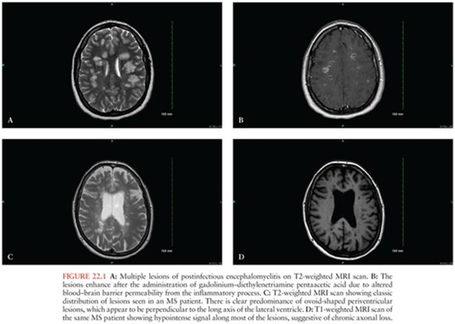
There is a long-standing controversy about whether a second episode of postinfectious encephalomyelitis should be called MS and treated accordingly. As a general rule, patients who develop clinical evidence of dissemination in space and time along with evidence of chronic demyelination in the CNS will likely develop MS. The classic scenario is the patient, typically a child, who is diagnosed with postinfectious encephalomyelitis following a viral infection, recovers, and then after some time develops recurrent symptoms with or without an antecedent viral infection. Two criteria are helpful in making the correct diagnosis: (a) The development of new symptoms representing distinct areas of demyelination not involved in the original episode favors the diagnosis of relapsing-remitting MS, and (b) the appearance of new lesions on neuroimaging supports the diagnosis of MS (45). It is worth remembering that brain lesions of MS patients usually increase in size and number during the course of the illness (41). In addition, chronic MS lesions appear as black holes on T1-weighted images, whereas black holes are not seen on T1-weighted images in patients with postinfectious encephalomyelitis. As patients recover from postinfectious encephalomyelitis, there is evidence of complete or partial resolution of lesions on neuroimaging. Oligoclonal bands should not persist in the cerebrospinal fluid (CSF) of patients with postinfectious encephalomyelitis, but will either persist or appear over time in the CSF of patients with MS (41).
Acute hemorrhagic leukoencephalitis is considered a hyperacute form of postinfectious encephalomyelitis and has been reported to occur in 2% of pediatric cases (46). On physical examination, there may be meningismus, obtundation, and lethargy, in addition to upper motor neuron signs, brainstem findings, transverse myelitis, and cranial neuropathies. In general, maximum deficits are reached in the first week from onset. Recovery usually starts to become clinically evident after the first week, with complete resolution of deficits and MRI lesions within 3 months (10). Although the prognosis is in general favorable, as high as 30% of patients require intensive care, with an estimated mortality rate of 20% (10). Despite the neuropathologic differences between postinfectious encephalomyelitis and acute hemorrhagic leukoencephalitis, it is possible that they represent a gradient of severity of the same pathologic process.
PATHOGENESIS
The pathology of postinfectious encephalomyelitis can be reproduced in the animal model of experimental allergic encephalomyelitis. This is a demyelinating disorder of the CNS induced in animals by immunization with myelin extracts, proteins, or peptides found in myelin. In an effort to reproduce in animal models the lesions described in postvaccinal encephalomyelitis, Rivers and colleagues (47) in 1933 injected homogenates of normal rabbit brains into monkeys. After 6 months, several monkeys developed a lymphocytic infiltration and demyelination of the CNS tissue (47).
Experimental allergic encephalomyelitis can be passively transferred to healthy animals by immune lymphocytes (48). These activated T cells assume a novel functional phenotype after transfer into a recipient animal that allows them to migrate to the CNS and pass through the blood–brain barrier. This migration briefly precedes the onset of clinical experimental allergic encephalomyelitis. There is a minimal interval of 3 days between the intravenous administration of pathogenic T cells and the onset of clinical experimental allergic encephalomyelitis (49).
T cells can potentially react with a wide variety of molecular structures, but normally they do not react against self-antigens. However, some encephalitogenic CD4 and CD8 T lymphocytes can be found in the blood, thymus, and secondary lymphoid tissues of apparently healthy individuals, but through the action of suppressive cytokines, they usually do not attack the CNS. T cells become pathogenic only if activated. One possible scenario to explain the development of postinfectious encephalomyelitis is that an infecting microbe expresses a peptide that is structurally similar to myelin basic protein (MBP). This epitope can trigger the activation of self-reactive T cells by a mechanism known as molecular mimicry. Once activated, these cells can multiply and mature into effector T cells, producing mediators and cytokines that can react to normal self-antigens. In addition, some T cells express more than one specific antigen receptor. One receptor type could be specific for the myelin antigen and the other for the microbial antigen. Exposure to the microbial antigen could activate the T cell, which by virtue of its myelin-specific alternative receptor could attack the CNS (49).
Another mechanism proposed to explain the pathogenesis of postinfectious encephalomyelitis is the activation of self-reactive immune cells by the release of cytokines by virus-mediated death of host cells. Penetration of these self-reactive immune cells into the brain or spinal cord leads to the characteristic pathology of postinfectious encephalomyelitis.
The role of circulating humoral factors in the pathogenesis of postinfectious encephalomyelitis is still unclear, but several lines of evidence suggest that antibody production by the host may aid in limiting or preventing the disease presumably by binding to MBP and inhibiting the access of autoreactive T lymphocytes. Prostaglandins of the E series secreted by blood monocytes and cerebral glial cells inhibit the immune response in experimental allergic encephalomyelitis by downregulation of monocytes and T cells and reduce the clinical and histologic abnormalities of experimental allergic encephalomyelitis in rats (12).
DIAGNOSIS
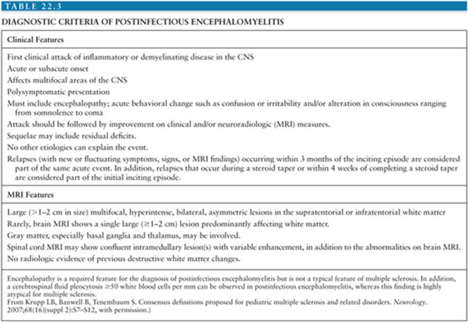
The diagnosis of postinfectious encephalomyelitis is based on clinical history, the findings on neurologic examination, neuroimaging abnormalities of demyelination, and CSF analysis. Several attempts have been made to establish a series of clinical features that will increase the likelihood of the diagnosis of postinfectious encephalomyelitis. The International Pediatric Multiple Sclerosis Study Group has developed a series of criteria for the diagnosis of postinfectious encephalomyelitis (Table 22.3). These criteria have been developed based on selected review of the literature and expert panel discussion. The specificity, sensitivity, and biologic validity to the diagnosis of postinfectious encephalomyelitis when using these guidelines have not been evaluated to date.
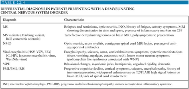
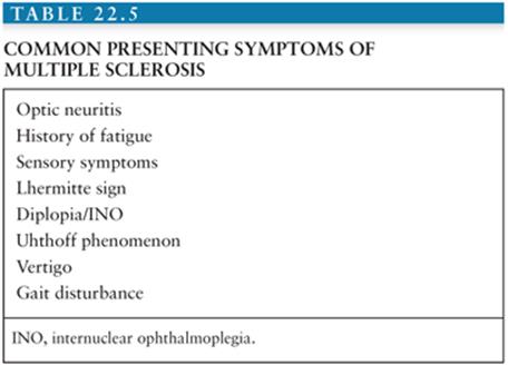
Due to the broad spectrum of differential diagnosis in patients presenting with a monophasic demyelinating CNS disorder (Table 22.4), the diagnosis of postinfectious encephalomyelitis usually requires a comprehensive, thorough investigation. The most common disorders in the differential diagnosis are MS (both pediatric as well as adult presentation), viral encephalitis, transverse myelitis, and neuromyelitis optica. Common symptoms of MS are listed in Table 22.5. Other disorders including CNS infections, neurometabolic CNS disorders (including mitochondrial disorders), neurosarcoidosis, systemic lupus erythematosus, primary CNS angiitis, NMO, anti-N-methyl-D-aspartate-receptor (anti-NMDA) encephalitis, chronic lymphocytic inflammation with pontine perivascular enhancement responsive to steroids (CLIPPERS), Bickerstaff encephalitis, and CNS malignancies should be considered.
Imaging
The typical computed tomography findings are areas of disseminated hypoattenuation in the subcortical white matter of the brain that enhance after the administration of contrast (35). They may or may not be surrounded by edema. MRI is a useful tool to aid in the diagnosis of postinfectious encephalomyelitis. In general, patients with postinfectious encephalomyelitis have a higher frequency of large confluent bilateral symmetrically oriented diffuse T2/fluid-attenuated inversion recovery (FLAIR) high signal lesions, ventral brainstem lesions, as well as the presence of monophasic pattern of enhancement (Fig. 22.1) (50). T2-weighted and FLAIR MRI scans demonstrate areas of increased signal in the subcortical white matter, brainstem, cerebellum, and periventricular white matter (43). These may have the appearance of large globular lesions (Fig. 22.1). Periventricular lesions are less frequently seen in postinfectious encephalomyelitis than MS, and if present, there is usually homogeneous contrast enhancement. The lesions are typically bilateral and asymmetric, vary in size and number, and could enhance in a nodular, spotty, ring, or heterogeneous pattern after the administration of intravenous gadolinium. There may be lesions in gray matter as well. Involvement of the deep gray matter may help distinguish postinfectious encephalomyelitis from MS (43). More advanced imaging techniques, such as magnetic resonance spectroscopy, have demonstrated elevation of lipids and reduction of the myoinositol:creatinine ratio during the acute phase, followed by reduction in lipids and increased myoinositol:creatinine ratios in the chronic setting (51). Fluorodeoxyglucose positron emission tomography scans usually show marked hypometabolism in the affected areas of the brain (45).
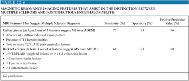
A recent review suggested that the use of Callen MS-ADEM criteria resulted in a high sensitivity (75%) and specificity (95%) for distinguishing MS at first attack from postinfectious encephalomyelitis (Table 22.6) (52). The authors also concluded that these MRI-based criteria were predictive of the diagnosis of MS versus postinfectious encephalomyelitis even in the absence of encephalopathy.
Laboratory Investigations
Given the possible similarities between postinfectious encephalomyelitis and MS, similar laboratory tests are performed as part of the workup of both conditions. MS is classically diagnosed by multiple lesions on MRI as well as the presence of oligoclonal IgG bands in the CSF (53). Unfortunately, in the majority of postinfectious encephalomyelitis cases, there are minor and nonspecific changes that occur that make diagnosis based on the standard of care laboratory testing difficult. However, there are some distinguishing features that do differentiate this disease from patients with MS and with healthy controls. These include elevated levels of total protein concentration (usually above 100 mg/dL) and increased cell count (usually more than 50 cells/mm3) (54,55). It has been shown that the presence of certain cytokines, such as tumor necrosis factor (TNF)-α, interleukin (IL)-2, IL-4, IL-5, IL-6, IL-8, IL-10, and interferon (IFN)-γ, are more common in postinfectious encephalomyelitis than MS (56,57). IL-17 has been reported to be increased in the CSF and blood of MS patients (58,59); however, this is not observed in patients with postinfectious encephalomyelitis. A study performed measuring 18 different chemokines in the blood and CSF of patients with suspected postinfectious encephalomyelitis, MS, or healthy controls revealed a distinct pattern of increased expression of specific chemokines in postinfectious encephalomyelitis patients not observed in patients with MS or controls. These changes were only observed in the CSF and not in the blood. Mean concentration of the chemokines (postinfectious encephalomyelitis, MS, healthy controls) CXCL7 (522 +/− 115 vs. 197 +/− 37 vs. 158 +/− 53), CCL1 (28.7 +/− 6.8, 18.5 +/− 3.4, 10.0 +/− 1.7), CCL22 (75.7 +/− 38.5, 9.4 +/− 1.8, 4.3 +/− 0.8), and CCL17 (18.6 +/− 3.4, 1.8 +/− 0.6, 7.2 +/− 0.6) were distinctly higher in the CSF from postinfectious encephalomyelitis patients. Based on these data, it may be possible to distinguish postinfectious encephalomyelitis patients from MS patients based on increased chemokine levels and lack of IL-17. However, more concrete studies will need to be performed to determine this. In order to facilitate the initial steps in the diagnosis of postinfectious encephalomyelitis, we propose a list of basic CSF studies that are useful in the differential diagnosis of postinfectious encephalomyelitis, MS, and viral encephalitis (Table 22.7).

PATHOLOGY
In rare circumstances, pathologic evaluation of involved tissue can assist in the diagnosis. Consistent with postinfectious encephalomyelitis clinical presentation, focal, perivenous, and subependymal changes dominate the pathologic pattern. Similar to MS, postinfectious encephalomyelitis lesions are mostly in the white matter at the cortical–subcortical junction but may also be seen in the cerebellum, spinal cord, and brainstem (43,60). Contrary to MS, early postinfectious encephalomyelitis involves infiltration of adaptive immune cells, mostly T lymphocytes, followed by innate monocytic cells. Inflammatory lesions in postinfectious encephalomyelitis spread radially outward from the vessels, whereas in MS, they border the plaques (43). Additionally, axons in areas of demyelination are relatively preserved in postinfectious encephalomyelitis, whereas in MS, there can be significant axonal loss. The major pathologic distinguishing factor is irregular borders observed in postinfectious encephalomyelitis lesions, whereas lesions from MS patients exhibit sharp distinct borders. Postinfectious encephalomyelitis is also distinguished from acute viral encephalitis by its pathology. Postinfectious encephalomyelitis is predominantly a disease of white matter, but gray matter may also be affected, particularly basal ganglia, thalami, and brainstem (42). Acute viral encephalitis is predominantly a disease of gray matter. The lesions characteristic of postinfectious encephalomyelitis are around small veins, in the cerebral white matter, brainstem, and spinal cord, and are composed of mononuclear cells and lymphocytes. Luxol fast blue stains that stain myelin reveal well-demarcated areas of loss of myelin. Staining for axons in the same areas that have loss of myelin reveals that the axon cylinders are relatively preserved. The neurons in the area show minor changes (4). The degree of the preservation of axon cylinders and the extent of the involvement of neurons determine prognosis.
DIFFERENTIAL DIAGNOSIS
The differential diagnosis is that of inflammatory, demyelinating diseases of the CNS. These include MS, viral encephalitis, transverse myelitis, and neuromyelitis optica (Devic syndrome) as the most common disorders. Table 22.7provides a list of CSF studies to determine the etiology. The leading disease in the differential diagnosis is MS. The first attack of MS can be difficult to differentiate from postinfectious encephalomyelitis. The specific neurologic symptoms are often very helpful in distinguishing between the two diseases, as is the history of the onset of symptoms within 2 to 31 days of a viral illness or vaccination. Although typical of postinfectious encephalomyelitis, a confusional state, headache, a decreased level of consciousness, and convulsions are also quite atypical of MS. MS is more likely than postinfectious encephalomyelitis to have a monosymptomatic presentation of optic neuritis, a brainstem syndrome, a cerebellar syndrome, or a transverse myelitis. The presence of bilateral optic neuritis is more suggestive of postinfectious encephalomyelitis than MS (41). Although both syndromes are primarily demyelinating diseases, the lesions of postinfectious encephalomyelitis are larger, more extensive, homogeneously enhance with contrast, and can involve gray matter, whereas the classic lesions of MS are ovoid-shaped, may or may not enhance, and usually involve the periventricular white matter, particularly the trigone and body of the lateral ventricle (41).
The Marburg variant of MS is a fulminant form of MS. Neuroimaging abnormalities may distinguish the Marburg variant from postinfectious encephalomyelitis, but more often it is the clinical course.
The viral encephalitides that may have a similar presentation and similar neuroimaging and CSF abnormalities to postinfectious encephalomyelitis include flavivirus infections (Japanese encephalitis virus, St. Louis encephalitis virus, and West Nile virus), herpesvirus infections (varicella-zoster virus [VZV], Epstein-Barr virus [EBV], herpes simplex virus [HSV], and other nonserotypable herpesviruses), progressive multifocal leukoencephalopathy due to JC virus, and HIV encephalitis. Japanese encephalitis virus classically infects the thalamus, brainstem, basal ganglia, substantia nigra, spinal cord, cerebral cortex, and cerebellum. The clinical presentation is characterized by fever, vomiting, convulsions, and coma. During the acute illness, patients may have movement disorders due to lesions in the basal ganglia and substantia nigra or flaccid paralysis due to lesions in the spinal cord (61,62). Hyperintense lesions on T2-weighted images in the substantia nigra and thalami can be seen in Japanese encephalitis (61,63). The diagnosis is made by demonstrating intrathecal production of specific antibodies.
West Nile virus encephalitis presents with fever, headache, and confusion and may have associated weakness in a “poliomyelitis-like syndrome.” The diagnosis is made by either the demonstration of West Nile virus nucleic acid in CSF, West Nile virus immunoglobulin M (IgM) antibody in CSF, or a fourfold increase in serum West Nile virus immunoglobulin G (IgG) antibodies between acute and convalescent sera.
VZV encephalitis presents with headache, malaise, and confusion days to weeks after the cutaneous eruption of zoster. The neuroimaging abnormalities of VZV encephalitis can be strikingly similar to those of postinfectious encephalomyelitis. VZV encephalitis may manifest as spherical subcortical white matter lesions with the typical appearance of demyelination (64).
In addition, there may be large and small ischemic and hemorrhagic infarctions of the cortical and subcortical gray and white matter. The diagnosis is made by demonstrating either VZV DNA in CSF, VZV IgM antibodies in CSF, or a positive CSF viral culture.
EBV may cause a meningoencephalitis during the course of mononucleosis. Following primary infection, the virus establishes latent infection in the CNS and can reactivate, causing encephalomyelitis. The clinical presentation includes fever, headache, focal neurologic deficits, an altered level of consciousness, and convulsions. This is a monophasic illness with neuroimaging evidence of lesions throughout the CNS. Diagnosis is made by demonstrating EBV DNA in CSF.
HSV-1 may reactivate from latent infection in the trigeminal ganglia and present as a brainstem encephalitis instead of causing the classic presentation of frontoorbital and temporal lobe dysfunction. There are also reports of HSV DNA in the CSF of patients with a clinical syndrome of encephalomyelitis and neuroimaging evidence of multiple hyperintense lesions in the thalamus, corpus striatum, pons, and deep white matter on T2-weighted MRI scans (65,66). The diagnosis of brainstem encephalitis or encephalomyelitis due to HSV-1 is made by the demonstration of HSV-1 DNA in CSF by polymerase chain reaction or by the demonstration of intrathecal production of HSV antibodies.
Progressive multifocal leukoencephalopathy is a demyelinating disease occurring in patients with severe cellular immunosuppression caused by a reactivation of latent JC virus likely acquired during childhood. This disease is progressive to death over the course of a few months. HIV patients and patients receiving immunosuppressive therapies are at higher risk of developing progressive multifocal leukoencephalopathy (PML). HIV encephalitis occurs in immunosuppressed individuals and presents with cognitive, motor, and behavioral abnormalities. T2-weighted MRI scans show areas of increased signal intensity in the subcortical white matter.
Subacute sclerosing panencephalitis (SSPE) is a degenerative disease of the brain due to measles virus, which presents after a latent period of several years or more from acute measles infection (67). This disease manifests with visual complaints, behavioral changes, and myoclonic jerks followed by hemiparesis, cogwheel rigidity, and dementia. There are elevated antibody titers against measles virus in CSF specimens and histopathologic evidence of extensive demyelination, glial proliferation, and neuronal and glial intranuclear inclusions (67,68). Although initially this disease may be a consideration in the differential diagnosis, SSPE is not a monophasic disorder, but a progressive illness.
Transverse myelitis is an inflammatory condition of the spinal cord due to a number of infectious and autoimmune etiologies. The thoracic cord is most commonly affected. There is a progressive weakness of lower extremities over the course of several hours to several days. Back pain may be present. Typically, there is a sensory level and bowel and bladder dysfunction. The maximum deficit is reached by definition within 4 weeks. Spinal MRI demonstrates swelling of the cord at the level of involvement. Devic syndrome refers to the combination of optic neuritis and myelitis and is now more commonly referred to as neuromyelitis optica.
Posttransplantation lymphoproliferative disease and leukoencephalopathies after chemotherapy and radiotherapy may have neuroimaging abnormalities that resemble those of postinfectious encephalomyelitis but are distinguished by the clinical setting in which they occur.
TREATMENT AND PREVENTION
Vaccination of infants against measles, mumps, and rubella has had a significant effect on decreasing the incidence of postinfectious encephalomyelitis caused by these viruses. Despite the inherent risk of acute neurologic complications following vaccination, the incidence of naturally occurring encephalomyelitis is still greater.
Currently, the only agent with proven efficacy in the prevention of postvaccinal encephalomyelitis is antivaccinia γ-globulin (AGG). The first clinical trial to prevent postvaccinal encephalitis was performed in 1956. At the time of primary smallpox vaccination, 53,630 Dutch military recruits were given an injection of 2 mL of 16% AGG, and 53,044 were given placebo. The donors of the AGG were healthy volunteers from the Royal Netherlands Army and the Royal Netherlands Air Force who had recently been vaccinated. Only 3 cases of postvaccinal encephalitis occurred in the treated group, compared with 13 cases in the control group (69). AGG is not effective in treating postvaccinal encephalitis once this complication has occurred.
Therapeutic recommendations for postinfectious encephalomyelitis are complicated by the lack of double-blind placebo-controlled clinical trials and the fact that postinfectious encephalomyelitis improves spontaneously. Several case series suggest that early high-dose corticosteroid therapy is beneficial (70,71). Early series used adrenocorticotropic hormone (ACTH) or dexamethasone (1 mg/kg per day), but recent series use intravenously administered methylprednisolone in a daily dose of 1000 mg per day for 3 to 5 days based on the experience with treating an acute exacerbation of MS. No firm guidelines exist on whether intravenously administered methylprednisolone therapy should be followed by an oral prednisone taper.
A randomized, sham-controlled clinical trial of plasma exchange in patients with either MS (12 patients) or other inflammatory demyelinating disease of the CNS (10 patients) was performed (72). The 10 patients with inflammatory demyelinating disease other than MS had transverse myelitis, ADEM, neuromyelitis optica, and focal cerebral demyelinating lesions. The patients were randomly assigned to receive either true or sham plasma exchange every other day for 2 weeks. All patients had a severe clinical deficit and had failed to improve over a period of 2 weeks from the initiation of high-dose intravenous corticosteroid therapy. Eight patients who were treated with true plasma exchange experienced moderate to marked improvement at the end of the 14 days. One patient who was treated with sham treatment had a moderate to marked improvement (72).
There are a number of case reports on the use of intravenous immune globulin (IVIG) therapy in ADEM (73–75). The mechanism of action of IVIG is not completely understood, but IVIG contains a wide spectrum of antibodies that have the potential to bind and neutralize pathogenic antibodies (73). These antibodies might bind to MBP and inhibit the access of autoreactive T lymphocytes.
In the initial days of the illness, postinfectious encephalomyelitis is often not distinguishable from acute viral encephalitis. A combination of intravenous acyclovir (10 mg/kg every 8 hours) and intravenous methylprednisolone (1000 mg per day) can be used until a definitive diagnosis is made. The addition of IVIG or plasma exchange may be beneficial either in the initial days of therapy or in those patients in whom a diagnosis of postinfectious encephalomyelitis is established, and the course is progressive despite intravenous methylprednisolone therapy.
PROGNOSIS
The mortality rate of postmeasles encephalomyelitis is 10% to 20%, and neurologic sequelae occur in 25% of survivors. The prognosis is related to age, with increasing mortality in those older than 16 years, and with the presence and duration of coma and convulsions (13). The mortality of postvaricella encephalomyelitis is approximately 10% (13). The mortality rate of postrubella encephalomyelitis was high, with 20% of children dying during the first week (13). Postinfectious encephalomyelitis in children that occurs as a complication of an upper respiratory tract infection or nonspecific febrile illness typically has a favorable prognosis. In adults, however, there are often neurologic sequelae. As described, prognosis is directly related to the degree of pathologic involvement of neurons and axon cylinders. To the extent that these are affected by the inflammatory process, there are varying degrees of cognitive deficits, movement disorders (dystonia), and spasticity. These deficits are often chronic.
References
1. Johnson RT. The pathogenesis of acute viral encephalitis and postinfectious encephalomyelitis. J Infect Dis. 1987;155(3):359–364.
2. Gurvich EB, Vilesova IS. Vaccinia virus in postvaccinal encephalitis. Acta Virol. 1983;27(2):154–159.
3. Lucas J. An account of uncommon symptoms succeeding the measles: with the additional remarks on the infection of measles and smallpox. London Med J. 1790;11:325–331.
4. Scott TF. Postinfectious and vaccinal encephalitis. Med Clin North Am. 1967;51(3):701–717.
5. Rivers TM. Viruses. JAMA. 1929;92:1147–1152.
6. Hurst EW. Acute hemorrhagic leukoencephalitis: a previously undefined entity. Med Aust. 1941;2:1–6.
7. Johnson RT, Griffin DE. Postinfectious encephalomyelitis. In: Kennedy PG, Johnson RT, eds. Infections of the Nervous System. London: Butterworth; 1987:209–226.
8. Leake JA, Albani S, Kao AS, et al. Acute disseminated encephalomyelitis in childhood: epidemiologic, clinical and laboratory features. Pediatr Infect Dis J. 2004;23(8):756–764.
9. Menge T, Hemmer B, Nessler S, et al. Acute disseminated encephalomyelitis: an update. Arch Neurol. 2005;62(11):1673–1680.
10. Pohl D, Tenembaum S. Treatment of acute disseminated encephalomyelitis. Curr Treat Options Neurol. 2012;14(3):264–275.
11. Johnson RT, Griffin DE, Hirsch RL, et al. Measles encephalomyelitis—clinical and immunologic studies. N Engl J Med. 1984;310(3):137–141.
12. Reder AT, Thapar M, Sapugay AM, et al. Prostaglandins and inhibitors of arachidonate metabolism suppress experimental allergic encephalomyelitis. J Neuroimmunol. 1994;54(1–2):117–127.
13. Gibbons JL, Miller HG, Stanton JB. Para-infectious encephalomyelitis and related syndromes: a critical review of the neurological complications of certain specific fevers. Q J Med. 1956;25(100):427–505.
14. Sacconi S, Salviati L, Merelli E. Acute disseminated encephalomyelitis associated with hepatitis C virus infection. Arch Neurol. 2001;58(10): 1679–1681.
15. Narciso P, Galgani S, Del Grosso B, et al. Acute disseminated encephalomyelitis as manifestation of primary HIV infection. Neurology. 2001;57(8): 1493–1496.
16. Allen SH, Malik O, Lipman MC, et al. Acute demyelinating encephalomyelitis (ADEM) in a patient with HIV infection. J Infect. 2002;45(1):62–64.
17. Johnson RT. Smallpox: the threat of bioterrorism and the risk of the vaccine. Neurology. 2003;60(8):1228–1229.
18. Fenichel GM. Neurological complications of immunization. Ann Neurol. 1982;12(2):119–128.
19. Ozawa H, Noma S, Yoshida Y, et al. Acute disseminated encephalomyelitis associated with poliomyelitis vaccine. Pediatr Neurol. 2000;23(2):177–179.
20. Ohtaki E, Matsuishi T, Hirano Y, et al. Acute disseminated encephalomyelitis after treatment with Japanese B encephalitis vaccine (Nakayama-Yoken and Beijing strains). J Neurol Neurosurg Psychiatry. 1995;59(3):316–317.
21. Boluksbasi O, Ozmenoglu M. Acute disseminated encephalomyelitis associated with tetanus vaccine. Eur Neurol. 1999;41:231–232.
22. Konstantinou D, Paschalis C, Maraziotis T, et al. Two episodes of leukoencephalitis associated with recombinant hepatitis B vaccination in a single patient. Clin Infect Dis. 2001;33(10):1772–1773.
23. Ito S, Nezu A, Matsumoto C, et al. Acute disseminated encephalomyelitis and poststreptococcal acute glomerulonephritis. Brain Dev. 2002;24(2): 88–90.
24. Sommer JB, Erbguth FJ, Neundorfer B. Acute disseminated encephalomyelitis following Legionella pneumophila infection. Eur Neurol. 2000;44(3): 182–184.
25. Alonso-Valle H, Muñoz R, Hernandez JL, et al. Acute disseminated encephalomyelitis following Leptospira infection. Eur Neurol. 2001;46: 104–105.
26. Krishna KK, Mitra DK, Diwan AG, et al. Acute disseminated encephalomyelitis with typhoid fever. J Assoc Physicians India. 1999;47(10):1017–1019.
27. Spieker S, Petersen D, Rolfs A, et al. Acute disseminated encephalomyelitis following Pontiac fever. Eur Neurol. 1998;40(3):169–172.
28. Wei TY, Baumann RJ. Acute disseminated encephalomyelitis after Rocky Mountain spotted fever. Pediatr Neurol. 1999;21(1):503–505.
29. Mohsen AH, McKendrick MW, Schmid ML, et al. Postmalaria neurological syndrome: a case of acute disseminated encephalomyelitis? J Neurol Neurosurg Psychiatry. 2000;68(3):388–389.
30. Riedel K, Kempf VA, Bechtold A, et al. Acute disseminated encephalomyelitis (ADEM) due to Mycoplasma pneumoniae infection in an adolescent. Infection. 2001;29(4):240–242.
31. Re A, Giachetti R. Acute disseminated encephalomyelitis (ADEM) after autologous peripheral blood stem cell transplant for non-Hodgkin’s lymphoma. Bone Marrow Transplant. 1999;24(12):1351–1354.
32. Dubreuil F, Cabre P, Smadja D, et al. Acute disseminated encephalomyelitis preceding cutaneous lupus [in French]. Rev Med Interne. 1998;19(2): 128–130.
33. Jaing TH, Lin KL, Chiu CH, et al. Acute disseminated encephalomyelitis in autoimmune hemolytic anemia. Pediatr Neurol. 2001;24(4):303–305.
34. Stonehouse M, Gupte G, Wassmer E, et al. Acute disseminated encephalomyelitis: recognition in the hands of general paediatricians. Arch Dis Child. 2003;88(2):122–124.
35. Lane JM, Millar JD, Neff JM. Smallpox and smallpox vaccination policy. Annu Rev Med. 1971;22:251–272.
36. Feery BJ. Adverse reactions after smallpox vaccination. Med J Aust. 1977;2(6):180–183.
37. Katz SL. The case for continuing “routine” childhood smallpox vaccination in the United States. Am J Epidemiol. 1971;93(4):241–244.
38. Lane JM, Ruben FL, Abrutyn E, et al. Deaths attributable to smallpox vaccination, 1959 to 1966, and 1968. JAMA. 1970;212(3):441–444.
39. Greenberg M. Complications of vaccination against smallpox. Am J Dis Child. 1948;76(5):492–502.
40. Tenembaum S, Chamoles N, Fejerman N. Acute disseminated encephalomyelitis: a long-term follow-up study of 84 pediatric patients. Neurology. 2002;59(8):1224–1231.
41. Dale RC, de Sousa C, Chong WK, et al. Acute disseminated encephalomyelitis, multiphasic disseminated encephalomyelitis and multiple sclerosis in children. Brain. 2000;123(pt 2):2407–2422.
42. Hartung HP, Grossman RI. ADEM: distinct disease or part of the MS spectrum? Neurology. 2001;56(10):1257–1260.
43. Hynson JL, Kornberg AJ, Coleman LT, et al. Clinical and neuroradiologic features of acute disseminated encephalomyelitis in children. Neurology. 2001;56(10):1308–1312.
44. Krupp LB, Banwell B, Tenembaum S. Consensus definitions proposed for pediatric multiple sclerosis and related disorders. Neurology. 2007;68(16)(suppl 2):S7–S12.
45. Stüve O, Zamvil SS. Pathogenesis, diagnosis, and treatment of acute disseminated encephalomyelitis. Curr Opin Neurol. 1999;12(4):395–401.
46. Barontini F, Di Lollo S, Toscani L. Acute hemorrhagic leukoencephalopathy: clinical and CT diagnosis and histopathological confirmation in an elderly patient. Ital J Neurol Sci. 1984;5(2):215–218.
47. Rivers TM, Sprunt DH, Berry GP. Observations on Attempts to produce acute disseminated encephalomyelitis in monkeys. J Exp Med. 1933;58(1):39–53.
48. Orgad S, Cohen IR. Autoimmune encephalomyelitis: activation of thymus lymphocytes against syngeneic brain antigens in vitro. Science. 1974;183(4129):1083–1085.
49. Wekerle H. Immune protection of the brain—efficient and delicate. J Infect Dis. 2002;186(suppl 2):S140–S144.
50. Lu Z, Zhang B, Qiu W, et al. Comparative brain stem lesions on MRI of acute disseminated encephalomyelitis, neuromyelitis optica, and multiple sclerosis. PLoS One. 2011;6(8):e22766.
51. Ben Sira L, Miller E, Artzi M, et al. 1H-MRS for the diagnosis of acute disseminated encephalomyelitis: insight into the acute-disease stage. Pediatr Radiol. 2010;40(1):106–113.
52. Ketelslegers IA, Neuteboom RF, Boon M, et al. A comparison of MRI criteria for diagnosing pediatric ADEM and MS. Neurology. 2010;74(18): 1412–1415.
53. Polman CH, Reingold SC, Banwell B, et al. Diagnostic criteria for multiple sclerosis: 2010 revisions to the McDonald criteria. Ann Neurol. 2011;69(2):292–302.
54. Stuve O, Nessler S, Hartung HP, et al. Acute disseminated encephalomyelitis. Pathogenesis, diagnosis, treatment, and prognosis [in German]. Der Nervenarzt. 2005;76(6):701–707.
55. Franciotta D, Salvetti M, Lolli F, et al. B cells and multiple sclerosis. Lancet Neurol. 2008;7(9):852–858.
56. Ishizu T, Minohara M, Ichiyama T, et al. CSF cytokine and chemokine profiles in acute disseminated encephalomyelitis. J Neuroimmunol. 2006; 175(1–2):52–58.
57. Ichiyama T, Shoji H, Kato M, et al. Cerebrospinal fluid levels of cytokines and soluble tumour necrosis factor receptor in acute disseminated encephalomyelitis. Eur J Pediatr. 2002;161(3):133–137.
58. Graber JJ, Allie SR, Mullen KM, et al. Interleukin-17 in transverse myelitis and multiple sclerosis. J Neuroimmunol. 2008;196(1–2):124–132.
59. Matusevicius D, Kivisakk P, He B, et al. Interleukin-17 mRNA expression in blood and CSF mononuclear cells is augmented in multiple sclerosis. Mult Scler. 1999;5(2):101–104.
60. Mikaeloff Y, Suissa S, Vallee L, et al. First episode of acute CNS inflammatory demyelination in childhood: prognostic factors for multiple sclerosis and disability. J Pediatr. 2004;144(2):246–252.
61. Pradhan S, Pandey N, Shashank S, et al. Parkinsonism due to predominant involvement of substantia nigra in Japanese encephalitis. Neurology. 1999;53(8):1781–1786.
62. Johnson RT, Burke DS, Elwell M, et al. Japanese encephalitis: immunocytochemical studies of viral antigen and inflammatory cells in fatal cases. Ann Neurol. 1985;18(5):567–573.
63. Misra UK, Kalita J. A comparative study of Japanese and herpes simplex encephalitides. Electromyogr Clin Neurophysiol. 1998;38(1):41–46.
64. Weaver S, Rosenblum MK, DeAngelis LM. Herpes varicella zoster encephalitis in immunocompromised patients. Neurology. 1999;52(1):193–195.
65. Kusuhara T, Nakajima M, Inoue H, et al. Parainfectious encephalomyeloradiculitis associated with herpes simplex virus 1 DNA in cerebrospinal fluid. Clin Infect Dis. 2002;34(9):1199–1205.
66. Nakamura K, Terasaki T, Tsuchiya T, et al. Herpes simplex encephalitis followed by myelopathy [in Japanese]. No To Shinkei. 1993;45:553–558.
67. Singer C, Lang AE, Suchowersky O. Adult-onset subacute sclerosing panencephalitis: case reports and review of the literature. Mov Disord. 1997;12(3):342–353.
68. Anlar B, Yalaz K, Oktem F, et al. Long-term follow-up of patients with subacute sclerosing panencephalitis treated with intraventricular alpha-interferon. Neurology. 1997;48(2):526–528.
69. Nanning W. Prophylactic effect of antivaccinia gamma-globulin against post-vaccinal encephalitis. Bull World Health Organ. 1962;27:317–324.
70. Pasternak JF, De Vivo DC, Prensky AL. Steroid-responsive encephalomyelitis in childhood. Neurology. 1980;30(5):481–486.
71. Straub J, Chofflon M, Delavelle J. Early high-dose intravenous methylprednisolone in acute disseminated encephalomyelitis: a successful recovery. Neurology. 1997;49(4):1145–1147.
72. Weinshenker BG. Plasma exchange for severe attacks of inflammatory demyelinating diseases of the central nervous system. J Clin Apher. 2001;16(1):39–42.
73. Marchioni E, Marinou-Aktipi K, Uggetti C, et al. Effectiveness of intravenous immunoglobulin treatment in adult patients with steroid-resistant monophasic or recurrent acute disseminated encephalomyelitis. J Neurol. 2002;249(1):100–104.
74. Nishikawa M, Ichiyama T, Hayashi T, et al. Intravenous immunoglobulin therapy in acute disseminated encephalomyelitis. Pediatr Neurol. 1999; 21(2):583–586.
75. Keliman M BP. Acute disseminated encephalomyelitis: response to intravenous immunoglobulin. J Child Neurol. 1995;10:481–483.