
BARRY J. HARTMAN AND DAVID C. HELFGOTT
CRANIAL SUBDURAL EMPYEMA
Empyema is an important form of intracranial suppuration, accounting for 15% to 25% of pyogenic intracranial infections (1–3,97). It represents an infectious process that occupies the space between the dura mater and arachnoid surrounding the brain. Left undiagnosed and untreated, subdural empyema is rapidly fatal, so early recognition is critical. In older children and adults, subdural empyema is most often a complication of otorhinologic infection (1,4–19,98). Subdural empyema may also occur as a result of head trauma or surgery, osteomyelitis of the skull, or bacteremic spread from a distant focus of infection. In infants, leptomeningitis is the most common predisposing cause of subdural empyema (11,20,21,97). Males predominate over females, and about 70% of patients are in their second or third decade of life (3–5,7–10,12,13,15,22–26). Patients will present with fever and headache in more than 90% of cases and many will have associated neurologic abnormalities (1,3–9,12,13, 15–19,21,23,24,26–30,99,100,101). The diagnosis of subdural empyema is confirmed using computed tomography (CT) or magnetic resonance imaging(MRI) scanning techniques. However, a strong clinical suspicion despite the lack of evidence for subdural empyema on scans warrants more invasive investigation. Definitive therapy consists of surgical drainage and systemic antibiotics, yet mortality remains as high as 40% in some series (14,21). Spinal subdural empyema also has been described; however, it is quite rare, with fewer than 150 cases reported in the literature (31,102). This condition is addressed briefly later in the chapter. Suppurative intracranial phlebitis is a serious complication of cranial and facial infections and results in thrombosis of the major dural venous sinuses. This is discussed at the end of this chapter.
Historical Perspective
The first comprehensive clinicopathologic descriptions of subdural empyema as a distinct entity were published in the 1940s, although the first definitive report of subdural empyema dates back to 1861 (32,33). This initial report was followed by several more around the turn of the century, with a compilation of 44 cases by Blegvad (32) in 1910. Early names for this disease included “pachymeningitis interna,” “purulent pachymeningitis,” “pia-arachnoid abscess,” “phlegmonous meningitis,” and “subdural abscess,” but these were rejected by Kubik and Adams in favor of “subdural empyema” (8,32). Interestingly, early publications reported a preponderance of subdural empyema secondary to otogenic infections (32). However, because of the compilation of 42 confirmed cases resulting from frontal sinusitis by Courville (33), it has become clear that paranasal sinusitis is the most important causative factor in the development of subdural empyema in older children and adults. During the past 50 years, much has changed in the areas of therapy and diagnosis of subdural empyema. Before the development of antibiotics, subdural empyema was almost always fatal (32,33). Antibiotics, improved diagnosis, and newer surgical techniques have combined to lower the mortality rate to 10% to 40% (1,4,5,7–15,21,22,24,28,29). Physicians depended on their clinical skills and plain roentgenograms of the sinuses and skull to direct their attention to the possibility of intracranial suppuration, until the development of cerebral angiography in the early 1960s, which proved an extremely sensitive method of detecting a subdural collection (13,34). The emergence of CT in the 1970s provided a noninvasive rapid means of visualizing the cranial contents; its reliability, safety, and ease of operation made it the first choice for diagnosis of suspected subdural empyema (2,10,26,35). Recently, MRI has proven even more sensitive than CT (36).
Pathogenesis and Anatomic Considerations
The clinical features of subdural empyema are easily understood if one considers the anatomy of the subdural space with respect to its surrounding structures (Fig. 33.1). The subdural space is normally a potential space rather than an actual space because the dura mater follows the contours of the skull and lies adjacent to the arachnoid and pia mater (37). The ability to form an actual space with fluid collection is greater around the convexities of the cerebral hemispheres where the brain does not approximate the skull as closely as it does around the basal areas (15,22,33).

Posteriorly, the tentorium cerebelli is a reflection of dura separating the cerebellum from the cerebral cortex (38). It is an effective barrier to the passage of subdural collection infratentorially, except at its free anterior margin where fluid may seep into the subdural space of the posterior fossa. Only about 10% of subdural empyemas are infratentorial (15,39,40). Medially, the falx cerebri is a reflection of dura extending the length of the cerebrum that separates the cerebral hemispheres (38). Subdural fluid that accumulates between the falx and the arachnoid are known as parasagittal, interhemispheric, or parafalcine subdural empyemas and are usually secondary to surface subdural collections but rarely may be primary (41–43). A subdural empyema may communicate with the contralateral side via the inferior free margin of the falx.
It therefore follows that infections or trauma of the head are the usual causes of subdural empyema. Table 33.1 reviews the conditions predisposing to subdural empyema in series reported during the past two decades (excluding series of only infants). Paranasal sinusitis overwhelmingly predominates as the precipitating factor for the development of subdural empyema (101). The sinusitis almost always involves a frontal sinus, often with other sinuses affected as well. The incidence of subdural empyema following frontal sinusitis is 1% to 2% (45). The frontal and sphenoid sinuses are intimately associated with the dura mater, separated from the dura only by a thin plate of bone.

These sinuses communicate with the maxillary and ethmoid sinuses, which are more anteriorly placed. Because of its position, the frontal sinus is almost always involved in paranasal sinus infection that spreads to the subdural space. In addition, the growing posterior wall of the frontal sinus during puberty has been offered as a possible explanation for the striking age susceptibility for the development of subdural empyema (7,26,46).
Two modes of extension have been proposed for the spread of infection from a frontal sinus to the subdural space: direct and indirect (33). Direct extension involves erosion of the posterior bony wall of the frontal sinus by infection, with further erosion of the underlying dura mater (33). In teenagers primarily, frontal bone osteomyelitis with subperiosteal abscesses (a Pott puffy tumor) can extend directly into the subdural space (47,103). The more likely route is indirect, with extension of infection and associated thrombophlebitis through the mucosal veins of the sinus to the emissary veins that link the facial and dural venous systems (15,33,48). From the dural sinuses, the infection establishes itself in the subdural space at the frontal pole and may spread posteriorly over the convexity, medially into the interhemispheric region, and contralaterally. This extension may create significant pressure on a large area of underlying brain tissue (15,22,49). As stated previously, it is unusual but possible for the empyema to spread infratentorially (15). Further retrograde thrombophlebitis often occurs, involving the valveless, deeper veins of the cerebrum, which in turn may lead to necrosis and infection of brain tissue (10,13,32,33). Subdural empyema secondary to an otogenic source of infection differs only in the site at which pus enters the subdural space. The tympanic cavity is bounded superiorly by the tegmen tympani, a thin plate of bone forming part of the temporal bone of the skull separating the tympanic cavity from the brain (37). Perforating veins pass through this plate of bone to communicate with the superior petrosal venous sinus of the dura (37). In addition, mastoid air cells within the temporal bone surrounding the middle ear communicate with the tympanic cavity and may lie very close to the posterior cranial fossa separated from the dura by slivers of bone (37). Otitis or mastoiditis may, therefore, extend directly into the subdural space via erosion of the tegmen tympani or bone adjacent to the air cells and dura mater or spread infection indirectly by way of a progressive thrombophlebitis of the perforating veins (27,32). Because the venous sinuses into which the veins from the middle ear and mastoid bone drain are within or beneath the tentorium (38), otogenic infection may result in posterior fossa subdural empyema (27). As opposed to subdural empyema secondary to frontal sinusitis, otitis-induced subdural empyema is initially localized posteriorly or on the tentorium (50). Hematogenous spread of bacteria to the subdural space from a distant site of infection is an uncommon cause of subdural empyema, accounting for fewer than 5% of all cases in most series (Table 33.1). Several reports describe the development of subdural empyema via hematogenous spread to a preexisting subdural hematoma (51,52). Subdural empyema is a rare complication following cranial surgery; in one series, subdural empyema occurred in slightly more than 1 in 1,000 craniotomies (53). In a large series of 16,540 craniotomy procedures from 1997 to 2007, only 0.5% had intracranial infection (only 7 cases were pure subdural infections) (104).
In adults, the extension of a subdural empyema from acute purulent meningitis is very unusual. Although there is a subarachnoid inflammatory exudate, the arachnoid is fairly impermeable to the bacterial process occurring adjacent to it (22). Bacterial meningitis in adults is a very unusual cause of subdural empyema, occurring in less than 1% of cases (21,105). However, in infants, meningitis is an important predisposing condition for the development of a subdural empyema (11,20,21,97,100). Subdural empyema occurs in about 2% of infants with bacterial meningitis (54). The pathogenesis is presumably infection of an initially sterile subdural effusion (13,20,21). Such sterile effusions are variably reported as occurring in up to 60% of infants with meningitis (13,20,97).
Clinical Features
A high clinical suspicion and rapid diagnosis of subdural empyema are critical for a successful outcome. Certainly, an adult with a recent history of sinusitis and a new presentation suggestive of central nervous system (CNS) infection warrants an investigation to exclude subdural empyema. However, in some cases, the antecedent infection is subtle enough to be unrecognized. In others, the concurrent complication of sinusitis with a subdural empyema delays the diagnosis of the latter because symptoms are attributed to the sinusitis. In other cases, the subdural empyema is not suspected because the precipitating cause for the subdural empyema is unknown or arises from a distant focus of infection. Although the clinical presentation may vary, there are key clinical features of subdural empyema that if present should result in its inclusion in one’s initial differential diagnosis. The sex and age distributions of patients with subdural empyema are striking. There is an overrepresentation of men reported in series of patients (mostly adults) with subdural empyema published during the last two decades. In those that report only children, males also predominate. However, in infants, this sex discrepancy may not be so marked (3–5,7–10,12,13,15,16,18,19,22–26,54,99,100,106). Figure 33.2 displays the age distribution of patients reported in series of consecutive patients with subdural empyema. It is clear that most cases occur during the second and third decades of life. As stated earlier, the significant growth of the frontal sinus during puberty has been proposed as an explanation for the uneven age distribution (7,26,46). However, confirmatory analyses comparing patients’ sex, age, and source of infection have not been reported.

The clinical features of adults with subdural empyema are presented in Table 33.2. Generally, patients have a nonspecific illness for a few days to a few weeks before presentation to the hospital acutely ill (1,5,12,13). However, if the infection is a result of head trauma or surgery, the symptoms may be milder and present more subacutely (10,23,55,56,104). The most common symptoms and signs are headache, fever, neurologic deficit, and stiff neck. Vomiting and malaise are often reported as well (8,15). Seizures, papilledema, and altered level of consciousness ranging from drowsiness and disorientation to coma also occur frequently. These neurologic changes may be presenting signs or, as is often the case, may develop during the course of the illness (6,7,15,28).

Diffuse neurologic signs such as altered level of consciousness, papilledema, and generalized seizures are a result of increased intracranial pressure (ICP) (33,57). Focal neurologic abnormalities such as hemiparesis, jacksonian seizures, dysphasia, and cranial neuropathies may be secondary to local pressure on the underlying cortex by the subdural process (21,27,32,33,41,49,58) and may be precipitated by cortical venous thrombosis with accompanying brain inflammation and infarction (21,22,57). Such focal neurologic signs may help to localize the empyema. This is particularly true in cases of infratentorial subdural empyema that occurs infrequently, but that is easily suspected if cerebellar signs such as ataxia and nystagmus are present (27). Interhemispheric (parasagittal, parafalcine) subdural empyema, usually associated with disease over the convexities but uncommonly occurring alone (43), characteristically produces contralateral leg symptoms, including weakness and focal seizures (41,43,49,58). As the interhemispheric suppuration extends backward over the tentorium and below the occipital lobes, homonymous hemianopia may result (49,58). Subdural empyema overlying one or both convexities yields the most nonspecific neurologic signs. Clues to the involved areas can be (a) contralateral paresis or seizures, (b) aphasia or dysphasia associated with left-sided infection, or (c) cranial neuropathies (32,33,49). The clinical signs of subdural empyema in infants are similar to those in adults. In addition, a bulging anterior fontanelle is a common finding in infants (11,20,54).
Differential Diagnosis
The cardinal features of headache, fever, stiff neck, and neurologic signs are not specific for subdural empyema. The differential diagnosis also includes brain abscess, epidural abscess, meningitis, meningoencephalitis, subdural hematoma, and intracerebral thrombophlebitis (21,48). Of these, the presence of focal neurologic signs makes meningitis much less likely. The presence of nuchal rigidity is unusual in brain abscess and subdural hematoma. Unfortunately, clinical grounds alone do not allow the exclusion of most of these possibilities. Therefore, more specific testing should be undertaken as soon as the diagnosis of subdural empyema is suspected.
Diagnostic Studies
Routine studies such as blood tests and plain roentgenograms are of very little value in patients with suspected subdural empyema. Most patients are found to have a peripheral blood leukocytosis (3,11,13,27,30,33). Plain films of the skull are not useful except to demonstrate a sinusitis or mastoiditis or to show widened sutures in infants (21). In infants, cranial ultrasonography can detect a subdural collection and may differentiate a reactive effusion from a subdural empyema (107). Before the development of CT, cerebral arteriography, with a diagnostic accuracy of 80% to 90%, was the procedure of choice to diagnose subdural empyema (13,15,21,27). Although nearly perfect for the detection of hemispheric and parafalcine subdural collections, the sensitivity of carotid angiograms for posterior fossa subdural empyema was not as great (27). Presently, the safety, ease of application, and reliability of CT and MRI make them the modalities of choice to diagnose subdural empyema.
Computed Tomography and Magnetic Resonance Imaging
The radiologic evaluation of patients with subdural and epidural empyemas has been revolutionized by the advent of CT in 1972 and MRI in 1984. The introduction of CT has had a major impact on the management and prognosis of subdural and epidural empyemas because CT allows (in a noninvasive manner) earlier and more accurate detection, delineation, and characterization of these extraaxial (extraparenchymal) inflammatory lesions and their associated intraaxial (parenchymal) sequelae when compared to carotid arteriography (10). In addition, CT provides an important adjunct to standard clinical parameters in the assessment of the adequacy of patient response to therapy. The CT, findings during the early stages of development of a subdural empyema may be subtle and easily overlooked (10). Noncontrast CT scans typically demonstrate a crescentic hypodense collection over one or both cerebral convexities and/or around the interhemispheric tissue (Fig. 33.3A). Contrast-enhanced CT increases the conspicuity of the collections that represent active inflammatory disease either in the leptomeninges or in the subjacent cerebral cortex (Fig. 33.3B).

Thick, irregular enhancement of the falx in association with a spindle-shaped collection is seen in interhemispheric subdural empyemas (43) (Fig. 33.4). Parenchymal changes at this early stage include thickening and hyperdensity of the underlying cortical gray matter and hypodensity of the white matter on noncontrast CT images; these changes indicate the presence of edema, hyperemia, and ischemia (10). Additionally, gyral enhancement subjacent to an extraaxial empyema on contrast CT scans is a common finding, indicative of meningitis, cerebritis, and/or venous thrombosis (Fig. 33.3B). Extensive mass effect on the ipsilateral cerebral hemisphere that is out of proportion to the small size of the extraaxial collection is invariably present, and it is manifested as ventricular compression, sulcal effacement, and midline shift. It is important to examine the paranasal sinuses, middle ear cavity, and orbits for the presence of inflammation, which may reflect the origin and extent of the intracranial abnormalities (26,29) (Figs. 33.5 and 33.6A).
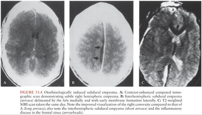

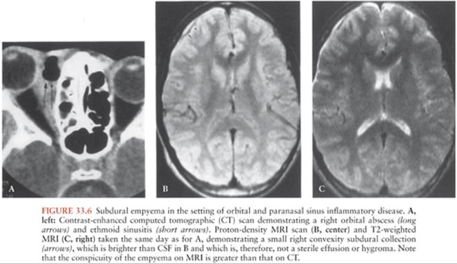
Unrecognized and untreated, the subdural empyema rapidly grows and develops loculations and the parenchymal abnormalities progress to cortical infarction and abscess formation. MRI is proving to have a greater sensitivity and specificity in the workup of patients with an extraaxial empyema; this is attributed to several inherent advantages of MRI over CT (36,59,60). MRI uses several standard pulse sequences referred to as “T1 weighted,” “proton-density weighted,” and “T2 weighted” (61). T1-weighted images emphasize contrast between the brain and cerebrospinal fluid (CSF), and proton-density–weighted and T2-weighted images emphasize contrast between brain and pathologic processes (61) (Fig. 33.6B and C).
MRI has been found to have six inherent advantages over CT. First, MRI permits excellent visualization of superficial brain anatomy, precise localization of extraaxial empyemas (Fig. 33.4C), and more definitive separation of extraaxial collections from their associated intraaxial complications such as edema, cerebritis, and venous thrombosis, which are more readily visualized on MRI than on CT (36,62). Second, streak artifacts from the bony calvaria, which are particularly problematic on CT, are not limitations on MRI. Third, MRI is superior to CT in differentiating noninfected subdural effusions and hygromas from infected empyemas. As with other proteinaceous fluids, the T1- and T2-weighted values of purulent collections are smaller than those of CSF. These collections are, therefore, mildly hyperintense to CSF on T1-weighted images and markedly hyperintense to CSF on T2-weighted images (36). Fourth, the unprecedented sensitivity of MRI to subtle changes in tissue water content is uniquely suited to the goal of early detection of the parenchymal abnormalities that can occur secondary to a subdural empyema. Fifth, MRI is more specific than CT in differentiating a subdural from an epidural empyema (36,63). A hypodense medial rim, representing inflamed displaced dura, is seen on MRI of an epidural empyema but not on that of a subdural empyema. Finally, the delineation of extraaxial inflammatory disease, leptomeningeal disease, and parenchymal abnormalities is improved with the use of the MRI contrast agent, gadolinium diethylenetriamine pentaacetic acid (64), and diffusion-weighted imaging (108,109).
CT and MRI play a role in the follow-up of patients with extraaxial empyemas (10,36,59). Residual or recurrent collections that may necessitate reexploration are particularly prone to occur in the parafalcine or subtemporal regions. These locations are well imaged on MRI because of its ability to obtain direct coronal sections of the brain. Long-term follow-up CT or MRI examinations frequently demonstrate cortical atrophy adjacent to a previous extraaxial empyema.
Lumbar Puncture
Lumbar puncture is often performed in patients who are subsequently diagnosed with subdural empyema, but it is neither sensitive nor specific for this disease. Recovery of a causative organism in the CSF is rare, except in infants in whom meningitis preceded the development of the subdural empyema (11,20,21,54). The CSF formula in children and adults with subdural empyema is unpredictable, as shown in Table 33.3. Typically, the white blood cell (WBC) count is elevated; however, many series report patients with zero to five CSF leukocytes per cubic milliliter (13,25,26,29,30). The differential cell count on the CSF is highly variable: although a polymorphonuclear pleocytosis is more common, the mononuclear cell predominates in close to 40% of patients. A normal protein concentration suggests the absence of a subdural empyema because there is an inflammatory response by the arachnoid to the overlying subdural process. However, because the arachnoid is generally impermeable to the infectious agent, CSF Gram stain and culture almost never demonstrate the bacterial cause of the subdural empyema and are, therefore, not helpful in choosing antibiotic therapy.

In addition to providing no valuable diagnostic information, lumbar puncture is a potentially dangerous procedure in patients with signs of increased ICP (66). Several deaths from cerebral herniation have been reported in patients with subdural empyema shortly after undergoing lumbar puncture (13,25,26,40,44). Certainly, patients with papilledema or a focal neurologic abnormality or patients with suspected increased ICP should not undergo a lumbar puncture.
Bacteriology
The microbiologic etiology of subdural empyema is established by Gram stain and culture of evacuated pus from the subdural space. Unfortunately, cultures of subdural pus are sterile in about one third of patients (25) because patients are almost always receiving antibiotics preoperatively. It has been suggested that the high number of negative cultures is also related to the lack of proper handling and culture for anaerobes (25). In one study in which paranasal sinus cultures and subdural cultures were compared, three of the four sinus isolates did not correlate with the subdural isolates (26). Blood cultures may provide additional diagnostic information in about 10% of cases in which the subdural fluid is sterile (11,21,26). The organisms cultured most often from subdural infections are aerobic and anaerobic streptococci. Staphylococci are cultured less often, followed by aerobic gram-negative bacilli and nonstreptococcal anaerobes (Table 33.4). In most patients, a single organism is responsible for subdural empyema. However, several series have included cases in which multiple organisms have been cultured (3,5,7,16,17,19,21,22,25,27).

Generally, the causative organism is predictable based on the anatomic focus from which the infection originated (3,5,8,13,45,55,56,100). Otorhinogenic subdural empyemas are most often due to aerobic and anaerobic streptococci and are less often due to coagulase-positive staphylococci and other anaerobes. Infections secondary to head trauma, surgery, or an indwelling foreign device are most often caused by coagulase-positive and coagulase-negative staphylococci and gram-negative bacilli. Four cases of postneurosurgical subdural empyema caused by Propionibacterium acnes, a gram-positive anaerobic bacillus, were reported often occurring several weeks after the surgical procedure (67,68).
Subdural empyemas originating from distant foci of infection are caused by a variety of organisms. In infants with leptomeningitis, subdural empyema is caused by the same organism responsible for the meningitis, usually Streptococcus pneumoniae or Haemophilus influenzae (21,54). Many organisms other than those mentioned have been reported to cause subdural empyema. These include Salmonellaspecies (69,70), Campylobacter fetus (52), Serratia marcescens (71), Neisseria meningitidis (72,73), Pasteurella multocida (52,74), Actinomyces israelii, and Actinobacillus actinomycetemcomitans (new genus name—Aggregatibacter) (75). In the Far East, a greater percentage of bacterial pathogens are gram-negative aerobes (Enterobacteriaceae), particularly Klebsiella pneumoniae (110).
Treatment and Outcome
The clinical suspicion of subdural empyema requires the immediate institution of parenteral antibiotic therapy. Antibiotics should be chosen based on the suspected source of the infection and on the organisms known to commonly cause subdural empyema. Although no prospective comparisons of antibiotic regimens for subdural empyema have been conducted, an acceptable empirical therapy includes a β-lactamase–stable penicillin, a third-generation cephalosporin, and metronidazole. Depending on the prevalence of methicillin-resistant Staphylococcus aureus or the likelihood of coagulase-negative staphylococci, vancomycin may be used in place of the β-lactamase–stable penicillin. Although there is no consensus, some advocate irrigation of the subdural space with antibiotics (2,8,9,12,14,49). No current data support a specific duration of antibiotic therapy; however, most patients are treated for 3 to 4 weeks after drainage (21). Empirical therapy for seizure prophylaxis has been advocated (8,13,28,45), and steroids and mannitol have been used successfully to decrease ICP in individual cases (5,13,24).
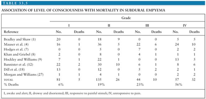
Although anecdotal cases have been successfully treated with antibiotics alone (76,77), surgical drainage of a subdural empyema is imperative. Disagreement exists, however, over the optimal mode of surgery. The comparative efficacy of multiple burr holes versus craniotomy is complicated by clinical factors that may contribute to outcome. Several parameters have been suggested to be important in predicting patient mortality, including age of patient (12), source of infection (12), microbiology (12), time from presentation to surgery (13), management of the primary source of infection (1), extent of spread of empyema (4), level of consciousness at presentation (1,4,7–9,12,27), and surgical technique (4,5,7–9,12,14,22,24,27). Analysis of the few reviews that correlate the primary source of infection with ultimate outcome suggest that subdural empyema secondary to paranasal sinusitis is associated with less overall mortality compared to other primary sources of infection (1,3,12–14,18). However, level of consciousness at presentation and surgical technique have correlated better with outcome in several studies. Table 33.5 compares patient mortality with level of consciousness at presentation. Those patients presenting awake and alert (grade I) have the greatest chance of survival and those presenting unresponsive to pain (grade IV) are least likely to survive. Patients who are drowsy and disoriented (grade II) or responsive only to painful stimuli (grade III) have intermediate survival statistics. Of the survivors, decreased level of consciousness at presentation correlates with more severe neurologic sequelae (4,7,8).
Several groups have advocated craniotomy over burr-hole drainage, citing increased survival in the group treated by craniotomy (4,5,7–9,12,14,22,24,27,44) (Table 33.6). The advantage of craniotomy is considered to be related to the greater ease of evacuating pus from a larger area. Few investigators, however, have considered the level of patients’ consciousness when evaluating mortality of the surgical groups. In several studies in which both the level of consciousness and the mode of surgery are established, it is notable that patients with grades III and IV coma were more likely to undergo burr-hole drainage (5,7,22,27). In fact, Mauser et al. (4) report a higher death rate in the craniotomy group when patients presenting with grades III and IV consciousness are considered but lower mortality in the craniotomy group among patients with grades I and II consciousness. Hence, the increased survival with craniotomy may be related, in part, to its more frequent use in a patient population starting with a better prognosis. A study by Nathoo et al. (44) of 699 patients with subdural empyema reported improved mortality rates using craniotomy rather than burr holes or limited craniectomy regardless of severity of disease. However, a good outcome was achieved in 71% of those undergoing burr-hole drainage and 86% undergoing craniotomy. Nathoo et al. (44) suggest that craniotomy provides the best decompression of the brain and the most complete evacuation of pus. The optimal treatment for all patients remains unclear; however, regardless of the initial surgical approach (multiple burr holes vs. craniotomy), several studies report a number of patients requiring reoperation (4,7,9,98). Several studies have documented increased reoperation rates in those patients treated with burr holes compared to craniotomy, with one study having a reoperation rate of 50% with burr holes compared to only 20% in the craniotomy group (4,98).

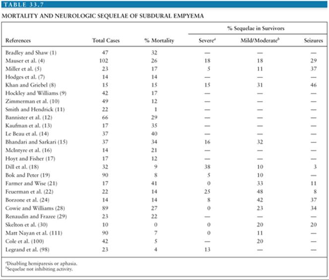
The overall mortality of subdural empyema and the extent of neurologic sequelae reported in survivors are summarized in Table 33.7. In the past, mortality rates averaged 11% to 12% and morbidity rates were 17% to 45% (98). Some recent studies with aggressive antibiotic and surgical approaches have reduced the mortality to less than 5% and morbidity to less than 13% (98). The potential extent of neuroanatomic sequelae is illustrated by necropsy studies (33). Venous sinus thrombosis is a common finding in patients with subdural empyema because the route of infection to the subdural space is generally via these venous sinuses. As a result of the absence of valves in the venous system of the brain, thrombophlebitis may extend to the cortical and subcortical veins of the cerebrum. Thrombosis of these vessels results in venous stasis and subsequent congestion and softening of adjacent brain tissue. Brain infarction and necrosis (Fig. 33.7), with or without abscess formation, may ensue. Therefore, clinical neurologic sequelae may be a consequence of brain abscess or brain infarction secondary to increased ICP or venous thrombosis. More than 10% of patients with subdural empyema develop venous sinus thrombosis or brain abscess (13,33,45).

SPINAL SUBDURAL EMPYEMA
Spinal subdural empyema is a rare condition, with fewer than 80 cases previously reported in the literature, with most reports from the Western Hemisphere in patients in their sixth and seventh decade of life. In 2013, Sandler et al. (112) reported 11 cases of spinal subdural empyema in children throughout the world and reviewed 73 additional cases. The median age was 6.5 years with 38% of cases younger than 3 years of age and 15% younger than 1 year of age. Most of the children were male (2:1 ratio) and 53% were associated with spinal congenital abnormalities. Fifty-six percent of cases lived in the Eastern Hemisphere with only 17% from the United States.
In adults, signs and symptoms include fever, back pain, and subsequent signs of spinal cord compression (78–80). It can be distinguished from spinal epidural abscess by the absence of tenderness to palpation in most cases of spinal subdural empyema (78,79,81,102). In children, however, spinal tenderness is more common (112). Therefore, in children, the clinical presentation may be difficult to differentiate from acute transverse myelitis. Spinal subdural empyema most commonly occurs in the thoracolumbar spine, although subdural infection of the cervical spine has been reported (31,80,82–85,112).
Spinal subdural empyema most often arises as a result of hematogenous spread of infection to the spinal subdural space, with S. aureus being the most common etiologic agent (78,102). Other reported pathogens include streptococci (31,86), coagulase-negative staphylococci (79,87), and gram-negative bacilli (78,80). Occasionally, this may arise from local trauma or surgical procedures including incidental dural tears (113,114) and even acupuncture (115). In a series of childhood spinal subdural empyema, the most common pathogens were Mycobacterium tuberculosis and Echinococcus granulosis (112).
Diagnosis is best accomplished by MRI with gadolinium with decreased signal on T1 and increased signal on T2. Fat-suppressive techniques with MRI help define the subdural space (112). Metrizamide-enhanced spinal CT can be used in areas where MRI is not available or is contraindicated. Treatment consists of empirical antibiotic therapy initially directed against S. aureus, streptococci, and gram-negative enteric bacilli, in association with laminectomy for drainage of the empyema. Antibiotics can then be adjusted based on specific culture results for a duration of 2 to 4 weeks.
SUPPURATIVE INTRACRANIAL PHLEBITIS
Infections within the sinuses or facial structures can lead to intracranial complications that include subdural empyema, cerebral abscesses, epidural abscesses, meningitis, and less commonly suppurative intracranial phlebitis of the dural veins. Suppurative phlebitis can occur as a primary complication or as a secondary complication to other intracranial infections such as subdural empyema (Table 33.8).

Pathogenesis
Infection usually spreads to the dura via venous drainage from the sinuses (91), middle ear, face, or scalp. Within the dura mater are seven paired venous sinuses and five unpaired sinuses, all of which are spaces between two layers of dura. They collect blood from the veins of the brain, skull, and face, and they empty into the internal jugular veins (38). Emissary (perforating) veins allow the passage of blood from the larger veins of the face and scalp into the dural venous sinuses, serving as a potential communication for more superficial infection of the head with the venous system of the brain (37). The venous system of the head and brain is valveless, allowing retrograde spread of thrombophlebitis from infected venous sinuses into dural and cortical venous channels (21,37,116).
Suppurative intracranial phlebitis can lead to infarction or brain inflammation (21,22,57) with subsequent neurologic sequelae such as seizures, hemiparesis, or cranial neuropathies.
Cavernous sinus thrombosis is more commonly associated with ethmoid and sphenoid disease, whereas occasionally, ophthalmic vein thrombophlebitis may extend posteriorly to cause cavernous sinus thrombosis (91).
Clinical Features
Most patients present with headache and fever associated with a toxic appearance and leukocytosis. Increased ICP and papilledema are present in 53% to 65% of cases of cavernous and lateral sinus thrombosis but less commonly with superior sagittal thrombosis (88,89). Focal neurologic findings involve cranial nerves primarily based on the location of some cranial nerves to the venous sinuses. Cranial nerves III, IV, and VI controlling the extraocular muscles are primarily involved with cavernous sinus thrombosis as they pass through or near the inflamed cavernous sinuses (89). Lateral sinus thrombosis is most often associated with unilateral sixth nerve palsy. On rare occasions, the ophthalmic and maxillary branches of the trigeminal nerve (V1 and V2, respectively) are involved with cavernous sinus thrombosis and less commonly with lateral sinus thrombosis. Hemiparesis may occur in up to 61% of patients with superior sagittal sinus thrombosis due to cerebral infarcts (88).
Microbiology and Pathogenesis
S. aureus is the most common bacterial pathogen, occurring in more than 60% to 70% of cases of cavernous sinus thrombosis (88–90). Other gram-positive bacteria such as streptococcal species and pneumococci also can be associated with acute suppurative intracranial phlebitis. In septic lateral sinus thrombosis, gram-negative bacteria such as Proteus mirabilis and Escherichia coli as well as anaerobes such as Bacteroides fragilis and anaerobic streptococci may play a larger role due to the pathogens involved in chronic otitis media, which is often its predisposing condition (88). Immunosuppression and hematologic malignancies may be associated with fungal infections such as mucormycosis (92). A study from Pakistan and the Middle East reviewed 109 cases of cerebral venous thrombosis of which 20 (18%) had infectious causes ranging from tuberculous meningitis, rhino-orbital fungal infection, bacterial meningitis, mastoiditis, malignant otitis, and others (117).
Diagnosis
High-resolution contrast-enhanced CT scans with fine cuts through the suspected areas have been used most extensively over the years. However, MRI has now become the modality of choice for diagnosing suppurative intracranial phlebitis, particularly cavernous sinus thrombosis, using thin-section coronal images as well as magnetic resonance angiography (MRA) and magnetic resonance venography (MRV) (91,93,117–120). In a recent review of cerebral venous thromboses (118), the advantages and disadvantages of CT and MRI modalities are discussed. Although the exact sensitivity and specificity for MRI techniques are unknown, it is estimated that CT/computed tomography venography (CTV) shows 95% sensitivity and 91% specificity and that MRI/MRV is equal or better than CT/CTV (118). In cases where MRI is not available or contraindicated, CT techniques are fairly accurate.
Cerebral angiography and venography are used only rarely (121) since the advent of CT and MRI. Lumbar puncture with CSF evaluation always is abnormal with neutrophilic pleocytosis and elevated pressure but is generally not diagnostic and may pose some risk in patients with severely elevated ICP.
Treatment
All intracranial suppurative infections require the use of appropriate high-dose antibiotics with good penetration of the blood–brain barrier. Appropriate surgical intervention to drain the sinuses or subdural collections is usually mandatory. In addition, anticoagulants may be added to the regimen for cavernous and sinus thrombosis using heparin or low-molecular-weight heparin followed by warfarin for a 6-week course or until the thrombus resolves radiographically (119). The use of anticoagulants, however, is controversial because of the risk of bleeding from carotid artery rupture or cerebral venous infarction (94). Direct thrombolytic therapy has been used for refractory cerebral sinus thrombosis, but its use in septic sinus thrombosis is not clear and may be hazardous (95).
Prognosis
Antibiotics have markedly reduced the morbidity and mortality of suppurative intracranial phlebitis. However, mortality remains as high as 78% for superior sagittal thrombosis, 30% for cavernous sinus thrombosis, and only 0% to 12% for lateral sinus thrombosis (88,96). Permanent neurologic sequelae result in 25% to 100% of those patients who survive and are most likely to occur in those with the most severe disease and those with superior sagittal sinus thrombosis.
References
1. Bradley PJ, Shaw MDM. Subdural empyema management of the primary source. Br J Clin Med. 1984;38:85–88.
2. Joubert MJ, Stephanov S. Computed tomography and surgical treatment in intracranial suppuration. J Neurosurg. 1977;47:73–78.
3. Harris LF, Haws FP, Triplett JN, et al. Subdural empyema and epidural abscess: recent experience in a community hospital. South Med J. 1987;80:1254–1258.
4. Mauser HW, Van Houwelingen HC, Tulleken CA. Factors affecting the outcome in subdural empyema. J Neurol Neurosurg Psychiatry. 1987;50:1136–1141.
5. Miller ES, Dias PS, Uttley D. Management of subdural empyema: a series of 24 cases. J Neurol Neurosurg Psychiatry. 1987;50:1415–1418.
6. Weisberg L. Subdural empyema: clinical and computed tomographic correlations. Arch Neurol. 1986;43:497–500.
7. Hodges J, Anslow P, Gillett G. Subdural empyema—continuing diagnostic problem in the CT scan era. Q J Med. 1986;59:387–393.
8. Khan M, Griebel R. Subdural empyema: a retrospective study of 15 patients. Can J Surg. 1984;27:283–288.
9. Hockley AD, Williams B. Surgical management of subdural empyema. Childs Brain. 1983;10:294–300.
10. Zimmerman RD, Leeds NE, Danziger A. Subdural empyema: CT findings. Radiology. 1984;150:417–422.
11. Smith HP, Hendrick EB. Subdural empyema and epidural abscess in children. J Neurosurg. 1983;58:392–397.
12. Bannister G, Williams B, Smith S. Treatment of subdural empyema. J Neurosurg. 1981;55:82–88.
13. Kaufman DM, Miller MM, Steigbigel NH. Subdural empyema: analysis of 17 recent cases and review of the literature. Medicine. 1975;54:485–498.
14. Le Beau J, Creissard P, Harispe L, et al. Surgical treatment of brain abscess and subdural empyema. J Neurosurg. 1973;38:198–203.
15. Bhandari YS, Sarkari NBS. Subdural empyema: a review of 37 cases. J Neurosurg. 1970;32:35–39.
16. McIntyre PB, Lavercombe PS, Kemp RJ, et al. Subdural and epidural empyema: diagnostic and therapeutic problems. Med J Aust. 1991;154:653–657.
17. Hoyt DJ, Fisher SR. Otolaryngologic management of patients with subdural empyema. Laryngoscope. 1991;101:20–24.
18. Dill SR, Cobbs CG, McDonald CK. Subdural empyema: analysis of 32 cases and review. Clin Infect Dis. 1995;20:372–386.
19. Bok APL, Peter JC. Subdural empyema: burr holes or craniotomy? J Neurosurg. 1993;78:574–578.
20. Curless RG. Subdural empyema in infant meningitis: diagnosis, therapy, and prognosis. Childs Nerv Syst. 1985;1:211–214.
21. Farmer TW, Wise GR. Subdural empyema in infants, children and adults. Neurology. 1973;23:254–261.
22. Feuerman T, Wackym PA, Gade GF, et al. Craniotomy improves outcome in subdural empyema. Surg Neurol. 1989;32:105–110.
23. Luken MG, Whelan MA. Recent diagnostic experience with subdural empyema. J Neurosurg. 1980;52:764–771.
24. Borzone M, Capuzzo T, Rivano C, et al. Subdural empyema: fourteen cases surgically treated. Surg Neurol. 1980;13:449–452.
25. Yoshikawa TT, Chow AW, Guze LB. Role of anaerobic bacteria in subdural empyema: report of four cases and review of 327 cases from the English literature. Am J Med. 1975;58:99–104.
26. Kaufman DM, Litman N, Miller MM. Sinusitis: induced subdural empyema. Neurology. 1983;33:123–132.
27. Morgan DW, Williams B. Posterior fossa subdural empyema. Brain. 1985;108:983–992.
28. Cowie R, Williams B. Late seizures and morbidity after subdural empyema. J Neurosurg. 1983;58:569–573.
29. Renaudin JW, Frazee J. Subdural empyema—importance of early diagnosis. Neurosurgery. 1980;7:447–479.
30. Skelton R, Maixner W, Isaacs D. Sinusitis-induced subdural empyema. Arch Dis Child. 1992;67:1478–1480.
31. Levy ML, Wieder BH, Schneider J, et al. Subdural empyema of the cervical spine: clinicopathologic correlates and magnetic resonance imaging. J Neurosurg. 1993;79:929–935.
32. Kubik CS, Adams RD. Subdural empyema. Brain. 1943;66:18–42.
33. Courville CB. Subdural empyema secondary to purulent frontal sinusitis. Arch Otolaryngol. 1944;39:211–230.
34. Kim KS, Weinberg PE, Magidson M. Angiographic features of subdural empyema. Radiology. 1976;118:621–625.
35. Kaufman DM, Leeds NE. Computed tomography (CT) in the diagnosis of intracranial abscesses. Neurology. 1977;27:1069–1073.
36. Weingarten K, Zimmerman RD, Becker RD, et al. Subdural and epidural empyemas: MR imaging. AJNR Am J Neuroradiol. 1987;10:81–87.
37. Warwick R, Williams PL, eds. Gray’s Anatomy. 35th ed. Philadelphia: WB Saunders; 1973.
38. Clemente CD. Anatomy: A Regional Atlas of the Human Body. 2nd ed. Baltimore: Urban & Schwarzenberg; 1981.
39. Weinman D, Samarasinghe HHR. Subdural empyema. Aust N Z J Surg. 1972;41:324–330.
40. van Alphen HAM, Dreissen JJR. Brain abscess and subdural empyema: factors influencing mortality and results of various surgical techniques. J Neurol Neurosurg Psychiatry. 1976;39:481–490.
41. Van Dellen JR, Boles DM, Van Der Heever CM. Interhemispheral subdural empyema. S Afr Med J. 1977;52:266–269.
42. Stephanov S, Joubert MJ, Welchman JM. Combined convexity and parafalx subdural empyema. Surg Neurol. 1979;11:147–151.
43. Holtzman RNN, Tepperberg J, Schwartz O. Parasagittal subdural empyema: a case report with computerized tomographic scan documentation. Mt Sinai J Med. 1980;47:62–67.
44. Nathoo N, Nadvi SS, Gouws E, et al. Craniotomy improves outcomes for cranial subdural empyemas: computed tomography-era experience with 699 patients. Neurosurgery. 2001;49:872–878.
45. Rosenbaum GS, Cunha BA. Subdural empyema complicating frontal and ethmoid sinusitis. Heart Lung. 1989;18:199–202.
46. Zizmor J, Noyak AM. Radiology of the nose and paranasal sinuses. In: Paparella MM, Shumrick DA, eds. Otolaryngology: Basic Science and Related Disciplines. Philadelphia: WB Saunders; 1980:1010–1066.
47. Grullen A, Brell M, Cardona E, et al. Pott’s puffy tumour: still not an eradicated entity. Childs Nerv Syst. 2001;17:359–362.
48. Layon J, McCulley D. Subdural empyema and group C Streptococcus. South Med J. 1985;78:64–66.
49. Osgood CP, Dujovny M, Holm E, et al. Delayed post-traumatic subdural empyema. J Trauma. 1975;15:916–921.
50. Hadj-Djilani M, Calliauw L. A contribution to the rapid diagnosis of subdural empyema. Acta Neurochir. 1982;61:187–199.
51. Casson IR, Patel P, Blair D, et al. Subdural empyema: caused by infection of preexisting subdural hematoma. N Y State J Med. 1981;81(3):389–391.
52. Khan MI, Chan R. Pasteurella multocida subdural empyema: a case report. Can J Neurol Sci. 1981;8:163–165.
53. Hlavin ML, Kaminski HJ, Fenstermaker RA, et al. Intracranial suppuration: a modern decade of postoperative subdural empyema and epidural abscess. Neurosurgery. 1994;34:974–981.
54. Jacobson PL, Farmer TW. Subdural empyema complicating meningitis in infants: improved prognosis. Neurology. 1981;31:190–193.
55. Hardy TL, Minor F, Phinney ES. Chronic subdural empyema. Surg Neurol. 1981;16:154–156.
56. Post EM, Modesti LM. “Subacute” postoperative subdural empyema. J Neurosurg. 1981;55:761–765.
57. Sadhu VK, Handel SF, Pinto RS, et al. Neuroradiologic diagnosis of subdural empyema and CT limitations. AJNR Am J Neuroradiol. 1980;1: 39–44.
58. List CF. Diagnosis and treatment of acute subdural empyema. Neurology. 1955;5:663–670.
59. Zimmerman RD, Weingarten K. Evaluation of intracranial inflammatory disease by CT and MRI. In: Theodore WH, ed. Clinical Neuroimaging. New York: Alan R. Liss; 1988:75–100.
60. Sze G, Zimmerman RD. The magnetic resonance imaging of infections and inflammatory diseases. Radiol Clin North Am. 1988;26:839–860.
61. Wehrli FW. Principles of magnetic resonance imaging. In: Stark DD, Bradley WG, eds. Magnetic Resonance Imaging. St. Louis: CV Mosby; 1988:3–23.
62. Rick PM, Deasy NP, Jarosz JM. Intracranial dural empyema. Br J Radiol. 2000;73:1329–1336.
63. Bockova J, Rigamonti D. Intracranial empyema. Pediatr Infect Dis J. 2000;19:735–737.
64. Bydder GM. Clinical applicability of gadolinium-DTPA. In: Stark DD, Bradley WG, eds. Magnetic Resonance Imaging. St. Louis: CV Mosby; 1988:182–200.
65. Farkas AG, Marks JC. Subdural empyema: an important diagnosis not to miss. Br Med J. 1986;293:118–119.
66. Duffy GP. Lumbar puncture in the presence of raised intracranial pressure. Br Med J. 1969;1:407–409.
67. Chu RM, Tummala RP, Halb WA. Local intracranial infections due to Propionibacterium acnes: report of three cases. Neurosurg. 2001;49: 717–720.
68. Nawashiro H, Uozumi Y. Letter to editor. Neurosurg. 2002;50:1172.
69. Okudera H, Yasutuki T, Kyoshima K. Bilateral subdural empyema due to Salmonella enteritidis in an infant. Childs Nerv Syst. 1989;5:45–46.
70. Grosinger L, Lauter CB. Salmonella subdural empyema in a patient with brain metastasis. Rev Infect Dis. 1986;8:830–831.
71. Safani MM, Ehrensaft DV. Successful treatment of subdural empyema due to Serratia marcescens. Drug Intell Clin Pharm. 1982;16:777–779.
72. Edwards MS, Baker CJ. Subdural empyema: an unusual complication of meningococcal meningitis. South Med J. 1982;75:68–69.
73. Nithyanandam S, D’Souza O, Rao S, et al. Rhinocerebral actinomycosis. Ophthal Plast Reconstr Surg. 2001;17:134–136.
74. Stern J, Bernstein CA, Whelan MA, et al. Pasteurella multocida subdural empyema. J Neurosurg. 1981;54:550–552.
75. Louie JA, Kusske JA, Rush JL, et al. Actinomycotic subdural empyema: case report. J Neurosurg. 1979;51:852–855.
76. Mauser HW, Ravijst RAP, Elderson A, et al. Nonsurgical treatment of subdural empyema: case report. J Neurosurg. 1985;63:128–130.
77. Rosazza A, de Tribolet N, Deonna T. Nonsurgical treatment of interhemispheric subdural empyemas. Helv Paediatr Acta. 1979;34:577–581.
78. Dacey RG, Winn R, Jane JA, et al. Spinal subdural empyema: report of two cases. Neurosurgery. 1978;3:400–403.
79. Theodotou B, Woosley RE, Whaley RA. Spinal subdural empyema: diagnosis by spinal computed tomography. Surg Neurol. 1984;21:610–612.
80. Bartels RH, De Jong R, Grotenhuis JA. Spinal subdural abscess: case report. J Neurosurg. 1992;76:307–311.
81. Fraser RAR, Ratzan K, Wolpert SM, et al. Spinal subdural empyema. Arch Neurol. 1973;28:235–238.
82. Hirson C. Spinal subdural abscess. Lancet. 1965;2:1215–1217.
83. Heindel CS, Ferguson JP, Kumarasamy T. Spinal subdural empyema complicating pregnancy: case report. J Neurosurg. 1974;40:654–656.
84. Negrin J Jr, Clark RA Jr. Pyogenic subdural abscess of the spinal meninges: report of two cases. J Neurosurg. 1952;9:95–100.
85. Scully RE, Mark EJ, McNeely BU. Case records of the Massachusetts General Hospital: case 47–1984. N Engl J Med. 1984;311:1365–1370.
86. Gelfand MS, Bakhtian BJ, Simmons BP. Spinal sepsis due to Streptococcus milleri: two cases and review. Rev Infect Dis. 1991;13:559–563.
87. Peacher WG, Robertson RCL. Neurologic complications following the use of continuous caudal anesthesia. Arch Neurol Psychiatry. 1944;52: 531–533.
88. Southwick FS, Richardson EP, Swartz MN. Septic thrombosis of the dural sinuses. Medicine. 1986;65:82–106.
89. Amran M, Sidek DS, Hamzah M, et al. Cavernous sinus thrombosis secondary to sinusitis. J Otolaryngol. 2002;31:165–169.
90. Ebright JR, Pace MT, Niazi AF. Septic thrombosis of the cavernous sinuses. Arch Intern Med. 2001;161:2671–2676.
91. Gallagher RM, Gross CW, Phillips CD. Suppurative intracranial complications of sinusitis. Laryngoscope. 1998;108:1635–1642.
92. Harril WC, Stewart MG, Lee AG, et al. Chronic rhinocerebral mucormycosis. Laryngoscope. 1996;106:1292–1297.
93. Eustis HS, Mafee MF, Walton C, et al. MR imaging and CT of orbital infections and complications in acute rhinosinusitis. Radiol Clin North Am. 1998;36:1165–1178.
94. Bhatia K, Jones NS. Septic cavernous sinus thrombosis secondary to sinusitis: are anticoagulants indicated? A review of the literature. J Laryngol Otol. 2002;116:667–676.
95. Smith TP, et al. Treatment of docral sinus thrombosis by urokinase infusion. AJNR Am J Neuroradiol. 1994;15:801–807.
96. DiNubile MJ. Septic thrombosis of the cavernous sinuses. Arch Neurol. 1988;45:567–571.
97. Liu ZH, Chen NY, Tu PH, et al. The treatment and outcome of postmeningitic subdural empyema in infants. J Neurosurg Pediatr. 2010;6:38–42.
98. Legrand M, Roujeau T, Meyer P, et al. Paediatric intracranial empyema: differences according to age. Eur J Pediatr. 2009;168:1235–1241.
99. Wu TJ, Chen NC, Huang FY. Subdural empyema in children—20-year experience in a medical center. J Microbiol Immunol Infect. 2008;41: 62–67.
100. Cole TS, Clark ME, Jenkins AF, et al. Pediatric focal intracranial suppuration: a UK single-center experience. Childs Nerv Syst. 2012;28(12):2109–2114.
101. Osborne M, Steinberg JP . Subdural empyema and other suppurative complications of paranasal sinusitis. Lancet Infect Dis. 2007;7(1):62–67.
102. Darouche RO. Spinal epidural and subdural empyema. Handb Clin Neurol. 2010;96:91–99.
103. Tsai B, Lin KL, Lin TY, et al. Pott’s puffy tumor in children. Childs Nerv Syst. 2010;26:53–60.
104. Dashti SR, Baharvadat H, Spetzler RF, et al. Operative intracranial infection following craniotomy. Neurosurg Focus. 2008;24(6):E10.
105. Van de Beek D, deGans J, Tunkel A, et al. Community-acquired bacterial meningitis. N Engl J Med. 2006;354:44–53.
106. Banerjee AD, Pandey P, Devi BI, et al. Pediatric supratentorial subdural empyemas: a retrospective analysis of 65 cases. Pediatr Neurosurg. 2009;45:11–18.
107. Chen CY, Huang CC, Chang YC, et al. Subdural empyema in 10 infants: US characteristics and clinical correlates. Radiology. 1998;207:609–617.
108. Fanning NF, Laffan EE, Shroff MM. Serial diffusion-weighted MRI correlates with clinical cause and treatment response in children with intracranial pus collections. Pediatr Radiol. 2006;36:26–37.
109. Foerster BR, Thurnher MM, Malane PN, et al. Intracranial infections: clinical and imaging characteristics. Acta Radiol. 2007;8:875–893.
110. Tsou TP, Lee PL, Lu CY, et al. Microbiology and epidemiology of brain abscess and subdural empyema in a medical center: a 10-year experience. J Microbiol Immunol Infect. 2009;42:405–412.
111. Matt Nayan SA, Mohd Herjani MS, Abd Latif AZ, et al. Two surgical methods used in 90 patients with intracranial subdural empyema. J Clin Neurosci. 2009;16:1567–1571.
112. Sandler AL, Thompson D, Goodrich JT, et al. Infections of the spinal subdural space in children: a series of 11 contemporary cases and review of all the published reports. A multinational collaborative effort. Childs Nerv Syst. 2013;29:105–117.
113. Wu AS, Griebel RW, Megurfo K, et al. Spinal subdural empyema after a dural tear. Neurosurg Focus. 2004;17:E10.
114. Vural M, Arslantas A, Adapinar B, et al. Spinal subdural Staphylococcus aureus abscess: case report and review of the literature. Acta Neurol Scand. 2005;112:343–46.
115. Chen MH, Chen MH, Huang JS. Cervical subdural empyema following acupuncture. J Clin Neurosci. 2004;11:908–911.
116. Syed MI, Baring D, Addidle M, et al. Lemmiere syndrome: two cases and a review. Laryngoscope. 2007;117:1605–1610.
117. Khealani BA, Wasay M, Saadah M, et al. Cerebral venous thrombosis. A descriptive multicenter study of patients in Pakistan and Middle East. Stroke. 2008;39:2707–2711.
118. Leach JL, Fortuna RB, Jones BV, et al. Imaging of cerebral venous thrombosis: current techniques, spectrum of findings, and diagnostic pitfalls. Radiographics. 2006;26:519–543.
119. Saposnik G, Barinagarrementeria F, Brown RD, et al. Diagnosis and management of cerebral venous thrombosis: a statement for the healthcare professionals from the American Heart Association and American Stroke Association. Stroke. 2011;42:1158–1192.
120. Hughes DC, Raghawan A, Mordekar SR, et al. Role of imaging and the diagnosis of acute bacterial meningitis and its complications. Postgrad Med J. 2010;86:478–485.
121. Rodallec MH, Krainek A, Feydy A, et al. Cerebral thrombosis and multidetector CT angiography: tips and tricks. Radiographics. 2006;26:S5–S18.