
STEFANO GIULIERI, RETO ANTOINE MEULI, AND MATTHIAS CAVASSINI
Central nervous system (CNS) complications of infective endocarditis (IE) occur in about 30% of patients, with the highest incidence among patients referred to tertiary care centers and intensive care units, as well as patients with mitral valve endocarditis and IE due to virulent microorganisms (e.g., Staphylococcus aureus, gram-negative bacilli, fungi). Ischemic stroke accounts for up to two thirds of CNS complications, followed by intracranial hemorrhage, meningitis, brain abscess, intracranial infectious aneurysm (ICIA), and encephalopathy. Embolization of infected vegetations is the main pathogenetic mechanism. Imaging (both computed tomography [CT] and magnetic resonance imaging [MRI], coupled with noninvasive angiography) plays a pivotal role in the diagnosis of CNS complications. Optimal management includes early institution of antimicrobial treatment, which has the highest impact on the risk of further emboli. Anticoagulation should be cautiously continued in patients with established cardiac indications (e.g., prosthetic valve). In the presence of an established indication, patients with IE and ischemic stroke should undergo cardiac surgery without delay, whereas it should be postponed for at least 4 weeks in case of intracerebral hematoma. Unruptured ICIA may be managed medically with radiologic follow-up, whereas enlarging or ruptured ICIA should be treated surgically or by an endovascular approach. Despite improvement in the management, patients with CNS complications generally carry a worse prognosis compared to patients without neurologic complications.
HISTORY
Infective endocarditis (IE) was probably recognized as early as 1646 by Rivière, but the full clinical spectrum of “malignant endocarditis” was first comprehensively described by Sir William Osler (1) in his Gulstonian lectures at the Royal College of Physicians in London in 1885. He not only described “the different modes of onset, and the extraordinary diversity of symptoms which may arise,” but carefully analyzed the pathophysiology of distant complications, particularly of central nervous system (CNS) manifestations such as meningitis: “The meningeal complications of endocarditis have not received much attention, considering the frequency with which it has occurred . . . somewhat over 12 per cent” (1). He also related IE and mycotic aneurysms to a common infectious etiology and later emphasized the central importance of blood cultures in diagnosis (2).
EPIDEMIOLOGY
In developed countries, the reported incidence of IE is 3 to 7 per 100,000 per year (3–6). This rate has remained fairly constant over time (7). Incidence of IE increases sharply with age, with a peak of 15 episodes per 100,000 per year in patients older than 70 years (4,8). The male:female ratio is 2:1. In-hospital and 6-month mortality in recent series are 15% to 20% and 25%, respectively (9). These apparently stable overall rates of incidence and mortality mask the continually evolving spectrum of IE and the contrasts that exist between various types of IE. For instance, subacute IE due to viridans streptococci has an early mortality rate of only 5% to 10%, whereas acute prosthetic valve endocarditis (PVE) in an elderly patient with congestive heart failure can be fatal in up to 70% of patients. The overall figures reflect shifting frequency of predisposing factors. In developed countries, the decreasing prevalence of chronic rheumatic heart disease is counterbalanced by an increased number of patients with prosthetic valves and intravascular devices, injection drug use, and elderly patients with degenerative heart disease (10). The mean age of patients presenting with IE has increased, from 34 to 43 years in the early antibiotic period to 52 to 55 years more recently, and has even passed 60 years in some reports (3,11,12).
PREDISPOSING FACTORS
Degenerative valve disease has replaced rheumatic heart disease as the main underlying condition in patients with IE (10). Degenerative valvular disease with calcified atheromatous deposits is associated with IE in a proportion of cases that increases with age and may be a predisposing factor in up to 50% of the IE cases in elderly patients (13). Mitral valve prolapse (MVP) associated with valve dysfunction is another recognized risk factor for IE, found in 10% to 30% of patients (14). In a case–control study, the risk of IE was eight times higher among patients with MVP (15), but, given the high incidence of MVP (5%) in the general population, the absolute risk per patient is low, estimated to be 0.0175% per year (14).
Although it remains a common predisposing condition in developing countries, rheumatic heart disease is associated with IE in less than 5% of cases in the Western countries (16). Congenital lesions are found in 6% to 20% of cases (17), mostly ventricular septal defect, bicuspid aortic valve, patent ductus arteriosus, and tetralogy of Fallot. A bicuspid aortic valve is an important factor in elderly persons in whom it may be present in up to 20% of IE cases; this is probably because of associated valvular sclerosis and abnormalities of blood flow (18).
Prosthetic valves account for 7% to 25% of the cases of IE and the annual incidence of IE in patients with prosthetic valves is approximately 1% (19). In-hospital mortality rate ranges from 20% to 30% (20).
IE related to hospitalization and ambulatory invasive treatments (health care associated) now accounts for one third of cases (21). The proportion of IE associated with other intracardiac devices such as pacemakers or implantable cardioverter-defibrillators was 6.4% in the ICE-PCS (22). Nosocomial bacteremias (mainly Staphylococcus aureus) represents as many as 25% of the cases in certain series (23,24). IE is reported in 2% to 6% of patients undergoing long-term hemodialysis (25). Intravenous drug users (IVDUs) are at high risk for IE. Moreover, 20% to 40% of IVDUs suffering from IE have preexisting cardiac lesions, often caused by previous infection (26).
PATHOGENESIS
Certain lesions of the endothelium of the valve or heart cavities can easily be infected should a bacteremia occur. This has been shown in animal models: An intravascular polyethylene catheter placed across the aortic or the tricuspid valve will induce the deposition of platelets and fibrin and lead to the formation of nonbacterial thrombotic endocarditis (NBTE) (27). Many factors can induce endothelial lesions, which promote the formation of NBTE. The most important are valvular organic lesions with associated perturbations of blood flow and prosthetic valves. Even microscopic lesions are prone to infection by circulating bacteria (28), so not surprisingly, up to 50% of patients with IE have no known predisposing heart condition at the time of diagnosis (29). Transient asymptomatic bacteremias occur frequently, often following minor mucosal trauma induced by daily domestic activities such as chewing or tooth brushing. Bacteremia may also be triggered by various iatrogenic procedures. Among the many bacterial species that can be recovered during transient bacteremias, streptococci account for most IE cases, probably because of the frequency with which they enter the bloodstream and the adherence properties of their surfaces.
Circulating bacteria readily attach to NBTE. The attachment process is probably mediated on the host side by receptor-like structures such as fibronectin, fibrinogen, laminin, or collagen, which interact with the surface of bacteria (8). Platelets also play a role in these initial events (30,31). On the microbial side, only bacteria with the ability to adhere to valvular lesions can produce IE. Gram-positive cocci (e.g., staphylococci, streptococci) adhere more strongly than enterobacteria to heart valves (32).
Once microorganisms have attached to NBTE, they start to multiply and stimulate further deposition of fibrin and platelets through the activation of tissue factors or procoagulant activity. This thrombotic process buries some bacterial colonies deep within the vegetation where they are protected from circulating neutrophils. This has led to the concept of the vegetation as an area of “localized agranulocytosis.” Bacteria deeply seated in the vegetation are metabolically inactive and thus resistant to the action of some antibiotics. Furthermore, some antibiotics do not fully penetrate into the vegetation. This combination of factors favors the bacteria and helps explain why cure of endocarditis requires the prolonged administration of bactericidal antibiotics.
Because the infected vegetation is located within the bloodstream, persistent bacteremia is a hallmark of bacterial endocarditis, permitting the diagnosis through blood cultures in most patients. Moreover, further deposition of platelets, fibrin, and circulating bacteria will compensate for the fragmentation, embolism, and/or resorption of the vegetation by the inflammatory response, in a delicate and complex balance. IE also induces immunologic responses that contribute to some clinical manifestations of IE such as glomerulonephritis or vasculitis (30).
ETIOLOGIC AGENTS
The list of microorganisms that can cause IE includes most human pathogenic bacteria, as well as rickettsiae, Bartonella, chlamydiae, and fungi (19). However, gram-positive cocci predominate. The pattern of distribution of these etiologic agents differs profoundly according to underlying risk factors, the valve type (native vs. prosthetic), and geography (33).
Native Valve Endocarditis
S. aureus is now as common as streptococci as a causative pathogen of native valve endocarditis (NVE) in developed countries (6). In the International Collaboration on Endocarditis-Prospective Cohort Study (ICE-PCS), S. aureusand streptococci both accounted for 32% of 1,881 cases of definite NVE (16). The increase in frequency of S. aureus IE is explained by several invasive medical procedures associated with S. aureus bacteremia (e.g., hemodialysis, long-term intravenous catheters) (24). The clinical course of S. aureus IE is usually acute and complicated. In-hospital mortality rate was 22% in a recent prospective study (24). Coagulase-negative staphylococci rarely cause NVE but appear to be increasing in frequency and can pursue an aggressive clinical course (34). Special attention has been drawn to Staphylococcus lugdunensis, being commonly associated with valve destruction (35).
In a recent French series of 497 patients with definite IE (79% with NVE), streptococci, although less frequent compared to a previous study (4), were still the most common cause of IE and were responsible for 48% of cases (6). The viridans group (e.g., Streptococcus sanguis, Streptococcus oralis, Streptococcus mutans, and Streptococcus mitis) accounts for at least two thirds of these cases. The clinical course is generally subacute and an underlying cardiac condition is often present. Streptococcus milleri, an occasional cause of IE, has a peculiar propensity to cause distant abscesses and local perivalvular invasion (36). Streptococcus bovis group (including S. gallolyticus subsp. gallolyticus, S. gallolyticus subsp. pasterianus, and S. infantarius subsp. coli) accounts for up to 20% of streptococcal IE (37) and is often associated with neoplasms of the gut, of which it may be the first manifestation (38,39). Streptococcus pneumoniae and streptococci of the Lancefield groups A, B, C, G may also occasionally cause endocarditis (40,41). Enterococci account for 10% of all IE (42); these strains are particularly resistant to the bactericidal activity of antibiotics. Older people are usually affected and outcome can be fatal in approximately 16% of patients (42).
Numerous other bacteria are well known but uncommon causes of NVE, for example, Coxiella burnetii, Brucella, and bacteria of the “HACEK” group (Haemophilus species, Actinobacillus actinomycetemcomitans, Cardiobacterium hominis, Eikenella corrodens, Kingella species) (43). Actinobacillus actinomycetemcomitans has now been reclassified as Aggregatibacter actinomycetemcomitans(44). Bartonella is an unusual cause of endocarditis in homeless people (45).
Prosthetic Valve Endocarditis
PVE cases with onset within 2 months after surgery are called early PVE (46). Early PVE cases are presumably related to intraoperative contamination. S. aureus is responsible for 35% of early PVE cases; coagulase-negative staphylococci are found in 17% of cases. The frequency of gram-negative bacilli (10% to 20%), diphtheroids (0% to 3%), and fungi (5% to 10%) is higher in PVE than in NVE (47), and they are usually isolated in early PVE.
Cases occurring 2 months or more after surgery are called late PVE and are largely community acquired. As compared to early PVE, streptococci are more common and are responsible for approximately 20% of late PVE cases (46).
Intravenous Drug Users
S. aureus accounts for more than 50% of IVDU cases probably because of its high frequency on the skin in local infections at the injection site (cellulitis, abscesses, suppurative thrombophlebitis) and on drug paraphernalia (48). In some regions, methicillin-resistant S. aureus (MRSA) have been responsible for IE among IVDUs. Several other bacterial species such as Pseudomonas aeruginosa, enterococci, and Serratia species or fungi are all more common in addicts than in the general population (49). In 40% to 70% of the cases, the tricuspid valve is affected (50). Right-sided IE among IVDUs has a mortality rate of less than 10% (48).
Culture-Negative Infective Endocarditis
Patients with a clinical presentation suggestive of IE but with negative blood cultures account for 5% to 10% of the cases (8). The most common cause for culture-negative IE is prior administration of antibiotics. In the absence of prior antibiotic administration and with appropriate blood culture media, most bacteria will eventually grow but may require repeated blood culture and prolonged incubation periods (51). Some microorganisms, such as Aspergillusspecies, C. burnetii, Legionella species (usually Legionella pneumophila), or Tropheryma whippelii, cannot be recovered from the blood, so diagnosis relies on other methods such as tissue culture, in situ hybridization, polymerase chain reaction (52), or special serologic tests (53). By applying a standardized diagnostic algorithm, Fournier et al. (33) were able to identify an etiologic agent among 476 out of 759 (63%) patients referred for blood culture–negative IE. C. burnetii (229 cases) and Bartonella sp. (86 cases) were the most common etiologic agents. A noninfectious origin of culture-negative IE (e.g., marantic endocarditis, systemic lupus erythematosus) was diagnosed in 19 patients.
CLINICAL MANIFESTATIONS
The signs and symptoms of IE are extremely variable because of the diversity of the etiologic agents and the various organs involved. Clinical presentation may range from a chronic disease with unspecific systemic symptoms to acute life-threatening sepsis. Fever with a heart murmur is the most common clinical presentation; it may be the only clue to the diagnosis, especially early during the course of the disease and with microorganisms of low virulence. In a prospective study of 109 episodes of IE, fever was present in 98% of cases (54). A new murmur and skin lesions were found in 59% and 32%, respectively. Splenomegaly and musculoskeletal manifestations were present in 16% (54). Congestive heart failure complicates one third of episodes in recent studies (4,16,54). Distant complications such as major emboli, lumbar pain, or rupture of a infectious aneurysm may be the first presenting manifestations of IE. All the various clinical manifestations of IE arise from one or more of four main mechanisms: bacteremia, embolization (55), immunologic manifestations (56), and local complications (57).
DIAGNOSIS
The diagnosis of IE requires the integration of clinical, microbiologic, and echocardiographic data. Hematologic abnormalities are frequently observed: Anemia, leukocytosis, and thrombocytopenia are present in 66%, 50%, and 18% of cases, respectively. Urinalysis shows abnormalities in 30% or more of patients, mainly evidence of proteinuria or microscopic hematuria, one third of patients has elevated creatinine (54). Circulating immune complexes may be detected, but they are nondiagnostic (56). Repeated electrocardiography can reveal new atrioventricular block, particularly in the setting of aortic valve endocarditis, suggesting perivalvular invasion (58).
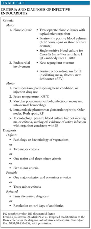
The Duke criteria combine predisposing factors to IE, the blood culture isolate or histologic examination, and echocardiographic findings (59). Based on the number of major and minor criteria, any case of suspected IE is classified as “definite,” “possible,” or “rejected” IE. These criteria have been primarily developed for research purpose, but they can help integrate diagnostic findings (Table 34.1)
Echocardiography has proven to be a valuable diagnostic tool in patients with suspected IE. Transesophageal echocardiography (TEE) has substantially better ability to detect vegetations, perivalvular extension, and myocardial abscesses than transthoracic echocardiography (TTE). TTE has a sensitivity of 40% to 63% for detecting vegetations in NVE (60). TEE increases the sensitivity for detecting vegetations in NVE to more than 90% (61). TEE is particularly useful in PVE, with which vegetations are detected in more than 80% of the cases compared with less than 30% with TTE. A negative TEE has a negative predictive value for IE of 86% to 97%.
TREATMENT AND PREVENTION
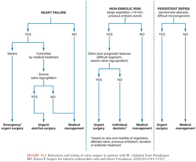
Once the diagnosis of IE has been made, antibiotic treatment should be started promptly to eradicate the infecting microorganisms as soon as possible. It is well established that a prolonged course of antibiotics is needed for cure even if the microorganisms are highly sensitive to the drug used. The antibiotic agent must be bactericidal and should be selected according to in vitro susceptibility test results and the experience gathered from experimental and clinical studies (10,62). Surgery has an important role in the management of IE, as an optimal therapeutic approach requires operative intervention during the course of medical treatment in almost 50% of patients with IE (63). In the ICE-PCS, the rate of surgical intervention (48%) was comparable among patients with NVE and PVE (16). Nearly all patients with fungal endocarditis require combined surgical and medical treatment (64). Hemodynamic deterioration, local complications, and uncontrolled infection are the major indications for surgery (10,65). Although the need for surgery in patients presenting with hemodynamic deterioration and uncontrolled infection is widely recognized (66), it remains controversial whether surgery is indicated for prevention of systemic embolism. In a randomized study from Korea, patients at high risk for systemic embolism (i.e., with severe valvular regurgitation and large vegetations) had significantly less systemic embolic events if operated early during the course of disease (67). Figure 34.1 summarizes current indications for valve surgery in patients with IE. Other interventions that should be considered in patients with IE are the removal of intravascular devices (e.g., pacemakers, intracardiac cardioverter defibrillators, central venous catheters) (68), and the drainage of extracardiac infections (e.g., septic arthritis).
Attempted prevention of IE is aimed at patients with known underlying predisposing factors who undergo a procedure that may result in bacteremia. Possible portals of entry, such as oral and dental lesions or urinary or gastrointestinal tract pathology, should be sought and treated if necessary. Recommendations for use of prophylactic antibiotics have been updated, and indications for prophylaxis have been considerably restricted to four high-risk conditions: prosthetic cardiac valve, history of previous IE, some forms of congenital heart disease (CHD) (e.g., unrepaired CHD), and heart transplantation recipients with valvulopathy (69).
CENTRAL NERVOUS SYSTEM COMPLICATIONS OF INFECTIVE ENDOCARDITIS
Among the various manifestations of IE, neurologic complications are particularly important for three main reasons: They occur often, they may be the first or the predominant manifestation of the disease, and they are a leading cause of death and complications due to IE (70).
Incidence
The reported overall incidence of CNS complications of IE varies greatly. In most series, the incidence of CNS involvement during the course of IE ranges between 20% and 40%, with an average of 30%. The incidence has not changed much over time, despite wide differences in the diagnostic criteria employed in various studies and many evolutionary changes that have occurred in the natural history of IE over the past 60 years. Among 743 patients pooled from seven series published before the advent of antibiotics, 176 (24%) were noted to have neurologic manifestations (1,71–76). Among 1,622 patients from eight studies reported between 1947 and 1978, 457 (28%) had such complications (77–85). Among 1,329 episodes of IE from seven series published between 1981 and 1993, 437 (33%) were accompanied by neurologic manifestations (11,86–93). And among 2,162 episodes of IE from eight series published between 1996 and 2007, 512 (24%) were complicated by neurologic events (70,94–101). Recent studies may indicate a lower rate of neurologic complications: The incidence of stroke in a French cohort of 390 patients and in the ICE-PCS was 18% and 17%, respectively (4,16). However, data on other neurologic complications were not reported. Finally, among 513 patients presenting with complicated native-valve IE, focal neurologic findings, and altered mental status were described in 18% and 16%, respectively (9).
Incidence of asymptomatic CNS events is much higher. By systematically using resonance imaging of the brain, Snygg-Martin et al. (102) identified cerebrovascular complications in 65% of patients; about half of them were asymptomatic. In another study of systematic brain imaging in IE, radiologic evidence of brain embolism was detected in 80% of patient, whereas 25% had clinical stroke (103).
Factors Influencing the Incidence of Central Nervous System Complications
The reported incidence is understandably higher in studies devoted primarily to the neurologic aspects of IE and in studies based on autopsies, because brain damage often causes or contributes to death in IE (11,78,79). In one autopsy series of 69 cases of IE, cerebral emboli were found in up to two thirds of the young adults and in 46% of the total cases (104). Frequency of CNS complications also tends to be higher in series gathered from referral centers (105) or specialized units. For example, among 198 patients with IE hospitalized in 33 intensive care units (ICUs) in France, 108 (55%) presented with neurologic complications (106). In contrast, the rate of CNS complications was only 22% in a series of IE observed in a large community hospital (11). This referral bias would alter not only the number but also the type and severity of the complications because referral is often precipitated by CNS involvement. On the other hand, in a series gathered from six hospitals of different types (university, private, and Veterans), the frequency of CNS complications was very similar (92).
Age and Sex
In many studies of adults with IE, the age of the patients did not appear to greatly influence the overall frequency of CNS complications. In a recent series, however, major neurologic events were recorded in 22%, 28%, and 34% of patients younger than 40 years, 40 to 60 years, and older than 60 years, respectively. In elderly patients, neurologic manifestations tend to be more common as a presenting clinical sign of IE (107); as many as one fourth to one third of them may present with neurologic signs (86), compared with 5% to 17% in the general population. If disorientation is included as a neurologic finding, the figure can be as high as 45%. Neurologic complications in children reported in five series ranged from 15% to 32% (108). Because major neurologic events are uncommon in children or young adults, everyone should recall the dictum, “In hemiplegia in young adults or children, always think of infective endocarditis.”
Generally, the sex distribution of the patients with or without neurologic complications appears to be similar in both sexes (83).
Location of Vegetations in the Heart
Neurologic complications are a hallmark of left-sided valvular abnormalities. This association was noted as early as 1852, before IE was recognized as a clinical entity (109). The incidence of neurologic complications is markedly lower in isolated right-sided IE. In a series of 40 IVDUs with tricuspid IE due to S. aureus, none presented with cerebral manifestations, whereas 2 of 5 of those with left-sided involvement had cerebral septic emboli (110). Of 97 episodes of IE in drug addicts, none of the nine major CNS events that were present on initial evaluation occurred in the 32 cases that involved the right side of the heart (111), although the infection was caused by highly virulent organisms such as S. aureus. CNS complications were observed in 54 (41%) of 133 patients with aortic and/or mitral valve involvement and in only 4 (12%) of 33 of those with tricuspid IE (92). When neurologic manifestations occur in right-sided IE, they present as meningitis, cerebral abscesses, or encephalopathy, rather than as strokes, and are most often related to virulent pathogens such as S. aureus or S. pneumoniae (112). Systemic embolization is rare in right-sided IE but does occasionally occur either from septic thrombi arising in the pulmonary veins or via paradoxical embolization through a patent foramen ovale, which is present in 18% of the normal population.
Mitral valve endocarditis is associated with a higher rate of neurologic complications (83,106,113,114). For example, in a recent French study of 198 patients with IE, mitral valve involvement was independently associated with neurologic complications (OR 1.54, CI 1.07 to 2.21) (106). A study from Duke University including 707 patients with IE found a twofold risk of stroke among patients with mitral valve endocarditis as compared to aortic valve endocarditis (114). This association was not reported by older studies (115), probably because of small size (88).
Neurologic complications in the context of PVE are of special concern because of the threat of superimposed intracerebral hemorrhage, which might be caused or promoted by anticoagulation. Most published data are derived from PVE complicating mechanical prosthetic valves. The overall incidence of focal neurologic events associated with PVE varies from 11% to 44% (46,116–122), which is considerably higher than the overall thromboembolic rate of 1% to 4% per year observed for anticoagulated patients with uninfected prosthetic valves (123).
Studies comparing the incidence of CNS complications between NVE and PVE have given conflicting results. In one study, 11 (13%) of 82 patients with NVE, compared to 6 (33%) of 18 with PVE, presented with such complications (117); however, another study reported 40 (35%) of 113 for patients with NVE and 24 (39%) of 62 for those with PVE (88). Other series did not report any statistically significant difference regarding the number of NVE and PVE CNS complications (46,98,124).
It is unclear whether the incidence of embolic events is affected by the time of onset of PVE in relation to the placement of the prosthetic valve. In one study, CNS embolic episodes were reported in 0% to 11% of patients with early onset PVE and in 23% to 28% of those with late-onset PVE (120). The higher rate in late-onset PVE was confirmed in another study in which peripheral manifestations occurred in 10% of early onset PVE cases, compared to 34% in late-onset PVE cases (125). However, in a recent prospective study of 78 cases of PVE, the rate of CNS complications did not differ between early and late PVE (126).
The incidence of CNS complications in PVE is influenced by anticoagulant therapy. In patients insufficiently anticoagulated, CNS complication rates of 38% to 71% were noted, compared to only 8% to 10% in patients on appropriate anticoagulant therapy (116,119). In one series of 61 patients, there was no difference in the rate of embolism or risk of bleeding between patients receiving no anticoagulation or subtherapeutic doses and those who were adequately anticoagulated (121). When anticoagulated patients develop neurologic complications, they are at high risk for major hemorrhage (83,120,127).
The type of prosthetic valve may influence the rate of CNS manifestations. Of 33 patients with bioprosthetic valve infection gathered from three studies, 4 (12%) had neurologic events (117,120,121), a figure lower than that with mechanical valves. However, in another study of 62 patients with PVE, there was no statistical difference when the type of prosthetic valve involved was compared among patients with and without neurologic complications (88).
In summary, patients with PVE do not appear to be at a much higher risk for CNS complications than patients with NVE, provided that anticoagulation is maintained and carefully controlled for complications associated with mechanical valves. However, those presenting a CNS complication are particularly threatened by the possibility of a massive cerebral hemorrhage (83,116,120).
Microbiology
An important factor in determining the rate, type, and severity of the neurologic complications is the nature of the microorganism causing the IE. This may account for some of the differences in the incidence noted between the various studies (Fig. 34.2). In series that have correlated the incidence of neurologic complications with the infectious agent causing IE, the frequency of CNS involvement ranged from 53% to 71% for S. aureus and was significantly higher than that observed with some other bacteria, particularly group D and non–group D streptococci, which ranged from 25% to 47% (83,88,92,97,113).
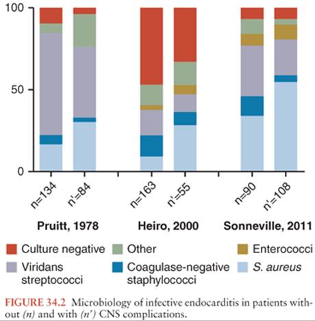
Some species of bacteria other than S. aureus, such as Enterobacteriaceae or anaerobic bacteria, have been associated with a high rate of neurologic complications (83,128). Moreover, certain microorganisms are prone to a high rate of certain types of neurologic involvement. For example, although S. pneumoniae has become rare in IE, causing only 1% to 3% of cases (129), in these few cases, associated pneumococcal meningitis is common, being found in 40% to 60% (130,131). Purulent complications are also frequently encountered with S. aureus IE, either as meningitis or as brain abscess. Certain bacteria such as the Haemophilus species, members of the genus Abiotrophia(formerly nutritionally variant streptococci), or fastidious organisms have been associated with large emboli; these emboli may occlude major vessels including cerebral arteries. In patients with culture-negative IE, major embolic phenomena, including those to the brain, were noted twice as often as in patients with culture-positive IE. Cases due to Candida spp. are also associated with large vegetations and a high rate of embolic phenomena (132,133). Of 27 patients with IE caused by Aspergillus species, a total of 14 neurologic manifestations secondary to emboli were recorded in 11 (41%) patients (134). In contrast, the rate of CNS complications in IE caused by some other bacteria, such as viridans streptococci or coagulase-negative staphylococci, appears to be lower. Of 128 patients with IE due to the latter bacteria, only 15 (12%) developed stroke (34).
Intravenous Drug Abuse
In IVDUs, as in all patients with IE, the frequency of cerebral manifestations is dependent on the side of the heart involved. Therefore, the reported incidence of neurologic complications will depend on the proportion of patients with exclusively right-sided involvement. Isolated right-sided involvement in IE has been reported in 9% to 72% in series of endocarditis in addicts (50,135). In a study comparing the clinical manifestations of S. aureus IE in addicts and nonaddicts, neurologic involvement was found in 51% of the nonaddicts, compared to only 9% of the addicts (110). Although neurologic complications were not categorized according to the side of the heart involved, 76% of the addicts had only tricuspid valve involvement, contrasted to 9% in the nonaddict population, indicating that the side involved plays an important role in the occurrence of neurologic complications (110). The frequency of neurologic complications in IVDUs with left-sided endocarditis appears to be higher than that in the nonaddict population, ranging from 45% to 58% (136). The higher complication rate may partly be due to the failure of some of the addicts to undergo a full course of antibiotics, or it may be due in part to a different pattern of etiologic agents.
Pathogenesis of Central Nervous System Complications
Neurologic complications of IE can arise through several mechanisms, as follows: occlusion of cerebral arteries by emboli, infected or not, derived from endocardial vegetations; infection of the meninges of the brain or of the walls of cerebral arteries by septic emboli or bacteremia; and microbial toxic effects or immune-mediated injuries. These events may result in various secondary lesions, including bland or hemorrhagic infarcts; intracerebral, subarachnoid, or subdural hemorrhages; focal expanding lesions such as abscesses or infectious aneurysms; and brain dysfunction due to one or multiple factors.
When embolization is the ultimate cause of the neurologic involvement, the lesions either may affect a single vessel and give focal signs or may affect multiple vessels and produce multifocal signs. Depending on whether ischemia is reversed before permanent changes occur, the clinical picture may be that of a transient ischemic attack (TIA) (137) or of a longer lasting obstruction resulting in brain damage. Because emboli of untreated or partially treated IE contain bacteria, the lesions produced may be ischemic, inflammatory, suppurative, or mixed. This may result in septic or aseptic meningitis, brain abscesses, microabscesses, or meningoencephalitis. If the wall of an artery or its vasa vasorum is involved, a infectious aneurysm may develop. Arterial rupture can occur in the absence of a detectable intracranial infectious aneurysm (ICIA) (88,138–140). Critical factors that may determine whether a septic embolus results in a bland infarct, a hemorrhagic infarct, a infectious aneurysm, or an abscess include the site where the embolus lodges, the virulence and number of the microorganisms, and importantly, the delay between the event and the initiation of antibiotic therapy.
Multiple other factors can cause or contribute to the neurologic manifestations of IE such as hypoxia, metabolic disturbances, drug toxicity, and toxic phenomena secondary to the systemic infection. Immune injury to small arteries is also likely to be involved (141), and proliferative endarteritis in the absence of local infection or embolization has been described.
In patients with tricuspid valve endocarditis, the sustained bacteremia rarely results in intracranial hemorrhage (ICH), even when due to virulent organisms, supporting the hypothesis that embolic fragments are a required factor in the pathogenesis of bleeding (138).
Clinical Presentation
Neurologic manifestations of IE constitute the presenting symptoms of IE in 16% to 23% of the cases (83,97,113). When a neurologic event is the presenting symptom, approximately two thirds are due to major cerebral emboli. The remaining third are divided among other manifestations including seizures, meningismus, subarachnoid, intracerebral, or subdural hemorrhage, personality change, visual disturbances, or weakness of the extremities (83,97).
Interestingly, the mean duration of prodromal symptoms prior to diagnosis was found to be similar in patients with and in those without neurologic complications (88).
Neurologic complications occur after the initiation of antibiotic treatment in 30% of patients (88). In most of these, the neurologic events tend to occur soon after treatment has begun, usually within the first 2 weeks (88). However, cerebral emboli or rupture of ICIA rarely occurs from several months to up to 2 years after the completion of successful treatment. More than one complication is often observed in a given patient; thus, a total of 160 neurologic manifestations were recorded among 84 patients studied by Pruitt et al. (83). The neurologic complications of IE are numerous (Tables 34.2 and 34.3) and can mimic many neurologic diseases of other etiologies.
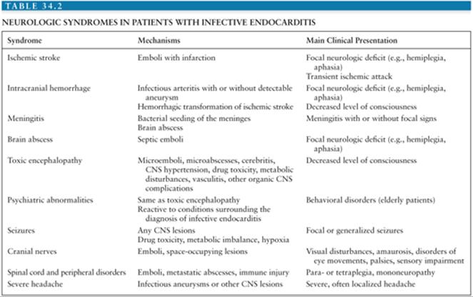
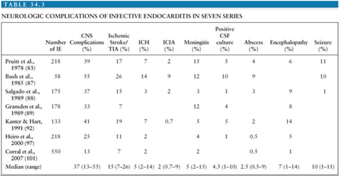
Stroke is the most common presentation and accounts for one half to two thirds of the neurologic manifestations (83,88,97,106,138). Most of these cases are due to cerebral emboli with infarction, but some are also due to intracerebral hemorrhage or even abscesses.
Meningitis, either septic or aseptic, is found in fewer than 10% of patients, with neurologic complications reported in most general reviews, although a rate as high as 37% has been reported (89). Meningeal symptoms or signs were encountered in 35 (42%) of 84 patients with neurologic complications reported by Pruitt et al. (83).
Decreased level of consciousness can be seen in association with embolism or hemorrhage. This can also occur without any specific identifiable cause, in which case nonspecific terms such as toxic encephalopathy or acute brain syndrome are often used (142). In one study of 63 patients with CNS complications of IE, 13% had a diffuse encephalopathy (i.e., alteration of the level of consciousness without focal brain lesions or meningitis) (101). These cases may be caused by various and often combined neurologic and nonneurologic mechanisms, such as microabscesses, microemboli, hypoxia, metabolic disturbances, bacterial toxins, or drug toxicity. The patients may present with symptoms of varying severity including impaired concentration, irritability, drowsiness, vertigo, or lethargy.
Seizures occur in 1.5% to 15% of the patients with neurologic manifestations (83,88). Among 141 children with IE, seizures were the most common neurologic manifestation, occurring in 10%. When seizures occur, they are often part of the presenting complex of symptoms (83). Generalized seizures occurred as the only neurologic symptom in 4 of 110 patients with CNS complications of IE (143). Three of these four patients also had focal components to the seizure activity. Focal seizures are usually the consequence of cerebral infarction. Generalized seizures can be the result of any of the organic lesions complicating IE and may be associated with other predisposing factors, such as hypoxia, metabolic disorders, or drug toxicity, especially when renal failure is present (83).
Mild, intermittent, diffuse headache is a common complaint in IE, occurring in 20% to 43% of the patients (87,144). However, severe or localized headache is found in only about 3% of patients with IE (92,144); this may be the initial symptom leading to the diagnosis of IE (145) or may indicate a disastrous complication. Indeed, 3 of 14 patients with IE and severe headache in one series (143), as well as 4 of 7 patients in another study (144), had ICIAs. Conversely, six of eight patients with ICIA in the latter study had severe localized headache, which should, therefore, prompt further investigation. This is also suggested by another study, in which 8 of 58 patients with IE complained of headache; a neurologic complication occurred in 7 of these 8 (87).
A wide variety of psychiatric abnormalities from minor personality changes to major psychiatric syndromes have been described in association with IE. This might be more common in elderly patients in whom confusion may be a presenting feature in up to 32% (96) or in patients with other underlying diseases, such as drug abuse or alcoholism (107). These psychiatric abnormalities may be caused by the same mechanisms that caused the toxic encephalopathy or they may only be reactive to the conditions surrounding the diagnosis of IE. In these cases, the neurologic examination and the cerebrospinal fluid (CSF) might be entirely normal, and fever and a cardiac murmur may be the only clue to the diagnosis of IE.
Various dyskinesias have been described, the most common being tremor, parkinsonism, ataxia, and myoclonus (146). Cases of chorea in the absence of evidence of rheumatic activity have been described (146).
Visual disturbance is a common manifestation of IE and may be due to retinal emboli or involvement of the peripheral or central pathways of cranial nerves II, III, IV, and VI. This may result in impairment in eye movements and varying degrees of visual loss (146,147). Iridocyclitis and panophthalmitis have also been described, especially in drug addicts. Other cranial nerve disorders can occur and pseudobulbar palsy has been described.
Abnormalities seen on funduscopic examination have been reported in 10% to 25% of patients with IE (86) and they have been observed in 35% of those with neurologic complications (143). These lesions are nonspecific. Papilledema was observed in 9 of 39 patients examined in a series of 110 cases of IE with CNS complications (143). It was probably related to various degrees of intracranial hypertension due to space-occupying lesions (148). Retinal hemorrhages are found in 10% to 25% of IE cases. They are thought to be the consequence of small emboli. Those with a white center are described as Roth spots, which occur in 2% to 9% of IE cases (149). They are probably due to a hypersensitivity reaction and not embolic. Microscopically, they consist of lymphocytes surrounded by edema and hemorrhage in the nerve fiber layer of the retina (150). In candidemia with or without associated endocarditis, funduscopic examination may reveal multiple white, cotton-like, circumscribed exudates with filamentous borders located in the chorioretina and extending into the vitreous cavity. They may initially be confused with Roth spots, but they may proceed to vitreous abscess and endophthalmitis (151).
Spinal cord involvement, mainly in relation with ischemic lesions but also secondary to extramedullary compression by metastatic abscesses, can be observed and may result in girdle pain and paraplegia.
Peripheral nerve involvement as a result of embolic or immunologic lesions may account for cases of localized pain or mononeuropathy (152,153).
Diagnostic Procedures
Imaging Studies
CT scan is of utmost importance for the diagnosis and management of CNS disorders associated with IE (88,154). Of 51 CT scans performed in 64 patients with neurologic complications of IE, 25 (39%) were abnormal. Focal lesions were discovered in 5 patients who presented with encephalopathy or headache but no focal deficits (88).
Brain MRI findings in IE play a major role in the diagnosis of multiple ischemic or hemorrhagic strokes, brain abscesses, and infectious aneurysms (Fig. 34.3)(155,156). MRI is very useful for the diagnosis of encephalopathy due to multiple microinfarcts and microabscesses that cannot be visualized by CT scan (155). MRI is more sensitive than the CT scan for most lesions including microhemorrhage. Magnetic resonance angiography (MRA) offers a noninvasive diagnostic method for intracranial aneurysms. Nevertheless, four-vessel cerebral angiography remains the method of reference (157). Diffusion-weighted imaging (DWI) allows the detection of ischemic lesions within minutes of symptom onset (158). DWI may be useful in differentiating cardioembolic stroke patterns originating from IE or NBTE (159). Patients with NBTE embolic events have multiple, widely distributed, small and large strokes, whereas patients with IE-associated embolic events exhibit a panoply of stroke patterns (159).
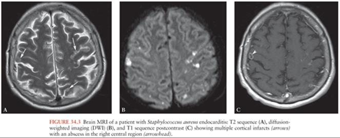
Asymptomatic lesions are frequently found among patients with IE, if MRI is systematically applied. For example, radiologic evidence of cerebral emboli can be shown among 65% to 80% of patients with IE, half of these lesions are asymptomatic (102,103). The clinical significance of asymptomatic emboli is unclear (160), and routine screening of patients with IE is not recommended. Asymptomatic microbleeds (i.e., cerebral microhemorrhages detected by T2 sequences) were identified among 57% of patients with IE in a French study (161). Given the strong association with IE in the case-control study (OR 6.12, CI 2.09 to 17.94), the authors suggest that this might be a new diagnostic criterion of IE. In a study of 26 patients with IE who underwent T2-weighted MRI, cerebral microbleeds were found in 14 patients (54%) and were strongly associated with subsequent ICH (162).
A French prospective study evaluated the impact of cerebral MRI on the management of IE. Among 130 patients, cerebral lesions were found in 82% and included ischemic lesions (52%), microbleeds (58%), and asymptomatic aneurysm (8%) (163). The systematic use of imaging modified the diagnostic or therapeutic management in 28% of patients. However, the authors did not study the impact of imaging on clinical outcome.
Cerebrospinal Fluid Examination
Despite similar rates of neurologic complications, the percentage of patients undergoing a lumbar puncture (LP) varied from 32% to 82% in four large series (83,88,92,145), with the most recent studies reporting the lowest rates. This is probably related to the recent availability of CT scan, which obviates the necessity for invasive procedures in many patients. Hence, there has been a decreasing number of LPs performed to investigate focal deficits in more recent studies (97).
The largest and most detailed study on CSF parameters in patients with neurologic complications of IE is from the Massachusetts General Hospital (83). CSF examination was performed in 69 (82%) of 84 patients. Neither the clinical setting nor any of the neurologic events were associated with a specific CSF formula, except for finding a purulent CSF more often in patients with meningeal signs. However, there was a good correlation between the CSF findings and the nature of the infecting microorganisms in that virulent bacteria such as S. aureus, enteric gram-negative bacilli, and S. pneumoniae were frequently associated with purulent CSF, whereas relatively avirulent bacteria such as viridans streptococci were usually associated with a normal or aseptic CSF. Positive culture of the CSF was found in only 11 of these 69 patients, and these were always associated with neutrophils in the CSF. There were eight cases of S. aureus and one case each of S. pneumoniae, Proteus mirabilis, and viridans streptococcal meningitis. The findings of this series are similar to those of other studies (88,92,143). Occasionally, a CSF analysis with an aseptic pattern will yield a pathogen such as a viridans streptococcus (164).
Beside diagnosis of meningitis, LP has little role in the diagnostic workup of IE. The clinical features and careful interpretation of the CT scan or MRI studies are better guides to patient management than the CSF findings alone (165).
Outcome
The overall mortality of patients with IE has decreased over the past 30 years, probably due to improved management (e.g., more widespread use of echocardiography, access to surgery, interdisciplinary care) (166). However, the mortality of patients with IE and neurologic complications has not changed appreciably, ranging from 34% to 74% (29,83,95,101), although some studies report rates as low as 20% (88,97). A mortality rate of 58% has been reported in patients with neurologic complications IE referred to the ICU (106). CNS complications were associated with higher mortality in several studies (24,83,92,101,145). For example, stroke was independently associated with in-hospital mortality (OR 3.67, CI 1.94 to 6.94) among patients with S. aureus endocarditis in the ICE-PCS (24). However, in a study of patients with IE admitted to the ICU, mortality was similar among patients with and without neurologic complications (106), whereas Glasgow Coma Scale (GCS) score less than 10 was a strong predictor of mortality. This suggests that severity of neurologic complications might be a more appropriate predictor of mortality than the occurrence of CNS complications per se, as confirmed by a study of 513 patients with complicated IE, where abnormal mental status at baseline was independently associated with 6-month mortality (9). Surgical treatment of IE does not appear to increase the CNS-related mortality of IE; among 55 patients with neurologic complications of IE, mortality did not differ between surgically and medically treated patients (97). In patients with PVE, the overall mortality was affected by the presence of neurologic complications in some studies (46,70) but not in others (167).
When only major cerebral events are considered, they are found to be the direct cause and often the only cause of death in more than 50% of fatal cases, both in NVE and in PVE. In many cases, the cause of death is multifactorial. Neurologic complications were found to be directly responsible for 8% to 20% of the deaths in patients with NVE (29,83) and for 10% to 40% in those with PVE (117). In a recent series of 55 cases, the mortality among patients with neurologic complications was not different between those with NVE and PVE (23% and 25%), and there was no difference in mortality between the episodes caused by various microorganisms (97).
The type of microorganism appears to play an important role in the outcome of patients with IE and neurologic complications, with S. aureus, Enterobacteriaceae, and fungi being associated with a higher mortality (83,88). Obviously, some neurologic complications such as major emboli, ICH, or purulent meningitis are associated with higher mortality rates than the less severe manifestations. Sequelae have been noted in up to 34% of patients with neurologic complications who survived (87).
Cerebral Emboli
Incidence
Emboli arising from the heart are responsible for 30% of strokes occurring in the general population (168–170). Emboli secondary to IE account for fewer than 1% of these episodes (171). The rate of CNS embolism due to IE is higher in autopsy series; for example, IE was found in 69 (1.5%) of 4,558 autopsies but was present in 32 (25%) of the 126 patients with cardiogenic cerebral emboli (104).
Occlusion of cerebral arteries by emboli is the most common neurologic complication of IE, comprising approximately half of the CNS complications (Table 34.3). Among 2,781 patients of the ICE-PCS, stroke complicated IE in 17% of cases (16). Lower incidence (9.6%) was reported in a recent study that used more stringent diagnostic criteria for stroke and the Duke criteria for IE (114). Stroke was present in up to 40% of patients with IE admitted to the ICU (106).
Approximately half the patients who present with major or minor cerebral embolism have clinically identifiable emboli to other organs as well (83). In contrast, evidence of systemic emboli was found in only 2% of unselected patients with stroke (172). In patients with IE and systemic emboli, cerebral emboli were found in 40% to 69% (83,154). It has been observed that the incidence of cerebral emboli associated with IE of the mitral valve was higher than when the aortic valve was involved despite a similar rate of peripheral emboli elsewhere (83,114,173). Except for the rare occurrence of paradoxical embolism, emboli are usually associated with lesions on the left side of the heart. In recent studies that compared NVE with PVE, no significant difference in the incidence of cerebral emboli was noted (46,88,97).
Pathogenesis
Most emboli related to IE are a result of dislodgment or disruption of cardiac vegetations into fragments, followed by lodgment of these fragments into peripheral vessels of various diameters, depending on size. Occasionally, emboli may be related to other concurrent disorders such as atrial arrhythmias. Vegetations are the result of complex interactions between various host components including serum, fibrin, platelets, fibroblasts, and inflammatory cells, as well as microbial factors including growth rate, adhesion, and production of extracellular proteases. These interactions influence the growth of the vegetation and the frequency of embolic episodes (174). Experimentally, the proteolytic capacity of the infecting organism was shown to influence the size of the vegetations and the course of the disease. In rabbits with IE induced by one of ten different strains of Enterococcus faecalis, proteolytic strains caused smaller vegetations with a soft and friable appearance, as well as an increased frequency of renal emboli, when compared to nonproteolytic strains (175). In humans, IE due to virulent microorganisms, particularly S. aureus, is associated with an increased frequency of systemic and cerebral emboli as compared to less virulent bacteria such as viridans streptococci. In a study of 52 patients with PVE, the calculated rate of stroke during uncontrolled infection ranged from 1% per day for nonvirulent streptococci to 9% per day for S. aureus (121). Pathologic examination provides some explanation for this difference: The vegetations of IE due to virulent bacteria are friable with little histologic evidence of healing, whereas in subacute disease, lesions progress more slowly with evidence of fibrotic reaction and early healing (176). In addition, certain microorganisms have been noted to produce large and mobile vegetations and are associated with an increased propensity to be complicated by major systemic and cerebral embolism. This has been described in IE due to microorganisms such as Haemophilus species or other slow-growing fastidious gram-negative rods, Abiotrophia, group B β-hemolytic streptococci, and fungi (Candida and Aspergillus species) (177,178). Occlusion of vessels by fragments of vegetations results in various degrees of ischemia and infarction depending on the vessels involved and the collateral blood flow. In addition, emboli occurring before the initiation or completion of successful antibiotic treatment may contain microorganisms capable of producing secondary infectious complications, such as abscesses of various sizes, meningitis, arteritis, or infectious aneurysms. In about 20% of the cerebrovascular episodes, hemorrhagic complications are observed (145). They may be due to the infarction itself or the erosion of the artery by the bacteria present in the emboli, with or without formation of a detectable infectious aneurysm (138). Concomitant anticoagulation appears to increase the risk of developing major hemorrhages at the sites of infarction (83,92,127,138). However, in patients with PVE, withholding anticoagulation has resulted in high rates of thromboembolism, and adequate anticoagulation has been shown to reduce the incidence of major CNS events (116).
As previously mentioned, macroscopic and microscopic cerebral emboli have been observed with greater frequency in patients with mitral valve infection as opposed to those with aortic valve involvement in most studies (55,83,106,179). Pathologic examination of operative or autopsy material shows that vegetations are not always found in patients who had an embolic episode; of 76 valves examined, a valvular vegetation was found in only 57% of patients with neurologic complications. Moreover, vegetations were found in 61% of the patients without CNS complications (88).
Clinical Presentation
Cerebral embolism associated with IE may present with protean clinical manifestations. Important determinants of the symptomatology are the number, location, and size of the emboli. Some patients may present with embolic occlusion of a single major cerebral artery, with symptomatology related to the territory involved. Other patients may have multiple microemboli with more diverse clinical presentations. In many patients, these two forms of emboli coexist (83,101).
By combining CT and clinical criteria, Hart et al. (154) found that 62% of 37 ischemic events involved the cortex or the cerebellar hemispheres, 16% were exclusively subcortical, 11% involved the retina, and 11% were in an uncertain location. Most infarcts were either small (58%) or moderately sized (33%). All three large hemispheric infarcts occurred in patients with S. aureus endocarditis (154).
Major cerebral embolism was the presenting symptom or first overt manifestation of IE in 22 (10%) of 218 patients (83) and in 12 (14%) of 86 patients (113) in two large series. The majority of the cerebral emboli occur before the initiation of antibiotic treatment or during the first 2 weeks of therapy (55,83,97,154,173,179–181). Among 109 episodes of stroke observed in 496 patients with IE, 31 (28%) occurred after the initiation of antibiotic treatment; the median time interval from onset of antibiotic treatment to the development of neurologic complications was 4 days, with a range of 1 to 21 days (179). In another study, 87% of ischemic events occurred either at the time of diagnosis (74%) or within 48 hours thereafter (13%) (154). Among the 25 initial ischemic events, 56% were simple and unifocal, 28% were multifocal, and 16% were associated with toxic encephalopathy. Embolization after the completion of successful antibiotic treatment is uncommon (55), but it has been described and can occur up to 2 years later (83). Recurrences of emboli events are rare. In one study of 55 neurologic events among 218 patients with IE, only one recurrent episode of cerebral embolism was reported (97). In another series of 64 patients with neurologic complications of IE, two recurrent embolic events occurred before initiation of antibiotic treatment (88).
More than 90% of large cerebral emboli affect the middle cerebral arteries and their branches, leading mainly to contralateral hemiparesis and/or hemisensory deficits. This localization may also produce parietal lobe signs including sensory loss, neglect, dyspraxia, hemianopia, and when the dominant hemisphere is involved, aphasia. Occlusion of anterior or posterior cerebral arteries may also produce similar symptoms, especially those involving the lower extremities. Posterior cerebral artery occlusion may produce homonymous hemianopia. The vertebrobasilar system was affected in 6 of 10 and 4 of 84 patients with CNS complications of IE in two series (83,143). Emboli affecting the vascular supply of the spinal cord or peripheral nerves may also occur (182).
TIAs are occasionally noted, sometimes as the presenting manifestation of IE. Such episodes were recorded in 15 (27%) of 55 patients with strokes associated with IE (143). Among 25 episodes of ischemic strokes reported in a study by Hart et al. (139), 3 were TIAs, all causing amaurosis fugax. In a patient with a TIA, the presence of fever or any other nonspecific signs of infection should raise the possibility of IE. Some of these patients may present with fluctuating neurologic signs, presumably from emboli that disintegrate after initial lodgment; autopsy may reveal multiple small or microscopic infarcts (137).
Multiple cerebral microemboli are common, causing about one third of all ischemic events (154). Of 45 patients autopsied because of a fatal neurologic complication, 38 had major embolism, but 23 were found to also have microscopic cerebral infarcts, presumably due to embolic occlusion of small vessels. Six also had microabscesses. These findings correlated with seizures or fluctuating neurologic signs in four patients and were clinically silent in four others. In 14 patients, multiple microscopic infarcts manifested as an altered level of consciousness not adequately explained by other abnormalities (83). Thus, encephalopathy among patients with IE, may partly be explained by multiple microemboli, resulting in multifocal microinfarcts, sometimes associated with cerebritis or microabscesses (145). Microabscesses were found in 6 of 23 patients with multiple microinfarcts at autopsy (83), demonstrating that a continuum exists between the ischemic cerebrovascular lesions and overtly infected CNS lesions complicating IE (145).
In summary, emboli may cause a wide variety of CNS symptoms and signs, ranging from hemiplegia to diffuse and fluctuating neurologic dysfunction, depending on their size, location, and number. Most emboli affect the middle cerebral artery and occur before or soon after the initiation of therapy.
Diagnostic Procedures
Cerebral CT scan and MRI are the most useful diagnostic procedures when emboli are suspected. They are helpful both in the differential diagnosis and in the distinction between nonhemorrhagic and hemorrhagic infarcts (Fig. 34.4). When acute hemorrhage is suspected, CT scan is preferred. MRI and MRA also provide a baseline in monitoring for the development of ICIA, which may arise at the site of a previous embolus. Follow-up imaging studies are also indicated when the secondary formation of an abscess is suspected.

The effects of the performance of cerebral angiography on survival were investigated and the authors concluded that it should not be routinely performed (183). A study comparing CT scan and angiography suggests that ICIA is unlikely to be present when the CT scan is normal (184). Because MRA is noninvasive, repeated studies with this technique would be more feasible than traditional angiography. The relatively new CT technique called CT angiography (CTA) allows the assessment of cerebral aneurysms of the circle of Willis with accuracy comparable to MRA (185). To our knowledge, there are no CTA studies focusing on ICIA. However, according to the available data, CTA could be a valuable diagnostic option and can be easily obtained at the same time as conventional CT.
As discussed earlier, LP is seldom helpful when IE is already suspected. A CSF leukocytosis may be the first hint suggesting the possibility of IE in certain patients with stroke, particularly if predisposing factors for endocarditis are present. However, this benefit is not sufficient to justify routine CSF examination in unselected patients with stroke because the yield might not be greater than 0.25% (186). Moreover, normal CSF values do not exclude CNS emboli originating from IE.
To exclude hemorrhagic transformation of a cerebral infarct, high-quality CT is a better procedure than CSF examination. Some patients with such a complication on CT may have normal CSF or CSF containing only a few red blood cells (RBCs). Furthermore, a few RBCs are often present in the CSF in nonhemorrhagic infarcts due to traumatic taps.
Echocardiography has a pivotal role for the diagnosis of IE in patients presenting with stroke. Its value in assessing the degree of valvular change and the status of left ventricular function is well established (60). The role of this technique in predicting the risk of embolization is more controversial. Some reports suggest that in patients with IE, vegetations detected by precordial echocardiography are associated with a higher risk of embolization (187,188). However, no significant difference in the overall incidence of embolism was found in a study of 77 patients, in which chart compilation and echocardiography readings were performed by separate investigators who had no knowledge of each other’s findings (189). Therefore, in some patients presenting with neurologic complications, it is possible that vegetations were not seen because embolization had already occurred before echocardiography, a well-documented possibility. Steckelberg et al. (55) found that detection of vegetations on two-dimensional echocardiography was not associated with a significantly higher risk for embolus, with the exception of patients with viridans streptococcal infection.
The vegetation size is a better predictor of systemic embolism (173,179,190–192). Thuny et al. (192) performed multivariate analysis of a study including 384 patients with IE and identified vegetation size larger than 10 mm, as well as vegetation mobility and infection by S. aureus and S. bovis as independent predictors of new embolic events after initiation of antibiotic therapy. In a study that employed both TTE and TEE, patients with large vegetations (>10 mm) had a significantly higher incidence of embolic events (47%) than those with small or no vegetations (19%) (193). Because an embolic event may reduce the size of vegetations, the analysis was repeated after exclusion of patients who had echocardiography after an embolic episode; the rate of new embolism was 36% in patients with large vegetations compared to 6% in those with small vegetations, a difference that was also significant. Patients with large vegetations of the mitral valve were at a significantly higher risk of embolism (193). Anterior as compared to posterior mitral leaflet vegetation was associated with an increased risk of embolization (194).
In conclusion, most studies suggest that vegetations seen on echocardiography, especially those larger than 10 mm, are associated with a higher risk of embolism. However, many patients with vegetations even of large size do not manifest clinically detectable emboli, and vegetations may persist on echocardiography after successful medical therapy.
Specific Management
Medical Treatment
For management of patients with IE and cerebral emboli, several aspects should be considered, namely, the primary prevention of embolization, the prevention of subsequent episodes, the optimal therapeutic strategy for already established lesions, and the detection of secondary complications (Fig. 34.5).
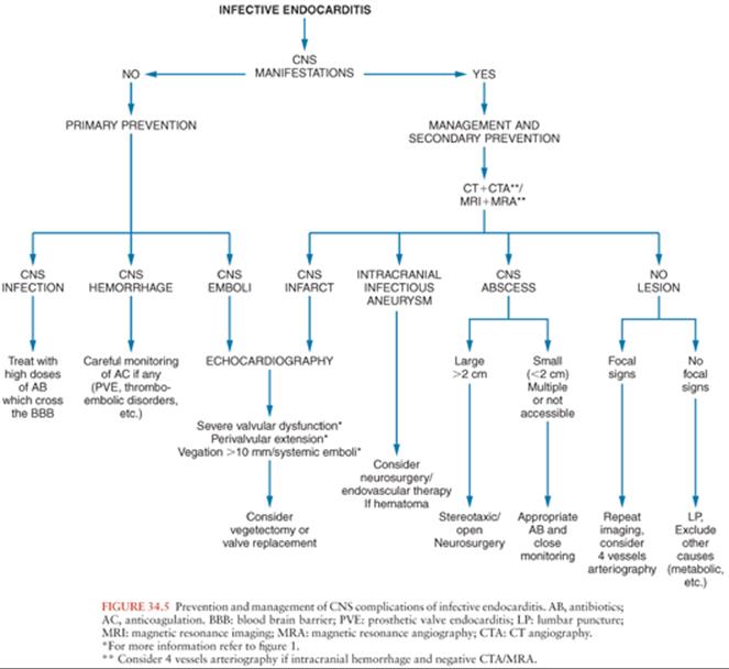
Efforts should be directed at adequate control of arterial blood pressure and provision of supportive care. Patients should be carefully followed clinically for the occurrence of secondary hemorrhages and repeated CT should be performed promptly if the patient’s condition deteriorates.
Prompt initiation of antibiotic treatment has a tremendous impact on the incidence of stroke in IE. In an analysis of the ICE-PCS including 1,437 patients, rate of stroke decreased from 4.82/1,000 patients-days during the first week of antibiotic treatment to 1.71/1,000 patients-days during the second week (195).
There is no specific antithrombotic therapy for embolic strokes in the patient with IE. This differs from noninfectious embolic cerebral infarction, which can be treated by thrombolysis in selected cases (196,197). Thrombolysis has been attempted anecdotally (198), sometimes because IE was not suspected at stroke diagnosis (199), but it is unlikely to be of equal value in the patient with IE given the different pathogenesis of vessel occlusion and the relatively high risk of ICH complication of this procedure in an infectious setting (200).
The proper use of anticoagulants has given rise to much controversy. Soon after the advent of sulfonamides in the 1930s and antibiotics in the 1940s, simultaneous treatment with heparin was tried with the idea that prevention of further deposition of platelet-fibrin thrombi on infected valves might favor antibiotic penetration and possibly prevent further emboli (201). It soon became apparent that antibiotics alone were able to cure IE and that there was no evidence that anticoagulants were a useful adjunct to antibiotic therapy (201). Moreover, cerebral hemorrhages were observed with a distressing frequency in anticoagulated patients and major emboli were not prevented (201). The antibiotic therapy of that time may have been inferior to currently recommended regimens. In the series of Pruitt et al. (83), seven patients with cerebral emboli were subjected to anticoagulation before or within 24 hours of embolization. Three of them (43%) developed major ICHs at the sites of infarction. In contrast, ICH was observed in only 10 (4.7%) of 211 patients not treated with anticoagulants (83). In another older study, 30 patients (29 with prosthetic valves) who were initially anticoagulated were analyzed. Among the 20 whose anticoagulation was continued, 7 developed a stroke, 6 being bland infarcts, and 1 a hematoma; anticoagulation was continued in 4 of them without further complications. In contrast, only one stroke was observed in the ten patients in whom anticoagulation was stopped (88). This deleterious effect of anticoagulants is also supported by experimental evidence. In an animal model of septic CNS embolism, anticoagulation was associated with an increased risk of hemorrhage (202). In experimental endocarditis, animals simultaneously treated with antibiotics and anticoagulants had a worse outcome than those who received antibiotics only (203). Therefore, routine use of anticoagulants is generally not recommended in the course of NVE because clinical and experimental data indicate that the rate of hemorrhagic CNS complications is high and because there is no proven benefit with regard to the course of the disease (10). However, in a Swedish study of 587 NVE episodes, cerebrovascular complications were significantly less frequent among patients on warfarin (6% vs. 26%), and the risk of hemorrhage was not increased among these patients (204). Among 332 patients with endocarditis in the series of Delahaye et al. (124), cerebrovascular accidents were significantly more common among anticoagulated patients (19/94 vs. 19/238), but the mortality rate in patients who had a cerebrovascular event was similar (124). This indicates that, although NVE is not an indication to start anticoagulation, antithrombotic treatment should not be withheld when there is an established previous indication (e.g., mitral valve disease and atrial fibrillation) and that anticoagulation with heparin should be maintained whenever a brain infarct is present unless it is large and/or hemorrhagic (124,205).
The issue of anticoagulant therapy in patients with PVE should be considered separately. With the exception of those patients with bioprostheses and in normal sinus rhythm, patients with prosthetic valves are at constant risk of thromboembolism, an important reason not to interrupt anticoagulant therapy. Patients who develop PVE are at high risk of presenting both thromboembolic and hemorrhagic complications and the indication for the maintenance or interruption of the anticoagulation therapy should be carefully balanced. In patients with PVE who were not anticoagulated, the incidence of CNS thromboembolic phenomena has ranged from approximately 50% to 71% (116). An incidence of CNS hemorrhage as high as 36% has been reported in a series of patients with PVE treated with anticoagulants (117). In the study of Wilson et al. (116) comparing patients with PVE receiving or not receiving anticoagulants, major CNS complications occurred in 3 (8%) of 38 patients who received adequate anticoagulant therapy as compared to 10 (71%) of 14 of those who received either inadequate or no anticoagulation. Mortality was 47% among the patients with adequate anticoagulation, not significantly different from 57% among those without. Among the patients treated adequately with anticoagulants, three deaths occurred; all were due to the CNS injury, but only one was attributed directly to the anticoagulant therapy. Among the patients without adequate anticoagulation, eight deaths were recorded, and CNS complications were thought to be the primary cause of death in five of them. Three of these patients were found to have massive intracerebral bleeding caused by thromboembolism (116). A recent Spanish study highlights the higher hemorrhagic risk associated with anticoagulation among patients with PVE due to S. aureus(127). In that study, six episodes of cerebral hemorrhage occurred in 21 patients with S. aureus PVE (90% of whom where anticoagulated), as compared to only one episode in 35 patients with S. aureus NVE.
Based on these varying data, most authors suggest that closely monitored anticoagulant therapy should be cautiously continued in patients with IE involving mechanical prosthetic valves (19,116,120). Although coumarin drugs are generally used, favorable experience with heparin has been reported (119). If the patient develops a CNS complication, the use of anticoagulants should be temporarily discontinued; in addition, the patient should be evaluated for evidence of ICH. If there is no evidence of hemorrhage or hemorrhagic infarct, carefully controlled anticoagulation with heparin may be reinstituted (10,116).
Because of the critical role played by the platelet-fibrin thrombus in the formation of the vegetations (206), antiplatelet agents may have a role in preventing subsequent embolization, as suggested by experimental models (207). In a randomized trial of aspirin at a dose of 325 mg/day, which included 115 patients with IE, patients treated with aspirin had similar rate of embolic events as compared to patients on placebo (208). By contrast, in a retrospective cohort study of 600 patients with IE, incidence of systemic embolism was significantly reduced among patients treated with antiplatelet therapy before diagnosis of IE (OR 0.36, CI 0.19 to 0.68), suggesting that chronic antiplatelet therapy during the early phase of IE may have an impact on the risk of embolism (209). However, another recent study was not able to confirm the positive effect of previous antiplatelet therapy on cerebrovascular events in patients with IE (210). Taken together, these data do not support the routine use of antiplatelet agents in IE.
Cardiac Surgery
The role of cardiac surgery in the prevention of embolism in IE is ill defined, although current recommendations mention systemic embolism, if vegetations have been visualized by echocardiography, as an indication for valve replacement (10,62,65).
Thus, many patients with large vegetations may actually be operated on for a combination of indications. The natural course of patients with large vegetations is difficult to assess because patients may undergo surgery before a potential embolic event. Further prospective data are required before management decisions can be based solely on the presence of echocardiographically detectable vegetations or on their dimensions. Given the operative risk and the postoperative complications of prosthetic valves, it seems reasonable not to operate on a patient solely because of the presence of a large vegetation seen on echocardiogram. This point of view might change with the advent of homografts and conservative surgery for valve repair, which carries less long-term risk than prosthetic valves.
The need for valve replacement in patients who have sustained a single embolic event and/or who have large vegetation demonstrated by echocardiography remains controversial. Embolic stroke is uncommon after the first 2 weeks of antibiotic therapy (97,101,179,195,199) and recurrent emboli are uncommon in patients who survive a first embolic episode and who are receiving appropriate antibiotics (83,88,179). When late embolic events after a cure of an episode of NVE are reported, they seem to be best explained by factors other than the past episode of IE (83). Thus, if surgery is considered solely for prevention of systemic embolism, it should be performed during the first days of antibiotic therapy. The impact on early surgery on the incidence of systemic emboli was highlighted by a recent randomized study from Korea (67). Seventy-six patients with NVE, severe mitral or aortic valve disease, and a vegetation larger than 10 mm were randomized to early surgery within 48 hours after randomization or surgery according to current guidelines. No embolic event occurred among patients assigned to early surgery, as compared to eight patients (21%) assigned to conventional treatment (67). However, among operated patients, rate of valve replacement with a mechanical valve was higher in the early-surgery group. Thus, early surgery seems to prevent systemic embolism (including stroke), but at the price of long-term anticoagulation (211).
Patients undergoing open heart surgery after a recent CNS complication pose a difficult management problem. There is always a risk that cardiopulmonary bypass and heparinization may exacerbate the recent neurologic injury by promoting hemorrhagic transformation (91,212). On the other hand, delay in surgery can have serious hemodynamic and CNS consequences, depending on the circumstances: Worsening neurologic symptoms can result from hypotension, hypoxemia, low cardiac output, and renal failure. Moreover, further embolization may occur, especially with microorganisms such as S. aureus, gram-negative bacilli, or fungi. Recent data indicate that the risk of surgery among patients with stroke is lower than previously estimated (Table 34.4). In one study, 65 patients with IE who underwent open heart surgery a few days following embolic stroke or TIA were analyzed (199). No significant difference in perioperative and long-term mortality was found between patients with and without preoperative stroke. However, patients with stroke associated with other neurologic complications (i.e., meningitis, abscess, or hemorrhage) had higher perioperative mortality as compared to patients with noncomplicated stroke (39% vs. 8%). Seventy percent of survivors had a full neurologic recovery (199). In another study of 109 patients with IE complicated by cerebrovascular events, valvular surgery was performed in 58% of cases early after the event (179). Postoperative neurologic deterioration was observed in only four patients (6%). Moreover, operated patients had a higher survival than patients treated conservatively (179). A large series of 198 patients undergoing cardiac surgery for IE after stroke was published by the ICE-PCS group (213). This study confirmed that early surgery (i.e., <7 days) was not associated with higher in-hospital mortality.
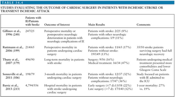
Thus, when there is an established indication for valvular surgery, the presence of an ischemic stroke does not appear to justify postponement of the procedure. However, because anticoagulation may precipitate hemorrhagic transformation of cerebral infarcts if administered during the first few hours after the episode (214), it would appear reasonable to postpone the operation for a few days if feasible. In a study of 33 patients with CNS complications of IE undergoing surgery, 2 developed fatal neurologic deterioration when surgery was performed within 5 days after the cerebral embolic event (91).
Outcome
Stroke increases the mortality of IE. For example, in the study by Thuny et al. (179), stroke was an independent predictor of death with a hazard ratio of 1.6. In another study of 68 patients with IE complicated by stroke, in-hospital mortality and 1-year mortality were 35% and 52%, respectively (114). Stroke was also associated with higher in-hospital mortality among patients with PVE (46) and S. aureus IE (24) in the ICE-PCS.
Mortality is particularly high in the case of hemorrhagic transformation, because it may occur in patients who are anticoagulated at the time of the embolic episode (83,88,116). In one study, middle cerebral artery stroke (both partial and complete) was associated with poorer neurologic recovery (199).
In conclusion, cerebral emboli are one of the major complications of IE. Emboli may cause further intracranial complications such as meningitis, abscesses, hemorrhages, and infectious aneurysms. To detect these secondary complications, CT scan and possibly angiography should be performed at appropriate intervals after an embolic episode. For these complications to be detected, investigations are best carried out 7 to 10 days after the embolic event. Although large vegetations seen on echocardiography seem to be associated with an increased risk of emboli, the precise role of valvular surgery for this indication is not yet determined, although recent data may indicate a benefit of early surgery for the prevention of systemic embolism. In patients with PVE, anticoagulation should not be stopped but may be interrupted briefly and, in any case, must be carefully monitored.
Intracranial Hemorrhage
Epidemiology
ICH occurs in 2% to 14% of cases of IE (Table 34.3) and represents 7% to 25% of neurologic complications. In selected populations of patients with IE, such as those hospitalized in an ICU, the incidence is higher. For example, it was 27% in the study by Sonneville et al. (106). The hemorrhage usually diffuses into the cerebral substance or into the subarachnoid space. Rarely, the subdural space may also be involved.
ICH complicating IE may be the result of different mechanisms. ICH have often been attributed to ruptured ICIA even when no aneurysms were demonstrable (83). In addition to ruptured ICIA, it has recently been recognized that ICH in IE can also result from the septic erosion of the arterial wall without a well-delineated aneurysm (138). Moreover, hemorrhagic transformation of ischemic brain infarcts can also result in ICH, particularly in anticoagulated patients (83,127,214). One study reported a 29% rate of ICH among patients with IE due to S. aureus and anticoagulated because of a prosthetic valve (83).
The proportion of ICH attributable to each of these mechanisms varies among studies. Of the 17 patients with ICH studied by Hart et al. (138), 4 presented a hemorrhagic infarct and 13 had a primary hemorrhage that could be attributed to a ruptured ICIA in only 2 patients and to a necrotic arteritis in 4 cases. In five other patients, an ICIA was excluded by arteriography or autopsy, and the exact cause of ICH could not be determined. In four other studies, the proportion of ICH due to ICIA ranged from 11% to 50% (83,97,106,113). Thus, less than one half of ICHs in IE are attributable to ruptured ICIA. Septic necrosis of the arterial wall and hemorrhagic transformation of cerebral infarcts each accounts for the other cases (83,138). Whatever mechanism is involved in the pathogenesis of ICH, the prognosis is poor with a mortality of 35% to 87% (113,138).
Intracranial Hemorrhage Due to Cerebral Emboli
Aseptic cardioembolic strokes may undergo spontaneous hemorrhagic transformation even in the absence of anticoagulation; pathologic examination reveals that this occurs to some degree in the majority of the episodes. This results from multifocal extravasations of confluent petechiae and is usually not associated with recognized clinical worsening. About 5% of the patients with embolic stroke who are not receiving anticoagulants have hemorrhagic infarcts visible on initial early CT scan with an additional 10% to 20% developing late, usually asymptomatic, hemorrhagic transformation (214). Hemorrhagic transformation of ischemic infarcts caused by septic emboli appears to be the most common mechanism leading to lethal intracerebral hemorrhage in patients with IE (139). The likelihood of developing spontaneous hemorrhagic transformation appears to be directly related to infarct size (215). Although exact figures are not available, hemorrhagic transformation occurred in 17% of cerebral infarcts associated with NVE in one study (114) and in 8% to 36% of those with PVE (120).
Intracranial Hemorrhage Due to Acute Necrotizing Arteritis or Intracranial Infectious Aneurysm
The spectrum of arterial injury leading to ICH can range from acute, pyogenic necrosis to large, aseptic aneurysms that may rupture weeks to months after bacteriologic cure. The extremes of this continuum appear to represent different clinical syndromes, although the term infectious (mycotic) aneurysm has usually been applied to both processes (138). For the purpose of clarity, true ICIAs are considered separately in this chapter (see later discussion).
Septic emboli appear to be necessary for the occurrence of septic arteritis and ICH, although clinically recognized embolism precedes ICH in only a fraction of the patients. Sustained bacteremia in tricuspid valve IE, even with virulent bacteria, does not result in ICH, indicating an important role for embolic fragments in pathogenesis (138).
Most ICHs due to acute erosive arteritis tend to occur early in the course of the disease and are often already present on admission. They are more likely to occur in S. aureus IE (113,138), although this microorganism can also be associated with the development of true ICIA at a later stage (83,139). Most of these patients have an acute form of IE of only a few days’ duration and present with severe and rapidly evolving neurologic signs that are related to the site of the intracerebral or subarachnoid hemorrhage (138). ICH may be the presenting manifestation of IE (138). Some patients have a preceding episode of clinical embolus or TIA (137,138). For example, 4 of 12 patients with IE and TIA subsequently sustained a fatal ICH (137). The precise sequence of events leading to ICH was demonstrated in a patient who died from a massive subarachnoid hemorrhage secondary to an erosive arteritis that developed at the site of a septic embolus visualized both at autopsy and on an arteriogram performed a few days before death (216).
Imaging by CT scan, CTA, MRI, and MRA (>24 hours after the acute event) plays a central role in the evaluation and management of these patients (Figs. 34.6 and 34.7). Conventional arteriography is still the technique of choice in cases of hemorrhage, because it allows the precise location of the arterial rupture and the visualization of associated ICIA and of the anatomy of the intracranial vessels. This may be particularly helpful in patients for whom surgery is considered. Surgical treatment is difficult, requiring sacrifice of the involved artery, sometimes with microvascular pedicle/bypass surgery, because there is not a well-delineated aneurysmal neck that can be readily clipped (138).


Intracranial Infectious Aneurysm
The term infectious (mycotic) aneurysm refers to localized dilation of an arterial wall due to an infective arteritis. This can be caused by almost any microorganism, so the term does not indicate fungal infection. To obviate the confusion with aneurysms of true fungal etiology, some authors have recently suggested that all types of aneurysms due to an infectious agent be grouped under the title of “infectious aneurysms” (217).
Incidence
ICIAs are uncommon. Although they represented 12% to 32% of all intracranial aneurysms before the advent of antibiotics, they appear to constitute only 2.6% to 6.4% in more recent series. More than 80% of all ICIAs occur as a complication of IE (218). In a review of 85 cases of documented ICIA reported from 1954 to 1977, 72 (85%) were due to IE (218). In patients with IE, the incidence of recognized ICIA ranges from 2% to 5% (83,88,106). As it has been documented that some patients with IE develop intracranial aneurysms that remain asymptomatic and undergo healing under appropriate antibiotic therapy (157,219,220), the true incidence of infectious aneurysm certainly exceeds the number of cases diagnosed. This is also suggested by a study in which systematic CT scan and four-vessel angiography were performed in patients with endocarditis and neurologic manifestations: 11 (31%) of 35 were found to have ICIA (184).
Multiple aneurysms have been documented in about 25% of the patients with ICIA (157), but the true figure is unknown because only a few of the reported patients have had full angiography analysis to show all the intracranial vessels (220). In the study just mentioned, 5 of 11 patients had multiple ICIAs (184). In another series, 17 patients had a total of 28 ICIAs (219). Multiple ICIAs may not occur simultaneously (218). Although it is conceivable that ICIAs and infectious aneurysms located elsewhere may develop in the same patient, it was not found in the one study that specifically investigated this possibility (83). ICIAs can occur at any age. Most reported cases of ICIA linked to IE were observed in patients with native valve infection, but ICIAs also occur in those with PVE.
Pathogenesis
ICIAs may develop by different mechanisms. When they occur in the context of IE, they result from septic embolization to the vasa vasorum or to the intraluminal space of the vessel itself. Early observations of infectious aneurysms indicated that the inflammation affected the adventitia first and then spread inward. In experimental studies, ICIAs could be produced in dogs by the injection in the carotid arteries of silicone rubber emboli coated with bacteria (221). Pathology showed aneurysmal dilation of the portion of the vessel immediately adjacent to the emboli; microscopically, the vasa vasorum were packed with inflammatory cells and the inflammatory response started first on the adventitial surface, penetrated the muscularis layer, and in ruptured aneurysms, destroyed the internal elastic membrane and the intima (221). However, peripheral arteries, which are frequently involved in ICIAs, are devoid of vasa vasorum, in which case it seems that bacteria may reach the adventitia via direct penetration through the wall. Moreover, ICIAs sometimes develop at the site of a previously documented embolic occlusion. Thus, both microembolization of vasa vasorum and direct penetration through the wall of the arteries appear to be possible routes of infection resulting in the formation of infectious aneurysms. The arterial wall may rupture at various stages of dilation, depending on the severity of the necrosis, which in turn is presumably related to the virulence of the infectious agent. Restoration of the blood flow through the damaged segment by recanalization of the initial septic embolus may contribute to subsequent rupture by increasing the intravascular pressure. Interestingly, treatment with an antibiotic to which the bacteria were sensitive did not prevent the development of an aneurysm but did prevent early rupture in experimental dogs (221). This parallels observations in humans who develop ICIAs while on appropriate antibiotic therapy (220).
Infected emboli, not just circulating bacteria, appear to be necessary for the formation of ICIAs, as suggested by the rarity of this complication in association with bacteremias caused by right-sided IE. Besides septic emboli, other pathogenic mechanisms may sometimes play a role in the occurrence of ICIA. ICIAs may result from a thrombophlebitis of the cavernous sinus (218). Rarely, ICIAs may be of extravascular origin and may be secondary to penetrating head trauma, otitis media, or tuberculous, syphilitic, fungal, or purulent meningitis (218,222). Superinfection of congenital aneurysm during the course of IE or during transient bacteremia has also been reported. This usually involves the vessels of the circle of Willis in elderly individuals.
The microbiologic spectrum of ICIAs of intravascular origin reflects that of IE. Viridans streptococci as well as S. aureus account for the vast majority of cases responsible for 89% of those with positive blood cultures during the period 1954 to 1977, reviewed by Bohmfalk et al. (218). The rest of the cases are due to various microorganisms such as Corynebacterium species, P. aeruginosa, bacteria of the HACEK group, or other bacteria. A similar distribution was found in a series of 17 patients (219). Among ten narcotic addicts with P. aeruginosa endocarditis, two had cerebral infectious aneurysms. In some patients, no organisms can be recovered from the blood, as a result of antibiotic treatment. In earlier days, pneumococci were also found and syphilitic aneurysms accounted for 5% of all intracranial aneurysms before the advent of antibiotics. S. aureus appears to be the most common offender in ICIAs associated with thrombophlebitis of the cavernous sinus. ICIAs have been associated with bacterial meningitis due to S. pneumoniae, N. meningitidis, M. tuberculosis, and other less common causes of meningitis (223,224). True fungal ICIAs have been described as the result of either a concomitant IE or direct or hematogenous spread from sinusitis. Most of them were caused by Aspergillus species. Cases associated with Candida species have also been described (222).
Pathology
Infectious aneurysm complicating IE may occur in any artery and at any location. The proportion of infectious aneurysms that are located in the CNS ranges from 15% to 54%. After the aorta, the brain is a leading location (144).
ICIAs are usually small and may be saccular or fusiform. Most are peripheral to the first bifurcation of a major cerebral artery; they involve the middle cerebral artery and its branches in more than 75% of the patients (219). This contrasts with congenital aneurysms, which are usually located near the circle of Willis. Vessel branching points are often affected, probably because these sites favor the impaction of emboli (144). This has been clearly documented in some cases by serial angiography showing the formation of an ICIA at the site of a previous embolus.
Histologically, infectious aneurysms are characterized by destruction of the normal architecture of the arterial wall, focal areas of necrosis, and infiltration by inflammatory cells. Strictly speaking, infectious aneurysms are pseudoaneurysms, because they result from destruction of the muscular layer. In acute lesions, polymorphonuclear cells are predominant and microabscesses are not uncommon. Organisms may be seen or cultured from the lesions but in many cases cannot be found, having been already eliminated from the focus of active inflammation by host defenses or by antibiotics. In more chronic lesions, or when rupture has not occurred, polymorphonuclear cells are mixed with lymphocytes and plasma cells in fibroblastic granulation tissue. The fibrotic reaction may contribute to the resistance of the wall and prevent rupture. Adhesions may form between the arachnoid and the brain in the region of the ICIA. With rupture of the aneurysm, these adhesions may prevent free escape of the blood into the subarachnoid space. Bleeding, therefore, tends to occur into the brain substance or into the subdural space.
Clinical Presentation
Most ICIAs occur in patients with IE. In many reported cases, ICIA is described as presenting with a sudden, often fatal subarachnoid or intracerebral hemorrhage without recognized warning signs (218). Among 58 patients with ICIA reviewed by Bohmfalk et al. (218), 33 already presented with major neurologic manifestations on admission, which were due to a subarachnoid hemorrhage in 19 cases.
Some authors have called attention to the fact that neurologic warning signs are often present before rupture occurs. For example, the presence of severe, localized headache in a patient with IE should raise the suspicion of an ICIA. Among 213 patients with IE seen at the Mayo Clinic from 1975 to 1979, 7 complained of severe localized headache and 4 had a proven ICIA; the other 3 also had an ICH, but ICIA could not be demonstrated (144). Focal neurologic events, such as seizures, ischemic deficits, or cranial nerve abnormalities, may often precede the development and the rupture of ICIA and should be regarded as serious warning signs, prompting further investigation (218,225). In a retrospective study of 25 patients with ICIAs, 40% of whom were associated with IE, headache was the most common symptom at admission, (86%) followed by fever (67%), vomiting (50%), ocular palsy (25%), and seizures (21%) (226). In 16 patients with ruptured ICIA reported by Pruitt et al. (83), 8 had a history suggesting embolization prior to the hemorrhage. At angiography, an occluded vessel was often found in association with the aneurysm. Among 81 cases of ICIA reviewed by Ojemann and Crowell (217), 65 were reported in enough detail to determine the initial neurologic event that led to angiography. This was a definite or probable hemorrhage in 42, an infarction in 16, an infarction followed by a hemorrhage in 5, and headache without hemorrhage in 2. The occurrence of a TIA may also precede subsequent rupture of an ICIA (137,225).
Although premonitory signs or symptoms may precede a catastrophic hemorrhage only by a few hours, the delay is generally of several days. Some aneurysms may leak slowly before rupture and produce a mild meningeal irritation; the CSF is sterile but shows an initial neutrophilic reaction and moderate numbers of RBCs (144,219). Overall, hemorrhage was documented by LP, surgery, or autopsies in 65% of the cases of ICIA gathered from the literature by Bohmfalk et al. (218). In approximately half of the cases, hemorrhage occurred before hospitalization. When rupture of ICIA occurs, it almost always causes either a subarachnoid or an intracerebral hemorrhage (218), but subdural hematomas have also been described. In some cases, ICIA may present as a space-occupying lesion or may be accompanied by a cerebral abscess (219). Finally and importantly, it has been documented that ICIAs may remain totally asymptomatic and resolve with antibiotic therapy alone (220,227).
ICIAs may become symptomatic or may even develop after appropriate antibiotic therapy for IE has been initiated (144,219) or completed (228). Delays from several months up to 2 years have been reported (138), and a high level of suspicion should be maintained in patients with a history of IE. Cases of ruptured ICIA during the postoperative period of cardiac valve replacement for IE have been reported and full systemic anticoagulation may constitute an aggravating factor (78).
Diagnosis
Because some ICIAs may benefit from surgical treatment, it is of utmost importance to maintain a high level of suspicion for ICIA in patients with active or treated IE who develop neurologic manifestations and to initiate appropriate diagnostic procedures.
Diagnosis of ICIA may be difficult to establish. According to a retrospective study of 25 patients, characteristics strongly associated with ICIAs were presence of a predisposing infection, specific radiologic features (e.g., multiplicity, distal location, fusiform shape), and other features such as younger age and fever (229).
MRI combined with MRA constitutes a major advance in the evaluation of cerebral aneurysms, especially when bleeding has not occurred. It is likely that the favorable diagnostic experience accumulated with noninfectious aneurysms will also apply to ICIA. This technique had a sensitivity of 100% and a specificity of 94% for intracranial aneurysms greater than 2 mm in diameter (230). In another prospective study, 5 mm appeared to be the critical size for detection of aneurysm with MRA (231).
CT scan with and without contrast enhancement is still the diagnostic procedure of choice when acute intracerebral or subarachnoid hemorrhage is suspected. In a systematic study comparing CT scan and conventional angiography in 34 patients with IE and neurologic manifestations, no infectious aneurysm was detected in the 14 patients with normal CT scans, whereas 11 cases with one or more ICIAs were diagnosed at angiography in the 20 patients who had abnormal CT scans (184). This suggests that ICIA is unlikely to be present when the CT scan is normal. Helical CTA is a noninvasive volumetric imaging technique. A recent prospective study concluded that CTA and MRA were of equal value for the detection of noninfectious intracranial aneurysms (185). In this study, the sensitivity of both techniques was excellent for aneurysms larger than 5 mm but decreased for smaller aneurysms, the latter being detected with higher sensitivity by regular angiography.
Digital substraction angiography remains the radiologic gold standard for the diagnosis of ICIAs. If hemorrhage is confirmed, and intervention is contemplated, it is the method of choice to pinpoint the location of the aneurysm and its relationship to the parent vessel (232). Because multiple aneurysms are common, examination of all four vessels is recommended.
To summarize, patients with focal CNS signs or with localized or severe headache should have an MRI and/or a CT scan, with contrast enhancement. CT scan is preferred if acute hemorrhage is suspected and can be combined with CTA. When patients with proven or suspected IE are referred to CT or MRI, CTA or MRA should be part of the examination, covering as much as possible the distal branches of the sylvian artery where most of the ICIA are located. In the presence of hematoma or subarachnoid hemorrhage and negative MRA or CTA, four-vessel conventional angiography is still the method of choice (184).
In patients in whom evidence of ICIA is the first manifestation of their illness, all efforts should be made to identify the exact underlying disease and to isolate the causative microorganism by blood and other pertinent cultures.
Specific Management
Suggested principles for management of patients with IE with proven or suspected ICIA are outlined in Figures 34.5 and 34.8. They are based on the suggestions of several groups and take into account the recent developments of diagnostic procedures and the endovascular therapeutic approach (144,218,219,233).
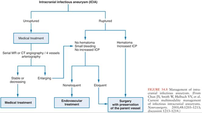
Optimal treatment of ICIA is based on small series and expert opinion (157). Part of the challenge in the treatment of these patients is derived from the choice of multiple treatment modalities, which currently consists of medical (antibiotics), endovascular (clot, glue, coils), and surgical therapies. Moreover, outcome with medical treatment alone is variable and unpredictable. Among 27 patients reviewed by Ojemann and Crowell (217) in whom follow-up angiography was performed during antibiotic therapy, complete disappearance of the lesions was observed in 8 patients (30%) and there was a decrease in size in five patients (19%). The lesions were unchanged in four patients (15%) and larger in six patients (22%). In four patients, a new ICIA was found (217). For a long time, it was believed that most ICIAs would rupture if not resected (144). Although the natural history of ICIAs treated with appropriate antibiotics is not known precisely, it has become increasingly apparent that a certain proportion of unruptured ICIAs may resolve with medical treatment alone (219,220,233,234) (Fig. 34.9)
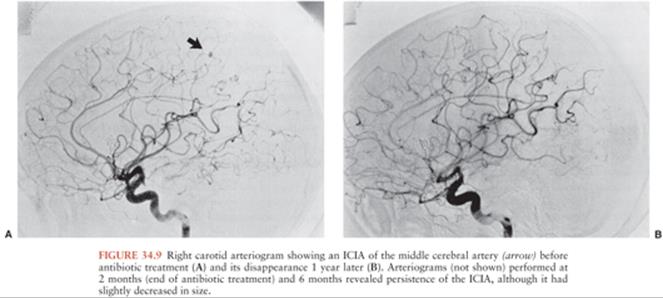
Thus, the most important aspect in the therapeutic approach to ICIAs is whether a rupture has occurred. By reviewing existing studies, Peters et al. (157) found that mortality among patients with unruptured ICIAs was very low, with no difference between patients treated with antibiotics alone and those treated with surgery. By contrast, outcome among patients with ruptured aneurysm was poor. Antibiotic therapy alone was associated with a mortality of 20% to 100% in this group, indicating that surgery or endovascular therapy should be attempted (157). Based on these figures, most authors favor a conservative approach in the management of patients with unruptured ICIA (157,220,233). However, some authors recommend surgical excision whenever possible (232), due to the unpredictable course of untreated ICIAs.
Chun et al. (233) proposed an algorithm for the assessment and treatment of patients with ICIA. Factors that guide the decisions are the general condition of the patient, aneurysm location, aneurysm rupture, hematomas with increased intracranial pressure (ICP), and the eloquence of brain tissue supplied by the parent artery. Eloquence is determined on the basis of radiographic anatomic features and, if needed, functional testing with amobarbital on a conscious patient. This technique consists of infusing amobarbital into the vessel that needs to be occluded and to evaluate clinically the neurologic deficit the patient would experience if the neuroradiologist proceeded with the occlusion of the artery harboring the ICIA (235). The size of the ICIA does not appear very helpful in deciding whether to operate immediately; some small ICIAs may rupture, whereas even large ones (>7 mm) may regress or even disappear under appropriate antibiotic therapy (220).
Medical Treatment
Antimicrobial therapy should be initiated as soon as possible in all situations. Although few data exist for ICIA, it is generally accepted that the regimen should be chosen according to the same principles and guidelines used for patients with IE; however, it has been recommended empirically that the duration of antibiotic treatment be extended to 6 to 8 weeks, even if the ICIA is resected surgically (236). Supportive measures such as control of the arterial pressure by antihypertensive agents, steroids, and avoidance of anticoagulation should be carefully considered.
Most authors agree that unruptured and stable ICIAs can be treated with medical treatment alone (157,220,233). Close clinical and radiologic follow-up has been advocated for unruptured ICIAs (157,217). Nowadays, MRA or CTA is probably preferable rather than conventional angiography for the follow-up of ICIAs. How often these investigations should be repeated has not been established, but an interval of 7 to 14 days appears reasonable. The patient should be monitored very closely for the development of any symptoms or signs suggestive of an increase in size, a leak, or an imminent rupture. Some authors also recommend surgery if the ICIA is still present at the end of antibiotic therapy (220). However, ICIAs may continue to regress after antibiotic therapy has been completed and complete resolution may take as long as 1 year (Fig. 34.9). Thus, if an ICIA has substantially decreased in size at the end of the antibiotic treatment but is still present, one may elect not to operate immediately but to continue to follow the patient with sequential CT and MRI.
Surgery
The primary goals of surgery are to eliminate the ICIA without further compromising cerebral function, to evacuate any associated hematoma, and to reduce ICP (233). Patients with infectious aneurysms are usually treated surgically if the aneurysm has ruptured and an intraparenchymal hematoma is producing a mass effect or increased ICP. Recent advances in image-guided surgical techniques have enhanced the ability of the neurosurgeon to opt for the best surgical treatment of the affected vessel (237–239).
Surgical treatment of ruptured ICIAs includes clipping, ligation, or excision (240). In some patients, the fibrotic process generated by the infection is sufficiently advanced to allow successful aneurysm clipping without sacrifice of the parent vessel. In other patients, the parent vessel may already be occluded by the embolus, which was presumably responsible for the formation of the ICIA, and therefore, ligation of the parent artery and excision of the ICIA may be performed. When surgical treatment is required for aneurysms located on proximal arteries or on important peripheral arteries, extracranial–intracranial bypass has been successfully attempted (241). It is important to take into consideration that administration of antibiotics during a certain period before surgery may facilitate resolution of arteritis with subsequent development of fibrosis in the wall of the aneurysm and the parent vessels. The lesion could then be handled more safely at surgery.
Endovascular Approach
Endovascular repair of ICIAs is less invasive than surgery and may be of interest before valve replacement. Small cerebral vessels can be cannulated and studied using small tracker wires. Various materials can be injected to occlude the vessel (242). The goal of this technique is to exclude the aneurysm from the circulation. A perioperative amobarbital test may help predict the vascular territory supplied by the parent artery prior to occlusion (243). Endovascular approaches have been used successfully to embolize aneurysmal vessels (242–246). Advantages of this endovascular approach include safety of the procedure, accessibility to distal aneurysms, and endovascular occlusion with minimal aneurysmal manipulation and low risk of rerupture. However, based on a metaanalysis of small series and case reports, endovascular treatment appeared more likely to imply parent artery sacrifice than surgical treatment (233). Therefore, a surgical approach is preferable for aneurysms located on eloquent parent arteries. Moreover, the development of stereotactic angiographic guidance for localization of distal ICIA allows the clipping of distal aneurysms using a minimally invasive procedure (237,247).
Special Issues
Few data are available to guide optimal timing of cardiac surgery in patients with IE complicated by ICIAs (Table 34.5). In a review of 34 patients with IE complicated by acute neurologic events who underwent valve replacement, three had a ruptured and four an unruptured ICIA (248). All patients with ruptured ICIAs underwent clipping before cardiac surgery. All patients with unruptured ICIAs had an uneventful postoperative course, whereas one patient with ruptured ICIA died after valve replacement. Thus, unruptured ICIAs do not seem to be associated with worse outcome after cardiac surgery and there is no indication for preoperative angiography (183). By contrast, ruptured ICIAs should undergo repair before cardiac surgery (240). According to recent case reports, cardiac surgery can be performed a few days after endovascular repair (246), thus this technique should be preferred if subsequent valve replacement is planned. Cardiac surgery should be postponed for at least 4 weeks in the presence of intracerebral hematoma (10).
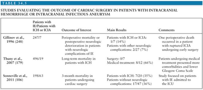
Multiple aneurysms present a complex problem. It seems appropriate to analyze each individual case along the same guidelines as for a single ICIA. If one or more of the aneurysms enlarge or bleed, prompt surgical excision should be attempted. The excision of the other aneurysms during the same operation will depend on their accessibility (217,234). Multiple aneurysms can be treated by endovascular techniques in one session and this may represent an important advantage over a surgical approach (233,242).
Outcome
The mortality of recognized ICIA is high. Among the 85 cases reviewed by Bohmfalk et al. (218), the figures for patients hospitalized for IE before neurologic symptoms occurred show a mortality of 80% for aneurysms that ruptured and of 30% if the aneurysm remained intact. The overall mortality was 46%.
Metastatic Infections
Infection of the CNS secondary to IE may present as meningitis, meningoencephalitis, cerebral microabscesses or macroabscesses, parameningeal abscesses, or infectious arteritis.
Meningitis
In the context of IE, the term meningitis is used to cover various conditions, ranging from full-blown bacterial infection with positive CSF cultures to sterile inflammatory reactions to infection, ischemia, or hemorrhage. Before the advent of antibiotics, meningitis was one of the more common neurologic complications of IE, accounting for up to two thirds of CNS complications in some studies (76). In more recent reviews of IE, meningitis was recorded in 2% to 15% of patients with IE, accounting for 15% to 40% of CNS manifestations of IE (83,97,101). The great variability of these percentages is partly due to the fact that some authors include all meningeal reactions whatever the underlying process, whereas others restrict the diagnosis to cases with positive CSF cultures. Moreover, the lower rate of meningitis in recent series may be related to a decrease in LPs because of improved cerebral imaging. Referral bias and a changing spectrum of IE may also account for some differences. For example, patients admitted to the ICU have higher rate of neurologic complications including meningitis: In a recent series of 198 patients, 41 (21%) were reported to have meningitis or meningeal reaction (106).
Although meningitis may be the presenting symptom of IE (83,249), IE is an uncommon cause of bacterial meningitis in general accounting for 3% in a large series (250). Microorganisms recovered in these cases are either Staphylococcus aureus or Streptococcus pneumoniae (251), but other pathogens (e.g., Streptococcus agalactiae, viridans streptococci, enterococci, gram-negative bacilli) are also found. When meningitis is due to bacterial species that do not normally cause primary meningitis, the proportion of cases associated with IE is higher (83,249). In a recent series of nine patients with community-acquired Staphylococcus aureus meningitis, endocarditis was found in five (252). Outcome was very poor with a mortality of 67%, while two of three survivors presented neurologic sequelae. In another study, IE was present in one quarter of 28 patients with community-acquired Staphylococcus aureusmeningitis. Mortality was 43% (253). In pneumococcal endocarditis, meningitis has been recorded in 40% to 60% of the patients (130,131). Austrian syndrome was first described in 1956 and represents the triad of Streptococcus pneumoniae pneumonia, endocarditis, and meningitis (254). Although chronic alcoholism is thought to be a risk factor, in a review of 31 published case reports of Austrian syndrome, only 38% were alcoholics (254).
Brain Abscess
Brain abscesses associated with IE are uncommon, especially in subacute cases. In a series of 218 patients with IE, 8 (3.6%) developed abscesses (83). In other large series, rates of 1% to 8.6% were recorded (88,92,97,101,143). In patients hospitalized in the ICU, the rate was 7% (106). In an autopsy study, cerebral abscesses were documented in 30% of cases (255). In large series of brain abscesses, IE is not a common cause. Among 314 patients with brain abscess collected from six series, 13 (4%) had IE (256,257). This reveals that IE should be suspected in the presence of brain abscess when there is no other obvious source. This suspicion must be particularly high when multiple abscesses are present (Fig. 34.10). Thus, IE was responsible for 2 of 5 patients who presented with multiple abscesses in a series of 41 patients with brain abscesses observed in our institution from 1977 to 1989 (257).
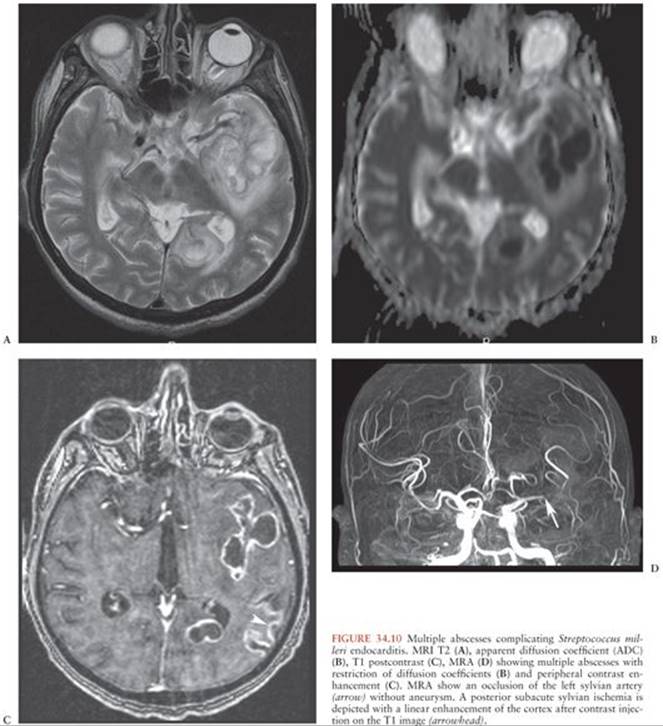
Obviously, the incidence of brain abscesses associated with IE will depend on the imaging techniques used and on whether the study included pathologic examination. In a series of patients with IE from the Massachusetts General Hospital, 9 of 218 had evidence of brain abscess, all cases of which were diagnosed at autopsy (83). In eight of the patients, the abscesses were less than 1 cm3 in size, not sufficient to create a mass effect. In six of these eight patients, multiple microscopic abscesses were present, usually in association with microabscesses in other organs. These patients also had multiple microscopic infarcts, demonstrating the interrelationship between vascular and infectious complications of IE. Seven of the eight patients had acute IE, four cases of which were due to S. aureus, and one had subacute IE due to viridans streptococci. Only one patient in this series had a large abscess, which was probably caused by the direct extension from S. aureus otitis media and mastoiditis, not by bacterial seeding from the bloodstream (83).
Because areas of cerebritis or cerebral abscesses of small size often resolve with appropriate antibiotic therapy alone (257), it is likely that the true incidence of these lesions is underestimated, even in recent studies that have benefited from CT. Thus, among 64 patients with neurologic complications of IE, abscesses were diagnosed in only 2 of the 51 patients who had a head CT scan (88). It is noteworthy that among these 51 patients, 15 presented with encephalopathy, a clinical presentation often associated with multiple microabscesses and/or microinfarcts (145). Likewise, 11 of these 51 had a focal deficit with a normal CT scan and some may have had undetected cerebritis (88). This suggests that small abscesses or foci of cerebritis may be below the level of detection of the CT scan. MRI has been shown to be superior to CT in the detection of early cerebritis. Patients with IE and “toxic” encephalopathy were described who had a normal CT scan but an MRI scan showing multiple lesions suggesting microembolization and/or microabscesses. For example, in prospective study of 130 patients who underwent systematic cerebral MRI, abscesses were found in 8 (6%) (163). Reports of abnormalities seen on MRI but undetected by CT have also included small infarcts. Thus, it is obvious that cerebritis, cerebral microinfarcts, and microabscesses associated with IE will be diagnosed with increased frequency if MRI is performed, especially in patients with toxic encephalopathy or focal deficit with a normal CT scan (163).
Pathogenesis
Clinical signs of meningitis may be associated with multiplication of the causative microorganism in the subarachnoid space or may be a reflection of various parameningeal lesions, such as brain microabscesses and macroabscesses, septic or nonseptic cerebral microemboli or macroemboli, leaking infectious aneurysms, subarachnoid hemorrhage, or immune-mediated arteritis. Several of these mechanisms can occur simultaneously or sequentially (83). Except for the immunologic arterial lesions, all these complications ultimately relate to embolization of infected material to cerebral or meningeal vessels. Critical factors that may determine whether a septic embolus results in a simple infarct, a infectious aneurysm, meningitis, cerebritis, an abscess, or a combination of these lesions include the size of the embolus, the site in which the embolus lodges, the virulence of the microorganism, and the defenses of the host, including the timing and adequacy of antibacterial therapy. As already mentioned, neurologic complications are characteristic of left-sided IE, indicating that emboli play a major role in the pathogenesis of cerebral manifestations including meningitis and abscesses (83). Although these emboli may be clinically silent, the development of macroscopic brain abscess 1 to 3 weeks after a contralateral hemiplegic stroke due to embolism has been well documented (113). However, with certain bacteria, meningitis can occur in the context of isolated right-sided IE, indicating that the mere presence of certain bacteria in the blood can cause meningeal seeding. This has been described with S. pneumoniae, not surprising given the well-known propensity of this organism to cause hematogenous meningitis. In right-sided S. aureus IE, meningitis has also been described, but it occurred in only 2 of 53 of such episodes. The CSF showed an “aseptic” formula. When S. aureus bacteremia is caused by IE, the incidence of CNS involvement is higher (29% to 54%) than in the absence of endocardial involvement (3% to 10%). In a recent study of 81 episodes of S. aureus IE, 22 neurologic complications were recorded. All but one occurred in the 42 patients with left-sided involvement; there were five brain abscesses and two cases of meningitis (110).
Thus, the occurrence of CNS infectious complications of IE depends on two major factors, as follows: the virulence characteristics of the microorganism and the side of the heart involved. The fact that right-sided and left-sided IE are both characterized by persistent bacteremia but are very much different with respect to the incidence of CNS infectious complications suggests that emboli associated with bacteria are more capable of disrupting the normal blood–brain barrier (BBB).
Clinical Presentation
There is a continuum in the clinical presentation of meningitis, meningoencephalitis, and microscopic and macroscopic brain abscesses. Patients may display a typical clinical presentation of primary meningitis. Meningoencephalitis characterized by confusion, decreased level of consciousness, stiff neck, and headache associated with normal or slightly abnormal CSF (minimal pleocytosis, normal glucose concentration, normal or slightly elevated protein concentration, and sterile culture) has been termed acute brain syndrome, acute encephalopathy, or toxic encephalopathy (142). Psychiatric manifestations may predominate, including personality change, disorientation, drowsiness, irritability, or even hallucinations. This may account for the frequent presence of confusion in elderly patients with IE in the absence of fever or metabolic abnormalities. This presentation is related to multiple microemboli and microinfarcts with or without microabscesses (83).
The clinical presentation of brain abscesses depends on the stage, size, number, and location of the lesions (258,259). This may result in clinical manifestations of a focal deficit, a space-occupying lesion, toxic encephalopathy, or meningitis. The triad of fever, headache, and focal neurologic signs is found in less than 50% of cases (260).The accompanying meningitis will usually be sterile unless the abscess ruptures into the subarachnoid space. Headache, confusion, increased ICP, and focal signs, often developing slowly over several days, are noted with large abscesses (261).
Parameningeal abscess (i.e., cerebral or spinal epidural abscess, spondylodiscitis) is another metastatic infection of IE that may cause neurologic manifestations. Back pain may be a symptom of IE in up to 43% of patients (262), whereas spondylodiscitis complicates 15% to 20% of cases (263,264). Conversely, IE is reported as the source of infection for vertebral osteomyelitis in up to 30% of patients (265). By contrast, epidural abscess was found to be associated with IE in only 3% of cases (266).
Management
The treatment of IE-associated meningitis and brain abscess is covered by the antibiotic regimens recommended for the underlying IE, provided that high doses of bactericidal antibiotics that cross the BBB are used. Among drugs commonly used for the treatment of IE, penicillins, third-generation cephalosporins and meropenem should be favored because they were shown to achieve bactericidal concentrations in the CSF, provided the highest intravenous dose is used (267).
Drainage of brain abscesses is only indicated if antibiotic treatment alone is expected to fail, as microbial etiology is usually already established by blood cultures. Most patients have small and multiple microabscesses and surgery is generally neither feasible nor desirable. However, a large abscess can be refractory to antibiotic treatment alone. Two centimeters appears to be the critical diameter above which surgery is strongly indicated (268). In patients with cerebritis, early therapy alone may also be curative, but surgery should be considered in case of neurologic deterioration (269). For CNS suppurative complications of IE, it is generally recommended that the duration of antimicrobial therapy be extended to 6 to 8 weeks (83).
Spinal Cord Lesions
Lesions of the spinal cord may be due to external compression by a parameningeal abscess or may be the consequence of emboli to the vascular supply. Despite the frequency of embolic phenomena in IE, there have been only rare case reports of embolic infarction of the spinal cord and no cases are reported in the major series of IE.
Cranial and Peripheral Nerve Lesions
In two large series of patients with neurologic manifestations of IE, mononeuropathies were reported in 5 of 110 and 5 of 84 patients (83,143). Five of these patients were reported in detail. Viridans streptococci were the responsible microorganisms in all five. All had an elevated erythrocyte sedimentation rate (ESR). In four patients, neuropathy was the initial manifestation of IE. Three had concomitant involvement of multiple nerves, emphasizing the need to consider IE in the differential diagnosis of mononeuritis multiplex (270). All patients improved after treatment, and only two of the eight nerves involved presented minimal residual deficit. Because of the temporal and spatial association with cutaneous emboli, embolic occlusion of the vasa vasorum was the postulated physiopathologic mechanism (152,271). Immune-complex–mediated vasculitis in the peripheral nerves is another possible mechanism. Peripheral neuropathy has been rarely reported as a complication of S. aureus endocarditis and meningitis (272) and as critical illness polyneuropathy (273).
References
1. Osler W. Gulstonian lectures on malignant endocarditis. Lancet. 1885;1: 415–508.
2. Contrepois A. Notes on the early history of infective endocarditis and the development of an experimental model. Clin Infect Dis. 1995;20:461–466.
3. Hogevik H, Olaison L, Andersson R, et al. Epidemiologic aspects of infective endocarditis in an urban population. A 5-year prospective study. Medicine. 1995;74:324–339.
4. Hoen B, Alla F, Selton-Suty C, et al. Changing profile of infective endocarditis: results of a 1-year survey in France. JAMA. 2002;288:75–81.
5. Tleyjeh IM, Steckelberg JM, Murad HS, et al. Temporal trends in infective endocarditis: a population-based study in Olmsted County, Minnesota. JAMA. 2005;293:3022–3028.
6. Selton-Suty C, Célard M, Le Moing V, et al. Preeminence of Staphylococcus aureus in infective endocarditis: a 1-year population-based survey. Clin Infect Dis. 2012;54:1230–1239.
7. Tleyjeh IM, Abdel-Latif A, Rahbi H, et al. A systematic review of population-based studies of infective endocarditis. Chest. 2007;132:1025–1035.
8. Moreillon P, Que YA. Infective endocarditis. Lancet. 2004;363:139–149.
9. Hasbun R, Vikram HR, Barakat LA, et al. Complicated left-sided native valve endocarditis in adults: risk classification for mortality. JAMA. 2003;289:1933–1940.
10. Habib G, Hoen B, Tornos P, et al. Guidelines on the prevention, diagnosis, and treatment of infective endocarditis (new version 2009): the Task Force on the Prevention, Diagnosis, and Treatment of Infective Endocarditis of the European Society of Cardiology (ESC). Endorsed by the European Society of Clinical Microbiology and Infectious Diseases (ESCMID) and the International Society of Chemotherapy (ISC) for Infection and Cancer. Eur Heart J. 2009;30:2369–2413.
11. Watanakunakorn C, Burkert T. Infective endocarditis at a large community teaching hospital 1980-1990. Medicine. 1993;72:90–102.
12. Correa de Sa DD, Tleyjeh IM, Anavekar NS, et al. Epidemiological trends of infective endocarditis: a population-based study in Olmsted County, Minnesota. Mayo Clin Proc. 2010;85:422–426.
13. McKinsey DS, Ratts TE, Bisno AL. Underlying cardiac lesions in adults with infective endocarditis. The changing spectrum. Am J Med. 1987;82: 681–688.
14. MacMahon SW, Roberts JK, Kramer-Fox R, et al. Mitral valve prolapse and infective endocarditis. Am Heart J. 1987;113:1291–1298.
15. Clemens JD, Horwitz RI, Jaffe CC, et al. A controlled evaluation of the risk of bacterial endocarditis in persons with mitral-valve prolapse. N Engl J Med. 1982;307:776–781.
16. Murdoch DR, Corey GR, Hoen B, et al. Clinical presentation, etiology, and outcome of infective endocarditis in the 21st century: the International Collaboration on Endocarditis-Prospective Cohort Study. Arch Intern Med. 2009;169:463–473.
17. Geva T, Frand M. Infective endocarditis in children with congenital heart disease: the changing spectrum, 1965-85. Eur Heart J. 1988;9:1244–1249.
18. Lamas CC, Eykyn SJ. Bicuspid aortic valve—a silent danger: analysis of 50 cases of infective endocarditis. Clin Infect Dis. 2000;30:336–341.
19. Mylonakis E, Calderwood SB. Infective endocarditis in adults. N Engl J Med. 2001;345:1318–1330.
20. Habib G, Thuny F, Avierinos JF. Prosthetic valve endocarditis: current approach and therapeutic options. Prog Cardiovasc Dis. 2008;50:274–281.
21. Benito N, Miro JM, de Lazzari E, et al. Health care-associated native valve endocarditis: importance of non-nosocomial acquisition. Ann Intern Med. 2009;150:586–594.
22. Athan E, Chu VH, Tattevin P, et al. Clinical characteristics and outcome of infective endocarditis involving implantable cardiac devices. JAMA. 2012;307:1727–1735.
23. Fang G, Keys TF, Gentry LO, et al. Prosthetic valve endocarditis resulting from nosocomial bacteremia. A prospective, multicenter study. Ann Intern Med. 1993;119:560–567.
24. Fowler VG Jr, Miro JM, Hoen B, et al. Staphylococcus aureus endocarditis: a consequence of medical progress. JAMA. 2005;293:3012–3021.
25. Robinson DL, Fowler VG, Sexton DJ, et al. Bacterial endocarditis in hemodialysis patients. Am J Kidney Dis. 1997;30:521–524.
26. Mathew J, Addai T, Anand A, et al. Clinical features, site of involvement, bacteriologic findings, and outcome of infective endocarditis in intravenous drug users. Arch Intern Med. 1995;155:1641–1648.
27. Garrison PK, Freedman LR. Experimental endocarditis I. Staphylococcal endocarditis in rabbits resulting from placement of a polyethylene catheter in the right side of the heart. Yale J Biol Med. 1970;42:394–410.
28. Durack DT. Experimental bacterial endocarditis. IV. Structure and evolution of very early lesions. J Pathol. 1975;115:81–89.
29. Lien EA, Solberg CO, Kalager T. Infective endocarditis 1973-1984 at the Bergen University Hospital: clinical feature, treatment and prognosis. Scand J Infect Dis. 1988;20:239–246.
30. Bayer AS, Theofilopoulos AN. Immunopathogenetic aspects of infective endocarditis. Chest. 1990;97:204–212.
31. Hoen B. Platelets and platelet inhibitors in infective endocarditis. Curr Infect Dis Rep. 2002;4:299–303.
32. Gould K, Ramirez-Ronda CH, Holmes RK, et al. Adherence of bacteria to heart valves in vitro. J Clin Invest. 1975;56:1364–1370.
33. Fournier PE, Thuny F, Richet H, et al. Comprehensive diagnostic strategy for blood culture-negative endocarditis: a prospective study of 819 new cases. Clin Infect Dis. 2010;51:131–140.
34. Chu VH, Woods CW, Miro JM, et al. Emergence of coagulase-negative staphylococci as a cause of native valve endocarditis. Clin Infect Dis. 2008;46:232–242.
35. Patel R, Piper KE, Rouse MS, et al. Frequency of isolation of Staphylococcus lugdunensis among staphylococcal isolates causing endocarditis: a 20-year experience. J Clin Microbiol. 2000;38:4262–4263.
36. Bert F, Bariou-Lancelin M, Lambert-Zechovsky N. Clinical significance of bacteremia involving the “Streptococcus milleri” group: 51 cases and review. Clin Infect Dis. 1998;27:385–387.
37. Francioli P, Etienne J, Hoigne R, et al. Treatment of streptococcal endocarditis with a single daily dose of ceftriaxone sodium for 4 weeks. Efficacy and outpatient treatment feasibility. JAMA. 1992;267:264–267.
38. Kupferwasser I, Darius H, Muller AM, et al. Clinical and morphological characteristics in Streptococcus bovis endocarditis: a comparison with other causative microorganisms in 177 cases. Heart. 1998;80:276–280.
39. Pergola V, Di Salvo G, Habib G, et al. Comparison of clinical and echocardiographic characteristics of Streptococcus bovis endocarditis with that caused by other pathogens. Am J Cardiol. 2001;88:871–875.
40. Lindberg J, Prag J, Schonheyder HC. Pneumococcal endocarditis is not just a disease of the past: an analysis of 16 cases diagnosed in Denmark 1986-1997. Scand J Infect Dis. 1998;30:469–472.
41. Sendi P, Ericsson M, Olaison L. Infective endocarditis caused by group B Streptococcus: the role of aminoglycoside-combination. J Infect. 2012;64:127–129.
42. Olaison L, Schadewitz K, Swedish Society of Infectious Diseases Quality Assurance Study Group for Endocarditis. Enterococcal endocarditis in Sweden, 1995-1999: can shorter therapy with aminoglycosides be used? Clin Infect Dis. 2002;34:159–166.
43. Das M, Badley AD, Cockerill FR, et al. Infective endocarditis caused by HACEK microorganisms. Annu Rev Med. 1997;48:25–33.
44. Nørskov-Lauritsen N, Kilian M. Reclassification of Actinobacillus actinomycetemcomitans, Haemophilus aphrophilus, Haemophilus paraphrophilus and Haemophilus segnis as Aggregatibacter actinomycetemcomitans gen. nov., comb. nov., Aggregatibacter aphrophilus comb. nov. and Aggregatibacter segnis comb. nov., and emended description of Aggregatibacter aphrophilus to include V factor-dependent and V factor-independent isolates. Int J Syst Evol Microbiol. 2006;56:2135–2146.
45. Raoult D, Fournier PE, Drancourt M, et al. Diagnosis of 22 new cases of Bartonella endocarditis. Ann Intern Med. 1996;125:646–652.
46. Wang A, Athan E, Pappas PA, et al. Contemporary clinical profile and outcome of prosthetic valve endocarditis. JAMA. 2007;297:1354–1361.
47. Nasser RM, Melgar GR, Longworth DL, et al. Incidence and risk of developing fungal prosthetic valve endocarditis after nosocomial candidemia. Am J Med. 1997;103:25–32.
48. Frontera JA, Gradon JD. Right-side endocarditis in injection drug users: review of proposed mechanisms of pathogenesis. Clin Infect Dis. 2000;30: 374–379.
49. Chambers HF, Korzeniowski OM, Sande MA. Staphylococcus aureus endocarditis: clinical manifestations in addicts and nonaddicts. Medicine. 1983;62:170–177.
50. Chan P, Ogilby JD, Segal B. Tricuspid valve endocarditis. Am Heart J. 1989;117:1140–1146.
51. Brouqui P, Raoult D. Endocarditis due to rare and fastidious bacteria. Clin Microbiol Rev. 2001;14:177–207.
52. Greub G, Lepidi H, Rovery C, et al. Diagnosis of infectious endocarditis in patients undergoing valve surgery. Am J Med. 2005;118:230–238.
53. Raoult D, Casalta JP, Richet H, et al. Contribution of systematic serological testing in diagnosis of infective endocarditis. J Clin Microbiol. 2005;43:5238–5242.
54. Bouza E, Menasalvas A, Munoz P, et al. Infective endocarditis—a prospective study at the end of the twentieth century: new predisposing conditions, new etiologic agents, and still a high mortality. Medicine. 2001;80:298–307.
55. Steckelberg JM, Murphy JG, Ballard D, et al. Emboli in infective endocarditis: the prognostic value of echocardiography. Ann Intern Med. 1991;114:635–640.
56. Brown M, Griffin GE. Immune responses in endocarditis. Heart. 1998; 79:1–2.
57. Choussat R, Thomas D, Isnard R, et al. Perivalvular abscesses associated with endocarditis; clinical features and prognostic factors of overall survival in a series of 233 cases. Perivalvular Abscesses French Multicentre Study. Eur Heart J. 1999;20:232–241.
58. Blumberg EA, Karalis DA, Chandrasekaran K, et al. Endocarditis-associated paravalvular abscesses. Do clinical parameters predict the presence of abscess? Chest. 1995;107:898–903.
59. Li JS, Sexton DJ, Mick N, et al. Proposed modifications to the Duke criteria for the diagnosis of infective endocarditis. Clin Infect Dis. 2000;30: 633–638.
60. Evangelista A, Gonzalez-Alujas MT. Echocardiography in infective endocarditis. Heart. 2004;90:614–617.
61. Erbel R, Rohmann S, Drexler M, et al. Improved diagnostic value of echocardiography in patients with infective endocarditis by transoesophageal approach. A prospective study. Eur Heart J. 1988;9:43–53.
62. Baddour LM, Wilson WR, Bayer AS, et al. Infective endocarditis: diagnosis, antimicrobial therapy, and management of complications: a statement for healthcare professionals from the Committee on Rheumatic Fever, Endocarditis, and Kawasaki Disease, Council on Cardiovascular Disease in the Young, and the Councils on Clinical Cardiology, Stroke, and Cardiovascular Surgery and Anesthesia, American Heart Association: endorsed by the Infectious Diseases Society of America. Circulation. 2005;111:e394–e434.
63. Tornos P, Iung B, Permanyer-Miralda G, et al. Infective endocarditis in Europe: lessons from the Euro heart survey. Heart. 2005;91:571–575.
64. Ellis ME, Al-Abdely H, Sandridge A, et al. Fungal endocarditis: evidence in the world literature, 1965-1995. Clin Infect Dis. 2001;32:50–62.
65. Prendergast BD, Tornos P. Surgery for infective endocarditis: who and when? Circulation. 2010;121:1141–1152.
66. Kiefer T, Park L, Tribouilloy C, et al. Association between valvular surgery and mortality among patients with infective endocarditis complicated by heart failure. JAMA. 2011;306:2239–2247.
67. Kang DH, Kim YJ, Kim SH, et al. Early surgery versus conventional treatment for infective endocarditis. N Engl J Med. 2012;366:2466–2473.
68. Baddour LM, Cha YM, Wilson WR. Clinical practice. Infections of cardiovascular implantable electronic devices. N Engl J Med. 2012;367:842–849.
69. Wilson W, Taubert KA, Gewitz M, et al. Prevention of infective endocarditis: guidelines from the American Heart Association: a guideline from the American Heart Association Rheumatic Fever, Endocarditis, and Kawasaki Disease Committee, Council on Cardiovascular Disease in the Young, and the Council on Clinical Cardiology, Council on Cardiovascular Surgery and Anesthesia, and the Quality of Care and Outcomes Research Interdisciplinary Working Group. Circulation. 2007;116:1736–1754.
70. Netzer RO, Zollinger E, Seiler C, et al. Infective endocarditis: clinical spectrum, presentation and outcome. An analysis of 212 cases 1980–1995. Heart. 2000;84:25–30.
71. Horder TJ. Influenzal endocarditis: with an account of two cases in which the influenza bacillus was repeatedly cultivated from the blood during life. Med Chir Trans. 1906;89:333–354.7.
72. Blumer G. Subacute bacterial endocarditis. Medicine. 1923;2:105.
73. DeJong RN. Central nervous system complications in subacute bacterial endocarditis. J Nerv Ment Dis. 1937;85:397–410.
74. Fetterman GL, Aske WF. Cerebral debut of certain cases of cardiac disease. Ohio Med J. 1938;34:1354.
75. Krinsky CM, Merritt HH. Neurological manifestations of subacute bacterial endocarditis. N Engl J Med. 1938;218:563–566.
76. Toone EC. Cerebral manifestations of bacterial endocarditis. Ann Intern Med. 1941;14:1551–1574.
77. Hickie J. Bacterial endocarditis in Sydney 1950-1959. Med JAust. 1961;1:929–934.
78. Bullock R, Van Dellen JR. Rupture of bacterial intracranial aneurysms following replacement of cardiac valves. Surg Neurol. 1982;17:9–11.
79. Pankey GA. Acute bacterial endocarditis at the University of Minnesota Hospitals. Am Heart J. 1962;64:583–591.
80. Cooper ES, Cooper JW, Schnabel TG. Pitfalls in the diagnosis of bacterial endocarditis. Arch Int Med. 1966;118:55–61.
81. Harrison MJ, Hampton JR. Neurological presentation of bacterial endocarditis A sixty- three year old woman with fulminant meningitis and convulsions. Am J Med. 1967;42:264–272.
82. Jones HRJ, Siekert RG, Geraci JE. Neurologic manifestations of bacterial endocarditis. Ann Int Med. 1969;71:21–28.
83. Pruitt AA, Rubin RH, Karchmer AW, et al. Neurologic complications of bacterial endocarditis. Medicine. 1978;57:329–343.
84. Lerner PI, Weinstein L. Infective endocarditis in the antibiotic era. Part I. N Engl J Med. 1966;274:199–206.
85. Pelletier LL, Petersdorf RG. Infective endocarditis. A review of 125 cases from the University of Washington Hospitals. Medicine. 1977;56:287–313.
86. Terpenning MS. Infective endocarditis. Clin Geriatr Med. 1992;8:903–912.
87. Bush M, Masferrer R, Teitel R, et al. Neurologic complications of infectious endocarditis. BNI Quarterly. 1985;1:13–18.
88. Salgado AV, Furlan AJ, Keys TF, et al. Neurologic complications of endocarditis: a 12-year experience. Neurology. 1989;39:173–178.
89. Gransden WR, Eykyn SJ, Leach RM. Neurological presentations of native valve endocarditis. Q J Med. 1989;73:1135–1142.
90. Mansur AJ, Grinberg M, Lemos da Luz P, et al. The complications of infective endocarditis. a reappraisal in the 1980s. Arch Int Med. 1992;152:2428–2432.
91. Matsushita K, Kuriyama Y, Sawada T, et al. Hemorrhagic and ischemmic cerebrovascular complications of active inefective endocarditis of native valve. Eur J Neurol. 1993;33:267–274.
92. Kanter MC, Hart GR. Neurologic complications of infective endocarditis. Neurology. 1991;41:1015–1020.
93. Von Reyn CF, Levy BS, Arbeit RD, et al. Infective endocarditis: an analysis based on strict case definition. Ann Int Med. 1981;94:505–518.
94. Sandre RM, Shafran SD. Infective endocarditis: review of 135 cases over 9 years. Clin Infect Dis. 1996;22:276–286.
95. Røder BL, Wandall DA, Espersen F, et al. A study of 47 bacteremic Staphylococcus aureus endocarditis cases: 23 with native valves treated surgically and 24 with prosthetic valves. Scand Cardiovasc J. 1997;31:305–309.
96. Gagliardi JP, Nettles RE, McCarty DE, et al. Native valve infective endocarditis in elderly and younger adult patients: comparison of clinical features and outcomes with use of the Duke criteria and the Duke Endocarditis Database. Clin Infect Dis. 1998;26:1165–1168.
97. Heiro M, Nikoskelainen J, Engblom E, et al. Neurologic manifestations of infective endocarditis: a 17-year experience in a teaching hospital in Finland. Arch Intern Med. 2000;160:2781–2787.
98. Castillo JC, Anguita MP, Ramirez A, et al. Long term outcome of infective endocarditis in patients who were not drug addicts: a 10 year study. Heart. 2000;83:525–530.
99. Cetinkaya Y, Akova M, Akalin HE, et al. A retrospective review of 228 episodes of infective endocarditis where rheumatic valvular disease is still common. Int J Microbial Agents 2001;18:1–7.
100. Tiurin VP, Odinak MM, Klimov IA, et al. Neurological complications of infectious endocarditis [in Russian]. Klin Med (Mosk). 2002;80:27–31.
101. Corral I, Martin-Davila P, Fortun J, et al. Trends in neurological complications of endocarditis. J Neurol. 2007;254:1253–1259.
102. Snygg-Martin U, Gustafsson L, Rosengren L, et al. Cerebrovascular complications in patients with left-sided infective endocarditis are common: a prospective study using magnetic resonance imaging and neurochemical brain damage markers. Clin Infect Dis. 2008;47:23–30.
103. Cooper HA, Thompson EC, Laureno R, et al. Subclinical brain embolization in left-sided infective endocarditis: results from the evaluation by MRI of the brains of patients with left-sided intracardiac solid masses (EMBOLISM) pilot study. Circulation. 2009;120:585–591.
104. Kane WC, Aronson SM. Cardiac disorders predisposing to embolic stroke. Stroke. 1970;1:164–172.
105. Kanafani ZA, Kanj SS, Cabell CH, et al. Revisiting the effect of referral bias on the clinical spectrum of infective endocarditis in adults. Eur J Clin Microbiol Infect Dis. 2010;29:1203–1210.
106. Sonneville R, Mirabel M, Hajage D, et al. Neurologic complications and outcomes of infective endocarditis in critically ill patients: the ENDOcardite en REAnimation prospective multicenter study. Crit Care Med. 2011;39:1474–1481.
107. Cantrell M, Yoshikawa TT. Aging and infective endocarditis. J Am Geriart Soc. 1983;31:216–222.
108. Groll A, Horlin A, Lang C, et al. Neurological complications in infectious endocarditis [in German]. Klin Padiatr. 1995;207:19–23.
109. Kirkes WS. Principal effects resulting from detachment of fibrinous deposits from the interior of the heart. Med Chir Trans. 1852;35:281–324.
110. Chambers HF, Miller RT, Newman MD. Right-sided Staphylococcus aureus endocarditis in intravenous drug abusers: two-week combination therapy. Ann Intern Med. 1988;109:619-624
111. Hubbell G, Cheitlin MD, Rapaport E. Presentation, management, and follow-up evaluation of infective endocarditis in drug addicts. Am Heart J. 1981;102:85–94.
112. Roberts WC, Buchbinder NA. Right-sided valvular infective endocarditis. A clinicopathologic study of twelve necropsy patients. Am J Med. 1972;53:7–19.
113. Le Cam B, Guivarch G, Boles JM, et al. Neurologic complications in a group of 86 bacterial endocarditis. Eur Heart J. 1984;5(suppl C):97–100.
114. Anderson DJ, Goldstein LB, Wilkinson WE, et al. Stroke location, characterization, severity, and outcome in mitral vs aortic valve endocarditis. Neurology. 2003;61:1341–1346.
115. Garvey GJ, Neu HC. Infective endocarditis—an evolving disease. A review of endocarditis at the Columbia-Presbyterian Medical Center, 1968-1973. Medicine. 1978;57:105–127.
116. Wilson WR, Geraci JE, Danielson GK, et al. Anticoagulant therapy and central nervous system complications in patients with prosthetic valve endocarditis. Circulation. 1978;57:1004–1007.
117. Carpenter JL, McAllister CK. Anticoagulation in prosthetic valve endocarditis. South Med J. 1983;76:1372–1375.
118. Quenzer RW, Edwards LD, Levin S. A comparative study of 48 host valve and 24 prosthetic valve endocarditis cases. Am Heart J. 1976;92:15–22.
119. Leport C, Vilde JL, Bricaire F, et al. Fifty cases of late prosthetic valve endocarditis: improvement in prognosis over a 15 year period. Br Heart J. 1987;58:66–71.
120. Keyser DL, Biller J, Coffman TT, et al. Neurologic complications of late prosthetic valve endocarditis. Stroke. 1990;21:472–475.
121. Davenport J, Hart RG. Prosthetic valve endocarditis 1976-1987. Antibiotics, anticoagulation, and stroke. Stroke. 1990;21:993–999.
122. Mullany CJ, Chua YL, Schaff HV, et al. Early and late survival after surgical treatment of culture-positive active endocarditis. Mayo Clin Proc. 1995;70:517–525.
123. Vongpatanasin W, Hillis LD, Lange RA. Prosthetic heart valves. N Engl J Med. 1996;335:407–416.
124. Delahaye JP, Poncet P, Malquarti V, et al. Cerebrovascular accidents in infective endocarditis: role of anticoagulation. Eur Heart J. 1990;11: 1074–1078.
125. Wilson WR, Danielson GK, Giuliani ER, et al. Prosthetic valve endocarditis. Mayo Clin Proc. 1982;57:155–161.
126. Castillo JC, Anguita MP, Torres F, et al. Long-term prognosis of early and late prosthetic valve endocarditis. Am J Cardiol. 2004;93:1185–1187.
127. Tornos P, Almirante B, Mirabet S, et al. Infective endocarditis due to Staphylococcus aureus: deleterious effect of anticoagulant therapy. Arch Intern Med. 1999;159:473–475.
128. Felner JM, Dowell VR Jr. Anaerobic bacterial endocarditis. N Engl J Med. 1970;283:1188–1192.
129. Kan B, Ries J, Normark BH, et al. Endocarditis and pericarditis complicating pneumococcal bacteraemia, with special reference to the adhesive abilities of pneumococci: results from a prospective study. Clin Microbiol Infect. 2006;12:338–344.
130. Aronin SI, Mukherjee SK, West JC, et al. Review of pneumococcal endocarditis in adults in the penicillin era. Clin Infect Dis. 1998;26:165–171.
131. Lefort A, Mainardi JL, Selton-Suty C, et al. Streptococcus pneumoniae endocarditis in adults. A multicenter study in France in the era of penicillin resistance (1991-1998). The Pneumococcal Endocarditis Study Group. Medicine. 2000;79:327–337.
132. Lefort A, Chartier L, Sendid B, et al. Diagnosis, management and outcome of Candida endocarditis. Clin Microbiol Infect. 2012;18:E99–E109.
133. Baddley JW, Benjamin DK Jr, Patel M, et al. Candida infective endocarditis. Eur J Clin Microbiol Infect Dis. 2008;27:519–529.
134. Woods GL, Wood RP, Shaw BW Jr. Aspergillus endocarditis in patients without prior cardiovascular surgery: report of a case in a liver transplant recipient and review. Rev Infect Dis. 1989;11:263–272.
135. Heldman AW, Hartert TV, Ray SC, et al. Oral antibiotic treatment of right-sided staphylococcal endocarditis in injection drug users: prospective randomized comparison with parenteral therapy. Am J Med. 1996;101:68–76.
136. Openshaw H. Neurological complications of endocarditis in persons taking drugs intravenously. West J Med. 1976;124:276–281.
137. Siekert RG, Jones HR Jr. Transient cerebral ischemic attacks associated with subacute bacterial endocarditis. Stroke. 1970;1:178–193.
138. Hart RG, Kagan-Hallet K, Joerns SE. Mechanisms of intracranial hemorrhage in infective endocarditis. Stroke. 1987;18:1048–1056.
139. Masuda J, Yutani C, Waki R, et al. Histopathological analysis of the mechanisms of intracranial hemorrhage complicating infective endocarditis. Stroke. 1992;23:843–850.
140. Chukwudelunzu FE, Brown RD Jr, Wijdicks EF, et al. Subarachnoid haemorrhage associated with infectious endocarditis: case report and literature review. Eur J Neurol. 2002;9:423–427.
141. Venger BH, Aldama AE. Mycotic vasculitis with repeated intracranial aneurysmal hemorrhage. Case report. J Neurosurg. 1988;69:775–779.
142. Sonneville R, Mourvillier B, Bouadma L, et al. Management of neurological complications of infective endocarditis in ICU patients. Ann Intensive Care. 2011;1:10.
143. Jones HR Jr, Siekert RG, Geraci JE. Neurologic manifestations of bacterial endocarditis. Ann Intern Med. 1969;71:21–28.
144. Wilson WR, Lie JT, Wayne Houser O, et al. The management of patients with mycotic aneurysm. Curr Clin Top Infect Dis. 1981;2:151–183.
145. Jones HR Jr, Siekert RG. Neurological manifestations of infective endocarditis. Review of clinical and therapeutic challenges. Brain. 1989;112(pt 5): 1295–1315.
146. Ziment I. Nervous system complications in bacterial endocarditis. Am J Med. 1969;47:593–607.
147. Lawrence-Friedl D, Bauer KM. Bilateral cortical blindness: an unusual presentation of bacterial endocarditis. Ann Emerg Med. 1992;21:1502–1504.
148. Hermans PE. The clinical manifestations of infective endocarditis. Mayo Clin Proc. 1982;57:15–21.
149. Weinstein L, Rubin RH. Infective endocarditis—1973. Prog Cardivasc Dis. 1973;16:239–274.
150. Silverberg HH. Roth spots. Mt Sinai J Med. 1970;37:77–79.
151. Oude Lashof AM, Rothova A, Sobel JD, et al. Ocular manifestations of candidemia. Clin Infect Dis. 2011;53:262–268.
152. Pamphlett R, Walsh J. Infective endocarditis with inflammatory lesions in the peripheral nervous system. Acta Neuropathol. 1989;78:101–104.
153. Caksen H, Uner A, Arslan S, et al. Severe peripheral polyneuropathy in a child with infective endocarditis caused by Staphylococcus aureus. Acta Neurol Belg. 2004;104:114–116.
154. Hart RG, Foster JW, Luther MF, et al. Stroke in infective endocarditis. Stroke. 1990;21:695–700.
155. Kim SJ, Lee JY, Kim TH, et al. Imaging of the neurological complications of infective endocarditis. Neuroradiology. 1998;40:109–113.
156. Bakshi R, Wright PD, Kinkel PR, et al. Cranial magnetic resonance imaging findings in bacterial endocarditis: the neuroimaging spectrum of septic brain embolization demonstrated in twelve patients. J Neuroimaging. 1999;9:78–84.
157. Peters PJ, Harrison T, Lennox JL. A dangerous dilemma: management of infectious intracranial aneurysms complicating endocarditis. Lancet Infect Dis. 2006;6:742–748.
158. Baird AE, Warach S. Magnetic resonance imaging of acute stroke. J Cereb Blood Flow Metab. 1998;18:583–609.
159. Singhal AB, Topcuoglu MA, Buonanno FS. Acute ischemic stroke patterns in infective and nonbacterial thrombotic endocarditis: a diffusion-weighted magnetic resonance imaging study. Stroke. 2002;33:1267–1273.
160. Baddour LM, Bayer AS. Cerebrovascular complications in patients with left-sided infective endocarditis: out of site, out of mind. Clin Infect Dis. 2008;47:31–32.
161. Klein I, Iung B, Labreuche J, et al. Cerebral microbleeds are frequent in infective endocarditis: a case-control study. Stroke. 2009;40:3461–3465.
162. Okazaki S, Sakaguchi M, Hyun B, et al. Cerebral microbleeds predict impending intracranial hemorrhage in infective endocarditis. Cerebrovasc Dis. 2011;32:483–488.
163. Duval X, Iung B, Klein I, et al. Effect of early cerebral magnetic resonance imaging on clinical decisions in infective endocarditis: a prospective study. Ann Intern Med. 2010;152:497–504, W175.
164. Kerr KG. Low back pain as the only presenting symptom in Streptococcus sanguis endocarditis. Rev Infect Dis. 1989;11:836–837.
165. Sila CA. Neurological complications of bacterial endocarditis. Handb Clin Neurol. 2010;96:221–229.
166. Botelho-Nevers E, Thuny F, Casalta JP, et al. Dramatic reduction in infective endocarditis-related mortality with a management-based approach. Arch Intern Med. 2009;169:1290–1298.
167. Ben Ismail M, Hannachi N, Abid F, et al. Prosthetic valve endocarditis. A survey. Br Heart J. 1987;58:72–77.
168. Kolominsky-Rabas PL, Weber M, Gefeller O, et al. Epidemiology of ischemic stroke subtypes according to TOAST criteria. Stroke. 2001;32:2735–2740.
169. Petty GW, Brown RD, Whisnant JP, et al. Ischemic stroke subtypes. Stroke. 2000;31:1062–1068.
170. Bejot Y, Caillier M, Ben Salem D, et al. Ischaemic stroke subtypes and associated risk factors: a French population based study. J Neurol Neurosurg Psychiatry. 2008;79:1344–1348.
171. Pujadas Capmany R, Arboix A, Casañas-Muñoz R, et al. Specific cardiac disorders in 402 consecutive patients with ischaemic cardioembolic stroke. Int J Cardiol. 2004;95:129–134.
172. Mohr JP, Caplan LR, Melski JW, et al. The Harvard cooperative stroke registry: a prospective registry. Neurology. 1978;28:754–762.
173. Vilacosta I, Graupner C, San Roman JA, et al. Risk of embolization after institution of antibiotic therapy for infective endocarditis. J Am Coll Cardiol. 2002;39:1489–1495.
174. Scheld WM, Strunk RW, Balian G. Microbial adhesion to fibronectin in vitro correlates with production of endocarditis in rabbits. Proc Soc Exp Biol Med. 1985;180:474–482.
175. Gutschik E, Moller S, Christensen N. Experimental endocarditis in rabbits. 3. Signifiance of the proteolytic capacity of the infecting strains of Streptococcus fecalis. Acta Pathol Microbiol Scand B. 1979;87:353–362.
176. McFarland MM. Pathology of infective endocarditis. In: Kaye D, ed. Infective Endocarditis. New York: Raven Press;1992:57–83.
177. Kalokhe AS, Rouphael N, El Chami MF, et al. Aspergillus endocarditis: a review of the literature. Int J Infect Dis. 2010;14:e1040-e1047.
178. Pierrotti LC, Baddour LM. Fungal endocarditis, 1995-2000. Chest. 2002;122:302–310.
179. Thuny F, Avierinos JF, Tribouilloy C, et al. Impact of cerebrovascular complications on mortality and neurologic outcome during infective endocarditis: a prospective multicentre study. Eur Heart J. 2007;28: 1155–1161.
180. Paschalis C, Pugsley W, John R, et al. Rate of cerebral embolic events in relation to antibiotic and anticoagulant therapy in patients with bacterial endocarditis. Eur Neurol. 1990;30:87–89.
181. Roder BL, Wandall DA, Espersen F, et al. Neurologic manifestations in Staphylococcus aureus endocarditis: a review of 260 bacteremic cases in nondrug addicts. Am J Med. 1997;102:379–386.
182. Bademosi O, Falase AO, Jaiyesimi F, et al. Neuropsychiatric manifestations of infective endocarditis: a study of 95 patients at Ibadan, Nigeria. J Neurol Neurosurg Psychiatry. 1976;39:325–329.
183. van der Meulen JH, Weststrate W, van Gijn J, et al. Is cerebral angiography indicated in infective endocarditis? Stroke. 1992;23:1662–1667.
184. Stilhart B, Aboulker J, Khouadja F, et al. Should the aneurysms of Osler’s disease be investigated and operated on prior to hemorrhage? [in French]. Neurochirurgie. 1986;32:410–417.
185. White PM, Teasdale EM, Wardlaw JM, et al. Intracranial aneurysms: CT angiography and MR angiography for detection prospective blinded comparison in a large patient cohort. Radiology. 2001;219:739–749.
186. Powers WJ. Should lumbar puncture be part of the routine evaluation of patients with cerebral ischemia ? Stroke. 1986;17:332–333.
187. Jaffe WM, Morgan DE, Pearlman AS, et al. Infective endocarditis, 1983-1988: echocardiographic findings and factors influencing morbidity and mortality. J Am Coll Cardiol. 1990;15:1227–1233.
188. Hickey AJ, Wolfers J, Wilcken DE. Reliability and clinical relevance of detection of vegetations by echocardiography in bacterial endocarditis. Br Heart J. 1981;46:624–628.
189. Lutas EM, Roberts RB, Devereux RB, et al. Relation between the presence of echocardiographic vegetations and the complication rate in infective endocarditis. Am Heart J. 1986;112:107–113.
190. Cabell CH, Pond KK, Peterson GE, et al. The risk of stroke and death in patients with aortic and mitral valve endocarditis. Am Heart J. 2001;142: 75–80.
191. Sanfilippo AJ, Picard MH, Newell JB, et al. Echocardiographic assessment of patients with infectious endocarditis: prediction of risk for complications. J Am Coll Cardiol. 1991;18:1191–1199.
192. Thuny F, Di Salvo G, Belliard O, et al. Risk of embolism and death in infective endocarditis: prognostic value of echocardiography: a prospective multicenter study. Circulation. 2005;112:69–75.
193. Mugge A, Daniel WG, Frank G, et al. Echocardiography in infective endocarditis: reassessment of prognostic implications of vegetation size determined by the transthoracic and the transesophageal approach. J Am Coll Cardiol. 1989;14:631–638.
194. Rohmann S, Erbel R, Gorge G, et al. Clinical relevance of vegetation localization by transoesophageal echocardiography in infective endocarditis. Eur Heart J. 1992;13:446–452.
195. Dickerman SA, Abrutyn E, Barsic B, et al. The relationship between the initiation of antimicrobial therapy and the incidence of stroke in infective endocarditis: an analysis from the ICE Prospective Cohort Study (ICE-PCS). Am Heart J. 2007;154:1086–1094.
196. Khaja AM, Grotta JC. Established treatments for acute ischaemic stroke. Lancet. 2007;369:319–330.
197. Lansberg MG, O’Donnell MJ, Khatri P, et al. Antithrombotic and thrombolytic therapy for ischemic stroke: Antithrombotic Therapy and Prevention of Thrombosis, 9th ed: American College of Chest Physicians Evidence-Based Clinical Practice Guidelines. Chest. 2012;141: e601S–636S.
198. Sontineni SP, Mooss AN, Andukuri VG, et al. Effectiveness of thrombolytic therapy in acute embolic stroke due to infective endocarditis. Stroke Res Treat. 2010. pii: 841797.
199. Ruttmann E, Willeit J, Ulmer H, et al. Neurological outcome of septic cardioembolic stroke after infective endocarditis. Stroke. 2006;37:2094–2099.
200. Bhuva P, Kuo SH, Claude Hemphill J, et al. Intracranial hemorrhage following thrombolytic use for stroke caused by infective endocarditis. Neurocrit Care. 2010;12:79–82.
201. Priest WS, Smith JM, McGee CJ. The effect of anticoagulants on the penicillin therapy and the pathologic lesions of subacute bacterial endocarditis. N Engl J Med. 1946;235:699–706.
202. Foote RA, Reagan TJ, Sandok BA. Effects of anticoagulants in an animal model of septic cerebral embolization. Stroke. 1978;9:573–579.
203. Thompson J, Eulderink F, Lemkes H, et al. Effect of warfarin on the induction and course of experimental endocarditis. Infect Immun. 1976;14:1284–1289.
204. Snygg-Martin U, Rasmussen RV, Hassager C, et al. Warfarin therapy and incidence of cerebrovascular complications in left-sided native valve endocarditis. Eur J Clin Microbiol Infect Dis. 2011;30:151–157.
205. Rasmussen RV. Anticoagulation in patients with stroke with infective endocarditis is safe. Stroke. 2011;42:1795–1796.
206. Widmer E, Que YA, Entenza JM, et al. New concepts in the pathophysiology of infective endocarditis. Curr Infect Dis Rep. 2006;8:271–279.
207. Kupferwasser LI, Yeaman MR, Nast CC, et al. Salicylic acid attenuates virulence in endovascular infections by targeting global regulatory pathways in Staphylococcus aureus. J Clin Invest. 2003;112:222–233.
208. Chan KL, Dumesnil JG, Cujec B, et al. A randomized trial of aspirin on the risk of embolic events in patients with infective endocarditis. J Am Coll Cardiol. 2003;42:775–780.
209. Anavekar NS, Tleyjeh IM, Anavekar NS, et al. Impact of prior antiplatelet therapy on risk of embolism in infective endocarditis. Clin Infect Dis. 2007;44:1180–1186.
210. Snygg-Martin U, Rasmussen RV, Hassager C, et al. The relationship between cerebrovascular complications and previously established use of antiplatelet therapy in left-sided infective endocarditis. Scand J Infect Dis. 2011;43:899–904.
211. Gabutti L, Montagna C, Mombelli G. Early surgery for infective endocarditis. N Engl J Med. 2012;367:1366; author reply 1366–1367.
212. Maruyama M, Kuriyama Y, Sawada T, et al. Brain damage after open heart surgery in patients with acute cardioembolic stroke. Stroke. 1989;20:1305–1310.
213. Barsic B, Dickerman S, Krajinovic V, et al. Influence of the timing of cardiac surgery on the outcome of patients with infective endocarditis and stroke. Clin Infect Dis. 2013;56:209–217.
214. Group CES. Cardioembolic stroke, early anticoagulation, and brain hemorrhage. Arch Intern Med. 1987;147:636–640.
215. Lee JH, Park KY, Shin JH, et al. Symptomatic hemorrhagic transformation and its predictors in acute ischemic stroke with atrial fibrillation. Eur Neurol. 2010;64:193–200.
216. Yock DH Jr. Septic saddle embolus causing basilar artery rupture without mycotic aneurysm. AJNR Am J Neuroradiol. 1984;5:822–824.
217. Ojemann J, Crowell RM. Infectious intracranial aneurysms. In: Ojemann RG, ed. Surgical Management of Cerebrovascular Disease. Baltimore:Williams & Wilkins; 1983:225–263.
218. Bohmfalk GL, Story JL, Wissinger JP, et al. Bacterial intracranial aneurysm. J Neurosurg. 1978;48:369–382.
219. Brust JC, Dickinson PC, Hughes JE, et al. The diagnosis and treatment of cerebral mycotic aneurysms. Ann Neurol. 1990;27:238–246.
220. Corr P, Wright M, Handler LC. Endocarditis-related cerebral aneurysms: radiologic changes with treatment. AJNR Am J Neuroradiol. 1995;16:745–748.
221. Molinari GF, Smith L, Goldstein MN, et al. Pathogenesis of cerebral mycotic aneurysms. Neurology. 1973;23:325–332.
222. Barrow DL, Prats AR. Infectious intracranial aneurysms: comparison of groups with and without endocarditis. Neurosurgery. 1990;27:562–572; discussion 572–573.
223. Bower SP, Tucker PE. Fungal meningitis and intracerebral hemorrhage complicating prosthetic valve endocarditis. Pathology. 1993;25: 87–89.
224. Ojemann RG, New PF, Fleming TC. Intracranial aneurysms associated with bacterial meningitis. Neurology. 1966;16:1222–1226.
225. Yamada M, Miyasaka Y, Takagi H, et al. Cerebral bacterial aneurysm and indications for cerebral angiography in infective endocarditis. Neurol Med Chir (Tokyo). 1994;34:697–699.
226. Kannoth S, Iyer R, Thomas SV, et al. Intracranial infectious aneurysm: presentation, management and outcome. J Neurol Sci. 2007;256:3–9.
227. Ahmadi J, Tung H, Giannotta SL, et al. Monitoring of infectious intracranial aneurysms by sequential computed tomographic/magnetic resonance imaging studies. Neurosurgery. 1993;32:45–49; discussion 49–50.
228. Bamford J, Hodges J, Warlow C. Late rupture of a mycotic aneurysm after :”cure” of bacterial endocarditis. J Neurol. 1986;233:51–53.
229. Kannoth S, Thomas SV, Nair S, et al. Proposed diagnostic criteria for intracranial infectious aneurysms. J Neurol Neurosurg Psychiatry. 2008;79:943–946.
230. Metens T, Rio F, Baleriaux D, et al. Intracranial aneurysms: detection with gadolinium-enhanced dynamic three-dimensional MR angiography-initial results. Radiology. 2000;216:39–46.
231. Huston J, 3rd, Nichols DA, Luetmer PH, et al. Blinded prospective evaluation of sensitivity of MR angiography to known intracranial aneurysms: importance of aneurysm size. AJNR Am J Neuroradiol. 1994;15: 1607–1614.
232. Kannoth S, Thomas S. Intracranial microbial aneurysm (infectious aneurysm): current options for diagnosis and management. Neurocrit Care. 2009;11:120–129.
233. Chun JY, Smith W, Halbach VV, et al. Current multimodality management of infectious intracranial aneurysms. Neurosurgery. 2001;48: 1203–1213; discussion 1213–1214.
234. Phuong LK, Link M, Wijdicks E. Management of intracranial infectious aneurysms: a series of 16 cases. Neurosurgery. 2002;51:1145–1151; discussion 1151–1142.
235. Khayata MH, Aymard A, Casasco A, et al. Selective endovascular techniques in the treatment of cerebral mycotic aneurysms. J Neurosurg. 1993;78:661–665.
236. Fowler VG, Scheld WM, Bayer AS. Endocarditis and intravascular infections. In: Mandell GL, Bennett JE, Dolin R, eds. Principles and Practice of Infectious Diseases. Philadelphia: Churchill Livingstone; 2010: 1067–1112.
237. Turtz AR, Yocom SS. Contemporary approaches to the management of neurosurgical complications of infective endocarditis. Curr Infect Dis Rep. 2001;3:337–346.
238. D’Angelo V, Fiumara E, Gorgoglione L, et al. Surgical treatment of a cerebral mycotic aneurysm using the stereo-angiographic localizer. Surg Neurol. 1995;44:263–264.
239. Elowiz EH, Johnson WD, Milhorat TH. Computerized tomography (CT) localized stereotactic craniotomy for excision of a bacterial intracranial aneurysm. Surg Neurol. 1995;44:265–269.
240. Ducruet A, Hickman Z, Zacharia B, et al. Intracranial infectious aneurysms: a comprehensive review. Neurosurg Rev. 2010;33:37–46.
241. Day AL. Extracranial-intracranial bypass grafting in the surgical treatment of bacterial aneurysms: report of two cases. Neurosurgery. 1981;9:583–588.
242. Chapot R, Houdart E, Saint-Maurice JP, et al. Endovascular treatment of cerebral mycotic aneurysms. Radiology. 2002;222:389–396.
243. Frizzell RT, Vitek JJ, Hill DL, et al. Treatment of a bacterial (mycotic) intracranial aneurysm using an endovascular approach. Neurosurgery. 1993;32:852–854.
244. Scotti G, Li MH, Righi C, et al. Endovascular treatment of bacterial intracranial aneurysms. Neuroradiology. 1996;38:186–189.
245. Utoh J, Miyauchi Y, Goto H, et al. Endovascular approach for an intracranial mycotic aneurysm associated with infective endocarditis. J Thorac Cardiovasc Surg. 1995;110:557–559.
246. Asai T, Usui A, Miyachi S, et al. Endovascular treatment for intracranial mycotic aneurysms prior to cardiac surgery. Eur J Cardiothorac Surg. 2002;21:948–950.
247. Steinberg GK, Guppy KH, Adler JR, et al. Stereotactic, angiography-guided clipping of a distal, mycotic intracranial aneurysm using the Cosman-Roberts-Wells system: technical note. Neurosurgery. 1992;30: 408–411.
248. Gillinov AM, Shah RV, Curtis WE, et al. Valve replacement in patients with endocarditis and acute neurologic deficit. Ann Thorac Surg. 1996;61:1125–1129; discussion 1130.
249. Angstwurm K, Halle E, Wetzel K, et al. Isolated bacterial meningitis as the key syndrome of infective endocarditis. Infection. 2004;32:47–50.
250. Durand ML, Calderwood SB, Weber DJ, et al. Acute bacterial meningitis in adults. A review of 493 episodes. N Engl J Med. 1993;328:21–28.
251. Brouwer MC, Thwaites GE, Tunkel AR, et al. Dilemmas in the diagnosis of acute community-acquired bacterial meningitis. Lancet. 2012;380: 1684–1692.
252. Brouwer MC, Keizerweerd GD, De Gans J, et al. Community acquired Staphylococcus aureus meningitis in adults. Scand J Infect Dis. 2009;41:375–377.
253. Lerche A, Rasmussen N, Wandall JH, et al. Staphylococcus aureus meningitis: a review of 28 consecutive community-acquired cases. Scand J Infect Dis. 1995;27:569–573.
254. Kanakadandi V, Annapureddy N, Agarwal SK, et al. The Austrian syndrome: a case report and review of the literature. Infection. 2013; 41(3):695–700.
255. Fernandez Guerrero ML, Alvarez B, Manzarbeitia F, et al. Infective endocarditis at autopsy: a review of pathologic manifestations and clinical correlates. Medicine. 2012;91:152–164.
256. Brewer NS, MacCarty CS, Wellman WE. Brain abscess: a review of recent experience. Ann Intern Med. 1975;82:571–576.
257. Seydoux C, Francioli P. Bacterial brain abscesses: factors influencing mortality and sequelae. Clin Infect Dis. 1992;15:394–401.
258. Carpenter J, Stapleton S, Holliman R. Retrospective analysis of 49 cases of brain abscess and review of the literature. Eur J Clin Microbiol Infect Dis. 2007;26:1–11.
259. Mathisen GE, Johnson JP. Brain abscess. Clin Infect Dis. 1997;25: 763–779; quiz 780–761.
260. Kastenbauer S, Pfister H, Wispelwey B, et al. Brain abscess. In: Scheld WM, Whitley RJ, Marra CM, eds. Infections of the Central Nervous System. Philadelphia: Lippincott Wiliams & Wilkins; 2004:479–507.
261. Tonon E, Scotton PG, Gallucci M, et al. Brain abscess: clinical aspects of 100 patients. Int J Infect Dis. 2006;10:103–109.
262. Levo Y, Nashif M. Musculoskeletal manifestations of bacterial endocarditis. Clin Exp Rheumatol. 1983;1:49–52.
263. Le Moal G, Roblot F, Paccalin M, et al. Clinical and laboratory characteristics of infective endocarditis when associated with spondylodiscitis. Eur J Clin Microbiol Infect Dis. 2002;21:671–675.
264. Tamura K. Clinical characteristics of infective endocarditis with vertebral osteomyelitis. J Infect Chemother. 2010;16:260–265.
265. Zimmerli W. Clinical practice. Vertebral osteomyelitis. N Engl J Med. 2010;362:1022–1029.
266. Sendi P, Bregenzer T, Zimmerli W. Spinal epidural abscess in clinical practice. QJM. 2008;101:1–12.
267. van de Beek D, Brouwer MC, Thwaites GE, et al. Advances in treatment of bacterial meningitis. Lancet. 2012;380:1693–1702.
268. Mamelak AN, Mampalam TJ, Obana WG, et al. Improved management of multiple brain abscesses: a combined surgical and medical approach. Neurosurgery. 1995;36:76–85; discussion 85–76.
269. Wispelwey B, Scheld WM. Brain abscess. Clin Neuropharmacol. 1987;10:483–510.
270. Lazzarino LG, Nicolai A, Mesiano T. Peripheral nervous system involvement as the only neurological manifestation of infective endocarditis. Ital J Neuro Sci. 1994;15:167–170.
271. Jones HR Jr, Siekert RG. Embolic mononeuropathy and bacterial endocarditis. Arch Neurol. 1968;19:535–537.
272. Corne P, Massanet P, Amigues L, et al. Acute motor axonal neuropathy and aseptic meningitis due to Staphylococcus aureus endocarditis [in French]. Rev Med Interne. 2001;22:660–663.
273. Chen HC, Tsai CS, Lee JT, et al. Acute quadriplegia complicating critical illness polyneuropathy in a patient with infective endocarditis: a case report. J Infect. 2005;50:153–157.