
JAMES D. MARKS
Botulism is a rare but life-threatening disease caused by spore-forming bacteria of the Clostridium genus, including Clostridium botulinum, Clostridium baratii, and Clostridium butyricum (1). The disease results from bacterial secretion of botulinum neurotoxin (BoNT), the most poisonous substance known (2). Approximately 7 pg of pure neurotoxin is the median lethal dose (lethal to 50% of the test group) (LD50) for a mouse, and it has been estimated that the human LD50 is approximately 0.09 to 0.15 µg intravenously, 0.7 to 0.9 µg inhalationally, and 70 µg orally (3–6).
Botulism is characterized by prolonged paralysis, which if not immediately fatal requires prolonged hospitalization in an intensive care unit and mechanical ventilation. The potent paralytic abilities of the neurotoxin have also resulted in its development as a biowarfare and biothreat agent (7), as well as a medicine to treat a range of overactive muscle conditions including cervical dystonias, cerebral palsy, posttraumatic brain injury, and poststroke spasticity (8). The toxin is also used cosmetically, for example, to treat wrinkles (9).
Clostridial organisms produce eight neurotoxins that differ significantly from each other in their amino acid sequences, resulting in the elicitation of different antibody responses. The different antibody responses allow the neurotoxins to be classified into different serotypes; antibodies that recognize one serotype do not recognize other serotypes. Of eight neurotoxin serotypes (A, B, C, D, E, F, G, and H) (10–12), five (A, B, E, F, and H) cause naturally occurring human botulism (7,12).
Naturally occurring botulism can result from ingestion of preformed toxin (food botulism) or from toxin produced in situ due to wound infection (wound botulism) or colonization of the gastrointestinal tract (infant or intestinal botulism). Botulism can also occur in exposed laboratory workers or from an overdose of therapeutic neurotoxin. In addition, the BoNTs are classified by the Centers for Disease Control and Prevention (CDC) as one of the six highest risk threat agents for bioterrorism, because of their extreme potency and lethality, ease of production and transport, and need for prolonged intensive care (7). Intoxication can occur via oral ingestion of toxin or inhalation of aerosolized toxin (13,14). Although only five of the neurotoxin serotypes cause natural human disease, aerosolized neurotoxin serotypes C, D, and G produce botulism in primates by the inhalation route (13) and would most likely also affect humans. Thus, any one of the eight neurotoxin serotypes can be used as a biothreat agent. Because of the severity of illness and the potential for outbreaks, both food-borne and biothreat botulism are public health emergencies.
ETIOLOGY
History and Types of Botulism
Each type of human botulism (food-borne, wound, infant, intestinal, inadvertent, or bioterror) is associated with different epidemiology and pathogenetic mechanisms. The name of the disease is derived from the Latin botulus(sausage), a food responsible for many early outbreaks of botulism. “Sausage poisoning” was recognized in Europe as early at the eighteenth century (15), with van Ermengem clearly describing the bacteriologic and toxicologic basis of the disease in 1897 in an outbreak in Belgium resulting from inadequately cured ham (16). The first recognized case of botulism in the United States occurred in 1899 and was caused by a beef tamale (15). Food botulism was the most common form of botulism in the United States prior to 1980.
Infant (or intestinal) botulism was first described in 1976 by two groups (17,18) and is now the most frequently reported type of botulism in the United States (http://www.cdc.gov/nationalsurveillance/PDFs/Botulism_CSTE_2011.pdf). Wound botulism was first described in the United States in 1951, with initial cases primarily due to traumatic wounds of the extremities (19). More recently, the incidence of this form of botulism has increased and has been associated with injection drug users injecting black tar heroin (20). An adult variant of infant botulism, varyingly called botulinal autointoxication, hidden, adult intestinal, or adult infectious botulism, was first described in 1979 (21,22). Inadvertent botulism results from unintentional exposure and typically occurs in laboratory workers (23) and in patients receiving therapeutic BoNT (24). Although successful use of neurotoxin as a bioterror agent has not occurred, the Japanese cult Aum Shinryko unsuccessfully released BoNT on at least three occasions (7).
Clostridial Bacteriology
C. botulinum can be classified into at least four genetically and phenotypically diverse groups (I through IV) (25,26). Although these groups are different enough to be classified as separate species, they have all been classified as C. botulinum because they share the common feature of neurotoxin production. The organisms in group I are referred to as proteolytic and the organisms in group II as nonproteolytic, based on their ability to digest complex proteins. All serotype A strains are group I, serotype B and F can be produced by either group, and serotype E is produced by group II strains. Serotypes C and D are both produced by group III organisms. Type C is found in avian species, occurring in domestic flocks and massive outbreaks in wild waterfowl (27–29). Type C also occurs in other animals such as dogs, mink, and cattle. Type D outbreaks are rare and associated with cattle (30). A single human outbreak of type C and type D food botulism have been reported (31,32). Group IV was created to accommodate an organism isolated from a soil sample in Argentina that produces a unique neurotoxin (type G) that causes a flaccid paralysis in mice (33,34). No human cases of type G botulism have been reported, although it has been isolated from autopsy specimens (35). Recently, the name Clostridium argentinense has been proposed for group IV clostridia (36). Two additional clostridial species, C. butyricum and C. baratiiproduce neurotoxins E (37,38) and F (39,40), respectively. Finally, rare stains of clostridia have been reported that cause human clinical disease and secrete more than one toxin, for example, A and B (Ab), A and F (Af), B and F (Bf), B and A (Ba), and B and H (Bh) (12,26,41–44).
Neurotoxin Structure and Function
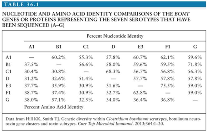
The protein neurotoxin is secreted as a single polypeptide chain of approximately 150 kDa, which is nicked by proteases to form a 100-kDa heavy chain and a 50-kDa light chain connected by a single disulfide bond. The sequences of the genes encoding neurotoxin serotypes A (45–47), B (48,49), C (50), D (51), E (52), F (53,54), and G (55) have been determined. Although these toxins differ by as much as 65% at the amino acid level (Table 36.1), it is likely that they share the same general protein structure (11). Significant sequence variability has also been observed within toxin serotypes, the so-called subserotypes (45,47–49,56–58). The most diverse subserotypes are those for serotype A (up to 16% different at the amino acid level) and serotype F (up to 36% different at the amino acid level) (57,58). Such changes account for the reported differences in the ability of monoclonal and polyclonal antibodies to bind and neutralize BoNTs from different strains (56,59–62).
The x-ray crystal structures of types A, B, and E neurotoxins have been solved at high resolution (Fig. 36.1) (63–66). The structural studies, combined with functional studies, provide clear insight into how the BoNTs interfere with normal release of the neurotransmitter acetylcholine resulting in flaccid paralysis (Fig. 36.2). The C-terminal portion of the heavy chain (HC) comprises the receptor binding domain, which binds to cellular receptors on presynaptic neurons, resulting in toxin endocytosis (67,68) (Figs. 36.2A and B). Specific cellular receptors have been identified for the BoNTs, with cellular binding and entry requiring two co-receptors, a protein and a sialoganglioside such as GD1b or GT1b (68–71) (Fig. 36.3). The binding domain consists structurally of an N-terminal subdomain (HCN) consisting of a jelly roll motif and a C-terminal subdomain (HCC) consisting of a β-trefoil motif. The HCC of the binding domain comprises the protein receptor-binding site as well as the ganglioside-binding site (63) (Fig. 36.3). In BoNT/A, B, E, F, and G, ganglioside binding occurs in a conserved binding pocket in the HCC(Fig. 36.3) (65,72,73). BoNT/C and BoNT/D have two ganglioside-binding sites, one of which is located in the conserved binding pocket used by the other BoNTs. After ganglioside binding, a second binding event occurs to one of several synaptosomal proteins. BoNT/A, BoNT/D, BoNT/E, and BoNT/F bind synaptic vesicle 2 (SV2), whereas BoNT/B and BoNT/G bind synaptotagmin-1 and -2 respectively (64,74–79).
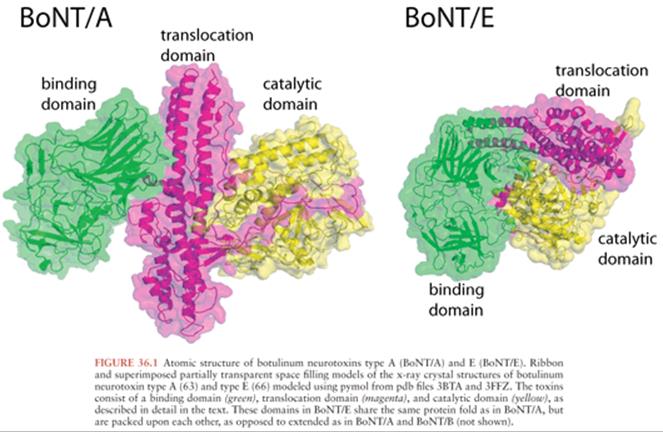
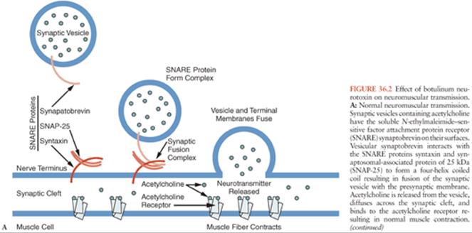


The N-terminal portion of the heavy chain (HN) (Fig. 36.1) comprises the translocation domain, which consists of alpha helices and is involved in pore formation. It is hypothesized that the lower pH level of the endosome induces a conformational change in this domain, which creates a pore allowing the light chain to escape the endosome (Fig. 36.2). The light chain (Fig. 36.1) is a zinc endopeptidase that, depending on serotype, cleaves different members of the soluble N-ethylmaleimide–sensitive factor attachment protein receptor (SNARE) family of proteins, resulting in blockade of neuromuscular transmission (80,81) (Fig. 36.2). The SNAREs are essential for normal fusion of the synaptic vesicle and acetylcholine release (Fig. 36.2). Toxin serotypes A and E cleave distinct sites within SNAP-25 (synaptosomal-associated protein of 25 kDa) (81–84), serotypes B, D, F, and G cleave distinct sites within vesicle-associated membrane protein (VAMP, also known as synaptobrevin) (80,81,85–88) and serotype C cleaves syntaxin and SNAP-25 (Fig. 36.2B) (89,90). These three SNARE proteins (syntaxin, SNAP-25, and synaptobrevin) interact to form a four-helix coiled coil in a step that precedes synaptic fusion (91) (Fig. 36.2). Cleavage of any one of these proteins blocks fusion and acetylcholine release leading to a flaccid paralysis.
BoNTs are secreted from clostridial species as progenitor toxin complexes (PTCs) ranging in size up to 900 kDa (92,93). These complexes consist of the neurotoxin and a number of proteins collectively called neurotoxin-associated proteins (NAPs). The NAPs include three proteins classified as hemagglutinins (HA70, HA17, and HA33) (94,95), because of their ability to agglutinate red blood cells, and other proteins termed nontoxinnonhemagglutinins (NTNH) (53,96). Recently, the x-ray crystal structure of NTNH complexed to BoNT/A was solved showing that NTNH has the same general structure as BoNT/A, packs tightly against BoNT/A in a manner like a “handshake,” and protects it from low pH denaturation and proteolysis in the gastrointestinal (GI) tract (Fig. 36.4) (97–99). The structure of a 760-kDa PTC has recently been solved showing a bimodular structure that the authors liken to an Apollo lunar module (Fig. 36.4). The “ascent stage” is a 280-kDa complex of BoNT/A and NTNH that protects BoNT from GI destruction, and the “descent stage” is a 470-kDa module composed of HA17, HA33, and HA70 that mediates absorption of BoNT by binding to host carbohydrate receptors in the GI tract (100,101).

DIFFERENTIAL DIAGNOSIS
Botulism is underdiagnosed and often misdiagnosed, often as Guillain-Barré or Miller-Fisher syndrome, myasthenia gravis, poliomyelitis, intoxications, or disease of the central nervous system (CNS) (Table 36.2) (7,102). Common and uncommon misdiagnoses are listed in Table 36.2, along with features that distinguish botulism from these diseases. Common signs and symptoms of botulism are listed in Table 36.3 and discussed further in the section on clinical diagnosis. Botulism is much more likely to be associated with outbreaks (cluster of cases) than other diseases with which it may be confused. This fact emphasizes the importance of prompt reporting of suspected botulism cases to the public health department. Botulism differs from other flaccid paralyses in (a) its prominent cranial nerve involvement disproportionate to weakness below the neck, (b) the symmetry of the weakness, and (c) the absence of sensory changes, although approximately 14% of patients report paresthesias (Table 36.3).


EPIDEMIOLOGY
In the most recent report from 2011 of the types of U.S. botulism cases, the CDC documented a total of 140 confirmed cases of botulism; 73% of cases were infant botulism, 20% food-borne, 13% wound, and 4% of unknown etiology (http://www.cdc.gov/nationalsurveillance/PDFs/Botulism_CSTE_2011.pdf).
Food-Borne Botulism
Food-borne botulism is usually associated with ingestion of preformed toxin in home canned products, most frequently foods low in acid such as vegetables, fish or marine mammals, condiments, and meat products. Fruits are rarely involved, because of their high natural acidity. Outbreaks have also been reported from commercially prepared products (103,104) and from food prepared in restaurants (105,106). In the United States, the incidence of food-borne botulism is highest in Alaska, where the vehicle is typically native Alaskan foods consisting of fermented or salted fish or marine mammal products (107). In other countries, different types of foods predominate (108,109). In Germany, Italy, France, and Poland, meats such as home-cured hams are the most frequently implicated foods. In Canada, Japan, and Scandinavia, outbreaks are usually associated with fish products.
Food-borne botulism usually occurs in outbreaks where multiple individuals ingest contaminated food. From 1899 to 1996, 921 outbreaks of food-borne botulism were reported to the CDC, with a relatively constant incidence of approximately 9.5 outbreaks per year, with an average of 2.5 cases per outbreak (1). The largest number of cases in a single food-borne outbreak was 59. From 1899 to 1996, 2,368 cases of food-borne botulism were reported; of these, 1,281 were reported between 1899 and 1949, and 1,087 were reported from 1950 to 1996. Between 1899 and 1949, the case-fatality ratio was approximately 60%. Since 1950, the mortality rate has decreased significantly to approximately 5.7% between 1990 and 1996. This decrease is attributed to improvements in respiratory intensive care and early administration of antitoxin.
Food-borne botulism is typically caused by type A, B, and E neurotoxins. Of the 1,087 cases of food-borne botulism reported between 1950 and 1996, the toxin type could be determined for 786; of these, 52% were type A, 22% were type B, 25% were type E, and less than 1% were type F (1). Type A botulism is most common west of the Mississippi River, type B is most common east of the Mississippi, and type E botulism predominates in Alaska (1,110). This distribution corresponds to the distribution of C. botulinum spores in the soil (111–114). Three outbreaks of type F botulism have been reported in the United States, with one of these due to home-prepared venison jerky (1). In 2011, 70% of the 20 reported food-borne botulism cases were of type A, 25% type E, and 5% type F (http://www.cdc.gov/nationalsurveillance/PDFs/Botulism_CSTE_2011.pdf).
Improvements in food-processing techniques have significantly reduced, but not eliminated, food-borne outbreaks from commercially prepared foods. Those that do occur are associated with inadequate pasteurization or breaks in the refrigeration chain (115). Outbreaks from home canning still occur and can best be prevented by adhering to the recommended canning techniques. C. botulinum spores are not killed by heating at 100°C, so pressure cooking is essential. BoNT, unlike spores, is heat labile and can be destroyed by heating to 80°C (176°F). Thus, thoroughly heating home-canned foods before eating them can reduce the risk of botulism.
Infant Botulism
Infant botulism was first reported in 1976 (17,18) but likely existed as a clinical entity before this date (116). This form of botulism is now the most frequent type of botulism in the United States. Infant botulism is distinct from food-borne botulism, being caused by the colonization (infection) of the intestines with clostridial strains and the subsequent in situ production of toxin, rather than the ingestion of preformed toxin. Between 1976 and 1996, 1,442 cases were reported to the CDC (1). The mean age at onset was 13 weeks, with a range of 1 to 63 weeks, and males and females were equally affected. The incidence of infant botulism has been essentially stable since 1980, with an average annual incidence of approximately 1.9 per 100,000 live births. Since 1976, the highest incidence of infant botulism has been in Delaware, Hawaii, Utah, and California, with almost half of reported cases from California. Reasons for the geographic variation are unknown.
Infant botulism has been reported from at least 14 countries worldwide including Argentina, Australia, Japan, Canada, Italy, the United Kingdom, Chile, the former Czechoslovakia, France, Spain, Switzerland, Sweden, and Taiwan (117; S.S. Arnon, personal communication, May, 2013). In the United States in 2011, 40% of the 102 reported infant botulism cases were type A, 60% were type B, and one case resulted from infection with a bivalent Ba strain. There have been rare reports of infant botulism caused by other serotypes and strains, including one case in Japan caused by type C toxin (118), multiple cases in the United States caused by C. baratii–producing type F toxin (26,39), and multiple cases in Italy caused by C. butyricum–producing type E toxin (37,38). Cases have also occurred due to production of multiple toxins; infant botulism cases have been reported from the United States and the United Kingdom where both types B and F are produced (Bf) (26,42) and from the United States where types B and A are produced (Ba) (41,43).
The primary source for clostridial spores is the environment. Clustering of some cases of infant botulism has been reported in the eastern United States and in small towns and rural areas of the western United States (119–122). In approximately 20% of cases, there was a history of honey ingestion prior to the development of botulism, and spores of the same type could be cultured from the honey (119,123,124). Infants with botulism are more likely to have been breast-fed (124–127), which in experimental models can alter the fecal flora differently than formula feeding and increase susceptibility to C. botulinumcolonization (128).
A tentative link exists between infant botulism and some cases of sudden infant death syndrome (SIDS). C. botulinum spores could be found in 5% of 211 necropsy specimens from SIDS cases in California, with toxin detected in 2 of 10 culture-positive cases (129,130). Toxin or clostridial organisms have been reported in a number of SIDS cases from Switzerland (131), Italy (132), and Germany (133), but not from Australia (134). Thus, infant botulism may account for a small percentage of SIDS cases, perhaps especially in North America and Europe.
Wound Botulism
Wound botulism, first described in 1951, is a rare disease caused by the growth of C. botulinum in contaminated wounds with in situ toxin production. Through 1985, 33 cases of wound botulism were reported in the United States; 25 of these cases were laboratory confirmed, with 17 cases type A and 7 cases type B (1). One case was a mixture of type A and B. Eighty-one percent of the cases occurred in males with a mean age of 21 years. Wounds were typically deep with necrotic areas and associated compound fractures. The median incubation period in trauma cases was 7 days, with a range of 4 to 21 days (135). Since 1980, most cases of wound botulism have occurred in users of illicit drugs. In these individuals, infection is associated with either drug injection or chronic cocaine sniffing with nasal or sinus involvement (136). From 1986 to 1996, 78 cases of wound botulism were reported in the United States, most linked to injection of “black tar” heroin (1). Sixty-six cases were type A, nine were type B, and the remainder of unknown type. The median age of the patients was 38 years and 60% were men. In 2011, all of the 13 reported wound botulism cases were type A and 11 of the 13 patients were injection drug users.
Child or Adult Botulism from Intestinal Colonization
Isolated cases of botulism in adults without a clear food-borne etiology have been reported to the CDC since 1978. These cases appear to be caused by colonization of the GI tract by C. botulinum or C. baratiiwith in vivo toxin production as occurs for infant botulism (21,137). In some cases, patients had a history of GI surgery or inflammatory bowel disease, which may have predisposed them to colonization (22).
Inadvertent or Intentional Botulism
A single incidence of botulism in three laboratory workers has been reported in the literature, which resulted from the inhalation of powdered BoNT (23). Inadvertent botulism can also occur when large doses of medicinal BoNT are injected for therapeutic indications (24,138). As the market for medicinal BoNT has increased, counterfeit BoNTs (139) and highly potent amounts of BoNTs intended for research purposes have made their way into clinical use, increasing the risk for overdose and the development of botulism (140).
Botulinum toxin has already been released as a bioterror agent, albeit unsuccessfully, by the Japanese cult Aum Shinryko (7). Both Iraq and the former Soviet Union produced BoNT for use as weapons (141,142). Iraq produced 19,000 L of concentrated BoNT, of which 10,000 L were weaponized in missile warheads or bombs (141,143). Exposure of even a small number of civilians to BoNT would overwhelm the health care delivery system of any metropolitan center. Treatment of botulism requires prolonged intensive care unit (ICU) hospitalization and mechanical ventilation. With the downsizing and closing of hospitals, most ICUs run at 80% to 100% occupancy. In San Francisco, for example, there are approximately 210 ICU beds, with an average occupancy rate of greater than 90%. As few as 30 cases of botulism would fill all empty ICU beds and occupy them for up to 6 weeks. This would eliminate availability of ICU beds for postoperative patients requiring ICU care, such as organ transplantation, neurosurgery, cardiac surgery, and traumatic injuries. Patients requiring such operations would represent “collateral damage,” with necessary surgery postponed or transferred to outlying hospitals. Major civilian exposure to BoNT would have catastrophic effects. One study estimated that aerosol exposure of 100,000 individuals to toxin, as could occur with an aerosol release over a metropolitan area, would result in 50,000 cases with 30,000 fatalities (144). Such exposure would result in 4.2 million hospital days and an estimated cost of $8.6 billion. In this study, the most important factors reducing mortality and cost were early availability of antitoxin and mechanical ventilation (144). Such treatment could reduce deaths by 25,000 and costs by $8.0 billion.
The intentional release of botulism is most likely to be associated with the outbreak of a large number of cases of flaccid paralysis with prominent bulbar palsies. Other features may include an outbreak with an unusual toxin type (C, D, F, or G), an outbreak with common geographic features but without a common dietary exposure, or multiple simultaneous outbreaks with no common source. The incubation period for intentional botulism is unknown but is likely related to the route of exposure and amount of toxin administered. For oral exposure to toxin, one may obtain some idea of the incubation period from the food-borne botulism literature (see earlier discussion). It is difficult to know precisely the incubation period for aerosol exposure to botulinum toxin due to the paucity of data. In one study, monkeys exhibited signs of intoxication 12 to 80 hours after aerosol exposure with 4 to 7 monkey LD50s (4). The incubation period for the three known cases of human botulism via the inhalation route was 72 hours (23).
CLINICAL HISTORY, SYMPTOMS, AND FINDINGS OF BOTULISM
The diagnosis of botulism is made clinically, with laboratory findings and confirmation not usually immediately available. The clinical syndrome of botulism is dominated by neurologic signs and symptoms resulting from blockade of cholinergic neurotransmission (102,127,135). Patients with botulism usually present with acute onset of weakness in muscles innervated by the cranial nerves, leading to diplopia, dysphonia, dysphagia, and dysarthria (Table 36.2). In mild cases, no other symptoms may develop. In more severe cases, symmetric weakness progresses in a descending manner, leading frequently to paralysis. If the illness is severe enough, the respiratory muscles are involved leading to ventilatory failure and death unless intubation and mechanical ventilation are instituted. In one series, intubation was required in 67% of type A botulism, 52% of type B botulism, and 39% of type E botulism (145). Patients may also have evidence of autonomic dysfunction including dry mouth, blurred vision, orthostatic hypotension, urinary retention, and constipation. Sensory abnormalities are usually absent, because only motor and autonomic nerves are affected. Similarly, mental function is usually not affected.
Paralysis from botulism can be quite long lasting. Mechanical ventilation may be required for 2 to 8 weeks with food-borne botulism, and paralysis lasting as long as 7 months has been reported (102). Symptoms of cranial nerve dysfunction and mild autonomic dysfunction may persist for more than a year (146–148). In infants, hospital stay averages 1 month, with serotype A causing longer lasting disease (5.4 weeks average hospitalization) than serotype B (3.8 weeks average hospitalization) (149). There is experimental evidence that neurotoxin catalytic activity persists at the nerve terminal for days to more than a month, especially for serotypes A, B, and C, and that recovery initially results from the sprouting of new neuromuscular connections (150,151).
Food-borne botulism has an incubation period of 6 hours to 10 days (152), with most cases developing evidence of disease between 18 and 72 hours after ingestion of contaminated food (102,145). GI symptoms are common with food-borne botulism, including abdominal pain, nausea, vomiting, and diarrhea. The GI symptoms may result from ingestion of other bacteria or their toxins. Death occurs in 5% to 10% of patients with food-borne botulism; early deaths result from failure to recognize the disease, and late deaths from complications of long-term mechanical ventilation and ICU care (102).
Infant botulism is initially characterized by constipation, which may precede the development of neurologic symptoms by 1 to 3 weeks, followed by lethargy, poor feeding, and increasing weakness. Other early symptoms include decreased suckling and crying, and neck and peripheral weakness. Because infants are not able to complain, mild symptoms may be missed, leading to a clinical presentation associated with a sudden onset of severe paralysis progressing rapidly to respiratory failure. Treatment with aminoglycoside antibiotics may promote or worsen neuromuscular weakness in infant botulism (153) and is associated with an increased need for mechanical ventilation. The mortality rate for treated infant botulism is less than 2% and in one large series was 0% (149).
Wound botulism presents similarly to food-borne botulism, but without the GI symptoms or signs. There is a history of a wound infection and usually injection or intranasal drug use. Inadvertent botulism occurs in laboratory workers who work with toxin or in patients who have been treated with medicinal BoNT. Bioterror release of toxin must be considered in any outbreak of botulism.
LABORATORY FINDINGS
The diagnosis of botulism should be based on the history and physical findings because routine laboratory tests are not particularly helpful in confirming the clinical suspicion of botulism and specific confirmation takes days. The complete blood count, electrolyte panel, renal and liver function tests, urinalysis, and electrocardiogram will all be normal unless complications have occurred. The cerebrospinal fluid (CSF) is typically normal in botulism, whereas the CSF protein is usually elevated in Guillain-Barré syndrome. The Tensilon test is usually, but not always (152), normal in botulism and may be helpful in distinguishing botulism from myasthenia gravis. The computed tomographic (CT) scan of the head is also normal in botulism and can be used to rule out stroke or other intracranial diseases.
Patients with botulism have normal motor nerve conduction velocities and distal latencies. The electromyogram (EMG), however, may be helpful in the diagnosis of botulism and in distinguishing it from other neuromuscular diseases such as myasthenia gravis and Guillain-Barré syndrome (154–157). In botulism, the EMG of involved muscle groups reveals decreased amplitude of the muscle action potential and facilitation during rapid repetitive or posttetanic stimulation, as can also be seen in patients with Eaton-Lambert syndrome.
Specific laboratory confirmation requires demonstration of toxin in the blood or GI tract and, in the case of food-borne, infant, or wound botulism, culture of clostridial species from stool or wounds (158). Currently, testing for the presence of toxin is available only at the CDC and approximately 20 state and municipal public health laboratories (1). The most sensitive assays for neurotoxin are the mouse bioassay and the use of mass spectrometry. The mouse bioassay is performed by injecting mice intraperitoneally with the toxin-containing sample (serum, stool, food extract, etc.) plus or minus polyclonal and type-specific antitoxin. The mice are observed for 4 days for the development of botulism, with the mice usually dying from botulism within 6 to 96 hours. Protection by simultaneous administration of antitoxin enables determination of serotype. The mouse bioassay can detect as little as 7 pg of toxin (5), the mouse LD50. Alternatively, mass spectrometry can be used for highly sensitive BoNT detection. BoNT is captured out of complex matrices such as blood or stool on antibody-coated beads and then incubated with substrate for the toxin catalytic domain. Substrate cleavage can be detected and quantitated with sensitivities equal to or better than the mouse bioassay (159–162). Such mass spectrometry assays are used alongside the mouse bioassay at the CDC and are likely to be rolled out to the Laboratory Response Network. Higher throughput in vitro tests to detect BoNT, especially variants of enzyme-linked immunosorbent assays (ELISA), are under development but have not been validated (163–165) and have sensitivities generally less than that of the mouse bioassay.
Simultaneously with collection of blood for serologic studies, stool or wound fluid can be cultured for the presence of clostridial species in the cases of botulism occurring via the oral or wound route. Unfortunately, current laboratory tests have not been particularly sensitive for the diagnosis of botulism. Clostridial cultures were positive for 51% of stool specimens collected from 309 patients with clinically suspected botulism (145). Toxin testing was positive in only 37% of sera and 23% of stool specimens. At least one laboratory test was positive in 65% of patients (145). Collecting samples early in the course of disease increases the likelihood of positive results. However, large outbreaks have occurred in which no specimens or a low percentage of specimens gave positive results (166). It can also take days for cultures or toxin testing results to be available. An alternative means of diagnosis is detection of bacteria or toxin in source material, such as contaminated food. Because toxin prepared by terrorists is likely to be crude and unpurified, it is possible that clostridial nucleic acids may be present on bioterror toxin preparations, which could be amplified by polymerase chain reaction for analysis. Cultures and nucleic acid testing would allow more specific classification of the precise Clostridial strain utilized. Because specific therapy with antitoxin must be administered as rapidly as possible to be effective (see later discussion) (167), specific antitoxin therapy must be based on the clinical diagnosis prior to laboratory confirmation.
TREATMENT
Treatment of botulism includes (a) early administration of botulinum antitoxin to prevent progression of moderate illness or reduce the duration of mechanical ventilation in patients with rapidly progressive severe botulism; (b) close monitoring of respiratory function (vital capacity and maximal inspiratory force [MIF]) (168); and (c) intensive care for patients with significant paralysis and evidence of respiratory insufficiency or failure. Vital capacity (VC) should be measured as soon as the diagnosis is suspected and followed closely. In one study, 10 of 11 patients requiring mechanical ventilation had VCs less than 30% of predicted value (169). Patients with VC less than 10 mL/kg should be monitored in an ICU for progression of respiratory failure requiring mechanical ventilation (170). Patients requiring mechanical ventilation obviously merit ICU care.
Treatment with antitoxin is the mainstay of therapy. In the United States, more than 80% of adults are treated with antitoxin. Antitoxin is most effective when administered early in the course of disease and prior to the development of respiratory failure (167). Once the toxin had entered the nerve terminal, antitoxin cannot bind and neutralize it (10). Thus, antitoxin only works on circulating toxin. However, antitoxin will remove any toxin remaining in the bloodstream as well as toxin that may continue to be absorbed from the gut or respiratory tract, thus preventing further intoxication. In infant botulism, antitoxin administered as late as 4 to 7 days after the clinical appearance of symptoms reduced morbidity, such as length of stay (171). Thus, there is rationale for administering antitoxin even a week after the appearance of symptoms. Antitoxin is immunoglobulin harvested from hyperimmunized horses (horse or equine antitoxin) from which the Fc portion has been enzymatically removed (despeciation) to reduce the incidence of side effects such as serum sickness and hypersensitivity reactions. The current licensed equine antitoxin is heptavalent, having activity against serotypes A, B, C, D, E, F, and G (172,173). The CDC should be contacted for information regarding these products. Efforts are underway to generate human monoclonal antibodies that could replace equine antitoxin therapy (174–177).
Equine antitoxins are foreign proteins, and despite despeciation, there is a risk of hypersensitivity reactions. For previous generations of equine antitoxin, the rate of these reactions was as high as 9% and included serum sickness and anaphylaxis (178). The latest generation heptavalent botulism antitoxin (HBAT) appears to be safer, with only one severe reaction (cardiac arrest) reported in the first 228 patients treated (http://www.fda.gov/downloads/BiologicsBloodVaccines/BloodBloodProducts/ApprovedProducts/LicensedProductsBLAs/FractionatedPlasmaProducts/UCM345147.pdf). There was one mild case of serum sickness, and it is recommended that patients be monitored for this and other types of delayed hypersensitivity reaction for up to 21 days. Antitoxin can be obtained via the CDC by contacting the local health department. A single 20-mL dose of antitoxin is recommended. The amount of antitoxin in one 20-mL vial administered intravenously is enough to neutralize toxin amounts many times in excess of those observed in patients with botulism. Antitoxin is diluted 1:10 in 0.9% saline and administered slowly by the intravenous route. To minimize allergic reactions, the infusion rate should be 0.5 mL/min for the first 30 minutes and then 1 mL/min for the next 30 minutes, followed by a rate of 2 mL/min for the remainder of the infusion. Clinicians should review the package insert with public health authorities before using antitoxin.
A second issue with equine antitoxins is that due to despeciation (removal of the Fc portion of the IgG), HBAT has a very short serum half-life that varies depending on the serotype, ranging from 7.3 hours for type E to 34 hours for type B. The half-life for type A is only 8.6 hours, and type F is 14 hours. As a result, it is possible for the initial dose to be cleared from the circulation while BoNT is still being absorbed systemically, for example, from the GI tract. This has led to relapse (rebound) botulism after treatment with HBAT (61). Patients should be monitored for relapse and consideration given to redosing, recognizing the potential for allergic reactions to the equine product.
Equine antitoxin is rarely administered to infants with botulism because of the risk of lifelong hypersensitivity to equine antigens (117). In addition, there is some evidence that anaphylaxis may be more severe in infants given equine antitoxin. As an alternative, human immunoglobulin prepared from volunteers immunized with the investigational botulinum toxoid vaccine has been developed. This U.S. Food and Drug Administration (FDA)–approved product, termed botulism immune globulin (BIG-IV), has been evaluated in a prospective randomized trial in infant botulism. Infants with the clinical diagnosis of botulism were randomized to receive either nonimmune human globulin or BIG-IV. Compared to nonimmune globulin, BIG-IV significantly reduced the duration and cost of hospitalization, and the duration of mechanical ventilation and tube feedings (149). This benefit appeared to accrue even in patients treated as late as 5 to 7 days after the onset of symptoms as well as in those with ongoing toxin production or slow clearance of toxin from the blood (S.S. Arnon, personal communication, May, 2013). BIG-IV is available for treatment of infant botulism from the Infant Botulism Treatment and Prevention Program (IBTPP; http://www.cdph.ca.gov/programs/ibtpp/Pages/default.aspx) of the California Department of Public Health (CDPH) at 510-231-7600.
Botulism is a reportable disease, and suspected cases should be reported immediately to the hospital epidemiologist or infection control practitioner as well as the local and state health departments. The phone number of the health department can usually be found in the phone directory under government listings or via the Internet at http://www.cdc.gov. If local or state health departments are not reachable, the CDC can be contacted directly at 800-CDC-INFO (800-232-4636).
With respect to prevention, the risk of food-borne botulism can be reduced by adhering to proper procedures for home canning, which are available from the U.S. Department of Agriculture Web site. The risk of wound botulism can be reduced by avoiding the injection or inhalation of illicit drugs. Infant younger than 12 months should not be fed honey to reduce the risk of infant botulism. For laboratory workers who might be exposed to large amounts of botulinum toxin, an investigational pentavalent (serotypes A, B, C, D, and E) toxoid used to be available from the CDC for immunization (179). This product has been removed from clinical use by the CDC due to the severity of injection reactions and lack of efficacy. A recombinant vaccine based on the toxin-binding domain for serotypes A and B is under development (180).
ACKNOWLEDGMENTS
This work was partially supported by NIAID grants U01 AI075443, R01AI104579, R21 AI101539, U54 AI065359 and HHS contract HHSN272201100031C.
References
1. Centers for Disease Control and Prevention. Botulism in the United States, 1899–1998. Handbook for Epidemiologists, Clinicians, and Laboratory Workers. Atlanta. U.S. Department of Health and Human Services, Public Health Service; 1998. http://www.bt.cdc.gov/agent/botulism/index.asp. Accessed June 2004.
2. Gill MD. Bacterial toxins: a table of lethal amounts. Microbiol Rev. 1982;46:86–94.
3. Herrero BA, Ecklung AE, Streett CS, et al. Experimental botulism in monkeys—a clinical pathological study. Exp Mol Pathol. 1967;6(1):84–95.
4. Franz DR, Pitt LM, Clayton MA, et al. Efficacy of prophylactic and therapeutic administration of antitoxin for inhalation botulism. In: DasGupta BR, ed. Botulinum and Tetanus Neurotoxins: Neurotransmission and Biomedical Aspects. New York: Plenum Press; 1993:473–476.
5. Schantz EJ, Johnson EA. Properties and use of botulinum toxin and other microbial neurotoxins in medicine. Microbiol Rev. 1992;56(1):80–99.
6. Scott AB, Suzuki D. Systemic toxicity of botulinum toxin by intramuscular injection in the monkey. Mov Disord. 1988;3(4):333–335.
7. Arnon SS, Schecter R, Inglesby TV, et al. Botulinum toxin as a biological weapon: medical and public health management. JAMA. 2001;285:1059–1070.
8. Mahant N, Clouston PD, Lorentz IT. The current use of botulinum toxin. J Clin Neurosci. 2000;7:389–394.
9. Kennedy D. Beauty and the beast. Science. 2002;295:1601.
10. Sugiyama H. Clostridium botulinum neurotoxin. Microbiol Rev. 1980;44(3):419–448.
11. Lacy DB, Stevens RC. Sequence homology and structural analysis of the clostridial neurotoxins. J Mol Biol. 1999;291:1091–1104.
12. Barash JR, Arnon SS. A novel strain of Clostridium botulinum that produces type B and type H botulinum toxins. J Infect Dis. 2014;209:183–191.
13. Middlebrook JL, Franz DR. Botulinum toxins. In: Sidell FR, Takafuji ET, Franz DR, eds. Textbook of Military Medicine Part 1, Warfare, Weaponry, and the Casualty: Medical Aspects of Chemical and Biological Warfare. Washington, DC: Office of the Surgeon General, Department of the Army, United States of America;1997:655–676.
14. Park JB, Simpson LL. Inhalational poisoning by botulinum toxin and inhalation vaccination with its heavy-chain component. Infect Immun. 2003;71(3):1147–1154.
15. Meyer KF. The rise and fall of botulism. Calif Med. 1973;118:63–64.
16. van Ermengem E. Classics in infectious diseases. A new anaerobic bacillus and its relation to botulism. E. van Ermengem. Clin Infect Dis. 1979;1(4):701–719. Originally published, in German, in: Zeitschrift fur Hygiene und Infektionskrankheiten. 1897;26:1–56.
17. Midura TF, Arnon SS. Infant botulism. Identification of Clostridium botulinum and its toxins in faeces. Lancet. 1976;2(7992):934–936.
18. Pickett J, Berg B, Chaplin E, et al. Syndrome of botulism in infancy: clinical and electrophysiologic study. N Engl J Med. 1976;295(14):770–772.
19. Davis JB, Mattman LH, Wiley M. Clostridium botulinum in a fatal wound infection. JAMA. 1951;146:646–648.
20. Werner SB, Passaro D, McGee J, et al. Wound botulism in California, 1951–1998: recent epidemic in heroin injectors. Clin Infect Dis. 2000; 31(4):1018–1024.
21. Chia JK, Clark JB, Ryan CA, et al. Botulism in an adult associated with food-borne intestinal infection with Clostridium botulinum. N Engl J Med. 1986;315(4):239–241.
22. Griffin PM, Hatheway CL, Rosenbaum RB, et al. Endogenous antibody production to botulinum toxin in an adult with intestinal colonization botulism and underlying Crohn’s disease. J Infect Dis. 1997;175(3):633–637.
23. Holzer E. Botulismus durch inhalation. Med Klin. 1962;41:1735–1740.
24. Cobb DB, Watson WA, Fernandez MC. Botulism-like syndrome after injection of botulinum toxin. Vet Hum Toxicol. 2000;42:163.
25. Hatheway CL. Toxigenic clostridia. Clin Microbiol Rev. 1990;3(1):66–98.
26. Hatheway CL. Botulism: the present status of the disease. Curr Top Microbiol Immunol. 1995;195:55–75.
27. Borland ED, Moryson CJ, Smith GR. Avian botulism and the high prevalence of Clostridium botulinum in the Norflok Broads. Vet Rec. 1977;100(6):106–109.
28. Galvin JW, Hollier TJ, Bodinnar KD, et al. An outbreak of botulism in wild waterbirds in southern Australia. J Wildl Dis. 1985;21(4):347–350.
29. Jensen WI, Price JI. The global importance of type C botulism in wild birds. In: Eklund MW, ed. Avian Botulism: An International Perspective. Springfield, IL: Charles C. Thomas; 1987:33–54.
30. Smith GR, Milligan RA. Clostridium botulinum type D in Britain. Vet Rec. 1977;100(6):121–122.
31. Demarchi J, Mourgues C, Orio J, et al. Existence du botulisme de type D. Bull Acad Nat Med. 1958;142:580–582.
32. Prevot AR, Terrasse J, Daumail J, et al. Existence en France du botulisme humain de type C. Bull Acad Natl Med. 1955;139:355–358.
33. Giménez DF, Ciccarelli AS. Studies on strain 84 of Clostridium botulinum. Zentralbl Bakteriol Orig. 1970;215(2):212–220.
34. Giménez DF, Ciccarelli AS. Clostridium botulinum in Argentina: present and future [in Spanish]. Rev Asoc Argent Microbiol. 1976;8(2):82–91.
35. Sonnabend O, Sonnabend W, Heinzle R, et al. Isolation of Clostridium botulinum type G and identification of type G botulinal toxin in humans: report of five sudden unexpected deaths. J Infect Dis. 1981;143(1):22–27.
36. Suen JC, Hatheway CL, Steigerwalt AG, et al. Clostridium argentinense, sp. nov: a genetically homogeneous group composed of all strains of Clostridium botulinum toxin type G and some nontoxigenic strains previously identified as Clostridium subterminale or Clostridium hastiforme. Int J Syst bacteriol. 1988;38:375–381.
37. Aureli P, Fenicia L, Pasolini B, et al. Two cases of type E infant botulism caused by neurotoxigenic Clostridium butyricum in Italy. J Infect Dis. 1986;154(2):207–211.
38. McCroskey LM, Hatheway CL, Fenicia L, et al. Characterization of an organism that produces type E botulinal toxin but which resembles Clostridium butyricum from the feces of an infant with type E botulism. J Clin Microbiol. 1986;23(1):201–202.
39. Hall JD, McCroskey LM, Pincomb BJ, et al. Isolation of an organism resembling which produces type F botulinal toxin from an infant with botulism. J Clin Microbiol. 1985;21(4):654–655.
40. McCroskey LM, Hatheway CL, Woodruff BA, et al. Type F botulism due to neurotoxigenic Clostridium baratii from an unknown source in an adult. J Clin Microbiol. 1991;29(11):2618–2620.
41. Hatheway CL, McCroskey LM, Lombard GL, et al. Atypical toxin variant of Clostridium botulinum type B associated with infant botulism. J Clin Microbiol. 1981;14(6):607–611.
42. Hatheway CL, McCroskey LM. Examination of feces and serum for diagnosis of infant botulism in 336 patients. J Clin Microbiol. 1987;25(12):2334–2338.
43. Giménez DF. Clostridium botulinum subtype Ba. Zentralbl Bakteriol Mikrobiol Hyg A. 1984;257:68–72.
44. Santos-Buelga JA, Collins MD, East AK. Characterization of the genes encoding the botulinum neurotoxin complex in a strain of Clostridium botulinum producing type B and F neurotoxins. Curr Microbiol. 1998;37(5):312–318.
45. Willems A, East AK, Lawson PA, et al. Sequence of the gene coding for the neurotoxin of Clostridium botulinum type A associated with infant botulism: comparison with other clostridial neurotoxins. Res Microbiol. 1993;144(7):547–556.
46. Thompson DE, Brehm JK, Oultram JD, et al. The complete amino acid sequence of the Clostridium botulinum type A neurotoxin, deduced by nucleotide sequence analysis of the encoding gene. Eur J Biochem. 1990;189(1):73–81.
47. Binz T, Kurazono H, Wille M, et al. The complete sequence of botulinum neurotoxin type A and comparison with other clostridial neurotoxins. J Biol Chem. 1990;265(16):9153–9158.
48. Whelan SM, Elmore MJ, Bodsworth NJ, et al. Molecular cloning of the Clostridium botulinum structural gene encoding the type B neurotoxin and determination of its entire nucleotide sequence. Appl Environ Microbiol. 1992;58(8):2345–2354.
49. Ihara H, Kohda T, Morimoto F, et al. Sequence of the gene for Clostridium botulinum type B neurotoxin associated with infant botulism, expression of the C-terminal half of heavy chain and its binding activity. Biochim Biophys Acta. 2003;1625(1):19–26.
50. Hauser D, Eklund MW, Kurazono H, et al. Nucleotide sequence of Clostridium botulinum C1 neurotoxin. Nucleic Acids Res. 1990;18(16):4924.
51. Binz T, Kurazono H, Popoff MR, et al. Nucleotide sequence of the gene encoding Clostridium botulinum neurotoxin type D. Nucleic Acids Res. 1990;18(18):5556.
52. Whelan SM, Elmore MJ, Bodsworth NJ, et al. The complete amino acid sequence of the Clostridium botulinum type-E neurotoxin, derived by nucleotide-sequence analysis of the encoding gene. Eur J Biochem. 1992;204(2):657–667.
53. East AK, Bhandari M, Stacey JM, et al. Organization and phylogenetic interrelationships of genes encoding components of the botulinum toxin complex in proteolytic Clostridium botulinum types A, B, and F: evidence of chimeric sequences in the gene encoding the nontoxic nonhemagglutinin component. Int J Syst Bacteriol. 1996;46(4):1105–1112.
54. Thompson DE, Hutson RA, East AK, et al. Nucleotide sequence of the gene coding for Clostridium baratii type F neurotoxin: comparison with other clostridial neurotoxins. FEMS Microbiol Lett. 1993;108(2):175–182.
55. Campbell K, Collins MD, East AK. Nucleotide sequence of the gene coding for Clostridium botulinum (Clostridium argentinense) type G neurotoxin: genealogical comparison with other clostridial neurotoxins. Biochim Biophys Acta. 1993;1216(3):487–491.
56. Smith TJ, Lou J, Geren IN, et al. Sequence variation within botulinum neurotoxin serotypes impacts antibody binding and neutralization. Infect Immun. 2005;73(9):5450–5457.
57. Raphael BH, Choudoir MJ, Lúquez C, et al. Sequence diversity of genes encoding botulinum neurotoxin type F. Appl Environ Microbiol. 2010;76(14):4805–4812.
58. Hill KK, Smith TJ, Helma CH, et al. Genetic diversity among botulinum neurotoxin-producing clostridial strains. J Bacteriol. 2007;189(3):818–832.
59. Gibson AM, Modi NK, Roberts TA, et al. Evaluation of a monoclonal antibody-based immunoassay for detecting type A Clostridium botulinum toxin produced in pure culture and an inoculated model cured meat system. J Appl Bacteriol. 1987;63(3):217–226.
60. Kozaki S, Kamata Y, Nishiki T, et al. Characterization of Clostridium botulinum type B neurotoxin associated with infant botulism in Japan. Infect Immun. 1998;66(10):4811–4816.
61. Fagan RP, Neil KP, Sasich R, et al. Initial recovery and rebound of type f intestinal colonization botulism after administration of investigational heptavalent botulinum antitoxin. Clin Infect Dis. 2011;53(9):e125–e128.
62. Garcia-Rodriguez C, Levy R, Arndt JW, et al. Molecular evolution of antibody cross-reactivity for two subtypes of type A botulinum neurotoxin. Nat Biotechnol. 2007;25(1):107–116.
63. Lacy DB, Tepp W, Cohen AC, et al. Crystal structure of botulinum neurotoxin type A and implications for toxicity. Nature Struct Biol. 1998;5:898–902.
64. Hanson MA, Stevens RC. Cocrystal structure of synaptobrevin-II bound to botulinum neurotoxin type B at 2.0 A resolution. Nat Struct Biol. 2000;7(8):687–692.
65. Eswaramoorthy S, Kumaran D, Swaminathan S. Crystallographic evidence for doxorubicin binding to the receptor-binding site in Clostridium botulinum neurotoxin B. Acta Crystallogr D Biol Crystallogr. 2001;57(pt 11):1743–1746.
66. Kumaran D, Eswaramoorthy S, Furey W, et al. Domain organization in Clostridium botulinum neurotoxin type E is unique: its implication in faster translocation. J Mol Biol. 2009;386(1):233–245.
67. Dolly JO, Black J, Williams RS, et al. Acceptors for botulinum neurotoxin reside on motor nerve terminals and mediate its internalization. Nature. 1984;307:457–460.
68. Montecucco C. How do tetanus and botulinum toxins bind to neuronal membranes? Trends Biochem Sci. 1986;11:315–317.
69. Simpson LL. Kinetic studies on the interaction between botulinum toxin type A and the chloinergic neuromuscular junction. J Pharmacol Exp Ther. 1980;212:16–21.
70. Nishiki T, Tokuyama Y, Kamata Y, et al. The high-affinity binding of Clostridium botulinum type B neurotoxin to synaptotagmin II associated with gangliosides GT1b/GD1a. FEBS Lett. 1996;378(3):253–257.
71. Rummel A. Double receptor anchorage of botulinum neurotoxins accounts for their exquisite neurospecificity. Curr Top Microbiol Immunol. 2013;364:61–90.
72. Rummel A, Mahrhold S, Bigalke H, et al. The HCC-domain of botulinum neurotoxins A and B exhibits a singular ganglioside binding site displaying serotype specific carbohydrate interaction. Mol Microbiol. 2004;51(3):631–643.
73. Stenmark P, Dupuy J, Imamura A, et al. Crystal structure of botulinum neurotoxin type A in complex with the cell surface co-receptor GT1b-insight into the toxin-neuron interaction. PLoS Pathog. 2008;4(8):e1000129.
74. Chai Q, Arndt JW, Dong M, et al. Structural basis of cell surface receptor recognition by botulinum neurotoxin B. Nature. 2006;444(7122):1096–1100.
75. Dong M, Yeh F, Tepp WH, et al. SV2 is the protein receptor for botulinum neurotoxin A. Science. 2006;312(5773):592–596.
76. Jin R, Rummel A, Binz T, et al. Botulinum neurotoxin B recognizes its protein receptor with high affinity and specificity. Nature. 2006;444(7122):1092–1095.
77. Mahrhold S, Rummel A, Bigalke H, et al. The synaptic vesicle protein 2C mediates the uptake of botulinum neurotoxin A into phrenic nerves. FEBS Lett. 2006;580(8):2011–2014.
78. Mahrhold S, Strotmeier J, Garcia-Rodriguez C, et al. Identification of the SV2 protein receptor-binding site of botulinum neurotoxin type E. Biochem J. 2013;453(1):37–47.
79. Rummel A, Eichner T, Weil T, et al. Identification of the protein receptor binding site of botulinum neurotoxins B and G proves the double-receptor concept. Proc Natl Acad Sci U S A. 2007;104(1):359–364.
80. Schiavo G, Benfenati F, Poulain B, et al. Tetanus and botulinum-B neurotoxins block neurotransmitter release by proteolytic cleavage of synaptobrevin. Nature. 1992;359:832–835.
81. Schiavo G, Rossetto O, Catsicas S, et al. Identification of the nerve terminal targets of botulinum neurotoxin serotypes A, D, and E. J Biol Chem. 1993;268:23784–23787.
82. Schiavo G, Santucci A, Dasgupta BR, et al. Botulinum neurotoxins serotypes A and E cleave SNAP-25 at distinct COOH-terminal peptide bonds. FEBS Lett. 1993;335(1):99–103.
83. Blasi J, Chapman ER, Link E, et al. Botulinum neurotoxin A selectively cleaves the synaptic protein SNAP-25. Nature. 1993;365(6442):160–163.
84. Binz T, Blasi J, Yamasaki S, et al. Proteolysis of SNAP-25 by types E and A botulinal neurotoxins. J Biol Chem. 1994;269(3):1617–1620.
85. Schiavo G, Shone CC, Rossetto O, et al. Botulinum neurotoxin serotype F is a zinc endopeptidase specific for VAMP/synaptobrevin. J Biol Chem. 1993;268(16):11516–11519.
86. Schiavo G, Malizio C, Trimble WS, et al. Botulinum G neurotoxin cleaves VAMP/synaptobrevin at a single Ala-Ala peptide bond. J Biol Chem. 1994;269(32):20213–20216.
87. Yamasaki S, Hu Y, Binz T, et al. Synaptobrevin/vesicle-associated membrane protein (VAMP) of Aplysia californica: structure and proteolysis by tetanus toxin and botulinal neurotoxins type D and F. Proc Natl Acad Sci U S A. 1994;91(11):4688–4692.
88. Yamasaki S, Binz T, Hayashi T, et al. Botulinum neurotoxin type G proteolyses the Ala81-Ala82 bond of rat synaptobrevin 2. Biochem Biophys Res Commun. 1994;200(2):829–835.
89. Blasi J, Chapman ER, Yamasaki S, et al. Botulinum neurotoxin C1 blocks neurotransmitter release by means of cleaving HPC-1/syntaxin. EMBO J. 1993;12(12):4821–4828.
90. Foran P, Lawrence GW, Shone CC, et al. Botulinum neurotoxin C1 cleaves both syntaxin and SNAP-25 in intact and permeabilized chromaffin cells: correlation with its blockade of catecholamine release. Biochemistry. 1996;35(8):2630–2636.
91. Sutton RB, Fasshauer D, Jahn R, et al. Crystal structure of a SNARE complex involved in synaptic exocytosis at 2.4 A resolution. Nature. 1998;395(6700):347–353.
92. Dasgupta BR, Boroff DA, Rothstein E. Chromatographic fractionation of the crystalline toxin of Clostridium botulinum type A. Biochem Biophys Res Commun. 1966;22(6):750–756.
93. DasGupta BR, Boroff DA. Separation of toxin and hemagglutinin from crystalline toxin of Clostridium botulinum type A by anion exchange chromatography and determination of their dimensions by gel filtration. J Biol Chem. 1968;243(5):1065–1072.
94. Balding P, Gold ER, Boroff DA, et al. Observations on receptor specific proteins. II. Haemagglutination and haemagglutination-inhibition reactions of Clostridium botulinum types A, C, D and E haemagglutinins. Immunology. 1973;25(5):773–782.
95. East AK, Stacey JM, Collins MD. Cloning and sequencing of a hemagglutinin component of the botulinum neurotoxin complex encoded by the Clostridium botulinum types A and B. J Syst Bacteriol. 1994;46:1105–1102.
96. Fujita R, Fujinaga Y, Inoue K, et al. Molecular characterization of two forms of nontoxic-nonhemagglutinin components of Clostridium botulinum type A progenitor toxins. FEBS Lett. 1995;376(1–2):41–44.
97. Gu S, Rumpel S, Zhou J, et al. Botulinum neurotoxin is shielded by NTNHA in an interlocked complex. Science. 2012;335(6071):977–981.
98. Ohishi I, Sugii S, Sakaguchi G. Oral toxicities of Clostridium botulinum toxins in response to molecular size. Infect Immun. 1977;16(1):107–109.
99. Chen F, Kuziemko GM, Stevens RC. Biophysical characterization of the stability of the 150-kilodalton botulinum toxin, the nontoxic component, and the 900-kilodalton botulinum toxin complex species. Infect Immun. 1998;66(6):2420–2425.
100. Amatsu S, Sugawara Y, Matsumura T, et al. Crystal structure of Clostridium botulinum whole hemagglutinin reveals a huge triskelion-shaped molecular complex. J Biol Chem. 2013;288(49):35617–35625.
101. Lee K, Gu S, Jin L, et al. Structure of a bimodular botulinum neurotoxin complex provides insights into its oral toxicity. PLoS Pathog. 2013;9(10):e1003690.
102. Hughes JM, Blumenthal JR, Merson MH, et al. Clinical features of types A and B food-borne botulism. Ann Intern Med. 1981;95(4):442–445.
103. Blake PA, Horwitz MA, Hopkins L, et al. Type A botulism from commercially canned beef stew. South Med J. 1977;70(1):5–7.
104. O’Mahony M, Mitchell E, Gilbert RJ, et al. An outbreak of foodborne botulism associated with contaminated hazelnut yoghurt. Epidemiol Infect. 1990;104(3):389–395.
105. Seals JE, Snyder JD, Edell TA, et al. Restaurant-associated type A botulism: transmission by potato salad. Am J Epidemiol. 1981;113(4):436–444.
106. Dodds KL. Restaurant-associated botulism outbreaks in North America. Food Control. 1990;1:139–141.
107. Wainwright RB, Heyward WL, Middaugh JP, et al. Food-borne botulism in Alaska, 1947–1985: epidemiology and clinical findings. J Infect Dis. 1988;157(6):1158–1162.
108. Lund BM. Foodborne disease due to Bacillus and Clostridium species. Lancet. 1990;336(8721):982–986.
109. Roblot P, Roblot F, Fauchère JL, et al. Retrospective study of 108 cases of botulism in Poitiers, France. J Med Microbiol. 1994;40(6):379–384.
110. Eisenberg MS, Bender TR. Botulism in Alaska, 1947 through 1974. Early detection of cases and investigation of outbreaks as a means of reducing mortality. JAMA. 1976;235(1):35–38.
111. Meyer KF, Dubovsky BJ. The distribution of the spores of C. botulinum in the United States. J Infect Dis. 1922;31:559–594.
112. Smith LD. The occurence of Clostridium botulinum and Clostridium tetani in the soil of the United States. Health Lab Sci. 1978;15:74–80.
113. Bott TL, Deffner JS, McCoy E, et al. Clostridium botulinum type E in fish from the Great Lakes. J Bacteriol. 1966;91(3):919–924.
114. Ward BQ, Carroll BJ, Garrett ES, et al. Survey of the U.S. Gulf Coast for the presence of Clostridium botulinum. Appl Microbiol. 1967;15(3): 629–636.
115. Sheth AN, Wiersma P, Atrubin D, et al. International outbreak of severe botulism with prolonged toxemia caused by commercial carrot juice. Clin Infect Dis. 2008;47(10):1245–1251.
116. Arnon SS, Werner SB, Faber HK, et al. Infant botulism in 1931. Discovery of a misclassified case. Am J Dis Child. 1979;133(6):580–582.
117. Arnon SS. Infant botulism. In: Feigen R, Cherry J, eds. Textbook of Pediatric Infectious Diseases. Philadelphia: WB Saunders; 1992:1095 –1102.
118. Oguma K, Yokota K, Hayashi S, et al. Infant botulism due to Clostridium botulinum type C toxin. Lancet. 1990;336(8728):1449–1450.
119. Arnon SS, Midura TF, Damus K, et al. Honey and other environmental risk factors for infant botulism. J Pediatr. 1979;94(2):331–336.
120. Istre GR, Compton R, Novotny T, et al. Infant botulism. Three cases in a small town. Am J Dis Child. 1986;140(10):1013–1014.
121. Long SS. Epidemiologic study of infant botulism in Pennsylvania: report of the Infant Botulism Study group. Pediatrics. 1985;75(5):928–934.
122. Thompson JA, Glasgow LA, Warpinski JR, et al. Infant botulism: clinical spectrum and epidemiology. Pediatrics. 1980;66(6):936–942.
123. Chin J, Arnon SS, Midura TF. Food and environmental aspects of infant botulism in California. Rev Infect Dis. 1979;1(4):693–697.
124. Morris JG Jr, Snyder JD, Wilson R, et al. Infant botulism in the United States: an epidemiologic study of cases occurring outside of California. Am J Public Health. 1983;73(12):1385–1388.
125. Arnon SS, Damus K, Thompson B, et al. Protective role of human milk against sudden death from infant botulism. J Pediatr. 1982;100(4):568–573.
126. Spika JS, Shaffer N, Hargrett-Bean N, et al. Risk factors for infant botulism in the United States. Am J Dis Child. 1989;143(7):828–832.
127. Wilson R, Morris JG Jr, Snyder JD, et al. Clinical characteristics of infant botulism in the United States: a study of the non-California cases. Pediatr Infect Dis. 1982;1(3):148–150.
128. Wells CL, Sugiyama H, Bland SE. Resistance of mice with limited intestinal flora to enteric colonization by Clostridium botulinum. J Infect Dis. 1982;146(6):791–796.
129. Arnon SS, Damus K, Chin J. Infant botulism: epidemiology and relation to sudden infant death syndrome. Epidemiol Rev. 1981;3:45–66.
130. Arnon SS, Midura TF, Damus K, et al. Intestinal infection and toxin production by Clostridium botulinum as one cause of sudden infant death syndrome. Lancet. 1978;1(8077):1273–1277.
131. Sonnabend OA, Sonnabend WF, Krech U, et al. Continuous microbiological and pathological study of 70 sudden and unexpected infant deaths: toxigenic intestinal Clostridium botulinum infection in 9 cases of sudden infant death syndrome. Lancet. 1985;1(8423):237–241.
132. Aureli P, Ferrini AM. Identification of C. botulinum spores in a case of sudden infant death in Italy. Description of a clinical case [in Italian]. Minerva Pediatr. 1988;40(2):125–126.
133. Böhnel H, Behrens S, Loch P, et al. Is there a link between infant botulism and sudden infant death? Bacteriological results obtained in central Germany. Eur J Pediatr. 2001;160(10):623–628.
134. Byard RW, Moore L, Bourne AJ, et al. Clostridium botulinum and sudden infant death syndrome: a 10 year prospective study. J Paediatr Child Health. 1992;28(2):156–157.
135. Merson MH, Dowell VR Jr. Epidemiologic, clinical and laboratory aspects of wound botulism. N Engl J Med. 1973;289(19):1105–1110.
136. MacDonald KL, Rutherford GW, Friedman SM, et al. Botulism and botulism-like illness in chronic drug abusers. Ann Intern Med. 1985;102(5):616–618.
137. McCroskey LM, Hatheway CL. Laboratory findings in four cases of adult botulism suggest colonization of the intestinal tract. J Clin Microbiol. 1988;26(5):1052–1054.
138. Ghasemi M, Norouzi R, Salari M, et al. Iatrogenic botulism after the therapeutic use of botulinum toxin-A: a case report and review of the literature. Clinical Neuropharmacol. 2012;35(5):254–257.
139. Coleman K, Zilinskas RA. Fake botox, real threat. Sci Am. 2010;302(6):84–89.
140. Chertow DS, Tan ET, Maslanka SE, et al. Botulism in 4 adults following cosmetic injections with an unlicensed, highly concentrated botulinum preparation. JAMA. 2006;296(20):2476–2479.
141. United Nations Security Council. Tenth Report of the Executive Committee of the Special Commision Established by the Secretary-General Pursuant to Paragraph 9(b)(I) of Security Council Resolution 687 (1991), and Paragraph 3 of Resolution 699 (1991) on the Activities of the Special Commision. New York: United Nations Security Council; 1995.
142. Bozheyeva G, Kunakbayev Y, Yeleukenov D. Former Soviet Biological Weapons Facilities in Kazakhstan: Past, Present, and Future. Monterey, CA: Center for Nonproliferation Studies, Monterey Institute of International Studies; 1999.
143. Zilinskas RA. Iraq’s biological weapons: the past as future? JAMA. 1997;278:418–424.
144. St John R, Finlay B, Blair C. Bioterrorism in Canada: an economic assessment of prevention and postattack exposure. Can J Infect Dis. 2001;12:275–284.
145. Woodruff BA, Griffin PM, McCroskey LM, et al. Clinical and laboratory comparison of botulism from toxin types A, B, and E in the United States, 1975–1988. J Infect Dis. 1992;166(6):1281–1286.
146. Maroon JC. Late effects of botulinum intoxication. JAMA. 1977;238(2): 129.
147. Mann JM, Martin S, Hoffman R, et al. Patient recovery from type A botulism: morbidity assessment following a large outbreak. Am J Public Health. 1981;71(3):266–269.
148. Ehrenreich H, Garner CG, Witt TN. Complete bilateral internal ophthalmoplegia as sole clinical sign of botulism: confirmation of diagnosis by single fibre electromyography. J Neurol. 1989;236(4):243–245.
149. Arnon SS. Clinical trial of human botulism immune globulin. In: DasGupta BR, ed. Botulinum and Tetanus Neurotoxins: Neurotransmission and Biomedical Aspects. New York: Plenum Press; 1993:477–482.
150. de Paiva A, Meunier FA, Molgó J, et al. Functional repair of motor endplates after botulinum neurotoxin type A poisoning: biphasic switch of synaptic activity between nerve sprouts and their parent terminals. Proc Natl Acad Sci U S A. 1999;96(6):3200–3205.
151. Foran PG, Mohammed N, Lisk GO, et al. Evaluation of the therapeutic usefulness of botulinum neurotoxin B, C1, E, and F compared with the long lasting type A. Basis for distinct durations of inhibition of exocytosis in central neurons. J Biol Chem. 2003;278(2):1363–1371.
152. St Louis ME, Peck SH, Bowering D, et al. Botulism from chopped garlic: delayed recognition of a major outbreak. Ann Intern Med. 1988;108(3):363–368.
153. L’Hommedieu C, Stough R, Brown L, et al. Potentiation of neuromuscular weakness in infant botulism by aminoglycosides. J Pediatr. 1979;95(6):1065–1070.
154. Cherington M, Ginsberg S. Type B botulism: neurophysiologic studies. Neurology. 1971;21(1):43–46.
155. Cherington M. Botulism. Ten-year experience. Arch Neurol. 1974;30(6): 432–437.
156. Cherington M. Clinical spectrum of botulism. Muscle Nerve. 1998;21(6): 701–710.
157. Maselli RA, Bakshi N. AAEM case report 16. Botulism. American Association of Electrodiagnostic Medicine. Muscle Nerve. 2000;23(7):1137–1144.
158. Dowell VR Jr, McCroskey LM, Hatheway CL, et al. Coproexamination for botulinal toxin and Clostridium botulinum. A new procedure for laboratory diagnosis of botulism. JAMA. 1977;238(17):1829–1832.
159. Barr JR, Moura H, Boyer AE, et al. Botulinum neurotoxin detection and differentiation by mass spectrometry. Emerg Infect Dis. 2005;11(10):1578–1583.
160. Boyer AE, Moura H, Woolfitt AR, et al. From the mouse to the mass spectrometer: detection and differentiation of the endoproteinase activities of botulinum neurotoxins A-G by mass spectrometry. Anal Chem. 2005;77(13):3916–3924.
161. Kalb SR, Lou J, Garcia-Rodriguez C, et al. Extraction and inhibition of enzymatic activity of botulinum neurotoxins/A1, /A2, and /A3 by a panel of monoclonal anti-BoNT/A antibodies. PLoS One. 2009;4(4):e5355.
162. Kalb SR, Moura H, Boyer AE, et al. The use of Endopep-MS for the detection of botulinum toxins A, B, E, and F in serum and stool samples. Anal Biochem. 2006;351(1):84–92.
163. Emanuel PA, Dang J, Gebhardt JS, et al. Recombinant antibodies: a new reagent for biological agent detection. Biosens Bioelectron. 2000;14(10–11):751–759.
164. Peruski AH, Johnson LH III, Peruski LF Jr. Rapid and sensitive detection of biological warfare agents using time-resolved fluorescence assays. J Immunol Methods. 2002;263(1–2):35–41.
165. Ferreira JL, Eliasberg SJ, Harrison MA, et al. Detection of preformed type A botulinal toxin in hash brown potatoes by using the mouse bioasssay and a modified ELISA test. J AOAC Int. 2001;84(5):1460–1464.
166. Terranova W, Breman JG, Locey RP, et al. Botulism type B: epidemiologic aspects of an extensive outbreak. Am J Epidemiol. 1978;108(2): 150–156.
167. Tacket CO, Shandera WX, Mann JM, et al. Equine antitoxin use and other factors that predict outcome in type A foodborne botulism. Am J Med. 1984;76(5):794–798.
168. Anderson MW, Sharma K, Feeney CM. Wound botulism associated with black tar heroin. Acad Emerg Med. 1997;4(8):805–809.
169. Schmidt-Nowara WW, Samet JM, Rosario PA. Early and late pulmonary complications of botulism. Arch Intern Med. 1983;143(3):451–456.
170. Hughes JM, Tacket CO. ‘Sausage poisoning’ revisited. Arch Intern Med. 1983;143(3):425–427.
171. Arnon SS, Schechter R, Maslanka SE, et al. Human botulism immune globulin for the treatment of infant botulism. N Engl J Med. 2006;354(5):462–471.
172. Centers for Disease Control and Prevention. Investigational heptavalent botulinum antitoxin (HBAT) to replace licensed botulinum antitoxin AB and investigational botulinum antitoxin E. MMWR. 2010;59(10):299.
173. Hill SE, Iqbal R, Cadiz CL, et al. Foodborne botulism treated with heptavalent botulism antitoxin. Ann Pharmacother. 2013;47(2):e12.
174. Amersdorfer P, Wong C, Chen S, et al. Molecular characterization of murine humoral immune response to botulinum neurotoxin type A binding domain as assessed using phage antibody libraries. Infect Immun. 1997;65:3743–3752.
175. Amersdorfer P, Marks JD. Phage libraries for generation of anti-botulinum scFv antibodies. Methods Mol Biol. 2000;145:219–240.
176. Amersdorfer P, Wong C, Smith T, et al. Genetic and immunological comparison of anti-botulinum type A antibodies from immune and non-immune human phage libraries. Vaccine. 2002;20(11–12):1640–1648.
177. Nowakowski A, Wang C, Powers DB, et al. Potent neutralization of botulinum neurotoxin by recombinant oligoclonal antibody. Proc Natl Acad Sci U S A. 2002;99(17):11346–11350.
178. Black RE, Gunn RA. Hypersensitivity reactions associated with botulinal antitoxin. Am J Med. 1980;69:567–570.
179. Siegel LS. Human immune response to botulinum pentavalent (ABCDE) toxoid determined by a neutralization test and by an enzyme-linked immunosorbent assay. J Clin Microbiol. 1988;26:2351–2356.
180. Byrne MP, Smith LA. Development of vaccines for prevention of botulism. Biochimie. 2000;82:955–966.