
ITZHAK BROOK
Tetanus is a serious but preventable nervous system disorder caused by the toxin produced by Clostridium tetani. Although the disease is rare in the industrial nations, it is still rampant in the developing world. The isolations of the causative organism and its toxin were cornerstones in the history of microbiology. Tetanus was one of the first bacterial conditions to be prevented by immunization, representing a triumph in the application of research and public health measures.
Tetanus is an intoxication manifested mainly by neuromuscular dysfunction expressed as muscle spasms and caused by tetanal exotoxin (tetanospasmin). Tetanus can present in one of four clinical forms: generalized, local, cephalic, and neonatal. The goals of treatment are interruption of the production of toxin, neutralization of the unbound toxin, controlling muscle spasms, management of autonomic dysfunction, and appropriate supportive management. Active immunization with tetanus toxoid is the most effective mean of protection.
HISTORICAL ASPECTS
Ancient physicians recognized the relationship between wounds and a disease producing spasticity, violent movements, and death. Case 7 in the Edwin Smith Surgical Papyrus discusses a patient with a penetrating skull wound who experiences trismus and nuchal rigidity (1). These findings were well known to the Egyptian physician who used them to help formulate the prognosis. This is commonly accepted as the earliest recorded description of tetanus. Hippocrates described the disorder clearly; and his relative contemporary Aretaeus observed that these manifestations were “apt to supervene on the wound of a membrane, or of muscles, or of punctured nerves, when, for the most part, the patients die; for, ‘spasm from a wound is fatal’” (2). Galen noted that cutting a nerve in tetanus stopped the movement but paralyzed the innervated part.
The ensuing millennia saw some refinements in clinical observation. John of Arderne (1307 to 1380), often thought to be the first English surgeon-author, described a case of tetanus in which trismus (“taken with the cramp on his cheeks”) began 11 days after a gardening injury. In the eighteenth century, tetanus was thought to be a consequence of nerve injury. However, the spasms of generalized tetanus were frequently confused with the convulsions of epilepsy. Sir Charles Bell, a noted illustrator and a surgeon, included a patient with tetanus infection in his 1824 text (3) (Fig. 37.1).

Neonatal tetanus (NT) was called the “7-day disease” in the Americas and was known as the “9-day fits” in Dublin. In 1846, Sims proposed the “congestive” theory of neonatal tetanus. He thought that this condition resulted from placing infants on their backs, which compressed the occiput and occluded the veins of the medulla. Beumer determined that the umbilicus was the portal of entry for NT in 1887.
The clinical advances of the nineteenth century culminated in this description by Sir William Gowers (4):
Tetanus is a disease of the nervous system characterized by persistent tonic spasm, with violent brief exacerbations. The spasm almost always commences in the muscles of the neck and jaw, causing closure of the jaws (trismus, lockjaw), and involves the muscles of the trunk more than those of the limbs. It is always acute in onset, and a very large proportion of those who are attacked die.
Gowers, and many of his contemporaries, felt that nontraumatic tetanus accounted for up to 20% of cases; he blamed these on a sudden chill or a frightening episode. Although he recognized the similarity between tetanus and strychnine poisoning, he disparaged the notion of pathophysiologic similarity. In one of his few failures of insight, Gowers dismissed Nicolaier’s first report of a strychnine-like toxin isolated from anaerobic soil bacteria (5). Six years later, Behring and Kitasato (6) proved that immunization with an inactivated derivative of this bacterial extract prevented tetanus.
Effective therapeutic suggestions for established tetanus also date from the nineteenth century. In 1829, Ceroli described the use of morphine as a treatment for tetanus. Based on the observations of Claude Bernard, curare was employed with some rare successes (and some dramatic failures) in France (7), Germany (8), the United States, and England. The lessons in long-term mechanical ventilation learned during the poliomyelitis epidemics of the 1950s finally made treatment with neuromuscular blockade feasible. Hutchinson and Jackson (9) of the National Hospital at Queen Square discussed the use of ether in tetanus in 1861. Meltzer and Auer (10) employed magnesium salts to treat tetanus patients at the dawn of the twentieth century. Gowers (4) gave the following description of autonomic dysfunction in tetanus in 1888: “The pulse is increased in frequency, especially during the paroxysms, and is often very small. There is some reason to believe that the small size of the pulse is due to generalized vasomotor spasm.” However, widespread recognition of the hypersympathetic state did not occur until the prolongation of survival of tetanus patients made possible by ventilatory management.
EPIDEMIOLOGY
Risks for acquired tetanus include infected wounds, infected surgical sites (include contaminated sutures, dressings, or plaster), burns and abscesses contaminated by C. tetani, animal-related injuries (bites and wounds), lack of active immunization against C. tetani, umbilical stump infections (tetanus neonatorum) when mud or feces are applied to the umbilical stump, and puerperal infections when nonsanitized instruments are used (11).
C. tetani has a worldwide distribution and has been recovered from diverse sites, including soil, feces, house dust, and contaminated heroin. It is one of the most common fatal infectious diseases throughout the world and in developing countries; it is an important cause of neonatal death. Natural immunity in communities that are not immunized is about 30% and increases with age (12). The attack rate and age-related mortality rate after the neonatal period is higher in males.
The illness is frequent in countries or in ethnic groups who are less likely to be immunized. In the United States, inadequate tetanus protection in rural elderly individuals was more common as compared to the entire population (13). Tetanus is still a major cause of mortality in those areas of the world with inappropriate hygiene and immunization programs. In 2006, an estimated 290,000 people worldwide died of tetanus, most of them in Asia, Africa, and South America (14).
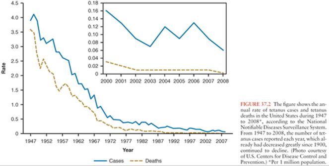
Published mortality figures underestimate the number of deaths but probably represent the most reliable data for much of the world. In the United States, reported cases per 100,000 populations fell from 0.28 in 1955 to 0.02 in 2001 (15). Reported mortality has declined from about 65% in the 1940s to about 20% in the 1990s, reflecting improvements in critical care. During 2001 to 2008, a total of 233 cases were reported in the USA, and the case-fatality rate was 13.2% among the 197 cases with known outcomes. Average annual incidence during that period was 0.10 per 1 million population overall and 0.23 among persons aged 65 years and older (Fig. 37.2). Case fatality was higher among persons older than age 65 years, diabetics, and among unvaccinated persons or those not up-to-date with vaccination (16). An average of 29 cases was reported each year (range: 19 to 40).
Sutter et al. (17) concluded that only 40% of cases are reported to the Centers for Disease Control and Prevention (CDC), 60% are reported to the National Center for Health Statistics, and almost 25% are reported to neither. As the disease becomes less common, more cases are likely to elude recognition, causing an artifactual decline in incidence. Most reported cases occur in patients older than 60 years (18), confirming that waning immunity is a serious problem in this population. Table 37.1 summarizes conditions before onset of tetanus among 130 reported U.S. cases (19).

About three fourth of cases of tetanus in the United States follow injuries. Infected wounds (both traumatic and surgical), abscesses, surgical wounds, major trauma, parenteral drug abuse, and animal-related injuries account for about 25% of the tetanus-associated injuries; about 20% of wounds are due to unknown causes, and in 5%, no source can be found (16).
In the developed world, about 25% of tetanus cases stem from occupational accidents, most frequently agricultural or sylvan. Immigrants to the United States were more than three times less likely to have been immunized than native New Yorkers in one study (20).
NT accounts for about half of all cases worldwide and has a 90% mortality (11). NT is rare in the United States. This is due to the efficacy of the intensive immunization program. Unhygienic childbirth, nonmedical abortion practices, inadequate immunization of mothers, and the lack of care of penetrating wounds explain most cases of neonatal and adult tetanus in the developing world. Furthermore, climate and soil pH in the tropics may contribute to the increased prevalence of C. tetani and its availability to contaminate wounds (11).
A recent estimate of worldwide causes of child mortality between 2000 and 2010 found that NT decreased from 146,000 to 58,000 at 9.5% per year (11). The incidence of NT and factors associated with NT mortality was evaluated in 416 cases seen in a district in rural Pakistan between 1993 and 2003. The overall case-fatality rate (CFR) for NT was 30.1% and fell from 42% in 1993 to 29% in 2003 (p = 0.377). NT incidence decreased from 0.90 per 1,000 livebirths (LB) in 1994 to 0.18 per 1,000 LB in 2003. Multivariable analysis showed that age at admission of 8 days or less with or without low birth weight was the strongest predictor of mortality. The rate of decline of NT incidence and case fatality was attributable to routine and supplementary immunization activities (21).
A study of neonatal mortality in Bangladesh revealed that tetanus caused 112 of 330 deaths (22). There is great geographic variability in incidence of and mortality from the neonatal form of the disease, with an inverse relationship between the extent of maternal immunization and incidence. In 1999, the World Health Organization (WHO) reported that Somalia had the highest reported rate, with 16.49 neonatal tetanus deaths per 1,000 livebirths (23). Poor prognostic factors include age younger than 10 days on admission, symptom duration of less than 5 days on admission, risus sardonicus, and fever (24). About 30% of neonatal tetanus cases occur in babies born to mothers who have already had at least one affected child; this dramatizes the failure to immunize mothers as a major contributor to this condition, because these women are known to lack immunity (25). Immunization programs are clearly effective in decreasing the mortality attributable to neonatal tetanus. Interestingly, tetanus appears to be rare at high altitudes (26).
ETIOLOGY
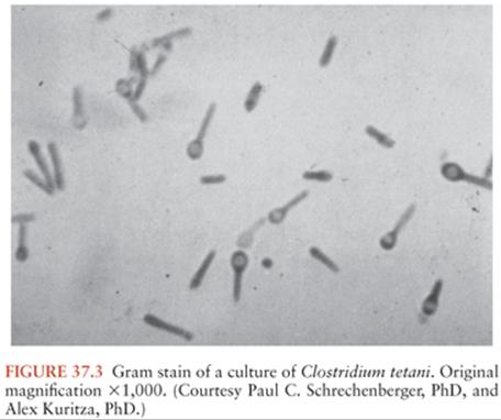
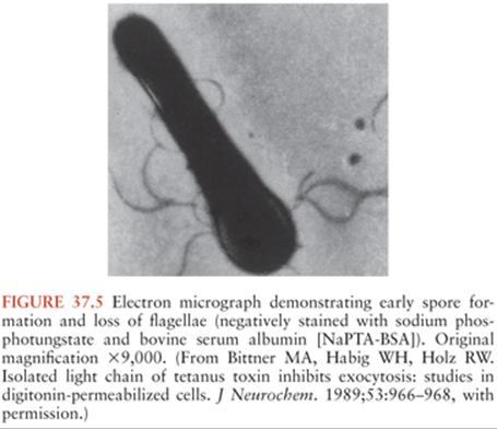
C. tetani (Fig. 37.3) is a slender, obligatively anaerobic bacillus measuring 0.5 to 1.7 µm by 2.1 to 18.1 µm (27). Usually classified as gram positive, it may stain variably, especially in tissues or in older cultures (28). Most strains are sluggishly motile and have abundant peritrichous flagellae during growth (Fig. 37.4). The mature organism loses its flagellae (Fig. 37.5) and forms a spherical terminal spore (29), producing a profile like that of a squash racket (Fig. 37.6). The spores resist extremes of temperature and moisture and are stable at ambient oxygen tension; in addition, they survive indefinitely. They are viable after exposure to ethanol, phenol, and formalin, but they are killed by iodine, glutaraldehyde, or hydrogen peroxide. Strains vary in resistance; exposure to 100°C for 4 hours or autoclaving at 121°C and 103 kPa (15 psi) for 15 minutes is necessary to ensure sterility. They can survive in soil for years and can be found in house dust, soil, salt, and fresh water (30).


Spores can be isolated from the feces of many animals and in small numbers are ubiquitous in soil and on carpets. Hence, any breach in skin defenses (e.g., wounds, burns, animal or human bites, or even insect bites) may result in inoculation. Injuries that favor growth conditions for anaerobic bacteria may lead to the development of tetanus. Tetanus-prone wounds include those contaminated with dirt, feces, or saliva; punctures (including the use of unsterile needles, or injections through unprepared skin); scratches, bites, missile wounds; burns; frostbite; avulsions; and crush injuries. Penetrating ocular injuries are considered tetanus prone based on experimental evidence, whereas nonperforating injuries are not (31). Sometimes, no clear portal of entry can be ascertained. Unusual infection sources include the alimentary tract, tonsils, ear lesions, and contaminated vaccines, sera, and catgut (32,33). Fatal tetanus has occurred in a patient with Stevens-Johnson syndrome. Other unusual sources of tetanus have been described (34). The importance of these observations lies not in their rarity, but in the much higher mortality rate associated with unusual portals of entry. This probably reflects a longer delay in considering the diagnosis. Between 7% and 21% of tetanus cases are cryptogenic (35).
In culture, growth occurs best at 37°C. C. tetani grows on several media if oxygen is excluded. When cultured on a solid medium such as blood agar, the organisms usually form a thin film over the entire surface (“swarming growth”). Routine anaerobic isolation techniques are sufficient if the tissue sample is rapidly placed in an anaerobic transport system. Clinical decisions should not be based on culture results, because (a) cultures are commonly negative in patients with tetanus, (b) isolation of the organism is of no consequence in an immune host, and (c) routine bacteriologic studies will not indicate whether a strain of C. tetani carries the plasmid required for toxin production.
The spores germinate when introduced into a wound and proliferate if the redox potential of the tissue is low. During growth, C. tetani produces two exotoxins: tetanospasmin (TS) and tetanolysin. The potential role of tetanolysin in human tetanus is unclear; at worst, it may damage otherwise viable tissue in the vicinity of an infected wound, lowering the redox potential and promoting the growth of anaerobic organisms. Tetanolysin can disrupt cell membranes, apparently by more than one mechanism. Although systemic administration of tetanolysin in animals produces electrocardiographic abnormalities and disseminated intravascular coagulation, the relevance of these findings to clinical tetanus in humans is uncertain.
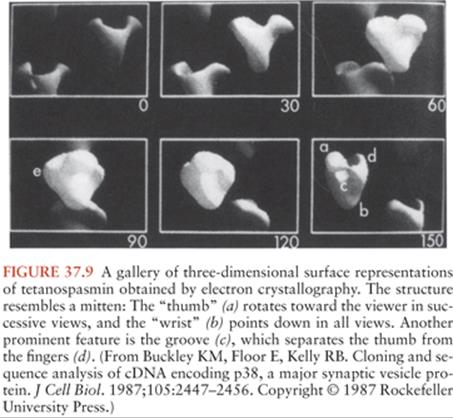
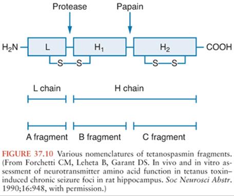
TS, the substance commonly called tetanus toxin, is synthesized as a single 151-kd, 1,315–amino-acid polypeptide chain (36) (Fig. 37.7). The genetic information for this molecule resides on a single large plasmid (37) (Fig. 37.8). Strains of C. tetani lacking this plasmid are not toxigenic and do not cause clinical tetanus. The native molecule has little or no activity but becomes potent when nicked at serine-458 by a bacterial protease (38). This produces one heavy (100-kd) chain and one light (50-kd) chain, connected by a disulfide bridge. This bridge, as well as another one on the heavy chain, is required for the activity of the toxin. These chains or their fragments affect different phases of toxin binding, cell entry, and toxicity. T cells commonly produce an immune response against two particular amino-acid sequences from the amphipathic alpha-helical portion of the molecule. The three-dimensional structure of TS has been determined (39) (Fig. 37.9). The genomic sequence of the bacterial chromosome and the plasmid is now available (40).


Investigators have described several enzymatic digestion products of TS (Fig. 37.10), but their clinical relevance is uncertain. Now that the toxin molecule has been sequenced, reference to the specific amino acids of a fragment is preferred (41). The more commonly investigated fragments are those derived from papain treatment, which cleaves the heavy chain at lysine-865, about 50 kd from the carboxy-terminal end (the C fragment) (42). The light chain and the amino-terminal end, still linked by the disulfide bridge, are variously called the B fragment or the A-B fragment. Attachment and internalization of TS into its target neurons is mediated by the heavy chain (43) or its C fragment (44), whereas the light chain is responsible for inhibiting transmitter release (45,46). The C fragment may also be responsible for the transsynaptic transport of the toxin (46). However, some disagreement remains about these distinctions.
The manifestations of tetanus result from the propensity of TS to inhibit neurotransmitter release by a presynaptic terminal (47). This is a three-step process (Fig. 37.11): binding to the presynaptic membrane, translation of the toxin to the active site, and induction of paralysis. Because TS avidly binds to gangliosides, these molecules were proposed as being the “receptors,” but several lines of evidence suggest that this binding is nonspecific (48). Other evidence implicates a nonganglioside receptor (49), but it has not been characterized. Similarly, the process by which the toxin breaches the cell membrane is not well understood. TS enters the neuron from the extracellular space via noncoated vesicles (50); Pelkmans and Helenius (51) stress the endocytosis of TS via caveolae, as is also the case for cholera toxin and some nonenveloped viruses. The discovery that bafilomycin A1 prevents the action of TS on cultured spinal cord neurons (52) implies that the toxin enters the cytoplasm in an acidic vacuole, analogous to that described for diphtheria toxin (53). Other clostridial neurotoxins appear to share this entry system. The internalization of TS is protease sensitive and appears to require glycosyl-phosphatidylinositol–anchored proteins, which are present in the vicinity of synaptic release site (54). The binding sites may differ biochemically in the central nervous system (CNS) and the peripheral nervous system (55).

Once inside the presynaptic terminal, TS exerts a local effect to inhibit transmitter release for several weeks. Although the synaptosomal content of γ-aminobutyric acid (GABA) is not altered in epileptogenic cortical foci induced by TS, the GABA release evoked by depolarization remains decreased for at least 3 weeks (56). Thus, inhibitory failure is due not to a decrease in available GABA, but to the inability of the presynaptic neuron to release the transmitter it contains.
In contrast to the lack of understanding of binding mechanisms, research in presynaptic function (much of it dependent on TS and other clostridial neurotoxins as tools for dissecting the molecular steps involved) paints an elegant and coherent picture of the mechanism by which these toxins prevent transmitter release (57).
The release of synaptic vesicles by an action potential is initiated by an abrupt rise in the intracellular free Ca2+ concentration, mediated by voltage-dependent calcium channels (Fig. 37.12). This increase in free Ca2+ triggers an interaction between synaptotagmin (in the vesicle membrane) and syntaxin (on the presynaptic cell membrane), clamping the vesicle to the presynaptic membrane. Synaptobrevin (also referred to as vesicle-associated membrane protein [VAMP] [58]) also binds to syntaxin and appears to dock the vesicle to the membrane at the proper location for fusion. There are different isoforms of synaptobrevin within neurons; a protein termed cellubrevin performs a similar function in nonneuronal secretory cells (59). Synaptophysin, the third major component of this mechanism, probably forms the fusion pore that allows release of the vesicle contents into the synaptic cleft (60).
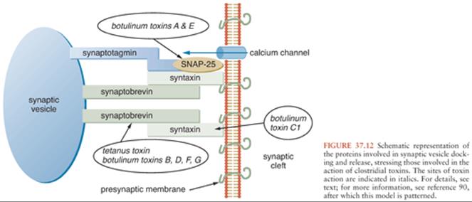
Clostridial neurotoxins inhibit vesicle release by cleaving peptide bonds in these proteins (59). Each toxin has a specific locus of activity (61). TS, along with botulinum neurotoxins B, D, F, and G, cleaves synaptobrevin (62). TS and botulinum neurotoxin B may share the same cleavage site on synaptobrevin. The toxins affect only the free proteins; once they have complexed to cause transmitter release, they are not subject to attack (63). Synaptobrevin and synaptotagmin cleavage also occurs normally, as an effect of an endogenous protease, and they are probably involved in organelle recycling (64). The endogenous protease does not appear homologous to the clostridial toxins.
From the medical standpoint, one of the most important properties of intraneuronal TS is its propensity to travel via the retrograde transport system back to the cell body, which allows access to various other neurons (65) (Fig. 37.13). This process extends from the periphery into the spinal cord as well across several orders of synaptically connected neurons in the brain. The particular clinical manifestations of tetanus depend on the classes and locations of the affected cells, as discussed in the section “Pathogenesis and Pathophysiology,” later in this chapter.

CLINICAL MANIFESTATIONS
Tetanus is traditionally classified into four clinical types: generalized, local, cephalic, and neonatal. These distinctions are useful diagnostically but do not reflect toxicologic differences. Rather, they reflect variations in the site of toxin action.
Although a portal of entry can usually be determined, the lack of a defined wound does not exclude tetanus. Similarly, bacterial stains and cultures of wounds are of no consequence in its diagnosis or management. A “protective” titer of antitetanus antibody may help exclude the diagnosis, but only in retrospect. This is an area of controversy.
The temporal development of symptoms in each form of tetanus is of great prognostic significance. The incubation period extends from the time of spore inoculation to the first symptom, and the period of onset marks the time from that first symptom to the first reflex spasm (i.e., a spasm produced in response to sensory stimulation, as opposed to an apparently spontaneous spasm). In a study of 176 patients from Brazil, the incubation period for patients with severe disease (N = 116) was 8.3 ± 4.7 days, whereas that for patients with moderate and mild disease severity (N = 60) was 11.0 ± 6.7 days. The corresponding periods of onset were 1.9 ± 1.5 days and 3.2 ± 1.9 days, respectively (66). Regardless of the clinical type of tetanus, shorter incubation and onset periods indicate a poorer prognosis.
A general assessment of the disease severity can be predictor of the outcome and can assist in determining the timing and need for airway protection. The portal of entry is another important prognostic factor, with burns, head and neck infections, umbilical stumps, surgical procedures, compound fractures, septic abortions, and intramuscular injections all associated with lesser chances for recovery. Injection drug users, especially those injecting narcotics and heroin, appear to develop particularly severe tetanus (67). Tetanus following the intramuscular injection of quinine, used in some third-world countries for malaria treatment, has an unusually high mortality rate and develops very quickly (68). This probably reflects the very acidic milieu produced by quinine injection. Fever and tachycardia, if reflecting autonomic dysfunction rather than wound infection, are similarly poor prognostic signs (69). The outcome is also influenced by age and comorbidity.
Several authors have developed rating systems for severity and prognosis (70–72), summarized in Table 37.2. The modified Ablett scale is frequently used for making decisions regarding tracheostomy, which is performed on moderate or severely affected patients (73).

Generalized Tetanus
This is the most common form of clinical tetanus. It may occur after relatively minor injuries. Patients generally have tonic contraction of their skeletal muscles and intermittent intense muscular spasms. Tonic and periodic spasmic muscular contractions account for the classic clinical findings: opisthotonus, stiff neck, risus sardonicus (sardonic smile) (Fig. 37.14), a boardlike rigid abdomen, periods of apnea caused by viselike contraction of the thoracic muscles and/or glottal or pharyngeal muscle, and dysphagia.

The onset can be insidious; however, the typical initial findings of trismus, or lockjaw, due to parapharyngeal and masseter muscles spasms is observed in about half of cases (32,33). Patients manifest tonic contraction of their skeletal muscles and intermittent intense muscular spasms. These may be triggered by sensory stimuli such as loud noises, contact, or light. Tonic and periodic spasms are responsible for most of the clinical findings of tetanus. Trismus is caused by rigidity of the masseter muscles, producing inability to open the mouth (see Fig. 37.14); its course can be followed by measuring the distance between the upper and the lower teeth with the mouth maximally open. Trismus is the most common presenting sign, although back or shoulder stiffness may have been present for hours. The most common complaints are pain, swallowing difficulty, and unilateral or bilateral stiffness of the neck and other muscle groups, such as those of the thorax or abdomen (74). Persistent trismus accounts for “risus sardonicus,” which is the classic finding of tetanus (see Fig. 37.14). Risus sardonicus is often a subtle finding that may best be diagnosed by the family or friends of the patient. Physicians can diagnose risus sardonicus by comparison with photographs or in retrospect.
As the illness progresses, more muscle groups get involved. One of the most significant findings occur with spasm of the paraspinal musculature, resulting in severe opisthotonos (see Fig. 37.1), where in young infants, the soles of the feet may touch their head. The typical generalized spasm resembles decorticate posturing, consisting of “a sudden burst of tonic contraction of muscle groups causing opisthotonos, flexion and adduction of the arms, clenching of the fists on the thorax, and extension of the lower extremities” (75). Although the spasms may be confused with posturing or epileptic seizures, they do not produce loss of consciousness and are extremely painful. The tetanic contractions progress further for several more days, with recruitment of additional muscle groups and significant aggravation of symptoms. Painful spasms and contraction can also contort and distort the patient’s posture. All voluntary muscles may be affected, and the disease can involve the larynx, which can be fatal. Fractures of vertebrae or other bones and hemorrhage into muscles can also take place. Even minor stimuli including light, drafts, noises or voices, and light touch may trigger reflex spasms. Because patients remain conscious throughout these spasms, anxiety and pain further complicate management and can contribute to the severity of disease.
Symptoms of autonomic overactivity are generally manifested in the early phases as irritability, restlessness, sweating, and tachycardia. In later phases of illness, profuse sweating, cardiac arrhythmias, labile hypertension or hypotension, and fever are commonly present (76–78). Episodes of bradycardia and hypotension can lead to cardiac arrest. Cardiac arrest has also been attributed to myocardial damage caused by the high catecholamine level (79) and toxic damage to the brainstem (80). Fever can be due to the sympathetic overactivity or superinfections, such as pneumonia (81). Cardiovascular instability due to spasms and inadequate sedation need to be excluded. Cardiovascular complications are managed in the intensive care setting, where ventilatory support and therapeutic paralysis are available. Spasms and cardiovascular complications occur most commonly during the first week and resolve slowly during the ensuing 2 to 4 weeks.
Respiratory compromise is the most serious early problem in generalized tetanus. Upper airway obstruction is common during spasms. The diaphragm and abdominal musculature are often involved, and they can produce apnea in inadequately treated patients in spite of mechanical ventilation. The neuromuscular junction (NMJ) effects of the toxin may produce diaphragmatic paralysis or bilateral vocal cord paralysis (82). Severity may continue to increase for 10 to 14 days after diagnosis, reflecting the transport time of intraneuronal toxin into the CNS. Recovery then begins, usually requiring about 4 weeks. This period probably reflects the time needed for synthesis and transport of presynaptic constituents. In the absence of antitoxin, disease persists as long as TS is produced. The total amount of toxin produced is so small that it is inadequate to prompt an immune response; therefore, patients with newly diagnosed tetanus must be actively immunized to prevent recurrence. Recurrent tetanus is well documented if this is not done (83).
Local Tetanus
This is an unusual presentation of tetanus that occurs when circulating antitoxin prevents general spread of the toxin but is insufficient to stop local uptake at a wound site (75,84). This causes mild, prolonged, steady without progression, and painful muscle contraction in the wounds’ region, which can last for several weeks to months, with subsequent complete spontaneous resolution. Localized tetanus may be unrecognized or mistaken for pain-induced muscle spasms. Neuromuscular transmission may be affected locally, producing weakness in addition to rigidity. Partial immunity to TS may contribute to the development of local tetanus by decreasing the hematogenous spread of toxin (85). More commonly, however, local tetanus is the harbinger of the generalized form, unless treated.
Cephalic Tetanus
This condition is a rare manifestation of tetanus that involves only the cranial nerves after C. tetani enters wound(s) or chronic infection(s) in the head and neck. Although any of the cranial nerves may be affected, singly or in combination, cranial nerve VII is most frequently involved, but involvement of cranial nerves VI, III, IV, and XII may also occur. Patients may present with confusing clinical findings including facial paresis, dysphagia, trismus, and other focal cranial neuropathies. Cephalic tetanus may precede generalized disease, and isolated cephalic tetanus can occur and follows a chronology similar to generalized disease.
Although most series suggest a poor prognosis, a large report from India described mild cases associated with chronic otitis media (86). This may represent colonization of the infected tissue with C. tetani and subsequently the production of toxin. Of 22 patients in one series with otogenic tetanus, 17 had acute otitis media (87). A coexisting aerobic infection was diagnosed in 85%, most commonly with Staphylococcus aureus. Rarely, extraocular movements are affected in patients with tetanus, causing “ophthalmoplegic tetanus” (88) or supranuclear oculomotor palsies (89). Horner syndrome has been reported as a presenting feature (90).
Neonatal Tetanus
This is a generalized form of the disease that often develops in infants delivered to mothers who have not been immunized. This is because passively transferred maternal antibody is protective. The lack of immunization of mothers and the birth practices in developing countries that include lack of aseptic techniques in managing the umbilical stump and application of mud, clarified butter, or feces to the umbilical stump increase the risk of acquiring this illness and are responsible for a large proportion of cases (91). Five factors are involved with NT: (a) the length of the stump (longer seems safer), (b) the care with which the cord is ligated, (c) the cleanliness of the instruments and dressings, (d) the cleanliness of the environment and of the patient’s and mother’s clothing, and (e) the application of mud, feces, or unclean material on the stump (92).
This form of tetanus usually occurs in infants within 14 days of birth and generally manifests initially by inability to suck, and then by tetanic spasms and rigidity producing the typical opisthotonic posture, and trismus (Fig. 37.15). The hypersympathetic state occurs commonly in these infants, and the mortality rate is high, with infants dying of complications such as CNS hemorrhage, pneumonia, pulmonary hemorrhage, and laryngeal spasms with asphyxia. One study found that 33% of infants with tetanus were also bacteremic, mostly with coliforms or S. aureus, most commonly from an infected umbilical cord stump (93). Developmental delay is common in survivors (94,95).

A Turkish study implicated unhygienic home deliveries in more than 90% of NT cases (94). A recent study in Pakistan showed that delivery onto soil compared to a clean surface resulted in a 3.2-fold increase in the odds ratio (OR) of death from NT (95). Other statistically significant risks included paternal illiteracy (OR = 3.2) and having sheep at home (OR = 2.0). Soil as a delivery surface accounted for 64% of the neonatal deaths. Although these concerns are important, active maternal immunization—or passive immunization of the mother before delivery and of the child at birth—would eliminate the disease.
PATHOGENESIS AND PATHOPHYSIOLOGY
After introduction into tissues, C. tetani spores convert to vegetative forms, multiply, and produce TS. In numerous instances, there is no associated inflammation or apparent local infection. At the site of entry, the toxin enters the peripheral nerve and reaches the CNS through the nerves (25,84,96).
The actions of TS predominantly involve three components of the nervous system: central motor control, autonomic function, and the NMJ (76). The central effects have been studied most intensively, and they provide paradigms for understanding the toxin’s effects on other nervous system components.
Central Motor Control Effects
To express its toxic potential, TS must gain access to its target neurons. The toxin appears to enter the nervous system predominantly through the NMJ of alpha motor neurons. Some toxin enters sensory and autonomic neurons, but the amount appears small and its contribution to symptoms is uncertain. It then moves, via the retrograde transport system, to the cell body (57). This system, consisting of microtubules and transport proteins, is normally used to bring signal molecules and exhausted presynaptic components back to the cell for processing. The heavy chain or the C fragment of the toxin is necessary for retrograde transport. TS also spreads hematogenously from its site of production, but it still must enter via neurons (97). Toxin already in transit within the neuron is inaccessible to antitoxin, which partly explains the progression of the disease for several days after treatment. The intrathecal administration of human tetanus immune globulin (HTIG) is an attempt to circumvent this by allowing access to TS moving across synapses in the CNS (see the section “Immunotherapy,” later in this chapter).
Once transported to the spinal cord or the brainstem, the toxin then migrates transsynaptically into presynaptic inhibitory cells (98), which use either glycine or GABA as transmitters (Fig. 37.16). The glycinergic cells are most important in the spinal cord, whereas the GABAergic cells are responsible for decreasing inhibition from the brainstem (99). By preventing glycine or GABA release, TS denies the alpha motor neuron its most essential inhibitory transmitters. This raises its resting firing rate, causing muscle rigidity. Moreover, the normal inhibition of other motor neurons during movements of a particular motor group depends on these inhibitory transmitters, as does the termination of reflexive contractions. Deprived of this inhibition, the motor system responds to an afferent stimulus with the intense, sustained contraction of a wide range of muscles that characterizes the tetanic spasm.

There may be some differences in the predominant effects of local and generalized tetanus regarding the loci of inhibition. Following intravenous injection, TS appears to affect supraspinal (i.e., GABAergic) inhibitory mechanisms exclusively, whereas local tetanus likely has more effect on the spinal glycinergic systems. Because the human disease is caused by spore inoculation rather than toxin administration, clinical generalized tetanus represents a variable combination of these mechanisms. In addition to the loss of inhibitory systems, excitatory transmission is also disrupted (100). This may partially explain the weakness associated with local tetanus.
Histologic abnormalities have been reported in the brainstem nuclei in fatal cases (101), but the significance of these changes is unknown. They most likely reflect terminal hypoxia and autonomic dysfunction. Reversible chromatolysis of motor neurons occurs in experimental tetanus when markedly suprathreshold doses of toxin are employed. Whether this is simply a manifestation of hypermetabolism or reflects another pathophysiologic process is unresolved.
Once transported into the CNS, the toxin continues its rostral movement by retrograde transport (97,102). This implies that structures above the brainstem could be affected by TS; however, there is no evidence that this occurs in the natural history of tetanus. It is likely that generalized tetanus does involve some vertical movement of toxin within the neuraxis, but the widespread manifestations in this form are mostly due to the hematogenous dissemination of toxin to NMJs throughout the body. TS does inhibit the evoked release of norepinephrine (103), acetylcholine (104), serotonin (105), and enkephalin (106) from brain tissue in vitro. TS is also epileptogenic, producing depolarizing shifts when added to CNS tissue in culture and partial seizures when injected intracerebrally. These acute seizures are associated with diminished extracellular GABA levels (107). However, the abnormal electrical activity outlasts the period of GABAA inhibition, indicating long-lasting reorganization of synaptic function (108).
Autonomic Nervous System Effects
Before modern intensive care, most patients with severe tetanus died quickly from ventilatory failure (109). Although autonomic involvement was mentioned in 1954, the first major report of this aspect of tetanus appeared in 1968 (74). The latter group described “a characteristic syndrome whose features include sustained but labile hypertension and tachycardia, irregularities of cardiac rhythm, peripheral vascular constriction, profuse sweating, pyrexia, increased carbon dioxide output, increased urinary catecholamine excretion, and, in some cases, the development of hypotension.” Although these signs are usually present toward the end of the first week, they may develop during the second (110). Most are associated with elevated catecholamine levels.
An elevated plasma epinephrine and norepinephrine concentration (74,111) and 24-hour urine catecholamine excretions (112) were reported in patients with tetanus. These manifestations resemble the effects of a pheochromocytoma; a similar cardiomyopathy is seen in both conditions (78).
Udwadia et al. (113) performed invasive hemodynamic monitoring of 27 patients with severe tetanus. The major finding was a hypersympathetic state, with tachycardia (mean, 131 beats per minute) and increased cardiac (5.48 liters per minute) and stroke volume (43.1 mL/m2) indexes. A total of 19 patients experienced episodes of sustained tachycardia (>150 beats per minute); 17 had hypertension of varying degree, whereas 8 experienced alternating hypertension and hypotension, and 5 had episodic hypotension only. Seven patients had paroxysmal supraventricular tachycardia, and three had brief runs of ventricular tachycardia. Despite the difficulties experienced by their patients, the mortality rate was 6.25%.
TS disinhibits sympathetic reflexes at the spinal level, implying that the hyperadrenergic findings are not dependent on hypothalamic or brainstem dysfunction (114). Parr et al. (115) later showed that cutaneous stimulation of animals with local tetanus increased firing rates in renal sympathetic nerves. Conversely, the development of inappropriate antidiuretic hormone secretion supports hypothalamic involvement (116).
The identification of TS as a Zn2+-dependent peptidase (117) leads to a new hypothesis to explain the hypertension seen in tetanus patients. Captopril, an inhibitor of Zn2+-dependent proteases, antagonizes the effect of TS on synaptobrevin and allows exocytosis to proceed normally (118). Antibodies directed against the Zn2+-binding domain of TS restore exocytosis in chromaffin cell cultures over 4 days (119); however, this model differs substantially from the synapse, so such rapid restoration of function would not be anticipated in clinical disease; chelation of Zn2+ by ethylenediaminetetraacetate (EDTA) and other chelators is also effective in experimental systems (120). Nonetheless, these results suggest that TS might, in addition to its other effects, have an angiotensin-converting enzyme (ACE)–like effect, and a component of the hypertension in patients with tetanus may be related to excess angiotensin-II effects rather than catecholamine effects.
Clinical evidence suggests some disruption of parasympathetic function as well. Bradycardia is occasionally noted as is hypotension (121) without previous evidence of increased sympathetic tone (122). Although both of these findings can be reproduced by injection of TS into a rat’s third ventricle (123), a more peripheral mechanism appears likely in human tetanus.
Neuromuscular Junction Effects
Because the central effects of TS are so dramatic, the peripheral disorder it produces received scant clinical attention until 1977. Investigators showed denervation changes in the muscle closest to the site of the injury that introduced C. tetani, which reflected failure of neuromuscular transmission (124). Experimental studies by Harvey (125) established the presence of neuromuscular blockade 40 years earlier. Single-fiber electromyography (EMG) confirmed a presynaptic defect of acetylcholine release qualitatively similar to that of botulism (126). The NMJ may be permanently disabled by TS; return of function requires sprouting of the terminal end to produce new synapses (124).
APPROACH TO DIAGNOSIS
Tetanus can be diagnosed only clinically. Generalized and cephalic tetanus are easily recognized if one thinks of the disease. Localized tetanus is more difficult to detect early. The neonatal form is less easily recognized on initial presentation, but it rapidly becomes obvious. Laboratory assistance is confined to the exclusion of other diagnoses and to the determination of immunity. Tetanus can follow an incubation period from 3 to 14 days after an injury.
EMG studies may be useful by showing evidence of denervation, reinnervation, and increased excitability of the motor neuron pool (127). The EMG differential diagnosis of tetanus has been reviewed (128). Blood counts are normal or slightly elevated; CSF measures are normal; and electroencephalogram (EEG) and EMG are normal and nonspecifically abnormal, respectively. Gram stains of wounds may reveal the characteristic gram-positive bacilli with terminal spores in as many as one third of tetanus patients. Even though a positive wound culture can support the clinical diagnosis, a positive culture in the absence of symptoms does not indicate that clinical tetanus will develop.
The spatula test is a simple test that can be diagnostic (129). The posterior pharyngeal wall is touched by a spatula, which induces a reflex contraction of the masseters in the presence of tetanus.
Gowers (4), again, provided an unequaled clinical description in 1888:
The first symptom is usually a sense of stiffness in the neck or jaw, sometimes difficulty in swallowing, or stiffness of the tongue. . . . In the course of a few hours, or at most a day or two, the difficulty in separating the jaws becomes greater, and is clearly due to increasing rigidity of the masseters. With this there is also more stiffness in the neck, and the head is slightly bent backward from the preponderance of spasm in the extensor muscles. As the rigidity in the neck increases, it passes down the spinal muscles. . . . The legs may also become extended and rigid, but the arms are little affected.
A few patients have atypical features on presentation. The lack of an easily identified portal of entry, discussed earlier, does not exclude the diagnosis but makes prognostication more difficult because the incubation period cannot be determined. These patients should be carefully examined for signs of parenteral drug abuse, otitis, or rectal or vaginal instrumentation. Recent injections or minor surgical procedures may be uncovered by questioning. Lesions of the gastrointestinal tract are occasionally implicated, especially among inhabitants of rural areas among whom the carriage rate of C. tetani may be 20-fold higher than that of urban dwellers (130).
Differential Diagnosis
Although many disorders have small areas of overlap with tetanus, strychnine intoxication is the only true mimic (131). Strychnine is a component of some rodenticides. This drug is a direct antagonist at the glycine receptor. Lack of abdominal rigidity between spasms may be more common with strychnine than with tetanus, and trismus may be absent in the former; otherwise, the clinical presentations are similar. For this reason, biochemical analyses of serum and urine for strychnine should be performed in suspected tetanus cases, and tetanus should be considered even when strychnine poisoning is likely. The initial symptomatic management of both conditions relies on benzodiazepines, but their subsequent treatments and complications differ substantially. Many other glycine and GABA antagonists have been exploited as experimental convulsant agents; although human poisoning has not been reported, conditions resembling tetanus may be expected.
Dystonic reactions to dopamine blockade usually involve torticollis, and oculogyric crises are common. Neither of these phenomena is part of tetanus, and reflex spasms are not seen. A rapid response to anticholinergic agents (benztropine, 1 to 2 mg; or diphenhydramine, 50 mg) and subsequent toxicologic studies help confirm the diagnosis of dystonic reaction. A trial of anticholinergic agents is reasonable in suspected tetanus if the diagnosis is in doubt. There are anecdotal reports of transient improvement in the symptoms of tetanus after anticholinergic drug administration, perhaps reflecting a sedative effect (132).
A report of opisthotonic posturing and risus sardonicus in a patient suffering from an amphetamine overdose is difficult to reconcile with the expected effects of amphetamines but reminds us of the need to keep an open mind in differential diagnosis (133).
The nuchal rigidity of meningitis may resemble the neck stiffness of tetanus, but the other manifestations of each disorder should resolve any confusion. The cerebrospinal fluid is normal in tetanus. The other infection that may raise a question of tetanus is an alveolar ridge abscess producing trismus. Oral pain and tenderness is not characteristic of tetanus; the patient with a dental disorder will not display spasms or rigidity. However, various dental infections may produce tetanus (134), and temporomandibular joint dislocation may be a symptom of the disease (135). Verma et al. (136) described a single case of unilateral trismus resulting from a tuberculoma of the brainstem.
Patients experiencing generalized convulsive status epilepticus may at first appear to have tetanic spasms, but the loss of consciousness and evolutionary movements of the former should quickly resolve any question.
Tetany precipitated by hypocalcemia or alkalosis will be accompanied by Chvostek and Trousseau signs. In contrast to tetanus, tetany involves the extremities more than the axial musculature.
Progressive fluctuating muscular rigidity (the “stiff person” syndrome) has been likened to a chronic form of tetanus (137). This idiopathic syndrome has an insidious onset, usually has minimal cranial nerve involvement, lacks trismus, and is relieved during sleep. Current evidence links this disorder to autoantibodies against GABAergic neurons (138).
Local tetanus is rarely confused with other disorders; the major problem is failure to consider the diagnosis. It has been confused with transverse myelitis (139).
“Pseudotetanus” has been used in the past to describe a broad variety of disorders (140) and may be due to malingering or a conversion disorder (141). In the former, the patient’s posture may be complex or inconsistent; rigidity is lacking or feigned, the patient is distractible, and some secondary gain is identified. Preexisting psychologic problems may also complicate the diagnosis and management of tetanus (142).
Determination of Immunity
Tetanus Toxoid Adsorbed, for intramuscular use, is a sterile suspension of alum-precipitated (aluminum potassium sulfate) toxoid in an isotonic sodium chloride solution containing sodium phosphate buffer to control pH. When properly administered and maintained, immunization with tetanus toxoid is highly effective, with an estimated failure rate of less than 4 per 100 million persons in those who are immunocompetent (143). Therefore, a confirmed history of active immunization almost eliminates tetanus as a diagnosis. This includes both an acceptable initial series and a booster within 10 years.
Human antibody against tetanus toxoid is predominantly in subclass immunoglobulin G1 (IgG1); there is a moderate amount of IgG3 and IgG4, and minimal IgG2 (144). The serum concentration of antitetanus antibodies can be measured by immunoassay or hemagglutination, but the results are seldom available in time to influence initial diagnosis and management. The results are quantitated in international units (IU) of antitoxin by reference to an arbitrary international standard, which has achieved international agreement (145). The development of a rapid test holds promise not only for the exclusion of tetanus but also for decisions regarding antitoxin administration (see the section “Therapy,” later in this chapter) (146).
In most epidemiologic studies, a level of 0.01 IU/mL is accepted as protective, based on one study in guinea pigs reported in 1937 (147). This level prevented death in the animals, but a few developed tetanus with concentrations between 0.1 and 0.5 IU/mL, from which they recovered. Nonetheless, cases of human tetanus have been reported with titers as high as 0.16 IU/mL (148–150). In one study of 64 tetanus patients, 24 had detectable antitoxin levels, and 10 had levels greater than 0.01 IU/mL (151). The severity of tetanus tended to be less in the patients with higher antibody levels, but six of the ten required tracheostomy and mechanical ventilation. In a study of 20 cases of NT in Nigeria, the disease developed in six infants whose mothers had been immunized; the mean antitetanus IgG concentration in these babies was 0.70 IU/mL, with a range of 0.16 to 2.83 (152). The authors speculated that the toxin load in neonatal tetanus may be greater than that in other forms of tetanus, suggesting that the antibody concentration needs to be higher to be protective.
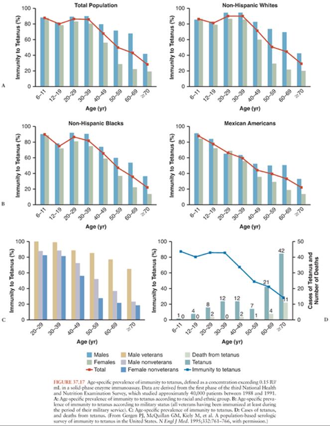
A population-based study, employing 0.15 IU/mL as the threshold for immunity, confirmed that antibody levels wane with aging (153). Individuals with histories of military service, higher educational attainment, and incomes exceeding the poverty level were more likely to have protective antibody titers. Women and members of several minority groups were less likely to have protective titers (Fig. 37.17), presumably because of failure to receive initial or booster vaccinations. An accompanying editorial reinforced the responsibility of physicians to maintain their patients’ immunity to tetanus (154). A more recent analysis confirms that failure to maintain tetanus immunity is an ongoing problem (155).
Disparity among studies regarding the level of protective antibody may reflect the insensitivity of the test systems to antibodies that are actually protective. In a bioassay, a mixture of several different monoclonal antibodies was necessary for protection; the most active single clone (that against the amino-terminal end of the heavy chain) was 100-fold less active than a polyclonal antiserum (156). A level of 0.5 IU/mL in the commercially available immunoassays should probably be set as the cutoff for protection until these questions are resolved (157). Studies characterizing the domains of the toxin molecule that must be recognized for protection by antibody (158) suggest that assays that measure protective antibody rather than total antibody may be on the horizon.
THERAPY
Appropriate treatment based on the clinical diagnosis is warranted even without specific confirmatory laboratory tests (Table 37.3). A patient with generalized tetanus requires the best possible intensive care. In this disease, the patient may become completely dependent on mechanical ventilation and the most effective pharmacotherapy yet stand a good chance of walking out of the hospital without deficits.

An analysis of 335 consecutive tetanus patients treated before the organization of an intensive care unit (ICU), compared with 306 consecutive patients managed after development of the ICU, revealed a decrease in mortality from 44% to 15% (159). The major improvement came from prevention of deaths resulting from acute respiratory failure. A recent study from Brazil showed a comparable decrease in mortality and in other complications with the introduction of an aggressive critical care protocol (160). However, CFR of tetanus has remained consistently high (43%) between 1996 and 2005 at a Nigerian tertiary hospital (161). Factors that were significantly associated with high mortality included older age, age older than 40 years, incubation period of less than 7 days, and higher degree of sedation with diazepam.
Table 37.3 presents a time-based protocol that we have found effective in generalized tetanus. Some of the more important or controversial aspects are discussed here. In local tetanus, a similar approach can be adjusted for the patient’s symptoms. Autonomic management in neonatal tetanus has not been studied, but it would likely be similar to that in older children and adults.
As the population at risk for tetanus becomes older, it becomes more important to recognize that elderly patients without serious chronic disease can survive generalized tetanus and its intensive therapy and return to their premorbid state. This is true even for those 80 years and older when aggressive treatment is started early in the course of the disease (162).
Portal of Entry
In most cases, the wound responsible for tetanus is still visible on presentation. However, in up to a third of the patients, there is no evidence of recent wound, the injury may be too trivial, or the portal of entry may be unusual (i.e., skin, dental or ear infection, septic abortion, or intramuscular injection). If there is a surgical indication, débridement under local anesthesia should be performed after spasms are controlled by benzodiazepines. There is no apparent benefit of débridement for tetanus itself. If no portal of entry is apparent, the patient should be carefully examined for ear infection, uterine disease, rectal pathology, and foreign bodies. Foreign bodies should be removed and wounds irrigated well and left open. Local antibiotic or instillation of HTIG is not indicated. Débridement of the umbilical stump is not recommended in NT.
Immunotherapy
Immunotherapy includes treatment to neutralize TS and active immunization to prevent recurrent tetanus (see later discussion). Specific therapy with intramuscular HTIG is aimed at neutralizing the circulating toxin before it binds to neuronal cell membranes. This should be given as soon as the diagnosis of tetanus is considered. Neutralization of TS that has not yet entered the nervous system will shorten the course of the disease and may reduce its severity. Early administration of antitoxin may prevent spread of the toxin within the CNS. The recommended dosage of HTIG ranges from 500 to 3,000 units. Although a dosage recommendation based on body weight is not available, it is reasonable to give a newborn a smaller dose, for example, a single vial of HTIG (250 units).
A retrospective study in which a group of patients who received the higher dose was compared to a later group receiving the lower dose suggested that the 500-IU dose was adequate (163). Blake et al. (164) also showed that a dose of 500 IU is as effective as the commonly recommended 3,000 to 5,000-IU dose. The smaller amount can be given as a single intramuscular injection. Because each injection is a potent stimulus for tetanic spasms, this is not a trivial point. In areas where HTIG is not available, equine antitetanus serum is used after testing for hypersensitivity and, if necessary, desensitization.
Once the toxin has entered the motor neuron, it is no longer available for neutralization by the antibody. Intrathecal administration may make specific immunoglobulin available where the toxin diffuses out of the motor neuron into other CNS structures. Gupta et al. (163) showed 250 units of intrathecal HTIG to be superior to 1,000 units administered intramuscularly. However, a subsequent study in NT failed to show any advantage for intrathecal treatment (165). A metaanalysis showed no benefit of intrathecal administration of HTIG (166). Because TS is irreversibly bound to tissues, only unbound toxin can be neutralized. Further research in this area may yet bear fruit (167). The HTIG preparations currently available in the United States are not licensed for intrathecal use, and they contain potentially neurotoxic preservatives.
Lee and Lederman (168) proposed that intravenous immune globulin (IVIG) would be a useful alternative to intramuscular HTIG. However, there is wide variation in antitetanus antibody content in commercial preparations of IVIG (168). One might reserve this treatment for patients in whom intramuscular injections are contraindicated, and then only if the product being used is known to have anti-TS activity (168). In countries where HTIG is not readily available, equine antitoxin is used in doses of 1,500 to 3,000 units intramuscularly or intravenously to achieve the minimal protective serum concentration of 0.1 IU/mL. However, patients should be tested for equine antitoxin hypersensitivity prior to its administration.
Patients should receive active immunization with a total of three doses of tetanus toxoid spaced at least 2 weeks apart because tetanus infection does not confer immunity following recovery from acute illness (169). Subsequent boosters with tetanus doses in the form of tetanus toxoid–containing vaccine should be given at 10-year intervals throughout life.
Corticosteroids
In 1954, Lewis et al. (170) attempted to show that steroids would diminish tetanus mortality. A group of ten patients treated with oral cortisone (in a dose “that appeared to be just sufficient to counteract or prevent a rise in the temperature”) experienced 60% survival; the 20 historical controls had only 15% survival. However, a group of five patients receiving intramuscular hydrocortisone all died. The question was later addressed by Paydas et al. (171); they found a trend toward a significant improvement in survival in 32 patients randomized to 40 mg of daily prednisolone when compared with 31 patients receiving placebo. Another group found that betamethasone (8 mg every 8 hours for 10 days) decreased the need for tracheostomy and mechanical ventilation in treated patients compared to double-blind controls (172). The mechanism by which steroid treatment might improve survival in tetanus is obscure, and despite these promising studies, its use should still be considered experimental.
Airway Control and Ventilation
Because the upper airway is often occluded during tetanic spasms, it must be protected rapidly and effectively preferably before spasms start. An endotracheal tube may be passed under sedation and neuromuscular blockade. A soft small-bore feeding tube should be placed concurrently. Because the endotracheal tube itself is a strong stimulus for spasms, some recommend that tracheostomy should be performed within 24 hours of diagnosis in patients predicted to develop mild to moderate tetanus (173). Early tracheostomy is often needed because of the likelihood of prolonged mechanical ventilation. Tracheostomy allows for better tracheal suctioning and pulmonary toilet and prevent laryngospasm, which greatly increases the mortality rate of the disease. It can also prevent aspiration and enable feeding. The patient may not require mechanical ventilation once the airway is secure and treatment has begun. Because pneumonia and pneumothorax occur commonly and produce fatalities, scrupulous pulmonary hygiene and ventilatory management are required (174). TS can inhibit macrophage function, but the relevance of this deficit to infectious complications is unknown (175).
Supportive Care
Transfer to ICU must be urgently done before spasms start. The goals of supportive care include prevention and management of nosocomial infection, decubitus ulcers, gastrointestinal hemorrhage, thromboembolic disease, and tracheal stenosis.
Patients with severe tetanus require prolonged immobility, much of which is during mechanical ventilation and which may last for weeks. Patients should be managed in an intensive care setting of a tertiary care center whenever possible. In one study, the length of ICU care ranged from 33 to 40 days (176). The patients must be managed by experienced caregivers skilled in ventilatory support and maintenance of cardiovascular stability. Minimizing external stimuli and maintaining intravenous hydration may be sufficient in the initial days of the illness. Prevention of thromboembolism can be attained with heparin, low-molecular-weight heparin, or other anticoagulants, and should be given early. Attention must be paid to skin care. Physical therapy should be initiated as soon as spasms have abated.
Antibiotic Management
In vitro, C. tetani is sensitive to metronidazole, penicillins, cephalosporins, imipenem, macrolides, and tetracycline. However, the utility of antibiotic therapy for what is essentially an intoxication has often been questioned. Penicillin, which is effective against most Clostridium spp., is no longer recommended for tetanus because it is a GABA antagonist and can aggravate the spasms of tetanus and diminish benzodiazepine efficacy (177). Furthermore, it can be inactivated by β-lactamase–producing polymicrobial flora that may also be present in the wound (178). High doses of penicillin might also lead to colonization of patients with resistant organisms, increasing morbidity from nosocomial infection.
Two studies compared intramuscular penicillin and oral metronidazole therapy for generalized tetanus. No placebo-controlled studies are available. The first study found less mortality in the metronidazole group as compared to the penicillin group (177). No difference in mortality was found in the second study, but patients receiving metronidazole required fewer muscle relaxants and sedatives (179). Alternative antimicrobials include clindamycin, tetracycline, and vancomycin.
Antispasticity Agents
Extensive muscle spasms can cause respiratory failure, aspiration, and lead to generalized exhaustion. Provocation of muscle spasms can be reduced by placing the patient in a dark and quiet room. This is especially important where the availability of neuromuscular blocking agents may be limited.
The benzodiazepines (diazepam, lorazepam, or midazolam) are the best agents available for the relief of spasms and rigidity. These agents’ mode of action is through GABAA antagonism, and they are therefore able to indirectly antagonize the effect of the toxin on inhibitory neurons (180), but they do not restore the glycinergic inhibition. Diazepam has received the greatest use, but lorazepam may be preferable because of its longer duration of action. The pharmacokinetics of these agents have not been studied at the exceptional doses and durations employed in treating tetanus. Doses in excess of 500 mg of diazepam (or 200 mg of lorazepam) may be required daily (181). Prolonged use can produce lactic acidosis, hypertriglyceridemia, and pancreatic dysfunction. Because such large doses, given intravenously, contain enough propylene and polyethylene glycol to rarely induce metabolic acidosis (182), administration of diazepam or lorazepam through a feeding tube should begin as soon as possible. Because midazolam does not require glycols for solubility, it may become the agent of choice. Benzodiazepine doses should be reduced gradually over a period of at least 14 days to avoid withdrawal signs. Diazepam has been shown to be useful in NT as well (183).
Continuous intrathecal administration of baclofen (a GABAB agonist) may diminish the need for sedation and ventilatory support by controlling spasms and rigidity and may shorten hospitalization (184). Intermittent intrathecal injections have also been employed successfully (185). In several patients, baclofen was used without the need for artificial ventilation (185–187). This approach holds promise in regions where tetanus is relatively common, but it should probably not be considered a standard treatment in the more developed countries. Dantrolene, a direct muscle relaxant, may also be valuable in selected cases (188). It does not appear to have advantages over other therapeutic modalities, however.
Although barbiturates and neuroleptics have been employed for sedation of tetanus patients, they are inferior agents for this indication and are best avoided. Propofol, a nonbarbiturate sedative, may prove to be a useful adjunct because of its short duration of action (189,190). However, because it lacks GABA agonist activity in the motor system, it should not be used as a single agent.
A prospective study of magnesium as an antispasticity agent suggests that this ion may have substantial efficacy both for control of spasms and for prevention of autonomic dysfunction (191). Spasms were controlled in most patients at a serum magnesium concentration of 4 to 8 mEq/L (2 to 4 mmol/L). Attempts to raise the concentration in two patients who did not respond resulted in hypotension and bradycardia.
If spasms are not adequately controlled by GABAergic agents, neuromuscular blocking agents are used. Neuromuscular blockade can be achieved with curariform drugs. The agents used most often are pancuronium and vecuronium. Pancuronium is a long-acting agent and may worsen autonomic instability because it is an inhibitor of catecholamine reuptake. Vecuronium is intermediate acting and is less likely to cause autonomic problems (192). However, because it is short acting, it is given as continuous infusion to provide adequate effects. The intervals between maintenance doses may be adjusted by the administration of smaller or larger doses. Patients who undergo therapeutic paralysis must be sedated to avoid anxiety and ideally monitored by EEG to confirm unconsciousness. The patient should be sedated to the point that the EEG background is slow and there is minimal reactivity to sensory stimuli.
Atracurium has also been recommended (193), but this drug is probably best avoided in tetanus patients because of its potentially epileptogenic metabolite laudanosine (194).
Administration of these agents requires close monitoring to avoid or recognize complications. Their administration should be stopped periodically (at least once a day) to assess the patient’s condition. In addition to clinical observation, neurophysiologic monitoring to ensure optimal dosing of the NMJ blocking agent is employed (e.g., no muscle contraction in response to train-of-four stimulation) (195).
Therapy for Autonomic Dysfunction
Suppression of excessive catecholamine release that induces the autonomic dysfunction can control the dysautonomia. Combined α- and β-adrenergic blockade with labetalol is the treatment of choice for the hypersympathetic state in tetanus (195). Isolated β-adrenergic blockade leaves the α-adrenergic vasoconstrictor response unopposed, and it should generally not be used. Beta blockade alone (i.e., with propranolol) should be avoided because of the danger of sudden death (196). Buchanan et al. (196) described a child with tetanus who died after propranolol administration, probably from myocardial failure in the setting of a catecholamine-induced myocarditis. However, the short-acting β-blocking agent esmolol may be useful (197); phentolamine should be available at the bedside in case esmolol elicits a hypertensive response (198). Esmolol has been used successfully to control severe tachycardia (>200 beats per minute, associated with arterial desaturation) in a case of NT (199). Clonidine may be a useful alternative (200), although its efficacy has been questioned (201).
Morphine sulfate is commonly used to control autonomic dysfunction as well as to induce sedation. It acts centrally by reducing sympathetic tone in the heart and the vascular system, inducing hypotension and bradycardia. It functions also as an adjunct to deep sedation and controls cardiac instability without cardiac compromise (202,203). Morphine is often effective, especially when used in concert with clonidine. Other agents available for the treatment of various autonomic events are atropine, clonidine, and epidural bupivacaine.
Magnesium should be part of the routine therapy for tetanus. It is a vasodilator by direct action and through inhibition of catecholamine-induced vasoconstriction. It also reduces catecholamine release from the adrenal medulla (204) and adrenergic nerve ending (205). Of the drugs used to produce adrenergic blockade and suppress autonomic hyperactivity in tetanus, only magnesium sulfate was evaluated in a randomized clinical trial (206) and in clinical series for the management of autonomic dysfunction and as adjunctive treatment for controlling spasms (191–210). In a randomized, double-blind study of 256 patients, magnesium sulfate infusion (loading dose 40 mg/kg over 30 minutes, followed by continuous infusion of either 2 g per hour for patients >45 kg or 1.5 g per hour for patients ≤45 kg) was compared to placebo (206). Magnesium sulfate significantly reduced the need for other drugs to control muscle spasms and patients were 4.7 times (95% CI, 1.4 to 15.9) less likely to require verapamil to treat cardiovascular instability than those in the placebo group.
Epidural anesthesia is also effective, perhaps by decreasing adrenal stimulation (210). Excessive parasympathetic function occurs rarely. If bradycardia or asystole is encountered, a pacemaker should be considered (211).
Nutrition
Maintenance of adequate nutrition and hydration is of outmost importance. Parenteral nutrition is usually required. Adequate nutritional support can minimize weight loss, maintain electrolyte balance, and prevent or improve management of arrhythmias. The nutritional requirements of tetanus patients may be extraordinarily high because of both their muscular activity and their excessive autonomic activity. The protein and calories required to maintain a positive nitrogen balance may exceed the maximum tolerable daily volume of enteral formulas (212). If gastric emptying is impaired, central venous nutrition may be necessary. Excretory functions must be monitored closely for urinary retention or serious constipation.
COMPLICATIONS OF TETANUS
Potential complications of tetanus include those due to direct effect of the toxin (e.g., laryngeal and phrenic nerves palsy, cardiomyopathy) and those that are secondary to spasms (e.g., respiratory compromise causing hypoxic cerebral injury, rhabdomyolysis causing acute renal failure, myositis ossificans, and vertebral compression fractured) as well as the psychologic impact.
Pulmonary aspiration and breathing or swallowing complications are treated by intubation, tracheostomy, constant suctioning, and care in an ICU. Early removal of tracheostomy can prevent tracheal stenosis. Skin care can prevent and treat skin conditions such as decubitus ulcers. Parenteral nutrition and enteral feeding deals with nutritional problems; prophylaxis of thromboembolism with heparin, low-molecular-weight heparin, or other anticoagulants can treat thromboembolic disease. Prophylactic treatment with sucralfate or acid blockers may avert gastrointestinal hemorrhage. Infection control measures are necessary to prevent nosocomial infection.
In the past, respiratory dysfunction was the most feared result of tetanus and was the major cause of death. The neuropathologic consequences of hypoxia (in concert with hyperthermia) probably accounted for earlier reports of brainstem neuronal destruction in this disease (101). However, the CNS and systemic consequences of hypoxia continue to occur, because of either delayed or inadequate treatment of respiratory problems (213). Cardiovascular consequences of autonomic instability, including cardiomyopathy, are still seen and may be less amenable to secondary prevention.
In the series of Vieira and Brauner (66) from 1993, the most common complications in patients with severe tetanus were respiratory and urinary tract infections related to the use of mechanical ventilation and indwelling catheters. The increased survival associated with the use of intensive care made autonomic hyperactivity the major cause of death. Other complications in this series included cardiac arrhythmias, pneumothoraxes, and atelectasis. Renal failure was uncommon, but special care was taken to ensure adequate hydration.
Phrenic and laryngeal neuropathies as a consequence of tetanus may occur (83,85). Other mononeuropathies probably also occur. As in any sedated or paralyzed patient, care must be taken to prevent common peroneal nerve compression at the fibular head, which produces footdrop.
Rhabdomyolysis, which may lead to acute renal failure (214), is very common in generalized tetanus. If the serum creatine kinase level exceeds 5,000 U/L or myoglobin is detected in the urine, hydration with normal saline and urinary alkalinization with sodium bicarbonate should be considered. Rarely, acute renal failure requiring dialysis may occur (215). Myositis ossificans circumscripta is a long-term complication of severe muscular hyperactivity, coupled with hematoma formation and anoxia. Vertebral compression fractures are common, especially in older patients. In a study from Finland, 13 of 20 tetanus survivors had subsequent rheumatologic disorders, predominantly hyperostoses at sites of tendon insertion and osteoarthritis of the elbows (216). About 40% of patients have psychologic aftereffects, and 25% feel that their health has permanently worsened in some manner after recovery from tetanus (213). Other late sequelae include mild developmental delay in children (217).
PREVENTION
Local wound care, including surgical debridement, is essential. Foreign bodies should be removed and wounds irrigated well and left open. Excision of necrotic tissue may be required, but excision of the umbilical stump is no longer recommended in cases of NT.
In the immunocompetent host, tetanus is an “inexcusable disease” (218). Active immunization with tetanus toxoid is one of the most effective preventive measures in medicine, and passive immunization may be performed at the time of any tetanus-prone wound. Preventing one case of tetanus saves enough health care expense to immunize several thousand people (219). The WHO, in concert with the United Nations Children’s Fund (UNICEF) and the United Nations Population Fund (UNFPA), set 2005 as the target date for the elimination of maternal and neonatal tetanus (defined as fewer than one case per thousand livebirths in every district of every country) (220). Zimbabwe and Namibia have recently achieved this goal (221,222). Although progress continues to be made, according to UNICEF, by November 2012, 31 countries have not reached maternal and neonatal tetanus elimination status (http://www.who.int/immunization_monitoring/diseases/MNTE_initiative/en/index.html).
Prophylactic Active Immunization
Active immunization with tetanus toxoid is the most effective mean of protection from the illness (223).A series of three intramuscular injections of tetanus toxoid (10 lyophilized units [Lf]; 0.5 mL) provides almost complete immunity to tetanus for at least 5 years. Other forms of toxoid are less immunogenic and should be avoided (224,225). In the United States, five doses of tetanus, diphtheria, and pertussis vaccines are recommended between 6 weeks and 7 years, and a booster dose is recommended starting at age 11 years (226). The vaccine to be used varies with age: children younger than 7 years should receive combined diphtheria-tetanus-pertussis vaccine; if pertussis vaccine is considered to be contraindicated, diphtheria-tetanus–adsorbed vaccine for pediatric use should be employed (226). In those older than 7 years, tetanus-diphtheria vaccine is recommended. Because of the increase in pertussis cases in the United States, the CDC and the American Academy of Pediatrics recommended in 2011 the use of tetanus toxoid–reduced diphtheria toxoid-acellular pertussis (Tdap) vaccine (226). This included a recommendation for off-label use of Tdap in children aged 7 through 10 years who are not fully vaccinated against pertussis (226). The complete series must be given; 30% of a group of elderly patients had inadequate antibody levels 4 years after a series of two injections (all who received three injections were protected) (227). Routine boosters are given every 10 years; giving them more frequently may increase the risk of an adverse reaction (228). This 10-year period has never been proven, and one study suggested that reimmunization may not be required this often (229). With the increasing interest in health care cost control, some analysts suggest that the routine decennial booster be replaced by a single booster at age 65 years (230). The authors recommended adopting this practice, despite their demonstration that the decennial strategy would be more effective, because the single booster approach was predicted to cost $4,527 per year of life saved, compared to $143,138 for the decennial regimen. Although this type of analysis deserves serious attention, we believe that clinicians would be well advised to let this debate play out before adopting this recommendation. The analyses involved many assumptions, such as that of protection at an antibody concentration of 0.01 IU/mL, which is probably incorrect; used inadequate estimates for tetanus incidence and for the costs associated with the care of tetanus patients; and omitted consideration of lost productivity for patients dying of the disease. As others have pointed out, eliminating decennial boosters also places the population at risk of a resurgence of diphtheria, as has occurred in Eastern Europe (231).
In the United Kingdom, the Department of Health states that boosters beyond five doses of adsorbed toxoid “are not recommended, other than at the time of tetanus prone injury, since they have been shown to be unnecessary and can cause considerable local reactions” (232). Others in the United Kingdom have tempered this recommendation because of concern regarding waning immunity in the elderly (233). The contention that further boosters are “unnecessary” remains to be studied adequately.
Substantial progress has been made in the effort to eliminate neonatal tetanus in the developing world, with a nearly 50% decrease in the number of reported cases between 1989 and 1993 (234). Although the reported cases are only a small fraction of the actual number, the reporting conditions have been relatively stable over this period, so the decrease is likely to be real. NT decreased from 0.146 million (uncertainty range [UR] 0.068 to 0.590) to 0.058 million (UR 0.020 to 0.276) at 9.5% (UR 0.9 to 17.8%) per year between 2000 and 2010 (11). In 1993, the WHO still estimated 515,000 deaths attributable to neonatal tetanus. The number was reduced to less than a third of a million by 2010 (11). In a novel approach, one group administered a single 250-Lf dose of tetanus toxoid to unimmunized third-trimester primigravid women and showed that both mothers and babies developed protective antibody titers (235). Although TS does not cross the placenta, it appears that small amounts of tetanus toxoid can; 78% of infants born to recently immunized mothers in one study showed immunoglobulin M (IgM) cord blood antibody responses to tetanus toxoid, although none developed an IgG response as maternal antibody waned (236). Topical antimicrobial treatment of the umbilical stump represents a novel approach that appears even more effective in preventing neonatal tetanus than either hand washing by the birth attendants or maternal immunization (237).
Immunization of patients before surgery is rarely considered, but case reports of tetanus following surgical procedures (summarized by Singh et al. [238]) raise the possibility that this should be considered, at least in cases in which antibiotic prophylaxis for other indications is not employed.
In a small number of patients with humoral immunodeficiencies, the prescribed toxoid regimen did not produce immunity (239). Although antitetanus antibody is usually of the IgG1 subclass, three of six children with isolated IgG2 deficiency demonstrated poor responses to tetanus toxoid (240). This may also be true of some patients infected with human immunodeficiency virus (HIV) (241). However, a study in Denmark showed that all of a cohort of 78 HIV-infected men had antitetanus antibody concentrations exceeding 0.01 IU/mL; most had concentrations more than 0.1 IU/mL (242). The peripheral blood CD4+ cell concentration did not appear to affect the antibody titer. Similarly, in a study of children who were HIV seropositive at birth, all who received three doses of tetanus toxoid achieved the 0.1 IU/mL antibody concentration previously deemed protective (243). However, children infected with HIV developed lower antibody concentrations than those who became HIV seronegative. Although limited in vitro evidence suggests that tetanus toxoid immunization may provoke HIV activation (244), there is no clinical evidence to suggest that patients with HIV infection should not receive tetanus toxoid when indicated. However, loss of response to soluble antigens like tetanus toxoid is one of the first immunologic consequences of HIV infection (245), suggesting that HIV-infected patients may not become immune after toxoid administration.
Acute leukemias produce a deficit in antibody concentrations to tetanus toxoid (246), and patients who undergo bone marrow transplantation lose their preexisting antitetanus immunity. This occurs by the fourth posttransplant month in patients with allogeneic transplants who develop graft-versus-host disease (GVHD), by the eighth month in similar patients without GVHD, and by the twentieth month in patients undergoing autologous transplantations (247). A series of three reimmunizations beginning 9 to 12 months posttransplantation will restore immunity (248). This appears to be the same for patients undergoing stem cell transplantation (249). In renal transplant recipients, in contrast, immunosuppression (at least with mycophenolate) does not appear to impair in vitro measures of antitetanus immunity (250).
Extremely premature infants (born before the twenty-ninth week of gestation and weighing <1 kg at birth) make presumably adequate responses to a primary immunization series against diphtheria, pertussis, and tetanus begun at the chronologic age of 2 months (251). Therefore, routine immunizations against these three diseases should not be delayed on the basis of prematurity.
Administration of the Haemophilus influenzae type b polysaccharide-tetanus protein conjugate vaccine in the same syringe as the diphtheria-tetanus-pertussis vaccine does not appear to impair antibody responses (252).
Whenever a question of immunity to TS exists (whether it is due to failure of immunization or an underlying disorder of immunity), passive immunization with HTIG is indicated after tetanus-prone injuries unless immunity to TS has been serologically demonstrated recently.
Immunization following Injury
All patients who last received tetanus toxoid more than 10 years before any injury that brings them to medical attention or who do not recall the date of their last immunization should receive active immunization with tetanus toxoid–containing vaccine. If their wounds are considered tetanus prone, they should be immunized if it has been more than 5 years since their last immunization. Patients in whom the prior immunization history is incomplete or unknown should receive a full series of three monthly injections.
Administration of a single booster dose of tetanus toxoid does not produce a rapid anamnestic response in patients who are more than 10 years from their last immunization (253). A tetanus-prone wound in a patient with an incomplete or uncertain initial immunization history should prompt passive immunization with HTIG (250 units, intramuscularly). HTIG should also be considered for those whose immune status is in question.
The standard recommendation of 250 units of HTIG has been questioned, based on the need to achieve antibody concentrations higher than 0.01 IU/mL. A dose of 500 units (or 8 IU/kg in children) is necessary to achieve an antibody concentration of 0.1 IU/mL (254).
Most authors suggest that HTIG and tetanus toxoid may be given at the same time as long as different sites are employed. A study of this question concluded that simultaneous administration impaired the immune response to tetanus toxoid in patients with low prevaccination antibody concentrations (it was still adequate; however, its long-term stability is uncertain) (255). Whether such patients should receive another dose of tetanus toxoid in 4 weeks is uncertain.
Adverse Reactions to Tetanus Toxoid
A mild degree of local tenderness and edema is common after tetanus toxoid booster injections, and fever is occasionally noted. Rare anaphylactoid responses and delayed hypersensitivity to the preservative thiomersal were documented in a large survey (256). Peripheral neuropathic complications (257), which may resemble the Guillain-Barré syndrome (258) or may be relapsing (259), have been the subject of case reports. CNS reactions have been reported rarely (260). In most of these reports, patients received more than one vaccine or had other confounding events such as injury, which make assignment of the apparent adverse event to a single cause difficult.
Doses of tetanus toxoid lower than the usually recommended 7.5 to 10.0 Lf (i.e., 0.75, 1.9, and 2.5 Lf) will produce an adequate anamnestic response at 1 month in patients who have had an adequate primary immunization series (261). In a study of 102 patients, there was no difference in the rate of adverse reactions among patients receiving different doses, but the sample size had inadequate power to detect such differences reliably.
ACKNOWLEDGMENTS
The author gratefully acknowledges Tomas P. Bleck and Janette Salles Brauner who wrote this chapter for the previous edition of this book.
References
1. Breasted JH. The Edwin Smith Surgical Papyrus. Chicago: University of Chicago Press; 1930.
2. Adams F. The Extant Works of Aretaeus, the Cappadocian. London: Publications of the Sydenham Society; 1856.
3. Bell C. Essays on the Anatomy and Physiology of Expression. 2nd ed. London: J Murray; 1824.
4. Gowers WR. A Manual of Diseases of the Nervous System. Philadelphia: Blackiston; 1888.
5. Nicolaier A. Über infectiösen tetanus. Dtsch Med Wochenschr. 1884; 10:842–844.
6. Behring E, Kitasato S. Über das zustandekommen der diphtherie-immunität und der tetanus-immunität bei thieren. Dtsch Med Wochenschr. 1890;16: 1113–1114. http://www.cdc.gov/vaccines/vpd-vac/tetanus/default.htm. Accessed December 30, 2013.
7. Mollaret P. Prélude, choral et fugue au XIXe siècle de la curarisation dans le tétanos. Presse Med. 1965;73:2313–2317.
8. Hoffman FA. Ein fall von tetanus traumaticus mit curare behandelt. Berl Klin Wochenschr. 1879;16:637–638.
9. Hutchinson J, Jackson JH. On cases of recovery from traumatic tetanus. Med Times Gazette. 1861;1:360–363.
10. Meltzer SJ, Auer J. The effects of intraspinal injection of magnesium salts upon tetanus. J Exp Med. 1906;8:692–706.
11. Liu L, Johnson HL, Cousens S, et al. Global, regional, and national causes of child mortality: an updated systematic analysis for 2010 with time trends since 2000. Lancet. 2012;379(9832):2151–2161.
12. Matzkin H, Regev S. Naturally acquired immunity to tetanus in an isolated community. Infect Imm. 1985;48:267–268.
13. Scher KS, Baldera A, Wheeler WE, et al. Inadequate tetanus protection among the rural elderly. South Med J. 1985;78:153–156.
14. Brunette GW. Tetanus. CDC Health Information for International Travel 2012: The Yellow Book. Oxford: Oxford University Press; 2011.
15. Centers for Disease Control and Prevention. Summary of notifiable disease, 2001. MMWR Morb Mortal Wkly Rep. 2003;50:80.
16. Centers for Disease Control and Prevention. Tetanus surveillance—United States, 2001–2008. MMWR Morb Mortal Wkly Rep. 2011;60: 365–369.
17. Sutter RW, Cochi SL, Brink EW, et al. Assessment of vital statistics and surveillance data for monitoring tetanus mortality, United States, 1979–1984. Am J Epidemiol. 1990;131:132–142.
18. Bleck TP. Tetanus: dealing with the continuing clinical challenge. J Crit Illness. 1987;2:41–52.
19. Sutter RW, Orenstein WA, Wassilak SG. Tetanus. In: Hoeprich PD, Jordan MC, Ronald AR, eds. Infectious Diseases. 5th ed. Philadelphia: JB Lippincott; 1994:1175–1185.
20. Jacobs DH, Tovar JM, Hung OL, et al. Behavioral risk factor and preventive health care practice survey of immigrants in the emergency department. Acad Emerg Med. 2002;9:599–608.
21. Lambo JA, Memon MI, Khahro ZH, et al. Epidemiology of neonatal tetanus in rural Pakistan. J Pak Med Assoc. 2011;61:1099–1103.
22. Hlady WG, Bennett JV, Samadi AR, et al. Neonatal tetanus in rural Bangladesh: risk factors and toxoid efficacy. Am J Public Health. 1992; 82:1365–1369.
23. World Health Organization. Tetanus vaccine: WHO position paper. Wkly Epidemiol Rec. 2006;81:198–208. http://www.who.int/wer/2006/wer8120.pdf. Accessed December 30, 2013.
24. Gürses N, Aydin M. Factors affecting prognosis of neonatal tetanus. Scand J Infect Dis. 1993;25(3):353–355.
25. Traverso HP, Kamil S, Rahim H, et al. A reassessment of risk factors for neonatal tetanus. Bull World Health Organ. 1991;69:573–579.
26. Ball K, Norboo T, Gupta U, et al. Is tetanus rare at high altitudes? Trop Doctor. 1994;24:78–80.
27. Cato EP, George WL, Finegold SM. Genus Clostridium prazemozski 1880, 23AL. In: Smeath PHA, Mair NS, Sharpe ME, et al., eds. Bergey’s Manual of Systematic Bacteriology. Vol 2. Baltimore: Lippincott Williams & Wilkins; 1986:1141–1200.
28. Hatheway CL. Bacterial sources of clostridial neurotoxins. In: Simpson LL, ed. Botulinum Neurotoxin and Tetanus Toxin. San Diego: Academic Press; 1989:4–24.
29. Hoeniger JFM, Tauschel HD. Sequence of structural changes in cultures of Clostridium tetani grown on a solid medium. J Med Microbiol. 1974;7:425–432.
30. LaForce FM, Young LS, Bennett JV. Tetanus in the United States (1965–1966): epidemiologic and clinical features. N Engl J Med. 1969;280:569–574.
31. Benson WH, Snyder IS, Granus V, et al. Tetanus prophylaxis following ocular injuries. J Emerg Med. 1993;11:677–683.
32. Fischer MGW, Sunakorn P, Duangman C. Otogenous tetanus: a sequelae of chronic ear infections. Am J Dis Child. 1977;131:445–446.
33. Nourmand A. Clinical studies on tetanus: notes on 42 cures in southern Iran with special emphasis on portal of entry. Clin Pediatr. 1973;12:652–653.
34. Luisto M. Unusual and iatrogenic sources of tetanus. Ann Chir Gynaecol. 1993;82:25–29.
35. Adams EB. The prognosis and prevention of tetanus. S Afr Med J. 1968; 42:739–743.
36. Matsuda M. The structure of tetanus toxin. In: Simpson LL, ed. Botulinum Neurotoxin and Tetanus Toxin. San Diego: Academic Press; 1989:69–92.
37. Eisel U, Jarausch W, Goretzki K, et al. Tetanus toxin: primary structure, expression in E. coli, and homology with botulinum toxins. EMBO J. 1986;5:2495–2502.
38. Bergey GK, Habig WH, Bennett JI, et al. Proteolytic cleavage of tetanus toxin increases activity. J Neurochem. 1989;53:155–161.
39. Robinson JP, Schmid M, Morgan DG, et al. Three-dimensional structural analysis of tetanus toxin by electron crystallography. J Mol Biol. 1988;200:367–375.
40. Bruggemann H, Baumer S, Fricke WF, et al. The genome sequence of Clostridium tetani, the causative agent of tetanus disease. Proc Natl Acad Sci U S A. 2003;100:1316–1321.
41. Fairweather NF, Lyness VA. The complete nucleotide sequence of tetanus toxin. Nucleic Acids Res. 1986;14:7809–7812.
42. Helting TB, Zwisler O. Structure of tetanus toxin. I. Breakdown of the toxin molecule and discrimination between polypeptide fragments. J Biol Chem. 1977;12:1147–1153.
43. Mochida S, Poulain B, Weller U, et al. Light chain of tetanus toxin intracellularly inhibits acetylcholine release at neuro-neuronal synapses, and its internalization is mediated by the heavy chain. FEBS Lett. 1989;253:47–51.
44. Simpson LL. Fragment C of tetanus toxin antagonizes the neuromuscular blocking properties of native tetanus toxin. J Pharmacol Exp Ther. 1984;228:600–604.
45. Bittner MA, Habig WH, Holz RW. Isolated light chain of tetanus toxin inhibits exocytosis: studies in digitonin-permeabilized cells. J Neurochem. 1989;53:966–968.
46. Fishman P, Savitt JM. Transsynaptic transfer of retrogradely transported tetanus protein-peroxidase conjugates. Exp Neurol. 1989;106:197–203.
47. Bleck TP. Pharmacology of tetanus. Clin Neuropharmacol. 1986;9:103–120.
48. Middlebrook JL. Cell surface receptors for protein toxins. In: Simpson LL, ed. Botulinum Neurotoxin and Tetanus Toxin. San Diego: Academic Press; 1989:95–119.
49. Yavin E, Nathan A. Tetanus toxin receptors on nerve cells contain a trypsin-sensitive component. Eur J Biochem. 1986;154:403–407.
50. Montesano R, Roth J, Robert A, et al. Non-coated membrane invaginations are involved in binding and internalization of cholera and tetanus toxins. Nature. 1982;296:651–653.
51. Pelkmans L, Helenius A. Endocytosis via caveolae. Traffic. 2002;3:311–320.
52. Williamson LC, Neale EA. Bafilomycin A1 inhibits the action of tetanus toxin in spinal cord neurons in cell culture. J Neurochem. 1994;63: 2342–2345.
53. Papini E, Rappuoli R, Murgia M, et al. Cell penetration of diphtheria toxin: reduction of the interchain disulfide bridge is the rate-limiting step of translocation in the cytosol. J Biol Chem. 1993;25:1567–1574.
54. Munro P, Kojima H, Dupont JL, et al. High sensitivity of mouse neuronal cells to tetanus toxin requires a GPI-anchored protein. Biochem Biophys Res Commun. 2001;289:623–629.
55. Herreros J, Blasi J, Arribas M, et al. Tetanus toxin mechanism of action in Torpedo electromotor system: a study on different steps in the intoxication process. Neuroscience. 1995;65:305–311.
56. Forchetti CM, Leheta B, Garant DS. In vivo and in vitro assessment of neurotransmitter amino acid function in tetanus toxin induced chronic seizure foci in rat hippocampus. Soc Neurosci Abstr. 1990;16:948.
57. Popoff MR, Bouvet P. Clostridial toxins. Future Microbiol. 2009;4: 1021–1064.
58. Trimble WS, Cowan D, Scheller RH. VAMP-1: a synaptic vesicle associated integral membrane protein. Proc Natl Acad Sci U S A. 1988;85:4538–4542.
59. McMahon HT, Ushkaryov YA, Edelmann L, et al. Cellubrevin is a ubiquitous tetanus-toxin substrate homologous to a putative synaptic vesicle fusion protein. Nature. 1993;364:346–349.
60. Buckley KM, Floor E, Kelly RB. Cloning and sequence analysis of cDNA encoding p38, a major synaptic vesicle protein. J Cell Biol. 1987;105: 2447–2456.
61. Blasi J, Binz T, Yamasaki S, et al. Inhibition of neurotransmitter release by clostridial neurotoxins correlates with specific proteolysis of synaptosomal proteins. J Physiol (Paris). 1994:88:235–241.
62. Schiavo G, Benfenati F, Poulain B, et al. Tetanus and botulinum-B neurotoxins block neurotransmitter release by proteolytic cleavage of synaptobrevin. Nature. 1992;359:832–835.
63. Hayashi T, McMahon H, Yamasaki S, et al. Synaptic vesicle membrane fusion complex: action of clostridial neurotoxins on assembly. EMBO J. 1994;13:5051–5061.
64. Hausinger A, Volknandt W, Zimmerman H. Calcium-dependent endogenous proteolysis of the vesicle proteins synaptobrevin and synaptotagmin. Neuroreport. 1995;6:637–641.
65. Lalli G, Schiavo G. Analysis of retrograde transport in motor neurons reveals common endocytic carriers for tetanus toxin and neurotrophin receptor p75NTR. J Cell Biol. 2002;156:233–239.
66. Vieira SRR, Brauner JS. Tetanus: following up 176 patients in the ICU. In: Proceedings from 6th World Congress on Intensive and Critical Care Medicine; June 1993; Madrid, Spain.
67. Palmateer NE, Hope VD, Roy K, et al. Infections with spore-forming bacteria in persons who inject drugs, 2000–2009. Emerg Infect Dis. 2013;19:29–34.
68. Yen LM, Dao LM, Day NPJ, et al. Role of quinine in the high mortality of intramuscular injection tetanus. Lancet. 1994;344:786–787.
69. Greco JB, Tavares-Neto J, Greco JB Jr. Accidental tetanus: prognosis evaluation in a historical series at a hospital in Salvador, Bahia, Brazil. Rev Inst Med Trop Sao Paulo. 2003;45:35–40.
70. Habermann E. Tetanus. In: Vinken PJ, Bruyn GW, eds. Handbook of Clinical Neurology. Vol. 33. Amsterdam: North-Holland; 1978:491–547.
71. Veronesi R, Focaccia R. The clinical picture. In: Veronesi R, ed. Tetanus: Important New Concepts. Amsterdam: Excerpta Medica; 1981:183–206.
72. Bleck TP. Clinical aspects of tetanus. In: Simpson LL, ed. Botulinum Neurotoxin and Tetanus Toxin. San Diego: Academic Press; 1989:379–398.
73. Brauner JS, Tedoldi CL, Vieira SRR. Tétano. In: Barreto SM, Brauner JS, Prado KF, et al, eds. Rotinas em Terapia Intensiva. Porto Alegre, Brasil: Editora Artes Médicas Sul Ltda; 1993:264–268.
74. Kerr JH, Corbett JL, Prys-Roberts C, et al. Involvement of the sympathetic nervous system in tetanus. Lancet. 1968;2:236–241.
75. Weinstein L. Tetanus. N Engl J Med. 1973;289:1293–1296.
76. Turton K, Chaddock JA, Acharya KR. Botulinum and tetanus neurotoxins: structure, function and therapeutic utility. Trends Biochem Sci. 2002;27:552–558.
77. Hassel B. Tetanus: pathophysiology, treatment, and the possibility of using botulinum toxin against tetanus-induced rigidity and spasms. Toxins (Basel). 2013;5:73–83.
78. Tsueda K, Oliver PB, Richter RW. Cardiovascular manifestations of tetanus. Anesthesiology. 1974;40:588–592.
79. Rose AG. Catecholamine-induced myocardial damage associated with phaeochromocytomas and tetanus. S Afr Med J. 1974;48:1285–1289.
80. Hollow VM, Clarke GM. Autonomic manifestations of tetanus. Anaesth Intensive Care. 1975;3:142–147.
81. Farrar JJ, Yen LM, Cook T, et al. Tetanus. J Neurol Neurosurg Psychiatry. 2000;69:292–301.
82. Bleck TP, Calderelli DD. Vocal cord paralysis complicating tetanus. Neurology. 1983;33(suppl 2):140.
83. Brust JCM, Richter RW. Tetanus in the inner city. N Y State Med J. 1974;74:1735–1742.
84. Bassin SL. Tetanus. Curr Treat Options Neurol. 2004;6:25–34.
85. Risk WS, Bosch EP, Kimura J, et al. Chronic tetanus: clinical report and histochemistry of muscle. Muscle Nerve. 1981;4:363–366.
86. Patel JC, Kale PA, Mehta BC. Otogenic tetanus: study of 922 cases. In: Patel JC, ed. Proceedings of an International Conference on Tetanus. Bombay. 1965;640–644.
87. deSouza CE, Karnard DR, Tilve GH. Clinical and bacteriologic profile of the ear in otogenic tetanus: a case control study. J Laryngol Otol. 1992;106:1051–1054.
88. Saltissi S, Hakin RN, Pearce J. Ophthalmoplegic tetanus. Br Med J. 1976;1:437.
89. Biglan AW, Ellis FD, Wade TA. Supranuclear oculomotor palsy and exotropia after tetanus. Am J Ophthalmol. 1978;86:666–668.
90. Syam B, Owens D. Horner’s syndrome in severe tetanus. Postgrad Med J. 1992;68:279–280.
91. Idema CD, Harris BN, Ogunbanjo GA, et al. Neonatal tetanus elimination in Mpumalanga Province, South Africa. Trop Med Int Health. 2002;7:622–624.
92. Schofield FD, Tucker VM, Westbrook GR. Neonatal tetanus in New Guinea: effect of active immunization in pregnancy. Br Med J. 1961;2: 785–789.
93. Antia-Obong OE, Ekanem EE, Udo JJ, et al. Septicaemia among neonates with tetanus. J Trop Pediatr. 1992;38:173–175.
94. Yalçin I, Güler N, Kebudi R, et al. A review of 43 cases of tetanus neonatorum. Turk J Pediatr. 1992;34:121–126.
95. Quddus A, Luby S, Rahbar M, et al. Neonatal tetanus: mortality rate and risk factors in Loralai District, Pakistan. Int J Epidemiol. 2002;31:648–653.
96. Montecucco C, Schiavo G. Mechanism of action of tetanus and botulinum neurotoxins. Mol Microbiol. 1994;13:1–8.
97. Price DL, Griffin J, Young A, et al. Tetanus toxin: direct evidence for retrograde axonal transport. Science. 1975;188:945–946.
98. Schwab ME, Thoenen H. Electron microscopic evidence for a transsynaptic migration of tetanus toxin in spinal cord motoneurons: an autoradiographic and morphometric study. Brain Res. 1976;105:213–227.
99. Davidoff RA, Hackman JC. Spinal inhibition. In: Davidoff RA, ed. Handbook of the Spinal Cord. Vols 2 and 3. New York: Marcel Dekker; 1984:385–459.
100. Takano K, Kirchner T, Tiebert B, et al. Presynaptic inhibition of the monosynaptic reflex during local tetanus in the cat. Toxicon. 1989;27:431–438.
101. Baker AB. The central nervous system in tetanus. J Neuropathol Exp Neurol. 1942;1:394–405.
102. Dumas M, Schwab ME, Thoenen H. Retrograde axonal transport of specific macromolecules as a tool for characterizing nerve terminal membranes. J Neurobiol. 1979;10:179–197.
103. Bigalke H, Heller I, Habermann E. Effects of tetanus and botulinum A toxin on uptake and release of centrally acting neurotransmitters. Naunyn Schmiedebergs Arch Pharmacol. 1980;313:R26.
104. Bigalke H, Ahnert-Hilber G, Habermann E. Tetanus toxin and botulinum A toxin inhibit acetylcholine release from but not calcium uptake into brain tissue. Naunyn Schmiedebergs Arch Pharmacol. 1981;316: 143–148.
105. Pelliccioni P, Gil C, Najib A, et al. Tetanus toxin modulates serotonin transport in rat-brain neuronal cultures. J Mol Neurosci. 2001;17:303–310.
106. Janicki PJ, Habermann E. Tetanus and botulinum toxins inhibit, and black widow spider venom stimulates the release of methionine-enkephalin-like material in vitro. J Neurochem. 1983;41: 395–402.
107. Garant DS, Chen YM, Bleck TP, et al. Long-term electrophysiologic and biochemical monitoring in the rat intra-amygdaloid tetanus toxin chronic model of epilepsy. Soc Neurosci Abstr. 1989;15:1032.
108. Whittington MA, Jeffreys JGR. Epileptic activity outlasts disinhibition after intrahippocampal tetanus toxin in the rat. J Physiol (Lond). 1994;481:593–604.
109. Lassen HCA, Bjornboe M, Ibsen B, et al. Treatment of tetanus with curarization, general anesthesia, and intratracheal positive pressure ventilation. Lancet. 1954;2:1040–1044.
110. Corbett JL, Harris PJ. Studies on the sympathetic nervous system in tetanus. Naunyn Schmiedebergs Arch Pharmacol. 1973;276:447–460.
111. Domenighetti GM, Savary S, Striker H. Hyperadrenergic syndrome in severe tetanus responsive to labetalol. Br Med J. 1984;288:1483–1484.
112. Thwaites CL, Yen LM, Cordon SM, et al. Urinary catecholamine excretion in tetanus. Anaesthesia. 2006;61:355–359.
113. Udwadia FE, Sunavala JD, Jain MC, et al. Haemodynamic studies in the management of severe tetanus. Q J Med. 1992;83:449–460.
114. Parr GH, Wellhoner HH. The action of tetanus toxin in preganglionic sympathetic reflex discharges. Naunyn Schmiedebergs Arch Pharmacol. 1973;276:437–445.
115. Parr GH, Wieland HJ, Wellhoner HH. The influence of tetanus toxin on sympathetic reflex discharges into the renal nerves. Naunyn Schmiedebergs Arch Pharmacol. 1978;281:383–390.
116. Potgieter PD. Inappropriate ADH secretion in tetanus. Crit Care Med. 1983;11:417–418.
117. Montecucco C, Schiavo G. Tetanus and botulinum neurotoxins: a new group of zinc proteases. Trends Biochem Sci. 1993;18:324–327.
118. Dayanithi G, Stecher B, Höhne-Zell B, et al. Exploring the functional domain and the target of the tetanus toxin light chain in neurohypophysial terminals. Neuroscience. 1994;58:423–431.
119. Bartels F, Bergel H, Bigalke H, et al. Specific antibodies against the Zn2+-binding domain of clostridial neurotoxins restore exocytosis in chromaffin cells treated with tetanus or botulinum A neurotoxin. J Biol Chem. 1994;269:8122–8127.
120. Simpson LL, Coffield JA, Bakry N. Chelation of zinc antagonizes the neuromuscular blocking properties of the seven serotypes of botulinum toxin as well as tetanus toxin. J Pharmacol Exp Ther. 1993;267:720–727.
121. Soumaré M, Seydi M, Ndour CT, et al. Cardiovascular events in the course of tetanus: a prospective study on 30 cases in the infectious diseases clinic, in the Fann teaching hospital, Dakar. Med Mal Infect. 2005;35:450–454.
122. Wasay M, Khealani BA, Talati N, et al. Autonomic nervous system dysfunction predicts poor prognosis in patients with mild to moderate tetanus. BMC Neurol. 2005;5:2.
123. Nistico G, Gratteri S, Donato Di Paola E, et al. Cardiovascular effects of tetanus toxin after systemic and intraventricular administration in rats. Neuropharmacology. 1985;24:341–343.
124. Duchen LW, Tonge DA. The effects of tetanus toxin on neuromuscular transmission and on the morphology of motor endplates in slow and fast skeletal muscle of the mouse. J Physiol (Lond). 1973;228:157–172.
125. Harvey AM. The peripheral action of tetanus toxin. J Physiol (Lond). 1939;96:348–365.
126. Fernandez JM, Ferrandiz M, Larrea L, et al. Cephalic tetanus studied with single fibre EMG. J Neurol Neurosurg Psychiatry. 1983;46:862–866.
127. Woo E, Yu YL, Huang CY. Local tetanus revisited: electrodiagnostic studies in 2 patients. Electromyogr Clin Neurophysiol. 1988;28:117–122.
128. Auger RG. AAEM mini monograph no. 44: diseases associated with excess motor unit activity. Muscle Nerve. 1994;17:1250–1263.
129. Apte NM, Karnad DR. Short report: the spatula test: a simple bedside test to diagnose tetanus. Am J Trop Med Hyg. 1995;53:386–387.
130. Heare BR, Shabot JM. Tetanus associated with carcinoma of the rectum. Am J Gastroenterol. 1990;85:105–106.
131. Boyd RE, Brennan PT, Deng JF, et al. Strychnine poisoning. Am J Med. 1983;74:507–512.
132. Bleck TP. Tetanus. In: Rakel RE, ed. Conn’s Current Therapy. Philadelphia: WB Saunders; 1996:144–147.
133. Humphreys A, Tanner AR. Acute dystonic drug reaction or tetanus? An unusual consequence of a “whizz” overdose. Hum Exp Toxicol. 1994;13:311–312.
134. Smith AT, Drew SJ. Tetanus: a case report and review. J Oral Maxillofac Surg. 1995;53:77–80.
135. Thacil RT, Philip B, Sridhar CB. Temporomandibular dislocation: a complication of tetanus. J Trop Med Hyg. 1993;96:60–61.
136. Verma A, Tandon PN, Maheshwari MC. Trismus caused by paradoxical activity of jaw-closing muscles in brainstem tuberculoma. Oral Surg Oral Med Oral Pathol. 1988;65:675–678.
137. Morsch FP, Woltman HW. Progressive fluctuating muscular rigidity (“stiff-man syndrome”): report of a case and some observations in 13 other cases. Mayo Clin Proc. 1956;31(15):421–427.
138. Solimena M, Folli F, Aparisi R, et al. Autoantibodies to GABA-ergic neurons and pancreatic beta cells in stiff-man syndrome. N Engl J Med. 1990; 322:1555–1560.
139. Dutta TK, Padmanabhan S, Hamide A, et al. Localised tetanus mimicking incomplete transverse myelitis. Lancet. 1994;343:983–984.
140. Stoddart JC. Pseudotetanus. Anaesthesia. 1979;34:877–881.
141. Barnes V, Ware MR. Tetanus, pseudotetanus, or conversion disorder: a diagnostic dilemma? South Med J. 1993;86:591–592.
142. Edwards RA, James B. Tetanus and psychiatry: unexpected bedfellows. Med J Aust. 1979;1:483–484.
143. Centers for Disease Control and Prevention. Updated recommendations for use of tetanus toxoid, reduced diphtheria toxoid, and acellular pertussis vaccine (Tdap) in pregnant women—Advisory Committee on Immunization Practices (ACIP), 2012. MMWR Morb Mortal Wkly Rep. 2013;62:131–135.
144. Maguire GA, Kumararatne DS, Joyce HJ. Are there any clinical indications for measuring IgG subclasses? Ann Clin Biochem. 2002;39:374–377.
145. Sesardic D, Wong MY, Gaines Das RE, et al. The first international standard for antitetanus immunoglobulin, human: pharmaceutical evaluation and international collaborative study. Biologicals. 1993;21:67–75.
146. Sesardic D, Winsnes R, Rigsby P, et al. Collaborative study for the validation of serological methods for potency testing of diphtheria toxoid vaccines—extended study: correlation of serology with in vivo toxin neutralisation. Pharmeuropa Bio. 2004;2003:69–76.
147. Sweath PAT, Kerslake EG, Scruby F. Tetanus immunity: resistance of guinea pigs to lethal spore doses induced by active and passive immunization. Am J Hyg. 1937;25:464–476.
148. Passen EL, Andersen BR. Clinical tetanus despite a protective level of toxin-neutralizing antibody. JAMA. 1986;255:1171–1173.
149. Abrahamian FM, Pollack CV Jr, LoVecchio F, et al. Fatal tetanus in a drug abuser with “protective” antitetanus antibodies. J Emerg Med. 2000;18:189–193.
150. Crone NE, Reder AT. Severe tetanus in immunized patients with high anti-tetanus titers. Neurology. 1992;42:761–764.
151. Goulon M, Girard O, Grosbius S, et al. Les anticorps antitétaniques: tirage avant séro-anatoxinothérapie chez 64 tétaniques. Nouv Presse Med. 1972;1:3049–3050.
152. de Moraes-Pinto MI, Oruamabo RS, Igbagiri FP, et al. Neonatal tetanus despite immunization and protective antibodies. J Infect Dis. 1995;171:1076–1077.
153. Gergen PJ, McQuillan GM, Kiely M, et al. A population-based serologic survey of immunity to tetanus in the United States. N Engl J Med. 1995;332:761–766.
154. Sanford JP. Tetanus—forgotten but not gone. N Engl J Med. 1995;332: 812–813.
155. McQuillan GM, Kruszon-Moran D, Deforest A, et al. Serologic immunity to diphtheria and tetanus in the United States. Ann Intern Med. 2002;136:660–666.
156. Trabaud MA, Lery L, Desgranges C. Human monoclonal antibodies with a protective activity against tetanus toxin. APMIS. 1989;97:671–676.
157. Bleck TP. Intravenous immune globulin for passive tetanus prophylaxis. J Infect Dis. 1993;167:498–499.
158. Matsuda M, Kamei M, Sugimoto N, et al. Characteristics of toxin-neutralization by antitetanus human monoclonal antibodies directed against the three functional domains [A], [B], and [C] of the tetanus toxin molecule and a reliable method for evaluating the protective effects of monoclonal antibodies. Eur J Epidemiol. 1992;8:1–8.
159. Trujillo MH, Castillo A, España J, et al. Impact of intensive care management on the prognosis of tetanus. Chest. 1987;92:63–65.
160. Brauner JS, Vieira SR, Bleck TP. Changes in severe accidental tetanus mortality in the ICU during two decades in Brazil. Intensive Care Med. 2002;28:930–935.
161. Chukwubike OA, God’s power AE. A 10-year review of outcome of management of tetanus in adults at a Nigerian tertiary hospital. Ann Afr Med. 2009;8:168–172.
162. Nolla-Salas M, Garcés-Brusés J. Severity of tetanus in patients older than 80 years: comparative study with younger patients. Clin Infect Dis. 1993;16:591–592.
163. Gupta PS, Kapoor R, Goyal S, et al. Intrathecal human tetanus immune globulin in early tetanus. Lancet. 1980;2:439–440.
164. Blake PA, Feldman RA, Buchanan TM, et al. Serologic therapy of tetanus in the United States. JAMA. 1976;236:42–44.
165. Begue RE, Lindo-Soriano I. Failure of intrathecal antitoxin in the treatment of neonatal tetanus. J Infect Dis. 1991;164:619–620.
166. Abrutyn E, Berlin JA. Intrathecal therapy in tetanus: a meta-analysis. JAMA. 1991;266:2262–2267.
167. Agarwal M, Thomas K, Peter JV, et al. A randomized double-blind sham-controlled study of intrathecal human anti-tetanus immunoglobulin in the management of tetanus. Natl Med J India. 1998;11:209–212.
168. Lee DC, Lederman HM. Anti-tetanus toxoid antibodies in intravenous gamma globulin: an alternative to tetanus immune globulin. J Infect Dis. 1992;166:642–645.
169. Pickering LK, Baker CJ, Kimberlin DW, et al. Tetanus (lockjaw). Red Book: 2012 Report of the Committee on Infectious Diseases. 29th ed. Evanston, IL: American Academy of Pediatrics; 2012:707.
170. Lewis RA, Satoskar RS, Joag GG, et al. Cortisone and hydrocortisone given parenterally and orally in severe tetanus. JAMA. 1954;156:479–484.
171. Paydas S, Akoglu TF, Akkiz H, et al. Mortality-lowering effect of systemic corticosteroid therapy in severe tetanus. Clin Ther. 1988;10:276–280.
172. Chandry ST, Peter JV, John L, et al. Betamethasone in tetanus patients: an evaluation of its effect on the mortality and morbidity. J Assoc Phys India. 1992;40:373–376.
173. Mukherjee DK. Tetanus and tracheostomy. Ann Otol. 1977;86:67–72.
174. Edmondson RS, Flowers MWW. Intensive care in tetanus: management, complications, and mortality in 100 patients. Br Med J. 1979;1401–1404.
175. Ho JL, Klempner MS. Tetanus toxin inhibits secretion of lysosomal contents from human macrophages. J Infect Dis. 1985;152:922–929.
176. Sun KO, Chan YW, Cheung RT, et al. Management of tetanus: a review of 18 cases. J R Soc Med. 1994,87:135–137.
177. Ahmadsyah I, Salim A. Treatment of tetanus: an open study to compare the efficacy of procaine penicillin and metronidazole. Br Med J. 1985;291:648–650.
178. Brook I. Presence of beta-lactamase-producing bacteria and beta-lactamase activity in abscesses. Am J Clin Pathol. 1986;86:97–101.
179. Yen LM, Dao LM, Day NPJ. Management of tetanus: a comparison of penicillin and metronidazole. In: Symposium on Antimicrobial Resistance in Southern Vietnam. Ho Chi Minh City, Vietnam; 1997.
180. Tallman JF, Gallagher DW. The GABAergic system: a locus of benzodiazepine action. Annu Rev Neurosci. 1985;8:21–44.
181. Vassa T, Yajnik VH, Joshi KR, et al. Comparative clinical trial of diazepam with other conventional drugs in tetanus. Postgrad Med J. 1974;50:755–758.
182. Kapoor W, Carey P, Karpf M. Induction of lactic acidosis with intravenous diazepam in a patient with tetanus. Arch Intern Med. 1981;141:944–945.
183. Okuonghae HO, Airede AI. Neonatal tetanus: incidence and improves outcome with diazepam. Dev Med Child Neurol. 1992;34:448–453.
184. Müller H, Börner U, Zierski J, et al. Intrathecal baclofen for treatment of tetanus-induced spasticity. Anesthesiology. 1987;66:76–79.
185. Saissy JM, Demazire J, Vitris M, et al. Treatment of severe tetanus by intrathecal injections of baclofen without artificial ventilation. Intensive Care Med. 1992;18:241–244.
186. Engrand N, Vilain G, Rouamba A, et al. Value of intrathecal baclofen in the treatment of severe tetanus in the tropical milieu. Med Trop. 2000; 60:385–388.
187. Santos ML, Mota-Miranda A, Alves-Pereira A, et al. Intrathecal baclofen for the treatment of tetanus. Clin Infect Dis. 2004;38:321–328.
188. Checketts MR, White RJ. Avoidance of intermittent positive pressure ventilation in tetanus with dantrolene therapy. Anaesthesia. 1993;48:969–971.
189. Langley MS, Heel RC. Propofol: a review of its pharmacodynamic and pharmacokinetic properties and use as an anaesthetic agent. Drugs. 1988; 35:334–372.
190. Orko R, Rosenberg PH, Himberg JJ. Intravenous infusion of midazolam, propofol and vecuronium in a patient with severe tetanus. Acta Anaesthesiol Scand. 1988;32:590–592.
191. Attygalle D, Rodrigo N. Magnesium as first line therapy in the management of tetanus: a prospective study of 40 patients. Anaesthesia. 2002; 57(8):811–817.
192. Powles AB, Ganta R. Use of vecuronium in the management of tetanus. Anaesthesia 1985;40:879–881.
193. Peat SJ, Potter DR, Hunter JM. The prolonged use of atracurium in a patient with tetanus. Anaesthesia. 1988;43:962–963.
194. Manthous CA, Chatila W. Atracurium and status epilepticus? Crit Care Med. 1995;23:1440–1442.
195. Prielipp RC, Coursin DB. Applied pharmacology of common neuromuscular blocking agents in critical care. New Horizons. 1994;2:34–47.
196. Buchanan N, Smit L, Cane RD, et al. Sympathetic overactivity in tetanus: fatality associated with propranolol. Br Med J. 1978;2:254–255.
197. King WW, Cave DR. Use of esmolol to control autonomic instability of tetanus. Am J Med. 1991;91:425–428.
198. Santhi R, Worthley L. Hypertension in the critically ill patient. Crit Care Resusc. 2003;5:24–42.
199. Luyt D, Dance M, Litmanovitch M, et al. Esmolol in the treatment of severe tachycardia in neonatal tetanus. Anaesth Intensive Care. 1994;22:303–304.
200. Sutton DN, Tremlett MR, Woodcock TE, et al. Management of autonomic dysfunction in severe tetanus: the use of magnesium sulphate and clonidine. Intensive Care Med. 1990;16:75–80.
201. Brown JL, Sinding H, Mathias CJ. Autonomic disturbance in severe tetanus: failure of parenteral clonidine to control blood pressure. J Infect. 1994:29:67–71.
202. Rocke DA, Wesley AG, Pather M, et al. Morphine in tetanus—the management of sympathetic nervous system overactivity. S Afr Med J. 1986;70:666–668.
203. Rie M, Wilson RS. Morphine therapy controls autonomic hyperactivity in tetanus. Ann Intern Med. 1978;88:653–654.
204. Lishajko F. Releasing effect of calcium and phosphate on catecholamines, ATP, and protein from chromaffin cell granules. Acta Physiol Scand. 1970;79:575–584.
205. von Euler US, Lishajko F. Effects of Mg2+ and Ca2+ on noradrenaline release and uptake in adrenergic nerve granules in differential media. Acta Physiol Scand. 1973;89:415–422.
206. Thwaites CL, Yen LM, Loan HT, et al. Magnesium sulphate for treatment of severe tetanus: a randomised controlled trial. Lancet. 2006;368:1436–1443.
207. James MF, Manson ED. The use of magnesium sulphate infusions in the management of very severe tetanus. Intensive Care Med. 1985;11:5–12.
208. Ceneviva GD, Thomas NJ, Kees-Folts D. Magnesium sulfate for control of muscle rigidity and spasms and avoidance of mechanical ventilation in pediatric tetanus. Pediatr Crit Care Med. 2003;4:480–484.
209. Lipman J, James MF, Erskine J, et al. Autonomic dysfunction in severe tetanus: magnesium sulfate as an adjunct to deep sedation. Crit Care Med. 1987;15:987–988.
210. Southorn PA, Blaise GA. Treatment of tetanus-induced autonomic nervous system dysfunction with continuous epidural blockade. Crit Care Med. 1986;14:251–252.
211. Brand GR, Breheny FX. Atrial pacing in tetanus. Anaesth Intensive Care. 1984;12:63–65.
212. O’Keefe SJD, Wesley A, Jiala I, et al. The metabolic response and problems with nutritional support in acute tetanus. Metabolism. 1984;33:482–487.
213. Flowers MW, Edmondson RS. Long-term recovery from tetanus: a study of 50 survivors. Br Med J. 1980;280:303–305.
214. Martinelli R, Matos CM, Rocha H. Tetanus as a cause of acute renal failure: possible role of rhabdomyolysis. Rev Soc Brasileira Med Trop. 1993;26:1–4.
215. Asherson AP, Ruttmann TG. Severe anuric renal failure in a patient with tetanus. Br J Anaesth. 2002;88:740–741.
216. Luisto M, Zitting A, Tallroth K. Hyperostosis and osteoarthritis in patients surviving after tetanus. Skeletal Radiol. 1994;23:31–35.
217. Tutuncuoglu S, Demir E, Koprubasi F, et al. The evaluation of late sequelae of tetanus infection. Indian J Pediatr. 1994;61:263–267.
218. Edsall G. The inexcusable disease. JAMA. 1976;235:62–63.
219. Bleck TP. Tetanus. Disease-A-Month. 1991;37:547–603.
220. Arístegui J, Usonis V, Coovadia H, et al. Facilitating the WHO expanded program of immunization: the clinical profile of a combined diphtheria, tetanus, pertussis, hepatitis B and Haemophilus influenzae type b vaccine. Int J Infect Dis. 2003;7:143–151.
221. Assessment of neonatal tetanus elimination, Namibia. Wkly Epidemiol Rec. 2002;77:121–123.
222. Cotter B, Bremer V, Stroh G, et al. Assessment of neonatal tetanus elimination in an African setting by lot quality assurance cluster sampling (LQA-CS). Epidemiol Infect. 2003;130:221–226.
223. Thayaparan B, Nicoll, A. Prevention and control of tetanus in childhood. Curr Opin Pediatr. 1998.10:4–8.
224. Levine L, McComb JA, Dwyer RC, et al. Active-passive tetanus immunization. N Engl J Med. 1966;274:186–190.
225. Jones AE, Johns A, Magrath DI, et al. Durability of immunity to diphtheria, tetanus and poliomyelitis after a three dose immunization schedule completed in the first eight months of life. Vaccine. 1989;7:300–302.
226. Centers for Disease Control and Prevention. Updated recommendations for use of tetanus toxoid, reduced diphtheria toxoid and acellular pertussis (Tdap) vaccine from the Advisory Committee on Immunization Practices, 2010. MMWR Morb Mortal Wkly Rep. 2011;60:13–15.
227. Ruben FL, Fireman P. Follow-up study: protective immunization in the elderly. Am J Public Health. 1983;73:1330.
228. Centers for Disease Control and Prevention. Updated recommendations for use of tetanus toxoid, reduced diphtheria toxoid, and acellular pertussis (Tdap) vaccine in adults aged 65 years and older—Advisory Committee on Immunization Practices (ACIP), 2012. MMWR Morb Mortal Wkly Rep. 2012;61:468.
229. Lackmann GM. Tetanus immunity in young German adults. Press Med. 1995;24:960.
230. Balestra DJ, Littenberg B. Should adult tetanus immunization be given as a single vaccination at age 65? A cost-effectiveness analysis. J Gen Intern Med. 1993;8:405–412.
231. Wagner KS, White JM, Lucenko I, et al. Diphtheria in the postepidemic period, Europe, 2000–2009. Emerg Infect Dis. 2012;18:217–225.
232. Salisbury DM, Begg NT, eds. Immunisation Against Infectious Disease. London: HMSO; 1996:208–213. http://www.doh.gov.uk/greenbook. Accessed December 30, 2013.
233. Adeboye KA, Sangowawa O. Tetanus prophylaxis in the A&E department. J R Soc Med. 2002;95:113.
234. Centers for Disease Control and Prevention. Progress toward the global elimination of neonatal tetanus, 1989–1993. MMWR Morb Mortal Wkly Rep. 1994;43:885–894.
235. Dastur FD, Awatramani VP, Chitre SK, et al. A single dose vaccine to prevent neonatal tetanus. J Assoc Physicians India. 1993;41:97–99.
236. Dastur FD, Shastry P, Iyer E, et al. The foetal immune response to maternal tetanus toxoid administration. J Assoc Physicians India. 1993;41:94–96.
237. Parashar UD, Bennett JV, Boring JR, et al. Topical antimicrobials applied to the umbilical cord stump: a new intervention against neonatal tetanus. Int J Epidemiol. 1998;27:904–908.
238. Singh K, Chhina RS, Kaul TK. Postoperative tetanus following cryosurgery for haemorrhoids. Trop Doctor. 1992;22:135–136.
239. Webster ADB, Latif AAA, Brenner MK, et al. Evaluation of test immunization in the assessment of antibody deficiency syndromes. Br Med J. 1984;288:1864–1866.
240. Shackelford PG, Granoff DM, Polmar SH, et al. Subnormal serum concentrations of IgG2 in children with frequent infections associated with varied patterns of immunologic dysfunction. J Pediatr. 1990;116: 529–538.
241. Furste W. The potential development of tetanus in wounded patients with AIDS: tetanus toxoid and tetanus immune globulin. Arch Surg. 1986;121:367.
242. Kurtzhals JAL, Kjeldsen K, Heron I, et al. Immunity against diphtheria and tetanus in human immunodeficiency virus-infected Danish men born 1950–1959. APMIS. 1992;100:803–808.
243. Barbi M, Biffi MR, Binda S, et al. Immunization in children with HIV seropositivity at birth: antibody response to polio vaccine and tetanus toxoid. AIDS. 1992;6:1465–1469.
244. Margolick JB, Volkman DJ, Folks TM, et al. Amplification of HTLV-III/LAV infection by antigen-induced activation of T cells and direct suppression of virus by lymphocyte blastogenic responses. J Immunol. 1987;138:1719–1723.
245. Abramczuk BM, Mazzola TN, Moreno YM, et al. Impaired humoral response to vaccines among HIV-exposed uninfected infants. Clin Vaccine Immunol. 2011;18:1406–1409.
246. Spickermann D, Gause A, Pfreundschuh M, et al. Impaired antibody levels to tetanus and pneumococcal polysaccharides in acute leukemias. Leuk Lymphoma. 1994;16:89–96.
247. Prager J, Baumer A, Hermann J, et al. Untersuchungen zur kinetik der impfantikörper gegen tetanustoxoid, diphtherietoxoid, masem-virus, poliomyelitis-virus und pneumokokken nach allogener und autologer knochenmarktransplantation und wiederholungsimpfung. Teil 1: kinetik der impfantikörper gegen tetanustoxoid nach allogener und autologer knochenmarktransplantation. Kinderärtzl Praxis. 1992;60:124–130.
248. Prager J, Baumer A, Thilo W, et al. Untersuchungen zur kinetik der impfantikörper gegen tetanustoxoid, diphtherietoxoid, masem-virus, poliomyelitis-virus und pneumokokken nach allogener und autologer knochenmarktranplantation und wiederholungsimpfung. Teil 3: kinetik der impfantikörper gegen tetanustoxoid und diphtherietoxoid nach allogener und autologer knochenmarktranplantation kombinierter wiederholsungsimpfung gegen diphtherie und tetanus. Kinderärtzl Praxis. 1992;60: 230–238.
249. Gandhi MK, Egner W, Sizer L, et al. Antibody responses to vaccinations given within the first two years after transplant are similar between autologous peripheral blood stem cell and bone marrow transplant recipients. Bone Marrow Transplant. 2001;28:775–781.
250. Rentenaar RJ, van Diepen FN, Meijer RT, et al. Immune responsiveness in renal transplant recipients: mycophenolic acid severely depresses humoral immunity in vivo. Kidney Int. 2002;62:319–328.
251. D’Angio CT, Maniscalco WM, Pichichero ME. Immunologic response of extremely premature infants to tetanus, Haemophilus influenzae, and polio immunizations. Pediatrics. 1995;96:18–22.
252. Avendano A, Ferreccio C, Lagos R, et al. Haemophilus influenzae type b polysaccharide-tetanus protein conjugate vaccine does not depress serologic responses to diphtheria, tetanus, or pertussis vaccine at two, four, and six months of age. Pediatr Infect Dis J. 1993:12: 638–643.
253. Porter JDH, Perkin MA, Corbel MJ, et al. Lack of early antitoxin response to tetanus booster. Vaccine. 1992;10:334–336.
254. Lindsay D. Tetanus prophylaxis—do our guidelines assure protection? J Trauma. 1984;24:1063–1064.
255. Dal-Ré R, Gil A, González A, et al. Does tetanus immune globulin interfere with the immune response to simultaneous administration of tetanus-diphtheria vaccine? A comparative clinical trial in adults. J Clin Pharmacol. 1995;35:420–425.
256. Jacobs RL, Lowe RS, Lanier BQ. Adverse reactions to tetanus toxoid. JAMA. 1982;247:40–42.
257. Baust W, Mayer D, Wachsmuth W. Peripheral neuropathy after administration of tetanus toxoid. J Neurol. 1979;222:131–133.
258. Halliday PL, Bauer RB. Polyradiculoneuritis secondary to immunization with tetanus and diphtheria toxoids. Arch Neurol. 1983;40:56–57.
259. Pollard JD, Selby G. Relapsing neuropathy due to tetanus toxoid. J Neurol Sci. 1978;37:113–125.
260. Abdul-Ghaffar NU, Achar KN. Brown-Séquard syndrome following diphtheria and tetanus vaccines. Trop Doctor. 1994;24:74–75.
261. Björkholm B, Wahl M, Granström M, et al. Immune status and booster effect of low doses of tetanus toxoid in Swedish medical personnel. Scand J Infect Dis. 1994;26:471–475.