
JOHN J. HALPERIN, SVEN BERGSTRÖM, AND GARY P. WORMSER
Relapsing fever and Lyme disease, infections most commonly acquired through bites of hematophagous arthropods, are caused by spirochetes in the genus Borrelia. Both can involve the nervous system; meningitis and cranial neuritis occur commonly. Relapsing fevers may cause more severe manifestations, leading in extreme cases to brain microhemorrhages and focal central nervous system (CNS) damage. Focal CNS involvement is quite rare in Lyme borreliosis; the details of its pathophysiologic mechanisms, although clearly inflammatory, remain to be elucidated.
RELAPSING FEVER
There are approximately 20 different borrelial species capable of causing relapsing fever (Fig. 39.1), an illness characterized by recurrent, several-day bouts of fever separated by periods of relative well-being. The episodic nature of this infection is due to sequential evasion of the host’s adaptive immunologic responses. In untreated patients, up to 10 to 15 recurrences may develop, although usually, the number is 1 to 3.

Epidemiology, Vectors, Agents, and Reservoirs
Borrelia recurrentis, for which the vector is the human body louse, Pediculosis humanus, is the only known species of relapsing fever Borrelia not vectored by ticks. This form of relapsing fever, referred to as louse-borne relapsing fever (LBRF), occurs in Africa (Fig. 39.2). Rather than being transmitted by an arthropod bite like other Borrelia, B. recurrentis infection occurs when scratching abraded skin inoculates organisms either from louse excrement or from lice crushed in the process of scratching (1,2). LBRF is the most severe form of relapsing fever, and in untreated patients, mortality rates may exceed 40%. There is no animal reservoir for B. recurrentis; epidemic transmission typically occurs when body lice spread extensively in badly overcrowded conditions such as in refugee camps resulting from war or famine. As the only relapsing fever that causes epidemics, LBRF is sometimes referred to as epidemic relapsing fever.

All other species of relapsing fever Borrelia are transmitted by tick bites. The resultant illness is referred to as endemic relapsing fever or tick-borne relapsing fever (TBRF). Soft-bodied Argasidae ticks transmit the vast majority of cases of TBRF. Transmission occurs within minutes of tick attachment as relapsing fever Borrelia persist in the tick salivary glands from which they are rapidly inoculated into a host. Most patients have no recollection of a tick bite because it is painless, typically occurs at night, and the blood meal is completed within 15 to 90 minutes (3).
Borrelia miyamotoi differs from other relapsing fever Borrelia in that it is transmitted by hard ticks in the Ixodes persulcatus family (Ixodidae). B. miyamotoi, first isolated in Japan from the hard tick I. persulcatus (4), has been found (5,6) in all Ixodes tick species that are epidemiologically important for transmission of Lyme disease, including I. ricinus and I. scapularis; the latter tick species frequently feed on humans.
B. miyamotoi infections are just being recognized as a cause of infection in the United States, where I. scapularis is the likely vector (7,8). Most cases of B. miyamotoi infection reported to date have not caused periodic fevers nor has B. miyamotoi been detected by examination of peripheral blood smears in the six infected patients for whom this diagnostic test was performed (9). B. miyamotoi infection has caused chronic meningoencephalitis in immunocompromised patients (8).
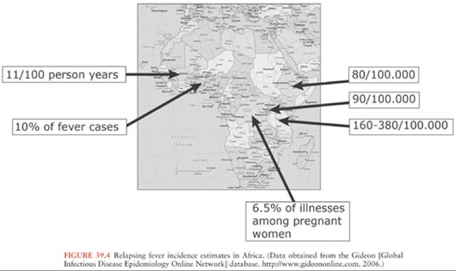
TBRF occurs nearly worldwide (Fig. 39.2). In North America, these infections occur primarily in the West and Southwest (Fig. 39.3) (3), where Borrelia hermsii, transmitted by the Ornithodoros hermsi tick, is the most common cause, followed by B. turicatae and B. parkeri. O. hermsi ticks are found in coniferous forests at altitudes of 1,500 to 8,000 feet (10). Consequently, in the United States, TBRF occurs primarily in the Rocky Mountain states, with infections acquired most commonly in rustic rodent-infested cabins. B. parkeri and B. turicatae infect ticks with corresponding names O. parkeri and O. turicata, which are found across a somewhat broader geography at lower altitudes, primarily in caves or burrows (3). In Eurasia, TBRF is most commonly caused by Borrelia persica but also by Borrelia caucasica and Borrelia latyschewii (11,12). In sub-Saharan Africa (Fig. 39.4), Borrelia duttonii(transmitted by Ornithodoros moubata) is the most common cause of TBRF (3). In rural sections of West Africa, Borrelia crocidurae is a prominent cause of infection, responsible for 5% to 25% of all febrile illnesses, depending on the year and location (13).

TBRF ecology varies among species. Most TBRF Borrelia infections are zoonoses, but no animal reservoir has been identified for B. duttonii, which is closely related genetically to B. recurrentis (1,3,14,15). For other species, the principal reservoirs are rodents, and other small animals. Infected Ornithodoros ticks may survive without a blood meal for more than a decade. That, along with the possibility of transovarial transmission of relapsing fever Borreliain the tick vector (3), suggests that ticks themselves may potentially serve as reservoirs.
Although vector-borne transmission is by far the most common means of transmission of relapsing fever Borrelia, infection has also been transmitted through blood transfusion, sharing of needles among intravenous drug users, laboratory accidents, and transplacentally (3,12).
General Clinical Features and Pathogenesis
Symptom severity varies widely in patients infected with relapsing fever Borrelia (16); some remain completely asymptomatic, whereas others become critically ill. The hallmark of relapsing fever is periodic fevers. Approximately 7 days after inoculation, patients develop the sudden onset of chills and fever, typically between 38.7° and 41°C (Fig. 39.5), frequently with rigors, headache, fatigue, altered sensorium, arthralgias, and myalgias (16). Some patients develop a cough, nausea, vomiting, diarrhea, icterus, a maculopapular or petechial truncal skin rash, neurologic and ocular involvement, pneumonia, myocarditis, acute respiratory distress syndrome, abdominal pain, hepatosplenomegaly, icterus, hepatic failure, and splenic rupture (3). Ocular manifestations of TBRF include iritis, iridocyclitis, and choroiditis (17). In contrast to TBRF, eye involvement occurs rarely if ever in LBRF(17).

The most common causes of death are myocarditis, cerebral hemorrhage, and liver failure. Relapsing fever in pregnancy can result in miscarriage in one third of cases (12,18,19). B. duttonii infection is often associated with low birth weight, preterm delivery, spontaneous abortion, and perinatal death (20–25).
The number of inoculated bacteria required to cause infection can be quite low. In mice, a single bacterium may be sufficient to establish infection (1). Once relapsing fever Borrelia enter the blood, they multiply to large numbers, potentially reaching concentrations of 106 to 108 organisms per milliliter, at which point symptoms appear. Symptoms resolve spontaneously within about 3 days (range 1 to 9 days), as bactericidal T-cell independent immunoglobulin (Ig) M antibodies (26) damage the borrelial outer membrane, resulting in clearance of spirochetes from the blood. Both B-cell receptor and toll-like receptor signaling are involved in the development of this rapid T-cell independent antibody response (27,28). Neither phagocytic cells nor complement activation play an essential role in spirochete clearance; relapsing fever Borrelia are able to inactivate the complement cascade (26,29). Fever recurs in 7 or more days (range typically 3 to 10 days) with the emergence of an antigenically different strain of Borrelia, resistant to the bactericidal activity of the previously produced antibodies.
These antigenically different strains arise from alteration in the expression of the variable major surface lipoprotein, Vmp, through genetic rearrangement whereby silent genes move into an expression locus (3,30,31). This antigenic variation is not random; certain Vmps are more likely than others to be expressed in early infection (32), perhaps explaining why some neurologic and ophthalmologic manifestations usually do not occur during the first bout of fever (see the following discussion). Aside from the change in Vmp, the new strain of Borrelia is otherwise identical to the original infecting strain (30,31,33). In theory, at least 25 antigenically distinct serotypes can be generated from a single bacterium (27). Emergence of each successive serotype is associated with a fever relapse. Recurrences may be shorter in duration and associated with less intense symptoms and fewer spirochetes in the blood, especially for LBRF. In experimental animals, different serotypes not only confer antibody resistance but also appear to vary in their capacity to cause brain infection (34,35). This may, at least in part, be explained by different serotypes’ varying ability to bind and cross the brain microvascular endothelium (33).
Laboratory Findings
In patients with relapsing fever, the white blood cell count, erythrocyte sedimentation rate, and liver function tests are usually elevated. Anemia and thrombocytopenia are common (1,3), and although usually mild, the thrombocytopenia can be severe, potentially resulting in bleeding complications. One possible mechanism proposed for the development of thrombocytopenia, suggested by animal models (36,37), is through binding of Borrelia to the outer surface of platelets, a phenomenon that may also play a role in eradicating relapsing fever Borrelia from the blood. Certain species of relapsing fever Borrelia—such as B. crocidurae, B. duttonii, and B. hispanica—bind to erythrocytes, causing erythrocyte rosetting, which may contribute to the development of anemia (1,12,38). The resulting aggregates of bacteria and red blood cells may also disrupt the microcirculation, resulting in hemorrhage, reduced blood flow, hypoxia, and cell death (1,12,38,39).
Neurologic Manifestations
The most commonly reported neurologic manifestations are meningeal symptoms, meningitis, and facial nerve palsy.Delirium (encephalopathy) is also common. In severe cases, CNS microhemorrhages, focal abnormalities, seizures, and coma can occur. The frequency of neurologic involvement depends on the infecting species of relapsing Borrelia. B. duttonii, B. crocidurae, and B. turicatae are the TBRF species most likely to cause neurologic complications (17), particularly meningitis and facial nerve palsy.
Facial nerve palsy, although frequent, does not typically appear during the first febrile episode. It usually resolves within 2 months with or without antibiotic therapy (17). Ocular involvement is unilateral in two thirds of cases and, like facial nerve palsy, almost never occurs during the first febrile episode.
Although there are reports of other cranial nerve palsies, optic neuritis, radicular symptoms, neuropsychiatric abnormalities, and other findings, no large population-based studies are available to provide clear data regarding the frequency of most of these disorders or even compelling evidence of a causal relationship.
Microscopically, meninges and brain may show perivascular infiltrates composed of monocytes, lymphocytes, and plasma cells (17). Cerebral edema and subarachnoid and parenchymal brain hemorrhage may occur in fatal cases of LBRF. In experimental animals, cerebral microgliosis is a prominent feature (33,40,41). Both meningitis and meningismus occur frequently in TBRF. In LBRF, although as many as 50% of patients develop meningismus, far fewer have actual meningitis (as confirmed by cerebrospinal fluid [CSF] examination) (17). In experimentally infected rodents, some strains of relapsing fever Borreliacan cause persistent brain infection (15) that can be cleared successfully with ceftriaxone treatment (42). Persistent brain infection in untreated humans, however, has not been documented.
The CSF white cell count is typically elevated in relapsing fever patients with CNS involvement. CSF examination demonstrates a mononuclear-predominant pleocytosis with cell counts in the hundreds/mm3and protein concentration of up to several hundred mg%. CSF glucose is typically normal. Organisms have been detected in CSF in a minority of cases by microscopic examination, culture, or animal inoculation. In fatal cases of TBRF, spirochetes have been detected histologically in perivascular areas of brain cortex (17). Testing for the production of intrathecal antibodies to Borrelia has not been established to be a useful diagnostic test in relapsing fever (32).
Diagnosis
During febrile episodes, there may be up to 108 spirochetes per milliliter of blood; none are detectable during afebrile periods. As a result, extracellular spirochetes may be directly visualized in blood obtained during a febrile episode, either on Giemsa (Fig. 39.6) or Wright-stained blood smears or by dark-field or phase-contrast microscopy of plasma wet mounts. The sensitivity of these techniques is 70% or less, but this can be increased using fluorescent microscopy of acridine orange–stained smears or by examination of buffy coat smears.

Detection of relapsing fever borrelial DNA by PCR appears to be more sensitive than detection by blood smear but is only offered by a few laboratories. Serologic assays including enzyme-linked immunosorbent assays (ELISAs) or immunofluorescence assays (IFAs), followed by supplemental immunoblots using antigens from particular species of relapsing fever Borrelia, are available from a few laboratories but only for some borrelial species. Relapsing fever and Lyme disease patients often produce cross-reactive antibodies given the similarity of flagellin and other antigens. A specific serologic assay for relapsing fever Borrelia infections uses the surface protein glycerophosphodiester phosphodiesterase (GlpQ) as the antigen (3). Because this protein is not present in Lyme Borrelia, Lyme disease patients will be seronegative using this assay. Other testing modalities with even more limited availability include culture and animal inoculation.
Treatment
TBRF is treated with a 5- to 10-day course of oral tetracycline, doxycycline, erythromycin, penicillin, amoxicillin, or chloramphenicol (3). The preferred therapy for LBRF is a single oral dose of tetracycline or erythromycin, although some authorities favor extending treatment to 7 days (1). Meningitis or encephalitis is treated with intravenous penicillin G, ceftriaxone, or cefotaxime for at least 14 days.
A Jarisch-Herxheimer reaction, thought to result from the rapid killing of relapsing fever Borrelia by antibiotics with the sudden release of lipoproteins, occurs frequently in the first few hours following initial treatment. The resultant induction of cytokines such as tumor necrosis factor-α (TNF-α), interleukin-6 and -8 (IL-6, IL-8), and other chemical mediators causes rigors, leukopenia, fever, and hypotension, signs similar to those of a classic endotoxin reaction (1,3). Because of the potential severity of such reactions, patients should be kept under observation for approximately 2 hours after initiating antibiotic therapy. This is an “all or nothing” reaction, occurs in more than 50% of patients, and usually resolves spontaneously within 12 to 24 hours. However, cardiovascular collapse may occur, with a fatality rate of about 5% (43,44). Several studies have suggested that the choice of antibiotic affects the severity, but such data are inconclusive. Treatment with anti–TNF-α agents prior to antibiotics may reduce the severity of the reaction but is not used clinically at this time (45).
When tick bites and/or tick exposure has occurred with ticks at high risk for transmitting relapsing fever Borrelia, a 5-day course of doxycycline chemoprophylaxis during the infection’s incubation period is highly effective in prevention of TBRF (46).
LYME DISEASE
Lyme disease is caused by a group of related spirochetes—Borrelia burgdorferi sensu lato or Lyme Borrelia. Lyme disease is the most commonly reported vector-borne infectious disease in the United States and is also of public health importance in numerous countries with temperate climates in Eurasia (47). The term “Lyme disease” was coined in the mid-1970s, with the recognition of a cluster of cases of what appeared to be juvenile rheumatoid arthritis (48) in Lyme, Connecticut. Europeans, however, have been familiar with this disorder since the early twentieth century (49) and have been treating it successfully with antibiotics since the 1950s, three decades before the isolation of the causative organism (50). The history of European versus United States Lyme disease has resulted in the perception that these infections behave quite differently. However, they actually share more similarities than differences, making the century of clinical experience with European neurologic Lyme disease informative for our understanding of this aspect of Lyme disease in the United States.
Epidemiology, Vectors, Agents, and Reservoirs of Lyme Borrelia
In North America, the only species of Lyme Borrelia known to cause human disease is B. burgdorferi sensu stricto (which will be referred to as B. burgdorferi). In Europe, at least five species of Lyme Borrelia can cause disease, of which B. afzelii, B. garinii, and B. burgdorferi are the most common. Because of this species diversity, the range of clinical manifestations appears to be greater in Europe than in North America (47). In Europe, B. garinii is the species of Lyme Borrelia most closely associated with neurologic manifestations (47).
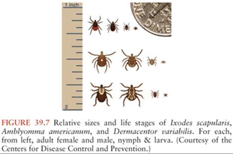
Lyme Borrelia is transmitted during feeding by Ixodes species ticks (Fig. 39.7). Only after ingested blood triggers spirochete proliferation and migration to the tick’s salivary glands can infection be transmitted. A feeding period of more than 36 hours is usually needed for transmission of B. burgdorferi by I. scapularis or I. pacificus ticks, whereas in experimental animals, transmission of B. afzelii by I. ricinus can occur in under 24 hours (47,51–54). The main vertebrate reservoirs for Lyme Borrelia are mice, other small mammals, and some species of birds.
In the United States, about 30,000 cases of Lyme disease are identified by the Centers for Disease Control and Prevention (CDC) annually; cases occur predominantly in the northeast, middle/south Atlantic, and north central states (Fig. 39.8). In Europe and Asia, with an estimated 85,000 cases per year (accurate numbers are difficult to obtain as this is not a reportable disease in many European countries), infection occurs broadly across temperate central regions (55).

General Clinical Features and Pathogenesis
Lyme Borrelia resides in the midgut of unfed Ixodes ticks. When an infected tick takes a blood meal, the spirochetes present there increase in number and undergo phenotypic changes, including the expression of a particular outer surface protein, OspC, which allows them to disseminate within the tick and enter its salivary glands (47). This process takes several days and explains why transmission occurs only after a delay. Expression of OspC plays an essential part in the establishment of infection in a mammalian host, although the mechanism by which this occurs is unknown (56,57).
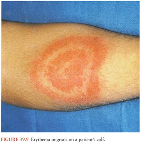
During feeding, the infected tick deposits spirochetes into the skin at the bite site. This may lead to the development of a type of bacterial cellulitis, referred to as erythema migrans (EM; Fig. 39.9), at that location. Lyme Borreliamay disseminate from that site to other locations in the body: through the blood or perhaps through tissue planes. The risk of hematogenous dissemination by B. burgdorferi is dependent on the strain of Borrelia (58).
Infection elicits innate and adaptive immune responses, resulting in both macrophage- and antibody-mediated killing of Lyme Borrelia. Despite a robust humoral and cellular immunologic response, however, infection with Lyme Borrelia can persist. Persistence is due in part to the spirochete’s ability to downregulate expression of particular immunogenic surface-exposed proteins, including OspC, and to alter by recombination the antigenic properties of another surface lipoprotein known as variable major protein-like sequence expressed (VlsE) (30,47). Lyme Borrelia may bind to various components of the extracellular matrix, which may also contribute to persistence (59). Localized infection is typically manifested by a single EM skin lesion. In early disseminated disease, there may be two or more EM skin lesions or an objective manifestation of neurologic or cardiac Lyme disease. The most frequently described manifestation of late Lyme disease in the United States is a mono- or pauciarticular arthritis.
Most patients develop EM between June and August (60). The seasonal distribution of extracutaneous manifestations of Lyme disease is less pronounced because the time from infection to onset of these manifestations is longer than it is for EM. Generally, the clinical features of Lyme disease are similar in children and in adults. However, acrodermatitis chronica atrophicans (a late skin manifestation seen primarily in Europe) is typically not seen in children (47).
Laboratory Findings
Patients with Lyme disease have a normal white blood cell count, hematocrit, and platelet count, unless there is coinfection with Anaplasma phagocytophilum, Babesia microti, or a tick-borne encephalitis virus (47). Slightly raised liver function tests can be seen in up to one third of patients with early disseminated Lyme borreliosis, especially in those with EM. Erythrocyte sedimentation rates may be slightly to moderately elevated. In patients with Lyme meningitis, CSF typically shows a pleocytosis with more than 90% lymphocytes, a slightly raised protein level, and a normal glucose level.
Neurologic Manifestations
Nervous system involvement, first described in 1922 in France (49), most commonly includes any or all elements of a clinical triad—lymphocytic meningitis, cranial neuritis, and painful radiculitis—often referred to in the European literature as Garin-Bujadoux-Bannwarth syndrome. This tends to occur relatively early in infection, and an EM skin lesion may still be present. Therefore, in patients with suspected neurologic Lyme disease, a complete skin examination should be done to look for EM skin lesions to help confirm the diagnosis (61).
Clinically, the symptoms associated with meningitis vary in severity. Comparative studies suggest that the onset is typically a little less acute than in viral meningitis, with symptoms evolving over a day or two rather than a few hours (62,63). In contrast to patients with Lyme meningitis, individuals with summertime viral meningitis, typically due to enteroviral infections, rarely develop cranial neuropathies. Another group of agents, tick-borne encephalitis viruses, seen in Eurasia but rarely in North America, can similarly cause a typical viral meningitis but can also lead to a more severe meningoencephalomyelitis that is particularly likely to cause segmental spinal cord involvement.
Of the patients who develop a cranial neuropathy, about 80% have facial nerve paralysis, and in one quarter of these patients, the facial palsy is bilateral (64). Other cranial neuropathies may include cranial nerves III, IV, or VI, which innervate the extraocular muscles (causing diplopia), the trigeminal nerve (causing paresthesias or numbness), or the vestibuloacoustic nerve (causing hearing loss or vertigo). Reports of involvement of the lower four cranial nerves are at best anecdotal. Although the eye itself can be involved in Lyme disease (similar to the eye involvement in relapsing fever), optic neuritis specifically is very rare, if it occurs at all (65).
The neurologic disorder that figured prominently in the 1922 case report, but is still underrecognized, is referred to as painful radiculitis (49,66). Patients develop severe, burning neuropathic type pain, often radicular in distribution but not uncommonly involving more than one dermatome, usually with corresponding sensory, motor, and reflex changes. Pain is typically worse at night. When involving truncal dermatomes, pain can be mistaken for visceral disease. Limb symptoms can precisely mimic those of a mechanical radiculopathy. The diagnosis should be considered in individuals with typical radicular symptoms without an antecedent injury, especially in those with negative imaging. Although CSF pleocytosis is not always present, when found, it should lead to consideration of this diagnosis in tick-exposed individuals. Some patients develop other forms of mononeuropathy multiplex, including more typical peripheral mononeuropathies or even plexopathies.
Parenchymal CNS infection is quite rare (67). Occasional patients with Lyme radiculitis may have evidence—clinically or by imaging—of involvement of the spinal cord at the affected nerve root level. Very rarely, patients develop parenchymal brain inflammation, a focal encephalitis (68–70).
At the other end of the spectrum, some individuals with milder but more prolonged untreated Lyme disease (something rarely seen now) may develop more indolent and disseminated peripheral nerve involvement (71,72). Not surprisingly, neurophysiologic studies indicate that this polyneuropathy is similarly due to a confluent mononeuropathy multiplex, the same basic pathophysiologic process seen in Garin-Bujadoux-Bannwarth syndrome. Although the mechanism responsible for this disorder is unknown, one could conjecture that these patients have a smaller spirochete load, infection with an antigenically different strain, or a different immunologic response to the infection. This manifestation of neurologic Lyme disease is now recognized rarely in the United States. Although not specifically described in European patients, a neurophysiologically similar disorder occurs in patients with acrodermatitis chronica atrophicans—patients in whom this primarily involves the limb affected with the skin lesion (73).
Over the years, possible associations between Lyme disease and a variety of serious neurologic disorders have been proposed.
Demyelinating disease: In the 1980s and 1990s, numerous reports, primarily from Europe, described patients with progressive inflammatory disorders of the brain and spinal cord (67–70). Far fewer reports have appeared in recent years. Whether this phenomenon was due to inadequacies of earlier serologic diagnostic tools, or to a change in the presentations of the illness as earlier recognition and treatment became widespread, is unclear (74). Regardless, it is now remarkably uncommon to see patients with Lyme disease presenting like multiple sclerosis.
Motor neuron disease: Several reports of patients (75,76) with what appeared to be motor neuron disease, serologic evidence of Lyme disease, and improvement following antimicrobial therapy led to an epidemiologic study demonstrating an excessive prevalence of antibodies to B. burgdorferi among amyotrophic lateral sclerosis (ALS) patients in a Lyme disease–endemic area compared both to ALS populations elsewhere and to a variety of other controls (77). No satisfactory explanation for this observation has ever been provided. However, the lack of treatment responsiveness among these patients, and the failure to replicate the observation elsewhere, suggest it was a chance observation.
Cerebral vasculitis: A number of isolated reports has described patients thought to have cerebral vasculitis attributable to Lyme disease (78–83). In some studies, vasculitis was inferred from the presence of areas of brain damage on magnetic resonance imaging (MRI) in the absence of vascular imaging. In others, the support for the diagnosis of Lyme disease did not meet current serologic criteria. Given that Lyme disease does not cause vasculitis in other organ systems, and the paucity of evidence in the cases that have been reported, this disorder seems unlikely.
Alzheimer disease, Parkinson, neuropsychiatric disorders: Numerous case reports have purported to show relationships between these disorders (84–87) and Lyme disease. No systematic data have ever indicated such an association; therefore, these linkages appear unlikely.
LYME ENCEPHALOPATHY
An entity that has caused tremendous confusion is the disorder termed “Lyme encephalopathy.” Originally described in individuals with symptoms of long-standing untreated Lyme disease (e.g., arthritis) (88–90), it rapidly became clear that few, if any, of these patients had objective evidence of CNS infection (91). Rather, this appeared to be an example of the encephalopathy or delirium seen in the context of innumerable other systemic infections and inflammatory states (pneumonia, urinary tract infections, influenza, active rheumatoid arthritis, etc.).
Initial interest in studying this as a possible model of encephalopathy in inflammatory states was widely misinterpreted in three critical ways. First, many assumed that this constituted evidence of either brain infection or of another neuropathologic state, something for which there is no scientific support. Second, some assumed that this disorder was specific to Lyme disease, despite it clearly being just one example of a state commonly occurring in a wide range of situations ranging from systemic illness to depression, sleep deprivation, and innumerable other conditions. Most problematic has been the notion that this encephalopathy is not only common but also often persists after antibiotic treatment, a state sometimes referred to as post–Lyme disease syndrome. Moreover, patients with similar symptoms, but no other evidence of Lyme disease—clinically or by laboratory studies—are sometimes, nevertheless, assumed to have Lyme disease and treated with antibiotics for extended periods of time. Because symptoms that are not related to infection with Lyme Borrelia would not be expected to respond to treatment for Lyme disease, treatment unresponsiveness in this group has contributed greatly to the widespread mistaken belief that Lyme disease does not respond to antimicrobial therapy.
Efforts to support the diagnosis of “Lyme encephalopathy” with brain single-photon emission computed tomography (SPECT) scanning—a technique so unreliable that it never even gained acceptance in the diagnosis of stroke, the purpose for which it was first developed—have only added to the confusion. This has all contributed tremendously to patient anxiety about Lyme disease. Clearly, the notion of a brain-damaging infection is terrifying to most people.
Studies of the general U.S. population, using well-validated self-reporting questionnaires, indicate that as many as 2% of the general population without Lyme Borrelia infection may experience the symptom complex attributed to “Lyme encephalopathy”—severe fatigue, cognitive slowing, and other nonspecific symptoms—at any given time (92). In contrast, identifying patients with “post–Lyme disease syndrome” for clinical trials has been remarkably difficult. One of the most systematic efforts (93), led by a group very well connected to Lyme disease patient advocacy groups throughout the country, recruited for 4 1/3 years and was able to enroll only 37 patients who appeared to have had Lyme disease previously and had persisting cognitive symptoms.
Pathogenesis of Neurologic Manifestations
Given the variety of clinical disorders associated with Lyme neuroborreliosis, it is likely that more than one mechanism is involved. Lyme meningitis can be attributed to B. burgdorferi infection within or in close proximity to the subarachnoid space because organisms can be detected in some patients’ CSF and because antibiotic treatment leads to rapid recovery. As challenging as it is to find organisms in Lyme meningitis, this is virtually impossible with most other neurologic manifestations of Lyme disease.
Peripheral nerve involvement, although highly variable in clinical presentation, seems to be uniformly attributable to a mononeuropathy multiplex, a mechanism typically associated with vasculopathic processes (e.g., vasculitis, diabetic microvascular changes). Neurophysiologic studies in patients with Lyme disease (71), and in experimentally infected animals, suggest this pathophysiology, whether the patient presents with facial nerve palsy, radicular symptoms, focal mononeuropathies, or “stocking glove” type distal symptoms (71). The few available nerve biopsies from patients (94–96), as well as nerve biopsies from experimentally infected rhesus macaques (97), consistently demonstrate perivascular inflammatory infiltrates, without evidence of vessel wall necrosis (which would be required to diagnose a vasculitis) or other vasculopathic changes (Fig. 39.10). None of the histopathologic studies in humans has demonstrated either intact spirochetes or even their antigens in the biopsied nerves.

Although there is evidence that the CNS may be invaded early in the course of Lyme disease (Fig. 39.11) (98–100), parenchymal brain involvement occurs very rarely in humans. The few human autopsy reports thought to reflect brain involvement have described primarily nonspecific changes such as glial proliferation and lymphocytic infiltrates (101). A few studies have identified spirochetes in patients thought to have parenchymal brain involvement. One study identified large areas of inflammation in brain tissue thought to be due to this infection and found B. burgdorferi DNA by PCR testing of tissue from involved areas (102), a finding that has never been replicated. The inability to demonstrate spirochetes in most cases has led to numerous studies exploring potential roles of the immune response, either by virtue of inadvertent cross reactivity or effect amplification by immunomodulators. Studies have shown that antibodies to B. burgdorferi flagellin cross-react with a heat shock protein (HSP60) present in peripheral nerve and can affect neuroblastoma cell neuritogenesis in vitro (103). Both B. burgdorferi bacteria themselves, and antibodies to B. burgdorferi, may also potentially interact with various myelin-associated glycoproteins (98,104). The most persuasive argument against a pathogenetic role for molecular mimicry, though, is the fact that the humoral response to B. burgdorferi usually persists for an extended period of time following successful antibiotic treatment, yet the neurologic disorders rapidly resolve.

Alternatively, the cytokine response to Lyme Borrelia infection could provide a more plausible explanation. Unlike antibodies, the level of these molecules does rise and fall rapidly with the presence of infecting microorganisms. Equally importantly, some cytokines can affect nervous system function (105,106) providing a potential mechanism. Over the years, a number of molecules have been investigated. CXCL13 is produced by brain glia and dorsal root ganglia early in infection and serves to attract B lymphocytes that then produce specific antibodies (107,108). Although there has been no evidence that this particular cytokine affects nervous system function, it may serve as a helpful marker of disease activity. Indirect evidence has suggested a possible role of interferon-γ (IFN-γ) (106) and interferon-α (IFN-α) (109); however, direct proof of an effect has been lacking. In an in vitro brain slice model (rhesus), brain oligodendroglia and astrocytes exposed to B. burgdorferi exhibited apoptosis, and glial transcription of genes for many inflammatory immunomodulators was altered (108,110). However, how this directly relates to neuroborreliosis remains to be elucidated.
In summary, despite numerous tantalizing suggestions regarding potential mechanisms underlying neuroborreliosis, this remains a topic in need of additional studies.
Diagnosis
EM usually has a distinctive enough appearance to allow a clinical diagnosis in the absence of a supporting laboratory test. Serologic assays for antibodies to Lyme Borrelia are negative at this stage in at least 50% of cases and thus should be obtained only in atypical cases and then in conjunction with convalescent phase serologic testing after 2 to 6 weeks (111).
For non-EM presentations of Lyme borreliosis, including neurologic Lyme disease, the mainstay of laboratory diagnosis is two-tier serologic testing in which the first-tier test is usually an ELISA. If the ELISA is reactive (positive or borderline), then separate IgM and IgG immunoblots are done on the same serum sample (112). If symptoms have persisted for at least 4 weeks, then the IgG immunoblot should be positive. Untreated patients who remain seronegative despite symptoms persisting for more than 6 weeks are unlikely to have illness attributable to Lyme borreliosis.
Omission of the first-tier ELISA or interpretation of the immunoblot with criteria that are not evidence based will potentially decrease specificity and possibly result in misdiagnosis (47). In addition, in clinical practice, there are often false-positive IgM immunoblot test results because of overreading of weak bands by some commercial laboratories (113).
As with most infections, patients with Lyme disease often continue to produce a specific immune response long after apparent microbiologic cure. Hence, a positive serology cannot be used to judge treatment efficacy or to establish proof of a current active infection.
Testing for anti-Borrelia antibodies that are produced locally in the CNS (i.e., intrathecal synthesis of specific antibodies) is a mainstay of the diagnosis of Lyme neuroborreliosis in Europe (114,115), where it is believed to be present in 70% to 90% of patients with acute neuroborreliosis and more than 90% of those with more long-standing infection (116). In the past, some European patients were found to have antibody in the CSF before they developed a positive serum serology (117). This observation has not been replicated with currently recommended serologic techniques. Importantly, intrathecal synthesis of antibodies can persist for months to years after successful antibiotic treatment and therefore cannot serve as a test for cure (118). (CSF cell counts and protein concentration can be more useful to gauge active infection, however.) Because there is no “gold standard” diagnostic tool for CNS infection, it is difficult to determine the test’s sensitivity; in American patients, estimates have ranged from 46% in individuals with more chronic symptoms (69) to 87% in those with acute Lyme meningitis (91). Finally, it is important to appreciate that the presence of intrathecal antibody to Lyme Borrelia is a marker of CNS infection specifically. In patients with neuroborreliosis limited to the peripheral nervous system (PNS), with encephalopathy due solely to systemic inflammation, or with no suggestion of nervous system involvement, there is no reason to expect that intrathecally produced antibodies to Lyme Borrelia will be present.
Cultures for Lyme Borrelia are not routinely done to diagnose Lyme disease. Such testing is unnecessary for patients with EM and too insensitive for patients with extracutaneous manifestations of Lyme disease. The positivity rate of PCR in CSF tends to be low in neurologic Lyme disease, being only about 5% in a recent study of children from the United States (119). A negative PCR result on CSF certainly does not exclude neurologic Lyme disease. PCR on blood or urine samples, tests designed to detect B. burgdorferi antigens in urine, tests for T-lymphocyte recognition of Borrelia antigens (as a measure of a cellular immune response to Lyme Borrelia), measurement of the number of CD57 natural killer cells, and use of live microscopy on blood to search for spirochetes have not been demonstrated to be reliable and are not recommended (111).
Treatment
In Europe and the United States, parenteral antibiotic therapy had been the preferred treatment for neurologic Lyme disease, especially for meningitis and radiculitis. Studies done in Europe, however, have provided convincing evidence that a 14-day course of oral doxycycline is as effective as ceftriaxone for most of the manifestations of neurologic Lyme disease (120). Although no systematic studies on this issue have been done in the United States, there is a growing favorable clinical experience with doxycycline. Other oral antibiotics such as amoxicillin have been used successfully to treat patients with uncomplicated seventh nerve palsy, but efficacy data for such drugs are limited. Seventh nerve palsy will resolve at the same rate with or without antibiotic treatment (121). Thus, one of the main reasons to treat such patients is to prevent the development of later complications, especially Lyme arthritis in the United States (122). At this point, it is reasonable in our judgment to reserve parenteral treatment—with at least a 14-day course of ceftriaxone, cefotaxime, or penicillin G—for patients with parenchymal CNS disease (both because of a lack of evidence of efficacy of oral regimens and out of an abundance of caution), patients who continue to have active disease following treatment with oral regimens, and for those rare patients who present acutely very ill.
There has been considerable uncertainty about the best approach for initiating antibiotic therapy for patients with meningitis or cranial or peripheral nerve involvement suspected but not yet proven to be due to Lyme disease. If an EM skin lesion is present, antimicrobial treatment, typically with oral doxycycline, can be started immediately. Because most patients with meningitis, cranial neuritis, and other disorders do not have Lyme disease, and because no clinical features reliably identify the subset of these patients who have Lyme disease, laboratory confirmation is essential before planning definitive treatment. If there are sound epidemiologic reasons to think the patient might have been exposed to B. burgdorferi–infected ticks, if test results can be obtained within 24 to 48 hours, and if there are no contraindications to several days of empirical treatment with oral doxycycline, then this can be started pending test results. On the other hand, the likelihood of disease progression during the 1 to 2 days required to obtain test results is small, making deferred treatment an equally valid option. Notably, if the patient presents with facial nerve palsy and is seen within the first 72 hours of symptoms, oral corticosteroids (typically prednisolone 50 to 60 mg per day [or equivalent]) for 5 days followed by a rapid taper (123) should be started immediately as such therapy has been shown to improve outcomes in Bell palsy, the most common cause of seventh nerve palsy in the general population (123). In this setting, concurrent initiation of empirical oral doxycycline is reasonable, pending test results, although there is no evidence that brief courses of steroids make neuroborreliosis more difficult to treat.
Outcome
Lyme disease is a curable bacterial infection. Appropriate antimicrobial therapy should stop both disease progression and the development of any new manifestations of the infection. As in many bacterial infections, some symptoms may resolve slowly following treatment, particularly nonspecific ones such as fatigue, malaise, arthralgias, and perceived cognitive and memory difficulty. If, prior to treatment, a patient has developed evidence of tissue damage—synovitis in patients with rheumatologic disease; facial nerve, nerve root, or other focal peripheral nerve damage in those with PNS disease—tissue repair may be slow and sometimes incomplete if sufficiently severe in the first place. Focal inflammatory CNS disease, initially described in a substantial number of European patients in the 1980s and 1990s but only rarely since then, can respond very well to antimicrobial therapy. As in PNS disease, however, symptoms improve with treatment, but if there has been irreparable damage to the brain or spinal cord, some residua would be expected.
CONCLUSION
Borrelia infections, both relapsing fevers and Lyme disease, can affect the nervous system. Meningitis—is probably the most common phenomenon in both groups of diseases. Focal and multifocal nervous system involvement can occur, but the frequency, severity, and mechanisms probably vary with the specific spirochete species. In relapsing fever, brain involvement probably is due to an ischemic and hemorrhagic vasculopathy. In Lyme disease, both PNS and CNS involvement tends to be much more focal; although it is presumed to occur on the basis of either local brain infection, multifocal vasculopathic changes secondary to the infection, and/or the immune response to the organism, much remains to be learned about the pathophysiology.
References
1. Larsson C, Andersson M, Bergstrom S. Current issues in relapsing fever. Curr Opin Infect Dis. 2009;22:443–449.
2. Houhamdi L, Raoult D. Excretion of living Borrelia recurrentis in feces of infected human body lice. J Infect Dis. 2005;191(11):1898–1906.
3. Dworkin MS, Schwan TG, Anderson DE, Jr, et al. Tick-borne relapsing fever. Infect Dis Clin North Am. 2008;22(3):449–468.
4. Fukunaga M, Takahashi Y, Tsuruta Y, et al. Genetic and phenotypic analysis of Borrelia miyamotoi sp. nov., isolated from the ixodid tick Ixodes persulcatus, the vector for Lyme disease in Japan. Int J Syst Bacteriol. 1995;45(4):804–810.
5. Scoles GA, Papero M, Beati L, et al. A relapsing fever group spirochete transmitted by Ixodes scapularis ticks. Vector Borne Zoonotic Dis. 2001;1(1):21–34.
6. Fraenkel CJ, Garpmo U, Berglund J. Determination of novel Borrelia genospecies in Swedish Ixodes ricinus ticks. J Clin Microbiol. 2002;40(9):3308–3312.
7. Krause PJ, Narasimhan S, Wormser GP, et al. Human Borrelia miyamotoi infection in the United States. N Engl J Med. 2013;368(3):291–293.
8. Gugliotta J, Goethert H, Berardi V, et al. Meningoencephalitis from Borrelia miyamotoi in an immunocompromised patient. N Engl J Med. 2013;368:240–245.
9. Platonov AE, Karan LS, Kolyasnikova NM, et al. Humans infected with relapsing fever spirochete Borrelia miyamotoi, Russia. Emerg Infect Dis. 2011;17(10):1816–1823.
10. Centers for Disease Control and Prevention. Tick-borne relapsing fever. Centers for Disease Control and Prevention Web site. http://www.cdc.gov /relapsing-fever. Accessed December 31, 2013.
11. Assous MV, Wilamowski A. Relapsing fever borreliosis in Eurasia—forgotten, but certainly not gone! Clin Microbiol Infect. 2009;15(5):407–414.
12. Rebaudet S, Parola P. Epidemiology of relapsing fever borreliosis in Europe. FEMS Immunol Med Microbiol. 2006;48(1):11–15.
13. Bottieau E, Verbruggen E, Aubry C, et al. Meningoencephalitis complicating relapsing fever in traveler returning from Senegal. Emerg Infect Dis. 2012;18(4):697–698.
14. Cutler SJ. Relapsing fever—a forgotten disease revealed. J Appl Microbiol. 2010;108(4):1115–1122.
15. Larsson C, Andersson M, Pelkonen J, et al. Persistent brain infection and disease reactivation in relapsing fever borreliosis. Microbes Infect. 2006;8(8):2213–2219.
16. Southern P, Sanford J. Relapsing fever: a clinical and microbiological review. Medicine. 1969;48:129–149.
17. Cadavid D, Barbour AG. Neuroborreliosis during relapsing fever: review of the clinical manifestations, pathology, and treatment of infections in humans and experimental animals. Clin Infect Dis. 1998;26(1):151–164.
18. Lawaczeck EW, Mead PS, Schriefer ME, et al. Tickborne relapsing fever in a mother and newborn child—Colorado, 2011. MMWR Morb Mortal Wkly Rep. 2012;61(10):174–176.
19. Larsson C, Andersson M, Guo BP, et al. Complications of pregnancy and transplacental transmission of relapsing-fever borreliosis. J Infect Dis. 2006;194(10):1367–1374.
20. Dupont HT, La Scola B, Williams R, et al. A focus of tick-borne relapsing fever in southern Zaire. Clin Infect Dis. 1997;25(1):139–144.
21. Melkert PW, Stel HV. Neonatal Borrelia infections (relapsing fever): report of 5 cases and review of the literature. East Afr Med J. 1991;68(12):999–1005.
22. Brasseur D. Tick-borne relapsing fever in a premature infant. Ann Trop Paediatr. 1985;5(3):161–162.
23. Goubau PF, Munyangeyo C. Congenital relapsing fever caused by Borrelia duttoni. Apropos of a case in Rwanda [in French]. Ann Soc Belg Med Trop. 1983;63(4):367–369.
24. Jongen VH, van Roosmalen J, Tiems J, et al. Tick-borne relapsing fever and pregnancy outcome in rural Tanzania. Acta Obstet Gynecol Scand. 1997;76(9):834–838.
25. van Holten J, Tiems J, Jongen VH. Neonatal Borrelia duttoni infection: a report of three cases. Trop Doct. 1997;27(2):115–116.
26. LaRocca TJ, Katona LI, Thanassi DG, et al. Bactericidal action of a complement-independent antibody against relapsing fever Borrelia resides in its variable region. J Immunol. 2008;180(9):6222–6228.
27. Benoit VM, Petrich A, Alugupalli KR, et al. Genetic control of the innate immune response to Borrelia hermsii influences the course of relapsing fever in inbred strains of mice. Infect Immun. 2010;78(2):586–594.
28. Dickinson GS, Piccone H, Sun G, et al. Toll-like receptor 2 deficiency results in impaired antibody responses and septic shock during Borrelia hermsii infection. Infect Immun. 2010;78(11):4579–4588.
29. Grosskinsky S, Schott M, Brenner C, et al. Borrelia recurrentis employs a novel multifunctional surface protein with anti-complement, anti-opsonic and invasive potential to escape innate immunity. PLoS One. 2009;4(3):e4858.
30. Norris SJ. Antigenic variation with a twist—the Borrelia story. Mol Microbiol. 2006;60(6):1319–1322.
31. Dai Q, Restrepo BI, Porcella SF, et al. Antigenic variation by Borrelia hermsii occurs through recombination between extragenic repetitive elements on linear plasmids. Mol Microbiol. 2006;60(6):1329–1343.
32. LaRocca TJ, Benach JL. The important and diverse roles of antibodies in the host response to Borrelia infections. Curr Top Microbiol Immunol. 2008;319:63–103.
33. Sethi N, Sondey M, Bai Y, et al. Interaction of a neurotropic strain of Borrelia turicatae with the cerebral microcirculation system. Infect Immun. 2006;74(11):6408–6418.
34. Liu H, Fitzgerald D, Gran B, et al. Induction of distinct neurologic disease manifestations during relapsing fever requires T lymphocytes. J Immunol. 2010;184(10):5859–5864.
35. Cadavid D, Sondey M, Garcia E, et al. Residual brain infection in relapsing-fever borreliosis. J Infect Dis. 2006;193(10):1451–1458.
36. Alugupalli KR, Michelson AD, Joris I, et al. Spirochete-platelet attachment and thrombocytopenia in murine relapsing fever borreliosis. Blood. 2003;102(8):2843–2850.
37. Shamaei-Tousi A, Martin P, Bergh A, et al. Erythrocyte-aggregating relapsing fever spirochete Borrelia crocidurae induces formation of microemboli. J Infect Dis. 1999;180(6):1929–1938.
38. Guo BP, Teneberg S, Munch R, et al. Relapsing fever Borrelia binds to neolacto glycans and mediates rosetting of human erythrocytes. Proc Natl Acad Sci U S A. 2009;106(46):19280–19285.
39. Shamaei-Tousi A, Collin O, Bergh A, et al. Testicular damage by microcirculatory disruption and colonization of an immune-privileged site during Borrelia crocidurae infection. J Exp Med. 2001;193(9):995–1004.
40. Gelderblom H, Londono D, Bai Y, et al. High production of CXCL13 in blood and brain during persistent infection with the relapsing fever spirochete Borrelia turicatae. J Neuropathol Exp Neurol. 2007;66(3): 208–217.
41. Andersson M, Nordstrand A, Shamaei-Tousi A, et al. In situ immune response in brain and kidney during early relapsing fever borreliosis. J Neuroimmunol. 2007;183(1–2):26–32.
42. Larsson C, Lundqvist J, Bergstrom S. Residual brain infection in murine relapsing fever borreliosis can be successfully treated with ceftriaxone. Microb Pathog. 2008;44(3):262–264.
43. Bryceson AD, Cooper KE, Warrell DA, et al. Studies of the mechanism of the Jarisch-Herxheimer reaction in louse-borne relapsing fever: evidence for the presence of circulating Borrelia endotoxin. Clin Sci. 1972;43(3): 343–354.
44. Bryceson AD, Parry EH, Perine PL, et al. Louse-borne relapsing fever. Q J Med. 1970;39(153):129–170.
45. Pound MW, May DB. Proposed mechanisms and preventative options of Jarisch-Herxheimer reactions. J Clin Pharm Ther. 2005;30(3):291–295.
46. Hasin T, Davidovitch N, Cohen R, et al. Postexposure treatment with doxycycline for the prevention of tick-borne relapsing fever. N Engl J Med. 2006;355(2):148–155.
47. Stanek G, Wormser GP, Gray J, et al. Lyme borreliosis. Lancet. 2012; 379(9814):461–473.
48. Steere AC, Malawista SE, Hardin JA, et al. Erythema chronicum migrans and Lyme arthritis. The enlarging clinical spectrum. Ann Intern Med. 1977;86(6):685–698.
49. Garin C, Bujadoux A. Paralysie par les tiques. J Med Lyon. 1922;71:765–767.
50. Hollstrom E. Successful treatment of erythema migrans Afzelius. Acta Derm Venereol. 1951;31(2):235–243.
51. des Vignes F, Piesman J, Heffernan R, et al. Effect of tick removal on transmission of Borrelia burgdorferi and Ehrlichia phagocytophila by Ixodes scapularis nymphs. J Infect Dis. 2001;183(5):773–778.
52. Peavey CA, Lane RS. Transmission of Borrelia burgdorferi by Ixodes pacificus nymphs and reservoir competence of deer mice (Peromyscus maniculatus) infected by tick-bite. J Parasitol. 1995;81(2):175–178.
53. Eisen L, Lane RS. Vectors of Borrelia burgdorferi sensu lato. In: Gray JS, Kahl O, Lane RS, et al, eds. Lyme Borreliosis: Biology, Epidemiology and Control. 1st ed. New York: CABI Publishing; 2002:91–115.
54. Kahl O, Janetzki-Mittmann C, Gray JS, et al. Risk of infection with Borrelia burgdorferi sensu lato for a host in relation to the duration of nymphal Ixodes ricinus feeding and the method of tick removal. Zentralbl Bakteriol. 1998;287(1–2):41–52.
55. European Concerted Action on Lyme Borreliosis. Epidemiology of European Lyme Borreliosis. European Concerted Action on Lyme Borreliosis Web site. http://meduni09.edis.at/eucalb/cms_15/index.php?option=com_content&view=article&id=118&Itemid=115&e4fe38f3d6071561264492cf534480ed=03c6607d8267768733e1cbce4d53d0ef. Updated August 16, 2009. Accessed January 20, 2013.
56. Steere AC, Coburn J, Glickstein L. The emergence of Lyme disease. J Clin Invest. 2004;113(8):1093–1101.
57. Rosa P. Lyme disease agent borrows a practical coat. Nat Med. 2005;11: 831–832.
58. Wormser GP, Brisson D, Liveris D, et al. Borrelia burgdorferi genotype predicts the capacity for hematogenous dissemination during early Lyme disease. J Infect Dis. 2008;198(9):1358–1364.
59. Cabello FC, Godfrey HP, Newman SA. Hidden in plain sight: Borrelia burgdorferi and the extracellular matrix. Trends Microbiol. 2007;15:350–354.
60. Bacon RM, Kugeler KJ, Mead PS. Surveillance for Lyme disease—United States, 1992–2006. MMWR Morb Mortal Wkly Rep. 2008;57(SS10):1–9.
61. Wormser GP. Clinical practice. Early Lyme disease. N Engl J Med. 2006;354(26):2794–2801.
62. Tuerlinckx D, Bodart E, Jamart J, et al. Prediction of Lyme meningitis based on a logistic regression model using clinical and cerebrospinal fluid analysis: a European study. Pediatr Infect Dis J. 2009;28(5):394–397.
63. Cohn KA, Thompson AD, Shah SS, et al. Validation of a clinical prediction rule to distinguish Lyme meningitis from aseptic meningitis. Pediatrics. 2012;129(1):e46–e53.
64. Halperin JJ. Facial nerve palsy associated with Lyme disease. Muscle & Nerve. 2003;28:516–517.
65. Sibony P, Halperin J, Coyle P, et al. Reactive Lyme serology in patients with optic neuritis and papilledema. J Neuroophthalmol. 2005;25(2):71–82.
66. Reik L, Steere AC, Bartenhagen NH, et al. Neurologic abnormalities of Lyme disease. Medicine. 1979;58(4):281–294.
67. Ackermann R, Rehse KB, Gollmer E, et al. Chronic neurologic manifestations of erythema migrans borreliosis. Ann N Y Acad Sci. 1988;539: 16–23.
68. Halperin JJ, Luft BJ, Anand AK, et al. Lyme neuroborreliosis: central nervous system manifestations. Neurology. 1989;39(6):753–759.
69. Logigian EL, Kaplan RF, Steere AC. Chronic neurologic manifestations of Lyme disease. N Engl J Med. 1990;323(21):1438–1444.
70. Kalina P, Decker A, Kornel E, et al. Lyme disease of the brainstem. Neuroradiology. 2005;47(12):903–907.
71. Halperin JJ, Luft BJ, Volkman DJ, et al. Lyme neuroborreliosis—peripheral nervous system manifestations. Brain. 1990;113:1207–1221.
72. Logigian EL, Steere AC. Clinical and electrophysiologic findings in chronic neuropathy of Lyme disease. Neurology. 1992;42(2):303–311.
73. Hopf HC. Peripheral neuropathy in acrodermatitis chronica atrophicans. J Neurol Neurosurg Psychiatry. 1975;38:452–458.
74. Schmutzhard E, Pohl P, Stanek P. Borrelia burgdorferi antibodies in patients with relapsing-remitting form and chronic progressive form of multiple sclerosis. J Neurol Neurosurg Psychiatry. 1988;51:1215–1218.
75. Fredrikson S, Link H. CNS-borreliosis selectively affecting central motor neurons. Acta Neurol Scand. 1988;78(3):181–184.
76. Hemmer B, Glocker FX, Kaiser R, et al. Generalised motor neuron disease as an unusual manifestation of Borrelia burgdorferi infection. J Neurol Neurosurg Psychiatry. 1997;63(2):257–258.
77. Halperin JJ, Kaplan GP, Brazinsky S, et al. Immunologic reactivity against Borrelia burgdorferi in patients with motor neuron disease. Arch Neurol. 1990;47:586–594.
78. Veenendaal-Hilbers JA, Perquin WV, Hoogland PH, et al. Basal meningovasculitis and occlusion of the basilar artery in two cases of Borrelia burgdorferi infection. Neurology. 1988;38(8):1317–1319.
79. Oksi J, Kalimo H, Marttila RJ, et al. Intracranial aneurysms in three patients with disseminated Lyme borreliosis: cause or chance association? J Neurol Neurosurg Psychiatry. 1998;64(5):636–642.
80. Schmitt AB, Kuker W, Nacimiento W. Neuroborreliosis with extensive cerebral vasculitis and multiple cerebral infarcts. Nervenarzt. 1999;70(2):167–171.
81. Heinrich A, Khaw AV, Ahrens N, et al. Cerebral vasculitis as the only manifestation of Borrelia burgdorferi infection in a 17-year-old patient with basal ganglia infarction. Eur Neurol. 2003;50(2):109–112.
82. Romi F, Krakenes J, Aarli JA, et al. Neuroborreliosis with vasculitis causing stroke-like manifestations. Eur Neurol. 2004;51(1):49–50.
83. Topakian R, Stieglbauer K, Aichner FT. Unexplained cerebral vasculitis and stroke: keep Lyme neuroborreliosis in mind. Lancet Neurol. 2007;6(9):756–757.
84. Miklossy J, Khalili K, Gern L, et al. Borrelia burgdorferi persists in the brain in chronic lyme neuroborreliosis and may be associated with Alzheimer disease. J Alzheimers Dis. 2004;6(6):639–649.
85. Mattsson N, Bremell D, Anckarsater R, et al. Neuroinflammation in Lyme neuroborreliosis affects amyloid metabolism. BMC neurology. 2010;10:51.
86. Cassarino DS, Quezado MM, Ghatak NR, et al. Lyme-associated parkinsonism: a neuropathologic case study and review of the literature. Arch Pathol Lab Med. 2003;127(9):1204–1206.
87. Fallon BA, Nields JA, Burrascano JJ, et al. The neuropsychiatric manifestations of Lyme borreliosis. Psychiatr Q. 1992;63(1):95–117.
88. Halperin JJ, Pass HL, Anand AK, et al. Nervous system abnormalities in Lyme disease. Ann N Y Acad Sci. 1988;539:24–34.
89. Halperin JJ, Krupp LB, Golightly MG, et al. Lyme borreliosis-associated encephalopathy. Neurology. 1990;40:1340–1343.
90. Krupp LB, Masur D, Schwartz J, et al. Cognitive functioning in late Lyme borreliosis. Arch Neurol. 1991;48:1125–1129.
91. Halperin JJ, Volkman DJ, Wu P. Central nervous system abnormalities in Lyme neuroborreliosis. Neurology. 1991;41:1571–1582.
92. Luo N, Johnson J, Shaw J, et al. Self-reported health status of the general adult U.S. population as assessed by the EQ-5D and Health Utilities Index. 2005;43(11):1078–1086.
93. Fallon BA, Keilp JG, Corbera KM, et al. A randomized, placebo-controlled trial of repeated IV antibiotic therapy for Lyme encephalopathy. Neurology. 2008;70:992–1003.
94. Vallat JM, Hugon J, Lubeau M, et al. Tick-bite meningoradiculoneuritis. Neurology. 1987;37:749–753.
95. Halperin JJ, Little BW, Coyle PK, et al. Lyme disease: a treatable cause of peripheral neuropathy. Neurology. 1987;37:1700–1706.
96. Jalladeau E, Pradat PF, Maisonobe T, et al. Multiple mononeuropathy and inflammatory syndrome manifested in Lyme disease. Rev Neurol (Paris). 2001;157(10):1290–1292.
97. England JD, Bohm RP, Roberts ED, et al. Mononeuropathy multiplex in rhesus monkeys with chronic Lyme disease. Ann Neurol. 1997;41(3): 375–384.
98. Garcia Monco JC, Villar BF, Alen JC, et al. Borrelia burgdorferi in the central nervous system: experimental and clinical evidence for early invasion. J Infect Dis. 1990;161(6):1187–1193.
99. Luft BJ, Steinman CR, Neimark HC, et al. Invasion of the central nervous system by Borrelia burgdorferi in acute disseminated infection. JAMA. 1992;267(10):1364–1367.
100. Garcia-Monco JC, Benach JL. Lyme neuroborreliosis. Ann Neurol. 1995;37:691–702.
101. Bertrand E, Szpak GM, Pilkowska E, et al. Central nervous system infection caused by Borrelia burgdorferi. Clinico-pathological correlation of three post-mortem cases. Folia Neuropathol. 1999;37(1):43–51.
102. Oksi J, Kalimo H, Marttila RJ, et al. Inflammatory brain changes in Lyme borreliosis. A report on three patients and review of literature. Brain. 1996;119(pt 6):2143–2154.
103. Sigal LH, Williams S. A monoclonal antibody to Borrelia burgdorferi flagellin modifies neuroblastoma cell neuritogenesis in vitro: a possible role for autoimmunity in the neuropathy of Lyme disease. Infect Immun. 1997;65(5):1722–1728.
104. Garcia-Monco JC, Fernandez-Villar B, Benach JL. Adherence of the Lyme disease spirochete to glial cells and cells of glial origin. J Infect Dis. 1989;160:497–506.
105. Heyes MP, Brew BJ, Martin A, et al. Quinolinic acid in cerebrospinal fluid and serum in HIV-1 infection: relationship to clinical and neurological status. Ann Neurol. 1991;29:202–209.
106. Halperin JJ, Heyes MP. Neuroactive kynurenines in Lyme borreliosis. Neurology. 1992;42(1):43–50.
107. Rupprecht TA, Pfister HW, Angele B, et al. The chemokine CXCL13 (BLC): a putative diagnostic marker for neuroborreliosis. Neurology. 2005;65(3):448–450.
108. Ramesh G, Borda JT, Gill A, et al. Possible role of glial cells in the onset and progression of Lyme neuroborreliosis. J Neuroinflammation. 2009;6:23.
109. Jacek E, Fallon BA, Chandra A, et al. Increased IFNα activity and differential antibody response in patients with a history of Lyme disease and persistent cognitive deficits. J Neuroimmunol. 2012;255(1–2):85–91.
110. Ramesh G, Borda JT, Dufour J, et al. Interaction of the Lyme disease spirochete Borrelia burgdorferi with brain parenchyma elicits inflammatory mediators from glial cells as well as glial and neuronal apoptosis. Am J Pathol. 2008;173(5):1415–1427.
111. Wormser GP, Dattwyler RJ, Shapiro ED, et al. The clinical assessment, treatment, and prevention of Lyme disease, human granulocytic anaplasmosis, and babesiosis: clinical practice guidelines by the Infectious Diseases Society of America. Clin Infect Dis. 2006;43:1089–1134.
112. Centers for Disease Control and Prevention. Recommendations for test performance and interpretation from the Second National Conference on Serologic Diagnosis of Lyme Disease. MMWR. 1995;44(31):590–591.
113. Seriburi V, Ndukwe N, Chang Z, et al. High frequency of false positive IgM immunoblots for Borrelia burgdorferi in clinical practice. Clin Microbiol Infect. 2011;18(12):1236–1240.
114. Henriksson A, Link H, Cruz M, et al. Immunoglobulin abnormalities in cerebrospinal fluid and blood over the course of lymphocytic meningoradiculitis (Bannwarth’s syndrome). Ann Neurol. 1986;20:337–345.
115. Hansen K, Lebech AM. Lyme neuroborreliosis: a new sensitive diagnostic assay for intrathecal synthesis of Borrelia burgdorferi—specific immunoglobulin G, A, and M. Ann Neurol. 1991;30(2):197–205.
116. Mygland A, Ljostad U, Fingerle V, et al. EFNS guidelines on the diagnosis and management of European Lyme neuroborreliosis. Eur J Neurol. 2010;17(1):8–16, e1–e4.
117. Hansen K, Lebech AM. The clinical and epidemiological profile of Lyme neuroborreliosis in Denmark 1985–1990. A prospective study of 187 patients with Borrelia burgdorferi specific intrathecal antibody production. Brain. 1992;115:399–423.
118. Hammers Berggren S, Hansen K, Lebech AM, et al. Borrelia burgdorferi-specific intrathecal antibody production in neuroborreliosis: a follow-up study. Neurology. 1993;43(1):169–175.
119. Avery RA, Frank G, Eppes SC. Diagnostic utility of Borrelia burgdorferi cerebrospinal fluid polymerase chain reaction in children with Lyme meningitis. Pediatr Infect Dis J. 2005;24(8):705–708.
120. Halperin JJ, Shapiro ED, Logigian EL, et al. Practice parameter: treatment of nervous system Lyme disease. Neurology. 2007;69(1):91–102.
121. Skogman BH, Croner S, Odkvist L. Acute facial palsy in children—a 2-year follow-up study with focus on Lyme neuroborreliosis. Int J Pediatr Otorhinolaryngol. 2003;67(6):597–602.
122. Kalish RA, Kaplan RF, Taylor E, et al. Evaluation of study patients with Lyme disease, 10–20-year follow-up. J Infect Dis. 2001;183(3):453–460.
123. Gronseth GS, Paduga R. Evidence-based guideline update: steroids and antivirals for Bell palsy: report of the Guideline Development Subcommittee of the American Academy of Neurology. Neurology. 2012;79(22): 2209–2213.
124. Centers for Disease Control and Prevention. (2013). Reported cases of Lyme disease—United States, 2011. Centers for Disease Control and Prevention Web site. http://www.cdc.gov/lyme/stats/maps/map2011.html. Published 2013. Accessed December 31, 2013.