
CHRISTINA M. MARRA
ETIOLOGY
Syphilis is caused by the bacterium Treponema pallidum subspecies pallidum, a pathogenic treponeme that cannot be cultured in vitro. The ability of this organism to invade the central nervous system (CNS) was carefully documented by investigators in the early 1900s who showed that cerebrospinal fluid (CSF) abnormalities such as mononuclear pleocytosis, elevated protein and globulin concentrations, and reactive CSF Wassermann test (the predecessor of the Venereal Disease Research Laboratory [VDRL] and the rapid plasma reagin [RPR] tests) were common findings in primary and secondary syphilis. In most of these studies, CSF was examined 6 months after at least one course of arsphenamine, the then-used syphilis therapy, because of concern that lumbar puncture could infect a previously sterile fluid with T. pallidum (1). Despite this delay in obtaining CSF and the potential effect of nonpenicillin treatment on the CSF formula, several studies showed that 10% to 20% of patients with primary and 30% to 70% of patients with secondary syphilis had CSF abnormalities. These abnormalities were more common in seropositive primary than seronegative primary syphilis, and reactive CSF Wassermann tests were more common with longer duration of infection (2–5). Additionally, T. pallidum could be identified in CSF from patients with early syphilis. For example, Chesney and Kemp (6) recovered T. pallidum from CSF by rabbit inoculation in 5 (15%) of 34 patients with untreated secondary syphilis who had no other CSF abnormality.
Modern investigations confirm the high likelihood of neuroinvasion by T. pallidum in early syphilis. In a study of 40 individuals with untreated primary and secondary syphilis by Lukehart and coworkers (7), 16 (40%) subjects had CSF pleocytosis, 8 (20%) had a reactive CSF-VDRL, and 12 (30%) had T. pallidum identified in CSF by rabbit inoculation. In a study that included 145 individuals with untreated primary, secondary, and early latent syphilis by Rolfs and coworkers (8), 44 (30%) of 145 subjects had CSF pleocytosis, 44 (30%) of 144 had a reactive CSF-VDRL, and 32 (24%) of 131 had T. pallidumidentified in CSF by rabbit inoculation or polymerase chain reaction (PCR). In a more recent study of 326 patients with early and late syphilis, including individuals who had been treated for syphilis before CSF collection, T. pallidum was detected in CSF by reverse transcriptase PCR in 26 (8%) patients (9). Detection was significantly more likely in patients with early syphilis, in those with serum RPR titers greater than or equal to 1:32, and in CSF samples that were VDRL-reactive or had higher white blood cell (WBC) counts (9).
Many early authors distinguished between CNS invasion and involvement by T. pallidum. They believed that the nervous system was invaded in most patients early in the course of infection, but that this was transient in many instances. Ravaut is credited with the statement that CSF abnormalities clear by the end of the secondary stage of syphilis in 70% of individuals (10). Those who do not clear CNS organisms were deemed to have CNS involvement as indicated by persistent CSF abnormalities, and these patients were at risk for symptomatic neurosyphilis (see later discussion). When the results of cross-sectional studies are pooled, the proportion with CSF abnormalities is lower in patients with late compared to early syphilis. However, not all studies of CSF abnormalities in syphilis supported the concept of spontaneous clearance. Wile and Marshall (11) showed that CSF pleocytosis was seen in 211 (28%) of 744 patients with primary and secondary syphilis and in 163 (29%) of 568 patients with latent syphilis. Moreover, the proportion of individuals with reactive CSF Wassermann tests was slightly higher in those with latent syphilis (20% vs. 28%). On the other hand, Hahn et al. (12) showed that in 290 patients with asymptomatic neurosyphilis (abnormal CSF findings but no neurologic abnormalities) who were treated with unspecified agents and had a known duration of infection, those who had syphilis for less time were more likely to normalize their CSF abnormalities. Specifically, CSF became normal in 54 (57%) of 94 individuals with syphilis for less than 2 years compared to 70 (36%) of 196 with syphilis for 2 years or more. The confounding factor in this analysis is that those with a less abnormal CSF profile, most notably a nonreactive CSF Wassermann test, were also more likely to normalize, and we know from the data outlined earlier that CSF Wassermann tests were more likely to be positive in later syphilis.
Regardless of whether CSF abnormalities clear spontaneously in some individuals, several studies in the preantibiotic era showed that the more abnormal the CSF profile in any stage of syphilis, the greater the risk of symptomatic neurosyphilis. For example, Moore and Hopkins (13) followed 123 individuals with CSF abnormalities due to syphilis for an average of 7 years; 55 patients had early and 68 had late syphilis and all were neurologically asymptomatic. Nineteen patients, 7 (13%) with early and 12 (21%) with late syphilis, developed clinically definite neurosyphilis, most often dementia. The patients with the most abnormal CSF were the most likely to develop symptomatic neurosyphilis despite nonpenicillin therapy. Hahn and coworkers (12) reviewed the records of 533 patients with asymptomatic neurosyphilis who had undergone at least two CSF examinations over periods ranging from 6 months to more than 10 years. Using life-table analysis, they demonstrated that progression to symptomatic neurosyphilis was five times more likely in those in whom CSF was unchanged or worse on repeat examination, compared to those in whom CSF normalized or improved. They concluded that 20% of individuals with asymptomatic neurosyphilis would develop symptomatic neurosyphilis over a 10-year period.
Conversely, a normal CSF examination suggested that symptomatic neurosyphilis would not develop. For example, Moore and Kemp (14) followed 54 patients who had a normal CSF examination after 6 months of nonpenicillin treatment for early syphilis. Subsequent examinations were usually performed 2 years later and were abnormal in only three patients; all three had evidence of reinfection or recurrence. Similarly, Hopkins (15) conducted 2- to 10-year follow-up on 161 patients with primary and 244 patients with latent syphilis who had a normal CSF at their first evaluation; 61 individuals with early and 61 with latent syphilis underwent repeat CSF examination. Only two patients (both with early syphilis) developed CSF abnormalities and both had evidence for relapse or reinfection. Overall, development of unequivocal neurosyphilis was uncommon and occurred in 10 (3%) of the 405 patients. O’Leary and coworkers examined the records of 5,293 patients who had undergone at least one CSF evaluation and had been followed for at least 2 years (16). Although actual numbers are not provided, these authors concluded that a normal CSF profile after 6 months of treatment indicated that the CSF would remain normal so as long as the blood Wasserman test remained nonreactive.
These data show that T. pallidum infects the CNS early in the course of disease in some but not all patients. Whether this is a consequence of differences in the host or of differences in neuroinvasive capacity of different T. pallidum strains is not known. Some data support the latter hypothesis. In their classic text on syphilis, Stokes et al. (10) cited experiments that showed that passage of T. pallidum in mouse brain rendered the organism more neurotropic in rabbits. In addition, they described several case reports of clinically identical forms of neurosyphilis developing in multiple sexual partners of a single individual, presumably due to transmission of a highly neurotropic strain. In the rabbit model, we have shown that T. pallidum strains vary in their ability to invade and infect the CSF after intravenous inoculation (17). Moreover, in an analysis of T. pallidum strain types in 83 patients with syphilis in Seattle, Washington, one type (14d/f) was significantly more common in patients with neurosyphilis defined as a reactive CSF-VDRL, CSF pleocytosis, or both abnormalities (18). Invasion is the substrate of subsequent symptomatic neurosyphilis and, in the preantibiotic era, the CSF profile in any stage of syphilis predicted the risk of symptomatic disease. As stated by Wile and Stokes (3), “The fate of every syphilitic, however, with regard to the incidence of cerebrospinal lues, whether this occurs early or late in the course of the disease, is in all probability determined in the first months of infection.” Our interpretation of these data in the current era must take into account the lower likelihood of CSF abnormalities after antibiotic treatment of uncomplicated (nonneurologic) syphilis (19–21) and the influence of concomitant HIV infection on the risk and course of neurosyphilis. Both issues are addressed in subsequent sections of this chapter.
DIFFERENTIAL DIAGNOSIS
The neurologic manifestations of syphilis are protean. Neurosyphilis should be considered in the differential diagnosis of any patient with acute aseptic meningitis, chronic meningitis, stroke involving the brain or spinal cord, transverse myelitis, chronic myelopathy, and dementia. Because they may share common neuroimaging findings, meningovascular or parenchymal syphilis should be considered in all patients in whom herpes encephalitis is a diagnostic consideration.
CLINICAL SYMPTOMS AND FINDINGS
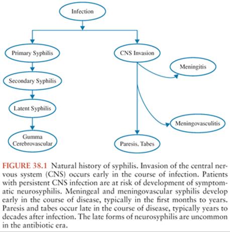
Neurosyphilis has been traditionally considered a late or “tertiary” manifestation of syphilis. In reality, asymptomatic or symptomatic neurosyphilis can occur at any time after infection. However, the forms of disease that are characterized by meningeal inflammation (asymptomatic, meningeal, and meningovascular) are most common in the first months to years after infection and are included under the category of “early neurosyphilis,” whereas parenchymal forms of neurosyphilis (general paresis and tabes dorsalis) most commonly occur years or decades after infection and are categorized as the forms of “late neurosyphilis” (Fig. 38.1). Ocular and otologic disease occur early and late in the course of syphilis, often, but not always, in combination with meningitis. These syndromes are considered separately in the following section.
ASYMPTOMATIC NEUROSYPHILIS
Asymptomatic neurosyphilis is diagnosed in a patient with serologic or clinical evidence of syphilis, CSF abnormalities due to T. pallidum infection, including pleocytosis, elevated protein and reactive CSF-VDRL, and no neurologic symptoms or signs. Asymptomatic neurosyphilis may occur very early in infection, even in patients with concomitant primary syphilis. Pleocytosis generally precedes development of reactive CSF-VDRL. Patients with asymptomatic neurosyphilis are at risk of progression to symptomatic forms of disease and about 20% of such individuals developed symptomatic neurosyphilis in the preantibiotic era (12). Thus, patients with asymptomatic neurosyphilis are treated to prevent progression to symptomatic disease.
SYMPTOMATIC NEUROSYPHILIS
Meningitis
Symptomatic syphilitic meningitis is indistinguishable from other causes of aseptic meningitis. Clinical findings include meningeal signs, nausea, and vomiting. Papilledema, convulsions, confusion, focal findings, and cranial nerve (CN) abnormalities, particularly involving CN VIII, VII, and II, were common in a large series of patients reported by Merritt and Moore (22) in 1935. Syphilitic meningitis was rare in the preantibiotic era, affecting less than 0.5% of individuals with syphilis (23) and was more commonly seen in patients who had been inadequately or incompletely treated for early syphilis (1,3,24). In Merritt and Moore’s (22) series, syphilitic meningitis most commonly occurred within a year of infection (range, 2 months to 26 years) and 7.5% of patients had secondary syphilis at the time that meningitis was diagnosed. Syphilitic meningitis is more common in the antibiotic era than it was in Merritt and Moore’s time (25).
Syphilitic meningitis may uncommonly affect the spinal cord, where it manifests as meningomyelitis or hyperplastic pachymeningitis. Adams and Merritt (26) described 15 cases of meningomyelitis and one case of hyperplastic meningitis among 2,231 syphilis cases seen at Boston City Hospital. Symptoms and signs in these patients included back pain, sensory loss, incontinence, leg weakness, and muscle atrophy. Occasional cases of syphilitic meningomyelitis (27–33), polyradiculopathy (34–38), and amyotrophy (39) are reported in modern times.
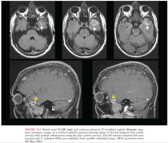
Localized syphilitic meningitis can produce one or more circumscribed masses of granulation tissue called gumma (26,40–44). Histopathologically, these consist of parenchymal and perivascular infiltration of lymphocytes and plasma cells with occlusive endarteritis and areas of necrosis (45–47). Gummas have been mistakenly deemed “tertiary” or late manifestations of neurosyphilis, but in reality, these can be seen early or late in the course of disease. These most commonly arise from the pia mater, especially over the convexities (48). Less commonly, parenchymal gumma may be seen without concomitant pial involvement. These may invade the substance of the brain and spinal cord (49) and may be mistaken for tumors, including meningioma, schwannoma, glioma, and lymphoma (50–54). CSF analysis often shows pleocytosis and a reactive CSF-VDRL but may be normal (48,55). Spirochetal organisms have been identified by histology (54–56), and T. pallidum has been amplified from resected gumma (46,56,57) (Fig. 38.2).
Ocular Syphilis
Ocular syphilis can befall any patient with syphilis, including those with normal immunity, and is seen in all stages of disease. Any part of the eye can be involved in T. pallidum infection. Ocular findings may be seen in patients with and without concomitant syphilitic meningitis; concomitant ocular disease and meningitis may be particularly common in patients also infected with HIV (58). Ocular manifestations include optic neuritis (59–61), which causes visual loss, and perineuritis (62–67) in which inflammation is localized to the optic nerve sheath and spares the nerve itself. Examination shows papilledema, generally normal visual acuity, except for an enlarged blind spot, and normal CSF pressure. Anterior uveitis affects the anterior uveal tract, which includes the iris and ciliary body. Symptoms include eye pain, redness, and photophobia. Asymptomatic involvement of the anterior chamber of the eye has been described in as many as one half of patients with secondary syphilis (68); a more recent case report of asymptomatic bilateral anterior uveitis in an HIV-infected man with secondary syphilis underscores this point (69).
Posterior uveitis is more common than anterior uveitis (70); a systematic review of ocular syphilis in HIV-infected patients showed that this form of ocular syphilis was more common in HIV-infected individuals with CD4+ T cells less than 200/µL (71). However, ocular syphilis, including posterior uveitis, is described in HIV-infected patients with well-controlled HIV infection on antiretroviral therapy (72,73). Posterior uveitis can involve the choroid, retina, and retinal pigment epithelium. Patients are described with chorioretinitis, neuroretinitis, retinal vasculitis, and retinal detachment, and syphilis should be considered in all patients with a diagnosis of acute retinal necrosis syndrome. Posterior uveitis is usually not painful, can be asymptomatic, or can cause severe visual loss. Visual outcome is generally good (74–79). A retrospective study of ocular syphilis in 35 eyes from 19 patients showed that poorer visual acuity at presentation and HIV infection predicted a poorer visual outcome, defined as acuity worse than 20/200 (70). Moore and Gieske (80) published a large series of patients with syphilitic uveitis in 1931 and showed that, like syphilitic meningitis, syphilitic uveitis occurred more commonly in patients who were inadequately treated for early syphilis. In this setting, uveitis was more often associated with meningitis. Worsening of unsuspected ocular syphilis has been observed after oral administration of prednisone (81) and after intravitreal administration of triamcinolone acetonide (82–84). T. pallidum has been identified by PCR in aqueous and vitreous fluid in patients with ocular syphilis (85–87).
Otologic Syphilis
Otosyphilis can occur in early and late syphilis. Hearing loss can be unilateral or bilateral, and it is often asymmetrical. Onset may be sudden or insidious, and speech discrimination may be preferentially affected (88). The symptoms and signs may mimic those of Meniere disease with episodic hearing loss, tinnitus, and vertigo (89). Hearing loss in otosyphilis may be seen in the presence or absence of syphilitic meningitis. The pathophysiology of hearing loss in meningitis is likely inflammation of the auditory (CN VIII) nerve within the subarachnoid space or a “meningo-neuro-labyrinthitis” from spread of CSF infection to the perilymph via the cochlear aqueduct (90). In the absence of meningitis, the pathophysiology is more likely to be obliterative vasculitis and osteitis of the cochleovestibular system and temporal bone with the end result being resorptive osteitis and productive periostitis with replacement by fat, marrow, or new bone (90–93). Meningo-neuro-labyrinthitis likely progresses to periostitis, but T. pallidum may also disseminate directly from blood to the inner ear, particularly the perilymph, without involvement of the CSF. Historically, compared to those with shorter duration of hearing loss, patients with longer duration of hearing loss due to syphilis were less likely to respond to otosyphilis treatment; this likely remains true today (94). This difference may reflect the underlying pathophysiology: in late disease, the cochleovestibular system is irreversibly damaged, arguing for identification of patients with otosyphilis as early in the disease course as is possible.
Meningovascular Syphilis
Syphilitic meningitis may cause arteritis affecting small (Nissl-Alzheimer endarteritis) or medium and large vessels (Heubner arteritis), the latter with adventitial inflammation and fibrosis, medial thinning, and intimal fibroblastic proliferation (95). Thrombosis and ischemia or infarction involving the brain or spinal cord can occur. Intracranial aneurysms (96,97), intraparenchymal hemorrhage (98), and carotid dissection have been uncommonly reported (99). Merritt and coworkers (40) described a series of 42 patients with meningovascular syphilis affecting the brain. These patients represented 3% of syphilis cases seen at their institution and were selected from a possible 250 individuals after excluding atherosclerotic cerebrovascular disease, cerebral embolism, other forms of neurosyphilis, and other nonsyphilitic neurologic diseases. Most patients were 30 to 50 years old, and meningovascular syphilis developed within months to years after infection, with an average of 7 years (23,40). Stroke in the distribution of the middle cerebral artery was the most common clinical finding, with hemiparesis, hemiplegia, or aphasia. Many patients experienced prodromal symptoms, such as headache, dizziness, and personality changes for days or weeks before the onset of stroke. In clinical practice, patients with meningovascular syphilis often have cognitive impairment and may even be frankly demented (100–108). The timing of onset after primary syphilis (early) and the presence of focal examination and imaging findings may help distinguish patients with meningovascular neurosyphilis from those with parenchymal neurosyphilis or general paresis, but sometimes the distinction can be difficult. As is the case with symptomatic meningitis, meningovascular syphilis is likely a relatively more common manifestation of symptomatic neurosyphilis today than it was in the preantibiotic era (25,109,110). Although uncommon, meningovascular syphilis can be seen in patients with nonreactive serum nontreponemal tests, but treponemal tests should be reactive to support the diagnosis (111). Recombinant tissue plasminogen activator has been used successfully in meningovascular syphilis (95,112).
Meningovascular syphilis involving the spinal cord is less common than involvement of brain. Adams and Merritt (26) described 16 cases of spinal meningovascular syphilis seen among 2,231 syphilis patients. As a result of thrombosis of spinal vessels, patients develop acute onset of transverse myelitis characterized by paraplegia, sensory level, usually in the thoracic region, and loss of sphincter control. Such cases continue to be reported in the modern era (27,113–117).
Parenchymal Neurosyphilis
In the preantibiotic era, the parenchymal or late forms of neurosyphilis were more common than the meningeal or early forms. Clinical experience suggests that the opposite is true today. Wolters (118) compared the spectrum of disease in 518 cases of neurosyphilis collected between 1930 and 1940 to 121 cases collected between 1970 and 1984 from the same neurologic clinic of the Academic Hospital of the University of Amsterdam. He found that parenchymal disease occurred in two thirds of the symptomatic individuals in 1930 to 1940, but that the proportions of symptomatic meningeal and parenchymal disease were equal in 1970 to 1984. The observation that late neurosyphilis is less common than early neurosyphilis is most striking in patients also infected with HIV. This finding may simply be due to the fact that syphilis is so commonly seen in HIV-infected individuals due to epidemiologic factors (see later discussion), or that, at least early in the HIV epidemic, HIV-infected patients did not live long enough to develop late neurosyphilis. Alternatively, therapy for early (nonneurologic) syphilis may be particularly ineffective in individuals also infected with HIV and may predispose them to develop early neurosyphilis. This issue is discussed in the section “Neurosyphilis and HIV” later in this chapter. Several authors have speculated that the shift in clinical presentation of neurosyphilis from late to early disease is due to inadvertent treatment with antibiotics prescribed for unrelated conditions. This contention is supported by observations collected in the preantibiotic era noted earlier that partial or incomplete therapy predisposes individuals infected with T. pallidum to develop the early forms of neurosyphilis, including meningitis, meningovasculitis, and ocular disease (1,3,24,80). Arguing against this hypothesis, in 2004, Timmermans and Carr (108) reported the clinical spectrum of neurosyphilis in 161 patients in South Africa, approximately 6% of whom were HIV-infected. These individuals had poor access to health care and were unlikely to have received “incidental” antibiotics, yet the proportion with tabes dorsalis (2 [1%] of 161) was much lower than reported in the preantibiotic era.
Syphilitic Dementia: General Paresis
General paresis, also known as general paralysis of the insane or dementia paralytica, was estimated by Merritt and coworkers (40) to develop in 5% of cases of syphilis. In their experience, it was most commonly seen in individuals 35 to 50 years of age and occurred from 5 to 25 years after primary infection (40). In a series by Hahn and coworkers (119), most patients with general paresis had been infected for 10 to 24 years (range 2 to more than 30 years), and in a series reported by Dewhurst (120), duration of infection was 4 to 15 years with a mean of 10.5 years.
Merritt et al. (40) stated that the clinical manifestations of general paresis mimic “every type of mental disorder” (p. 194). Early in the course of this chronic, progressive, dementing illness, patients are forgetful and have personality changes. With time, they may develop psychiatric symptoms, such as mania, depression, or psychosis. However, most patients simply experience worsening of deficits in memory and judgment progressing to frank dementia. In its latest stages, patients become immobile and incontinent and may have seizures. The most frequent neurologic examination findings are pupillary abnormalities; facial and limb hypotonia; intention tremors of the face, tongue, and hands that can cause dysarthria and handwriting abnormalities; and reflex abnormalities. Early in the course of disease, the neurologic examination can be normal. The average survival after diagnosis in Merritt and coworkers’ (40) experience was 2.5 years. Intercurrent infection was the most common cause of death in the preantibiotic era. With the availability of antibiotics for syphilis and other infections, as well as better supportive care, survival is longer.
Cases of syphilitic dementia continue to be reported in the modern era, generally characterized by rapidly progressive dementia with or without psychiatric features (121,122). A retrospective series of 116 patients with general paresis diagnosed in three Chinese hospitals identified dementia, personality change, abnormal behavior, and emotional problems as the most common findings (123). As noted earlier, there is clinical overlap between syphilitic dementia and meningovascular disease (124,125). Patients with syphilitic dementia may show background slowing on electroencephalogram (EEG), periodic lateralized epileptiform discharges (PLEDS) or epileptiform discharges (123,124,126). In the large Chinese series discussed earlier, seizures were seen in 16% of patients with syphilitic dementia. Of note, neurosyphilis was not suspected initially in 36% of patients in that series (123). A smaller series from China compared performance on comprehensive neuropsychological testing in 12 patients with mild syphilitic dementia to 24 patients with mild Alzheimer disease matched with regard to age, education, and Mini Mental State scores (127). The two groups had very similar patterns of impairment.
Tabes Dorsalis
Tabes dorsalis or locomotor ataxia was the most common form of neurosyphilis described in the preantibiotic era. Merritt and coworkers (40) established the diagnosis of tabes in 9% of 2,231 patients with syphilis seen in the outpatient department of the Boston City Hospital. Tabes was typically seen in patients between 44 and 60 years of age, and onset ranged from 3 to 47 years after primary infection, with an average of 21 years (40).
The most common symptoms of tabes are pupillary abnormalities, optic atrophy, lancinating pains, sensory changes, progressive ataxia, and bowel and bladder dysfunction. Pupillary abnormalities were described in 94% of cases reported by Merritt and coworkers (40). They defined the Argyll Robertson pupil as a small pupil that does not respond to light but does contract normally to accommodation–convergence, dilates imperfectly to mydriatics, and does not dilate in response to painful stimuli. Using this rather stringent definition, 48% of patients with tabes had Argyll Robertson pupils (40). Optic atrophy was seen in 16% of Merritt and coworkers’ series (40). The clinical findings in optic atrophy include gradual decrease in visual acuity with constriction of the peripheral visual fields and central scotomata on examination. Untreated, this disorder progresses to complete blindness over months to years (128). Lightning or lancinating pains, seen in 75% of 150 cases of tabes collected by Merritt and coworkers (40), are sudden, brief stabs of pain that may affect the legs, back, arms, and face. They may last for minutes to days and occur unpredictably, sometimes separated by long remissions. Visceral crises occurred in 10% to 15% of patients with tabes (40). The most common type was the gastric crisis, characterized by recurrent attacks of severe epigastric pain, nausea, and vomiting. Early sensory changes include paresthesias or hyperesthesias in radicular distributions. Later, pain, vibration, and tactile sensation become impaired, and reflexes are lost. Sensory ataxia, usually involving the lower more than the upper extremities, was a feature in 42% of Merritt and coworkers’ patients (40). Bladder dysfunction may occur early with urinary retention and overflow incontinence. A similar process affects the bowel, although fecal incontinence is unusual. As noted earlier, tabes is now an uncommon form of neurosyphilis, but cases continue to be described (129–131).
Congenital Neurosyphilis
As in adults, T. pallidum may invade the CNS in infants exposed to the organism in utero or at birth. For example, T. pallidum was identified by rabbit inoculation in CSF from 19 (13%) of 148 infants born to mothers with syphilis (132). The manifestations and clinical course of congenital neurosyphilis parallel that of acquired disease, and early and late forms of symptomatic neurosyphilis have similar latent periods. Thus, congenital meningeal neurosyphilis is seen in infancy and meningovascular disease in the first few years of life. Parenchymal disease usually has its onset around puberty or in early adulthood (40).
LABORATORY AND IMAGING STUDIES
Cerebrospinal Fluid
The diagnosis of symptomatic neurosyphilis is based on clinical evidence and is supported by CSF abnormalities, particularly pleocytosis and reactive CSF-VDRL. As noted earlier, the diagnosis of asymptomatic neurosyphilis is based on the presence of CSF abnormalities alone. CSF WBC concentration in neurosyphilis is generally greater than 10 cells/µL with a lymphocytic predominance (Table 38.1). Higher cell counts are seen in early compared to late neurosyphilis. For example, in an examination of CSF from 100 patients with tabes dorsalis, Merritt and coworkers (40) found that 50% had less than 5 WBC/µL. Mild elevations in CSF protein, ranging between 45 and 200 mg/dL are also common, again with higher values in early compared to late neurosyphilis. The CSF-VDRL is considered to be the gold standard test for diagnosis of neurosyphilis. However, depending on the diagnostic criteria chosen, the CSF-VDRL may be reactive in 0% to 100% of individuals with neurosyphilis; the generally accepted sensitivity is 30% to 70%. The CSF-VDRL test is very specific. False-positive results may be seen when the CSF is visibly blood tinged (133,134) and rarely in the absence of blood contamination (135). Thus, a reactive CSF-VDRL establishes the diagnosis of neurosyphilis, but a nonreactive test does not exclude the diagnosis.

The CSF-VDRL test is not available in some parts of the world, and the test method is technically cumbersome. Larsen and colleagues (136) suggested that the CSF-RPR and CSF-toluidine red unheated serum test (TRUST), two alternative nontreponemal tests that are less logistically complicated to perform than the CSF-VDRL, should not be used to diagnose neurosyphilis because of false-positive results. However, two more recent studies suggested that the CSF-RPR (137) or CSF-TRUST (138) could be suitable alternatives to the CSF-VDRL, reporting sensitivities of 75% and 95% and specificities of 99% and 100% for laboratory-defined neurosyphilis. We examined the diagnostic performance of the CSF-RPR compared to the CSF-VDRL. Although the CSF-RPR was significantly more specific for the diagnosis of symptomatic neurosyphilis, compared to the CSF-VDRL, the CSF-RPR was negative in 36% of CSF-VDRL-reactive samples (139). A large study from China examined concordance of CSF-VDRL and CSF-TRUST and found that qualitative results were the same 97% of the time. However, of 204 CSF-VDRL reactive samples, 22 (11%) were CSF-TRUST nonreactive (140). Thus, the problem of low diagnostic sensitivity of the CSF-VDRL is likely greater for alternative CSF nontreponemal tests.
In contrast to the CSF-VDRL, several studies have shown that CSF treponemal antibody tests, such as the CSF fluorescent treponemal antibody-absorption (FTA-ABS) or the CSF-Treponema pallidumparticle agglutination assay (TPPA), are sensitive but not specific for the diagnosis of neurosyphilis (141–146). As noted in a systemic review of such work, the sensitivity of these tests is highest when the diagnosis of neurosyphilis is based on CSF-VDRL reactivity (147). Thus, nonreactive CSF treponemal antibody tests exclude the diagnosis of asymptomatic neurosyphilis with a high degree of certainty, but a negative result is less able to exclude symptomatic neurosyphilis. Sera from patients with reactive CSF treponemal antibody tests have higher treponemal antibody titers than sera from those with nonreactive CSF treponemal tests, suggesting that reactive CSF treponemal tests may simply be a surrogate for higher serum antibody concentration (141,148). However, one study suggested that a CSF-Treponema pallidum hemagglutination (TPHA) test titer greater than 1:320 (the TPHA is an alternative treponemal test that is not available in the United States) was sensitive and specific for the diagnosis of neurosyphilis (149), suggesting that there may be a CSF treponemal antibody concentration above which intrathecal antibody production is more likely.
T. pallidum may be identified in CSF by inoculation into rabbits (150). However, rabbit inoculation is too cumbersome and expensive to be clinically useful. PCR or RT-PCR can be used to detect T. pallidumin CSF with a limit of detection similar to that of rabbit inoculation (9,151). T. pallidum can be detected in CSF from 25% to 30% of patients with early syphilis; detection is much less likely in late syphilis (7–9). In our experience, T. pallidum is most often detectable in samples with reactive CSF-VDRL or CSF pleocytosis, limiting its usefulness in neurosyphilis diagnosis (9).
The proportion of CSF lymphocytes that are B cells (145), and the CSF concentration of chemoattractant chemokine (C-X-C motif) ligand 13 (CXCL13), a B-cell chemoattractant, are elevated in patients with neurosyphilis compared to patients with uncomplicated syphilis (152,153). Although these tests show promise for improving neurosyphilis diagnosis, particularly in patients infected with HIV (152), they are not in routine clinical use. A study in HIV-uninfected patients that included 12 with late neurosyphilis, 17 with uncomplicated syphilis, and 14 normal controls showed that a CSF total tau protein concentration higher than 300 pg/mL, a cutoff that has been used to diagnose Alzheimer disease, had a diagnostic sensitivity of 83% and a diagnostic specificity of 94% (154). The diagnostic performance in HIV-infected individuals or in those with early neurosyphilis is not known, and these results remain to be replicated.
Several studies have examined predictors of abnormal CSF in neurologically asymptomatic and symptomatic patients with syphilis. The odds of neurosyphilis are higher when serum RPR titers are greater than or equal to 1:32 (approximately 11-fold in HIV-uninfected and sixfold in HIV-infected patients, regardless of syphilis stage or previous syphilis treatment [9]), and in HIV-infected individuals, when peripheral blood CD4+ T cells are less than or equal to 350 cells/µL (9,155,156). Also, HIV-infected patients with syphilis who are taking antiretrovirals may be at lower risk of neurosyphilis than those who are not taking them (157). A small, retrospective study suggested that high plasma HIV RNA concentration increased the risk of neurosyphilis among individuals with peripheral blood CD4+ T cells greater than 350 cells/µL (158).
As noted earlier, asymptomatic neurosyphilis is defined solely by CSF abnormalities, and, in the preantibiotic era, neurologically asymptomatic patients with syphilis of any stage who had the most abnormal CSF measures were at highest risk for developing symptomatic neurosyphilis. This observation prompted universal lumbar puncture (LP) in all stages of syphilis. The subsequent observation that CSF abnormalities were uncommon in patients with early syphilis who were treated with penicillin (19–21,159) led to recommendations to reserve the procedure for neurologically asymptomatic patients with late syphilis. With the advent of HIV, the appreciation that benzathine penicillin G (BPG) used to treat uncomplicated syphilis does not achieve treponemicidal penicillin levels in the CSF (160), and concerns regarding neurorelapse in this patient population (see later discussion), the issue of which patients with syphilis should undergo LP again arose and has remained an area of ongoing controversy. A panel of experts convened by the Centers for Disease Control and Prevention (CDC) has provided guidelines for LP in syphilis. Although they have consistently recommended LP for patients with neurologic, ocular, or otologic symptoms and signs, and for patients who have failed treatment for uncomplicated syphilis, over the years, changes have been made, sometimes in the absence of new data. For example, in the 2002 guidelines, LP was recommended for all HIV-infected patients with late latent syphilis or syphilis of unknown duration (161). The 2006 guidelines additionally indicated that some experts recommended LP when the serum RPR titer was greater than or equal to 1:32 or when the peripheral blood CD4+ T cells were less than or equal to 350 cells/µL (162). In the 2010 version of the guidelines, however, the recommendation for LP in HIV-infected patients with late syphilis or syphilis of unknown duration was removed. In addition, consideration of LP in HIV-infected patients with high serum RPR titer or low CD4 was rescinded, stating, “Unless neurologic symptoms are present, CSF examination in this setting has not been associated with improved clinical outcomes” (163). This language is repeated in the 2013 Guidelines for the prevention and treatment of opportunistic infections in HIV-infected adults and adolescents (http://aidsinfo.nih.gov/contentfiles/lvguidelines/adult_oi.pdf). In contrast, the 2008 European Guidelines on the Management of Syphilis (164) state that LP is indicated for HIV-infected patients with syphilis, especially those who have serum RPR titers greater than 1:32 or peripheral blood CD4+ T cells less than 350 cells/µL; a footnote adds “not obligatory, but may be indicated in late latent syphilis or syphilis of unknown duration or in treatment failure,” perhaps underscoring the lack of consensus regarding this issue. There are no modern day clinical outcome data to guide decisions regarding which HIV-infected or HIV-uninfected patients with syphilis should undergo LP. The safest or most conservative approach is to perform LP in patients with high serum RPR titers or low peripheral blood CD4+ T-cell concentrations. However, the magnitude of averted morbidity in terms of preventing symptomatic neurologic, ocular, or otologic disease of this approach is unknown. Suggested recommendations for LP in patients with syphilis are shown in Figure 38.3A and B.


Neuroimaging
Neuroimaging in patients with asymptomatic neurosyphilis is usually normal (165). Neuroimaging in syphilitic meningitis involving brain or spinal cord may show enhancement of meninges, spinal fluid, cranial nerves, or spinal roots (35, 166–169). Enhancement of the cochlea (likely early) (169,170) and lucency within the otic capsule and ossicular chain (late) (93) may be seen in syphilitic hearing loss.
Cerebral gummas are low intensity on T1-weighted MR sequences, isointense or high intensity on T2-weighted sequences, enhance homogenously, sometimes with a nodular component and often with a dural tail, and have associated edema (42,48,49,165,171,172) (Fig. 38.2). They can sometimes have the appearance of nodular basilar meningitis (44). One report described restricted diffusion in the region of cortical enhancement and the dural tail (172).
Brain computed tomographic (CT) and magnetic resonance (MR) scans in meningovascular syphilis show one or more areas of infarction (102,165,168,173,174); infarctions in multiple vascular distributions are often seen with diffuse vasculitis (175,176). Catheter, CT, or MR angiographic findings in meningovascular neurosyphilis include segmental arterial narrowing, focal narrowing and dilation, or “beading,” and occlusion, which may sometimes mimic vasospasm (97,100,177–179).
Meningitis or meningovasculitis affecting the spinal cord may show intramedullary high signal on T2-weighted images; enhancement may or may not be seen on T1-weighted images after administration of contrast material (30,32,117,180,181). Rare cases of syrinx in association with meningeal syphilis have been reported (31,33).
Brain MR in patients with syphilitic dementia may show cerebral or cortical atrophy with ventriculomegaly, focal or diffuse white matter changes, and low signal intensity on T2-weighted sequences in the globus pallidus, putamen, caudate head, and thalamus (124,125,168,182). In one study of 20 patients with neurosyphilis defined by CSF abnormalities and psychiatric symptoms and signs, measures of brain atrophy on MR were significantly worse in patients with lower scores on the Mini Mental State (125). Medial temporal lobe atrophy may be particularly common (127) and hippocampal atrophy on MR mimicking Alzheimer disease has been reported (183). A study of seven patients with syphilitic dementia showed that the three with medial temporal lobe atrophy had poorer outcome after treatment than the four patients who did not have this finding; of note, those with temporal lobe atrophy also had atrophy of the frontal lobes and dilation of the ventricles out of proportion to the overall degree of atrophy (184).
Several reports of meningovascular or parenchymal neurosyphilis with high signal on T2-weighted MR sequences mimicking herpes encephalitis have been published (101,103–106, 126,168,185,186). In most, but not all, instances, patients had seizures or PLEDS, which could explain the abnormal imaging findings. In one report, biopsy of the affected temporal lobe showed astrocytosis and scant plasma cell infiltration with detection of T. pallidumDNA by PCR (187).
Spinal cord MR in patients with tabes dorsalis may show increased intramedullary T2 signal, particularly in the posterior columns and cord atrophy (35,131,168).
Evoked Potentials
In tabes dorsalis, motor and sensory nerve conductions are normal. Somatosensory evoked potentials elicited from the median nerve are normal, whereas those elicited by stimulation of the tibial nerve show abnormalities consistent with dysfunction of the caudal dorsal roots and posterior columns (35,188–190).
TREATMENT AND PREVENTION
There has never been a large study of currently recommended (163) penicillin regimens for the treatment of neurosyphilis. These regimens are based on the observation that intravenous (IV) crystalline penicillin G in doses from 5 to 24 million units (MU) per day achieves treponemicidal CSF levels (160,191,192). Similarly, most (but not all) reports document treponemicidal CSF penicillin levels after intramuscular procaine penicillin, 2.4 MU per day, with oral probenecid (193–195). The CDC recommends high-dose IV penicillin G as the first-line therapy for neurosyphilis; IM procaine penicillin with oral probenecid is an alternative in adults, but not children (163) (Table 38.2). Ocular and otosyphilis are treated as for neurosyphilis; steroids are sometimes recommended as an adjunctive therapy.

Case reports in HIV-uninfected and HIV-infected individuals with early and late neurosyphilis and ocular syphilis (28,196–198), and a small study in HIV-infected patients with early neurosyphilis (199) suggest that ceftriaxone, 1 to 2 g IV every day, may be an acceptable alternative to penicillin. Based on CSF drug concentrations and pharmacologic properties, doxycycline may be an effective alternative oral treatment for neurosyphilis. The European (164) and United Kingdom (200) syphilis guidelines recommend doxycycline 200 mg orally twice daily for 28 days as an alternative neurosyphilis treatment regimen. The European guidelines reserve this regimen for patients who are allergic to penicillin or who refuse parenteral therapy. Although such recommendations acknowledge that the evidence for the efficacy of doxycycline for neurosyphilis is weak (164,201), case reports describe instances of treatment success (202). The CDC guidelines do not recommend doxycycline as an alternative treatment for neurosyphilis. Rather, they recommend penicillin desensitization for penicillin-allergic patients with neurosyphilis in whom ceftriaxone is not an option (163).
The success of neurosyphilis therapy is judged by resolution or stabilization of clinical abnormalities and by resolution of CSF abnormalities. Clinical abnormalities are more likely to resolve when there has not been structural CNS damage. Patients with syphilitic meningitis can be expected to recover completely, although if they have concomitant ocular or otologic injury, deficits may persist. Complete resolution is the rule after antibiotic therapy for intracranial gumma; these lesions may also resolve after steroid therapy alone (48), but this is not recommended as it will not treat the underlying infection. Patients with meningovascular syphilis will likely resolve meningeal symptoms and signs but will be left with residual stroke symptoms and signs. Similarly, treatment of patients with paresis or tabes will arrest disease progression but is unlikely to reverse dementia or sensory ataxia.
The CDC guidelines state that CSF WBC count should decline at 6 months and all CSF abnormalities should resolve by 2 years after treatment (163). Careful follow-up after neurosyphilis therapy is mandatory for all patients. We recommend that CSF be reexamined 3 months after therapy, rather than 6 months, to decrease patient loss to follow-up and because the median time for normalization of CSF WBC, CSF-VDRL, and serum RPR was approximately 4 months in our prospective study of 59 individuals with neurosyphilis (203). In our experience, CSF protein concentration is slow to normalize and may remain elevated even with other objective evidence of treatment success (203,204), thus we do not base a decision to re-treat solely on failure of CSF protein concentration to normalize. Thus, if CSF WBC count is normal and the CSF-VDRL is nonreactive at 3 months, no further LPs are required. For those with persistent CSF abnormalities at 3 months after therapy (excluding elevated protein concentration), CSF should be reexamined at 6 months after therapy and every 6 months thereafter until CSF WBC and CSF-VDRL normalize. Failure of the CSF WBC count to decrease 6 months after therapy or failure of CSF-VDRL to decline fourfold (or to nonreactive if the initial titer is <1:2) 1 year after therapy are indications for retreatment. Serum RPR or VDRL tests should be obtained at 3, 6, and 12 months after therapy and every 3 to 6 months thereafter until they are nonreactive. Failure of the serum RPR or VDRL to decline fourfold (or to nonreactive if the initial titer is <1:2) at 1 year after therapy is an indication for retreatment.
Because some patients may be reluctant to undergo follow-up LPs after neurosyphilis therapy, we examined whether normalization of serum RPR, defined as a fourfold decline in titer or reversion to nonreactive, could predict normalization of clinical and CSF abnormalities in 110 patients treated for asymptomatic syphilitic meningitis, symptomatic syphilitic meningitis, or syphilitic eye disease (204). Normalization of serum RPR predicted normalization of CSF abnormalities (except CSF protein) and clinical abnormalities with a high degree of certainty. However, using the serum RPR criterion (and excluding CSF protein), 12% of individuals were misclassified as treatment successes. Among HIV-infected individuals, misclassification was most common in those not taking antiretrovirals.
Prevention of neurosyphilis rests on first preventing syphilis. As discussed earlier, LP in syphilis patients at highest risk for neurosyphilis (based on serum RPR titer and HIV-associated disease factors) and treatment of those with CSF abnormalities consistent with asymptomatic neurosyphilis will prevent progression to symptomatic disease. However, the magnitude of averted morbidity of this approach is currently undefined.
NEUROSYPHILIS AND HIV
In the developed world, syphilis is most common in individuals who are at risk for or infected with HIV, including children and adolescents. Patients who have both HIV and syphilis may be more likely to fail therapy for early syphilis and to develop early neurosyphilis. Although some have argued that neurosyphilis has a more rapid or aggressive course in HIV-infected compared to uninfected individuals, careful review of the preantibiotic literature suggests that this is not the case. However, neurosyphilis may be more difficult to diagnose in the setting of concomitant HIV and HIV-infected patients may be more likely to fail therapy for neurosyphilis.
Epidemiology of Syphilis and Neurosyphilis in HIV-Infected Individuals
In 2000, the rate of infectious syphilis in the United States was the lowest since reporting began in 1941. However, since 2001, the number of cases of syphilis in the United States has increased steadily. Between 2001 and 2011, the rates of primary and secondary syphilis more than doubled (205); similar increases have been seen in Europe (206). In the last two decades, there has been a remarkable resurgence of syphilis in China. Syphilis is now among the top five reportable communicable diseases in many regions in China, with an estimated rate of 25 per 100,000 in 2009 (207). In 2011, 72% of primary and secondary syphilis cases reported to the CDC occurred in men who have sex with men (MSM) (205). HIV is particularly common in MSM with syphilis (206,208,209). In the U.S. STD Surveillance Network, of MSM with primary and secondary syphilis, a median of 40% were infected with HIV (205).
Neurosyphilis is not a reportable disease, and thus it is harder to estimate its incidence or prevalence in HIV-infected people. The CDC conducted a retrospective study of definite or probable symptomatic early neurosyphilis in HIV-infected MSM in four U.S. cities from January 2002 to June 2004 (210). Forty-nine patients were identified: 34 had cranial nerve dysfunction (25 ocular dysfunction, 6 auditory dysfunction, 1 had both abnormalities and 2 had other cranial nerve abnormalities); 6 had symptomatic meningitis; 2 had strokes; and 7 had headache, altered mental status, or both abnormalities. The estimated risk of symptomatic neurosyphilis in the study population was 1.7%. Taylor and colleagues (211) retrospectively reviewed 109 cases of neurosyphilis identified by chart review of patients reported with syphilis in Los Angeles between 2001 and 2004 who received a diagnosis of neurosyphilis or had CSF findings consistent with neurosyphilis. Overall, the highest proportion of syphilis cases with neurosyphilis was seen among those with secondary syphilis (1.9%). Among HIV-infected patients with early syphilis, the incidence of neurosyphilis was 2.1%, which was higher than the 0.6% estimate in HIV-uninfected individuals. As is the case with the CDC study (210), this study suffers from ascertainment bias in that patients underwent neurosyphilis evaluation at the discretion of the treating provider and therefore cases were likely missed.
Neurorelapse
Cases of asymptomatic and early symptomatic neurosyphilis or syphilitic ocular disease in HIV-infected individuals were reported early in the HIV epidemic (58,212–219). Many of these patients had been previously treated for early syphilis with appropriate doses of intramuscular BPG, suggesting that they experienced neurologic or ocular relapse (212). Of note, in the CDC study described earlier, 9 (18%) of the 49 individuals with symptomatic early neurosyphilis had received recommended BPG treatment for early syphilis before developing symptomatic neurosyphilis (210). In addition to neurorelapse presenting as neurologic or ocular disease, it may also present as otologic syphilis (220).
As described earlier, in the preantibiotic era, syphilitic meningitis, meningovasculitis, and uveitis were most commonly seen in patients who were inadequately treated for early syphilis. Merritt and Moore (22,24) argued that incomplete or inadequate therapy was sufficient to eradicate peripheral T. pallidum, but not sufficient to kill organisms that had invaded the CNS or the eye. Nonetheless, this treatment led to an appropriate attenuation of the immune response. The organisms remaining in the CNS or eye were then free to multiply and produce neurologic or ocular disease. A parallel has been made to the HIV-infected patient treated with benzathine penicillin for early syphilis (212). This treatment does not clear T. pallidum from the CNS, the eye, or the inner ear (7,221). Because of impaired cell-mediated immunity, the HIV-infected patient would not be able to eradicate persistent organisms and would thus be more likely to suffer relapse with neurologic, ocular, or otologic disease. As noted earlier, HIV-infected patients with low peripheral blood CD4+ T-cell concentrations or who are not taking antiretrovirals are at increased risk for neurosyphilis (9,156,157). In addition, among HIV-infected patients with neurosyphilis, peripheral blood CD4+ T cell concentrations are lower in those with symptomatic compared to asymptomatic disease (211,222). Taken together, these findings support the contention that HIV-induced immunosuppression may impair clearance of CNS T. pallidum.
Diagnosis of Neurosyphilis in HIV-Infected Individuals
Because the CSF-VDRL may be nonreactive in neurosyphilis, the diagnosis of neurosyphilis in an HIV-infected person may need to be based solely on identification of CSF pleocytosis. CSF pleocytosis in neurosyphilis is generally mild. As did many experts in the early 1900s, Moore and Hopkins (13) deemed a CSF WBC greater than 10 cells/µL to be consistent with neurosyphilis, even if the CSF Wassermann test was not reactive. Mild CSF pleocytosis is also common in HIV-infected individuals, particularly in those who are relatively immunocompetent. For example, Marshall and coworkers (223) showed that 15.6% of CSF samples from 649 HIV-infected individuals had greater than 10 WBC/µL with progressive decline in CSF WBC count with more advanced stages of disease. Because HIV can cause mild mononuclear CSF pleocytosis, we have used a cutoff of greater than 20 WBCs/µL as diagnostic of neurosyphilis in HIV-infected individuals (9); a lower cutoff has been used by others (157) and is appropriate when the peripheral blood CD4+ T cell concentration is low or patients are on antiretrovirals (ARVs), because these lower the risk of HIV-related CSF pleocytosis (224).
Neurosyphilis Treatment Failure in HIV
HIV-infected individuals may be more likely than HIV-uninfected patients to fail neurosyphilis therapy based on clinical and serologic criteria as well as on failure to normalize CSF abnormalities (157,203,210,225–228). In a prospective analysis of 13 HIV-uninfected and 46 HIV-infected patients with neurosyphilis, those who were HIV-infected were 2.5 times less likely to normalize CSF-VDRL after therapy (203). Among the HIV-infected subjects, those with peripheral blood CD4+ T cells less than or equal to 200 cells/µL were 3.3 times less likely to normalize CSF-VDRL (203). These data again lend support to the contention that HIV-induced immunosuppression may impair clearance of CNS T. pallidum and indicate that follow-up after treatment for neurosyphilis in HIV-infected individuals should be particularly diligent.
References
1. Moore J. Studies in asymptomatic neurosyphilis II. The classification, treatment, and prognosis of early asymptomatic neurosyphilis. Bull Johns Hopkins Hosp. 1922;33:231–246.
2. Ravaut P. Le liquide céphalo-rachidien des syphiliques en période secondaire. Ann Dermatol Syphiligr. 1903;4:537–554.
3. Wile UJ, Stokes JH. A study of the spinal fluid with reference to involvement of the nervous system in secondary syphilis. J Cutan Dis. 1914;32:607–623.
4. Fildes P, Parnell RJG, Maitland HB. The occurrence of unsuspected involvement of the central nervous system in unselected cases of syphilis. Brain. 1918;41:255–301.
5. Mills CH. Routine examination of the cerebrospinal fluid in syphilis: its value in regard to more accurate knowledge, prognosis, and treatment. BMJ. 1927;2:527–532.
6. Chesney AM, Kemp JE. Incidence of Spirochaeta pallida in cerebrospinal fluid during early stage of syphilis. JAMA. 1924;83:1725–1728.
7. Lukehart SA, Hook EW III, Baker-Zander SA, et al. Invasion of the central nervous system by Treponema pallidum: implications for diagnosis and treatment. Ann Intern Med. 1988;109(11):855–862.
8. Rolfs RT, Joesoef MR, Hendershot EF, et al. A randomized trial of enhanced therapy for early syphilis in patients with and without human immunodeficiency virus infection. The Syphilis and HIV Study Group. N Engl J Med. 1997;337(5):307–314.
9. Marra CM, Maxwell CL, Smith SL, et al. Cerebrospinal fluid abnormalities in patients with syphilis: association with clinical and laboratory features. J Infect Dis. 2004;189(3):369–376.
10. Stokes JH, Beerman H, Ingraham NR. Modern Clinical Syphilology. Diagnosis, Treatment, Case Study. 3rd ed. Philadelphia: WB Saunders; 1944.
11. Wile UJ, Marshall CH. A study of the spinal fluid in one thousand eight hundred and sixty-nine cases of syphilis in all stages. Arch Derm Syphilol. 1921;3:272–278.
12. Hahn RD, Clark EG, Felsovanyi A, et al. Asymptomatic neurosyphilis: prognosis. Am J Syph Gonorrhea Vener Dis. 1946;30(6):513–548.
13. Moore JE, Hopkins H. Asymptomatic neurosyphilis. VI. The prognosis of early and late asymptomatic neurosyphilis. JAMA. 1930;95:1637–1641.
14. Moore JE, Kemp JE. The treatment of early syphilis II. Clinical results in 402 patients. Bull Johns Hopkins Hosp. 1926;39:16–35.
15. Hopkins HH. Prognostic import of a negative spinal fluid in early and in latent syphilis. Arch Derm Syphilol. 1931;24:404–408.
16. O’Leary PA, Cole HN, Moore JE, et al. Cooperative clinical studies in the treatment of syphilis: asymptomatic neurosyphilis. Venereal Disease Information. 1937;18:45–65.
17. Tantalo LC, Lukehart SA, Marra CM. Treponema pallidum stain-specific differences in neuroinvasion and clinical phenotype in a rabbit model. J Infect Dis. 2005;191(1):75–80.
18. Marra CM, Sahi SK, Tantalo LC, et al. Enhanced molecular typing of Treponema pallidum: geographical distribution of strain types and association with neurosyphilis. J Infect Dis. 2010;202(9):1380–1388.
19. Altshuler L, Karpinos BD, Leifer W, et al. Efficacy of penicillin treatment of early syphilis in the army. Am J Syph. 1949;33:126–138.
20. Fernando WL. Cerebrospinal fluid findings after treatment of early syphilis with penicillin. Br J Vener Dis. 1965;41(3):168–169.
21. Fernando WL. Cerebrospinal fluid findings after treatment of early syphilis with penicillin. A further series of 80 cases. Br J Vener Dis. 1968;44(2): 134–135.
22. Merritt HH, Moore M. Acute syphilitic meningitis. Medicine (Baltimore). 1935;14:119–183.
23. Merritt HH. The early clinical and laboratory manifestations of syphilis of the central nervous system. N Engl J Med. 1940;223:446–450.
24. Moore JE. The relation of neurorecurrences to late syphilis. A clinical study of eighty-one cases. Arch Neurol and Psychiatry. 1929;21:117–136.
25. Burke JM, Schaberg DR. Neurosyphilis in the antibiotic era. Neurology. 1985;35(9):1368–1371.
26. Adams RD, Merritt HH. Meningeal and vascular syphilis of the spinal cord. Medicine (Baltimore). 1944;23:181–214.
27. Berger JR. Spinal cord syphilis associated with human immunodeficiency virus infection: a treatable myelopathy. Am J Med. 1992;92(1):101–103.
28. Gentile JH, Viviani C, Sparo MD, et al. Syphilitic meningomyelitis treated with ceftriaxone: case report. Clin Infect Dis. 1998;26(2):528.
29. Matijosaitis V, Vaitkus A, Pauza V, et al. Neurosyphilis manifesting as spinal transverse myelitis. Medicina. 2006;42(5):401–405.
30. Chilver-Stainer L, Fischer U, Hauf M, et al. Syphilitic myelitis: rare, nonspecific, but treatable. Neurology. 2009;72(7):673–675.
31. Mebrouk Y, Chraa M, McMaughey C, et al. Syringomyelia associated with syphilitic spinal meningitis: real complication or possible association? Spinal Cord. 2011;49(6):757–760.
32. Kikuchi S, Shinpo K, Niino M, et al. Subacute syphilitic meningomyelitis with characteristic spinal MRI findings. J Neurol. 2003;250(1):106–107.
33. Bulundwe KK, Myburgh CJ, Gledhill RF. Syringomyelia complicating syphilitic spinal meningitis: a case report. Eur J Neurol. 2000;7(2): 231–236.
34. Lanska MJ, Lanska DJ, Schmidley JW. Syphilitic polyradiculopathy in an HIV-positive man. Neurology. 1988;38(8):1297–1301.
35. Stepper F, Schroth G, Sturzenegger M. Neurosyphilis mimicking Miller-Fisher syndrome: a case report and MRI findings. Neurology. 1998;51(1): 269–271.
36. Caponnetto C, de Maria A, Solaro C, et al. Late symptomatic neurosyphilis presenting as a motor polyradiculoneuropathy. Ital J Neurol Sci. 1997;18(1):62.
37. Winston A, Marriott D, Brew B. Early syphilis presenting as a painful polyradiculopathy in an HIV positive individual. Sex Transm Infeect. 2005;81(2):133–134.
38. Avenel G, Goeb V, Abboud P, et al. Atypical forms of syphilis: two cases. Joint Bone Spine. 2009;76(3):293–295.
39. Etgen T, Bischoff C, Resch M, et al. Obstacles in the diagnosis and treatment of syphilitic amyotrophy. Neurology. 2003;60(3):509–511.
40. Merritt HH, Adams RD, Solomon HC. Neurosyphilis. New York: Oxford University Press; 1946.
41. Berger JR, Waskin H, Pall L, et al. Syphilitic cerebral gumma with HIV infection. Neurology. 1992;42(7):1282–1287.
42. Roeske LC, Kennedy PR. Images in clinical medicine. Syphilitic gummas in a patient with human immunodeficiency virus infection. N Engl J Med. 1996;335(15):1123.
43. Suarez JI, Mlakar D, Snodgrass SM. Cerebral syphilitic gumma in an HIV-negative patient presenting as prolonged focal motor status epilepticus [letter]. N Engl J Med. 1996;335(15):1159–1160.
44. Case records of the Massachusetts General Hospital. Weekly clinicopathological exercises. Case 32-1991. A 35-year-old man with changed mental status and multiple intracerebral lesions. N Engl J Med. 1991;325(6):414–422.
45. Yoon YK, Kim MJ, Chae YS, et al. Cerebral syphilitic gumma mimicking a brain tumor in the relapse of secondary syphilis in a human immunodeficiency virus-negative patient. J Korean Neurosurg Soc. 2013;53(3): 197–200.
46. Darwish BS, Fowler A, Ong M, et al. Intracranial syphilitic gumma resembling malignant brain tumour. J Clin Neurosci. 2008;15(3):308–310.
47. Gyori E, Lew EO. Unsuspected central nervous system gummas in a case of “cerebral infarct” associated with cocaine use. Am J Forensic Med Pathol. 2007;28(3):208–211.
48. Fargen KM, Alvernia JE, Lin CS, et al. Cerebral syphilitic gummata: a case presentation and analysis of 156 reported cases. Neurosurgery. 2009;64(3):568–575; discussion 75–76.
49. Dhasmana D, Joshi J, Manavi K. Intracerebral and spinal cord syphilitic gummata in an HIV-negative man: a case report. Sex Transm Dis. 2013;40(8):629–631.
50. Noel CB, Moeketsi K, Kies B. Cavernous sinus syndrome, an atypical presentation of tertiary syphilis: case report and review of the literature. Clin Neurol Neurosurg. 2011;113(1):65–67.
51. Bandou N, Kamakura N, Yamanishi K, et al. Intracranial gumma mimicking a cerebral tumour. Aust N Z J Psychiatry. 2008;42(9):838–839.
52. Ances BM, Danish SF, Kolson DL, et al. Cerebral gumma mimicking glioblastoma multiforme. Neurocrit Care. 2005;2(3):300–302.
53. Li JC, Mahta A, Kim RY, et al. Cerebral syphilitic gumma: a case report and review of the literature. Neurol Sci. 2012;33(5):1179–1181.
54. McVey M, Cameron W, MacPherson P. When infections collide—gummatous syphilis in an HIV-infected individual. Int J Infect Dis. 2010;14(suppl 3):e283–e286.
55. Land AM, Nelson GA, Bell SG, et al. Widening the differential for brain masses in human immunodeficiency virus-positive patients: syphilitic cerebral gummata. Am J Med Sci. 2013;346(3):253–255.
56. Morshed MG, Lee MK, Maguire J, et al. Neurosyphilitic gumma in a homosexual man with HIV infection confirmed by polymerase chain reaction. Int J STD AIDS. 2008;19(8):568–569.
57. Cordato DJ, Djekic S, Taneja SR, et al. Prevalence of positive syphilis serology and meningovascular neurosyphilis in patients admitted with stroke and TIA from a culturally diverse population (2005–09). J Clin Neurosci. 2013;20(7):943–947.
58. Katz DA, Berger JR, Duncan RC. Neurosyphilis. A comparative study of the effects of infection with human immunodeficiency virus. Arch Neurol. 1993;50(3):243–249.
59. Lorentzen SE. Syphilitic optic neuritis. A case report. Acta Ophthalmol (Copenh). 1967;45(6):769–772.
60. Bandettini di Poggio M, Primavera A, Capello E, et al. A case of secondary syphilis presenting as optic neuritis. Neurol Sci. 2010;31(3):365–367.
61. Sivaram M, Radcliffe KW. Acute visual loss as a presenting manifestation of syphilis. Int J STD AIDS. 2007;18(6):429–30.
62. Rush JA, Ryan EJ. Syphilitic optic perineuritis. Am J Ophthalmology. 1981;91:404–406.
63. Toshniwal P. Optic perineuritis with secondary syphilis. J Clin Neuro Ophthalmol. 1987;7:6–10.
64. Gartaganis S, Georgiou S, Monastirli A, et al. Asymptomatic bilateral optic perineuritis in secondary syphilis. Acta Derm Venereol. 2000;80(1):75–76.
65. O’Connell K, Marnane M, McGuigan C. Bilateral ocular perineuritis as the presenting feature of acute syphilis infection. J Neurol. 2012;259(1):191–192.
66. Low GS, Edis RH. Syphilitic perioptic neuritis mimicking papilloedema. Med J Aust. 2009;191(4):236.
67. Meehan K, Rodman J. Ocular perineuritis secondary to neurosyphilis. Optom Vis Sci. 2010;87(10):E790–E796.
68. Zwink FB, Dunlop EM. Clinically silent anterior uveitis in secondary syphilis. Trans Ophthalmol Soc U K. 1976;96(1):148–150.
69. Kitson MT, Yong MK, Hoy JF. Ocular syphilis: are we seeing all there is to see? Med J Aust. 2008;189(7):411.
70. Yang P, Zhang N, Li F, et al. Ocular manifestations of syphilitic uveitis in Chinese patients. Retina. 2012;32(9):1906–1914.
71. Tucker JD, Li JZ, Robbins GK, et al. Ocular syphilis among HIV-infected patients: a systematic analysis of the literature. Sex Transm Infect. 2011;87(1):4–8.
72. Balba GP, Kumar PN, James AN, et al. Ocular syphilis in HIV-positive patients receiving highly active antiretroviral therapy. Am J Med. 2006;119(5):448.e21–448.e25.
73. Rose-Nussbaumer J, Goldstein DA, Thorne JE, et al. Uveitis in HIV-infected persons with CD4+ T-lymphocyte count over 200 cells per microliter. Clin Experiment Ophthalmol. 2013. In press.
74. Joseph A, Rogers S, Browning A, et al. Syphilitic acute posterior placoid chorioretinitis in nonimmuno-compromised patients. Eye. 2007;21(8): 1114–1119.
75. Chao JR, Khurana RN, Fawzi AA, et al. Syphilis: reemergence of an old adversary. Ophthalmology. 2006;113(11):2074–2079.
76. Doris JP, Saha K, Jones NP, et al. Ocular syphilis: the new epidemic. Eye. 2006;20(6):703–705.
77. Parc CE, Chahed S, Patel SV, et al. Manifestations and treatment of ocular syphilis during an epidemic in France. Sex Transm Dis. 2007;34(8):553–556.
78. Li SY, Birnbaum AD, Tessler HH, et al. Posterior syphilitic uveitis: clinical characteristics, co-infection with HIV, response to treatment. Jpn J Ophthalmol. 2011;55(5):486–494.
79. Eandi CM, Neri P, Adelman RA, et al. Acute syphilitic posterior placoid chorioretinitis: report of a case series and comprehensive review of the literature. Retina. 2012;32(9):1915–1941.
80. Moore JE, Gieske M. Syphilitic iritis. A study of 249 patients. Am J Ophthalmol. 1931;14:110–126.
81. Zamani M, Garfinkel RA. Corticosteroid-induced modulation of acute syphilitic posterior placoid chorioretinitis. Am J Ophthalmol. 2003;135(6):891–894.
82. Erol N, Topbas S. Acute syphilitic posterior placoid chorioretinitis after an intravitreal triamcinolone acetonide injection. Acta Ophthalmol Scand. 2006;84(3):435.
83. Song JH, Hong YT, Kwon OW. Acute syphilitic posterior placoid chorioretinitis following intravitreal triamcinolone acetonide injection. Graefes Arch Clin Exp Ophthalmol. 2008;246(12):1775–1778.
84. Mushtaq B, Gupta R, Elsherbiny S, et al. Ocular syphilis unmasked following intravitreal triamcinolone injection. Ocul Immunol Inflamm. 2009;17(3):213–215.
85. Booth J, Rodger A, Singh J, et al. Syphilitic panuveitis with retinal necrosis in an HIV positive man confirmed by Treponema pallidum PCR. J Infect. 2009;59(5):373–375.
86. Cornut PL, Sobas CR, Perard L, et al. Detection of Treponema pallidum in aqueous humor by real-time polymerase chain reaction. Ocul Immunol Inflamm. 2011;19(2):127–128.
87. Muller M, Ewert I, Hansmann F, et al. Detection of Treponema pallidum in the vitreous by PCR. Br J Ophthalmol. 2007;91(5):592–595.
88. Zoller M, Wilson WR, Nadol JB Jr. Treatment of syphilitic hearing loss. Combined penicillin and steroid therapy in 29 patients. Ann Otol Rhinol Laryngol. 1979;88(2, pt 1):160–165.
89. Pasricha JM, Read TR, Street AC. Otosyphilis: a cause of hearing loss in adults with HIV. Med J Aust. 2010;193(7):421–422.
90. Fayad JN, Linthicum FH Jr. Temporal bone histopathology case of the month: otosyphilis. Am J Otol. 1999;20(2):259–260.
91. Miller ME, Makary C, Lopez IA, et al. Endolymphatic hydrops in otologic syphilis: a temporal bone study. Otol Neurotol. 2010;31(4):681–686.
92. Becker GD. Late syphilitic hearing loss: a diagnostic and therapeutic dilemma. Laryngoscope. 1979;89(8):1273–1288.
93. Sonne JE, Zeifer B, Linstrom C. Manifestations of otosyphilis as visualized with computed tomography. Otol Neurotol. 2002;23(5):806–807.
94. Bradshaw D, Pallawela S, Nelson M, et al. Otosyphilis: missed opportunities for early treatment? Sex Transm Infect. 2012;88(8):573.
95. Feng W, Caplan M, Matheus MG, et al. Meningovascular syphilis with fatal vertebrobasilar occlusion. Am J Med Sci. 2009;338(2):169–171.
96. Opina AD, Tafur A. A night with Venus, a lifetime with Mercury: a case of multiple intracranial aneurysms. Am J Med Sci. 2012;343(6):498–500.
97. Asdaghi N, Muayqil T, Scozzafava J, et al. The re-emergence in Canada of meningovascular syphilis: 2 patients with headache and stroke. CMAJ. 2007;176(12):1699–700.
98. Zhang X, Xiao GD, Xu XS, et al. A case report and DSA findings of cerebral hemorrhage caused by syphilitic vasculitis. Neurol Sci. 2012;33(6): 1411–1414.
99. Bourazza A, Kerouache A, Reda R, et al. Meningovascular syphilis: study of five cases [in French]. Rev Neurol. 2008;164(4):369–373.
100. Holmes MD, Brant-Zawadzki MM, Simon RP. Clinical features of meningovascular syphilis. Neurology. 1984;34(4):553–536.
101. Denays R, Collier A, Rubinstein M, et al. A 51-year-old woman with disorientation and amnesia. Lancet. 1999;354(9192):1786.
102. Fox PA, Hawkins DA, Dawson S. Dementia following an acute presentation of meningovascular neurosyphilis in an HIV-1 positive patient. AIDS. 2000;14(13):2062–2063.
103. Silberstein P, Lawrence R, Pryor D, et al. A case of neurosyphilis with a florid Jarisch-Herxheimer reaction. J Clin Neurosci. 2002;9(6):689–690.
104. Lauria G, Erbetta A, Pareyson D, et al. Parenchymatous neurosyphilis. Neurol Sci. 2001;22(3):281–282.
105. Bash S, Hathout GM, Cohen S. Mesiotemporal T2-weighted hyperintensity: neurosyphilis mimicking herpes encephalitis. AJNR Am J Neuroradiol. 2001;22(2):314–316.
106. Szilak I, Marty F, Helft J, et al. Neurosyphilis presenting as herpes simplex encephalitis. Clin Infect Dis. 2001;32(7):1108–1109.
107. Kyebambe PS. Neurosyphilis masquerading as hemiparesis and Jacksonian epilepsy in an HIV positive patient: a case report. Afr Health Sci. 2010;10(2):211–214.
108. Timmermans M, Carr J. Neurosyphilis in the modern era. J Neurol Neurosurg Psychiatry. 2004;75(12):1727–1730.
109. Liu LL, Zheng WH, Tong ML, et al. Ischemic stroke as a primary symptom of neurosyphilis among HIV-negative emergency patients. J Neurol Sci. 2012;317(1–2):35–39.
110. Conde-Sendín MA, Amela-Peris R, Aladro-Benito Y, et al. Current clinical spectrum of neurosyphilis in immunocompetent patients. Eur Neurol. 2004;52(1):29–35.
111. Dharmasaroja PA, Dharmasaroja P. Serum and cerebrospinal fluid profiles for syphilis in Thai patients with acute ischaemic stroke. Int J STD AIDS. 2012;23(5):340–345.
112. Han JH, Lee CC, Crupi RS. Meningovascular syphilis and improvement with tissue-plasminogen activator (T-PA). Am J Emerg Med. 2004;22(5):426–427.
113. Silber MH. Syphilitic myelopathy. Genitourin Med. 1989;65(5):338–341.
114. Harrigan EP, McLaughlin TJ, Feldman RG. Transverse myelitis due to meningovascular syphilis. Arch Neurol. 1984;41(3):337–338.
115. Lowenstein DH, Mills C, Simon RP. Acute syphilitic transverse myelitis: unusual presentation of meningovascular syphilis. Genitourin Med. 1987;63(5):333–338.
116. Terry PM, Glancy GR, Graham A. Meningovascular syphilis of the spinal cord presenting with incomplete Brown-Séquard syndrome: case report. Genitourin Med. 1989;65(3):189–91.
117. Tsui EY, Ng SH, Chow L, et al. Syphilitic myelitis with diffuse spinal cord abnormality on MR imaging. Eur Radiol. 2002;12(12):2973–2976.
118. Wolters EC. Neurosyphilis: a changing diagnostic problem? Eur Neurol. 1987;26(1):23–28.
119. Hahn RD, Webster B, Weickhardt G, et al. Penicillin treatment of general paresis (dementia paralytica). AMA Arch Neurol Psychiatry. 1959;81:557–590.
120. Dewhurst K. The neurosyphilitic psychoses today. A survey of 91 cases. Br J Psychiatry. 1969;115(518):31–38.
121. Lessig S, Tecoma E. Perils of the prozone reaction: neurosyphilis presenting as an RPR-negative subacute dementia. Neurology. 2006;66(5):777.
122. Gilad R, Lampl Y, Blumstein G, et al. Neurosyphilis: the reemergence of an historical disease. Isr Med Assoc J. 2007;9(2):117–118.
123. Zheng D, Zhou D, Zhao Z, et al. The clinical presentation and imaging manifestation of psychosis and dementia in general paresis: a retrospective study of 116 cases. J Neuropsychiatry Clin Neurosci. 2011;23(3):300–307.
124. Luo W, Ouyang Z, Xu H, et al. The clinical analysis of general paresis with 5 cases. J Neuropsychiatry Clin Neurosci. 2008;20(4):490–493.
125. Russouw HG, Roberts MC, Emsley RA, et al. Psychiatric manifestations and magnetic resonance imaging in HIV-negative neurosyphilis. Biol Psychiatry. 1997;41(4):467–473.
126. Yu Y, Wei M, Huang Y, et al. Clinical presentation and imaging of general paresis due to neurosyphilis in patients negative for human immunodeficiency virus. J Clin Neurosci. 2010;17(3):308–310.
127. Wang J, Guo Q, Zhou P, et al. Cognitive impairment in mild general paresis of the insane: AD-like pattern. Dement Geriatr Cogn Disord. 2011;31(4):284–290.
128. Hahn RD. Tabes dorsalis with special reference to primary optic atrophy. Br J Vener Dis. 1957;33:139–148.
129. Lai W, Xue H, Han G. Rapidly progressive tabetic neurosyphilis. Chin Med J. 2003;116(9):1432–1434.
130. Nishimura S, Miura H, Yamada H, et al. Rapidly progressive tabes dorsalis associated with selective IgA deficiency. Intern Med. 2001;40(9): 972–975.
131. Pandey S. Magnetic resonance imaging of the spinal cord in a man with tabes dorsalis. J Spinal Cor Med. 2011;34(6):609–611.
132. Michelow IC, Wendel GD Jr, Norgard MV, et al. Central nervous system infection in congenital syphilis. N Engl J Med. 2002;346(23):1792–1798.
133. Izzat NN, Bartruff JK, Glicksman JM, et al. Validity of the VDRL test on cerebrospinal fluid contaminated by blood. Br J Vener Dis. 1971;47(3):162–164.
134. Davis LE, Sperry S. The CSF-FTA test and the significance of blood contamination. Ann Neurol. 1979;6(1):68–69.
135. Madiedo G, Ho KC, Walsh P. False-positive VDRL and FTA in cerebrospinal fluid. JAMA. 1980;244(7):688–689.
136. Larsen SA, Hambie EA, Wobig GH, et al. Cerebrospinal fluid serologic test for syphilis: treponemal and nontreponemal tests. In: Morisset R, Kurstak E, eds. Advances in Sexually Transmitted Diseases. Utrecht, The Netherlands: VNU Science Press; 1986:157–162.
137. Castro R, Prieto ES, da Luz Martins Pereira F. Nontreponemal tests in the diagnosis of neurosyphilis: an evaluation of the Venereal Disease Research Laboratory (VDRL) and the Rapid Plasma Reagin (RPR) tests. J Clin Lab Anal. 2008;22(4):257–261.
138. Jiang Y, Chen X, Ma X, et al. The usefulness of toluidine red unheated serum test in the diagnosis of HIV-negative neurosyphilis. Sex Transm Dis. 2011;38(3):244–245.
139. Marra CM, Tantalo LC, Maxwell CL, et al. The rapid plasma reagin test cannot replace the venereal disease research laboratory test for neurosyphilis diagnosis. Sex Transm Dis. 2012;39(6):453–457.
140. Gu W, Yang Y, Wu L, et al. Comparing the performance characteristics of CSF-TRUST and CSF-VDRL for syphilis: a cross-sectional study. BMJ Open. 2013;3(2):e002204.
141. Jaffe HW, Larsen SA, Peters M, et al. Tests for treponemal antibody in CSF. Arch Intern Med. 1978;138(2):252–255.
142. McGeeney T, Yount F, Hinthorn DR, et al. Utility of the FTA-Abs test of cerebrospinal fluid in the diagnosis of neurosyphilis. Sex Transm Dis. 1979;6(3):195–198.
143. Davis LE, Schmitt JW. Clinical significance of cerebrospinal fluid tests for neurosyphilis. Ann Neurol. 1989;25(1):50–55.
144. Marra CM, Critchlow CW, Hook EW III, et al. Cerebrospinal fluid treponemal antibodies in untreated early syphilis. Arch Neurol. 1995;52(1): 68–72.
145. Marra CM, Tantalo LC, Maxwell CL, et al. Alternative cerebrospinal fluid tests to diagnose neurosyphilis in HIV-infected individuals. Neurology. 2004;63(1):85–8.
146. Castro R, Prieto ES, Aguas MJ, et al. Evaluation of the Treponema pallidum particle agglutination technique (TP.PA) in the diagnosis of neurosyphilis. J Clin Lab Anal. 2006;20(6):233–238.
147. Harding AS, Ghanem KG. The performance of cerebrospinal fluid treponemal-specific antibody tests in neurosyphilis: a systematic review. Sex Transm Dis. 2012;39(4):291–297.
148. De Silva Y, Walzman M, Shahmanesh M. The value of serum TPHA titres in selecting patients for lumbar puncture. Genitourin Med. 1991;67(1):37–40.
149. Luger AF, Schmidt BL, Kaulich M. Significance of laboratory findings for the diagnosis of neurosyphilis. Int J STD AIDS. 2000;11(4):224–234.
150. Turner TB, Hardy PH, Newman B. Infectivity tests in syphilis. Br J Vener Dis. 1969;45(3):183–196.
151. Marra CM, Castro CD, Kuller L, et al. Mechanisms of clearance of Treponema pallidum from the CSF in a nonhuman primate model. Neurology. 1998;51(4):957–961.
152. Marra CM, Tantalo LC, Sahi SK, et al. CXCL13 as a cerebrospinal fluid marker for neurosyphilis in HIV-infected patients with syphilis. Sex Transm Dis. 2010;37(5):283–287.
153. Rupprecht TA, Kirschning CJ, Popp B, Kastenbauer S, Fingerle V, Pfister HW, et al. Borrelia garinii induces CXCL13 production in human monocytes through Toll-like receptor 2. Infect Immun. 2007;75(9):4351–4356.
154. Paraskevas GP, Kapaki E, Kararizou E, et al. Cerebrospinal fluid tau protein is increased in neurosyphilis: a discrimination from syphilis without nervous system involvement? Sex Transm Dis. 2007;34(4):220–223.
155. Libois A, De Wit S, Poll B, et al. HIV and syphilis: when to perform a lumbar puncture. Sex Transm Dis. 2007;34(3):141–144.
156. Ghanem KG, Moore RD, Rompalo AM, et al. Lumbar puncture in HIV-infected patients with syphilis and no neurologic symptoms. Clin Infect Dis. 2009;48(6):816–821.
157. Ghanem KG, Moore RD, Rompalo AM, et al. Neurosyphilis in a clinical cohort of HIV-1-infected patients. AIDS. 2008;22(10):1145–1151.
158. Wang YJ, Chi CY, Chou CH, et al. Syphilis and neurosyphilis in human immunodeficiency virus-infected patients: a retrospective study at a teaching hospital in Taiwan. J Microbiol Immunol Infect. 2012;45(5):337–342.
159. Bauer TJ, Price EV, Cutler JC. Spinal fluid examinations among patients with primary or secondary syphilis. Am J Syph Gonorrhea Vener Dis. 1952;36(4):309–318.
160. Polnikorn N, Witoonpanich R, Vorachit M, et al. Penicillin concentrations in cerebrospinal fluid after different treatment regimens for syphilis. Br J Vener Dis. 1980;56(6):363–367.
161. Centers for Disease Control and Prevention. Sexually transmitted diseases treatment guidelines 2002. MMWR Morb Mortal Wkly Rep. 2002;51: 18–30.
162. Centers for Disease Control and Prevention, Workowski KA, Berman SM. Sexually transmitted diseases treatment guidelines, 2006. MMWR Recomm Rep. 2006;55(RR-11):1–94.
163. Workowski KA, Berman SM; Centers for Disease Control and Prevention. Sexually transmitted diseases treatment guidelines, 2010. MMWR Recomm Rep. 2010;59(RR-12):1–110.
164. French P, Gomberg M, Janier M, et al. IUSTI: 2008 European Guidelines on the Management of Syphilis. Int J STD AIDS. 2009;20(5):300–309.
165. Brightbill TC, Ihmeidan IH, Post MJ, et al. Neurosyphilis in HIV-positive and HIV-negative patients: neuroimaging findings. AJNR Am J Neuroradiol. 1995;16(4):703–711.
166. Good CD, Jäger HR. Contrast enhancement of the cerebrospinal fluid on MRI in two cases of spirochaetal meningitis. Neuroradiology. 2000;42(6):448–450.
167. Smith MM, Anderson JC. Neurosyphilis as a cause of facial and vestibulocochlear nerve dysfunction: MR imaging features. AJNR Am J Neuroradiol. 2000;21(9):1673–1675.
168. Nagappa M, Sinha S, Taly AB, et al. Neurosyphilis: MRI features and their phenotypic correlation in a cohort of 35 patients from a tertiary care university hospital. Neuroradiology. 2013;55(4):379–88.
169. Weder S, Senn P, Caversaccio M, et al. Cochleovestibular deficit as first manifestation of syphilis in a HIV-infected patient. Case Rep Neurol. 2013;5(1):62–67.
170. Chan SY, Medhi M, Ekbote A, et al. Syphilis causing hearing loss. Int J STD AIDS. 2008;19(10):721–722.
171. Herrold JM. A syphilitic cerebral gumma manifesting as a brain-stem mass lesion that responded to corticosteroid monotherapy. Mayo Clin Proc. 1994;69(10):960–961.
172. Soares-Fernandes JP, Ribeiro M, Maré R, et al. Diffusion-weighted magnetic resonance imaging findings in a patient with cerebral syphilitic gumma. J Comput Assist Tomogr. 2007;31(4):592–594.
173. Holland BA, Perrett LV, Mills CM. Meningovascular syphilis: CT and MR findings. Radiology. 1986;158(2):439–442.
174. Gabay EL, Hallinan J, Lovett MA. Computerized tomographic findings in meningovascular syphilis: a case report. Sex Transm Dis. 1983;10(1): 39–40.
175. Albuquerque LA, Souza RB, Christo PP. Syphilitic meningoencephalitis associated with vasculitis. Rev Assoc Med Bras. 2011;57(5):498.
176. Bucher JB, Golden MR, Heald AE, et al. Stroke in a patient with human immunodeficiency virus and syphilis treated with penicillin and antiretroviral therapy. Sex Transm Dis. 2011;38(5):442–444.
177. Vatz KA, Scheibel RL, Keiffer SA, et al. Neurosyphilis and diffuse cerebral angiopathy: a case report. Neurology. 1974;24(5):472–476.
178. Landi G, Villani F, Anzalone N. Variable angiographic findings in patients with stroke and neurosyphilis. Stroke. 1990;21(2):333–338.
179. Gaa J, Weidauer S, Sitzer M, et al. Cerebral vasculitis due to Treponema pallidum infection: MRI and MRA findings. Eur Radiol. 2004;14(4): 746–747.
180. Byrne TN, Bose A, Sze G, et al. Syphilitic meningitis causing paraparesis in an HIV-negative woman. J Neurol Sci. 1991;103(1):48–50.
181. Tashiro K, Moriwaka F, Sudo K, et al. Syphilitic myelitis with its magnetic resonance imaging (MRI) verification and successful treatment. Jpn J Psychiatry Neurol. 1987;41(2):269–271.
182. Alam F, Yasutomi H, Fukuda H, et al. Diffuse cerebral white matter T2-weighted hyperintensity: a new finding of general paresis. Acta Radiol. 2006;47(6):609–611.
183. Mehrabian S, Raycheva M, Traykova M, et al. Neurosyphilis with dementia and bilateral hippocampal atrophy on brain magnetic resonance imaging. BMC Neurol. 2012;12:96.
184. Kodama K, Okada S, Komatsu N, et al. Relationship between MRI findings and prognosis for patients with general paresis. J Neuropsychiatry Clin Neurosci. 2000;12(2):246–250.
185. Taylor SM, Lou JY, Cox GM. Thanks for the memories. Am J Med. 2009;122(9):827–829.
186. Jantzen SU, Ferrea S, Langebner T, et al. Late-stage neurosyphilis presenting with severe neuropsychiatric deficits: diagnosis, therapy, and course of three patients. J Neurol. 2012;259(4):720–728.
187. Lee JW, Wilck M, Venna N. Dementia due to neurosyphilis with persistently negative CSF VDRL. Neurology. 2005;65(11):1838.
188. Donofrio PD, Walker FO. Tabes dorsalis: electrodiagnostic features. J Neurol Neurosurg Psychiarty. 1988;51(8):1097–1099.
189. Mochizuki H, Kamakura K, Kanzaki M, et al. Somatosensory evoked potential in neurosyphilis. J Neurol. 2002;249(9):1220–1222.
190. Sonoo M, Katayama A, Miura T, et al. Tibial nerve SEPs localized the lesion site in a patient with early tabes dorsalis. Neurology. 2005;64(8):1452–1454.
191. Mohr JA, Griffiths W, Jackson R, et al. Neurosyphilis and penicillin levels in cerebrospinal fluid. JAMA. 1976;236(19):2208–2209.
192. Schoth PE, Wolters EC. Penicillin concentrations in serum and CSF during high-dose intravenous treatment for neurosyphilis. Neurology. 1987;37(7):1214–1216.
193. Dunlop EM, Al-Egaily SS, Houang ET. Penicillin levels in blood and CSF achieved by treatment of syphilis. JAMA. 1979;241(23):2538–2540.
194. Dunlop EM, Al-Egaily SS, Houang ET. Production of treponemicidal concentration of penicillin in cerebrospinal fluid. Br Med J (Clin Res Ed). 1981;283(6292):646.
195. van der Valk PG, Kraai EJ, van Voorst Vader PC, et al. Penicillin concentrations in cerebrospinal fluid (CSF) during repository treatment regimen for syphilis. Genitourin Med. 1988;64(4):223–225.
196. Hook EW III, Baker-Zander SA, Moskovitz BL, et al. Ceftriaxone therapy for asymptomatic neurosyphilis. Case report and Western blot analysis of serum and cerebrospinal fluid IgG response to therapy. Sex Transm Dis. 1986;13(3)(suppl):185–188.
197. Milger K, Fleig V, Kohlenberg A, et al. Neurosyphilis manifesting with unilateral visual loss and hyponatremia: a case report. BMC Infect Dis. 2011;11:17.
198. Shann S, Wilson J. Treatment of neurosyphilis with ceftriaxone. Sex Transm Infect. 2003;79(5):415–416.
199. Marra CM, Boutin P, McArthur JC, et al. A pilot study evaluating ceftriaxone and penicillin G as treatment agents for neurosyphilis in human immunodeficiency virus-infected individuals. Clin Infect Dis. 2000;30(3):540–544.
200. Kingston M, French P, Goh B, et al. UK National Guidelines on the Management of Syphilis 2008. Int J STD AIDS. 2008;19(11):729–740.
201. Yim CW, Flynn NM, Fitzgerald FT. Penetration of oral doxycycline into the cerebrospinal fluid of patients with latent or neurosyphilis. Antimicrob Agents Chemother. 1985;28(2):347–348.
202. Kang-Birken SL, Castel U, Prichard JG. Oral doxycycline for treatment of neurosyphilis in two patients infected with human immunodeficiency virus. Pharmacotherapy. 2010;30(4):119e–22e.
203. Marra CM, Maxwell CL, Tantalo L, et al. Normalization of cerebrospinal fluid abnormalities after neurosyphilis therapy: does HIV status matter? Clin Infect Dis. 2004;38(7):1001–1006.
204. Marra CM, Maxwell CL, Tantalo LC, et al. Normalization of serum rapid plasma reagin titer predicts normalization of cerebrospinal fluid and clinical abnormalities after treatment of neurosyphilis. Clin Infect Dis. 2008;47(7):893–899.
205. Centers for Disease Control and Prevention. Sexually Transmitted Disease Surveillance 2011. Atlanta: Department of Health and Human Services; 2012.
206. Van de Laar M, Spiteri G. Increasing trends of gonorrhoea and syphilis and the threat of drug-resistant gonorrhoea in Europe. Euro Surveill. 2012;17(29):20225.
207. Tucker JD, Yin YP, Wang B, et al. An expanding syphilis epidemic in China: epidemiology, behavioural risk and control strategies with a focus on low-tier female sex workers and men who have sex with men. Sex Transm Infect. 2011;87(suppl 2):ii16–ii18.
208. Buchacz K, Klausner JD, Kerndt PR, et al. HIV incidence among men diagnosed with early syphilis in Atlanta, San Francisco, and Los Angeles, 2004 to 2005. J Acquir Immune Defic Syndr. 2008;47(2):234–240.
209. Gao L, Zhang L, Jin Q. Meta-analysis: prevalence of HIV infection and syphilis among MSM in China. Sex Transm Infect. 2009;85(5):354–358.
210. Centers for Disease Control and Prevention. Symptomatic early neurosyphilis among HIV-positive men who have sex with men—four cities, United States, January 2002–June 2004. MMWR Morb Mortal Wkly Rep. 2007;56(25):625–628.
211. Taylor MM, Aynalem G, Olea LM, et al. A consequence of the syphilis epidemic among men who have sex with men (MSM): neurosyphilis in Los Angeles, 2001–2004. Sex Transm Dis. 2008;35(5):430–434.
212. Musher DM. Syphilis, neurosyphilis, penicillin, and AIDS. J Infect Dis. 1991;163(6):1201–1216.
213. Berger JR. Neurosyphilis in human immunodeficiency virus type 1-seropositive individuals. A prospective study. Arch Neurol. 1991;48(7): 700–702.
214. Flood JM, Weinstock HS, Guroy ME, et al. Neurosyphilis during the AIDS epidemic, San Francisco, 1985–1992. J Infect Dis. 1998;177(4):931–940.
215. Inungu J, Morse A, Gordon C. Neurosyphilis during the AIDS epidemic, New Orleans, 1990–1997 [letter]. J Infect Dis. 1998;178(4):1229.
216. Schöfer H, Imhof M, Thoma-Greber E, et al. Active syphilis in HIV infection: a multicentre retrospective survey. The German AIDS Study Group (GASG). Genitourin Med. 1996;72(3):176–181.
217. Kuo IC, Kapusta MA, Rao NA. Vitritis as the primary manifestation of ocular syphilis in patients with HIV infection. Am J Ophthalmol. 1998;125(3):306–311.
218. McLeish WM, Pulido JS, Holland S, et al. The ocular manifestations of syphilis in the human immunodeficiency virus type 1-infected host. Ophthalmology. 1990;97(2):196–203.
219. Shalaby IA, Dunn JP, Semba RD, et al. Syphilitic uveitis in human immunodeficiency virus-infected patients. Arch Ophthalmol. 1997;115(4): 469–473.
220. Mishra S, Walmsley SL, Loutfy MR, et al. Otosyphilis in HIV-coinfected individuals: a case series from Toronto, Canada. AIDS Patient Care STDS. 2008;22(3):213–219.
221. Tramont EC. Persistence of Treponema pallidum following penicillin G therapy. Report of two cases. JAMA. 1976;236(19):2206–2207.
222. Poliseli R, Vidal JE, Penalva De Oliveira AC, et al. Neurosyphilis in HIV-infected patients: clinical manifestations, serum venereal disease research laboratory titers, and associated factors to symptomatic neurosyphilis. Sex Transm Dis. 2008;35(5):425–429.
223. Marshall DW, Brey RL, Cahill WT, et al. Spectrum of cerebrospinal fluid findings in various stages of human immunodeficiency virus infection. Arch Neurol. 1988;45(9):954–958.
224. Marra CM, Maxwell CL, Collier AC, et al. Interpreting cerebrospinal fluid pleocytosis in HIV in the era of potent antiretroviral therapy. BMC Infect Dis. 2007;7:37.
225. Gordon SM, Eaton ME, George R, et al. The response of symptomatic neurosyphilis to high-dose intravenous penicillin G in patients with human immunodeficiency virus infection. N Engl J Med. 1994;331(22): 1469–1473.
226. Malone JL, Wallace MR, Hendrick BB, et al. Syphilis and neurosyphilis in a human immunodeficiency virus type-1 seropositive population: evidence for frequent serologic relapse after therapy. Am J Med. 1995;99(1):55–63.
227. Dibbern DA Jr, Ray SC. Recrudescence of treated neurosyphilis in a patient with human immunodeficiency virus. Mayo Clin Proc. 1999;74(1):53–56.
228. Marra CM, Longstreth WT Jr, Maxwell CL, et al. Resolution of serum and cerebrospinal fluid abnormalities after treatment of neurosyphilis. Influence of concomitant human immunodeficiency virus infection. Sex Transm Dis. 1996;23(3):184–189.