
SHANNON MOONAH AND WILLIAM A. PETRI, JR.
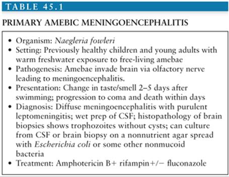
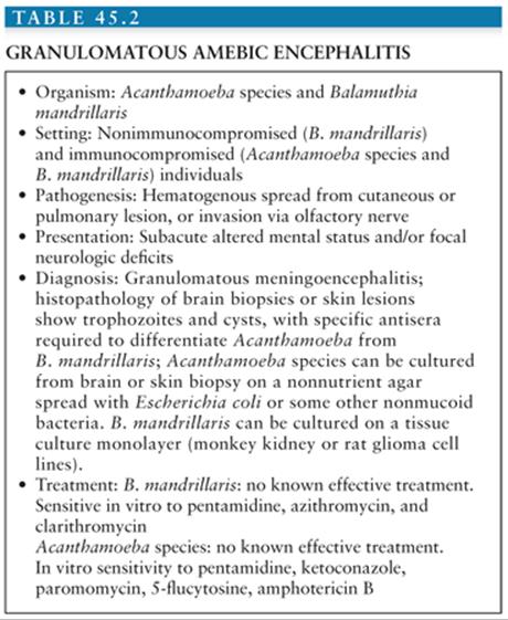
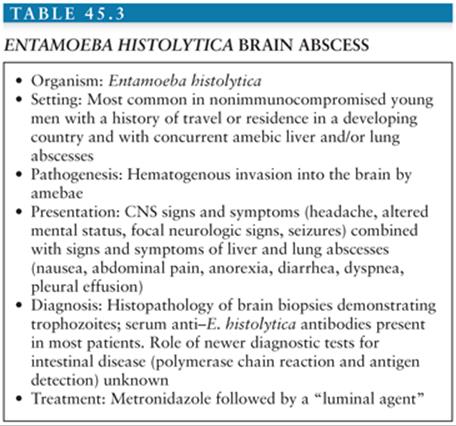
Central nervous system (CNS) infection with free-living and parasitic amebae is rare but life threatening. Three separate clinical syndromes occur from infection with free-living amebae (1). Primary amebic meningoencephalitis is due to infection with Naegleria fowleri and occurs almost exclusively in previously healthy children and young adults following exposure to freshwater. Pathologic examination of brain tissues is consistent with infection via direct invasion of the brain from the olfactory nerves (2–11) (Table 45.1). Granulomatous amebic encephalitis is due to hematogenous or direct infection of the brain with Acanthamoeba species (including Acanthamoeba castellani, Acanthamoeba polyphaga, Acanthamoeba culbertsoni, Acanthamoeba palestinensis, Acanthamoeba astronyxis, Acanthamoeba hatchetti, and Acanthamoeba rhysodes) (12,13) or Balamuthia mandrillaris (formerly misidentified as leptomyxid amebae) (14–27). Although granulomatous amebic encephalitis due to Acanthamoeba has been observed predominantly in patients with known immunodeficiencies, B. mandrillaris infection has also been observed in nonimmunocompromised individuals (Table 45.2). Acanthamoeba keratitis is seen predominantly in individuals who wear contact lenses and is not discussed further in this chapter (28,29). Brain abscess is a rare complication of Entamoeba histolytica infection, an otherwise common infectious disease in the developing world. E. histolytica brain abscess is most commonly seen in association with E. histolytica hepatic and/or lung abscess (30–40) (Table 45.3). As opposed to infection with the parasitic ameba E. histolytica, infection with free-living amebae is not linked to fecally contaminated food or water and is therefore no more common in developing nations than in the industrialized West. Encephalitis may also be caused by ameba belonging to the genus Sappinia. Only one case of amebic encephalitis due to Sappinia pedata infection has been reported (41,42).
ORGANISMS AND EPIDEMIOLOGY
N. fowleri is the only recognized pathogenic species of Naegleria. Trophozoites of N. fowleri are 10 to 30 µm in diameter and the nucleus has a prominent central dense nucleolus (Fig. 45.1A and B). The trophozoites can transform into a flagellated form (seen in vitro upon transfer to distilled water). The cyst form of the parasite is an approximately 9-µm diameter sphere with two pores in the cyst wall that serve as an egress point for the trophozoite upon excystation. N. fowleri have been isolated from freshwater lake and river water and soil in all parts of the world (3). Warm temperatures appear to favor their growth; for example, in Florida, it is not uncommon to isolate N. fowlerifrom freshwater lakes and streams. Most cases of N. fowleri infection have occurred in the summertime, and in the winter, the organism is identified in lake and river sediments. The cyst form of the parasite is stable for months in the environment but is not found in human tissues. It is estimated that in Florida, there are more than 100 million exposures to the parasite for every case of primary amebic meningoencephalitis (3). In fact most adults in the United States appear to have been exposed to the parasite as evidenced by the presence of serum antibodies against N. fowleri. The factors leading to N. fowleri infection in only a tiny minority of exposed individuals is unknown. Primary amebic meningoencephalitis has been reported in the central and southern United States, Central America, Europe, Japan, Africa, Australia, and New Zealand. Outbreaks have been linked to common environmental exposures (2,6,10).

Acanthamoeba species consist of cyst and trophozoite stages, with the trophozoites approximately 14 to 40 µm in diameter and cysts with diameters of 12 to 16 µm (Fig. 45.1C and D). Both cysts and trophozoites can be observed in infected brain. The nucleus has a central nucleolus similar in appearance to that of Naegleria. Acanthamoeba species have also been isolated from soil, water, and air from diverse geographic locations (1,12,13). In contrast to N. fowleri, Acanthamoeba may be spread through the air. For example, 2% of nasopharyngeal swabs from healthy individuals were culture positive for Acanthamoeba species in one study. Again, it is common to find antibodies against Acanthamoeba in healthy individuals, although almost all cases of granulomatous amebic encephalitis due to Acanthamoebaspecies have been observed in immunocompromised individuals (1,12,13). The cause of immunosuppression has included HIV/AIDS, renal and bone marrow transplantation, cancer chemotherapy, steroid therapy, diabetes mellitus, and liver disease.
B. mandrillaris trophozoites have an average size of 30 µm with cysts of 15 µm and therefore can be distinguished from Acanthamoeba only by hematoxylin-eosin stains using specific antisera (1,20,26). It is an inhabitant of soil and freshwater with the highest concentration of organisms observed in freshwater in the spring and fall. Unique to B. mandrillaris is that it can cause CNS infection in both healthy and immunocompromised individuals (14–17,19–21,25,26).
E. histolytica trophozoites have an average diameter of 15 to 30 µm, and the 10-µm quadrinucleate cysts of the parasite have never been found in brain. E. histolytica infection is estimated to occur in hundreds of millions of people annually (30,31). With the use of modern diagnostic tests, the burden of amebic disease is becoming better appreciated. In Dhaka, Bangladesh, preschool-aged children had a 2.2% frequency of amebic dysentery during 3 years of prospective community observation (32). In Hue City, Vietnam, an incidence of amebic liver abscess of 21 cases per 100,000 inhabitants was observed (33). In the United States where fecal-oral transmission is unusual, amebiasis is most commonly seen in immigrants from and travelers to developing countries. Brain abscess is seen usually in association with an amebic liver or lung abscess (32–40). Amebic liver abscess in turn is predominantly (but not exclusively) a disease of young men (30,31), leaving one to have the highest suspicion for an E. histolytica brain abscess in a male patient with hepatic abscess, risk factors of E. histolytica infection, and CNS signs or symptoms.
PATHOLOGY
N. fowleri appears to enter the CNS after disrupting the olfactory mucosa. It produces a diffuse meningoencephalitis with purulent leptomeningitis. Cortical edema and hemorrhage with cerebellar or uncal herniation are observed. The olfactory bulbs are commonly hemorrhagic or necrotic and trophozoites can be observed in the olfactory nerves and in the perivascular spaces and adventitia of arterioles and midsize arteries. A neutrophilic myocarditis has been observed in some cases (2,5,6,8). Trophozoites of N. fowleri can be observed in wet preparations of cerebrospinal fluid (CSF) but are commonly misidentified as monocytes.
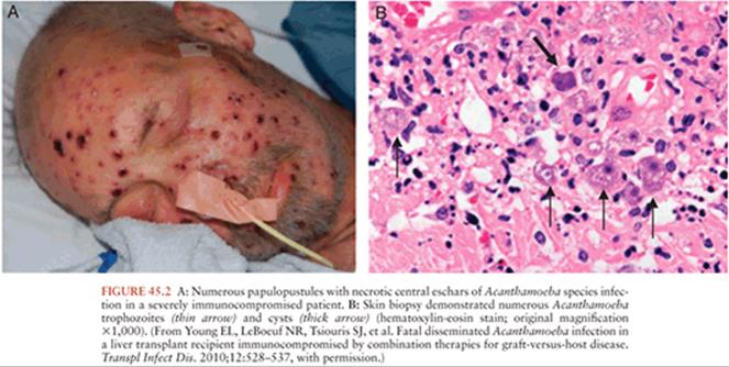
Granulomatous amebic encephalitis due to Acanthamoeba species usually presents with focal neurologic deficits in immunocompromised individuals. Cerebral edema resulting in bilateral cerebellar or uncal herniation occasionally occurs. Areas of cortical involvement include the cerebellum, midbrain, and brainstem (12,13). Histologically, the lesions spare the leptomeninges and contain necrotizing granulomas with amebic cysts and trophozoites. Multinucleated giant cells may be seen within the granulomas. Occasionally in immunodeficient individuals, the granulomatous reaction may not be present. The parasites are observed most often in a perivascular location. Identification of Acanthamoeba in the skin (Fig. 45.2), lung, adrenals, and lymph nodes of patients with CNS infection suggests hematogenous dissemination of infection to the brain.
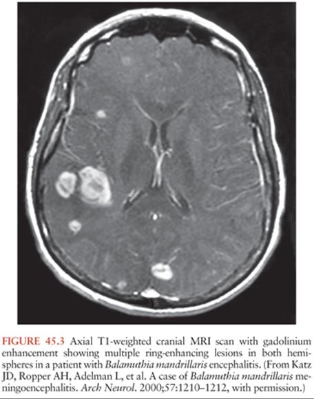
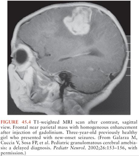
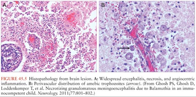
B. mandrillaris infection of the CNS results in a subacute or chronic granulomatous meningoencephalitis that can closely resemble the pathologic lesions of Acanthamoeba infection (Figs. 45.3 to 45.6). Both immunocompromised and normal individuals can be infected. A chronic inflammatory process is observed in the CNS that includes lymphocytes, mononuclear cells, and giant and plasma cells. Granulomas may be absent, and parasites (cysts and trophozoites) are observed in a perivascular location (Figs. 45.5 and 45.6). Angiitis and hemorrhagic necrosis of the meninges and brain tissue have been observed.

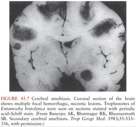
In most cases, brain lesions due to E. histolytica infection are multiple and are most common in the basal ganglia, followed by the frontal and occipital lobes. Lesions vary widely in size from 2 mm to 5 cm and are associated with a polymorphonuclear infiltrate that may also be present in the CSF. Hemorrhage may be observed at the junction of the gray and white matter (35–40) (Fig. 45.7).
CLINICAL MANIFESTATIONS
Primary amebic meningoencephalitis is an almost uniformly fatal infection of previously healthy children (2,5,7). A history of exposure to freshwater lakes, ponds, or streams in the week preceding the illness is obtained in most cases, with an average incubation period of 2 to 5 days. The first symptom may be a change in taste or smell. This is followed by the abrupt onset of fever, nausea, vomiting, or anorexia. Meningismus is present in more than 85% of patients and two thirds have alterations in mental status at the time of admission. Progression to coma and death occurs without the development of focal neurologic signs in most patients, usually within a week of illness onset (2,5,7).
Granulomatous amebic encephalitis due to Acanthamoeba species is a subacute or chronic illness of the immunocompromised and debilitated. Its onset is insidious, with presenting signs and symptoms including altered mental status, seizures, fever, headache, hemiparesis, meningismus, visual disturbances, and ataxia. Underlying illnesses include HIV/AIDS, liver and renal disease, solid organ transplants, neoplasm, steroid therapy, pregnancy, and diabetes mellitus (4). The average time from onset of CNS symptoms to death is a month. Acanthamoeba also causes ulcerative or nodular skin lesions on the trunk or extremities that may develop over a period of weeks to months and may occur at the time of or before the development of CNS symptoms or signs. The average time from onset of CNS symptoms to death is 1 month.
B. mandrillaris can cause CNS infection in both immunocompetent and immunodeficient individuals (14,16–21,25,26). Otherwise the clinical presentation is similar to that of CNS Acanthamoeba infection. Skin involvement with Balamuthia frequently involves the face, including papulonodular, erythematous, plate-like lesions that are painless and begin most frequently on the nose. Hydrocephalus has been reported as a complication.
E. histolytica brain abscess is almost always seen in an individual who also has an amebic liver abscess and occasionally an amebic lung abscess. Thus, abdominal pain, dyspnea, pleural effusion, anorexia, and weight loss commonly accompany the neurologic findings of headache, meningismus, seizure, and altered mental status (36–40). Patients with amebic abscess are commonly males younger than 50 years who have immigrated from or traveled to an endemic developing country.
DIAGNOSIS
Primary amebic meningoencephalitis should be included in the differential diagnosis of children and young adults with meningoencephalitis, especially in patients with a recent history of freshwater exposure. The CSF pressure is elevated, with hemorrhagic CSF, elevated white blood cell count, and a neutrophilic predominance. If no bacteria are demonstrated in a purulent CSF, then it is important to examine the CSF for amebic trophozoites, which can be accomplished by preparing a wet mount of fresh CSF (1,7–9). Fixation of CSF for Gram strain disrupts the trophozoites, making diagnosis difficult or impossible. Motile trophozoites have been observed in most CSF specimens from patients with primary amebic meningoencephalitis but are frequently misidentified as white blood cells. Modern diagnostic approaches using polymerase chain reaction (PCR), monoclonal antibodies, and DNA probes have also been applied to diagnosis. N. fowleri can be successfully cultured from CSF and brain biopsies; therefore, the microbiology lab should be informed about suspected cases so that appropriate processing of specimens can be carried out.
Granulomatous amebic encephalitis due to Acanthamoeba species or B. mandrillaris is diagnosed by brain biopsy. Amebae have never been observed in CSF. CSF findings are nonspecific but include elevated protein and decreased glucose concentration. Lumbar puncture is contraindicated if the patient is at risk of herniation. Computed tomographic and magnetic resonance imaging (Figs. 45.3 and 45.4) show single or more often multiple enhancing mass lesions that are most commonly found in the anterior cortex and subcortical white matter, with only rare involvement of the cerebellum and brainstem (12–14,16–18,21). Skin lesions should be biopsied and examined for Acanthamoeba species in patients suspected of having granulomatous amebic encephalitis. Morphologically, Acanthamoeba species and B. mandrillaris appear similar, and specific antisera is necessary to distinguish them by immunohistochemistry. Culture has successfully been used to isolate Acanthamoeba species and B. mandrillaris from fresh (not frozen or fixed) brain biopsy materials (1). A multiplex real-time PCR assay has been developed for the simultaneous detection of Acanthamoeba spp., B. mandrillaris, and N. fowleri. The assay facilitates rapid detection with a turn-around time of 5 hours. This allows for an early diagnosis and therefore prompt initiation of therapy (43).
The diagnosis of E. histolytica brain abscess has classically been made by the identification of E. histolytica trophozoites by periodic acid–Schiff stain in the periphery of abscesses (36–40,44). Most patients will have detectable serum antibodies against the parasite at the time of diagnosis. The use of immunoperoxidase staining of tissue sections with specific E. histolytica antibodies is helpful in the diagnosis of colonic amebiasis and could potentially be applied to cerebral amebiasis (45). The sensitivity and specificity of modern diagnostic tests for intestinal infection, such as detection of E. histolytica–specific antigen or DNA, for the diagnosis of brain infection are unknown.
TREATMENT
The only patients known to have survived primary amebic meningoencephalitis received amphotericin B, but the overall mortality remains greater than 95%. A few survivors received rifampin in addition to amphotericin, and some experts, therefore, recommend combination therapy with these two agents (46,47). Other agents used have included miconazole, rifampin, and sulfisoxazole (7,11). In two recent cases, both patients recovered after treatment with a combination of amphotericin B, rifampin, and fluconazole (48,49).
The optimal treatment of granulomatous amebic encephalitis is unknown. Different species and strains have different in vitro drug sensitivities. In general, diamidines such as propamidine, pentamidine, and dibromopropamidine are active against Acanthamoeba species (12,13). In vitro susceptibility to ketoconazole, paromomycin, 5-flucytosine, and amphotericin B has been observed. The combination of hyperbaric oxygen therapy and multiple antimicrobial agents (pentamidine, fluconazole, metronidazole, trimethoprim-sulfamethoxazole, and miltefosine) was successful in one patient (50). For B. mandrillaris, in vitro sensitivity has been demonstrated for pentamidine, ketoconazole, paromomycin, 5-flucytosine, amphotericin B, and phenothiazine derivatives (24,27,51). A recent report described two patients who survived following treatment with flucytosine, pentamidine, fluconazole, sulfadiazine, a macrolide antibiotic (azithromycin or clarithromycin), and phenothiazine derivatives (51).
E. histolytica brain abscess in the past has almost always been fatal. In the absence of any data, but in light of the seriousness of this infection, a combination of aspiration and drainage of the brain abscess combined with antiamebic drug therapy is prudent. The nitroimidazole metronidazole is the mainstay of therapy for invasive amebiasis (52–55). E. histolytica persists in the intestine in many nitroimidazole-treated patients. Therefore, intestinal infection should be eradicated by following nitroimidazole treatment with paromomycin or the second-line agent diloxanide furoate (52–55).
In one reported case of amebic encephalitis due to Sappinia pedata infection, the patient was successfully treated with a combination of azithromycin, pentamidine, itraconazole, and flucytosine (41,42).
References
1. Schuster FL. Cultivation of pathogenic and opportunistic free-living amebas. Clin Microbiol Rev. 2002;15:342–354.
2. Shenoy S, Wilson G, Prashanth HV, et al. Primary meningoencephalitis by Naegleria fowleri: first reported case from Mangalore, South India. J Clin Microbiol. 2002;40:309–310.
3. Cabanes PA, Wallet F, Pringuez E, et al. Assessing the risk of primary amoebic meningoencephalitis from swimming in the presence of environmental Naegleria fowleri. Applied Environ Microbiol. 2001;67:2927–2931.
4. Visvesvara GS. Amebic meningoencephalitides and keratitis: challenges in diagnosis and treatment.Curr Opin Infect Dis. 2010;23(6):590–594.
5. Jarolim KL, McCosh JK, Howard MJ, et al. A light microscopy study of the migration of Naegleria fowleri from the nasal submucosa to the central nervous system during the early stage of primary amebic meningoencephalitis in mice. J Parasitol. 2000;86:50–55.
6. Sugita Y, Fujii T, Hayashi I, et al. Primary amebic meningoencephalitis due to Naegleria fowleri: an autopsy case in Japan. Pathol Int. 1999;49:468–470.
7. Parija SC, Jayakeerthee SR. Naegleria fowleri: a free living amoeba of emerging medical importance. J Commun Dis. 1999;31:153–159.
8. Kidney DD, Kim SH. CNS infections with free-living amebas: neuroimaging findings. AJR Am J Roentgenol. 1998;171:809–812.
9. De Jonckheere JF, Brown S. Primary amebic meningoencephalitis in a patient with AIDS: unusual protozoological findings. Clin Infect Dis. 1997;25:943–944.
10. DeNapoli TS, Rutman JY, Robinson JR, et al. Primary amoebic meningoencephalitis after swimming in the Rio Grande. Texas Med. 1996;92:59–63.
11. Barnett ND, Kaplan AM, Hopkin RJ, et al. Primary amoebic meningoencephalitis with Naegleria fowleri: clinical review. Pediatr Neurol. 1996;15:230–234.
12. Sell JJ, Rupp FW, Orrison WW Jr. Granulomatous amebic encephalitis caused by Acanthamoeba. Neuroradiology. 1997;39(6):434–436.
13. Walker CW. Acanthamoeba: ecology, pathogenicity and laboratory detection. Br J Biomed Sci. 1996;53(2):146–151.
14. Shirabe T, Monobe Y, Visvesvara GS. An autopsy case of amebic meningoencephalitis: the first Japanese case caused by Balamuthia mandrillaris. Neuropathology. 2002;22:213–217.
15. Schuster FL, Glaser C, Gilliam S, et al. Survey of sera from encephalitis patients for Balamuthia mandrillaris antibody. J Eukaryotic Microbiol. 2001;(suppl):10S–12S.
16. Martinez AJ, Schuster FL, Visvesvara GS. Balamuthia mandrillaris: its pathogenic potential. J Eukaryotic Microbiol. 2001;(suppl):6S–9S.
17. Martinez AJ, Visvesvara GS. Balamuthia mandrillaris infection. J Med Microbiol. 2001;50:205–207.
18. Healy JF. Balamuthia amebic encephalitis: radiographic and pathologic findings. AJNR Am J Neuroradiol. 2002;23:486–489.
19. Galarza M, Cuccia V, Sosa FP, et al. Pediatric granulomatous cerebral amebiasis: a delayed diagnosis. Pediatr Neurol. 2002;26:153–156.
20. Deol I, Robledo L, Meza A, et al. Encephalitis due to a free-living amoeba (Balamuthia mandrillaris): case report with literature review. Surg Neurol. 2000;53:611–616.
21. Katz JD, Ropper AH, Adelman L, et al. A case of Balamuthia mandrillaris meningoencephalitis. Arch Neurol. 2000;57:1210–1212.
22. Huang ZH, Ferrante A, Carter RF. Serum antibodies to Balamuthia mandrillaris, a free-living amoeba recently demonstrated to cause granulomatous amoebic encephalitis. J Infect Dis. 1999;179:1305–1308.
23. Recavarren-Arce S, Velarde C, Gotuzzo E, et al. Amoeba angeitic lesions of the central nervous system in Balamuthia mandrillaris amoebiasis. Human Pathol. 1999;30:269–273.
24. Schuster FL, Visvesvara GS. Efficacy of novel antimicrobials against clinical isolates of opportunistic amebas. J Eukaryotic Microbiol. 1998;45:612–618.
25. Kodet R, Nohynkova E, Tichy M, et al. Amebic encephalitis caused by Balamuthia mandrillaris in a Czech child: description of the first case from Europe. Pathol Res Practice. 1998;194:423–429.
26. Denney CF, Iragui VJ, Uber-Zak LD, et al. Amebic meningoencephalitis caused by Balamuthia mandrillaris: case report and review. Clin Infect Dis. 1997;25:1354–1358.
27. Schuster FL, Visvesvara GS. Axenic growth and drug sensitivity studies of Balamuthia mandrillaris, an agent of amebic meningoencephalitis in humans and other animals. J Clin Microbiol. 1996;34:385–388.
28. Sharma S, Garg P, Rao GN. Patient characteristics, diagnosis, and treatment of non-contact lens related Acanthamoeba keratitis. Br J Ophthalmol. 2000;84:1103–1108.
29. McCulley JP, Alizadeh H, Niederkorn JY. The diagnosis and management of Acanthamoeba keratitis. CLAO J. 2000;26:47–51.
30. Haque R, Huston CD, Hughes M, et al. Current concepts: amebiasis. N Engl J Med. 2003;348:1565–1573.
31. Stanley SL Jr. Amoebiasis. Lancet. 2003;361:1025–1034.
32. Haque R, Duggal P, Ali IM, et al. Innate and acquired resistance to amebiasis in Bangladeshi children. J Infect Dis. 2002;186:547–552.
33. Blessmann J, Van Linh P, Nu PA, et al. Epidemiology of amebiasis in a region of high incidence of amebic liver abscess in central Vietnam. Am J Trop Med Hyg. 2002;66:578–583.
34. Ohnishi K, Murata M. Present characteristics of symptomatic amebiasis due to Entamoeba histolytica in the east-southeast area of Tokyo. Epidemiol Infect. 1997;119:363–367.
35. Ohnishi K, Murata M, Kojima H, et al. Brain abscess due to infection with Entamoeba histolytica. Am J Trop Med Hyg. 1994;51:180–182.
36. Shah AA, Shaikh H, Karim M. Amoebic brain abscess: a rare but serious complication of Entamoeba histolytica infection. J Neurol Neurosurg Psychiatry. 1994;57(2):240–241.
37. Orbison JA, Reeves N, Leedham CL, et al. Amebic brain abscess: review of the literature and report of five additional cases. Medicine. 1951;30:247–282.
38. Lombardo L, Alonso P, Arroyo LS, et al. Cerebral amebiasis: report of 17 cases. J Neurosurg. 1964;21:704–709.
39. Brandt H, Perez-Tamayo R. Pathology of human amebiasis. Human Pathol. 1970;1:351.
40. Hughes FB, Faehnle ST, Simon JL. Multiple cerebral abscesses complicating hepatopulmonary amebiasis. J Pediatr. 1975;86:95–96.
41. Gelman BB, Rauf SJ, Nader R, et al. Amoebic encephalitis due to Sappinia diploidea. JAMA. 2001;285(19):2450–2451.
42. Qvarnstrom Y, da Silva AJ, Schuster FL, et al. Molecular confirmation of Sappinia pedata as a causative agent of amoebic encephalitis. J Infect Dis. 2009;199(8):1139–1142.
43. Qvarnstrom Y, Visvesvara GS, Sriram R, et al. Multiplex real-time PCR assay for simultaneous detection of Acanthamoeba spp., Balamuthia mandrillaris, and Naegleria fowleri. J Clin Microbiol. 2006;44(10): 3589–3595.
44. Banerjee AK, Bhatmagar RK, Bhusnurmath SR. Secondary cerebral amebiasis. Trop Geogr Med. 1983;35:333–336.
45. McCarthy JS, Peacock D, Trown KP, et al. Endemic invasive amoebiasis in northern Australia. Med J Australia. 2002;177:570.
46. Wang A, Kay R, Poon WS, et al. Successful treatment of amoebic meningoencephalitis in a Chinese living in Hong Kong. Clin Neurol Neurosurg. 1993;95:249–252.
47. Jain R, Prabhakar S, Modi M, et al. Naegleria meningitis: a rare survival. Neurol India. 2002;50:470–472.
48. Vargas-Zepeda J, Gómez-Alcalá AV, Vásquez-Morales JA, et al. Successful treatment of Naegleria fowleri meningoencephalitis by using intravenous amphotericin B, fluconazole and rifampicin. Arch Med Res. 2005;36(1): 83–86.
49. Yadav D, Aneja S, Dutta R, et al. Youngest survivor of Naegleria meningitis. Indian J Pediatr. 2012;80(3):253–254.
50. Maritschnegg P, Sovinz P, Lackner H, et al. Granulomatous amebic encephalitis in a child with acute lymphoblastic leukemia successfully treated with multimodal antimicrobial therapy and hyperbaric oxygen. J Clin Microbiol. 2011;49(1):446–448.
51. Deetz TR. Successful treatment of Balamuthia amoebic encephalitis: presentation of 2 cases. Clin Infect Dis Epub. 2003;37:1304–1312.
52. Powell SJ, MacLeod I, Wilmot AL, et al. Metronidazole in amoebic dysentery and amoebic liver abscess. Lancet. 1967;1:1329–1331.
53. Blessmann J, Tannich E. Treatment of asymptomatic intestinal Entamoeba histolytica infection. N Engl J Med. 2002;347:1384.
54. McAuley JB, Herwaldt BL, Stokes SL, et al. Diloxanide furoate for treating asymptomatic Entamoeba histolytica cyst passers: 14 years’ experience in the United States. Clin Infect Dis. 1992;15:464–468.
55. McAuley JB, Juranek DD. Paromomycin in the treatment of mild to moderate intestinal amebiasis. Clin Infect Dis. 1992;15:551–552.