
JOSE A. SERPA, WON K. CHUNG, AND A. CLINTON WHITE, JR.
The helminths include a broad range of organisms that are common human parasites. There are three major groups of helminths: the nematodes (or roundworms, phylum Nematoda), the trematodes (or flatworms or flukes, phylum Platyhelminthes, subphylum Trematoda), and the cestodes (tapeworms, phylum Platyhelminthes, subphylum Cestoda). Numerous members of each group cause human central nervous system (CNS) infection (Table 46.1). Helminths are part of the animal kingdom and are eukaryotic multicellular organisms with well-developed organs including reproductive and digestive tracts (nematodes and trematodes) or digestive surface (cestodes). Helminths are often large enough to be visible to the human eye. They are extracellular parasites. The host response to helminths is dominated by mechanisms aimed at extracellular organisms, resulting in the development of eosinophilia.

Most helminths have complex life cycles, with different morphologic forms and often involving multiple hosts. Most reproduce primarily by sexual reproduction. The host in which sexual reproduction occurs is termed the definitive host. The host in the life cycle in which no or only asexual reproduction occurs is termed the intermediate host. In many cases, humans are a dead-end host and not part of the parasite life cycle. In that case, humans are termed an incidental host.
CENTRAL NERVOUS SYSTEM DISEASE CAUSED BY CESTODES
Neurocysticercosis
Taenia solium is a cestode parasite with a wide distribution globally (1–3). The parasite has two forms: an intestinal tapeworm, found only in human hosts, and the cystic larval form, termed the cysticercus, which is typically found in pigs. Humans can also be host to the cysticercus. Cysticercus infection of the CNS is termed neurocysticercosis (NCC), which is now recognized as a common cause of neurologic disease worldwide (1–3).
Epidemiology
The endemic regions for T. solium infection include most areas of the world where pigs are raised including Latin America, sub-Saharan Africa, India, Southeast Asia, China, Indonesia, and other regions less well characterized such as New Guinea and Eastern Europe (1–3). However, the burden of disease was not recognized until the availability of neuroimaging studies in the early 1980s resulted in identification of NCC as a common neurologic problem among immigrants to the United States and other developed countries and later among residents of endemic areas. Review of studies from endemic areas around the world document NCC in approximately 30% of patients with seizures (3). Several studies have documented a high prevalence of infection throughout Latin America (4,5) with estimates ranging between 15% and 38%. Similar studies have also demonstrated NCC in a high proportion of African patients with seizures, with the disease prevalent throughout the region (6,7). The prevalence of seizures due to NCC in sub-Saharan Africa has been estimated at 1.7 to 24.8 per 1,000 persons.
When computed tomographic (CT) scans were initially used in adults with new-onset seizures in India, most patients were noted to have abnormalities, including single calcifications or single enhancing lesions. In a series of 991 patients with symptomatic localization-related seizures in south India, 40% of patients were found to have either active NCC, calcifications on CT scan consistent with prior cysticercosis, or single enhancing CT lesions, consistent with cysticercal granulomas (8). These findings were confirmed in a study of children with seizures regardless of features of localization and in a study from northern India (9,10). The single enhancing CT lesions were initially attributed to tuberculosis or to the effects of seizures. However, when excisional biopsies were performed on these patients, nearly all showed histopathologic evidence of cysticercosis (11). More than half of 401 patients presenting with a single enhancing lesion were subsequently proven to have NCC (12). As mentioned earlier, NCC is also prevalent in other parts of Asia including Indonesia, Southeast Asia, China, and Korea (13). Recent data suggest an average prevalence of T. solium infection in China of 0.11% (range, 0.05% to 15%); which translates to about 1.26 million cases of taeniasis and 3 to 6 million cases of cysticercosis. In the United States, estimates range from 0.2 to 0.6 cases of NCC per 100,000 general population and 1.5 to 5.8 cases per 100,000 Hispanics (14).
Parasitology
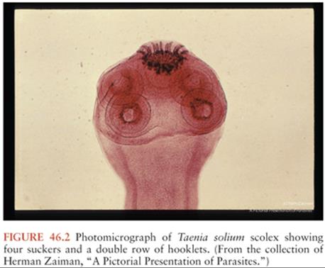
The T. solium life cycle requires two hosts and two major forms of the parasite (Fig. 46.1). The cysticerci are found primarily in muscle of the pig, the intermediate host. Humans are the host for the intestinal tapeworm form. Humans become infected with the tapeworm form (taeniasis) by ingesting undercooked pork infested with T. solium cysticerci. After ingestion, the scolex (or head) evaginates and attaches to the small intestines by means of hooks and suckers (Fig. 46.2). Proglottids (segments) develop from the base of the scolex. As new segments arise at the base of the scolex, the older proglottids form a chain that can reach a length of up to 30 feet, with the larger more mature proglottids at the distal end. The mature proglottids are off-white, opaque, and approximately 1 cm wide, 1 to 2 cm long, and 1 to 3 mm thick. The proglottids are hermaphroditic and mate with themselves to produce ova. The ova or proglottids are shed intermittently in the stool. Most tapeworm carriers note few or no symptoms other than possibly noting proglottids in the stool. Excretion is intermittent, so stool examinations for the ova or parasites are usually negative.

Porcine cysticercosis is endemic in regions where pigs consume human fecal material. Pigs, when raised under these conditions, ingest the ova or proglottids from the tapeworm carrier. In the intestines, the eggs hatch, release the invasive larvae (also termed oncospheres), which penetrate the intestinal mucosa using hooklets and excretory proteases, enter the bloodstream, migrate to the tissues, and mature into cysticerci. In the muscle, the cysticerci appear as thin-walled, translucent, oval cysts, approximately 1 cm in diameter. The invaginated scolex is found as a 1- to 2-mm opaque nodule attached to one side of the wall.
Human cysticercosis occurs when people ingest ova shed by a tapeworm carrier. As in pigs, the eggs hatch in the intestine, enter the bloodstream, migrate to the tissues, including muscle, brain, and eye, and mature into cysticerci. Most epidemiologic studies suggest that close personal contact with or perhaps food preparation by a tapeworm carrier is associated with NCC. The tapeworm carriers can also be infected themselves, probably by the fecal-oral route, but cysticercosis is not acquired directly from pork. This is illustrated by an outbreak that occurred in an Orthodox Jewish community in New York City (15,16). In all the exposed households, there was a history of employment of live-in housekeepers who had recently emigrated from Latin American countries. Examination of six housekeepers revealed an active taeniasis in one and a positive serologic test result in another. There have been a total of 78 autochthonous cases of cysticercosis reported in the United States between 1954 and 2005 (17). A confirmed or presumptive source of infection was identified among household members or close personal contacts of 16 (21%) case patients.
Pathogenesis and Pathology
Despite the fact cysticerci reach their final size in a few weeks, there is a period of several years between infection and onset of symptoms (18,19). Thus, the presence of the parasites in the CNS does not explain symptoms. Autopsy studies of individuals in whom cysticerci were an incidental finding demonstrate viable cysticerci in brain that appear similar to the viable cysticerci from pig muscle (20) (Fig. 46.3A and B). By contrast, autopsies of patients with cysticercosis with seizures demonstrate a prominent inflammatory infiltrate. Studies have elaborated a number of molecular mechanisms used by the parasites to suppress the host inflammatory and immune responses (21). Similarly, our group has demonstrated that parasite granulomas—not the parasite itself—are the cause of seizures in an animal model. Thus, seizures likely result from the host response rather than from the parasitic infection per se (22). After months to years, the cysticerci lose their ability to control the host response. The cysticerci become infiltrated and surrounded by host inflammatory cells composed primarily of mononuclear cells, with variable numbers of eosinophils and neutrophils, and pass through a series of stages of inflammation (23,24). This response is associated with elaboration of substance P and type 1 cytokines such as interleukin-12 (IL-12), interferon-γ (IFN-γ), and IL-2 (25,26). Recent findings indicate that substance P (SP) is a critical mediator of seizures in NCC, and this may be prevented with SP-receptor antagonists (27). The walls of the cysticercus degrade. The cyst cavity is invaded by inflammatory cells (colloid stage). As the host response progresses, fibrosis encompasses the cysticercus with collapse of the cyst cavity (granular nodular stage). Eventually, the parasite is replaced by progressive fibrosis, which may calcify (calcific stage). This stage may have a minimal lymphocytic infiltrate but may also become more inflamed if the granulomas break down.

Findings similar to these pathologic stages have been noted on neuroimaging studies (28,29). Invasion of the CNS and early development of the cysticercus may present as an area of focal edema or enhancement. The viable cysticercus appears as a cystic area that is isodense with cerebrospinal fluid (CSF) (Fig. 46.4A). The cyst wall has the same density as brain parenchyma and lacks surrounding edema or contrast enhancement. As the cysticercus becomes inflamed, the density of the cyst wall increases and may enhance with contrast (Fig. 46.4B and C). Associated edema or enhancement is noted in the brain parenchyma. The cyst fluid increases in density. As the cysticercus becomes fibrotic or collapses, neuroimaging studies reveal an area of focal enhancement, suggestive of a granuloma. Finally, the calcific stage is defined by the appearance of a focal area of calcification, typically several millimeters in diameter (Fig. 46.4C). Recent studies have demonstrated edema and contrast enhancement associated with symptomatic calcified lesions (30,31). This likely results from breakdown of the calcified granulomas, triggering host inflammation.

Cysticerci can also be found in the cerebral ventricles and typically float within the CSF (Fig. 46.4A). However, cysticerci in the ventricles can cause hydrocephalus, primarily by mechanical obstruction of CSF flow (32). In most cases, the obstruction is caused by viable cysticerci (33). When the cysticerci become inflamed, the ependymitis and accompanying astrocytosis cause the cysticercus to adhere to the walls of the ventricles or block CSF flow directly (especially in the aqueduct of Sylvius).
Cysticerci in the basilar cisterns are associated with a form of NCC termed subarachnoid cysticercosis. Some of the cysticerci within the subarachnoid space may enlarge and occasionally lose the scolex (Figs. 46.3 and 46.4C). The cysticerci without a scolex were termed racemose. The term racemose cysticercosis was used to describe subarachnoid disease. Because the clinical presentation, pathogenesis, and treatment are the same, whether the scolex is present or not, the term racemose may not be ideal. Furthermore, many of the so-called racemose cysticerci actually contain hooks, evidence that they previously contained a scolex.
Subarachnoid NCC is characterized by a prominent basilar arachnoiditis (34). Patients may have meningeal signs, communicating hydrocephalus, or vasculitis. The communicating hydrocephalus is presumed to be caused by CSF outflow obstruction or blocked ventricular outflow (32). The accompanying vasculitis may present as lacunar infarctions, or occasionally, erosion into larger vessels may cause large vessel strokes (35,36).
Cysticerci within the subarachnoid space can grow to sizes larger than the 1 to 2 cm typical of parenchymal cysticerci (Fig. 46.4C). These giant cysticerci may expand to sizes up to 10 cm in diameter and can cause mass effects (37). The mass effects are especially prominent when the cysticerci are inflamed either from spontaneous degeneration or after drug treatment.
Classification and Clinical Manifestations
The clinical manifestations of NCC depend on the number and location of the cysticerci, as well as the associated host response. Clinical classifications are usually based on neuroimaging (38–40). The most common scheme separates NCC into parenchymal and extraparenchymal. Extraparenchymal NCC is subdivided into ventricular and subarachnoid (including spinal). Experts agree that the term inactive NCC be used to describe cases in which there is no longer evidence of either a viable or a degenerating parasite. CT scans may reveal one or more calcifications, typically 2 to 10 mm in diameter. These correspond to the calcific stage described pathologically. Often, these lesions are symptomatic causing recurrent seizures associated with contrast enhancement (perilesional edema) (30,41–44). Some patients may also present with hydrocephalus without evidence of cysts. These patients have had prior arachnoiditis or granular ependymitis resulting in obstruction of CSF flow (e.g., aqueductal stenosis) or CSF outflow obstruction.
Symptomatic parenchymal infection typically results from the host inflammatory response and carries a favorable prognosis. Extraparenchymal disease tends to have a worse prognosis, with associated hydrocephalus (usually requiring surgical therapy), and can be fatal if not properly managed (45,46).
Parenchymal Neurocysticercosis. Parenchymal infection is the most common form of disease, and in most series, the majority of cases are inflamed cystic lesions. By contrast, noninflamed cysts rarely cause symptoms. For example, Garcia and Del Brutto (47) have described a group of patients with numerous, noninflamed cysticerci but few, if any, symptoms. Thus, symptomatic infection likely occurs when the cysticercus can no longer control the host inflammatory and immune responses. Neuroimaging studies reveal round cysts, typically 4 to 20 mm in diameter. The cyst fluid is often isodense with CSF. In viable cysticerci, the cyst wall is thin (<1 mm thick) and is usually not visible. If seen, the scolex appears as a round or tubular mass 1 to 3 mm long on the side of the cyst wall. The inflamed cyst wall becomes denser and may display contrast enhancement of the wall (ring enhancement), cyst fluid, or surrounding tissues. There is often edema surrounding the cysticercus, especially on T2-weighted magnetic resonance imaging (MRI) scans. Later stage cysticerci appear as focal areas of enhancement or granulomas. The number of cysticerci varies from one to several thousand. However, in areas where most patients with new-onset seizures undergo CT scanning (e.g., India and the United States), most cases of parenchymal NCC have only a single cyst (48–50).
A subgroup of patients with active parenchyma disease present with diffuse cerebral edema and a clinical presentation resembling encephalitis (51). This form, termed cysticercal encephalitis, is more common in children than adults. Cysticercal encephalitis results from an inflammatory reaction to large numbers of cysticerci in the brain parenchyma. The accompanying inflammatory response causes diffuse cerebral edema.
Ventricular Neurocysticercosis. In some series, 5% to 30% of patients with NCC had cysticerci in the ventricles (1,40,50). These cysticerci can obstruct CSF flow causing hydrocephalus. The cysticerci are found in any of the ventricles. Older series noted most of the ventricular cysticerci in the fourth ventricle, but this may have reflected more severe disease when the cysticerci are in that location. In most cases, the cysticerci cause symptoms while still viable (33). Because viable cysticerci have thin walls with cyst fluid isodense with CSF, they may be difficult to detect by CT scanning, which usually only shows evidence of obstructive hydrocephalus or distortion of the shapes of the involved ventricle. The cysticerci are frequently visible on MRI (52,53). However, the findings are subtle and can be missed unless an expert familiar with cysticercosis reviews the scan. These patients may also have parenchymal cysticerci.
Subarachnoid Neurocysticercosis. When patients have cysticerci in the gyri of the cerebral convexities, the radiologic appearance, clinical presentation, and pathogenesis overlap with active parenchymal NCC. However, the cysts may be slightly larger and are more likely to persist after antiparasitic chemotherapy.
With cysticerci in the fissures (especially the sylvian fissure), the cysticerci can enlarge to several centimeters in diameter, termed giant cysticerci (37). Isolated cysticerci in the fissures carry a similar prognosis to parenchymal cysts with symptoms resulting from parenchymal inflammation. In some patients, however, the cysts may enlarge to the point of causing mass effects or midline shift. Most of the time, giant cysticerci are accompanied by cysticerci in the parenchyma or basilar cisterns. The giant cysticerci are readily visualized by CT or MRI, but the accompanying cysticerci in the basilar cisterns may not be seen on CT or MRI. Cysticercosis of the basilar cisterns carries a grave prognosis. Most patients are infected with numerous cysticerci that fill the basilar cisterns. Cisternal cysticercosis is characterized by arachnoiditis, which may be seen as focal or diffuse meningeal enhancement or vasculitis with strokes (34,36). Patients often develop communicating hydrocephalus. The spinal subarachnoid space is commonly involved in patients with basal subarachnoid NCC (54).
Spinal Neurocysticercosis. Only a small percentage of patients with NCC have recognized spinal involvement (1,40). Most cases of spinal NCC result from cysticerci in the subarachnoid space (45). Initially, the cysticerci are free floating, and they may move between levels. When the cysticerci degenerate, they eventually become fixed at one level. The accompanying inflammation may cause cord compression from mass effect with obstruction of flow on myelogram. Cysticerci are rarely intramedullary and may cause symptoms or signs from mass effect or accompanying inflammation.
Other Forms of Cysticercosis. Cysticercosis can involve the eye, with either intravitreal or subretinal cysticerci (55). Cysticerci may involve the muscles. Only rarely do they cause more than minor symptoms. Cysticerci can involve the subcutaneous tissues, where they present as a palpable cystic lesion that is often confused with a sebaceous cyst.
Mixed Forms. Clinical cases, especially those involving large numbers of cysticerci, often include more than one of the aforementioned forms. The pathogenesis and presentation may reflect each location. For example, patients who present with seizures will usually have parenchymal cysticerci. However, some also have cysticerci in the basilar cisterns that can progress to hydrocephalus later if not properly managed.
Clinical Manifestations
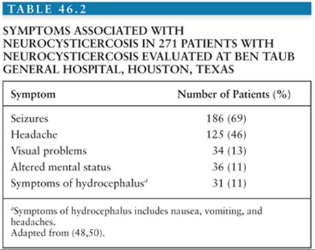
The most common clinical manifestation of NCC is seizures (56–58) (Table 46.2). The seizures may be either focal, focal with secondary generalization, or generalized (56,57,59). Most patients with seizures have evidence of parenchymal cysticerci with associated edema or enhancement, corresponding to the colloid or granular nodular stages. Electroencephalographic (EEG) studies for patients with active disease may reveal focal abnormalities. The seizures are usually easily controlled and often resolve as the inflammation subsides (57).
Some patients have seizures from calcified parenchymal NCC (60). The calcified lesions are thought to represent scarring from prior active infection (calcific stage). In spite of the presentation with seizures, few will have focal abnormalities on EEG studies. Patients who develop calcifications are more likely to have recurrence of seizures if anticonvulsants are withdrawn (61,62). Thus, patients with calcifications are classified as having remote symptomatic seizures (i.e., epilepsy) (56,57). They usually require continuous treatment with antiepileptic therapy. Recent MRI studies have documented that some patients with NCC and seizures have calcified lesions with associated enhancement (30,41,43,60,63). There is no evidence that these lesions are associated with viable parasites. Instead, the enhancement may result from breakdown of the calcified granulomas, with antigen release resulting in restimulation of host inflammation (63).
Headaches are common among patients with NCC and may be seen with parenchymal, ventricular, or cisternal NCC. Headaches may be hemicranial or bilateral (64,65). The headaches can be confused with uncomplicated migraines or tension headaches. The pathogenesis is also variable. In some cases, headache is the initial symptom of raised intracranial pressure (ICP). The strong association of parenchymal NCC with migraine-like headaches suggests vascular involvement (64). Headaches may also reflect vasculitis associated with cisternal cysticercosis.
Patients may also present with symptoms or signs of raised ICP. Symptoms may include nausea or vomiting, altered mental status, visual changes, or dizziness. In adults, raised ICP usually results from obstructive hydrocephalus from cysticerci obstructing CSF flow in the ventricles. Patients with numerous cysticerci in the basilar cisterns can also present with communicating hydrocephalus. Some pediatric patients present with large numbers of inflamed cysticerci, causing cerebral edema (51,66). These patients with cysticercal encephalitis may have altered mental status, raised ICP, or seizures.
Neurocognitive defects and decreases in quality of life have been described with cysticercosis (67,68). Infected children are thought to suffer from learning disabilities (69). There also appears to be an association of cysticercosis with depression and perhaps psychosis (70). By contrast, acute alterations of mental status usually reflect ongoing seizures or hydrocephalus. In our experience, altered mental status that does not resolve after a brief postictal period usually results from hydrocephalus.
Less common manifestations include spinal cysticerci, which can present with radicular symptoms or myelopathy, ocular disease with visual changes, subcutaneous nodules, which may be confused with sebaceous cysts, and muscular disease presenting as a myopathy.
Diagnosis
Because the symptoms of NCC are similar to those of several other diseases and the parasite material is not readily accessible, diagnosis has proven problematic (71). A consensus panel proposed diagnostic criteria based on neuroimaging studies, serologic tests, clinical history, and exposure (71). Patients with either an absolute criterion or two major criteria alone with two minor or epidemiologic criteria were considered to have a definite diagnosis. One major criterion plus two other criteria or two minor criteria plus exposure were considered to establish a probable diagnosis (Table 46.3).

Demonstration of T. solium infection in biopsy or autopsy material makes a conclusive diagnosis but is rarely available. Parasites may be visualized directly when there is ocular involvement (55). Neuroimaging studies may reveal a cystic lesion with an associated scolex (demonstrated as a 1- to 3-mm mural nodule) (Fig. 46.4A and B). This is thought to be pathognomonic for cysticercosis, although this has never been rigorously tested (53,72). All of the aforementioned criteria were considered absolute and diagnostic of NCC by Del Brutto et al. (71).
Neuroimaging studies that do not reveal a scolex, although suggestive of NCC, are usually not diagnostic. Neuroimaging studies highly suggestive of NCC were considered major diagnostic criteria. These included cystic lesions, single or multiple ringlike or nodular enhancing lesions, and typical parenchymal brain calcifications. Parenchymal cysts are typically round and 5 to 20 mm in diameter (Fig. 46.4B). They are usually found in the cerebral cortex or the basal ganglia. Cysts in the subarachnoid space are often larger, attaining a diameter of up to 60 mm. They may be either round or lobulated. Cysticerci may also be located in the cerebral ventricles. Ring-enhancing or nodular enhancing lesions are also highly suggestive of NCC. However, a number of other diseases including tuberculomas, brain abscesses, and tumors can cause similar lesions. Cysticercal lesions are usually smaller than 20 mm in diameter and rarely cause midline shift (12). Parenchymal brain calcifications are a common CT finding in NCC. In cysticercosis, the calcifications tend to be solid, dense, supratentorial, and 2 to 10 mm in diameter, and in the absence of evidence of other illnesses should be considered as highly suggestive of NCC. Thus, characteristic calcifications are highly suggestive of NCC (71).
Which imaging modality to use depends on a number of factors. CT scanning is more sensitive than MRI at detecting intracerebral calcifications (53,72). By contrast, MRI is much better at detecting cysticerci in the CSF (e.g., ventricular or cisternal cysticercosis) (52,53,73,74). MRI may also reveal the scolex, which is usually not visible on CT scans (75). Either method is adequate for imaging intraparenchymal cysticerci or hydrocephalus.
Clinical criteria have not generally proved helpful in making a diagnosis. Rajshekhar and Chandy (12) identified combined clinical and radiologic criteria for cysticercosis for patients who presented with seizures and single enhancing lesions. The constellation of a single, round enhancing lesion less than 20 mm in diameter with no midline shift in patients without increased ICP, focal neurologic deficits, or evidence of systemic disease was highly suggestive of NCC. In a cohort of more than 400 patients with a single enhancing lesion, the criteria were about 99% sensitive and specific for NCC in an endemic population. The diagnosis is further supported by resolution of the lesion spontaneously or after anticysticercal therapy or by precipitation of symptoms by antiparasitic drugs (76). Thus, the consensus panel felt that satisfying these criteria was a major diagnostic criterion (71).
Serologic tests have been examined as additional diagnostic criteria. Antibody-detection assays that employ crude antigen have proven problematic in cysticercosis because of cross reactions to other common parasites and nonspecific binding of antibody by the parasite. Tsang et al. (77) at the Centers for Disease Control and Prevention (CDC) developed an immunoblot assay employing semipurified membrane antigens (termed the enzyme-linked immunotransfer blot [EITB]). Binding to any one of seven bands is considered positive. This EITB assay was initially reported to be 98% sensitive and 100% specific for cysticercosis (77). Subsequent studies have confirmed nearly 100% specificity, but the sensitivity is limited in patients with either a single lesion or only calcified lesions (5,78–80). Thus, detection of antibody by the EITB assay is another major diagnostic criterion (71). Other serologic tests are less accurate and are generally not considered major diagnostic criteria. However, studies mostly published since the consensus conference suggest that assays to detect parasite antigens may prove to be important diagnostic studies (81–87) in serum, CSF, and even urine. A number of antigen-detection tests are being developed.
Minor criteria include lesions compatible with NCC, but less suggestive. These include isolated basilar meningitis, hydrocephalus, or filling defects in the spinal subarachnoid space without discrete cysticerci. Each of these may be the only neuroradiologic finding in NCC. However, the radiographic picture is not distinct, with similar findings observed in other diseases. Clinical symptoms of seizures or hydrocephalus, though present in most symptomatic cases, can also be caused by other diseases. Thus, the clinical pattern is also a minor criterion. Finally, evidence of cysticercosis outside the CNS (subcutaneous nodules demonstrated to be due to T. solium, cigar-shaped muscle calcifications) is not diagnostic of NCC and hence is only a minor criterion. Epidemiologic criteria include residence or prolonged visits to endemic areas or contact with a tapeworm carrier.
Management
Symptomatic therapy, antiparasitic drugs, antiinflammatory drugs, and surgical therapy are all important in the management of different forms of NCC. Their use should be guided by knowledge of the pathogenesis of different forms of disease. Thus, treatment of NCC should be individualized based on the clinical presentation, the number, location, and viability of cysticerci, and the host response.
Symptomatic Therapy. Symptomatic therapy plays a critical role in the management of NCC. Seizures should be treated with anticonvulsant drugs and are generally easily controlled (57,59,88). Most published experience is with phenytoin and carbamazepine, but other agents such as levetiracetam and oxcarbazepine are likely effective. Among patients in whom neuroimaging studies normalize and seizures are controlled, antiepileptic treatment can often be tapered off (56,57,59). However, the presence of residual calcifications is a marker for a high risk of recurrent seizures and an indication for continued treatment (61).
Hydrocephalus should generally be treated surgically. If obstruction is due to parasites in the lateral or third ventricles, primary endoscopic removal may be both symptomatic and definitive therapy. Obstructive hydrocephalus can be treated acutely by CSF diversion procedures such as ventriculoperitoneal shunting. In the past, there was a high rate of shunt failure, partly because of aspiration of the cysticerci into the shunt. Recent studies have demonstrated that antiparasitic drugs or treatment with corticosteroids decreases the rate of shunt failure (1,33,89).
Antiinflammatory Drugs. Corticosteroids are frequently used in NCC to control inflammation. The main indications for corticosteroid use are for forms of the disease in which the host response causes life-threatening reactions. These include cysticercal encephalitis, subarachnoid NCC, and spinal intramedullary NCC. Meningitis, vasculitis with stroke, and communicating hydrocephalus can result from the host inflammatory response and accompanying arachnoiditis in subarachnoid NCC. Chronic antiinflammatory therapy is a key component of treatment in these cases. Steroids are also used along with antiparasitic drugs to treat parenchymal NCC to prevent the inflammatory reaction when the parasites die. Methotrexate is increasingly being used as a steroid-sparing agent when antiinflammatory therapy needs to be prolonged (90).
Antiparasitic Drugs. Considerable controversy has existed on the role of antiparasitic drugs in the treatment of NCC. Prior to the use of CT and MRI scanning, diagnosis of NCC was difficult. Thus, the spectrum of cases diagnosed tended to be weighted toward more severe disease. Two developments led to a dramatic change in the prognosis. First, the development and dissemination of CT and subsequently MRI scanning led to more widespread diagnosis of milder cases. Secondly, praziquantel and later albendazole were recognized as antiparasitic agents that could kill the parasites. In Latin America, initial uncontrolled trials of antiparasitic drugs were accompanied by resolution of neuroimaging abnormalities and improved prognosis (91). It was generally assumed that the improvements were a consequence of drug effects. By contrast, in the United States, neuroimaging studies were readily available, but because of regulatory hurdles, antiparasitic drugs were available only later. Thus, U.S. clinicians recognized that many cases of NCC resolved spontaneously without antiparasitic therapy (88). By contrast, when antiparasitic drugs became available, there were reports of worsening symptoms with treatment (92). The first randomized controlled trials were published in the 1990s (93). A metaanalysis of randomized clinical trials evaluating the use of antiparasitics in cystic parenchymal or subarachnoid convexity lesions demonstrated a slightly higher likelihood of radiologic resolution with antiparasitics. Antiparasitics also seemed to be associated with higher resolution of enhancing lesions, although this difference did not reach statistical significance. In addition, antiparasitics were associated with a reduction in seizure recurrence in patients with enhancing lesions (94–96).
Praziquantel was the first antiparasitic drug reported to be effective in NCC. It has been widely available since the early 1980s. Praziquantel is absorbed well after oral administration, but with extensive first-pass metabolism. Metabolism is induced by antiepileptic drugs (carbamazepine, phenytoin, and probably phenobarbital) and corticosteroids (97,98). This induction can be inhibited by cimetidine (e.g., 400 mg orally three times a day), which may increase efficacy when praziquantel is coadministered with anticonvulsants or corticosteroids (99–102). Dose-ranging studies have shown greater effects at doses of 50 mg/kg day in three daily doses for 14 days. Subsequent studies demonstrated that doses as high as 100 mg/kg per day could be given safely (103). Studies with praziquantel in parenchymal NCC given in three doses of 25 mg/kg separated by only 2 hours suggest similar efficacy as with longer courses of therapy (102,104,105).
Adverse reactions to praziquantel in NCC mainly include worsening neurologic function (e.g., headaches, dizziness, seizures, and increased ICP) thought to be caused by the host inflammatory response to the dying parasite. Based on these observations, some observers recommend use of corticosteroids along with antiparasitic drugs (92), but routine use of corticosteroids may decrease efficacy.
Albendazole is a benzimidazole anthelminthic agent with broad-spectrum activity versus most nematodes and cestodes. Albendazole is given is a dose of 15 mg/kg per day in two daily doses. Side effects include minor gastrointestinal (GI) side effects and neurologic effects similar to those described for praziquantel. Imaging studies demonstrated resolution of parenchymal cysticerci as good or better than those noted with praziquantel (106,107). Controlled trials in parenchymal NCC showed no difference in neuroradiologic resolution with treatment for 7 days versus longer courses (107–109). Albendazole was subsequently studied in cases of extraparenchymal disease and was associated with improvement (37,110–114). However, none of these trials was controlled. The only randomized controlled trial that included evaluation of extraparenchymal lesions showed higher resolution with albendazole, but this was not statistically significant (115).
Recent studies have employed albendazole in combination with praziquantel. Animal studies suggest that the combination is synergistic (116). Pharmacologic studies have demonstrated higher levels of albendazole sulfoxide (117). A small, randomized trial of combination therapy in single enhancing lesions noted slightly more rapid resolution, but this was not statistically significant. Two trials of combination therapy suggest enhanced parasiticidal activity, but these trials have not yet been published.
Antiparasitic drugs are not recommended in patients with calcified parenchymal lesions, because there are no signs of viable parasites. These patients typically present with seizures. Seizures are clustered near the time of diagnosis, but they may recur years later (56,57,61). Thus, these patients may require chronic antiepileptic therapy. If patients maintain therapeutic levels of the drugs, the risk of recurrent seizures is very low (59). Some patients will have contrast enhancement or edema on imaging studies (30,41,63). Whether they would benefit from antiinflammatory treatment is unclear. Some patients have hydrocephalus related to scarring from prior infection (e.g., aqueductal stenosis) (118). In this case, hydrocephalus can be corrected by ventriculoperitoneal shunting without the need for antiparasitic drugs.
Parenchymal Neurocysticercosis. Patients with cystic parenchymal lesions are usually asymptomatic. In most symptomatic cases, neuroimaging studies demonstrate evidence of edema or contrast enhancement. Thus, in most cases, symptoms are associated with transitional parasites that are in early stages of degeneration. Neuroimaging studies will typically normalize within 1 to 2 years regardless of whether the patients receive antiparasitic drugs (88,119–123). Thus, seizures tend to cluster near the time of presentation (57). On the other hand, seizures are more common while there is residual parasite material present (57) and most studies demonstrate more rapid radiographic normalization with antiparasitic therapy (120). Accordingly, antiparasitics have shown to decrease the rate of seizure recurrence in patients with parenchymal NCC. Although there is no consensus, most experts also agree that the prognosis for patients with a single inflamed lesion is generally favorable with only symptomatic therapy (88,93,119,124). Many experts think that the prognosis is worse when there are multiple lesions (93,119). Such patients are more likely to have noninflamed viable parasites, and resolution may not be synchronized. Thus, experts are more likely to recommend antiparasitic drugs (47,93,119). In cases with numerous inflamed lesions with associated diffuse cerebral edema (cysticercal encephalitis), antiparasitic drugs are contraindicated (93). Instead, the management of cerebral edema with high-dose corticosteroids (e.g., dexamethasone) or in some cases, mannitol is of utmost importance.
Ventricular Neurocysticercosis. Ventricular NCC usually presents with hydrocephalus. Management should focus on reversing hydrocephalus, reducing the risk for recurrence, and minimizing treatment-associated morbidity (106). The traditional treatment is with emergent surgical removal of the cysticercus from the ventricle. Because open brain surgery is associated with significant morbidity, alternative approaches have been tried. Recent studies have demonstrated that cysticerci can usually be removed by endoscopic surgery (125–131). Cysticerci in the lateral and third ventricles can be removed with rigid endoscopes and the fourth ventricle can be approached with flexible endoscopes. When possible, this is considered the preferred treatment for ventricular cysticerci (93).
Some patients require emergent CSF diversion by placement of a ventriculoperitoneal shunt. In older series, up to 75% of patients with shunts developed shunt failure requiring revision or replacement. Recent studies have demonstrated that shunt revision is infrequently required among patients who are also treated with antiparasitic drugs (33,48,113, 114,132). Similarly, corticosteroids may decrease the risk of shunt obstruction (89).
Subarachnoid Neurocysticercosis and Giant Cysticerci. NCC with involvement of the basilar cisterns is a rare form of NCC associated with a poor prognosis. In one series of patients with this form of NCC, those treated with only CSF diversion had a 90% fatality rate at 10 years (133). Complications include mass effect, communicating hydrocephalus, vasculitis with stroke, and basilar meningitis (34,118).
No controlled trials have been performed on the management of subarachnoid NCC. However, recent case series employing antiparasitic drugs, corticosteroids, and shunting for hydrocephalus have demonstrated a markedly improved prognosis compared to earlier studies (37,45,111,112). A large observational study that investigated medical treatment of patients with giant cysts in the sylvian fissure documented good responses to repeated courses of antiparasitics (37). Thus, most experts consider subarachnoid NCC a clear indication for more intensive antiparasitic therapy. This may take the form of prolonged treatment or repeated courses of albendazole, higher doses of albendazole, or combinations of praziquantel with albendazole.
Other Forms of Neurocysticercosis. NCC in the spine can be located in the subarachnoid space or can be intramedullary. Because of the risks of paralysis from cord swelling, intramedullary infection is usually approached surgically (93). Spinal subarachnoid cysticerci may respond to antiparasitic drugs (45). Ocular disease may also respond to antiparasitic treatment, but the standard therapy is still surgical removal (93).
Prevention
Transmission of T. solium in developed countries has been largely eliminated by meat inspection, improved sanitation, and better animal husbandry. Human infection with adult tapeworms can also be prevented by destruction, freezing, or adequate cooking of infected (measly) pork. Treatment of the tapeworm carrier might result in elimination of the disease. However, detection of tapeworm carriers has been problematic. Mass chemotherapy of humans has been tried with limited short-term success as a field intervention. Development of an effective animal vaccine against cysticercosis may provide the best potential tool toward the eradication of the disease. A recombinant vaccine has been developed for Taenia ovis that can provide nearly complete protection. This vaccine is commercially available in several countries. T. solium antigens have been cloned, and initial study results suggest vaccines based on these proteins may be protective (134). The optimal control measure for cysticercosis may require mass chemotherapy of tapeworm carriers with praziquantel or niclosamide, treatment of pigs with oxfendazole, and vaccination (135).
Echinococcus
Human infection with the different echinococcal species is termed hydatid disease (136–139). The word hydatid refers to the fluid-filled larval forms found in the intermediate hosts (hydatid is derived from a Latin hydatis, or drop of water). Echinococcus granulosus and related species cause cystic hydatid disease, which is characterized by cystic lesions found mainly in the liver or lungs. Isolated brain lesions or second lesions in the brain occur in less than 5% of patients. Echinococcus multilocularis causes alveolar hydatid disease, characterized by tumor-like collections of vesicular parasites in the liver. Isolated lesions in the brain are very rare, but metastatic lesions from the liver to the brain may occur. Echinococcus vogeli and Echinococcus oligarthrus cause polycystic hydatid disease, a rare cause of visceral organ infection noted only in Latin America. CNS involvement has not been described.
The Parasites, Life Cycles, and Epidemiology
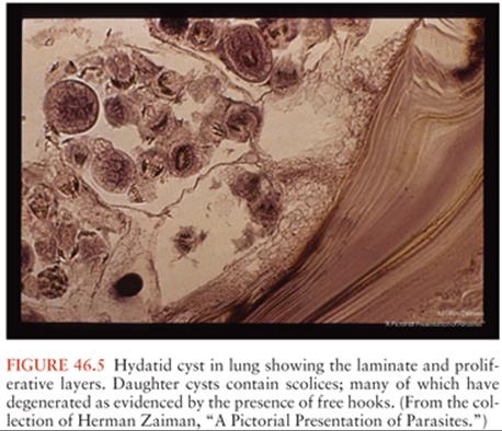
E. granulosus has two obligate mammalian hosts (136–139). The dog and other canines are the main definitive host containing the tapeworm form. The tapeworms are 2 to 5 mm in length and contain only three to four proglottids. The normal intermediate hosts are ruminants, which are infected when eating material contaminated with ova passed in the feces of the canines. In the intermediate host, the eggs hatch and release the invasive larva (termed the oncosphere), which then penetrates the intestine and migrates to the tissues, primarily the liver. The larva develops into a large cystic lesion containing an external laminar membrane, a germinal layer (the brood capsule), and a central fluid layer. Within the cyst fluid are numerous protoscolexes, which form from the brood capsule (Fig. 46.5). When ingested by the definitive host, the protoscolexes develop into tapeworms. If the cyst ruptures, however, the protoscolexes can develop into additional hydatid cysts. A number of separate life cycles have been noted for what was previously assumed to be a single species (140). However, recent molecular evidence suggests that there may be separate species infecting sheep, cattle, horses, and perhaps camels. The sheep strains appear to be most pathogenic for humans, hence the association between human disease and sheep raising. E. granulosus has a wide geographic distribution. Highly endemic areas include countries surrounding the Mediterranean Sea, the Middle East, East Africa, parts of Russia, and many countries in South America. Locally acquired cases are occasionally noted in Australia, New Zealand, North America, China, and South Asia.
E. multilocularis is mainly found in alpine and arctic zones. Foxes and wolves are the main definitive hosts. However, domestic dogs can also be infected. The normal intermediate hosts are rodents. Cases are increasing in Central Europe. Endemic areas also include Russia, parts of China, Canada, and Alaska. In contrast to E. granulosus, the parasite develops as multiple adjacent vesicles, usually without internal protoscolexes. However, the vesicles may bud and may spread as metastatic lesions.
Pathology and Pathogenesis
Humans are infected by ingesting the ova shed by dogs or other canines. After hatching in the intestine, the invasive larva (or oncosphere) is released, attaches to and penetrates the intestine, and spreads hematogenously (136–140). In the case of E. granulosus, approximately 70% of the parasites develop in the liver. Another one third develops in the lung. Less common sites include bone, pelvis, spleen, and the CNS. The echinococcal cysts slowly expand and generally remain asymptomatic until symptoms result from their expanding size or their space-occupying effect. Because years may elapse before cysts enlarge sufficiently to cause symptoms, cysts may be discovered incidentally on routine x-ray or ultrasound studies.
E. multilocularis characteristically presents as a slowly growing hepatic tumor, with progressive destruction of the liver and extension into vital structures (138,141). Patients commonly complain of right upper quadrant and epigastric pain, and obstructive jaundice may be apparent. The liver lesions metastasize to brain or lung.
Clinical Manifestations
Patients with hepatic echinococcosis who are symptomatic most often present with abdominal pain or a palpable mass in the right upper quadrant. Rupture or leakage from a hydatid cyst may produce fever, pruritus, urticaria, eosinophilia, or anaphylaxis. CNS disease typically presents as a mass lesion. In this case, the slowly enlarging lesion may cause headaches, seizures, or focal neurologic abnormalities (137,142–145).
Diagnosis
Diagnosis is primarily made by imaging studies. In the case of E. granulosus, CT or MRI scans of brain typically demonstrate a spherical cystic lesion with smooth borders (144,146) (Fig. 46.6). The cyst wall may or may not be visible. Daughter cysts within the main cyst and degenerative forms are occasionally visualized by MRI. When the internal images from the protoscolexes (or “hydatid sand”) are seen, the image can be diagnostic. Midline shift and distortion of the ventricles are common. Lesions are usually not inflamed and thus do not have surrounding edema or enhancement. Cysts may be found outside of the CNS, which suggests that the CNS lesion is also due to hydatid disease. The CT appearance of E. multilocularis is less distinct. It may present as an ill-defined mass lesion. Most cases will also have evidence of liver involvement.

In doubtful cases, serologic assays can be diagnostic. Enzyme-linked immunosorbent assay (ELISA) or indirect hemagglutination assays are readily available and can be confirmed by immunoblot assays. However, the sensitivity is not optimal for extrahepatic disease. For E. multilocularis, the EM2 ELISA with purified antigen is the confirmation test.
Treatment
The treatment of choice for CNS hydatid disease is surgical removal. In the case of E. granulosus, care must be taken not to rupture the cyst with resultant spread of the protoscolexes (Fig. 46.7). Complications can include recurrent disease and death (147,148). This is more readily accomplished if the diagnosis is made before surgery. Antiparasitic drugs, particularly albendazole, are routinely recommended before surgery in hepatic cases (138,149). Although the role of chemotherapy in CNS disease is not clear, data suggest that adjuvant chemotherapy for CNS disease is associated with an improved outcome (143). The practice of aspirating the cyst and injecting scolicidal agents is no longer recommended because of the increased risk of spillage of the protoscolexes. Drapes and potentially contaminated surfaces should be covered with hypertonic saline.

In the case of E. multilocularis, CNS involvement is often a manifestation of advanced disease (141). Few cases of alveolar hydatid disease can be cured with surgery alone, so chemotherapy is now routinely recommended postoperatively (149). In patients in whom the lesion is completely resected, albendazole is recommended for 2 years after surgery. In other patients, albendazole should be continued indefinitely. There are also reports of treatment of cerebral disease with radiosurgery plus albendazole (150).
Prevention
Peridomestic transmission of E. granulosus is linked to canine infection, which can be prevented by not feeding dogs on viscera of ruminants. People should also limit exposure to material potentially contaminated by dog feces. The vaccine has already been developed and licensed in New Zealand. E. multilocularis may be acquired from foxes or other animals. Care should be taken to avoid exposure to material potentially contaminated by feces (e.g., wearing gloves when gardening).
Sparganosis
Sparganosis, caused by Spirometra mansoni, Spirometra ranarum, and Spirometra erinaei, is a parasitic infection caused by the migratory larvae of these tapeworms. The adult tapeworm parasitizes the intestines of domestic and wild carnivores, such as dogs and cats. The intermediate hosts are usually frogs or snakes. Humans are an incidental host.
Epidemiology
Sparganosis infections occur worldwide but are most commonly reported in China, Korea, Japan, and Southeast Asia (24,151–157). Most cases have a history of eating frogs or snakes (152,153,155). Topical application of frog or snake poultices is also an important risk factor. Less than 70 cases have been reported from the United States. The most common species causing human infections in the Western Hemisphere is Spirometra mansonoides. In the Far East, the most common species is S. ranarum followed by S. mansoni and S. erinaei (24).
Parasitology
Adult tapeworms (measuring 3 to 40 cm in length) are found in the small intestines of domestic and wild carnivores (24). Eggs are shed in the animal’s feces into freshwater. Two intermediate hosts are involved in the sparganosis infection cycle. The first includes an aquatic copepod crustacean, Cyclops, where the procercoid larva develops. A second host, such as frogs, snakes, mammals, and birds, can ingest these infected copepods. The procercoid larva develops into a motile form, called the sparganum, in the second intermediate host’s GI tract. From the GI tract, the sparganum is able to attach to the mucosa, penetrate the intestinal wall, and migrate into various tissues. The life cycle of Spirometra species is completed when domestic or wild carnivores eat the infected tissues of the second intermediate host. Humans serve as accidental secondary intermediate hosts when ingesting polluted freshwater containing Cyclops; eating raw or inadequately cooked infected tissues of secondary intermediate hosts, such as frogs, fish, and snakes; or by the direct application of infected tissues to the eye or an open wound as in traditional medicines (24).
Pathogenesis and Pathology
The spargana are off-white, motile, ribbon-shaped larvae with a broad evaginated anterior end (future scolex) that can protrude and retract (24,158) (Fig. 46.8). This protruding and retracting motion helps it traverse the host’s tissues. When the larvae reach the host tissues, they will either remain coiled or can become elongated. Nodules (2 to 3 cm) are then formed in the subcutaneous tissues and superficial muscles and less commonly the abdominal cavity, perirenal fat, breast, scrotum, ureter, lymphatics, orbit, and the CNS. These nodules can be tender, inflamed, painful, or pruritic. Larva can be dissected out of fibroadipose tissue in extracted nodules. When the parasites die, a strong inflammatory reaction is elicited, leading to an abscess containing the Spirometra larvae.

This organism rarely enters the brain parenchyma, ventricular system, or spinal canal. Cerebral sparganosis usually involves the cerebral hemispheres, with occasional extension to the external and internal capsules and basal ganglia (158–162). White matter degeneration occurs, probably caused by migration of the surviving larvae along the fiber tracts. The cerebellum, brainstem, and spinal cord are less commonly involved. In the CNS, the larva is surrounded by a collagenous capsule, granulomatous layer of inflammatory cells, and the formation of new capillaries. Degenerated worms develop into punctate calcifications.
A rare and lethal form of sparganosis, termed proliferative sparganosis, has been described in Japan in which spargana proliferate in tissues and throughout the body, ultimately leading to death (24,163).
Clinical Manifestations of Central Nervous System Sparganosis
CNS involvement with sparganosis occurs in variable proportions of cases (152,153,155,159). CNS involvement may be subacute or chronic. The most common neurologic manifestations included seizures, hemiparesis, and headache (152,159). When the migrating worms penetrate the ventricles, they can lead to intraventricular hematomas and obstructive hydrocephalus. Cerebral sparganosis can result in vasculitis leading to cerebral hemorrhage or infarction.
Diagnosis
Sparganosis should be suspected in patients from endemic areas with a history of eating frogs, fish, or snakes, or characteristic findings on imaging studies (159–162). Serodiagnostic tests for sparganum-specific immunoglobulin G (IgG) in either serum or CSF can help support the diagnosis but are not widely available. An ELISA developed in Korea was reported to have a sensitivity of 80% to 85% (151). Definitive diagnosis is made by the finding of a live worm or a lesion consisting of the remains of a sparganum. Stereotactic biopsy has also been used in making the diagnosis (164).
CNS imaging by CT or MRI may show characteristic slightly hypointensity on T1 images and hyperintense on T2 (151,159–161,165). The CT triad of white matter hypodensity with adjacent ventricular dilation, irregular or nodular enhancing lesion, and small punctate calcifications is specific for cerebral sparganosis, occurring in most patients (151,160). Movement of nodules or serpiginous tubular tracts, as shown by repeated CT or MRI of the head, represents larval migration in the brain (159,165). This may occur in one third of patients. MRI is less sensitive than CT for detecting small punctate calcifications. However, MRI is more sensitive in detecting inflammatory changes, degenerated white matter, thickening of the meninges, and nodular and subcortical lesions. Intracerebral petechial hemorrhage or hematoma may also be seen. These lesions represent capillary or venous injury from the migrating worm. MRI may show linear or curvilinear contrast-enhancing lesions, conforming to the shape of the worm. Cerebral sparganosis can be confused with primary or metastatic malignancies and granulomatous diseases.
Treatment
Treatment consists of surgical removal of nodules for patients with migrating nodules or lesions or worsening of CT findings (Fig. 46.8) (151,159,164). Whether patients with nonmigrating nodules, lesions, or calcifications should undergo surgical removal of lesions is not clear. Medical therapy with praziquantel (50 mg/kg per day for 14 days) can kill the parasites and might provide benefit for patients with surgically inaccessible CNS sparganosis (141). When patients have seizures, antiepileptic drugs should be given. Whether there are any benefits to administering antiepileptic drugs to asymptomatic patients is not known.
Prevention
Individuals who live in or are traveling to endemic areas should avoid ingestion of frogs, fish, snakes, and contaminated water.
CENTRAL NERVOUS SYSTEM DISEASES CAUSED BY NEMATODES
Angiostrongyliasis
Angiostrongylus cantonensis, the rat lungworm, is responsible for most human cases of eosinophilic meningitis and meningoencephalitis. This parasite is found in many Pacific islands and in Southeast Asia, but has recently spread to Hawaii, the Caribbean, and the Gulf of Mexico (157,166–172). The mortality rate of A. cantonensis in human eosinophilic meningitis is low, but fatalities do occur. Human CNS infections with this nematode are characterized by headache, paresthesias, and CSF leukocytosis with striking eosinophilia. The related species Angiostrongylus costaricensis causes eosinophilic gastroenteritis.
Epidemiology
Angiostrongyliasis occurs worldwide. The disease was first described in rats in 1933, and the first human case was described in 1944 (166,167). Initial reports came from Southeast Asia (Thailand, Malaysia, Vietnam, Indonesia, and southern China) (166,167,173,174). Subsequent cases have been noted from the Pacific Islands, Egypt, the Caribbean, and Brazil (166,167,170,175). In the United States, angiostrongyliasis is endemic in Hawaii and has been reported along the Gulf coast (171,172). The wide geographic spread of infection is attributed to rats that are carried on ships. Increasing transcontinental travel, influx of refugees, and importation of food from Southeast Asian countries are thought to be factors. The endemic focus in the Caribbean likely represents spread from Asia (175).
Parasitology
The definitive hosts for A. cantonensis are rats (Fig. 46.9). The adult worm (2 to 3 cm long) lives and deposits eggs in the pulmonary arteries of rats and other rodents (176). The eggs reach the lung and hatch into first-stage larvae in the terminal branches of the pulmonary arteries. The larvae then migrate up the rat’s trachea to reach the pharynx. The rat swallows the larvae, allowing access to the GI tract and subsequent passage in the feces.
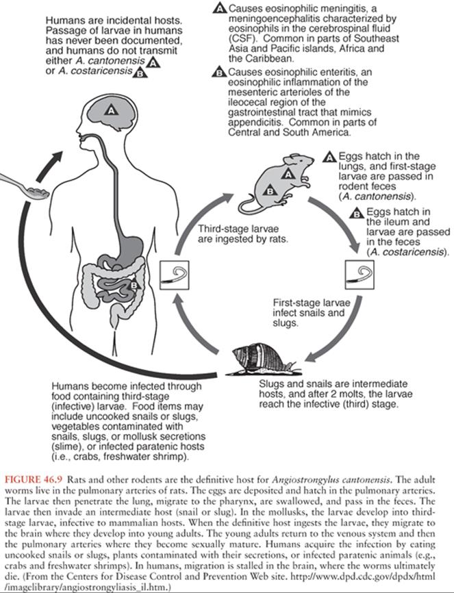
Intermediate hosts are mollusks such as freshwater snails and slugs, which may be exposed to infected rat feces. Within mollusks, larvae proliferate, molt two times, and mature into second- and third-stage larvae. Humans can be infected after ingestion of infected mollusks or paratenic hosts, such as frogs, freshwater prawns, crabs, fish, and planaria. A less common path of infection is ingestion of contaminated vegetables, water, or fruit juice containing third-stage larvae (Fig. 46.9). When the infective larva is ingested, it penetrates the intestinal wall and reaches the bloodstream. Infective larvae may die in the meningeal vessels or gain access to the CNS and eyes by tissue migration. In humans, the third-stage larvae may migrate through the brain parenchyma and molt a third time into a fourth-stage larvae where they ultimately die. In rats, but not humans, the fourth-stage larvae can again invade the venous system and return to the right side of the heart and colonize the pulmonary artery where they become sexually mature.
Pathogenesis and Pathology
Pathologic studies show relatively small worm tracks (0.1 to 2.0 mm in size) and microcavities in the brain and spinal cord; edema with dilated meningeal vessels and diffuse subarachnoid hemorrhages; leptomeningitis with perivascular inflammatory cells in white matter; and abscesses containing inflammatory cells, parasitic worms, and debris in the meninges, brain tissue, and sometimes in blood vessels or perivascular spaces (169,174,176–178). The small worm tracks and lack of tissue destruction likely explain the low mortality, clinical severity, and neurologic sequelae in patients with angiostrongyliasis.
Clinical Manifestations of Central Nervous System Angiostrongyliasis
CNS manifestations typically begin 1 to 35 days after ingestion of snails, contaminated vegetables, or exposure to infected rat urine. The symptoms can persist up to 10 weeks (166, 168–170,173,174,179–181). Nearly all patients report headache, often associated with photophobia. Most will demonstrate neck pain or nuchal rigidity, nausea or vomiting, and paresthesias or hyperesthesia. Fever, if present, tends to be modest. High fever, older age, and longer duration of fever are associated with Angiostrongylus encephalitis, which carries a poor prognosis (180). Focal findings including cranial nerve palsies, focal weakness, and hyporeflexia are rare except with encephalitis. Neurologic symptoms are related to larval migration and local host reactions. Ocular involvement may result in conjunctivitis, retinal hemorrhage, retinal detachment, or blindness.
Diagnosis
Mild peripheral blood eosinophilia is usually seen. CSF studies typically show a variable leukocytosis (median 455 cells, range 0 to 1,660), with a predominance of lymphocytes but with some eosinophils (median 16%) (170,173,181,182). Most will have increased protein concentration and a normal glucose concentration. Unlike with NCC and gnathostomiasis, which are the other causes of eosinophilic meningitis, CT or MRI scans of the brain are usually normal or lacking focal findings (with just meningeal enhancement, cerebral edema, or enlarged ventricles) (182–185). Less common findings on MRI include T1-hypointense/T2-hyperintense tracks and nodular enhancement. Definitive diagnosis made by isolating larvae from brain (at biopsy or autopsy) or the CSF is rarely possible (173,182). In most patients, the diagnosis is made by detection of specific antibody (169,170,181,186,187). An immunoblot assay is the main test used in the United States. Antibody binding to a 31-kd antigen is considered diagnostic. A variety of molecular methods are in development including polymerase chain reaction (PCR), loop-mediated isothermal amplification (LAMP), and immunoPCR (188–191).
Treatment
Angiostrongylus infections are self-limited with good prognosis and low mortality (166,169,170,173,174,181,182). Resolution of clinical symptoms takes about 1 to 2 weeks, with occasional chronic symptoms and long-term neurologic sequelae. Corticosteroids (e.g., prednisone at 60 mg per day for 2 weeks) significantly hastens symptomatic improvement (166, 169,182,192–194). Symptomatic therapy may also include analgesics and interval removal of CSF. Treatment with antiparasitic drugs has been tried, including albendazole. In several small controlled trials, there was no clear proof of benefit, but trends suggested a better response with dual therapy (166,168, 169,182,192,195).
Prevention
Travelers to endemic areas (including Southeast Asia and the Caribbean) should avoid ingestion of raw or cooked snails and unwashed raw leafy vegetables. Contact with infected rats should also be avoided in endemic areas.
Gnathostomiasis
Gnathostomiasis, most commonly caused by Gnathostoma spinigerum, is a human zoonotic disease caused by migrating immature worms (157,158,168,169,196–199). Three other species of Gnathostoma are known to cause human diseases: Gnathostoma hispidum, Gnathostoma nipponicum, and Gnathostoma doloresi. These worms are parasites of mammalian carnivores, especially dogs and cats. Humans are infected after ingestion of raw or pickled freshwater fish.
Epidemiology
Although gnathostomiasis is endemic worldwide, the most common areas include Southeast Asia, Japan, Central America, Ecuador, Peru, and Mexico (157,169,197,199–201). Neurognathostomiasis is largely confined to Thailand and Southeast Asia (197). Most cases have been associated with eating raw fish, frogs, snakes, and raw or undercooked ducks or chickens.
Parasitology
Gnathostoma worms parasitize the gastric mucosa of mammalian carnivores (pigs, cats, dogs, and wild animals) (Fig. 46.10) (157,158,168,169,196–200). The head of these worms (2 to 3 cm long) penetrates the gastric mucosa of these animals while the tails protrude into the lumen of the stomach and release eggs. The eggs then traverse the entire intestinal system and pass with feces into freshwater, where they can hatch and mature into small first-stage larva. The larvae are then ingested by freshwater crustacean copepods (genus Cyclops) where they undergo further maturation into second-stage larvae.
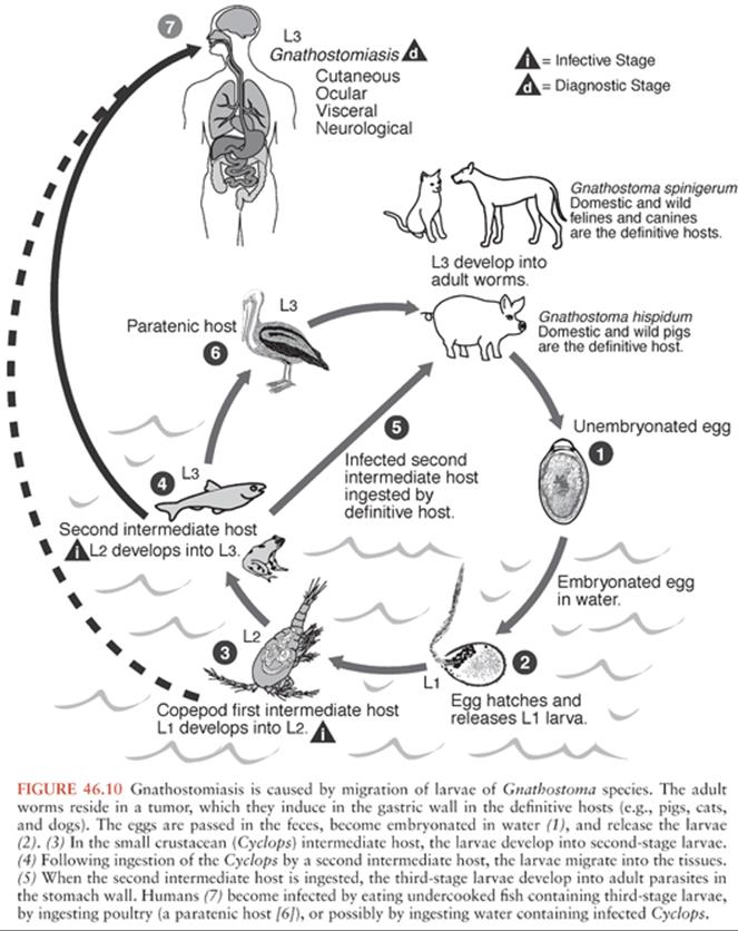
Secondary intermediate hosts such as fish, frogs, snakes, and eels ingest Cyclops containing second-stage larvae, which encyst in the host tissues and develop into third-stage larvae. When infected, secondary intermediate hosts are eaten by definitive hosts (pigs, cats, dogs, and wild animals), and larvae penetrate the host’s stomach wall and develop into adult parasites before migrating to the gut to complete their life cycle. In humans, however, larvae do not find their way back to the gastric mucosa but continue migrating through tissues and organs. Ducks, chickens, pigs, and other species that ingest secondary intermediate hosts may also develop infections with third-stage larvae in tissues and organs, which may be as infective to humans. Drinking water contaminated with second-stage larvae in Cyclops may also cause human gnathostomiasis.
Pathogenesis and Pathology
The adult worm is 1 to 2 cm long with a retractable head carrying four circumferential rows of spines (158,196,197,199). The anterior half of the worm body also carries rows of spines directed toward its tail to assist in migration through tissues. Inflammation, hemorrhage, and necrosis result along the tracks of Gnathostoma migration. Hemolysin, hyaluronidase, and acetylcholine-like substances are released in response to the larvae migration through tissues. Transient painful, pruritic, edematous, creeping eruptions, or abscesses develop in various tissues and vessels through which worms migrate.
When infected tissues containing Gnathostoma cysts are ingested, digestive enzymes from the stomach free the larvae. The larvae then penetrate the gastric mucosa and travel to the circulation of the liver and lungs before reaching skeletal muscles, subcutaneous tissues, and CNS.
Clinical Manifestations of Central Nervous System Gnathostomiasis
The invasive Gnathostoma larva readily penetrates tissues and is able to access any area of the CNS. Second-stage larvae from the gastric mucosa can invade the lumbar, thoracic, cervical, and cranial nerve roots through the spinal foramina, allowing entrance into the spinal canal and cord, causing sudden onset of excruciating radiculopathy (157,169,174,197–199). The classic features of cerebral gnathostomiasis include sudden onset of extreme severe radicular pain and/or headache followed by paralysis of the extremities and/or the cranial nerves with migration signs. The radiculopathy may resolve over 1 to 5 days as the larvae continue to migrate cephalad into the brain parenchyma where they can leave tracks of hemorrhage and necrosis. The larvae are long lived, and intermittent symptoms can occur for 10 to 15 years. Manifestations of CNS invasion included (in order of decreasing frequency) meningitis, multiple cranial nerve palsies, encephalitis, subarachnoid or intracerebral hemorrhage, transverse myelitis, radiculitis, optic nerve lesion, radiculomyelitis or radiculomyelitis terminating with encephalitis, and transient obstructive hydrocephalus (157,168,197,202). When the worm migrates along the spinal cord, hemiplegia, paraplegia, and monoplegia may occur. When the eye is involved, anterior uveitis, holes in the iris, subretinal hemorrhages, vitreous haze, and secondary glaucoma can occur (203–205).
Other symptoms experienced by infected patients with gnathostomiasis include nausea and vomiting, pruritus, urticaria, and upper abdominal discomfort or pain within 1 to 2 days of ingesting larvae. Once the larvae enter the stomach and penetrate the gastric mucosa, they can migrate to subcutaneous tissues for 1 to 2 months causing intermittent migratory erythema, pruritus, warmth, and swelling. The edema may resolve over 1 to 2 weeks but may recur at 2- to 6-week intervals characterized by marked peripheral blood eosinophilia. The characteristic skin lesion is a localized or migratory “creeping eruption” or swelling of the subcutaneous skin (cutaneous larva migrans) and various visceral organs (visceral larva migrans). Occasionally, breaks in the surface of the skin occur, facilitating removal, and diagnosis. The most commonly affected areas are the head, trunk, upper limbs, and thighs.
Up to 25% of cases with CNS involvement are fatal (168,174,197,198). Human death occurs with direct extensive involvement of vital centers of the brainstem, intracranial hemorrhage, or complications such as pneumonia or sepsis. Of survivors, two thirds have no neurologic sequelae, and the remaining third of patients had residual paresis, nerve root lesions, and cranial nerve lesions. Multiple cranial nerve palsies is a poor prognostic sign.
Diagnosis
Definitive diagnosis is made when the worm is identified in biopsies of infected tissues or by removal of the parasite from areas of broken skin. ELISA tests have been developed to look for antigen and antibody associated with Gnathostoma but are not widely available (206–208). Immunoblot assays containing specific G. spinigerum antigen have been developed to support the diagnosis of human gnathostomiasis. Patients may have marked systemic eosinophilia (168,169,197,199). Those with CNS invasion may have elevated CSF pressure (median opening pressure, 200 mm Hg [range, 90 to 350 mm Hg]), with CSF findings including a leukocytosis with a median white blood cell count of 920 cells/mm3 (110 to 3,000), eosinophilia of 54% (range, 15 to 90), elevated protein concentration at 0.80 mg/L (0.43 to 1.80), and a normal glucose level 0.51 mg/L (0.18 to 1.00) (168,169,197,199). Larvae have not been isolated in the CSF. CNS imaging studies are nonspecific but often reveal hemorrhage or myelitis (168,185,209,210).
Treatment
The treatment of symptomatic CNS gnathostomiasis is not clearly defined. Surgical removal or antiparasitic therapy has been tried in cutaneous and ocular disease (169,197,199,211, 212). Albendazole at 400 mg twice a day for 21 days or ivermectin at 0.2 mg/kg daily for 2 days is more than 93% effective in treating dermal and subcutaneous involvement with gnathostomiasis. However, relapses are common (213). No treatment studies have been done on patients with CNS involvement with gnathostomiasis, but a trial of albendazole or ivermectin might be tried in an attempt to decrease long-term sequelae. Surgical removal of larvae from the brain and eye is technically challenging. The worms are extremely small and may be difficult to find because they migrate rapidly through tissues. Patients with radiculopathy may benefit from analgesia and those with inflammation and edema may benefit from systemic steroids.
Prevention
Prevention of gnathostomiasis includes avoidance and proper cooking of foods contaminated with Gnathostoma cysts or larvae, such as fish, snakes, and poultry in endemic areas. Drinking potentially contaminated water should also be avoided.
Trichinosis
Trichinosis, classically caused by Trichinella spiralis, causes disease when humans ingest undercooked meat (especially pork) containing Trichinella cysts (214,215). Several other species of Trichinella cause human infection, including Trichinella pseudospiralis, Trichinella nativa, Trichinella nelsoni, Trichinella murreli, and Trichinella britovi.
Epidemiology
Trichinosis occurs worldwide, but is most commonly diagnosed in Europe (215–217). Since the implementation of effective sanitary and public health measures, the incidence of trichinosis has been dramatically reduced. Only a total of 66 cases of trichinosis were reported to CDC from 2002 to 2007 (218). Although trichinosis from domestic food supplies has been decreasing for many years, resurgence has occurred in cases derived from the consumption of wild game meat, especially bear. In Europe, meats of horses and wild boars have played a significant role during outbreaks (215,217,219). Trichinosis has reemerged as an important clinical problem in parts of Europe.
Parasitology
When mammals ingest infected skeletal muscles containing Trichinella cysts, gastric acid and pepsin free the larvae, which are then able to complete their life cycle by colonizing the duodenum and jejunum where they mature into adult worms. The female adult (2.2 mm long and 60 to 95 µm in diameter) and male adult worm (half the size of the female) colonize and reproduce in the intestinal crypts and mucous layer lining the duodenum and jejunum. Within the body cavity of the female adult, eggs are fertilized and develop into motile larvae (100 to 160 µm in length and 6 to 60 µm in width), which are released and are able to cross the bowel wall into the lymphatics. Each adult worm has a life span of 3 to 5 weeks. In that time span, the female adult worm is able to produce 1,000 to 5,000 larvae.
These motile larvae are able to gain access to the bloodstream, passing into the right side of the heart, the lungs, and into the arterial circulation. Larvae can cause inflammatory reactions in any organ where they encyst. However, they are only able to further develop in skeletal muscle. An eosinophilic inflammatory reaction, maturation into cysts, and subsequent dormancy for several years can occur in skeletal muscle, especially the masseter and diaphragm.
Humans acquire trichinosis by ingestion of infected undercooked pork or other meats contaminated with Trichinella cysts (215,216). Pigs are primarily infected when they ingest infected rats and other rodents. Other animals such as birds (T. pseudospiralis), wild boars, polar bears (T. nativa), dogs, walruses, cows, whales, other herbivores, and carnivores (T. nelsoni, T. britovi) become infected when they feed on infected animal flesh. Horse meat is an increasing source of infection in Europe.
Pathogenesis and Pathology
The migration of larvae in tissues elicits an eosinophilic inflammatory response (215,220,221). When skeletal muscle invasion occurs, a sarcolemma surrounds the larvae, creating an appearance of a basophilic halo around the larvae. Mononuclear cells and polymorphonuclear leukocytes are stimulated and surround the sarcolemma, creating a granuloma containing Langerhans giant cells and eosinophils. Inflammation generally subsides over 3 months as the larvae encystment is completed. These cysts may eventually calcify over a period of months to years.
Pathologic studies demonstrate few if any parasites within the brain (215,220,221). Focal areas of infarction may be seen. Punctate parenchymal hemorrhages, granulomas, and calcifications may be seen. The perivascular and Virchow-Robin space may become edematous, dilated, thrombosed, and ruptured with hemorrhage within small vessels. However, the CSF is usually without an inflammatory reaction.
Clinical Manifestations of Central Nervous System Trichinosis
Most infections with trichinosis are asymptomatic. Symptoms may develop 2 to 10 days after ingestion of infected pork or meat. The severity of symptoms is associated with the number of ingested larval parasites. Patients may present with fever, headaches, conjunctivitis, subconjunctival hemorrhages, petechiae, periorbital or facial edema, abdominal cramps, vomiting, diarrhea, peripheral edema, rashes, or myalgias (215,216,218,222,223). Once larval tissue migration begins, high fevers (38°C to 40°C) and intense myalgias can develop, especially with involvement of the extraocular and jaw muscles. The muscles are often firm and tender from inflammation and edema.
Severe manifestations of trichinosis may occur with cardiac (myocarditis), pulmonary (pneumonitis), and CNS involvement (214,222,224,225). Symptomatic CNS involvement occurs only in a small proportion of patients. Symptoms develop in the third week after infection with Trichinella. Although disease could be caused by larval invasion or encystment, most of the neurologic disease is thought to be due to vascular complications of hypereosinophilia. The neurologic findings in these patients include encephalitis, delirium, hyporeactive or absent deep tendon reflexes, findings suggesting meningitis, polyneuritis with motor weakness in the extremities, seizures, monoplegia, hemiplegia, paraplegia, cranial nerve palsies, and organic psychoses.
Diagnosis
With CNS involvement, CSF eosinophilia or larvae are found only in 8% to 28% of patients. CT or MRI scans of the brain often demonstrate cerebral infarction (162,214). They may show multiple small (a few millimeters in size), contrast-enhancing nodular or ring lesions and focal calcifications. Most patients with CNS trichinosis (90%) have marked peripheral blood eosinophilia. When muscle involvement occurs, there are elevated serum concentrations of creatine phosphokinase and lactic dehydrogenase. Antibody tests may help aid in the diagnosis of trichinosis. Indirect immunofluorescence, bentonite flocculation, IgM, IgG, and IgE ELISAs have sensitivities in the 75% to 85% range (215,226–228). These test results become positive 2 to 4 weeks after initial infection. Antigen-detection and PCR assays have also been developed but are not widely available. Definitive diagnosis is made by muscle biopsy revealing Trichinella cysts by microscopy.
Treatment
Corticosteroids can decrease the amount of inflammation and edema in patients with severe symptoms involving the CNS, cardiac, or pulmonary systems. Thus, most patients with CNS involvement should be treated with steroids (214,229–231). The antiparasitic treatment for trichinosis includes albendazole (400 mg two times daily for 8 to 14 days) or mebendazole (400 to 1,200 mg per day in two to three doses for 10 to 14 days) (214,229,230,232–234).
Prevention
Individuals should avoid ingestion of raw or undercooked meat of animals that are known hosts of Trichinella species.
Strongyloidiasis
Strongyloidiasis, caused by Strongyloides stercoralis, causes disease when infective larvae penetrate the skin. The adult female worms live in the wall of the duodenum, but larvae can reinvade the host causing cutaneous, GI, pulmonary, and neurologic disease.
Epidemiology
Strongyloides infection has a global distribution with areas of high prevalence in most developing countries as well as economically depressed areas of Europe and the United States (235,236).
Parasitology
The Strongyloides species have both free-living and parasitic life cycles. In the parasitic cycle, filariform larvae are highly motile (up to 10 cm per hour) and are able to penetrate the skin by both mechanical and enzymatic factors with collagenolytic and elastase activity. After penetrating the skin, the larvae travel into the lymphatic system, gain access to the venous system, and are transported to the pulmonary vessels (237,238). The larvae invade the alveoli, migrate up the bronchial tree, and then are swallowed. In the small bowel, the filariform larvae molt twice to become adult female worms. Hermaphroditic female adult worms colonize the intestinal wall of the duodenum and upper jejunum where they can produce 40 eggs daily. Eggs develop into rhabditiform larvae in the bowel wall, which enter the lumen. Rhabditiform larvae are passed in the stool. Most rhabditiform larvae are not infective until they pass outside the body and spend days to weeks in the soil where they transform into infective larvae. However, some of the rhabditiform larvae mature into filariform larvae before they are expelled in the feces. These invasive forms can then reinfect the host (termed autoinfection). The parasites may reinvade through the rectum or intestinal wall (internal autoinfection) or perirectal skin (external autoinfection). In hyperinfection, reinvasion may take place in the intestines (237). Because of this efficient life cycle, S. stercoralis infection can persist within a human host for decades after leaving an endemic area (239,240).
In the free-living cycle, the rhabditiform larvae mature in moist soil into filariform infective larvae. Then, after molting two to four times, they become free-living adult males and females. The free-living adults mate and produce eggs from which rhabditiform and then filariform larvae develop.
Pathogenesis and Pathology
Chronic strongyloidiasis occurs because of autoinfection and can last for many years. This process is usually controlled with limited numbers of organisms produced. However, autoinfection can also lead to a high parasite burden, termed the hyperinfection syndrome (238,241–243). Hyperinfection is most commonly associated with corticosteroid treatment. Hyperinfection is also associated with underlying diseases, especially human T lymphotrophic virus type 1 (HTLV-1) infection, but also renal failure, alcoholism, malnutrition, and chemotherapy. Hyperinfection likely results from defective host antibody, eosinophil, and neutrophil responses (235,244). The high burden of larvae in the hyperinfection syndrome leads to dissemination with multisystem involvement including the intestines, lungs, CNS, liver, peritoneal cavity, and adrenals (237,238,241,242,245,246). Mobile larvae are able to carry enteric bacteria during migration, and polymicrobial bacteremia or meningitis can ensue (241,247). Intestinal stasis, achlorhydria, and constipation may also predispose the patient to dissemination of Strongyloides.
Clinical Manifestations of Central Nervous System Strongyloidiasis
Most Strongyloides infections are asymptomatic or associated with only minor GI or dermatologic symptoms (248,249). Dermatologic findings include larva currens, a rapidly moving intermittent serpiginous, itchy skin eruption, and chronic urticaria commonly found on the buttock and waist areas. GI symptoms include nausea, bloating, pain, diarrhea, and malabsorption. Disseminated Strongyloides should be suspected in patients with fever, abdominal pain and distention, cough, diffuse pneumonia, diarrhea, or polymicrobial bacteremia. Disseminated strongyloidiasis may cause shock, pulmonary and neurologic complications, and sepsis.
Cerebral strongyloidiasis occurs either by direct larval invasion or by larval-induced enteric bacteremia with subsequent pyogenic meningitis. CNS involvement by infective larvae may also cause small blood vessel infarction with associated inflammatory infiltrates, granulomatous ependymitis, brain edema, demyelinization, microvacuolation, and focal hemorrhage with necrosis (242,245,247,250). Patients present with acute or subacute meningitis, abscess, cerebritis, cerebral vasculitis, or chronic meningoencephalitis. CNS findings include headache, altered mental status, meningismus, focal or generalized seizures, cranial nerve palsies, and motor weakness. The most common presentation is bacterial meningitis, most commonly due to enteric organisms, including gram-negative organisms and streptococci (247).
Diagnosis
During the chronic stages of strongyloidiasis, peripheral eosinophilia is common. However, eosinophilia is unusual in the hyperinfection syndrome. Cerebral abscess may be visualized on CT scans of the brain (251), but imaging studies are not typically abnormal and are not diagnostic. Definitive diagnosis depends on identification of larvae (rhabditiform and occasionally filariform). These should be sought by microscopic examination of stool, sputum (disseminated strongyloidiasis), and duodenal fluid via aspiration or an enteric string test (252–254). Examination of serial samples increases the yield of identifying larvae. Occasionally, larvae can be identified from bacterial cultures, in which tracks of bacterial colonies trace the larval migration on culture plates. Larvae are occasionally found in CSF and in the brain and meninges from biopsies or autopsy (255). Serologic assays for IgG antibodies to Strongyloides are available, and an enzyme immunoassay is available from the CDC (256).
Treatment
Strongyloidiasis has traditionally been treated with thiabendazole (25 mg/kg orally twice daily). This regimen has been used for patients with CNS Strongyloides infection (242,246). Treatment must be continued until consecutive stool samples are free of larvae for several days (238). Ivermectin (200 µg/kg per day given on 2 consecutive days and repeated in 2 weeks in compromised hosts) is now the treatment of choice for infection outside of the CNS (257–260). Efficacy is similar to thiabendazole with fewer side effects. However, there are few data on its use in CNS infection. Albendazole is a less effective alternative therapy. Ideally, patients taking immunosuppressive drugs should discontinue the agent or reduce the dose as much as possible.
Prevention
Those exposed to soil in endemic areas should wear protective footwear. Those who are living in endemic areas or who have traveled to such areas should have the possibility of strongyloidiasis evaluated before initiation of immunosuppressive therapy.
Toxocariasis
Toxocara species are intestinal ascarid nematodes of dogs and cats. Toxocariasis is a common and important cause of larva migrans syndrome worldwide, especially in developing countries (261). Toxocarawas first described in 1952 as a parasite that caused hepatomegaly and eosinophilia in children. Toxocara canis and Toxocara cati live as adult worms in the intestines of dogs and cats, respectively. Most human infections are caused by T. canis. More than 90% of puppies are infected within the first week of life. This highly adapted nematode infects dogs by transplacental and transmammary migration of larvae from bitch to puppy. Dogs are also infected by the oral route with ingestion of larvae in vomitus or of embryonated eggs in contaminated soil. These routes are also similar for larvae in infected cats, except no placental transfer of larvae has been observed. Toxocara eggs can be found in up to 30% of soil samples (262). The egg is the infective stage for humans. Once the egg is ingested and hatches, the larva (350 to 450 µm by 16 to 20 µm) can migrate through various tissues.
Epidemiology
More than half of all U.S. households have dogs or cats. The intimate association between children, especially 18 months to 3 years of age, and dogs and cats leads to environmental contamination with infective eggs and human infections with Toxocara species (261–263). Most infected children with toxocariasis have had a dog in the house or yard within 1 year of developing illness. Pica is an important risk factor in children 1 to 6 years of age (264). The seroprevalence by ELISA varies by age and by region. Seroprevalence of Toxocara antibody is much higher in young children than older individuals (265). Serosurvey results suggest widespread infection, with 6.4% of individuals seropositive in the United States, 3.6% in Japan, 51% in Taiwan, and 83% in Saint Lucia (263). Many new infections are asymptomatic, which suggests that Toxocara larva migrans is underrecognized and underreported.
Parasitology
Toxocara species are large (8 to 18 cm) and heavy-bodied nematodes. The adult stage of these parasites resides in the host’s small intestine, with a life span averaging 4 months. The female Toxocara are able to produce 200,000 eggs per day. The fertilized eggs require a minimum of 14 to 21 days to develop into larvae. However, most larvae can remain dormant for months to years in humid soil. The larvae are unable to survive in direct sunlight and dry soil.
Pathogenesis and Pathology
Humans are infected when they ingest Toxocara eggs or infective-stage larvae in contaminated soil. Consumption of raw or undercooked meat of paratenic animals can also result in transmission (261,266). Raw liver seems to be particularly infective in this setting (266,267). The disease produced in humans by Toxocara is known as visceral larva migrans (VLM). This term was used to describe the prolonged migration and persistence of larvae in infected hosts. The eggs of this nematode hatch and produce larvae in the proximal small intestine. The larvae then penetrate the small intestinal mucosa and migrate to various tissues, most commonly the liver and lung, then into the systemic circulation. Other sites infected include the eye, which can cause blindness known as ocular larva migrans (OLM) (268–270), heart, and the brain (271–273).
The migration of larvae to various tissues causes pathologic and immunologic responses eliciting granulomatous inflammation with associated eosinophilia (272,273). The rapid migration of the larvae of Toxocara through tissue causes tissue necrosis along the route with minimal inflammation (272). The larvae are able to survive and protect themselves from the host’s immunologic response by producing and shedding surface excretory-secretory antigens. Once migration stops, necrosis and hemorrhage develop in the surrounding tissues with infiltration of various inflammatory cells such as polymorphonuclear leukocytes, eosinophils, histiocytes, and lymphocytes. Collagenous capsules are formed around the larvae and larvae tracks by 1 month. Chronic infections with Toxocara cause infiltration of cells, leading to a delayed hypersensitivity reaction to larval proteins. A granuloma with a core of multinucleated cells and leukocytes is then formed.
Granulomatous responses limiting larval migration may be less likely to occur in the CNS as compared to other sites (272). Similar to migration through other tissues, larvae leave tracks of necrosis with infiltrates of inflammatory cells when infecting the brain. In contrast to other sites of infection, however, the larvae continue to accumulate progressively in the brain, likely because of decreased inflammatory and other systemic immunologic responses from the host (274). Few pathologic descriptions of CNS Toxocara infections have been described (272), but case reports describe live larvae in the biopsy of the infected brain.
Clinical Manifestations of Central Nervous System Toxocariasis
Most infections with Toxocara are asymptomatic (272,273,275). The symptoms of VLM are dependent on the numbers of eggs or larvae ingested, frequency of reinfection, which tissues are infected, and the host inflammatory and immunologic responses. VLM, most often infecting children at the average age of 2 years, is manifested by symptoms of fever, sore throat, cough, wheezing, cervical adenitis, lymphadenopathy, pulmonary infiltrate or pneumonia, abdominal pain, anorexia, nausea, vomiting, hepatomegaly, limb pains, lethargy, sleep, and behavior disturbances (272).
The average age of patients with OLM is older (8 years) (269,270). Disease typically involves only one eye. Patients can develop visual loss, strabismus, and eye pain as larvae can cause severe exudative endophthalmitis with retinal detachment, posterior and peripheral retinochoroiditis, optic papillitis, and uveitis (269,270). Funduscopic examination may reveal raised, unilateral, whitish or gray solitary posterior pole or peripheral granulomatous lesions.
OLM is associated with lower numbers of ingested Toxocara, whereas VLM is associated with an increased number of ingested larvae (261). When the number of ingested larvae is increased even further, OLM can develop concurrently with VLM. Concurrent infection is associated with more severe symptoms. Toxocara serum antibody titers are lower in individuals with OLM compared to those with VLM. Neurologic manifestations of toxocariasis have been reported in up to 28% of infected patients. Symptoms and signs include encephalopathy, meningoencephalitis, transverse myelitis, psychiatric disturbances, focal or generalized seizures, epilepsy, and death (272). Several studies have identified an association of antibody to T. canis and seizures, particularly focal seizures (262,264,276,277). At present, however, whether this association is causal or merely a marker for another infection spread by the oral route is not clear.
Diagnosis
Patients with marked or persistent eosinophilia should be evaluated for VLM and OLM by specific serologic tests. Some cases may not develop marked eosinophilia. Imaging studies by ultrasound, CT, or MRI may show the migratory tracks or granulomas of Toxocara larvae in various tissues, but they are usually normal.
In a patient with clinical signs and a history of geophagia and exposure to dogs or cats, it may be helpful to order Toxocara serologic tests. The most useful serologic test uses excretory-secretory antigens from infective-stage larvae in an ELISA (278,279). Although an elevated ELISA titer in an infected patient supports the diagnosis of Toxocara VLM and OLM, definitive diagnosis relies on identifying characteristic larvae in biopsies of the infected tissue. A positive Toxocara ELISA titer does not distinguish between a recent and prior infection because titers may be elevated for years after initial infection. Seroprevalence studies have shown that 1% to 10% of asymptomatic children may have positive ELISA titers to Toxocara (263). Although nonspecific, other clinical and laboratory findings may include leukocytosis, hypergammaglobulinemia, and blood group isohemagglutinins in individuals with VLM. MRI abnormalities, including T2-hyperintense lesions primarily in the white matter and basilar enhancement, have also been reported in CNS disease (280–282). In cases of spinal cord involvement, MRI may reveal nontumorous myelopathy mimicking transverse myelitis, single or multiple T2-hyperintense lesions with focal nodular enhancement on posterior or posterolateral segments of the spinal cord, short segmental involvement, and migration of lesions (283).
Treatment
Asymptomatic toxocariasis does not require anthelminthic treatment. Most of these patients will have spontaneous resolution of their eosinophilia and ELISA titers will decline without adverse sequelae (284). In patients with symptomatic toxocariasis, the treatment is primarily supportive. To suppress the inflammatory response in ocular, pulmonary, myocardial, or CNS involvement, systemic corticosteroid therapy (e.g., prednisone, 30 to 60 mg per day by mouth for 2 to 4 weeks) and intraocular corticosteroids with eye involvement may be warranted (273,277,285). The indications for use of anthelminthic therapy have not been clearly defined (273,275,285). Albendazole (15 mg/kg per day orally for 5 to 15 days) is the treatment of choice. Mebendazole (20 to 25 mg/kg per day orally), diethylcarbamazine (50 to 150 mg by mouth three times a day for 1 to 3 weeks), and thiabendazole (25 to 50 mg/kg per day by mouth for 1 to 3 weeks) have also been used (285). In OLM, anthelminthic drugs have not been shown to have any benefits in addition to corticosteroid therapy. Laser photocoagulation has been shown to be an effective way of destroying migrating larva detected by funduscopic examination. Chronic (>8 weeks) OLM requires corticosteroids for the treatment of exacerbations, relapse, and progression of ocular disease. Surgical intervention such as pars plana vitrectomy and scleral buckling are common ophthalmologic procedures used to manage intraocular fibrous adhesions, retinal traction and detachment, retrolental plaques, and chronic vitreal inflammation.
Prevention
Increased education, timely deworming of pets, emphasis on careful personal hygiene in pet owners, preventing children from playing in and eating contaminated soil, and avoiding ingestion of raw meat and liver of animals will minimize the development of Toxocara infections.
Baylisascaris procyonis
The raccoon ascarid Baylisascaris procyonis has recently been recognized as an important cause of visceral, ocular, and neural larva migrans syndromes (NLM) in the United States (286–289). Infection usually occurs in children or mentally retarded adults with variable onset of neurologic deterioration. Exposure occurs mainly at raccoon latrines, where large numbers of infective eggs may be accidentally ingested. CT and MRI scans often reveal atrophy and may reveal periventricular and deep white matter changes (287,290,291). Definitive diagnosis requires demonstration of the parasite in biopsy material. Serologic tests have also been described. Treatment with corticosteroids and albendazole has been attempted with some improvement. However, long-term outcomes have generally been poor.
Pathogenesis and Pathology
Humans are accidental intermediate hosts for B. procyonis. Infection follows the ingestion of B. procyonis eggs containing infective second-stage larva. B. procyonis undergo aggressive tissue migration that includes the CNS and eyes. Continued larval growth and migration following entry of even a few B. procyonis larvae into the CNS may have potentially severe consequences. Animal and limited human autopsy data suggest that B. procyonis hatch in the small intestine, penetrate the bowel mucosa, and pass presumably via the portal circulation, through the liver and along vascular channels to the lungs (292,293). In the lungs, B. procyonis larvae rupture pulmonary capillaries, enter pulmonary veins, return to the left side of the heart, and gain access to the systemic circulation. Larvae most likely access the brain by penetrating cerebral blood vessels. On autopsies, the brain was the most severely affected organ. Massive larval invasion of the CNS is characteristic, with estimates of more than 3,200 larvae being isolated from the brain of a single case (292). Macroscopic findings included marked swelling and softening of the brain, leptomeningeal congestion and thickening, and evidence of cerebellar herniation. Histologic findings for acute fatal cases demonstrate necrosis and inflammation with numerous macrophages, eosinophils, lymphocytes, and occasional plasma cells concentrated in cerebral periventricular white matter and leptomeninges. Large numbers of eosinophils and eosinophilic granules and deposits of extracellular eosinophilic material (the Splendore-Hoeppli phenomenon) are present around necrotic migration tracks and cerebral blood vessels (292,293). Immunofluorescence studies suggest that eosinophilic material surrounding migration tracks, larvae, and perivascular area consists of eosinophil major basic protein arising from eosinophil degranulation (294). Subacute cases of NLM demonstrate well-defined granulomata, composed of relatively few intact eosinophils surrounded by a chronic fibrotic reaction, and the absence of the acute inflammatory reaction (293).
Clinical Manifestations of Central Nervous System Baylisascaris
The majority of human patients with B. procyonis neural larva migrans present with an acute fulminant eosinophilic meningoencephalitis (286,288,292,293,295). Early features include low-grade fever, ataxia, increasing lethargy, somnolence, and periods of increased irritability. Over time, there is regression and loss of developmental milestones, progression to extensor posturing, increasing spasticity with hemi- or quadriparesis, and ocular or cranial nerve involvement. Seizures occur commonly and may be difficult to control. Neurologic status may deteriorate rapidly to stupor, coma, and death. To date, all survivors have been left in a persistent vegetative state or with severe residual deficits. Most infants and children with clinical visceral larva migrans and neural larva migrans also have evidence of ocular disease. Visual impairment or blindness results from widespread larval migration, with destruction of the visual cortex, or from larval migration within the eye itself (293,296).
Diagnosis
In the absence of a brain biopsy, the diagnosis of B. procyonis NLM is dependent on serology. Demonstration of anti-B. procyonis antibodies in serum and CSF, particularly in the setting of a compatible clinical case and epidemiologic history, is the mainstay of diagnosis. ELISA using larval excretory-secretory antigens is available in United States (291). A diagnosis of ocular baylisascariasis is supported by characteristic chorioretinal lesions, and especially by observation of a larva in the eye (286,296).
Treatment
The prognosis is grave with or without treatment. The majority of cases have been treated with anthelminthics and corticosteroids without intact survivors. The role of prophylactic anthelminthic treatment for asymptomatic children is unclear but could be potentially beneficial (297). Given the potentially devastating sequela of untreated infection or late treatment and the availability of well-tolerated anthelminthics, prophylaxis appears warranted in select cases with documented exposure to infected raccoons, raccoon feces, or contaminated environments (291,297). Systemic corticosteroids could be beneficial and have been used in the majority of B. procyonis NLM cases.
Prevention
Most cases of B. procyonis infection are preventable by relatively simple measures. Education of the public regarding the potential dangers of contact with raccoons or their feces is the most important preventive step. Young infants and toddlers, particularly those with pica or geophagia, should be kept away from potentially contaminated areas.
Lagochilascariasis
Lagochilascaris minor and other species are found in opossums, raccoons, ocelots, dogs, and cats. Cases of human infection include abscesses of the neck, tonsils, mastoid, nasal sinus, and lesions of the lung, cervical and sacral spine, and subcutaneous tissues (298). Lagochilascariasis has rarely been reported in Brazil and other countries in Latin America. Lagochilascariasis is most commonly seen in individuals who live in or close to forested areas and in rural areas and individuals of the lowest socioeconomic status.
Parasitology, Pathogenesis, and Pathology
Little is known about the life cycle of Lagochilascaris. Wild rodents can act as intermediate hosts or paratenic hosts of Lagochilascaris minor. An autoinfecting cycle may occur in felines, which also serve as definitive hosts. Humans are infected when they ingest infected rodents (299).
Clinical Manifestations of Central Nervous System Lagochilascariasis
The clinical manifestation of Lagochilascaris may vary from mild to severe symptoms. L. minor lesions in animals and humans characteristically result in tumors and fistulas with cutaneous and subcutaneous abscesses localized in the cervical region and surrounding tissues (300). Cases have reported parasite lesions of mastoids, jaw, tonsils, maxillary and paranasal sinuses, middle ear, pharynx, deep pharyngeal space, ocular globe, and meninges (300,301). One report of a fatal case of encephalopathy with lagochilascariasis minor has been described (298). The patient had signs of headache and meningismus. His physical examination was significant for papilledema, hemiparesis, and mental deterioration, which progressed to coma then death. Laboratory findings showed mild eosinophilia, increased erythrocyte sedimentation rate, and increased protein concentration in CSF. A later lumbar puncture showed progressive increases in CSF white and red blood cell counts. CT scan of the brain showed areas of hemorrhage and infarction. Numerous larvae and adult worms were identified in the brain at autopsy.
Diagnosis
Definitive diagnosis of human infection is made by biopsy and findings of adult worms in the involved tissues.
Treatment
There is no proven therapy for lagochilascariasis. Two case reports described successful treatment with ivermectin (302,303). Thiabendazole, mebendazole, and levamisole have been tried with varying success (300,301,304–306).
Prevention
Prevention likely consists of careful observation of proper hygiene, including hand washing and use of clean water and sanitation.
CENTRAL NERVOUS SYSTEM DISEASES CAUSED BY TREMATODES
Schistosomiasis
Schistosomiasis, depending on the region of the world, is caused by Schistosoma japonicum, Schistosoma mansoni, and Schistosoma hematobium. Two minor species (Schistosoma mekongi and Schistosoma intercalatum) can also cause human infection in circumscribed foci in Southeast Asia and Africa, respectively. Human contact with water is necessary for infection by schistosomes.
Epidemiology
Schistosomiasis affects more than 200 million people worldwide (307,308). S. japonicum is found mainly in China, the Philippines, and Southeast Asia. S. mansoni is found in Africa, Southwest Asia, the Caribbean, the Middle East, and part of South America. S. hematobium is found in Africa, the Middle East, and Southwest Asia. No nonhuman reservoirs have been found for S. mansoni and S. hematobium, although these species are rarely found in other hosts. Domestic animals are important reservoirs for S. japonicum.
Parasitology
Humans are the definitive hosts. When the parasite eggs are passed into freshwater, they can release ciliated miracidia, which swim and penetrate freshwater snails (307,308). Once inside the snail, the miracidium is able to mature into a mother sporocyst, which can produce motile daughter sporocysts that target the snail’s hepatic and gonadal tissues. The daughter sporocysts develop into cercariae within the snail’s hepatic and gonadal tissues, migrate to the vascular sinuses, and exit through the mantle. Humans, particularly children and adolescents, become infected when released motile cercariae attach to and penetrate skin. The cercariae shed their forked tail and develop into the schistosomula, which migrates to the heart, lung, and the liver. From the liver, each Schistosoma species is able to migrate to its preferred sites and to its residence in venules. Nevertheless, the parasites occasionally migrate to other sites.
The adult worms of Schistosoma live in the human vasculature. S. mansoni lives in the inferior mesenteric veins draining the large intestine, S. hematobium lives in the venous plexus surrounding the bladder and in the rectal venules, and S. japonicum lives in the superior mesenteric veins draining the small intestine. After the adult worms mate in the blood vessels, the female worm (7 to 20 mm) travels against the flow of the blood to reach the vessels surrounding the intestine or bladder. There, she releases hundreds to thousands of eggs in the small venules of the portal (S. mansoni and S. japonicum) and periportal (S. hematobium) systems. The eggs may attach to the venule endothelium and migrate through tissues to reach the intestine or bladder. Both eggs in the small venules and those attached to the endothelium elicit a granulomatous inflammatory reaction, which facilitates migration of the eggs into the lumen and excretion in feces or urine.
Pathogenesis and Pathology
Early in the course of infection, adult worms may localize in the spinal cord vasculature or cerebral vessels (307–309). More commonly and later in the course of infection, CNS schistosomiasis results from ectopic deposition of the parasite eggs. In heavy infection, the ova are able to reach the CNS by retrograde flow from the iliac veins and inferior vena cava by the valveless venous plexus of Batson (307,308). Eggs reach the brain and spinal cord by embolization from this vertebral venous plexus. Cerebral schistosomiasis is thought to be more common with S. japonicum because S. japonicum releases small eggs, which are able to reach the brain. The eggs of S. mansoni are larger, containing a lateral spine, and are found more commonly in the spinal cord. The eggs of S. hematobium are of intermediate size and are found more commonly in the brain parenchyma compared to S. mansoni but less commonly than S. japonicum. The different schistosomal species affect different regions of the CNS. S. japonicum affects the brain, and S. hematobium and S. mansoni more commonly affect the spinal cord (307,308).
Schistosome eggs have been identified in leptomeninges; parietal, occipital, and temporal lobes; basal ganglia; hippocampus; brainstem; cerebellum; choroid plexus; and spinal cord. Eggs that localize to the brain or spinal cord may cause inflammation with perivascular infiltration of lymphocytes, eosinophils, and macrophages, focal and diffuse vasculitis, microinfarction, or granuloma formation (307,308,310). Granulomas are less commonly seen in the brain and in chronic infection. This may reflect modulation of the granulomatous response in late infection.
Clinical Manifestations
Schistosome infection consists of three phases (307,308). The first stage is cercarial dermatitis, where cercariae penetrate the skin eliciting an acute inflammatory reaction. This may result in a transient rash at the site of penetration. The second stage is termed acute schistosomiasis (or Katayama fever). It begins at the time the adult worms begin to lay eggs, 2 to 8 weeks after the initial infection. Acute disease is now recognized with all Schistosoma species. This stage is due to immune complexes formed in reaction to schistosomal antigens. Clinical manifestations include fever, abdominal pain, diarrhea, hepatosplenomegaly, muscle pains, and urticaria with peripheral blood eosinophilia. The third stage is chronic schistosomiasis, which may involve many organ systems including the hepatosplenic, intestinal, urinary, cardiopulmonary, and CNS. All species except S. hematobium may cause colonic polyposis and bloody diarrhea. S. hematobium is associated with cystitis and ureteritis with hematuria, which can progress to bladder cancer. S. mansoni, S japonicum, and S. mekongi are associated with portal hypertension with hematemesis, splenomegaly, and pulmonary hypertension. The chronic states occur because of the inflammatory, granulomatous, and fibrotic reactions to antigens in schistosome eggs.
Involvement of the CNS occurs in about 1% to 2% of patients, but most patients do not have neurologic symptoms (307–309). Neurologic symptoms include mental confusion, meningitis, encephalitis, headache, vertigo, seizures, coma, visual changes, optic neuritis, papilledema, hemiplegia, opisthotonos, and tremors. Schistosomal myelopathy, characterized by lumbar pain (often radicular), followed by muscle weakness, sensory deficits, and loss of sphincter control, most commonly involves the conus medullaris from intramedullary granuloma. Other spinal cord involvement includes multiple nodules on the spinal cord, cord compression, and cord necrosis and less likely involvement of the cauda equina and thoracic portion of the spinal cord. Involvement of the spinal cord can cause flaccid paraplegia, sphincter dysfunction, areflexia, spasticity, sensory deficit, radiculopathy, back pain, and transverse myelitis (310,311). Additional CNS syndromes include cerebellar and vestibular syndromes, tumor-like mass lesions, cerebral edema, and intracerebral and subarachnoid hemorrhage.
Diagnosis
Peripheral blood eosinophilia may occur, but other general laboratory studies are not particularly helpful. The CSF may show pleocytosis typically with lymphocyte predominance. Eosinophils are present only in a few patients. Protein concentrations are increased, glucose concentrations are normal, and ICP may be increased (307,308). CSF from patients with schistosomal myelopathy may be xanthochromic.
CT and MRI scans of the brain may show cerebral edema or atrophy. Many patients present with large granulomas, which can cause focal lucencies, enhancing lesions, tumor-like lesions, or intracerebral hematomas (307–309,312). Some patients will display a characteristic central linear enhancement surrounded by multiple enhancing punctate nodules (313,314) (Fig. 46.11). This treelike pattern is thought to be highly suggestive of schistosomiasis. Myelography with CT may show schistosomiasis involvement with intramedullary cord swelling and partial or complete spinal cord block (310,311,315). MRI scan may reveal a nodular surface to the lesion (315).
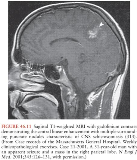
Definitive diagnosis is made by identification of the characteristic ova in stool (S. mansoni and S. japonicum) or urine (S. hematobium) (316). Because eggs are intermittently passed, repeated examinations increase the yield of detection. Centrifugation and examination of urine sediment can also aid in isolating eggs. Rectal biopsy may be a more sensitive way to detect the ova. Ova are not found in the CSF. Although demonstration of ova in biopsies is possible, it is preferable to make the diagnosis with less invasive tests. Serologic tests using egg or other parasite-derived antigens are available to support the diagnosis of schistosomiasis. An ELISA using adult worm microsomal antigens is available at the CDC and is more than 99% specific for Schistosoma infection and more than 95% sensitive for S. mansoniand S. haematobium (317,318). However, the sensitivity is poor for S. japonicum infection. Species-specific immunoblot assays are also available using adult microsomal antigens. Antigen-detection assays have also been developed but are not readily available in the United States (319).
Treatment
Medical therapy for cerebral schistosomiasis of all species is with praziquantel (60 mg/kg orally in three divided doses over a single day) (320). Patients who have schistosomiasis myelopathy can be treated at a lower dose of praziquantel (40 mg/kg in two divided doses over 1 day) (307,308,321). Although no data are available, corticosteroids are often given to reduce the edema associated with myelopathy and cerebral lesions.
Surgical therapy is an important adjunctive therapy in patients who have tumor-like masses (322). Laminectomy has been used in some patients with schistosomal myelopathy and still may be required in the case of acute paraplegia. With recent improvement in prompt diagnosis plus treatment with corticosteroids and praziquantel, surgical therapy is now less frequently required (307,308,321).
Prevention
Prevention of schistosomiasis includes avoiding contact with freshwater in endemic regions. Cercaria can be eliminated by chlorination or allowing water to stand for at least 24 hours before contact.
Paragonimiasis
Paragonimiasis is caused by trematode parasites of the genus Paragonimus. Initially, most cases were attributed to the species, Paragonimus westermani (307,308,321), which is also called the oriental lung fluke. However, recent molecular data have demonstrated that there are over 40 species in the genus and at least 8 Paragonimus species cause human infection (323–326).
Epidemiology
The first case of cerebral paragonimiasis was reported in Japan in 1887 in a man who had a chronic cough and hemoptysis and developed seizures that subsequently led to coma and death. Postmortem examination revealed adult flukes in cystic lesions in the right frontal and occipital lobes. Paragonimiasis is endemic in Southeast Asia, Korea, Japan, and China. Other foci include Africa (Cameroon and Nigeria), the west coasts of Central and South America, and central North America.
Parasitology
P. westermani causes classical pulmonary disease in China and Korea. A second species, P. skrjabina is associated with extrapulmonary disease including cerebral infections. P. heterophyes is the main species in Southeast Asia and India. P. africanus is endemic in Cameroon and Nigeria. In Latin America, Paragonimus mexicanus causes both classical and atypical infections. Paragonimus kellicotti has been associated with human infections in the United States. The reservoirs for paragonimiasis are tigers, other felines, pigs, dogs, foxes, opossums, humans, and other mammals. Adult worms reside in cysts of the host’s lungs. The hermaphroditic adults produce eggs, which are coughed up and excreted unembryonated in the sputum or the feces. When the eggs reach freshwater, they become embryonated and release miracidia. The miracidia penetrate snails, which serve as first intermediate hosts, and then develop into cercariae. The cercariae are released into water and are then ingested by freshwater crayfish and crabs where they encyst and become metacercariae. Humans and other animals are then infected when freshwater crayfish and crabs containing metacercariae are ingested. The metacercariae excyst in the small intestines. The larvae penetrate the bowel wall, migrate into the peritoneal cavity, through the diaphragm, and into the lungs.
Pathogenesis and Pathology
The most common extrapulmonary site of paragonimiasis is the brain. The adult worm (7 to 12 mm by 4 to 6 mm) reaches the CNS through the jugular foramen and causes arachnoiditis, granulomas, or abscesses (327,328). Lesions are most common in the temporal and occipital lobes and less commonly in the frontal and parietal lobes. Abscess cavities, containing giant cell granulomas and Charcot-Leyden crystals, are often formed. The wall of the cavity is composed of highly vascular glial mesenchymal capsules with numerous inflammatory cells.
Clinical Manifestations of Paragonimiasis
Most patients with paragonimiasis are asymptomatic or have mild symptoms. Pulmonary symptoms include chronic cough with rusty sputum, intermittent hemoptysis, or pleurisy. Extrapulmonary symptoms include abdominal pain, diarrhea, fever, urticaria, and hepatosplenomegaly.
The frequency of cerebral disease varies markedly by species, from less than 1% for most species to 31% of P. skrjabini cases (328). The main clinical syndromes for cerebral paragonimiasis are epilepsy, hemiplegia or other mass lesion, aphasia, and headache (327). The most common neurologic symptom with paragonimiasis is epilepsy, which occurs in up to 80% of patients with neurologic disease. Some patients have symptoms of meningitis, but the symptoms are usually mild and self-limiting, but they may be recurrent and chronic. Chronic infections may persist for 20 years. Basilar meningitis can involve the optic nerves, causing visual disturbances and optic atrophy. Therefore, it is important to perform an ophthalmologic examination.
Diagnosis
General laboratory tests are usually not helpful, although patients often have peripheral blood eosinophilia. With pulmonary involvement, 70% may have multiple nodular lesions, fibrosis, cavities, or calcifications by chest x-ray. With CNS involvement, lumbar puncture usually reveals CSF pleocytosis with increased eosinophils, low glucose concentration (50%), and increased protein concentration. Skull roentgenograms may reveal intracranial calcium deposits, characteristically described as “soap bubble” calcifications (157,162). CT scans of the brain may show soap bubble calcification, cerebral atrophy, solitary ringlike lesions or irregular enhancing lesions, localized hemorrhage with or without enhancing lesions and poorly defined nonhemorrhagic nonenhancing lesions, and ventricular dilation. The most characteristic imaging finding is a conglomerate of ring-enhancing grapelike cysts with surrounding edema in one cerebral hemisphere that may be evident on CT or MRI scan. MRI scans may also show edema and inflammatory changes.
Definitive diagnosis requires demonstration of the parasites or their ova. With pulmonary disease, ova are often present in sputum or stool. However, ova are not typically excreted in isolated CNS infection. Ova or parasites may be identified in operative specimens from CNS lesions.
Serologic tests are an important adjunct to diagnosis. Although serum antibody may not distinguish acute from past infection in endemic areas, positive serologic test results are predictive of disease in a low prevalence area. ELISA and immunoblot assays are available from the CDC. Reaction with an 8-kd band by immunoblot is 96% sensitive and more than 99% specific for paragonimiasis (283). Complement fixation tests and skin tests are available in some countries. Antigen-detection assays are also being developed.
Treatment
Treatment of cerebral paragonimiasis may involve surgical or medical therapy. Hydrocephalus requires surgical removal of cysts and relief of the hydrocephalus (327). Medical therapy with praziquantel (25 mg/kg three times per day for 2 to 3 days) is highly effective in pulmonary disease (327). However, response rates in neurologic infections are only available from retrospective studies. Corticosteroids may be added to praziquantel to reduce the inflammatory changes occurring with treatment. Acute disease responds well to treatment. In contrast, the prognosis of chronic CNS disease, even with treatment, is poorer.
Prevention
Prevention of paragonimiasis infections is achieved by avoiding ingestion of infected water or foods in endemic areas.
References
1. Garcia HH, Coyle CM, White AC Jr. Cysticercosis. In: Guerrant RL, Walkder DH, Weller PF, eds. Tropical Infectious Diseases: Principles, Pathogens, and Practice. Philadelphia: Saunders/Elsevier; 2011:815–823.
2. Coyle CM, Mahanty S, Zunt JR, et al. Neurocysticercosis: neglected but not forgotten. PLoS Negl Trop Dis. 2012;6(5):e1500.
3. Ndimubanzi PC, Carabin H, Budke CM, et al. A systematic review of the frequency of neurocyticercosis with a focus on people with epilepsy. PLoS Negl Trop Dis. 2010;4(11):e870.
4. Ramos-Kuri M, Montoya RM, Padilla A, et al. Immunodiagnosis of neurocysticercosis: disappointing performance of serology (ELISA) in an unbiased sample of neurologic patients. Arch Neurol. 1992;49:633–636.
5. Schantz PM, Sarti E, Plancarte A, et al. Community-based epidemiological investigation of cysticercosis due to Taenia solium: comparison of serological screening tests and clinical findings in two populations in Mexico. Clin Infect Dis. 1994;18:879–885.
6. Druet-Cabanac M, Ramanankandrasana B, Bisser S, et al. Taenia solium cysticercosis in Africa. In: Singh G, Prabhakar S, eds. Taenia solium Cysticercosis: From Basic to Clinical Science. Oxford: CABI Publishing; 2002:129–137.
7. Andriantsimahavandy A, Lesbordes JL, Rasoaharmalala B, et al. Neurocysticercosis: a major aetiological factor of late-onset epilepsy in Madagascar. Trop Med Int Health. 1997;2:741–746.
8. Murthy JM,Yangala R. Etiological spectrum of symptomatic localization related epilepsies: a study from South India. J Neurol Sci. 1998;158: 65–70.
9. Murthy JM,Yangala R. Etiological spectrum of localization-related epilepsies in childhood and the need for CT scan in children with partial seizures with no obvious causation—a study from south India. J Trop Pediatr. 2000;46(4):202–206.
10. Sawhney IM, Lekhra OP, Shashi JS, et al. Evaluation of epilepsy management in a developing country: a prospective study of 407 patients. Acta Neurol Scand. 1996;94(1):19–23.
11. Rajshekhar V. Etiology and management of single CT lesions in patients with seizures: understanding a controversy. Acta Neurol Scand. 1991; 84:465–470.
12. Rajshekhar V, Chandy MJ. Validation of diagnostic criteria for solitary cerebral cysticercus granuloma in patients presenting with seizures. Acta Neurol Scand. 1997;96(2):76–81.
13. Singh G, Prabhakar S, Ito A, et al. Taenia solium taeniasis and cysticercosis in Asia. In: Singh G, Prabhakar S, eds. Taenia solium Cysticercosis: From Basic to Clinical Science. Oxford: CABI Publishing; 2002:111–128.
14. Serpa JA, White AC Jr. Neurocysticercosis in the United States. Pathog Glob Health. 2012;106(5):256–260.
15. Schantz PM, Moore AC, Muñoz JL, et al. Neurocysticercosis in an Orthodox Jewish community in New York City. N Engl J Med. 1992;327:692–695.
16. Moore AC, Lutwick LI, Schantz PM. Seroprevalence of cysticercosis in an Orthodox Jewish community. Am J Trop Med Hyg. 1995;53:439–442.
17. Sorvillo F, Wilkins P, Shafir S, et al. Public health implications of cysticercosis acquired in the United States. Emerg Infect Dis. 2011;17(1):1–6.
18. de la Garza Y, Graviss EA, Daver NG, et al. Epidemiology of neurocysticercosis in Houston, Texas. Am J Trop Med Hyg. 2005;73:766–770.
19. Dixon HBF, Lipscomb FM. Cysticercosis: an analysis and follow-up of 450 cases. Medical Research Council Special Report Series. Vol. 299. London: Her Majesty’s Stationary Service; 1961:1–58.
20. Ridaura Sanz C. Host response in childhood neurocysticercosis. Childs Nerv Syst. 1987;3:206–207.
21. White AC Jr, Robinson P, Kuhn R. Taenia solium cysticercosis: hostparasite interactions and the immune response. In: Freedman DO, ed. Immunopathogenetic Aspects of Disease Induced by Helminth Parasites. Basel, Switzerland: Karger Medical & Scientific Publishers; 1997: 209–230.
22. Stringer JL, Marks LM, White AC Jr, et al. Epileptogenic activity of granulomas associated with murine cysticercosis. Exp Neurol. 2003;183(2):532–536.
23. Escobar A. The pathology of neurocysticercosis. In: Palacios E, Rodriguez-Carbajal J, Taveras J, eds. Cysticercosis of the Central Nervous System. Springfield, IL: Charles C Thomas, 1983:27–54.
24. Gutierrez Y. Cysticercosis, coenurosis, and sparganosis. In: Gutierrez Y, ed. Diagnostic Pathology of Parasitic Infection with Clinical Correlation. Oxford: Oxford University Press; 2000:635–674.
25. Restrepo BI, Llaguno P, Sandoval MA, et al. Analysis of immune lesions in neurocysticercosis patients: central nervous system response to helminth appears Th1-like instead of Th2. J Neuroimmunol. 1998;89:64–72.
26. Restrepo BI, Alvarez JI, Castano JA, et al. Brain granulomas in neurocysticercosis patients are associated with a Th1 and Th2 profile. Infect Immun. 2001;69(7):4554–4560.
27. Robinson P, Garza A, Weinstock J, et al. Substance P causes seizures in neurocysticercosis. PLoS Pathog. 2012;8(2):e1002489.
28. Kramer LD, Locke GE, Byrd SE. Cerebral cysticercosis: documentation of natural history by CT. Radiology. 1989;171:459–462.
29. Dumas JL, Visy JM, Belin C, et al. Parenchymal neurocysticercosis: followup and staging by MRI. Neuroradiology. 1997;39:12–18.
30. Nash TE, Pretell J, Garcia HH. Calcified cysticerci provoke perilesional edema and seizures. Clin Infect Dis. 2001;33(10):1649–1653.
31. Sheth TN, Lee C, Kucharczyk W, et al. Reactivation of neurocysticercosis: case report. Am J Trop Med Hyg. 1999;60(4):664–667.
32. Estañol B, Kleriga E, Loyo M, et al. Mechanisms of hydrocephalus in cerebral cysticercosis: implications for therapy. Neurosurgery. 1983;13:119–123.
33. Kelley R, Duong DH, Locke GE. Characteristics of ventricular shunt malfunctions among patients with neurocysticercosis. Neurosurgery. 2002;50(4):757–761; discussion 761–762.
34. Del Brutto OH. Meningeal cysticercosis. In: Singh G, Prabhakar S, eds. Taenia solium Cysticercosis: From Basic to Clinical Science. Oxford: CABI Publishing; 2002:177–188.
35. Cantú C, Barinagarrementeria MD. Cerebrovascular complications of neurocysticercosis: clinical and neuroimagining spectrum. Arch Neurol. 1996;53:233–239.
36. Barinagarrementeria F, Cantu C. Frequency of cerebral arteritis in subarachnoid cysticercosis: an angiographic study. Stroke. 1998;29(1):123–125.
37. Proano JV, Madrazo I, Avelar F, et al. Medical treatment for neurocysticercosis characterized by giant subarachnoid cysts. N Engl J Med. 2001;345(12):879–885.
38. Sotelo J, Guerrero V, Rubio F. Neurocysticercosis: a new classification based on active and inactive forms. Arch Intern Med. 1985;145:442–445.
39. Carpio A, Placencia M, Santillán F, et al. A proposal for classification of neurocysticercosis. Can J Neurol Sci. 1994;21:43–47.
40. Salgado P, Rojas R, Sotelo J. Cysticercosis. Clinical classification based on imaging studies. Arch Intern Med. 1997;157:1991–1997.
41. Sheth TN, Pillon L, Keystone J, et al. Persistent MR contrast enhancement of calcified neurocysticercosis lesions. AJNR Am J Neuroradiol. 1998;19:79–82.
42. Nash TE, Patronas NJ. Edema associated with calcified lesions in neurocysticercosis. Neurology. 1999;53(4):777–781.
43. Nash TE, Pretell EJ, Lescano AG, et al. Perilesional brain oedema and seizure activity in patients with calcified neurocysticercosis: a prospective cohort and nested case-control study. Lancet Neurol. 2008;7(12): 1099–1105.
44. Nash TE, Del Brutto OH, Butman JA, et al. Calcific neurocysticercosis and epileptogenesis. Neurology. 2004;62:1934–1938.
45. Bandres J, White AC Jr, Samo T, et al. Extraparenchymal neurocysticercosis: report of five cases and review of the literature on management. Clin Infect Dis. 1992;15:799–822.
46. Estañol B, Corona T, Abad P. A prognostic classification of cerebral cysticercosis: therapeutic implications. J Neurol Neurosurg Psychiatry. 1986; 49:1131–1134.
47. Garcia HH, Del Brutto OH. Heavy nonencephalitic cerebral cysticercosis in tapeworm carriers. The Cysticercosis Working Group in Peru. Neurology. 1999;53(7):1582–1584.
48. Shandera WX, White AC Jr, Chen JC, et al. Neurocysticercosis in Houston, Texas: a report of 112 cases. Medicine. 1994;73:37–52.
49. Varma A, Gaur KJ. The clinical spectrum of neurocysticercosis in the Uttaranchal region. J Assoc Physicians India. 2002;50:1398–1400.
50. Serpa JA, Graviss EA, Kass JS, et al. Neurocysticercosis in Houston, Texas: an update. Medicine (Baltimore). 2011;90(1):81–86.
51. Rangel R, Torres B, Del Bruto O, et al. Cysticercotic encephalitis: a severe form in young females. Am J Trop Med Hyg. 1987;36:387–392.
52. Zee CS, Segall HD, Destian S, et al. MRI of intraventricular cysticercosis: surgical implication. J Comput Assist Tomogr. 1993;17:932–939.
53. Zee CS, Go JL, Kim PE, et al. Imaging of neurocysticercosis. Neuroimaging Clin N Am. 2000;10(2):391–407.
54. Callacondo D, Garcia HH, Gonzales I, et al. High frequency of spinal involvement in patients with basal subarachnoid neurocysticercosis. Neurology. 2012;78(18):1394–1400.
55. Cardenas F, Quiroz H, Plancarte, et al. Taenia solium ocular cysticercosis: findings in 30 cases. Ann Opthalmol. 1992;24:25–28.
56. Carpio A, Escobar A, Hauser WA. Cysticercosis and epilepsy: a critical review. Epilepsia. 1998;39:1025–1040.
57. Carpio A, Hauser WA. Prognosis for seizure recurrence in patients with newly diagnosed neurocysticercosis. Neurology. 2002;59(11):1730–1734.
58. Carabin H, Ndimubanzi PC, Budke CM, et al. Clinical manifestations associated with neurocysticercosis: a systematic review. PLoS Negl Trop Dis. 2011;5(5):e1152.
59. Del Brutto OH, Santibañez R, Noboa CA, et al. Epilepsy due to neurocysticercosis: analysis of 203 patients. Neurology. 1992;42:389–392.
60. Antoniuk SA, Bruck I, Dos Santos LH, et al. Seizures associated with calcifications and edema in neurocysticercosis. Pediatr Neurol. 2001;25(4): 309–311.
61. Del Brutto OH. Prognostic factors for seizure recurrence after withdrawal of antiepileptic drugs in patients with neurocysticercosis. Neurology. 1994; 44:1706–1709.
62. Garg RK, Karak B, Mohan Kar A. Neuroimaging abnormalities in Indian patients with uncontrolled partial seizures. Seizure. 1998;7(6):497–500.
63. Gupta RK, Kumar R, Chawla S, et al. Demonstration of scolex within calcified cysticercus cyst: its possible role in the pathogenesis of perilesional edema. Epilepsia. 2002;43(12):1502–1508.
64. Cruz ME, Cruz I, Preuz PM, et al. Headaches and cysticercosis in Ecuador, South America. Headache. 1995;35:93–97.
65. Rajshekhar V. Severe episodic headache as the sole presenting ictal event in patients with a solitary cysticercus granuloma. Acta Neurol Scand. 2000; 102(1):44–46.
66. Thomson AJG. Neurocysticercosis—experience at the teaching hospitals of the University of Cape Town. S Afr Med J. 1993;83:332–334.
67. Levav M, Mirsky AF, Cruz ME, et al. Neurocysticercosis and performance on neuropsychologic tests: a family study in Ecuador. Am J Trop Med Hyg. 1995;53:552–557.
68. Wallin MT, Pretell EJ, Bustos JA, et al. Cognitive changes and quality of life in neurocysticercosis: a longitudinal study. PLoS Negl Trop Dis. 2012; 6(1):e1493.
69. Ruiz-Garcia M, Gonzalez-Astiazaran A, Rueda-Franco R. Neurocysticercosis in children: clinical experience in 122 patients. Childs Nerv Syst. 1997; 13:608–612.
70. Forlenza OV, Filho AH, Nobrega JP, et al. Psychiatric manifestations of neurocysticercosis: a study of 38 patients from a neurology clinic in Brazil. J Neurol Neurosurg Psychiatry. 1997;62:612–616.
71. Del Brutto OH, Rajshekhar V, White AC Jr, et al. Proposed diagnostic criteria for neurocysticercosis. Neurology. 2001;57(2):177–183.
72. Zee CS, Segall HD, Boswell W, et al. MR imaging of neurocysticercosis. J Comput Assist Tomogr. 1988;12:927–934.
73. Jena A, Sanchetee PC, Gupta RK, et al. Cysticercosis of the brain shown by magnetic resonance imaging. Clin Radiol. 1988;39(5):542–546.
74. Noujaim SE, Rossi MD, Rao SK, et al. CT and MR imaging of neurocysticercosis. AJR Am J Roentgenol. 1999;173(6):1485–1490.
75. Gulati P, Jena AN, Tripathi RP, et al. MRI (Magnetic resonance imaging) spectrum of epilepsy. J Indian Med Assoc. 1994;92:110–112.
76. Rajshekhar V. The story behind solitary cysticercal granuloma. In: Singh G, Prabhakar S, eds. Taenia solium Cysticercosis: From Basic to Clinical Science. Oxford: CABI Publishing; 2002:241–250.
77. Tsang VC, Brand JA, Boyer AE. An enzyme-linked immunoelectrotransfer blot assay and glycoprotein antigens for diagnosing human cysticercosis (Taenia solium). J Infect Dis. 1989;159(1):50–59.
78. Wilson M, Bryan RT, Fried JA, et al. Clinical evaluation of the cysticercosis enzyme-linked immunoelectrotransfer blot in patients with neurocysticercosis. J Infect Dis. 1991; 164:1007–1009.
79. Gekeler F, Eichenlaub S, Mendoza EG, et al. Sensitivity and specificity of ELISA and immunoblot for diagnosing neurocysticercosis. Eur J Clin Microbiol Infect Dis. 2002;21(3):227–229.
80. Proano-Narvaez JV, Meza-Lucas A, Mata-Ruiz O, et al. Laboratory diagnosis of human neurocysticercosis: double-blind comparison of enzyme-linked immunosorbent assay and electroimmunotransfer blot assay. J Clin Microbiol. 2002;40(6):2115–2118.
81. Garcia HH, Harrison LJ, Parkhouse RM, et al. A specific antigen-detection ELISA for the diagnosis of human neurocysticercosis. The Cysticercosis Working Group in Peru. Trans R Soc Trop Med Hyg. 1998;92:411–414.
82. Garcia HH, Parkhouse RM, Gilman RH, et al. Serum antigen detection in the diagnosis, treatment, and follow-up of neurocysticercosis patients. Trans R Soc Trop Med Hyg. 2000;94(6):673–676.
83. Garcia HH, Gonzalez AE, Gilman RH, et al. Circulating parasite antigen in patients with hydrocephalus secondary to neurocysticercosis. Am J Trop Med Hyg. 2002;66(4):427–430.
84. Espindola NM, Vaz AJ, Pardini AX, et al. Excretory/secretory antigens (ES) from in-vitro cultures of Taenia crassiceps cysticerci, and use of an anti-ES monoclonal antibody for antigen detection in samples of cerebrospinal fluid from patients with neurocysticercosis. Ann Trop Med Parasitol. 2002;96(4):361–368.
85. Pardini AX, Vax AJ, Dos Ramos Machado L, et al. Cysticercus antigens in cerebrospinal fluid samples from patients with neurocysticercosis. J Clin Microbiol. 2001;39(9):3368–3372.
86. Das S, Mahajan RC, Ganguly NK, et al. Detection of antigen B of Cysticercus cellulosae in cerebrospinal fluid for the diagnosis of human neurocysticercosis. Trop Med Int Health. 2002;7(1):53–58.
87. Castillo Y, Rodriguez S, García HH, et al. Urine antigen detection for the diagnosis of human neurocysticercosis. Am J Trop Med Hyg. 2009; 80(3):379–383.
88. Mitchell WG, Crawford TO. Intraparenchymal cerebral cysticercosis in children: diagnosis and treatment. Pediatrics. 1988;82:76–82.
89. Suastegui Roman R, Soto-Hernandez J, Sotelo J. Effects of prednisone on ventriculoperitoneal shunt function in hydrocephalus secondary to cysticercosis: a preliminary study. J Neurosurg. 1996;84:629–633.
90. Mitre E, Talaat KR, Sperling MR, et al. Methotrexate as a corticosteroid-sparing agent in complicated neurocysticercosis. Clin Infect Dis. 2007; 44(4):549–553.
91. Del Brutto OH, Sotelo J, Roman GC. Therapy for neurocysticercosis: a reappraisal. Clin Infect Dis. 1993;17:730–735.
92. De Ghetaldi LD, Norman RM, Couville AW Jr. Cerebral cysticercosis treated biphasically with dexamethasone and praziquantel. Ann Intern Med. 1983;99:179–181.
93. Garcia HH, Evans CA, Nash TE, et al. Current consensus guidelines for treatment of neurocysticercosis. Clin Microbiol Rev. 2002;15(4):747–756.
94. Del Brutto OH, Roos KL, Coffey CS, et al. Meta-analysis: cysticidal drugs for neurocysticercosis: albendazole and praziquantel. Ann Intern Med. 2006;145(1):43–51.
95. Baird RA, Wiebe S, Zunt JR, et al. Evidence-based guideline: treatment of parenchymal neurocysticercosis: report of the Guideline Development Subcommittee of the American Academy of Neurology. Neurology. 2013;80(15):1424–1429.
96. Otte WM, Singla M, Sander JW, et al. Drug therapy for solitary cysticercus granuloma: a systematic review and meta-analysis. Neurology. 2013; 80(2):152–162.
97. Vazquez ML, Jung H, Sotelo J. Plasma levels of praziquantel decrease when dexamethasone is given simultaneously. Neurology. 1987;37(9): 1561–1562.
98. Bittencourt PR, Gracia CM, Martins R, et al. Phenytoin and carbamazepine decreased oral bioavailability of praziquantel. Neurology. 1992;42(3, pt. 1):492–496.
99. Dachman WD, Adubofour KO, Bikin DS, et al. Cimetidine-induced rise in praziquantel levels in a patient with neurocysticercosis being treated with anticonvulsants. J Infect Dis. 1994;169:689–691.
100. Yee T, Barakos JA, Knight RT. High-dose praziquantel with cimetidine for refractory neurocysticercosis: a case report with clinical and MRI follow-up. West J Med. 1999;170:112–115.
101. Jung H, Medina R, Castro N, et al. Pharmacokinetic study of praziquantel administered alone and in combination with cimetidine in a single-day therapeutic regimen. Antimicrob Agents Chemother. 1997;41:1256–1259.
102. Lopez-Gomez M, Castro N, Jung H, et al. Optimization of the single-day praziquantel therapy for neurocysticercosis. Neurology. 2001;57(10): 1929–1930.
103. Bittencourt PR, Gracia CM, Gorz AM, et al. High-dose praziquantel for neurocysticercosis: efficacy and tolerability. Eur Neurol. 1990;30(4): 229–234.
104. Pretell EJ, Garcia HH, Custodio N, et al. Short regimen of praziquantel in the treatment of single brain enhancing lesions. Clin Neurol Neurosurg. 2000;102(4):215–218.
105. Del Brutto OH, Campos X, Sanchez J, et al. Single-day praziquantel versus 1-week albendazole for neurocysticercosis. Neurology. 1999;52(5): 1079–1081.
106. Sotelo J, Escobedo F, Penagos P. Albendazole vs praziquantel for therapy for neurocysticercosis: a controlled trial. Arch Neurol. 1988;45:532–534.
107. Sotelo J, del Brutto OH, Penagos P, et al. Comparison of therapeutic regimens of anticysticercal drugs from parenchymal brain cysticercosis. J Neurol. 1990;237:69–72.
108. Garcia HH, Gilman RH, Horton J, et al. Albendazole therapy for neurocysticercosis: a prospective double-blind trial comparing 7 versus 14 days of treatment. Cysticercosis Working Group in Peru. Neurology. 1997; 48:1421–1427.
109. Singhi P, Dayal D, Khandelwal N. One week versus four weeks of albendazole therapy for neurocysticercosis in children: a randomized, placebo-controlled double blind trial. Pediatr Infect Dis J. 2003;22(3):268–272.
110. Del Brutto OH, Sotelo J. Albendazole therapy for subarachnoid and ventricular neurocysticercosis. J Neurosurg. 1990;72:816–817.
111. Del Brutto OH, Sotelo J, Aguirre R, et al. Albendazole therapy for giant subarachnoid cysticerci. Arch Neurol. 1992;49(5):535–538.
112. Del Brutto OH. Albendazole therapy for subarachnoid cysticerci: clinical and neuroimaging analysis of 17 patients. J Neurol Neurosurg Psychiatry. 1997;62:659–661.
113. Proano JV, Madrazo I, Garcia L, et al. Albendazole and praziquantel treatment in neurocysticercosis of the fourth ventricle. J Neurosurg. 1997; 87:29–33.
114. Zhu L, Weng X, Shi Y, et al. CSF-VP shunt placement and albendazole therapy for cerebral cysticercosis. Chin Med J (Engl). 2002;115(6):936–938.
115. Carpio A, Kelvin EA, Bagiella E, et al. Effects of albendazole treatment on neurocysticercosis: a randomised controlled trial. J Neurol Neurosurg Psychiatry. 2008;79(9):1050–1055.
116. Gonzalez AE, Bustos JA, Jimenez JA, et al. Efficacy of diverse antiparasitic treatments for cysticercosis in the pig model. Am J Trop Med Hyg. 2012;87(2):292–296.
117. Garcia HH, Lescano AG, Lanchote VL, et al. Pharmacokinetics of combined treatment with praziquantel and albendazole in neurocysticercosis. Br J Clin Pharmacol. 2011;72(1):77–84.
118. McCormick GF. Cystcercosis: review of 230 patients. Bull Clin Neurosci. 1985;50:76–101.
119. Singhi P, Ray M, Singhi S, et al. Clinical spectrum of 500 children with neurocysticercosis and response to albendazole therapy. J Child Neurol. 2000;15(4):207–213.
120. Baranwal AK, Singhi PD, Khandelwal et al. Albendazole therapy in children with focal seizures and single small enhancing computerized tomographic lesions: a randomized, placebo-controlled, double blind trial. Pediatr Infect Dis J. 1998;7:696–700.
121. Padma MV, Behari M, Misra NK, et al. Albendazole in neurocysticercosis. Natl Med J India. 1995;8:255–258.
122. Padma MV, Behari M, Misra NK, et al. Albendazole in single CT ring lesions in epilepsy. Neurology. 1994;44(7):1344–1346.
123. Carpio A, Santillán F, León P, et al. Is the course of neurocysticercosis modified by treatment with antihelminthic agents. Arch Intern Med. 1995; 155:1982–1988.
124. Del Brutto OH. Single parenchymal brain cysticercus in the acute encephalitic phase: definition of a distinct form of neurocysticercosis with a benign prognosis. J Neurol Neurosurg Psychiatry. 1995;58(2):247–249.
125. Bergsneider M, Nieto JH. Endoscopic management of intraventricular cysticercosis. In: Singh G, Prabhakar S, eds. Taenia solium Cysticercosis: From Basic to Clinical Science. Oxford: CABI Publishing; 2002:399–410.
126. Anandh B, Mohanty A, Sampath S, et al. Endoscopic approach to intraventricular cysticercal lesions. Minim Invasive Neurosurg. 2001;44(4): 194–196.
127. Bergsneider M. Endoscopic removal of cysticercal cysts within the fourth ventricle. Technical note. J Neurosurg. 1999;91(2):340–345.
128. Cudlip SA, Wilkins PR, Marsh HT. Endoscopic removal of a third ventricular cysticercal cyst. Br J Neurosurg. 1998;12:452–454.
129. Neal JH. An endoscopic approach to cysticercosis cysts of the posterior third ventricle. Neurosurgery. 1995;36:1040–1043.
130. Bergsneider M, Holly LT, Lee JH, et al. Endoscopic management of cysticercal cysts within the lateral and third ventricles. J Neurosurg. 2000; 92(1):14–23.
131. Proaño JV, Torres-Corzo J, Rodríguez-Della Vecchia R, et al. Intraventricular and subarachnoid basal cisterns neurocysticercosis: a comparative study between traditional treatment versus neuroendoscopic surgery. Childs Nerv Syst. 2009;25(11):1467–1475.
132. Cuetter A, Garcia-Bobadilla J, Guerra LG, et al. Neurocysticercosis: focus on intraventricular disease. Clin Infect Dis. 1997;24:157–164.
133. Sotelo J, Marin C. Hydrocephalus secondary to cysticercotic arachnoiditis: a long-term follow-up review of 92 cases. J Neurosurg. 1987; 66:686–689.
134. Plancarte A, Flisser A, Gauci CG, et al. Vaccination against Taenia solium cysticercosis in pigs using native and recombinant oncosphere antigens. Int J Parasitol. 1999;29(4):643–647.
135. Gilman RH, Gonzalez AE, Llanos-Zavalaga F, et al. Prevention and control of Taenia solium taeniasis/cysticercosis in Peru. Pathog Glob Health. 2012;106(5):312–318.
136. Eckert J, Deplazes P. Biological, epidemiological, and clinical aspects of echinococcosis, a zoonosis of increasing concern. Clin Microbiol Rev. 2004;17(1):107–135.
137. Eckert J, Gemmell MA, Meslin FX, et al, eds. WHO/OIE Manual on Echinococcus in Humans and Animals: A Public Health Problem of Global Concern. Paris: World Organization for Animal Health; 2001.
138. Schantz PM, Kern P, Brunetti E. Echinococcosis. In: Guerrant RL, Walker DH, Weller PF, eds. Tropical Infectious Diseases: Principles, Pathogens, and Practice. Philadelphia: Elsevier Churchill Livingstone; 2011:824–838.
139. Brunetti E, White AC Jr. Cestode infestations: hydatid disease and cysticercosis. Infect Dis Clin North Am. 2012;26(2):421–435.
140. Thompson RC. The taxonomy, phylogeny and transmission of Echinococcus. Exp Parasitol. 2008;119(4):439–446.
141. Kern P, Bardonnet K, Renner E, et al. European echinococcosis registry: human alveolar echinococcosis, Europe, 1982–2000. Emerg Infect Dis. 2003;9(3):343–349.
142. Duishanbai S, Jiafu D, Guo H, et al. Intracranial hydatid cyst in children: report of 30 cases. Childs Nerv Syst. 2010;26(6):821–827.
143. Altinors N, Bavbek M, Canner HH, et al. Central nervous system hydatidosis in Turkey: a cooperative study and literature survey analysis of 458 cases. J Neurosurg. 2000;93(1):1–8.
144. Abdel Razek AA, El-Shamam O, Abdel Wahab N. Magnetic resonance appearance of cerebral cystic echinococcosis: World Health Organization (WHO) classification. Acta Radiol. 2009;50(5):549–554.
145. Gutierrez Y. Echinococcus—hydatid disease. In: Gutierrez Y, ed. Diagnostic Pathology of Parasitic Infection with Clinical Correlation. Oxford: Oxford University Press; 2000:675–705.
146. Tuzun M, Altinörs N, Arda IS, et al. Cerebral hydatid disease CT and MR findings. Clin Imaging. 2002;26(5):353–357.
147. Duishanbai S, Geng D, Liu C, et al. Treatment of intracranial hydatid cysts. Chin Med J (Engl). 2011;124(18):2954–2958.
148. Tuzun Y, Solmaz I, Sengul G, et al. The complications of cerebral hydatid cyst surgery in children. Childs Nerv Syst. 2010;26(1):47–51.
149. Brunetti E, Kern P, Vuitton DA, et al. Expert consensus for the diagnosis and treatment of cystic and alveolar echinococcosis in humans. Acta Trop. 2010;114(1):1–16.
150. Schmid M, Pendl G, Samonigg H, et al. Gamma knife radiosurgery and albendazole for cerebral alveolar hydatid disease. Clin Infect Dis. 1998;26(6):1379–1382.
151. Kim DG, Paek SH, Chang KH, et al. Cerebral sparganosis: clinical manifestations, treatment, and outcome. J Neurosurg. 1996;85(6):1066–1071.
152. Chang KH, Chi JG, Cho Sy, et al. Cerebral sparganosis: analysis of 34 cases with emphasis on CT features. Neuroradiology. 1992;34(1):1–8.
153. Anantaphruti MT, Nawa Y, Vanvanitchai Y. Human sparganosis in Thailand: an overview. Acta Trop. 2011;118(3):171–176.
154. Wiwanitkit V. A review of human sparganosis in Thailand. Int J Infect Dis. 2005;9(6): 312–316.
155. Li MW, Song HQ, Li C, et al. Sparganosis in mainland China. Int J Infect Dis. 2011; 15(3):e154–e156.
156. Lee MK, Hong SJ, Kim HR. Seroprevalence of tissue invading parasitic infections diagnosed by ELISA in Korea. J Korean Med Sci. 2010; 25(9):1272–1276.
157. Lv S, Zhang Y, Steinmann P, et al. Helminth infections of the central nervous system occurring in Southeast Asia and the Far East. Adv Parasitol. 2010;72:351–408.
158. Pittella JE, Pathology of CNS parasitic infections. Handb Clin Neurol. 2013;114:65–88.
159. Gong C, Liao W, Chineah A, et al. Cerebral sparganosis in children: epidemiological, clinical and MR imaging characteristics. BMC Pediatr. 2012;12:155.
160. Song T, Wang WS, Zhou BR, et al. CT and MR characteristics of cerebral sparganosis. AJNR Am J Neuroradiol. 2007;28(9):1700–1705.
161. Chu S, Lu X, Wang Y, et al. Magnetic resonance imaging features of pathologically proven cerebral sparganosis. J Int Med Res. 2013;41(3): 867–877.
162. Abdel Razek AA, Watcharakorn A, Castillo M. Parasitic diseases of the central nervous system. Neuroimaging Clin N Am. 2011;21(4):815–841, viii.
163. Koonmee S, Intapan PM, Yamasaki et al. Molecular identification of a causative parasite species using formalin-fixed paraffin embedded (FFPE) tissues of a complicated human pulmonary sparganosis case without decisive clinical diagnosis. Parasitol Int. 2011;60(4):460–464.
164. Deng L, Xiong P, Qian S. Diagnosis and stereotactic aspiration treatment of cerebral sparganosis: summary of 11 cases. J Neurosurg. 2011; 114(5):1421–1425.
165. Li YX, Ramsahye H, Yin B, et al. Migration: a notable feature of cerebral sparganosis on follow-up MR imaging. AJNR Am J Neuroradiol. 2013;34(2):327–333.
166. Wang QP, Wu ZD, Wei J, et al. Human Angiostrongylus cantonensis: an update. Eur J Clin Microbiol Infect Dis. 2012;31(4):389–395.
167. Kliks MM, Palumbo NE. Eosinophilic meningitis beyond the Pacific Basin: the global dispersal of a peridomestic zoonosis caused by Angiostrongylus cantonensis, the nematode lungworm of rats. Soc Sci Med. 1992;34(2): 199–212.
168. Ramirez-Avila L, Slome S, Schuster FL, et al. Eosinophilic meningitis due to Angiostrongylus and Gnathostoma species. Clin Infect Dis. 2009;48(3):322–327.
169. Graeff-Teixeira C, da Silva AC, Yoshimura K. Update on eosinophilic meningoencephalitis and its clinical relevance. Clin Microbiol Rev. 2009; 22(2):322–348.
170. Slom TJ, Cortese MM, Gerber SI, et al. An outbreak of eosinophilic meningitis caused by Angiostrongylus cantonensis in travelers returning from the Caribbean. N Engl J Med. 2002;346(9):668–675.
171. Hochberg NS, Blackburn BG, Park SY, et al. Eosinophilic meningitis attributable to Angiostrongylus cantonensis infection in Hawaii: clinical characteristics and potential exposures. Am J Trop Med Hyg. 2011; 85(4):685–690.
172. Diaz JH. Helminthic eosinophilic meningitis: emerging zoonotic diseases in the South. J La State Med Soc. 2008;160(6):333–342.
173. Punyagupta S, Juttijudata P, Bunnag T. Eosinophilic meningitis in Thailand. Clinical studies of 484 typical cases probably caused by Angiostrongylus cantonensis. Am J Trop Med Hyg. 1975;24(6, pt 1):921–931.
174. Punyagupta S, Bunnag T, Juttijudata P. Eosinophilic meningitis in Thailand. Clinical and epidemiological characteristics of 162 patients with myeloencephalitis probably caused by Gnathostoma spinigerum. J Neurol Sci. 1990;96(2–3):241–256.
175. Lindo JF, Waugh C, Hall J, et al. Enzootic Angiostrongylus cantonensis in rats and snails after an outbreak of human eosinophilic meningitis, Jamaica. Emerg Infect Dis. 2002; 8(3):324–326.
176. Gutierrez Y. Strongylida—hookworms, oesophagostomum, Trichostrongylus, Angiostrongylus, and others. In: Gutierrez Y, ed. Diagnostic Pathology of Parasitic Infection with Clinical Correlation. Oxford: Oxford University Press; 2000:314–342.
177. Tangchai P, Nye SW, Beaver PC. Eosinophilic meningoencephalitis caused by angiostrongyliasis in Thailand. Autopsy report. Am J Trop Med Hyg. 1967;16(4):454–461.
178. Sonakul D. Pathological findings in four cases of human angiostrongyliasis. Southeast Asian J Trop Med Public Health. 1978;9(2):220–227.
179. Wang X, Huang H, Dong Q, et al. A clinical study of eosinophilic meningoencephalitis caused by angiostrongyliasis. Chin Med J (Engl). 2002;115(9):1312–1315.
180. Sawanyawisuth K, Takahashi K, Hoshuyama T, et al. Clinical factors predictive of encephalitis caused by Angiostrongylus cantonensis. Am J Trop Med Hyg. 2009;81(4): 698–701.
181. Tseng YT, Tsai HC, Sy CL, et al. Clinical manifestations of eosinophilic meningitis caused by Angiostrongylus cantonensis: 18 years’ experience in a medical center in southern Taiwan. J Microbiol Immunol Infect. 2011;44(5):382–389.
182. Wang QP, Lai DH, Zhu XQ, et al. Human angiostrongyliasis. Lancet Infect Dis. 2008; 8(10):621–630.
183. Jin EH, Ma Q, Ma DQ, et al. Magnetic resonance imaging of eosinophilic meningoencephalitis caused by Angiostrongylus cantonensis following eating freshwater snails. Chin Med J (Engl). 2008;121(1):67–72.
184. Tsai HC, Tseng YT, Yen CM, et al. Brain magnetic resonance imaging abnormalities in eosinophilic meningitis caused by Angiostrongylus cantonensis infection. Vector Borne Zoonotic Dis. 2012;12(2):161–166.
185. Wiwanitkit V. Neuroimaging features in neuro-gnathostomiasis and angiostrongyliasis. Neurol Sci. 2013;34(1):123.
186. Eamsobhana P, Yong HS. Immunological diagnosis of human angiostrongyliasis due to Angiostrongylus cantonensis (Nematoda: Angiostrongylidae). Int J Infect Dis. 2009; 13(4):425–431.
187. Eamsobhana P, Gan XX, Ma A, et al. Dot immunogold filtration assay (DIGFA) for the rapid detection of specific antibodies against the rat lungworm Angiostrongylus cantonensis (Nematoda: Metastrongyloidea) using purified 31-kDa antigen [published online ahead of print]. J Helminthol. 2013:1–6.
188. Chen R, Tong Q, Zhang Y, et al, Loop-mediated isothermal amplification: rapid detection of Angiostrongylus cantonensis infection in Pomacea canaliculata. Parasit Vectors. 2011;4:204.
189. Chye SM, Lin SR, Chen YL, et al. Immuno-PCR for detection of antigen to Angiostrongylus cantonensis circulating fifth-stage worms. Clin Chem. 2004;50(1):51–57.
190. Qvarnstrom Y, Sullivan JJ, Bishop HS, et al. PCR-based detection of Angiostrongylus cantonensis in tissue and mucus secretions from molluscan hosts. Appl Environ Microbiol, 2007;73(5):1415–1419.
191. Eamsobhana P, Wanachiwanawin D, Dechkum N, et al. Molecular diagnosis of eosinophilic meningitis due to Angiostrongylus cantonensis (Nematoda: Metastrongyloidea) by polymerase chain reaction-DNA sequencing of cerebrospinal fluids of patients. Mem Inst Oswaldo Cruz. 2013;108(1):116–118.
192. Sawanyawisuth K, Sawanyawisuth K. Treatment of angiostrongyliasis. Trans R Soc Trop Med Hyg. 2008;102(10):990–996.
193. Thanaviratananich S, Thanaviratananich S, Ngamjarus C. Corticosteroids for parasitic eosinophilic meningitis. Cochrane Database Syst Rev. 2012;(10):CD009088.
194. Chotmongkol V, Sawadpanitch K, Sawanyawisuth K, et al. Treatment of eosinophilic meningitis with a combination of prednisolone and mebendazole. Am J Trop Med Hyg. 2006;74(6):1122–1124.
195. Jitpimolmard S, Sawanyawisuth K, Morakote N, et al. Albendazole therapy for eosinophilic meningitis caused by Angiostrongylus cantonensis. Parasitol Res. 2007;100(6):1293–1296.
196. Gutierrez Y. Dracunculus, Gnathostoma, and others. In: Gutierrez Y, ed. Diagnostic Pathology of Parasitic Infection with Clinical Correlation. Oxford: Oxford University Press; 2000:415–432.
197. Katchanov J, Sawanyawisuth K, Chotmongkoi V, et al. Neurognathostomiasis, a neglected parasitosis of the central nervous system. Emerg Infect Dis. 2011;17(7):1174–1180.
198. Rusnak JM, Lucey DR. Clinical gnathostomiasis: case report and review of the English-language literature. Clin Infect Dis. 1993;16(1):33–50.
199. Herman JS, Chiodini PL. Chiodini, Gnathostomiasis, another emerging imported disease. Clin Microbiol Rev. 2009;22(3):484–492.
200. Ligon BL Gnathostomiasis: a review of a previously localized zoonosis now crossing numerous geographical boundaries. Semin Pediatr Infect Dis. 2005;16(2):137–143.
201. Díaz Camacho SP, Willms K, de la Cruz Otero Mdel C, et al. Acute outbreak of gnathostomiasis in a fishing community in Sinaloa, Mexico. Parasitol Int. 2003;52(2): 133–140.
202. Schmutzhard E, Boongird P, Vejjajiva A. Eosinophilic meningitis and radiculomyelitis in Thailand, caused by CNS invasion of Gnathostoma spinigerum and Angiostrongylus cantonensis. J Neurol Neurosurg Psychiatry. 1988;51(1):80–87.
203. Bhattacharjee H, Das D, Medhi J. Intravitreal gnathostomiasis and review of literature. Retina. 2007;27(1):67–73.
204. Pillai GS, Kumar A, Radhakrishnan N, et al. Intraocular gnathostomiasis: report of a case and review of literature. Am J Trop Med Hyg. 2012;86(4):620–623.
205. Biswas J, Gopal L, Sharma T, et al. Intraocular Gnathostoma spinigerum. Clinicopathologic study of two cases with review of literature. Retina. 1994;14(5):438–444.
206. Diaz Camacho SP, Zazueta Ramos M, Ponce Torrecillas E, et al. Clinical manifestations and immunodiagnosis of gnathostomiasis in Culiacan, Mexico. Am J Trop Med Hyg. 1998;59(6):908–915.
207. Intapan PM, Khotsri P, Kanpittaya J, et al. Immunoblot diagnostic test for neurognathostomiasis. Am J Trop Med Hyg. 2010;83(4):927–929.
208. Sawanyawisuth K, Sawanyawisuth K, Intapan PM, et al. Specificity of immunoblotting analyses in eosinophilic meningitis. Mem Inst Oswaldo Cruz. 2011;106(5):570–572.
209. Sawanyawisuth K, Chlebicki MP, Pratt E, et al. Sequential imaging studies of cerebral gnathostomiasis with subdural hemorrhage as its complication. Trans R Soc Trop Med Hyg. 2009;103(1):102–104.
210. Sawanyawisuth K, Tiamkao S, Kanpittaya J, et al. MR imaging findings in cerebrospinal gnathostomiasis. AJNR Am J Neuroradiol. 2004;25(3):446–449.
211. Bussaratid V, Desakorn V, Krudsood S, et al. Efficacy of ivermectin treatment of cutaneous gnathostomiasis evaluated by placebo-controlled trial. Southeast Asian J Trop Med Public Health. 2006;37(3):433–440.
212. Nontasut P, Clasesson BA, Dekumyoy P, et al. Double-dose ivermectin vs albendazole for the treatment of gnathostomiasis. Southeast Asian J Trop Med Public Health. 2005;36(3):650–652.
213. Strady C, Dekumyoy P, Clement-Rigolet M, et al. Long-term follow-up of imported gnathostomiasis shows frequent treatment failure. Am J Trop Med Hyg. 2009;80(1):33–35.
214. Bruschi F, Brunetti E, Pozio E. Neurotrichinellosis. Handb Clin Neurol. 2013;114:243–249.
215. Gottstein B, Pozio E, Nockler K. Epidemiology, diagnosis, treatment, and control of trichinellosis. Clin Microbiol Rev. 2009;22(1):127–145.
216. Murrell KD, Pozio E. Worldwide occurrence and impact of human trichinellosis, 1986–2009. Emerg Infect Dis. 2011;17(12):2194–2202.
217. Pozio E. World distribution of Trichinella spp. infections in animals and humans. Vet Parasitol. 2007;149(1–2):3–21.
218. Kennedy ED, Hall RL, Montgomery SP, et al. Trichinellosis surveillance—United States, 2002–2007. MMWR Surveill Summ. 2009;58(9):1–7.
219. Blaga R, Durand B, Antoniu S, et al. A dramatic increase in the incidence of human trichinellosis in Romania over the past 25 years: impact of political changes and regional food habits. Am J Trop Med Hyg. 2007;76(5):983–986.
220. Gutierrez Y. Trichinelloidea and diotophymatodidea. In: Gutierrez Y, ed. Diagnostic Pathology of Parasitic Infection with Clinical Correlation. Oxford: Oxford University Press; 2000:507–534.
221. Taratuto AL, Venturiello SM. Trichinosis. Brain Pathol. 1997;7(1): 663–672.
222. Capo V, Despommier DD. Clinical aspects of infection with Trichinella spp. Clin Microbiol Rev. 1996;9(1):47–54.
223. Neghina R, Neghina AM, Marincu I, et al. Trichinellosis in children and adults: a 10-year comparative study in Western Romania. Pediatr Infect Dis J. 2011;30(5):392–395.
224. Bălescu A, Nemet C, Zamfir C, et al. Identifying risk factors for symptoms of severe trichinellosis—a case study of 143 infected persons in Brasov, Romania 2001–2008. Vet Parasitol. 2013;194(2–4):142–144.
225. Neghina R, Neghina AM, Marincu I, et al. Reviews on trichinellosis (II): neurological involvement. Foodborne Pathog Dis. 2011;8(5): 579–585.
226. Gajadhar AA, Pozio E, Gamble HR, et al. Trichinella diagnostics and control: mandatory and best practices for ensuring food safety. Vet Parasitol. 2009;159(3–4):197–205.
227. Neghina R, Neghina AM, Marincu I, et al. Epidemiological and diagnostic findings during a 16-year-long trichinellosis surveillance in Timis County, Romania. Vet Parasitol. 2009;159(3–4):328–331.
228. Nockler K, Reckinger S, Broglia A, et al. Evaluation of a Western Blot and ELISA for the detection of anti-Trichinella-IgG in pig sera. Vet Parasitol. 2009;163(4): 341–347.
229. Dupouy-Camet J, Kociecka W, Bruschi F, et al. Opinion on the diagnosis and treatment of human trichinellosis. Expert Opin Pharmacother. 2002;3(8):1117–1130.
230. Watt G, Silachamroon U. Areas of uncertainty in the management of human trichinellosis: a clinical perspective. Expert Rev Anti Infect Ther. 2004;2(4):649–652.
231. Shimoni Z, Klein Z, Weiner P, et al. The use of prednisone in the treatment of trichinellosis. Isr Med Assoc J. 2007;9(7):537–539.
232. Cabie A, Bouchaud O, Houze S, et al. Albendazole versus thiabendazole as therapy for trichinosis: a retrospective study. Clin Infect Dis. 1996;22(6):1033–1035.
233. Siriyasatien P, Yingyourd P, Nuchprayoon S. Efficacy of albendazole against early and late stage of Trichinella spiralis infection in mice. J Med Assoc Thai. 2003;86(suppl 2):S257–S262.
234. Watt G, Saisorn S, Jongsakul K, et al. Blinded, placebo-controlled trial of antiparasitic drugs for trichinosis myositis. J Infect Dis. 2000;182(1):371–374.
235. Montes M, Sawhney C, Barros N. Strongyloides stercoralis: there but not seen. Curr Opin Infect Dis. 2010;23(5):500–504.
236. Woll F, Gotuzzo E, Montes M. Strongyloides stercoralis infection complicating the central nervous system. Handb Clin Neurol. 2013;114: 229–234.
237. Haque AK, Schandiq V, Rubin SA, et al. Pathogenesis of human strongyloidiasis: autopsy and quantitative parasitological analysis. Mod Pathol. 1994;7(3):276–288.
238. Scowden EB, Schaffner W, Stone WJ. Overwhelming strongyloidiasis: an unappreciated opportunistic infection. Medicine (Baltimore). 1978; 57(6):527–544.
239. Gyorkos TW, MacLean JD, Viens P, et al. Intestinal parasite infection in the Kampuchean refugee population 6 years after resettlement in Canada. J Infect Dis. 1992;166(2):413–417.
240. Pelletier LL Jr, Baker CB, Gam AA, et al. Diagnosis and evaluation of treatment of chronic strongyloidiasis in ex-prisoners of war. J Infect Dis. 1988;157(3):573–576.
241. Igra-Siegman Y, Kapila R, Sen P, et al. Syndrome of hyperinfection with Strongyloides stercoralis. Rev Infect Dis. 1981;3(3):397–407.
242. Wachter RM, Burke AM, MacGregor RR. Strongyloides stercoralis hyperinfection masquerading as cerebral vasculitis. Arch Neurol. 1984; 41(11):1213–1216.
243. Cook GC. Strongyloides stercoralis hyperinfection syndrome: how often is it missed? Q J Med. 1987;64(244):625–629.
244. Montes M, Sanchez C, Verdonck K, et al. Regulatory T cell expansion in HTLV-1 and strongyloidiasis co-infection is associated with reduced IL-5 responses to Strongyloides stercoralis antigen. PLoS Negl Trop Dis. 2009;3(6):e456.
245. Morgello S, Soifer FM, Lin CS, et al. Central nervous system Strongyloides stercoralis in acquired immunodeficiency syndrome: a report of two cases and review of the literature. Acta Neuropathol. 1993;86(3):285–288.
246. Schindzielorz A, Edberg SC, Bia FJ. Strongyloides stercoralis hyperinfection and central nervous system involvement in a patient with relapsing polychondritis. South Med J. 1991;84(8):1055–1057.
247. Link K, Orenstein R. Bacterial complications of strongyloidiasis: Streptococcus bovis meningitis. South Med J. 1999;92(7):728–731.
248. Milder JE, Walzer PD, Kilgore G, et al. Clinical features of Strongyloides stercoralis infection in an endemic area of the United States. Gastroenterology. 1981;80(6):1481–1488.
249. Tsai HC, Lee SS, Liu YC, et al. Clinical manifestations of strongyloidiasis in southern Taiwan. J Microbiol Immunol Infect. 2002;35(1):29–36.
250. Dutcher JP, Marcus SL, Tanowitz HB, et al. Disseminated strongyloidiasis with central nervous system involvement diagnosed antemortem in a patient with acquired immunodeficiency syndrome and Burkitts lymphoma. Cancer. 1990;66(11):2417–2420.
251. Masdeu JC, Tantulavanich S, Gorelick PP, et al. Brain abscess caused by Strongyloides stercoralis. Arch Neurol. 1982;39(1):62–63.
252. Siddiqui AA, Berk SL. Diagnosis of Strongyloides stercoralis infection. Clin Infect Dis. 2001;33(7):1040–1047.
253. Sato Y, Kobayashi J, Toma H, et al. Efficacy of stool examination for detection of Strongyloides infection. Am J Trop Med Hyg. 1995; 53(3):248–250.
254. Kramer MR, Gregg PA, Goldstein M, et al. Disseminated strongyloidiasis in AIDS and non-AIDS immunocompromised hosts: diagnosis by sputum and bronchoalveolar lavage. South Med J. 1990;83(10):1226–1229.
255. Takayanagui OM, Lofrano MM, Araúgo MB, et al. Detection of Strongyloides stercoralis in the cerebrospinal fluid of a patient with acquired immunodeficiency syndrome. Neurology. 1995;45(1):193–194.
256. Loutfy MR, Wilson M, Keystone JS, et al. Serology and eosinophil count in the diagnosis and management of strongyloidiasis in a non-endemic area. Am J Trop Med Hyg. 2002;66(6):749–752.
257. Toma H, Sato Y, Shiroma Y, et al. Comparative studies on the efficacy of three antihhelminthics on treatment of human strongyloidiasis in Okinawa, Japan. Southeast Asian J Trop Med Public Health. 2000;31(1):147–151.
258. Marti H, Haji HJ, Savioli L, et al. A comparative trial of a single-dose ivermectin versus three days of albendazole for treatment of Strongyloides stercoralis and other soil-transmitted helminth infections in children. Am J Trop Med Hyg. 1996;55(5):477–481.
259. Gann PH, Neva FA, Gam AA. A randomized trial of single- and two-dose ivermectin versus thiabendazole for treatment of strongyloidiasis. J Infect Dis. 1994;169(5):1076–1079.
260. Datry A, Hilmarsdottir I, Mayorga-Sagastume R, et al. Treatment of Strongyloides stercoralis infection with ivermectin compared with albendazole: results of an open study of 60 cases. Trans R Soc Trop Med Hyg. 1994;88(3):344–345.
261. Glickman LT, Schantz PM. Epidemiology and pathogenesis of zoonotic toxocariasis. Epidemiol Rev. 1981;3:230–250.
262. Arpino C, Gattinara GC, Piergili D, et al. Toxocara infection and epilepsy in children: a case-control study. Epilepsia. 1990;31(1):33–36.
263. Schantz PM. Toxocara larva migrans now. Am J Trop Med Hyg. 1989;41(3)(suppl):21–34.
264. Jones WE, Schantz PM, Foreman K, et al. Human toxocariasis in a rural community. Am J Dis Child. 1980;134(10):967–969.
265. Taylor MR, Keane CT, O’Connor P, et al. The expanded spectrum of toxocaral disease. Lancet. 1988;1(8587):692–695.
266. Kwon NH, Oh MJ, Lee SP, et al. The prevalence and diagnostic value of toxocariasis in unknown eosinophilia. Ann Hematol. 2006;85(4):233–238.
267. Hoffmeister B, Glaeser S, Flick H, et al. Cerebral toxocariasis after consumption of raw duck liver. Am J Trop Med Hyg. 2007;76(3):600–602.
268. Hotez PJ. Visceral and ocular larva migrans. Semin Neurol. 1993;13(2): 175–179.
269. Shields JA. Ocular toxocariasis. A review. Surv Ophthalmol. 1984;28(5): 361–381.
270. Molk R. Ocular toxocariasis: a review of the literature. Ann Ophthalmol. 1983;15(3):216–219, 222–227, 230–231.
271. Hill IR, Denham DA, Scholtz CL. Toxocara canis larvae in the brain of a British child. Trans R Soc Trop Med Hyg. 1985;79(3):351–354.
272. Gutierrez Y. Visceral larva migrans. In: Gutierrez Y, ed. Diagnostic Pathology of Parasitic Infection with Clinical Correlation. Oxford: Oxford University Press; 2000:402–414.
273. Pawlowski Z. Toxocariasis in humans: clinical expression and treatment dilemma. J Helminthol. 2001;75(4):299–305.
274. Dunsmore JD, Thompson RC, Bates IA. The accumulation of Toxocara canis larvae in the brains of mice. Int J Parasitol. 1983;13(5):517–521.
275. Magnaval JF, Glickman LT, Dorchies P, et al. Highlights of human toxocariasis. Korean J Parasitol. 2001;39(1):1–11.
276. Nicoletti A, Bartoloni A, Reggio A, et al. Epilepsy, cysticercosis, and toxocariasis: a population-based case-control study in rural Bolivia. Neurology. 2002;58(8):1256–1261.
277. Magnaval JF, Galindo V, Glickman LT, et al. Human Toxocara infection of the central nervous system and neurological disorders: a case-control study. Parasitology. 1997;115(pt 5):537–543.
278. Jacquier P, Gottstein B, Stingelin Y, et al. Immunodiagnosis of toxocarosis in humans: evaluation of a new enzyme-linked immunosorbent assay kit. J Clin Microbiol. 1991;29(9):1831–1835.
279. Glickman LT, Grieve RB, Lauria SS, et al. Serodiagnosis of ocular toxocariasis: a comparison of two antigens. J Clin Pathol. 1985;38(1):103–107.
280. Duprez TP, Bigaignon G, Delgrange E, et al. MRI of cervical cord lesions and their resolution in Toxocara canis myelopathy. Neuroradiology. 1996;38(8):792–795.
281. Sommer C, Ringlstein EB, Biniek R, et al. Adult Toxocara canis encephalitis. J Neurol Neurosurg Psychiatry. 1994;57(2):229–231.
282. Ruttinger P, Hadidi H. MRI in cerebral toxocaral disease. J Neurol Neurosurg Psychiatry. 1991;54(4):361–362.
283. Lee IH, Kim ST, Oh DK, et al. MRI findings of spinal visceral larva migrans of Toxocara canis. Eur J Radiol. 2010;75(2):236–240.
284. Bass JL, Mehta KA, Glickman LT, et al. Asymptomatic toxocariasis in children. A prospective study and treatment trial. Clin Pediatr (Phila). 1987;26(9):441–446.
285. Magnaval JF, Dorchies P, Glickman L. Toxocara species (toxocariasis). In: Yu VL, Weber R, Raoult D, eds. Antimicrobial Therapy and Vaccines. New York: Apple Tree Productions, LLC; 2002:1669–1678.
286. Gavin PJ, Kazacos KR, Tan TQ, et al. Neural larva migrans caused by the raccoon roundworm Baylisascaris procyonis. Pediatr Infect Dis J. 2002;21(10):971–975.
287. Rowley HA, Uht RM, Kazacos KR, et al. Radiologic-pathologic findings in raccoon roundworm (Baylisascaris procyonis) encephalitis. AJNR Am J Neuroradiol. 2000;21(2):415–420.
288. Park SY, Glaser C, Murray WJ, et al. Raccoon roundworm (Baylisascaris procyonis) encephalitis: case report and field investigation. Pediatrics. 2000;106(4):E56.
289. Cunningham CK, Kazacos KR, McMillan JA, et al. Diagnosis and management of Baylisascaris procyonis infection in an infant with nonfatal meningoencephalitis. Clin Infect Dis. 1994;18(6):868–872.
290. Gavin PJ, Kazacos KR, Shulman ST. Baylisascariasis. Clin Microbiol Rev. 2005;18(4):703–718.
291. Murray WJ, Kazacos KR. Raccoon roundworm encephalitis. Clin Infect Dis. 2004;39(10):1484–1492.
292. Fox AS, Kazacos KR, Gould NS, et al. Fatal eosinophilic meningoencephalitis and visceral larva migrans caused by the raccoon ascarid Baylisascaris procyonis. N Engl J Med. 1985;312(25):1619–1623.
293. Huff DS, Neafie RC, Binder MJ, et al. Case 4. The first fatal Baylisascaris infection in humans: an infant with eosinophilic meningoencephalitis. Pediatr Pathol. 1984;2(3):345–352.
294. Hamann KJ, Kephart GM, Kazacos KR, et al. Immunofluorescent localization of eosinophil granule major basic protein in fatal human cases of Baylisascaris procyonis infection. Am J Trop Med Hyg. 1989;40(3):291–297.
295. Wise ME, Sorvillo FJ, Shafir SC, et al. Severe and fatal central nervous system disease in humans caused by Baylisascaris procyonis, the common roundworm of raccoons: a review of current literature. Microbes Infect. 2005;7(2):317–323.
296. Goldberg MA, Kazacos KR, Boyce WM, et al. Diffuse unilateral subacute neuroretinitis. Morphometric, serologic, and epidemiologic support for Baylisascaris as a causative agent. Ophthalmology. 1993;100(11): 1695–1701.
297. Peters JM, Madhavan VL, Kazacos KR, et al. Good outcome with early empiric treatment of neural larva migrans due to Baylisascaris procyonis. Pediatrics. 2012;129(3):e806–e811.
298. Rosemberg S, Lopes MB, Masuda Z, et al. Fatal encephalopathy due to Lagochilascaris minor infection. Am J Trop Med Hyg. 1986;35(3):575–578.
299. Paco JM, Campos DM, Oliveira JA. Wild rodents as experimental intermediate hosts of Lagochilascaris minor Leiper, 1909. Mem Inst Oswaldo Cruz. 1999;94(4):441–449.
300. Barrera-Perez M, Manrique-Saide P, Reyes-Novelo E, et al. Lagochilascaris minor Leiper, 1909 (Nematoda: Ascarididae) in Mexico: three clinical cases from the Peninsula of Yucatan. Rev Inst Med Trop Sao Paulo. 2012;54(6):315–317.
301. Aquino RT, Magliari ME, Vital Filho J, et al. Lagochilascariasis leading to severe involvement of ocular globes, ears and meninges. Rev Inst Med Trop Sao Paulo. 2008; 50(6):355–358.
302. Bento RF, Mazza Cdo C, Motti EF, et al. Human lagochilascariasis treated successfully with ivermectin: a case report. Rev Inst Med Trop Sao Paulo. 1993;35(4):373–375.
303. Calvopina M, Guevara AG, Herrera M, et al. Treatment of human lagochilascariasis with ivermectin: first case report from Ecuador. Trans R Soc Trop Med Hyg. 1998;92(2):223–224.
304. Veloso MG, Faria MC, de Freitas JD, et al. Human lagochilascariasis: 3 cases encountered in the Federal District, Brazil [in Spanish]. Rev Inst Med Trop Sao Paulo. 1992;34(6):587–591.
305. Oostburg BF. The sixth case of lagochilascariasis minor in Surinam. Trop Geogr Med. 1992;44(1–2):154–159.
306. Botero D, Little MD. Two cases of human Lagochilascaris infection in Colombia. Am J Trop Med Hyg. 1984;33(3):381–386.
307. Ross AG, McManus DP, Farrar J, et al. Neuroschistosomiasis. J Neurol. 2012;259(1):22–32.
308. Ferrari TC, Moreira PR. Neuroschistosomiasis: clinical symptoms and pathogenesis. Lancet Neurol. 2011;10(9):853–864.
309. Wu L, Wu M, Tian D, et al. Clinical and imaging characteristics of cerebral schistosomiasis. Cell Biochem Biophys. 2012;62(2):289–295.
310. Ferrari TC, Moreira PR, Cunha AS. Spinal cord schistosomiasis: a prospective study of 63 cases emphasizing clinical and therapeutic aspects. J Clin Neurosci. 2004;11(3):246–253.
311. Carod Artal FJ, Vargas AP, Horan TA, et al. Schistosoma mansoni myelopathy: clinical and pathologic findings. Neurology. 2004;63(2): 388–391.
312. Preidler KW, Riepl T, Szolar D, et al. Cerebral schistosomiasis: MR and CT appearance. AJNR Am J Neuroradiol. 1996;17(8):1598–1600.
313. Sanelli PC, Lev MH, Gonzalez RG, et al. Unique linear and nodular MR enhancement pattern in schistosomiasis of the central nervous system: report of three patients. AJR Am J Roentgenol. 2001;177(6):1471–1474.
314. Case records of the Massachusetts General Hospital. Weekly clinicopathological exercises. Case 21-2001. A 31-year-old man with an apparent seizure and a mass in the right parietal lobe. N Engl J Med. 2001; 345(2):126–131.
315. Bennett G, Provenzale JM. Schistosomal myelitis: findings at MR imaging. Eur J Radiol. 1998;27(3):268–270.
316. Whitty CJ, Mabey DC, Armstrong M, et al. Presentation and outcome of 1,107 cases of schistosomiasis from Africa diagnosed in a non-endemic country. Trans R Soc Trop Med Hyg. 2000;94(5):531–534.
317. Al-Sherbiny MM, Osman AM, Hancock K, et al. Application of immunodiagnostic assays: detection of antibodies and circulating antigens in human schistosomiasis and correlation with clinical findings. Am J Trop Med Hyg. 1999;60(6):960–966.
318. Tsang VC, Wilkins PP. Immunodiagnosis of schistosomiasis. Screen with FAST-ELISA and confirm with immunoblot. Clin Lab Med. 1991; 11(4):1029–1039.
319. van Lieshout L, Polderman AM, Deelder AM. Immunodiagnosis of schistosomiasis by determination of the circulating antigens CAA and CCA, in particular in individuals with recent or light infections. Acta Trop. 2000;77(1):69–80.
320. Watt G, Adapon B, Long GW, et al. Praziquantel in treatment of cerebral schistosomiasis. Lancet. 1986;2(8506):529–532.
321. Vale TC, de Sousa-Pereira SR, Ribas JG, et al. Neuroschistosomiasis mansoni: literature review and guidelines. Neurologist. 2012;18(6): 333–342.
322. Cabral G, Pittella JE. Tumoural form of cerebellar schistosomiasis mansoni. Report of a surgically treated case. Acta Neurochir (Wien). 1989;99(3–4):148–151.
323. Blair D, Agatsuma T, Wang W. Paragonimiasis. In: Murrell KD, Fried B, eds. Food-Borne Parasitic Zoonoses: Fish And Plant-Borne Parasites. New York: Springer; 2007:117–150.
324. Mahanty S, MacLean JD, Cross JH. Liver, lung, and intestinal fluke infections. In: Guerrant RL, Walkder DH, Weller PF, eds. Tropical Infectious Diseases: Principles, Pathogens, and Practice. Philadelphia: Saunders/Elsevier; 2011:854–867.
325. Furst T, Keiser J, Utzinger J. Global burden of human food-borne trematodiasis: a systematic review and meta-analysis. Lancet Infect Dis. 2012;12(3):210–221.
326. Sripa B, Kaewkes S, Intapan PM, et al. Food-borne trematodiases in Southeast Asia epidemiology, pathology, clinical manifestation and control. Adv Parasitol. 2010;72:305–350.
327. Chen J, Chen Z, Lin J, et al. Cerebral paragonimiasis: a retrospective analysis of 89 cases. Clin Neurol Neurosurg. 2013;115(5):546–551.
328. Zhang XL, Wang Y, Wang GX, et al. Distribution and clinical features of paragonimiasis skrjabini in Three Gorges Reservoir Region. Parasitol Int. 2012;61(4):645–649.