
CAROL GLASER AND ARUN VENKATESAN
Encephalitis is one of the most challenging syndromes for clinicians to manage. Patients are often critically ill, and there are many potential etiologies. Despite exhaustive testing, an etiology is only identified in 40% to 70% of cases. Even when a cause is identified, there may be no effective treatment (1–3). Mortality rates vary substantially across studies and range from 3% to 15% (4,5). The frequency of sequelae, including cognitive and motor impairment as well as seizures, is also variable; some case series report severe disability in 20% to 40% of patients (4,6,7). Not surprisingly, given the severity of the disease as well as the complexity of diagnosis and clinical management, substantial health care costs are associated with encephalitis.
The term encephalitis generally refers to inflammation of the brain parenchyma. However, without the identification of a neurotropic agent or confirmation of inflammation in brain tissue, the diagnosis of encephalitis is presumptive and based on clinical features. Clinically, patients with encephalitis often present with fever, headache, and altered mental status. Seizures or focal neurologic deficits may also be present. In principle, alteration in mental status distinguishes encephalitis from uncomplicated meningitis as meningitis symptoms typically include fever, headache, and nuchal rigidity but lack global or focal neurologic dysfunction. In practice, however, the distinction between these two entities is not always readily apparent, and in descriptions of central nervous system (CNS) infections with mental status changes due to agents such as enteroviruses, rabies, West Nile virus or herpesviruses, the terms encephalitis or meningoencephalitis are often broadly applied.
In contrast to encephalitis, encephalopathy refers to any diffuse disease of the brain that results in changes in function; the clinical hallmark of encephalopathy is an altered mental state. Many entities including metabolic or mitochondrial dysfunction, toxins, trauma, poor nutrition, or lack of oxygen or blood flow to the brain can lead to encephalopathy.
This chapter focuses predominantly on the immunocompetent host and pathogens in North America that either cause encephalitis or an encephalitis-like syndrome. Moreover, many patients with encephalitis also have meningeal inflammation, thus demonstrating the overlap of encephalitis and meningoencephalitis. For the purposes of this chapter, the terms encephalitis and meningoencephalitis are used interchangeably. Other regions of the CNS may be variably affected, including the spinal cord (myelitis), nerve roots (radiculitis), and nerves (neuritis).
GENERAL: ETIOLOGIC AGENTS AND EPIDEMIOLOGY
Although the term encephalitis is often used in conjunction with a viral etiology, many other infections and noninfectious entities can cause encephalitis or encephalitis-like symptomatology. The incidence of encephalitis varies throughout the world and is contingent upon the population under study, the geographic region, the availability of vaccines for some causes of encephalitis, and differences in case definitions and surveillance activities. In tropical regions of the world, the minimum estimated incidence of encephalitis is 6.3 per 100,000 (8). In the Western world, the incidence ranges between 0.7 and 13.8 per 100,000 (8–10). Most reports find the incidence of encephalitis higher in the pediatric age-group than in adults. For example, a study in England of hospitalized patients with encephalitis over a 10-year period demonstrated an overall incidence of 1.5 per 100,000 population, a rate of 2.8 per 100,000 in children, and a rate of 8.7 per 100,000 in infants (11). Somewhat higher rates in children were reported from Finland (8.8 per 100,000 from 1973 to 1987) (12) and in Slovenia (6.7 per 100,000 from 1979 to 1991) (13).
The epidemiology of encephalitis is a dynamic process. In countries where vaccines are widely used for measles, mumps, rubella, and varicella infections, the incidence of encephalitis due to these viruses has decreased (14,15). However, there is a growing list of emerging and reemerging pathogens such as Nipah virus, enterovirus 71, Balamuthia mandrillaris, European tick-borne encephalitis virus, Hendra virus, Baylisascaris procyonis, and Chandipura virus that can cause encephalitis. Moreover, some agents are now identified in previously nonendemic regions of the world. Notable is West Nile virus, which has expanded its geographic region from Africa to North and South America, Europe, the Middle East, Western Asia, and Australia (16). Chikungunya virus is yet another striking example of a virus that has spread from its origin in Africa to nearly 40 countries including a number of countries adjacent to the Indian Ocean: La Reunion Island, Madagascar, the Maldives, the Seychelles, and India. (17).
The increasing recognition of specific autoimmune causes, as discussed later in this chapter, has also had a tremendous impact on our understanding of the epidemiology of encephalitis.
INFECTIOUS CAUSES: SPECIFIC EPIDEMIOLOGIC AND CLINICAL FEATURES
Viruses
Many cases of viral encephalitis are either an uncommon complication of a common infection, such as a herpesvirus or enterovirus infection, or a predictable presentation of a rare pathogen such as rabies or lymphocytic choriomeningitis virus. The clinical manifestations of encephalitis are variable and reflect the degree of brain involvement, host factors, and the inherent pathogenicity of the offending agent. Most patients with encephalitis have headache and fever, followed by altered mental status. Seizures, behavioral changes, impaired cognition, aphasia, hemiparesis, and other focal neurologic signs may be seen. Arboviruses are often associated with global CNS dysfunction, whereas agents such as herpes simplex encephalitis typically result in focal manifestations. Although there is significant overlap in the clinical presentation of various agents and diagnosis can rarely be made on clinical grounds alone, the most typical and/or characteristic features of some of the causes are highlighted in the following sections.
Herpes Simplex Virus
Herpesviruses are enveloped DNA viruses that are among the most common causes of infections in humans. At least eight herpesvirus types are known to infect humans. Herpes simplex virus type 1 (HSV-1) is one the most common causes of sporadic encephalitis in the world (see Chapters 9 and 10). The epidemiology and clinical features of neonatal herpes CNS infections differ from children and adults and are not covered in this chapter. The incidence of herpes simplex encephalitis (HSE) caused by HSV-1 is estimated to be 4 per 1,000,000 (11,18). HSE is responsible for 10% to 20% of adult encephalitis cases (3,19). HSE is less common in children than in adults; in a large cohort of over 300 pediatric encephalitis patients over a 12-year period, only 5% were due to HSE. In the pediatric age-group, HSE is often a result of a primary infection, whereas most HSE infections in adults are the result of HSV reactivation. Importantly, the presence of herpes labialis has no diagnostic specificity for encephalitis causality but does serve as a marker of HSV infection.
The characteristic clinical presentation for HSE includes altered mental status (97%), fever (90%), and headache (81%) (20). Other common neurologic findings include personality change (85%), aphasia (40%), ataxia (40%), hemiparesis (38%), cranial nerve deficits (32%), and seizures (31%) (4,21). Children are more likely to have extratemporal involvement as manifested by clinical symptoms as well as neuroimaging (22).
Unlike HSV-1, HSV-2 is more likely to cause disseminated encephalitis and does not generally localize to the temporal and inferior frontal regions of the brain (23). Most neurologic CNS HSV-2 infections present with lymphocytic meningitis. Relapsing meningitis, encephalitis, and myelitis can also occur.
HSV-1 and HSV-2 can also cause brainstem encephalitis. A recent comprehensive literature review of HSV brainstem encephalitis identified 24 cases: 79% due to HSV-1 and 29% due to HSV-2 (24). The most prominent features were neuro-ophthalmologic manifestations; these were seen in over 80% of patients and included nystagmus, impaired ocular movements, anisocoria, ptosis, oscillopsia, or spasmodic movements (24). Other cranial deficits, altered mentation, ataxia, and corticospinal tract findings (e.g., hyperreflexia) were also described. Although not common, quadriplegia was also present in some (19%) of the patients (24).
Varicella-Zoster Virus
Both primary infections with varicella-zoster virus (VZV) and endogenous reactivation (herpes zoster) can lead to encephalitis (25) (see Chapter 10). The most characteristic manifestation of VZV encephalitis in children is acute cerebellar ataxia (e.g., nystagmus, dysarthria, and ataxia), which usually occurs 1 week after rash onset (26). VZV encephalitis, once a leading cause of encephalitis in children, is much less common due to the widespread use of VZV vaccine (27).
However, VZV encephalitis is relatively common in adults, and its incidence rivals that of HSE (28–31). The clinical presentation in adults is different than in children and includes diffuse brain dysfunction, seizures, cranial nerve palsies, and other focal neurologic signs (4,32). The presence of a diffuse varicella rash or a vesicular rash in a dermatome distribution can be an important clue to diagnosing VZV encephalitis. Notably, however, as many as 44% of patients lack cutaneous findings, a condition termed herpes sine zoster or preeruptive varicella (32–36). Although the pathophysiology of VZV-associated encephalitis remains unclear, some cases appear to be due to a medium and large vessel vasculopathy (25).
Epstein-Barr Virus
Epstein-Barr virus (EBV), another herpesvirus, is most often associated with “mononucleosis” but can also cause several distinct neurologic syndromes including aseptic meningitis, Guillain-Barré syndrome, Bell palsy, transverse myelitis, cerebellitis, and encephalitis (37,38) (see Chapter 12). Most neurologic complications due to EBV occur during primary infection, typically in childhood. Importantly, many patients with EBV-associated encephalitis do not have classic mononucleosis symptoms (39,40). In a case series of 216 pediatric encephalitis patients in Canada, 21 (9.7%) were identified with EBV-associated encephalitis (40). Of these, only one patient had classic mononucleosis symptoms; most others had a nonspecific prodrome of fever (81%) and headache (66%) (40). Seizures occurred in almost half (48%) (40). Some individuals with EBV-associated encephalitis experience micropsia, macropsia, and/or size distortion. This pattern of unusual images of body and objects is referred to as the “Alice in Wonderland syndrome” (41). There are sporadic reports in the literature that reactivation of chronic EBV infection in adults may cause neurologic manifestations, including encephalitis (42).
Human Herpesviruses 6 and 7
Human herpesvirus-6 (HHV-6), the primary cause of roseola infantum in young children, has been identified as the causal agent of 10% to 20% of febrile seizures and is also occasionally associated with encephalitis (43,44) (see Chapter 13). Several studies of encephalitis in children have identified HHV-6 as a causative agent, with the incidence of HHV-6 encephalitis ranging from 1% to 11% of cases (45–49). In one study of nine children with HHV-6 CNS infections, characteristic clinical features included fever, gastroenteritis, rash, seizures, and ataxia (50). Another case series reported three pediatric patients with HHV-6–associated rhombencephalitis; clinical manifestations included encephalopathy, seizures, ataxia, and myoclonus (51). HHV-7, a recently described herpesvirus, is occasionally associated with roseola and is typically acquired in the first few years of life. Recent studies from the United Kingdom suggest HHV-7 may be an important cause of febrile seizures and encephalitis in young children (52).
Enteroviruses and Parechoviruses
Enteroviruses (EVs) are small, nonenveloped, single-stranded RNA picornaviruses. Similar to herpesvirus infections, EV infections are very common, and neurologic complications, including encephalitis, represent a rare complication of EV infection. Because EV infections occur frequently in children, they are a leading cause of encephalitis in children and are responsible for 10% to 15% of encephalitis cases for which an etiology is identified (53). In general, EVs cause a milder clinical illness than many other etiologies of encephalitis. In the California Encephalitis Project, EV encephalitis patients had less severe manifestations, including lower frequencies of coma and shorter hospitalization stays than those due to other agents (54). CNS infections with EV-71 are an important exception to the decreased severity of EVs (55,56). In addition to causing acute flaccid paralysis (aka polio-like syndrome), EV-71 has also been associated with a distinctive form of encephalitis initially described in Taiwan and Malaysia (55,57). Most cases were in young children (younger than 5 years of age), with a characteristic hand, foot, and mouth rash along with ataxia, nystagmus, myoclonus, and oculomotor palsies (55). The predominant neurologic presentations included myoclonus (68%), vomiting (53%), and ataxia (35%) (57). Many of the fatalities associated with EV-71 are due to pulmonary edema and hemorrhage, which are thought to be a consequence of pronounced autonomic instability due to lesions in the medulla and spinal cord (58,59). Sporadic outbreaks involving substantial numbers of EV-71 encephalitis cases have been observed among young children in Europe and Asia over the past several decades (60–62).
The reclassification of former EVs, echovirus 21 and echovirus 22, resulted in the human parechovirus (HPeV) genus. Echoviruses 21 and 22 are currently classified as HPeV-1 and HPeV-2, respectively. At least 12 HPeV serotypes have been described to date; nearly all have been associated with encephalitis, typically in children younger than 2 years of age (63,64). Clusters of HPeV-3 CNS infections have been reported (65). Young children and infants with HPeV encephalitis develop fever, seizures, irritability, feeding problems, and rash (66). The relative frequency of HPeV encephalitis is unknown, particularly because HPeV testing has only recently become available.
Arboviruses
Arboviruses, viruses transmitted by an arthropod vector, are well-recognized causes of encephalitis. The vast majority of neurologic illnesses seen in humans are caused by three arbovirus families: Togaviridae, Flaviviridae, and Bunyaviridae (see Chapter 15).
West Nile virus (WNV), a flavivirus, was first detected in the Western Hemisphere in 1999 in New York City and rapidly spread across North America from the Atlantic to the Pacific coasts and into Mexico and Canada. It is the now the most common cause of arboviral encephalitis in the United States (67). Most individuals infected with WNV will experience subclinical infection (70% to 80%) or febrile illness (20% to 30%). Less than 1% of infected individuals develop West Nile neuroinvasive disease (WNND), which includes meningitis, encephalitis, and/or acute flaccid paralysis.
WNND is more common in older individuals, with an incidence of 1.35 per 100,000 in persons 70 years of age or older compared with 0.05 per 100,000 in persons younger than 10 years of age (67). Other risk factors for WNND include male gender, hypertension, diabetes, renal disease, and immunosuppression (68,69). Characteristic presentations of WNND include altered mental status or lethargy with or without movement disorders (tremors, Parkinsonism, or myoclonus). Acute flaccid paralysis is also a feature of WNV infection and can be seen along with encephalitis or may occur in isolation (70,71).
Although the number of WNND cases has far surpassed the number of cases due to other arboviruses in recent years, other arboviruses in the United States cause seasonal outbreaks and sporadic cases of neurologic disease. These include La Crosse virus (LACV), eastern equine encephalitis virus (EEEV), Powassan virus (POWV), and St. Louis encephalitis virus (SLEV). In 2012, over 2,500 WNND cases were recognized in the United States compared with 78 LAC cases (71 neuroinvasive), 15 EEE cases (all neuroinvasive), 7 POW cases (all neuroinvasive), and 2 SLE cases (1 neuroinvasive) (72).
LACV, primarily found in the upper Midwestern, mid-Atlantic, and Southeastern regions of the United States, is the second most common cause of arbovirus-associated CNS infections in the United States. Unlike WNV, most neuroinvasive LACV cases occur in the pediatric population rather than in adults; in 2012, 86% of cases were younger than 20 years of age (73).
Outside the United States, other arboviruses predominate. Japanese encephalitis virus (JEV) is the most common cause of mosquito-borne encephalitis worldwide. An estimated 50,000 cases of JEV clinical disease occur annually, primarily in children younger than 10 years of age in Asia, South Asia (east of Pakistan), and Southeast Asia (74–76).
Similar to many other arboviruses, most JEV infections are asymptomatic, with less than 1% of infections leading to clinical disease. When symptoms occur, encephalitis is the most common presentation. After a characteristic febrile prodrome including headache and vomiting, mental status changes, seizures, focal neurologic deficits, and movement disorders develop. Similar to patients with WNV, those affected by JEV can also develop acute flaccid paralysis (77).
In Europe, tick-borne encephalitis virus (TBEV), another flavivirus, is the most common cause of arboviral encephalitis (78). It is also found in China and Japan. Analogous to WNV, neuroinvasive disease is more common in older populations (79). Growing numbers of cases have been recognized in recent years as a result of improvements in diagnostics and case reporting as well as increased recreational activities in tick-infested areas (80). In Europe and Russia, there was an average of 8,755 reported cases per year from 1990 to 2007 compared to an average of 2,755 cases per year from 1976 to 1989 (81). TBEV is characterized by three different subtypes: European (TBEV-Eu), Siberian (TBEV-Sib), and Far Eastern (TBEV-Fe). The TBEV-Eu subtype circulates predominantly in Western, Central, Northern, and Eastern Europe; the TBEV-Sib circulates predominantly in Asian parts of Russia; and TBEV-Fe circulates predominantly in China, Japan, and Eastern Russia. The clinical spectrum of disease ranges from mild meningitis to severe meningoencephalitis with or without paralysis (82). In individuals affected with the European virus subtype, the illness is often biphasic, with the first stage characterized by fever, fatigue, general malaise, headache, and body pain. During the second phase of the illness, clinical manifestations range from mild meningitis to severe encephalitis, with or without myelitis and paralysis. Seizures are uncommon. The disease associated with TBEV-Fe subtype is the most severe, with a case fatality of 20% to 40% and higher rates of neurologic sequelae compared with other subtypes (82).
Although JEV is recognized to cause more cases of encephalitis than any other mosquito-borne virus worldwide, dengue viruses are the most prevalent arboviruses that infect humans and result in an estimated 390 million infections every year (83). Infections with dengue typically result in dengue fever, dengue hemorrhagic fever, and dengue shock syndrome. Unlike many of the aforementioned arboviruses, neurologic manifestations of dengue have traditionally been considered to be the result of an encephalopathy rather than encephalitis. However, detection of dengue viral RNA in brain tissue, virus isolation in CSF, and the presence of dengue-specific CSF antibody suggesting intrathecal synthesis have been described in recent studies and strongly suggest the neuroinvasive potential of dengue (84–86).
Rabies
Rabies virus is one of the oldest known infectious diseases and is considered to be the most deadly of all infectious diseases (see Chapter 17). The number of rabies encephalitis cases in the United States has declined dramatically from an average of 100 or more cases per year before 1940 to only 2 to 3 cases per year (87). Although rabies is rare in the United States, an estimated 50,000 rabies cases occur annually worldwide; most are acquired via rabid dog contact (88). In Asia, Africa, and Latin America, animal rabies control programs and postexposure prophylaxis are limited. A recent report of 49 rabies cases in the United States, from 1995 to 2011, identified 10 imported and 39 cases acquired in the United States; of the cases acquired in the United States, one was associated with a raccoon strain of rabies, and the rest were due to bat exposures. The incubation period is generally between 20 and 60 days but can range from a few days to several years (89). Paresthesia at the site of the bite is unique to rabies and can be an important clue to the diagnosis. Approximately 80% of human rabies cases develop the encephalitic (“furious”) form characterized by unusual behavior, extreme agitation, hydrophobia, delirium, and seizures. The remainder of cases develops the paralytic (“dumb”) form which is characterized by ascending paralysis followed by confusion and coma. Patients generally have a predominance of one form, but many affected individuals have components of both forms.
Lymphocytic Choriomeningitis Virus
Lymphocytic choriomeningitis (LCM) virus is an Old World arenavirus that can be acquired from infected house mice, hamsters, and guinea pigs. Humans become infected with LCM virus when aerosolized saliva, respiratory secretions, or urine from rodents or virus-contaminated dust are inhaled or possibly ingested. Infections occur more frequently in the winter months when rodents migrate indoors (90). The incidence of LCM is unknown but appears to have decreased substantially in the last several decades due to improvements in housing, which have resulted in less contact between house mice and humans. However, it is likely that cases continue to occur but are not recognized (91). LCM often results in a biphasic illness with an initial phase of fever, anorexia, headache, muscle aches, nausea, and vomiting. Several days later, CNS symptoms can occur with either meningitis or encephalitis. Extra-CNS manifestations may also be present, including orchitis, parotitis, arthritis, or alopecia (92,93).
Hendra Virus
Hendra virus is a paramyxovirus first recognized in Hendra, Australia where it was associated with an outbreak of respiratory and neurologic disease in horses and humans in 1994. The natural reservoir of the virus is thought to be flying foxes (bats of the genus Pteropus). The virus is transmitted from bats to horses and then transmitted to humans as a result of direct contact with infected horses. More than 60 equine and 4 human fatalities have been reported (94). The high fatality rate of this infection in horses and people, as well as the large reservoir species, underscores the potential of this virus, and other similar viruses, to emerge and cause outbreaks of severe illness. Human illness due to Hendra virus is characterized by influenza-like symptoms often followed by acute encephalitis. A relapsing neurologic syndrome has also been described in a few individuals (94).
Nipah Virus
Nipah virus, another emerging paramyxovirus, was first recognized in 1999 and associated with an encephalitis outbreak among pig farmers in Malaysia. This virus has also caused outbreaks in Singapore, Australia, Bangladesh, and India (95–97). Human infections can range from asymptomatic infection to fatal encephalitis. When neurologic illness occurs, individuals often experience influenza-like illness followed by dizziness, excess drowsiness, and altered consciousness.
Measles Virus
Measles virus infection causes acute encephalitis in approximately 1 in 1,000 cases, often resulting in permanent brain injury (see Chapter 8). Although indigenous transmission of measles was eliminated in the United States in 2000, it is still a common infection in much of the world. In addition to acute encephalitis, measles is associated with subacute sclerosing panencephalitis (SSPE), an indolent, progressive, and often fatal form of encephalitis that typically occurs 7 to 12 years after the initial infection and usually affects children between 10 and 14 years of age (98,99). A history of prior measles vaccination does not preclude the diagnosis of SSPE because an unrecognized measles infection may occur at an early age prior to immunization. This is supported by molecular studies that have identified wild type measles virus (rather than vaccine-type virus) from brain specimens of SSPE cases (100). Early clinical features of SSPE include personality and behavior changes, lethargy, decline in school performance, and hyperactivity. More pronounced neurologic manifestations such as aphasia, difficulty walking, and involuntary movements (e.g., tremors, myoclonic jerks, and choreoathetosis) later ensue. In the final stages, neurologic deterioration resulting in a vegetative state occurs in most affected patients (99).
Bacteria
While many infectious encephalitis cases have a viral etiology, bacterial causes are important to consider in the diagnosis either as a “mimicker” of encephalitis or as an actual cause of encephalitis. For example, Neisseria meningitidis and Streptococcus pneumoniae do not cause encephalitis per se but can cause clinical manifestations that are indistinguishable from encephalitis. Mental confusion, drowsiness, convulsions, and coma are not uncommon manifestations of N. meningitidis and S. pneumoniae bacterial meningitides (see Chapter 24).
Mycobacterium tuberculosis
Although meningitis is the most common form of neurotuberculosis, Mycobacterium tuberculosis was the third leading cause of encephalitis in a French study where it was identified in 15% of cases (101). In England, from 2005 to 2006, M. tuberculosis was the causative agent in 12% of encephalitis cases with an identified cause (29). Analogously, in a multicenter study of encephalitis in Taiwan, M. tuberculosiswas the third most common cause of encephalitis in both pediatric and adult patients (31). Of 20 patients with M. tuberculosis encephalitis referred to California Encephalitis Project (CEP) between 1998 and 2005, many had features in common with patients with viral encephalitis, including fever (75%), altered consciousness (65%), personality change (45%), and hallucinations (16%) (102). Because the base of the brain is often affected by M. tuberculosis, signs referable to cranial nerves are often seen along with fever, headache, irritability, and drowsiness. Diffuse meningeal irritation may also result in impairment of CSF resorption with accompanying hydrocephalus (see Chapter 29).
Listeria monocytogenes
Unlike S. pneumoniae and N. meningitidis that rarely cause parenchymal brain infections, Listeria monocytogenes has tropism for the brain parenchyma itself as well as the meninges (103). The most common CNS manifestation of listeriosis is of an isolated meningitis, but approximately 10% of patients present with brainstem encephalitis, encephalitis, diffuse cerebritis, or abscess in cerebral cortex or spinal cord (104). In the French study cited earlier, L. monocytogenes was the fourth most common etiology identified (101). Reported risk factors for CNS Listeria include male gender, immunosuppression, chronic illness, and advanced age (35,105–108). Conversely, an in-depth review of Listeria rhombencephalitis found that it was reported primarily in healthy, nonimmunosuppressed middle-aged adults and affected both genders equally (109). Clinically, a biphasic disease course may be seen; a nonspecific prodrome lasting several days is typical of the first phase. The prodrome itself is similar to many viral illnesses with fever, headache, nausea, and vomiting, but the duration of prodrome is longer in CNS Listeria illnesses compared with viruses. The second phase is characterized by progressive asymmetric cranial nerve palsies, cerebellar signs (e.g., ataxia or dysmetria), hemiparesis, and altered level of consciousness (109).
Treponema pallidum
Treponema pallidum, the cause of syphilis, is yet another bacterial infection that can potentially be confused with CNS viral etiologies. Syphilis, also known as the “great mimicker,” can be difficult to recognize; this is particularly true for neurologic presentations. Neurologic manifestations can occur during any stage of the infection and include meningeal syphilis, meningovascular syphilis, paretic neurosyphilis, and tabes dorsalis (see Chapter 38).
Borrelia Species
Borrelia burgdorferi, the causative agent of Lyme disease, is primarily endemic to the eastern United States, although reservoirs are also present in the Pacific Northwest and Midwestern states. Notably, the geographic range is expanding. Lyme disease can affect both the peripheral and central nervous system; CNS involvement is typically characterized by meningitis, although encephalitis can rarely occur (see Chapter 39).
Rickettsia
Rickettsial infections can also cause encephalitis. Of the rickettsial diseases, Rocky Mountain spotted fever (RMSF) and epidemic typhus are most commonly associated with neurologic manifestations (110). In patients with RMSF, an intense headache along with restlessness, irritability, confusion, and delirium often occur. General or focal neurologic impairment including vertigo, seizures, hemiparesis, and ataxia may also be present (111). In a study of 92 children hospitalized with RMSF in the southeastern and south central United States from 1990 through 2002, 33% had altered mental status, 18% photophobia, 17% seizures, and 10% coma (112). Ophthalmic features including photophobia, conjunctivitis, petechiae of the bulbar conjunctiva, exudates and retinal venous engorgement, papilledema, and ocular palsies are frequently described (113,114). Acute temporary hearing impairment may also occur (115). Similar neurologic complications have also been described for other rickettsial infections, although generally are not as severe (116) (see Chapter 27).
Parasites and Free-Living Ameba
A number of parasites can cause encephalitis via direct invasion of the brain. Helminthes including various ascaris, hookworms, Gnathostoma spinigerum, Angiostrongylus cantonensis, Spirometra spp., Alaria spp., and others can cause larva migrans, which refers to the prolonged migration and persistence of helminth larvae in the tissues of humans (117,118). Larva migrans can result in visceral (VLM), ocular (OLM), neural (NLM), and cutaneous larva migrans (CLM) based on the organ systems involved (118). VLM and NLM are usually diseases of childhood, affecting children ages 1 to 8 years old.
B. procyonis, a common ascarid roundworm in raccoons, causes an eosinophilic encephalitis in humans and other animals. It is most commonly identified in children and, although it is rare, often results in a severe and fatal illness. B. procyonis occurs in raccoons in North America, Europe, and parts of Asia (119). More than 90% of juvenile raccoons are infected in some areas of the United States (118). Humans become infected by ingesting raccoon roundworm eggs in raccoon feces, by soil or water contaminated with raccoon feces, or via contaminated hands. Small children are particularly vulnerable to infection because of their propensity to place dirt and other objects in their mouths.
Other important neurotropic helminthes associated with eosinophilic encephalitis or meningitis includes G. spinigerum and Angiostrongylus species. Gnathostomiasis, most commonly caused by the nematode G. spinigerum, is a cause of eosinophilic myeloencephalitis. G. spinigerum is endemic in Southeast Asia and is increasingly being recognized in Central and South America (120,121). Most cases are associated with the ingestion of raw or undercooked fish, frogs, snakes, chickens, or ducks. The median time from ingestion of infected food to onset of symptoms may be several weeks to several months (122). Common early symptoms may include sporadic episodes of cutaneous larva migrans (“creeping eruption”) with localized pain and pruritus. When CNS involvement occurs, it may result in the sudden onset of radicular pain or headache. Paralysis of the extremities and loss of bladder control may also occur (123). Cranial nerve abnormalities are also described. Intermittent symptoms can occur for 10 to 15 years after exposure because the larvae are long lived (124).
A. cantonensis, the rat lungworm, is the principal cause of human eosinophilic meningitis worldwide, and although many cases are self-limiting, severe forms of the disease occur (125,126). Angiostrongylusspp. have been reported in Louisiana and Hawaii as well as the South Pacific, Asia, Australia, and the Caribbean (127,128). Humans become infected by ingestion of the third stage larvae in the molluscan intermediate host (e.g., snails, crabs, freshwater prawns) or contaminated vegetables. Following ingestion, the larvae penetrate the intestinal wall and reach the CNS via the bloodstream. Clinical illness often consists of severe headache, photophobia, meningeal signs, hyperesthesia, and paresthesia. Coma, paralysis of extremities, and seizures are seen in the severe forms of the disease (125,126). Conjunctivitis, periorbital swelling, retinal hemorrhage, retinal detachment, or blindness may occur if the eye is infected (129,130) (see Chapter 46).
Free-living ameba are ubiquitous in nature, and a few have been associated with human disease. Those causing encephalitis are generally divided into two clinical entities: (a) primary amebic meningoencephalitis due to Naegleria fowleri (also known as “the brain-eating ameba”), and (b) granulomatous amebic encephalitis.
Primary amebic meningoencephalitis (PAM) is a fulminant disease occurring in children and young adults. The disease has been reported in Australia, Europe, Asia, Africa, and North America. In the United States, most of the cases have developed in the southern tier of the country, where warm water conditions are more likely to be encountered. Typically, humans become infected with N. fowleri while swimming or washing in warm, fresh water containing the ameba. Recent cases associated with the use of “neti” pots where N. fowleri–contaminated tap water were implicated as the source of infection have also been described (131). The onset of PAM is usually within 2 to 3 days after exposure, and symptoms include severe headache, fever, stiff neck, nausea, vomiting, diplopia, seizures, behavioral changes, and coma (132–135). Distortion of taste or smell may also be a clinical feature (136,137). The case fatality rate is very high; of 111 reported cases in the United States between 1962 and 2008, only one individual survived (131).
Two different closely related ameba cause granulomatous encephalitis: Acanthamoeba spp. and B. mandrillaris. Acanthamoeba granulomatous encephalitis is an opportunistic, chronic disease that may have a prodromal period of weeks to months. Predisposing factors to acanthamebiasis include steroid treatments, autoimmune conditions, organ transplants, chemotherapy, radiation therapy, alcoholism, and pregnancy (134,138). A small number of Acanthamoeba granulomatous encephalitis cases have been described in immunocompetent children. Clinical features of CNS Acanthamoeba infections are variable but typically have a subacute to chronic presentation with fever, headache, seizures, personality change, lethargy, or confusion. Cranial nerve palsies, meningeal signs, or hemiparesis may be seen on physical exam (138). Children infected with Acanthamoeba have exhibited headache, stiff neck, vomiting, abnormal behavior, fever, ataxia, and tonic-clonic seizures (139–141).
When first described, B. mandrillaris was considered to be very rare, but recent reports suggest it may be more common than previously recognized (142–145). Symptoms of Balamuthia granulomatous encephalitis include fever, headache, vomiting, ataxia, hemiparesis, tonic-clonic seizures, cranial nerve palsies (third and sixth cranial nerves), and diplopia (146). Otitis media has preceded the onset of Balamuthia granulomatous encephalitis in several pediatric cases (146,147). Hydrocephalus develops in many cases (147,148). Interestingly, two independent case reports involving Balamuthia encephalitis patients describe associated CNS aneurysms (149,150) (see Chapter 45).
Fungi
Although fungal CNS infections do not cause encephalitis per se and often manifest as meningitis and abscesses, some patients present with encephalitis-like symptoms. These illnesses are more common in immunocompromised individuals, but fungal neurologic infections can be seen in immunocompetent individuals. Important fungal causes of CNS infections in the United States include Cryptococcus neoformans, Coccidioides immitis, Histoplasmacapsulatum, and Blastomyces dermatitidis. Cryptococcus gattii is an emerging fungal infection in the United States, particularly in the Pacific Northwest, and has a greater tendency to affect normal hosts than Cryptococcus neoformans (151) (see Chapter 40).
Other Agents
Hundreds of other infectious agents have been associated with encephalitis, but the frequency and significance of many of them are unknown. This is especially true when a nonneurotropic agent is found in a patient with encephalitis, particularly when the agent is identified outside the CNS. For example, Mycoplasma pneumoniae is one of the most commonly diagnosed infections among children with encephalitis (see Chapter 25). However, the significance of this association is unclear, particularly as most of the diagnoses are based on a positive immunoglobulin M (IgM) antibody to M. pneumoniae. M. pneumoniae is a ubiquitous pathogen, and there is a high background incidence of acute infection. Furthermore, there are many limitations of Mycoplasma serologic testing (152). Other similar examples of nonneurotropic agents implicated as causative agents include parvovirus B19, rotavirus, and human metapneumovirus (153–155).
Although the association of influenza viruses and encephalitis is better documented and more accepted than parvovirus B19, rotavirus, or human metapneumovirus, the mechanism by which influenza leads to neurologic illness is poorly understood. Influenza-associated encephalitis (IAE) and encephalopathy is typically characterized by a rapidly progressive neurologic illness. Cases have been described sporadically and follow the seasonal influenza pattern, with illnesses typically occurring during winter months in temperate climates. IAE is more common in the pediatric population than in adults. Evidence of neuroinvasion by influenza is rarely seen; these cases are better characterized as an encephalopathy rather than encephalitis. Many cases of IAE, especially acute necrotizing encephalopathy (ANE), have been reported from Japan, but cases of encephalitis and encephalopathy have been reported throughout the world, including the United States (156,157). Several case reports and case series describe neurologic illness associated with the pandemic H1N1 influenza virus, including encephalitis/encephalopathy (158–160).
Similarly, the neurologic illness associated with Bartonella is often an encephalopathy rather than encephalitis. Cat-scratch disease (CSD), typically caused by Bartonella henselae, is usually a self-limited infection associated with fever, regional lymphadenopathy, and malaise. As the name implies, many affected individuals have contact with a cat, often a kitten. Atypical presentations can occur especially in children and young adults and immunocompromised individuals. In a case series of 130 CSD cases in Japan, 19 (15%) had encephalopathy (161). Although lymphadenopathy is a hallmark of CSD, it is not always present in encephalopathy cases (161). Neuroretinitis can be an associated feature as well (162).
ACUTE DISSEMINATED ENCEPHALOMYELITIS
The incidence of acute disseminated encephalomyelitis (ADEM) is estimated to be 0.4 to 0.8 per 100,000 and it accounts for 10% to 15% of encephalitis cases in the United States (163–165). It is more common in children than adults, and many cases of ADEM have an identifiable trigger such as a recent illness or vaccination. Neurologic symptoms typically develop 2 to 4 weeks after the trigger with rapid progression of symptoms (165). Prior to the widespread use of vaccine-preventable diseases in the United States, measles and mumps were common triggers of ADEM. In regions of the world where vaccines are widely used to prevent measles and mumps, such as the United States and Canada, upper respiratory infections are now the most commonly identified triggers of ADEM.
ADEM affects multiple regions of the brain and spinal cord and is characterized by the rapid onset of encephalopathy along with multifocal neurologic deficits. Up to three quarters of patients with ADEM will have altered mental status, whereas seizures occur in 10% to 35% of patients (165). Motor deficits (e.g., acute hemiparesis), ataxia, decreased verbal output or mutism, cranial neuropathies, and urinary disorders are common (166). Recovery can be complete, although residual deficits can occur in up to 30% of patients.
NONINFECTIOUS ETIOLOGIES
There is growing recognition that immune-mediated conditions result in a substantial proportion of cases of encephalitis. Anti–N-methyl-D-aspartate receptor (NMDAR) encephalitis, a relatively newly recognized neuronal antibody-associated encephalitis, deserves special mention because of the seemingly high frequency of the syndrome. In the California Encephalitis Project, the frequency of anti-NMDAR encephalitis surpasses that of any single viral entity in the pediatric population and also contributes to cases in adulthood (167).
Clinically, anti-NMDAR encephalitis is characterized by abnormal behavior, seizures, and movement disorders followed by decreased level of consciousness and autonomic instability and may be associated with ovarian teratoma (168). Importantly, the development of prodromal symptoms of headache, low-grade fever, or a nonspecific viral-like illness prior to the onset of neurologic symptoms in many patients may initially suggest the diagnosis of an infectious, rather than autoimmune, encephalitis (168).
Another important cause of immune-mediated encephalitis is anti–voltage-gated potassium channel (VGKC) encephalitis. Patients are typically older than 50 years of age, present with symptoms of limbic encephalitis (memory dysfunction, behavioral changes, and seizures) and hyponatremia, and rarely have an underlying neoplasm. Although antibodies were initially thought to recognize the VGKC receptor, subsequent studies have demonstrated that the target of autoimmunity is usually a different antigen (i.e., LGI1 or CASPR2) that is tightly associated with the VGKC complex (168,169). Several other antibodies are also associated with limbic encephalitis, including those that recognize glutamic acid decarboxylase (GAD), AMPA receptor, and the γ-aminobutyric acid (GABAb) receptor. Intracellular onconeural antibodies associated with paraneoplastic conditions (e.g., anti-Hu, Yo, Ma2, CV2, amphiphysin, CRMP5, etc.) also need to be considered (170).
PATHOLOGY
Given the many different etiologies of encephalitis, the pathology is highly variable and dependent not only on the underlying etiology but also the relative severity of the infection. The characteristic histology of patients with viral encephalitis includes perivascular mononuclear cell inflammation, phagocytosis of neurons, and microglial nodules. Distinctive characteristic histopathologic features are seen with some viral infections; intranuclear inclusion bodies are sometimes seen in herpes simplex and varicella zoster, whereas Negri bodies (eosinophilic cytoplasmic inclusions) are found within Purkinje cells and are pathognomonic for rabies (Fig. 6.1).

EVs, parechoviruses, herpesviruses, arboviruses, and rabies have well-established neurotropic potential where the virus directly invades the CNS and primarily affects the gray matter of the brain. Other viruses, such as measles and rubella viruses, primarily affect the white matter of the brain by triggering an autoimmune reaction and result in a postinfectious encephalitis (e.g., ADEM). Symptoms indistinguishable from viral encephalitis can be seen in patients with bacterial meningitis and rickettsial infections where associated vasculitis and elaboration of toxins can lead to CNS dysfunction. Intense inflammatory responses to fungi, free-living ameba, and parasites can also lead to CNS dysfunction.
Organisms enter the CNS by different routes. Most enter via the bloodstream, as is the case for EVs, HPeVs, and arboviruses as well as several bacteria, rickettsia, and fungal agents (171). Once the agent reaches the CNS, the blood–brain barrier is penetrated via the choroid plexus or through vascular endothelium (172).
The proposed mechanism for entry into brain of the amebae varies by organism. For example, Balamuthia is thought to enter the CNS via a hematogenous route, with ameba entering the bloodstream either from the lungs or from cutaneous lesions. Naegleria ameba, on the other hand, enter the nasal passages and directly extend into the CNS by penetrating the olfactory mucosa, entering the submucosal nervous plexus, migrating along the olfactory nerves, and traversing the cribriform plate. A distinct mechanism of entry via axon transport resulting in intraneuronal route is used by some viruses such as rabies and HSV-1.
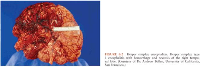
Within the CNS, the pathogen often targets specific cells and, depending on the brain region affected, variable clinical manifestations ensue (173). Agents with specific predilection to areas such as the brainstem (e.g., EV-71 and Listeria) can cause rapid decompensation with coma or respiratory failure. Herpes simplex encephalitis characteristically affect the temporal lobes and cause hemorrhage and necrotizing lesions (174) (Fig. 6.2).
WNND has a predilection for the gray matter of the brainstem and spinal cord, but cerebellum, temporal lobe, basal ganglia, and thalamus may also be affected (175). Indeed, many of the neuroinvasive flaviviruses such as JEV, TBEV, and WNV have a predilection for specific regions of the brain, including those regions important for motor control (thalamus, basal ganglia, brainstem, and anterior horn cells of spinal cord) (176).
In other instances, the infectious agents do not necessarily infect neurons. Nonneuronal cells, such as oligodendroglia, may be infected, with resultant demyelination (177). Alternatively, an infection may cause immune changes that result in damage. EBV-associated encephalitis, for example, may be a result of an immunologic phenomena rather than acute neuroinvasion. There is typically a 1- to 3-week delay in the onset of neurologic symptoms after acute EBV infection (40). Further, the virus itself is often not found in the CSF (40). In one cohort of five EBV encephalitis patients with CNS demyelination, four had prodromal symptoms for 2 weeks or more and did not have EBV detected by polymerase chain reaction (PCR) in CSF (40). Conversely, the development of neurologic symptoms either in the absence of, or within a few days of prodrome onset in the presence of, EBV in the CSF is suggestive of direct invasion. In the pediatric series cited previously, one such patient had EBV detected by PCR in brain tissue, suggesting that direct invasion of the brain may occur in some cases.
Viruses such as influenza are well known to be associated with CNS manifestations, but the mechanisms by which they cause neurologic signs and symptoms are not well understood. The lack of viral detection in most IAE cases in the CNS strongly points to a different pathogenesis; a number of potential mechanisms have been invoked, including excessive production of proinflammatory cytokines, vascular endothelial dysfunction, and mitochondrial dysregulation (178).
As discussed earlier, Listeria may be associated either with a pure meningitis or encephalitis. Pathologically, a suppurative reaction is seen in the meningitic form, whereas a granulomatous response is seen in the meningoencephalitis form (104).
Rickettsial agents invade and multiply in vascular endothelial cells, leading to vasculitis both within and outside the CNS (179,180). Vasculitis in the small vessels in the brain leads to meningeal irritation with perivascular mononuclear infiltrates. Characteristic pathologic lesions within the CNS include multifocal glial nodules and arteriolar microinfarctions (181).
In partially treated bacterial meningitis and in tuberculous and fungal meningitis, a chronic basilar meningeal inflammation can cause a subarachnoid exudate, leading to obstruction of CSF reabsorption with resultant communicating hydrocephalus and cranial nerve palsies. CNS vasculitis can also lead to infarcts and focal neurologic deficits.
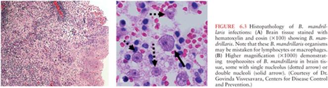
N. fowleri, a free-living ameba, causes destruction of gray matter and devastation of the olfactory bulbs with purulent meningitis and pronounced brain edema (182). In Balamuthia and Acanthamoeba CNS infections, a granulomatous reaction occurs with affected areas including the cerebrum, cerebellum, and brainstem, where the amebae produce hemorrhagic necrotic lesions. Multinucleated giant cells, focal necrosis, and hemorrhage are seen in brain histopathology. In some instances, large sheets of ameba can be found in the perivascular areas of brain tissue (Fig. 6.3).
B. procyonis and other nematodes that invade the CNS cause damage by CNS larval migration, with histology demonstrating inflammation and necrosis with “track”-like spaces. Pathologic findings of fatal cases demonstrate necrosis and inflammation with eosinophils, macrophages, lymphocytes, and plasma cells concentrated in periventricular white matter and leptomeninges in brain tissue. A characteristic of NLM/Baylisascaris infection is a large number of eosinophils and eosinophil granules surrounding the nematode migration track and blood vessels (183).
DIAGNOSTIC APPROACH OF PATIENTS WITH ENCEPHALITIS
Identification of a specific etiology, even if there is no available treatment, is important for counseling of patients and families, potential postexposure prophylaxis of contacts, and other public health interventions. Additionally, the identification of a specific agent may lead to withdrawal of unnecessary antimicrobial agents and reduce further testing. Knowledge of limitations of testing, appropriate test selection, and timing of sample collection is crucial to optimal diagnosis.
A thorough assessment of exposures highlighting ill contacts, occupational exposures, vector and animal exposures, outdoor activities, and ingestions should be ascertained. Both recent (e.g., for arbovirus) and remote (e.g., rabies, fungal) travel history are important. Any recent or current respiratory, gastrointestinal, or rash illness should be investigated. Based on this information, specific assays can be performed (Tables 6.1 and 6.2) (184).


A complete blood count, renal function tests, hepatic enzyme levels, and coagulation studies should be included in the evaluation of a patient with suspected encephalitis. An initial chest radiograph should also be performed as focal infiltrates are suggestive of certain pathogens (e.g., fungal or mycobacterial infections) and might lead to other diagnostic studies (e.g., bronchoscopy). A thorough eye exam by an ophthalmologist may identify a migratory nematode which would considerably narrow the differential.
Unless there is a specific contraindication, a lumbar puncture (LP) with cerebrospinal fluid (CSF) analysis (cell count with differential analysis, glucose and protein concentrations) and a measurement of the opening pressure should be performed (185). A simultaneous peripheral glucose should also be measured and is particularly important in a diabetic patient because what appears to “normal” CSF glucose might actually be low if the ratio of CSF to serum glucose is not taken into consideration (102). The CSF sample should be analyzed promptly, particularly because neutrophils degrade within a few hours (186). Additionally, if the CSF cell counts are performed by an automated cell counter, eosinophils can be mistaken for neutrophils. Accurate identification of eosinophils is best done by Giemsa or Wright stain of CSF (128). The identification of CSF oligoclonal bands, representing intrathecal antibody synthesis, is a nonspecific finding but can be helpful in corroborating an infectious or inflammatory etiology.
The CSF profile offers important diagnostic clues on whether a patient has an infectious or noninfectious etiology and, within the infectious sphere, whether the cause is likely viral, bacterial, fungal, or parasitic. The CSF profile in most viral encephalitis patients demonstrates CSF mononuclear cell pleocytosis, with cell counts ranging from 10 to 200 mg/dL. However, several studies have shown that pleocytosis can be absent or there may be an elevation in neutrophils early in the course. Although many textbooks mention that polymorphonuclear leukocytes (PMNs) can predominate the first 24 hours of viral CNS infections and suggest that a shift to mononuclear cells occurs after that time period, there have been studies showing that PMNs may still predominate well beyond this 24-time period (187). Therefore, a repeat LP a few days following the first LP may be useful (188,189). In viral encephalitis, the CSF protein generally is elevated but is typically less than 100 mg/dL, whereas the glucose level is almost always normal with a few important exceptions (e.g., LCM and mumps; Table 6.3).
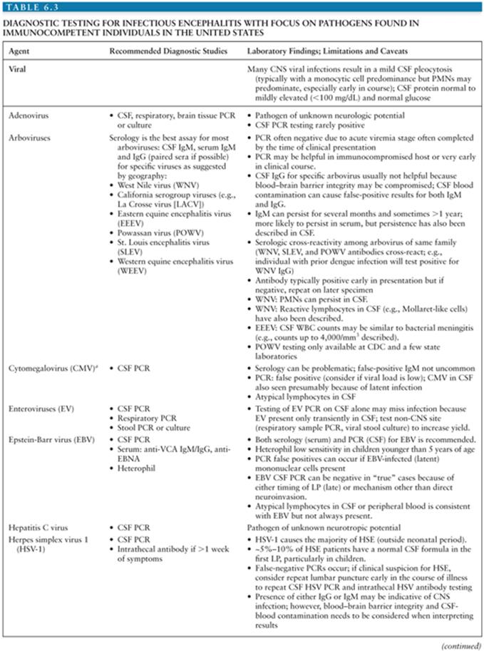


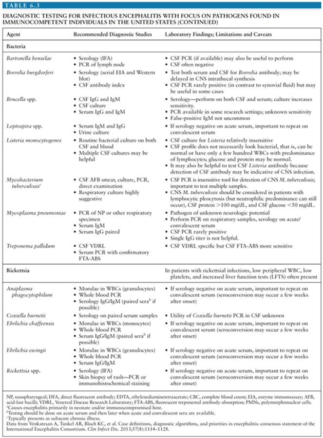
As outlined earlier, viral infections can sometimes be associated with a neutrophilic predominance; however, when a neutrophilic pleocytosis is observed, particularly in cases where CSF white blood cell (WBC) count is more than 1,000 cells/mm3, protein more than 100 mg/dL, or CSF glucose level less than 2/3 of serum levels, a nonviral entity should be strongly considered. For most bacterial pathogens, the CSF shows a WBC of 1,000 to 5,000 cells/mL with predominance of neutrophils (80% to 95%), glucose less than 40 mg/dL in over half the cases, and a CSF glucose level less than 2/3 serum levels. In fungal infections, moderate lymphocytic pleocytosis usually is found along with elevated protein and low CSF glucose.
Eosinophils in the CSF are suggestive of a helminth infection (e.g., B. procyonis, Angiostrongylus spp., and G. spinigerum). Eosinophils can also be seen in patients with neurococcidiomycosis or neuro-Mycobacteriumtuberculosis. In patients with PAM, the CSF typically contains a very high CSF cell count with a predominance of neutrophils, a slight to pronounced decrease in glucose concentration, and an increased protein content (75 to 970 mg/dL). The amebae can sometimes be seen in wet-mount preparations of spinal fluid (139,140,190). In patients with CNS Balamuthia infections, elevated CSF WBC is often seen along with mild to markedly elevated protein (>1,000 mg/dL) and normal or slightly decreased glucose.
Although brain biopsy in the setting of suspected encephalitis has become less common, examination of brain tissue still has utility because of limitations in both molecular and serologic methods. The use of targeted biopsy was recently demonstrated by a report of 16 patients with undiagnosed CNS illness where brain biopsy detected bacterial abscess (6), toxoplasmosis (3), HSV (1), Aspergillus infection (2), and M. tuberculosis infection (2). Biopsies can also be particularly helpful for the diagnosis of noninfectious entities such as small vessel vasculitis and intravascular lymphoma (191). An autopsy should be encouraged to determine the cause of death in patients who die with unexplained encephalitis.
In addition to studies done acutely, an extra red top serum tube should be drawn during the acute phase of illness and held for later serologic studies. A convalescent serum should be collected 10 to 21 days later. Similarly, extra spinal fluid should also be frozen for future testing. Specific caveats of testing are outlined in Table 6.3 and selected agents are outlined in the following sections.
TESTING OF SELECTED ETIOLOGIES
Viral Testing
Specific studies for viral agents often employ molecular testing. Molecular testing by nucleic acid amplification (e.g., PCR) testing provides more timely and typically more sensitive results than culture for viral CNS infections (116,192,193). However, the limitations of these tests are often not recognized by physicians, particularly false-negative and false-positive results as discussed in the following sections for specific agents. Testing for intrathecal antibody synthesis of specific pathogens such as arboviruses and herpesviruses can be useful adjunct for diagnosis, particularly in the later stages of disease (see the following discussion).
In patients with either preceding or concurrent respiratory symptoms, a viral culture from the respiratory tract should be performed early in the hospital course to optimize the virus recovery. When a particular agent such as influenza or another virus is suspected but the viral culture is negative, agent-specific PCR testing should also be performed. Similarly, if there is a history of preceding or concurrent diarrhea, a stool culture for viral and, possibly, bacterial pathogens should be performed. In children, a stool viral culture and/or EV PCR should be routinely performed to enhance EV detection, regardless of the presence of diarrhea.
Herpesviruses
The diagnostic test of choice for HSE is CSF HSV-1 DNA nucleic acid (PCR) testing. If testing from the first LP is negative and HSE is still suspected (e.g., temporal lobe involvement seen on neuroimaging), a second LP should be performed within 24 to 48 hours as a number of studies have shown that HSV PCR can be falsely negative, particularly early in course of illness and in the pediatric age-group (22,189). For HSE, intrathecal antibodies can be performed as a complement to molecular testing, and this may be particularly helpful in establishing the diagnosis later in the course. Intrathecal synthesis of HSV-specific IgG antibodies can often be detected 10 to 14 days after the onset of illness (194).
As mentioned in the clinical and epidemiology section, the clinical spectrum of CNS infections associated with HSV-2 often differs from HSV-1. Molecular testing of CSF for the presence of nucleic acid is the most reliable method for diagnosis for HSV-2; intrathecal antibody testing can be used as an adjunct as described previously for HSV-1.
Because VZV encephalitis is one of the most common causes of encephalitis in adults, VZV testing should be performed in all adult patients, with or without skin lesions. Testing should include both CSF VZV PCR and CSF VZV antibody (195). Although detection of VZV antibody in the CSF can be more sensitive than PCR, intrathecal synthesis may be delayed by a week or more after onset of neurologic symptoms (196,197). In the event of a traumatic tap, distinguishing intrathecal antibody synthesis versus blood contamination of CSF may be accomplished by calculation of an antibody index (198).
For the diagnosis of EBV encephalitis, both serology and molecular testing is recommended (40). Quantitative viral load may be helpful, as a high viral load supports a significant CNS infection, whereas a low positive PCR in CSF may represent latent virus and may only be an incidental finding (199,200). Conversely, the absence of a positive PCR in the setting of acute EBV serology does not rule out acute infection, and a negative PCR may be a result of the timing of LP (i.e., after virus has cleared from CSF) or may reflect a mechanism other than direct infection.
CSF PCR testing is recommended for the diagnosis of HHV-6 and HHV-7; however, the finding of a positive CSF PCR for HHV-6 or HHV-7 is not necessarily equivalent to a diagnosis of HHV-6 or HHV-7 encephalitis. This is because HHV-6 DNA can be present in normal brain tissue and HHV-6 may be identified in CSF as an incidental “bystander” rather than a cause of infection (201). Additionally, chromosomal integration must be considered as a potential reason for a positive PCR in the CSF. If a positive HHV-6 CSF PCR is obtained, an evaluation of HHV-6 in the blood should be done to distinguish between chromosomal integration and acute infection (48).
Enteroviruses and Parechoviruses
PCR testing of both CSF and a throat specimen should be performed; if only CSF is tested, the diagnosis may be missed. For example, in an outbreak of EV-71 in the United States, EV PCR of CSF was positive in only 5 (31%) of 16 cases, whereas PCR testing of throat specimens were uniformly positive (202,203). Because EVs may be shed in the stool for weeks after infection, culturing the stool for EV may increase the diagnostic yield; for the same reason, a positive stool is suggestive, but not confirmatory, of the diagnosis. Testing for parechoviruses should be considered in all children younger than 3 years of age with the same range of biologic samples tested as EVs. Parechoviruses can only be detected by parechovirus-specific PCR assays.
Arbovirus
For most arboviruses, serologic testing of CSF and serum is preferred to molecular testing because peak viremia generally occurs prior to symptom onset (204). The diagnosis of WNV infection is typically made via detection of IgM capture enzyme-linked immunosorbent assay (MAC-ELISA) or a fourfold or greater change in antibody titer on paired or convalescent serum. Approximately 90% of patients with neuroinvasive disease will have detectable CSF IgM antibody by days 8 to 10 after symptom onset (205). However, WNV IgM may persist for prolonged periods, with detectable IgM titers persisting more than 500 days after presentation in some individuals with neuroinvasive disease (206). Furthermore, there is serologic cross-reactivity between the flaviviruses (SLEV, dengue virus, yellow fever virus, or JEV), such that individuals infected with, or vaccinated against, one of these agents could test positive for WNV by IgM MAC-ELISA. Differentiation of WNV from other flaviviruses is achieved by plaque reduction neutralization testing (PRNT) (207).
For other arboviruses, the standard method for diagnosis is similar, namely through detection of IgM or a fourfold or greater change in antibody titer on paired acute and convalescent serum (208).
Other Viral Testing
The diagnosis of rabies should be considered in any rapidly progressing encephalitis. Antemortem testing for rabies requires highly specialized testing in a public health laboratory using a combination of different assays and specimen types (209). Whenever the diagnosis of rabies is entertained, consultation with public health authorities is strongly recommended. Testing for other viruses should be performed as suggested by exposures or travel history.
Bacterial Testing
In most instances, detection of routine bacterial pathogens via Gram stain and/or culture is fairly straightforward. The CSF bacterial culture for L. monocytogenes, however, is relatively insensitive compared with many other bacterial agents. Additionally, in contrast to many bacterial pathogens, the CSF profile of CNS listeriosis patients, particularly those with rhombencephalitis, can appear normal or similar to viral illnesses with only a mild pleocytosis, a lymphocytic predominance rather than polynuclear predominance, and a normal to minimal protein elevation (109) (see Table 6.3).
Neurotuberculosis can also be difficult to diagnose. The sensitivity of CSF tuberculosis (TB) PCR is very poor; reported sensitivity has been less than 25% in some studies (102). This may be because of low bacillary load in the CSF, relatively small CSF volumes submitted for testing, or PCR inhibitors in samples (210). Testing from both CNS and extra-CNS sites should be done for the workup of neuro-TB (see Table 6.3).
Fungal Testing
Fungal meningoencephalitis is typically a subacute illness. Fungal culture of the CSF should be performed in individuals with indolent symptoms, immunocompromised persons, or when the CSF findings are suggestive (e.g., CSF lymphocytic pleocytosis, high protein, and low glucose). If a fungal etiology is suspected, several different tests including culture, antigen test, and serology on both CNS as well as non-CNS specimens should be done (see Table 6.3) (211).
Rickettsial Testing
Diagnostic testing is similar for most rickettsial infections. Serology is the most widely used method, and the indirect fluorescent antibody (IFA) assay is considered the gold standard. Because serologic tests may be negative early in the illness, detection of the rickettsial agent in blood or tissues by molecular assays (e.g., PCR) may be useful. Confirmation of disease is best accomplished by evaluation of paired serum specimens collected during the acute and convalescent phases of the illness for a fourfold or greater change in antibody titer. Immunohistochemistry of patients’ tissues (e.g., brain) is another methodology that can be used to diagnose rickettsial infection (212).
Parasitic and Free-Living Ameba Testing
The diagnosis of Baylisascaris is often suggested by a history of exposure to raccoons (or their feces), clinical presentation, and persistent eosinophilia in the blood and spinal fluid. Serologic assays are available at the Centers for Disease Control and Prevention (CDC). In patients with a history of travel outside the United States with presence of CSF eosinophilia, both gnathostomiasis and Angiostrongylusshould be considered. An ELISA test has been developed but is not widely available (213). Molecular tests for Angiostrongylus are available at the CDC.
Molecular technology is also available for the identification of Balamuthia and Acanthamoeba DNA in tissues and CSF (CSF PCR is less sensitive than brain tissue PCR) but is largely limited to research laboratories and the CDC.
NEUROIMAGING MODALITIES
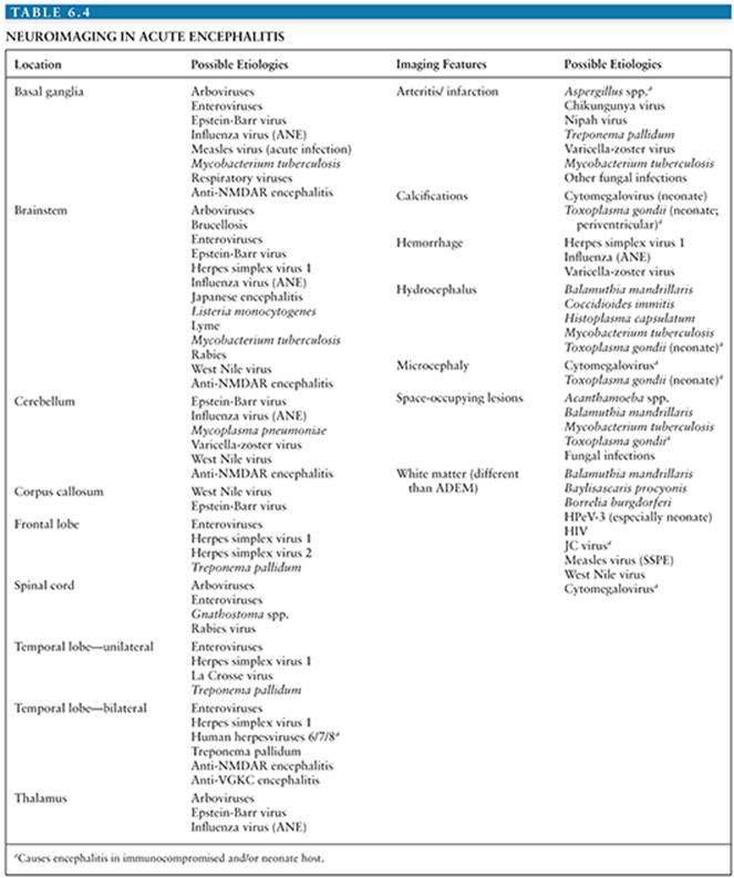
Neuroimaging plays a crucial role in the evaluation of patients with suspected encephalitis, as it may support the diagnosis of a specific etiology or identify alternate conditions that mimic encephalitis (Table 6.4). Routinely available studies to evaluate patients with suspected encephalitis include computed tomography (CT) and magnetic resonance imaging (MRI). CT scanning involves the application of x-rays to produce tomographic images, resulting in excellent delineation of bony structures; conventional MRI uses nuclear magnetic resonance (MR) to detect hydrogen nuclei and provides good contrast between soft tissues. Of the two, MRI is far more sensitive and specific for the evaluation of infectious encephalitis (214–216). Conventional MRI imaging entails acquisition of various sequences that provide complementary information. For example, T1-weighted imaging (T1W) is well suited to define structural abnormalities; T2-weighted imaging (T2W) and fluid-attenuated inversion recovery (FLAIR) sequences may identify areas of edema, demyelination, or inflammation; and susceptibility-weighted imaging (SWI) is particularly useful in identifying areas of hemorrhage. T1W imaging following gadolinium administration can identify parenchymal or meningeal regions where the blood–brain barrier has been compromised, as occurs in active inflammation. Of particular note is diffusion-weighted imaging (DWI), which identifies brain regions where diffusion of water is restricted and may be more sensitive than other conventional sequences for detection of early abnormalities in encephalitis (217,218).
Several additional imaging modalities have emerged for evaluation of encephalitis. MR spectroscopy can identify a number of metabolites, including N-acetylaspartate (NAA), choline, creatine, and lactate, based on unique proton chemical shifts. Quantification of the ratios of these metabolites can provide information regarding inflammation, neuronal loss, and demyelination. Both single-photon emission computed tomography (SPECT) imaging and fluorodeoxyglucose positron emission tomography (FDG-PET) involve detection of radioisotopes within the brain following injection into the bloodstream and can provide information regarding metabolism. Other MR-based techniques, including magnetization transfer and diffusion tensor imaging, as well as molecular imaging techniques that allow for the detection of disease-specific molecules, are currently being optimized and may eventually show promise in evaluation of patients with encephalitis (219).
Imaging of Selected Etiologies
Herpesviruses
The neuroimaging findings of encephalitis caused by HSV-1 reflect its predilection for the limbic system, with frequent involvement of the mesial temporal and inferior frontal lobes (Fig. 6.4).
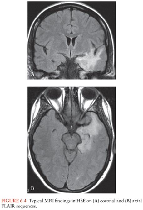
In the acute stage, HSE typically results in edema, inflammation, or hemorrhage. Although initial CT scans are normal in up to 25% of affected individuals, greater than 90% of patients with HSE documented by CSF PCR will have MRI abnormalities (220–223). Abnormalities on conventional MRI appear within 48 hours and include isointense or hypointense lesions on T1W images and hyperintense lesions on T2W or FLAIR images. Changes in DWI, which may reflect the rapid accumulation of intracellular water in the setting of impaired ion transport during acute infection, are seen even earlier (218,224,225). MRI is superior to CT in the detection of subacute (>1 week) hemorrhage. Contrast enhancement on MRI is typically in a cortical gyral pattern and often lags behind symptom onset (226). Extratemporal abnormalities occur in over half of cases and may involve the cerebral hemispheres, thalamus, and brainstem (227–229). Notably, in up to 15% of patients, extratemporal involvement may be the sole-observed neuroimaging abnormality (229) and is more likely to occur in pediatric and immunocompromised populations (230–232). MR spectroscopic findings have also been described, although the clinical use of this imaging modality in the evaluation of HSE remains undefined. Abnormalities, which typically include decreased NAA and increased choline, myoinositol, and lactate, are thought to represent loss of neuronal integrity and necrosis with accompanying macrophage infiltration and gliosis (233–235).
In several case series of VZV encephalitis, the most notable finding was the absence of brain imaging abnormalities in the majority of patients. In a minority of patients, affected regions included the temporal lobe and brainstem. A notable caveat is that many patients in these series underwent CT scanning only (32,236). VZV vasculopathy is often associated with T2W hyperintensities in the white matter and at the gray–white matter junctions on MRI, with evidence of either ischemia or hemorrhage (25). The most commonly reported imaging abnormalities in EBV-associated encephalitis include multiple, diffuse hyperintensities on T2W and FLAIR imaging, often transient and with involvement of the splenium of the corpus callosum (237–239).
HHV-6 associated limbic encephalitis typically involves the mesial temporal lobe (hippocampus, entorhinal cortex, and amygdala). Additional cortical and subcortical areas can be involved and may be associated with specific clinical subtypes (240,241). Involved areas are characterized by T2W hyperintensity and variable degrees of enhancement. In addition, increased hippocampal uptake of glucose, suggestive of increased metabolic activity, has been detected by FDG-PET (242,243). Although both HSE and HHV-6 limbic encephalitis are associated with mesial temporal abnormalities, a recent study suggested that early extratemporal involvement was more likely in HSE (244). Rarely, HHV-6 infection of the CNS has been associated with a syndrome similar to ANE (51,245).
Enteroviruses
Although poliovirus has almost completely disappeared in developed countries, nonpolio EVs are an important cause of diffuse, generalized encephalitis. In such cases, neuroimaging is typically normal. Certain EVs, however, may result in similar clinical and radiographic manifestations as poliovirus. EV-71, for example, is characterized by symmetric bilateral T2W hyperintensities in the dorsal brainstem, cerebellar dentate nuclei, and anterior horns of the cervical spinal cord on MRI (246,247) (Fig. 6.5).
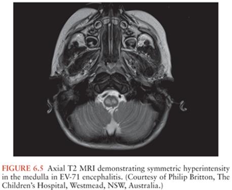
As in many cases of acute encephalitis, DWI appears to be more sensitive than other conventional sequences for detection of EV-71 lesions (248). Echovirus 7-associated encephalomyelitis is also associated with a similar topographical distribution on MRI (249). Isolated reports of unilateral and bilateral hippocampal lesions in the setting of acute enteroviral infection have also been described (250,251).
Arboviruses
Neuroimaging abnormalities in WNV often involve the deep gray matter, although findings are highly variable and up to half of patients may have normal brain MRI (233,252,253). In those who develop a flaccid paralysis, spinal cord imaging may demonstrate T2W hyperintensities in the anterior cord and gadolinium enhancement of the cauda equina, conus medullaris, and nerve roots (252,254). Limited studies of other arboviruses, including SLE, EEE, TBE, and Murray Valley encephalitis, suggest involvement of the substantia nigra and other deep gray structures, and the latter three viruses may also resemble WNV acute flaccid paralysis both clinically and radiographically (255,256). The considerable overlap in the neuroimaging spectrum of these arboviruses prevents discrimination between these etiologies based on imaging alone.
JEV is most commonly associated with T2W hyperintensities on MRI in the thalami, with variable involvement of the substantia nigra, basal ganglia, brainstem, cerebellum, and cortex (257). Although the presence of thalamic abnormalities is highly sensitive for JEV in the appropriate clinical setting, their absence does not rule out JEV (258). Mesial temporal lobe involvement has also been reported, although unlike herpes encephalitis, the insula and anterior portion of the temporal lobe are usually spared, while additional thalamic lesions are typically present (259,260). In JEV cases with biphasic illness, regression of prior lesions and the appearance of new lesions in typical areas are described (261,262). In cases of dengue encephalopathy, focal MRI abnormalities in a number of brain areas have been found. In addition, generalized cerebral edema has been reported and may be related to the prominent metabolic disturbances observed in affected individuals (263).
Bilateral frontoparietal white matter lesions with restricted diffusion, suggestive of subcortical strokes, have been reported in two cases of Chikungunya encephalitis (264). However, in another case series of children with various CNS manifestations including encephalitis, MRI abnormalities were only rarely observed (265).
Rabies
The often rapid clinical course of rabies encephalitis presents a challenge in obtaining neuroimaging. The limited available data demonstrate diffuse T2W hyperintensities affecting both the deep and cortical gray matter of the brain and spinal cord in both the paralytic and encephalitic forms of rabies (266). Although abnormalities may also be seen in the white matter, the predominant gray matter involvement distinguishes rabies encephalitis from ADEM. Initially nonenhancing, the gray matter lesions may demonstrate gadolinium enhancement once the patient becomes comatose (267).
Nipah
In the acute phase, Nipah encephalitis is associated with small, punctate T2W hyperintensities in the subcortical white matter, often restricting diffusion (268). This imaging evidence of microinfarction is congruent with the main pathologic findings of vasculitis-associated thrombosis and presence of Nipah virus antigen in endothelial and smooth muscle cells of blood vessels (269). For the subset of patients who suffer from a relapse of neurologic symptoms, MRI shows patchy areas of confluent cortical involvement on MRI and focal hypoperfusion on SPECT imaging (270).
Hendra
In contrast to Nipah virus, the closely related Hendra virus is associated predominantly with gray matter abnormalities in the setting of meningoencephalitis. In three reported cases, T2W hyperintense lesions involved the cortical gray matter, with relative sparing of the deep white matter tracts and cerebellum. Cortical lesions may be confluent or multifocal, and restriction of diffusion can be seen on DWI (271).
Measles
Measles virus can cause differing CNS syndromes, including acute measles encephalitis and SSPE. Neuroimaging in acute measles encephalitis typically demonstrates bilateral, symmetric T2W hyperintense lesions involving the white matter and deep gray matter structures in a pattern that can resemble ADEM (272). Cortical gyral swelling may also be present in the acute setting, and hemorrhage may develop in gray matter areas over days to weeks (273).
The earliest stages of SSPE are often not characterized by abnormalities on conventional MRI, although FDG-PET can demonstrate cortical hypometabolism and MR spectroscopy may show decreased NAA levels (274). As the disease progresses, T2W hyperintensities in the subcortical and periventricular white matter can develop, as does brain atrophy. By the late stages of SSPE, widespread changes in the cortex, brainstem, and cerebellum can be seen (275).
Mumps
Encephalomyelitis following mumps is associated with white matter changes on brain MRI and a longitudinally extensive myelopathy on spine imaging that may be indistinguishable from ADEM; rarely, hemorrhagic white matter lesions have been reported (276–278).
Influenza
Neuroimaging findings in influenza-associated encephalopathy/encephalitis are diverse. MRI can be normal or may demonstrate diffuse brain edema or symmetric involvement of the thalami (233,279,280). Severe cases of encephalitis are characterized by T2W hyperintensities, hemorrhage, and restricted diffusion in the thalami, basal ganglia, and cerebellum, consistent with ANE (281). Milder cases may be associated with reversible lesions of the splenium of the corpus callosum (282). Notably, imaging abnormalities caused by the 2009 H1N1 virus do not appear to differ significantly from other influenza strains (283–285).
Mycobacterium tuberculosis
Cranial TB can present as tuberculous meningitis (TBM), tuberculomas, or abscesses—each of which have differing neuroimaging characteristics. TBM is associated with basal meningeal enhancement, often accompanied by hydrocephalus, strokes in the basal ganglia and internal capsule, and a focal or diffuse pachymeningitis (286–288). Intracranial tuberculomas consisting of granulomatous tissue may have variable appearance on T2W imaging depending on the pathologic stage of the lesion; noncaseating tuberculomas are T2W hyperintense, whereas those with solid caseation are isointense or hypointense. When central liquefaction ensues, tuberculomas may take on an appearance more similar to that of an abscess. Such lesions exhibit T2W hyperintensity surrounded by a peripheral rim of hypointensity and demonstrate peripheral rim enhancement on contrast imaging (289).
Fungal Infections
Neuroimaging findings in the setting of CNS fungal infections are often nonspecific and may be mistaken for TBM, pyogenic abscess, or brain tumor (290,291). Fungal infections may result in basilar meningitis, hydrocephalus, vasculitis, or abscess, and any combination of these may be present on neuroimaging. In immunocompetent individuals, fungal abscesses are hypointense on T1W imaging and hyperintense on T2W imaging, with a well-defined rim enhancement on postcontrast images, similar to pyogenic or tubercular abscesses (290). Recent progress has been made in distinguishing fungal abscesses from other abscesses. In one study, all of the fungal abscesses showed intracavitary projections directed centrally from the wall, a finding not seen in the other abscess types. Overall, a ring-enhancing lesion with irregular walls and nonenhancing intracavitary projections was likely to be a fungal abscess (292). Detection of the disaccharide trehalose on MR spectroscopy may also distinguish fungal from nonfungal abscesses (292–294).
MRI is frequently normal in the setting of cryptococcal meningitis, although evidence of a basilar meningitis and/or hydrocephalus indistinguishable from TBM may be observed. The adjacent brain parenchyma may also be affected, giving rise to cryptococcomas, most commonly seen in the midbrain and basal ganglia. These lesions are of variable density on CT scan and, on MRI, are hypointense on T1W imaging and hyperintense on T2W imaging. Contrast enhancement is variable and more commonly seen in immunocompetent individuals. Unlike pyogenic abscesses, cryptococcomas typically do not demonstrate restricted diffusion on DWI. MR spectroscopy demonstrates elevated lactate and decreased NAA, choline, and creatine (26,290,295).
Imaging abnormalities in patients with CNS coccidioidomycosis may reflect strokes, granulomas, and white matter disease, in addition to basilar meningitis and hydrocephalus. Ischemia has been reported in over half of cases and typically results in deep cerebral infarcts seen as areas of hyperintensity on T2W imaging. Less commonly, focal enhancing lesions in the white matter or deep grey matter representing granulomas are observed (292,296,297).
Listeria monocytogenes
Rhombencephalitis in the setting of L. monocytogenes infection is characterized by the presence of multiple, small, medullary T2W hyperintensities that demonstrate rim enhancement on T1W imaging, reflecting a combination of microabscesses and associated edema in the lower brainstem (298) (Fig. 6.6).

Borrelia Species
Acute CNS Lyme disease is associated with small T2W hyperintense and T1W isointense lesions in the subcortical white matter, which may mimic those seen in multiple sclerosis (299). Multifocal lesions may also be observed in the brainstem and spinal cord (300).
Treponema Pallidum
The clinical and MRI features of neurosyphilis are highly variable. The MRI of patients with clinical encephalitis is characterized by diffuse brain atrophy, evidence of subcortical white matter infarcts, or T2W hyperintensity in the mesial temporal lobe (301,302).
Acute Disseminated Encephalomyelitis
MRI findings in ADEM typically include multiple, asymmetrically distributed areas of T2W hyperintensity in the subcortical and deep white matter (Fig. 6.7).

Lesions in the deep gray matter, which are characteristic of ADEM but atypical for multiple sclerosis and other demyelinating disorders, often have a symmetric appearance. In approximately one third of cases, the cerebral cortex is also involved. Infratentorial involvement occurs in over half of cases, with lesions seen in the brainstem, middle cerebellar peduncles, and cerebellar white matter (303–305). The size and morphology of lesions is highly variable, although they tend to have poorer margination as compared to lesions in other demyelinating diseases. In most cases, lesions appear simultaneously, and all lesions will exhibit a similar degree of contrast enhancement. Restricted diffusion may be observed on DWI sequences in the acute stage, potentially representing swelling of myelin sheaths, myelin vacuolation, or infiltration of inflammatory cells with concomitant free radical production (306,307). Rare variants of ADEM include hemorrhagic, necrotizing, and relapsing forms (308).
Anti–N-Methyl-D-Aspartate Receptor Encephalitis
One third to one half of patients may have abnormalities on brain MRI. Affected areas, seen as T2W or FLAIR hyperintensities, include the mesial temporal lobes, periventricular white matter, cortex, cerebellum, brainstem, or basal ganglia. Notably, on follow-up, many of these abnormalities improve, although atrophy may develop. Less than 20% of affected individuals will have evidence of contrast enhancement (168,309).
Anti–Voltage-Gated Potassium Channel Receptor Encephalitis
The spectrum of diseases associated with antibodies to the VGKC complex has been steadily growing. In the setting of encephalitis, the most common finding is unilateral or bilateral T2W hyperintensity in the mesial temporal lobe (169) (Fig. 6.8).
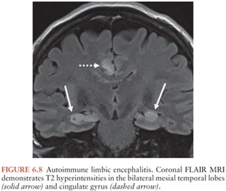
Recent reports have suggested additional or exclusive involvement of the basal ganglia (310,311).
MANAGEMENT
Clinicians should focus first on treatable and common causes of encephalitis. Empirical treatment for bacterial meningitis (vancomycin plus a third-generation cephalosporin) should be started because the clinical presentation may overlap with encephalitis. Therapy with ampicillin should also be considered when the demographics (i.e., older individuals), clinical presentation (i.e., rhombencephalitis), or CSF profile is suggestive of Listeria infection. Antiviral therapy is generally restricted to treatment of herpesviruses (especially HSV-1 and VZV) and the unusual instance of HIV infection (312). Therapy with acyclovir should be started and continued until HSV-1 has been reasonably excluded as a diagnosis, which may require testing serial CSF samples (see prior discussion). Therapy for M. tuberculosis or fungal meningitis should be initiated when clinical and laboratory testing is compatible. If rickettsial or Ehrlichia infections are suspected, doxycycline should be initiated empirically.
For IAE, oseltamivir may be beneficial (313). Corticosteroids or intravenous immune globulin may also be helpful in some IAE cases to combat the proposed hyperintense cytokine response. There is no evidence that treatment of presumed CNS Mycoplasma infection alters outcome.
In addition to directed therapy, aggressive supportive care is critical, and minimizing secondary brain injury should be made a high priority (314). Seizures, status epilepticus, and cerebral edema are important complications of encephalitis and encephalopathy and should be monitored closely in patients who are not improving. An elevated CSF opening pressure may serve as a harbinger for impending complications. Repeat neuroimaging to monitor for cerebral edema is particularly important in comatose patients. Typical indicators of elevated intracranial pressure, such as poorly reactive dilated pupils, decorticate or decerebrate posture, or Cushing triad (systolic hypertension, bradycardia, and shallow respirations) are late findings. Patients should also be monitored for the syndrome of inappropriate secretion of antidiuretic hormone (SIADH). SIADH and extrapontine myelinolysis (EPM) are important examples by which acquired metabolic derangements can both complicate, and mimic, encephalitis (315).
Conditions that mimic infectious encephalitis should be considered, particularly if no etiology is identified in the first week of hospitalization. Metabolic and toxic disorders causing encephalopathy and seizures should be excluded. Anti-NMDAR encephalitis is of particular importance given its apparently high incidence; when identified, immunotherapy and removal of the tumor, if present, have been associated with improvement.
OUTCOME
In general, the prognosis of encephalitis is highly dependent on the underlying cause. Rabies and N. fowleri, for instance, have an almost 100% fatality rate. Two of the most well-studied viral causes of encephalitis are HSV-1 and EV. HSV-1 encephalitis has been reported to have a worse prognosis than EV, with greater than 35% of patients with HSE suffering severe sequelae or death (4). Although EV encephalitis patients often have a good outcome, EV-71 is associated with fatalities and neurologic sequelae (58). Other viral etiologies are less well studied, with information limited to case reports and small series. Persistent neurologic deficits after EBV encephalitis are reported to be rare (316). Influenza has been associated with a severe type of encephalitis with high mortality particularly in Japan and Taiwan, including several case reports of ANE (317,318). In the United States, cases appear to be less severe with better outcomes, although further studies are needed (319).
Deaths and complications caused by arboviruses are better documented as a result of comprehensive reporting to the CDC. WNV infections are less severe in children than in adults; children accounted for only 4% of WNND reported to the CDC from 1999 to 2007, with 63% of cases older than 10 years of age and only 15% younger than 4 years of age. There were only three pediatric fatalities over this time period (1% of all cases of WNND), a case fatality rate substantially lower than for older adults (14% for adults 50 years of age or older) (320). In a study of 127 children with LACV, 12% had neurologic deficits at discharge (321). EEE is also known to have a higher mortality (almost 30%) and potentially severe neurologic complications.
Bacterial infections also have variable outcomes. Both L. monocytogenes and M. tuberculosis may have relatively high morbidity and mortality (109,322). In a French encephalitis study, these two etiologies accounted for the majority of fatalities due to encephalitis (together, 12 of 26 fatalities) (101). On the other hand, infection with bacterial agents, such as Bartonella spp., which typically causes encephalopathy rather than encephalitis, has an excellent outcome; over 90% of patients recover completely without sequelae. There are limited studies reporting outcomes specifically on encephalitis of unknown etiology. One study, however, reported significant sequelae in up to 53% of survivors hospitalized with unknown causes of encephalitis (323).
SUMMARY
It is unfortunate that in this era of modern medicine, so little progress has been made in the field of encephalitis. An etiology is only identified in about half of the cases and for those patients who receive a viral diagnosis; very few specific antivirals are available for treatment. Given the significant morbidity and mortality of this syndrome along with the financial costs of hospitalization, rehabilitation, and sequelae, research to address gaps in our understanding of this entity are urgently needed as well as development of effective antiviral agents.
References
1. Glaser CA, Gilliam S, Schnurr D, et al. In search of encephalitis etiologies: diagnostic challenges in the California Encephalitis Project, 1998–2000. Clin Infect Dis. 2003;36(6):731–742.
2. Kolski H, Ford-Jones EL, Richardson S, et al. Etiology of acute childhood encephalitis at the Hospital for Sick Children, Toronto, 1994–1995. Clin Infect Dis. 1998;26(2):398–409.
3. Rantalaiho T, Färkkilä M, Vaheri A, et al. Acute encephalitis from 1967 to 1991. J Neurol Sci. 2001;184(2):169–177.
4. Raschilas F, Wolff M, Delatour F, et al. Outcome of and prognostic factors for herpes simplex encephalitis in adult patients: results of a multicenter study. Clin Infect Dis. 2002;35(3):254–260.
5. Rautonen JM, Koskiniemi M, Vaheri A. Prognostic factors in childhood acute encephalitis. Pediatr Infect Dis. 1991;10(6):441–446.
6. Fowler A, Stödberg T, Eriksson M, et al. Childhood encephalitis in Sweden: etiology, clinical presentation and outcome. Eur J Pediatr Neurol. 2008;12(6):484–490.
7. Fowler A, Stödberg T, Eriksson M, et al. Long-term outcomes of acute encephalitis in childhood. Pediatrics. 2010;126(4):e828–835.
8. Jmor F, Emsley HC, Fischer M, et al. The incidence of acute encephalitis syndrome in Western industrialised and tropical countries. Virol J. 2008;5:134.
9. Michael BD, Sidhu M, Stoeter D, et al. Acute central nervous system infections in adults—a retrospective cohort study in the NHS North West region. QJM. 2010;103(10):749–758.
10. Granerod H, Cunningham R, Zuckerman M, et al. Causality in acute encephalitis: defining aetiologies. Epidemiol Infect. 2010;138(6):783–800.
11. Davison KL, Crowcroft NS, Ramsay ME, et al. Viral encephalitis in England, 1989–1998: what did we miss? Emerg Infect Dis. 2003;9(2):234–240.
12. Rantala H, Uhari M, Tuokko H, et al. Poliovaccine virus in the cerebrospinal fluid after oral polio vaccination. J Infect. 1989;19(2):173–176.
13. Cizman M, Jazbec J. Etiology of acute encephalitis in childhood in Slovenia. Pediatr Infect Dis J. 1993;12(11):903–908.
14. Koskiniemi M, Korppi M, Mustonen K, et al. Epidemiology of encephalitis in children: a prospective multicenter study. Eur J Pediatr. 1997;156(7):541–545.
15. Beghi E, Nicolosi A, Kurland LT, et al. Encephalitis and aseptic meningitis, Olmsted County, Minnesota, 1950–1981: I. Epidemiology. Ann Neurol. 1984;16(3):283–294.
16. Gubler DJ. The continuing spread of West Nile virus in the western hemisphere. Clin Infect Dis. 2007;45(8):1039–1046.
17. Thiboutot MM, Kannan S, Kawalekar OU, et al. Chikungunya: a potentially emerging epidemic? PLoS Negl Trop Dis. 2010;4(4):e623.
18. Tyler KL. Update on herpes simplex encephalitis. Rev Neurol Dis. 2004;1(4):169–178.
19. Corey L, Spear PG. Infections with herpes simplex viruses (2). N Engl Med. 1986;314(12):749–757.
20. Whitley RJ, Soong SJ, Linneman C Jr, et al. Herpes simplex encephalitis. Clinical assessment. JAMA. 1982;247(3):317–320.
21. Schleede L, Bueter W, Baumgartner-Sigl S, et al. Pediatric herpes simplex virus encephalitis: a retrospective multicenter experience. J Child Neurol. 2013;28(3):321–331.
22. Elbers JM, Bitnun A, Richardson SE, et al. A 12-year prospective study of childhood herpes simplex encephalitis: is there a broader spectrum of disease? Pediatrics. 2007;119(2):e399–e407.
23. Mccabe K, Tyler K, Tanabe J. Diffusion-weighted MRI abnormalities as a clue to the diagnosis of herpes simplex encephalitis. Neurology. 2003;61(7):1015–1016.
24. Livorsi D, Anderson E, Qureshi S, et al. Brainstem encephalitis: an unusual presentationof herpes simplex virus infection. J Neurol. 2010;257(9):1432–1437.
25. Nagel MA, Cohrs RJ, Mahalingam R, et al. The varicella zoster virus vasculopathies: clinical, CSF, imaging, and virologic features. Neurology. 2008;70(11):853–860.
26. Ho TL, Lee HJ, Lee KW, et al. Diffusion-weighted and conventional magnetic resonance imaging in cerebral cryptococcoma. Acta Radiol. 2005;46(4):411–414.
27. Seward JF, Watson BM, Peterson CL, et al. Varicella disease after introduction of varicella vaccine in the United States , 1995–2000. JAMA. 2002;287(5):606–611.
28. Quist-Paulsen E, Kran AM, Dunlop O, et al. Infectious encephalitis: a description of a Norwegian cohort. Scand J Infect Dis. 2013;45(3):179–185.
29. Granerod J, Ambrose HE, Davies NW, et al. Causes of encephalitis and differences in their clinical presentations in England: a multicentre, population-based prospective study. Lancet Infect Dis. 2010;10(12):835–844.
30. Kulkarni MA, Lecocq AC, Artsob H, et al. Epidemiology and aetiology of encephalitis in Canada, 1994–2008: a case of underdiagnosed arboviral agents? Epidemiol Infect. 2013;141(11):2243–2255.
31. Lee TC, Tsai CP, Yuan CL, et al. Encephalitis in Taiwan: a prospective hospital-based study. Jpn J Infect Dis. 2003;56(5–6):193–199.
32. De Broucker T, Mailles A, Chabrier S, et al. Acute varicella zoster encephalitis without evidence of primary vasculopathy in a case-series of 20 patients. Clin Microbiol Infect. 2012;18(8):808–819.
33. Ibraheem M, Marin M, Leung J, et al. Fatal wild type varicella-zoster virus encephalitis without a rash in a vaccinated child. Pediatr Infect Dis J. 2013;32(2):183–185.
34. Koskiniemi M, Piiparinen H, Rantalaiho T, et al. Acute central nervous sytem complications in varicella zoster virus infections. J Clin Virol. 2002;25(3):293–301.
35. Pollak L, Dovrat S, Book M, et al. Varicella zoster vs. herpes simplex meningoencephalitis in the PCR era. A single center study. J Neurol Sci. 2012;314(1–2):29–36.
36. Echevarría JM, Casas I, Tenorio A, et al. Detection of varicella-zoster virus-specific DNA sequences in cerebrospinal fluid from patients with acute aseptic meningitis and no cutaneous lesions. J Med Virol. 1994;43(4):331–335.
37. Grose C, Henle W, Henle G, et al. Primary Epstein-Barr-virus infections in acute neurologic diseases. N Engl J Med. 1975;292(8):392–395.
38. Connelly KP, DeWitt LD. Neurologic complications of infectious mononucleosis. Pediatr Neurol. 1994;10(3):181–184.
39. Häusler M, Ramaekers VT, Doenges M, et al. Neurological complications of acute and persistent Epstein-Barr virus infection in paediatric patients. J Med Virol. 2002;68(2):253–263.
40. Doja A, Bitnun A, Jones EL, et al. Pediatric Epstein-Barr virus-associated encephalitis: 10-year review. J Child Neurol. 2006;21(5):385–391.
41. Liaw SB, Shen EY. Alice in Wonderland syndrome as a presenting symptom of EBV infection. Pediatr Neurol.1991;7(6):464–466.
42. Fujimoto H, Asaoka K, Imaizumi T, et al. Epstein-Barr virus infections of the central nervous sytem. Intern Med. 2003;42(1):33–40.
43. Hall CB, Long CE, Schnabel KC, et al. Human herpesvirus-6 infection in children. A prospective study of complications and reactivation. N Engl Med. 1994;331(7):432–438.
44. Zerr DM, Meier AS, Selke SS, et al. A population-based study of primary human herpesvirus 6 infection. N Engl J Med. 2005;352(8):768–776.
45. de Ory F, Avellón A, Echevarría JE, et al. Viral infections of the central nervous system in Spain: a prospective study. J Med Virol. 2013;85(3):554–562.
46. Beaver H. The need is here. Detroit Dent Bull. 1969;38(2):16.
47. Xu Y, Zhaori G, Vene S, et al. Viral etiology of acute childhood encephalitis in Beijing diagnosed by analysis of single samples. Pediatr Infect Dis J. 1996;15(11):1018–1024.
48. Ward KN, Thiruchelvam AD, Couto-Parada X. Unexpected occasional persistence of high levels of HHV-6 DNA in sera: detection of variants A and B. J Med Virol. 2005;76(4):563–570.
49. Virtanen JO, Färkkilä M, Multanen J, et al. Evidence for human herpesvirus 6 variant A antibodies in multiple sclerosis: diagnostic and therapeutic implications. J Neurovirol. 2007;13(4):347–352.
50. Mannonen L, Herrgård E, Valmari P, et al. Primary human herpesvirus-6 infection in the central nervous sytem can cause severe diasease. Pediatr Neurol. 2007;37(3):186–191.
51. Crawford JR, Kadom N, Santi MR, et al. Human herpesvirus 6 rhombencephalitis in immunocompetent children. J Child Neurol. 2007;22(11): 1260–1268.
52. Ward KN, Andrews NJ, Verity CM, et al. Human herpesviruses-6 and -7 each cause significant neurological morbidity in Britain and Ireland. Arch Dis Child. 2005;90(6):619–623.
53. Modlin JF, Dagan R, Berlin LE, et al. Focal encephalitis with enterovirus infections. Pediatrics. 1991;88(4);841–845.
54. Fowlkes AL, Honarmand S, Glaser C, et al. Enterovirus-associated encephalitis in the California encephalitis project, 1998–2005. J Infect Dis. 2008;198(11):1685–1691.
55. Ho M, Chen ER, Hsu KH, et al. An epidemic of enterovirus 71 infection in Taiwan. Taiwan Enterovirus Epidemic Working Group. N Engl J Med. 1999;341(13):929–935.
56. Gilbert GL, Dickson KE, Waters MJ, et al. Outbreak of enterovirus 71 infection in Victoria, Australia, with a high incidence of neurologic involvement. Pediatr Infect Dis. 1988;7(7):484–488.
57. Wang SM, Liu CC, Tseng HW, et al. Clinical spectrum of enterovirus 71 infection in children in southern Taiwan, with an emphasis on neurological complications. Clin Infect Dis. 1999;29(1):184–190.
58. Chang LY, Huan YC, Lin TY. Fulminant neurogenic pulmonary oedema with hand, foot, and mouth disease. Lancet. 1998;352(9125):367–368.
59. Lum LC, Wong KT, Lam SK, et al. Fatal enterovirus 71 encephalomyelitis. J Pediatr. 1998;133(6):795–798.
60. Chumakov M, Voroshilova M, Shindarov L, et al. Enterovirus 71 isolated from cases of epidemic poliomyelitis-like disease in Bulgaria. Arch Virol. 1979;60(3–4):329–340.
61. Lin TY, Chang LY, Hsia SH, et al. The 1998 enterovirus 71 outbreak in Taiwan: pathogenesis and management. Clin Infect Dis. 2002;34(suppl 2):S52–S57.
62. Kung CM, King CC, Lee CN, et al. Differences in replication capacity between enterovirus 71 isolates obtained from patients with encephalitis and those obtained from patients with herpangina in Taiwan. J Med Virol. 2007;79(1):60–68.
63. Alam MM, Khurshid A, Shaukat S, et al. Identification of human parechovirus genotype, HPeV-12, in a paralytic child with diarrhea. J Clin Virol. 2012;55(4):339–342.
64. Harvala H, Wolthers KC, Simmonds P. Parechoviruses in children: understanding a new infection. Curr Opin Infect Dis. 2010;23(3):224–230.
65. Selvarangan R, Nzabi M, Selvaraju SB, et al. Human parechovirus 3 causing sepsis-like illness in children from midwestern United States. Pediatr Infect Dis. 2011;30(3):238–242.
66. Verboon-Maciolek MA, Krediet TG, Gerards LJ, et al. Severe neonatal parechovirus infection and similarity with enterovirus infection. Pediatr Infect Dis J. 2008;27(3):241–245.
67. Centers for Disease Control and Prevention. Summary of notifiable diseases—United States, 2008. MMWR Morb Mortal Wkly Rep. 2010;57(54):1–94.
68. Jean CM, Honarmand S, Louie JK, et al. Risk factors for West Nile virus neuroinvasive disease, California, 2005. Emerg Infect Dis. 2007;13(12): 1918–1920.
69. Tu Y. The discovery of artemisinin (qinghaosu) and gifts from Chinese medicine. Nat Med. 2011;17(10):1217–1220.
70. Davis LE, DeBias R, Goade DE, et al. West Nile virus neuroinvasive disease. Ann Neurol. 2006;60(3):286–300.
71. Sejvar JJ, Haddad MB, Tierney BC, et al. Neurologic manifestations and outcome of West Nile virus infection. JAMA. 2003;290(4):511–515.
72. Centers for Disease Control and Prevention. West Nile virus and other arboviral diseases—United States, 2012. MMWR Morb Mortal Wkly Rep. 2013;62(25):513–517.
73. Reimann CA, Hayes EB, DiGuiseppi C, et al. Epidemiology of neuroinvasive arboviral disease in the United States, 1999–2007. Am J Trop Med Hyg. 2008;79(6):974–979.
74. Tyler KL. Emerging viral infections of the central nervous system: part 1. Arch Neurol. 2009;66(8):939–948.
75. Solomon T. Control of Japanese encephalitis—within our grasp? N Engl J Med. 2006;355(9):869–871.
76. Parida M, Dash PK, Tripathi NK, et al. Japanese encephalitis outbreak, India, 2005. Emerg Infect Dis. 2006;12(9):1427–1430.
77. Solomon T, Kneen R, Dung NM, et al. Poliomyelitis-like illness due to Japanese encephalitis virus. Lancet. 1998;351(9109):1094–1097.
78. Mansfield KL, Johnson N, Phipps LP, et al. Tick-borne encephalitis virus—a review of an emerging zoonosis. J Gen Virol. 2009;90(pt 8): 1781–1794.
79. Lindquist L, Vapalahti O. Tick-borne encephalitis. Lancet. 2008;371 (9627):1861–1871.
80. Amicizia D, Domnich A, Panatto D, et al. Epidemiology of tick-borne encephalitis (TBE) in Europe and its prevention by available vaccines. Hum Vaccin Immunother. 2013;9(5):1163–1171.
81. Suss J. Tick-borne encephalitis in Europe and beyond—the epidemiological situation as of 2007. Euro Surveill. 2008;13(26):pii=18916.
82. Bogovic P, Lotric-Furlan S, Strle F. What tick-borne encephalitis may look like: clinical signs and symptoms. Travel Med Infect Dis. 2010;8(4):246–250.
83. Bhatt S, Gething PW, Brady OJ, et al. The global distribution and burden of dengue. Nature. 2013;496(7446):504–507.
84. Araújo FM, Araújo MS, Nogueira RM, et al. Central nervous system involvement in dengue: a study in fatal cases from a dengue endemic area. Neurology. 2012;78(10):736–742.
85. Chen WJ, Hwang KP, Fang AH. Detection of IgM antibodies from cerebrospinal fluid and sera of dengue fever patients. Southeast Asian J Trop Med Public Health. 1991;22(4):659–663.
86. Miagostovich MP, Ramos RG, Nicol AF, et al. Retrospective study on dengue fatal cases. Clin Neuropathol. 1997;16(4):204–208.
87. Petersen BW, Repprecht CE. Human rabies epidemiology and diagnosis. In: Tkachev S, ed. Non-Flavivirus Encephalitis. Winchester, United Kingdom: InTech; 2011.
88. Dürr S, Naïssengar S, Mindekem R, et al. Rabies diagnosis for developing countries. PLoS Negl Trop Dis. 2008;2(3):e206.
89. Johnson N, Fooks A, McColl K. Human rabies case with long incubation, Australia. Emerg Infect Dis. 2008;14(12):1950–1951.
90. Peters C. Arenaviruses. In: Richman DD, Whitley RJ, Hayden FG, eds. Clinical Virology. Washington, DC: American Society of Microbiology Press; 2009:1009–1029.
91. Jahrling PB, Peters CJ. Lymphocytic choriomeningitis virus. A neglected pathogen of man. Arch Pathol Lab Med. 1992;116(5):486–488.
92. Baum SG, Lewis AM Jr, Rowe WP, et al. Epidemic nonmeningitic lymphocytic-choriomeningitis-virus infection. An outbreak in a population of laboratory personnel. N Engl J Med. 1966;274(17):934–936.
93. Lewis JM, Utz JP. Orchitis, parotitis and meningoencephalitis due to lymphocytic choriomeningitis virus. N Engl J Med. 1961;265:776–780.
94. Mahalingam S, Herrero LJ, Playford EG, et al. Hendra virus: an emerging paramyxovirus in Australia. Lancet Infect Dis. 2012;12(10):799–807.
95. Chew MH, Arquin PM, Shay DK, et al. Risk factors for Nipah virus infection among abattoir workers in Singapore. J Infect Dis. 2000; 181(5):1760–1763.
96. Wong KT. Emerging epidemic viral encephalitides with a special focus on henipaviruses. Acta Neuropathol. 2010;120(3):317–325.
97. Montgomery JM, Hossain MJ, Gurley E, et al. Risk factors for Nipah virus encephalitis in Bangladesh. Emerg Infect Dis. 2008;14(10):1526–1532.
98. Garg RK. Subacute sclerosing panencephalitis. J Neurol. 2008;255 (12):1861–1871.
99. Dyken PR. Subacute sclerosing panencephalitis. Current status. Neurol Clin. 1985;3(1):179–196.
100. Miki K, Komase K, Mgone CS, et al. Molecular analysis of measles virus genome derived from SSPE and acute measles patients in Papua, New Guinea. J Med Virol. 2002;68(1):105–112.
101. Mailles A, Stahl JP. Infectious encephalitis in France in 2007: a national prospective study. Clin Infect Dis. 2009;49(12):1838–1847.
102. Christie LJ, Loeffler AM, Honarmand S, et al. Diagnostic challenges of central nervous sytem tuberculosis. Emerg Infect Dis. 2008;14(9):1473–1475.
103. Painter J, Slutsker L. Listeriosis in humans. In: Ryser ET, Marth EH, eds. Listeria, Listeriosis, and Food safety. 3rd ed. Boca Raton, FL: CRC Press Taylor & Francis Group; 2007:85–109.
104. Pollock SS, Pollock TM, Harrison MJ. Infection of the central nervous system by Listeria monocytogenes: a review of 54 adult and juvenile cases. Q J Med. 1984;53(211):331–340.
105. Bouvet E, Suter F, Gibert C, et al. Severe meningitis due to Listeria monocytogenes. A review of 40 cases in adults. Scand J Infect Dis. 1982;14(4):267–270.
106. Peeters A, Waer M, Michielsen P, et al. Listeria monocytogenes meningitis. Clin Neurol Neurosurg. 1989;91(1):29–36.
107. Louria DB, Hensle T, Armstrong D, et al. Listeriosis complicating malignant disease. A new association. Ann Intern Med. 1967;67(2):260–281.
108. Cherubin CE, Marr JS, Sierra MF, et al. Listeria and gram-negative bacillary meningitis in New York City, 1972–1979. Frequent causes of meningitis in adults. Am J Med. 1981;71(2):199–209.
109. Armstrong RW, Fung PC. Brainstem encephalitis (rhombencephalitis) due to Listeria monocytogenes: case report and review. Clin Infect Dis. 1993;16(5):689–702.
110. Silpapojakul K, Ukkachoke C, Krisanapan S, et al. Rickettsial meningitis and encephalitis. Arch Intern Med. 1991;151(9):1753–1757.
111. Marrie TJ, Raoult D. Rickettsial infections of the central nervous sytem. Semin Neurol. 1992;12(3):213–224.
112. Buckingham SC, Marshall GS, Schutze GE, et al. Clinical and laboratory features, hospital course, and outcome of Rocky Mountain spotted fever in children. J Pediatr. 2007;150(2):180–184.
113. Raab EL, Leopold IH, Hodes HL. Retinopathy in Rocky Mountain spotted fever. Am J Ophthalmol. 1969;68(1):42–46.
114. Smith TW, Burton TC. The retinal manifestations of Rocky Mountain spotted fever. Am J Ophthalmol. 1977;84(2):259–262.
115. Kelsey DS. Rocky Mountain spotted fever. Pediatr Clin North Am. 1979;26(2):367–376.
116. Glaser C, Christie L, Bloch KC. Rickettsial and erhlichial infections. Handb Clin Neurol. 2010;96:143–158.
117. Beaver PC. The nature of visceral larva migrans. J Parasitol. 1969;55(1):3–12.
118. Kazacos KR, Boyce WM. Baylisascaris larva migrans. J Am Vet Med Assoc. 1989;195(7):894–903.
119. Gavin PJ, Kazacos KR, Shulman ST. Baylisascariasis. Clin Microbiol Rev. 2005;18(4):703–718.
120. Rusnak JM, Lucey DR. Clinical gnathostomiasis: case report and review of the English-language literature. Clin Infect Dis. 1993;16(1):33–50.
121. Moore DAJ, McCroddan J, Dekumyoy P, et al. Gnathostomiasis: an emerging imported disease. Emerg Infect Dis. 2003;9(6):647–650.
122. Ogata K, Nawa Y, Akahane H, et al. Short report: gnathostomiasis in Mexico. Am J Trop Med Hyg. 1998;58(3):316–318.
123. Boongird P, Phuapradit P, Siridej N, et al. Neurological manifestations of gnathostomiasis. J Neurol Sci. 1977;31(2):279–291.
124. Herman JS, Chiodini PL. Gnathostomiasis, another emerging imported disease. Clin Microbiol Rev. 2009;22(3);484–492.
125. Fuller AJ, Munckhof W, Ricrs L, et al. Eosinophilic meningitis due to Angiostrongylus cantonensis. West J Med. 1993;159(1):78–80.
126. Yii CY, Chen CY, Chen ER, et al. Epidemiologic studies of eosinophilic meningitis in southern Taiwan. Am J Trop Med Hyg. 1975;24(3):447–454.
127. Slom TJ, Cortese MM, Gerber SI, et al. An outbreak of eosinophilic meningitis caused by Angiostrongylus cantonensis in travelers returning from the Carribean. N Engl J Med. 2002;346(9):668–675.
128. Kuberski T, Wallace GD. Clinical manifestations of eosinophilic meningitis due to Angiostrongylus cantonensis. Neurology. 1979;29(12):1566–1570.
129. Punyagupta S, Juttijudata P, Bunnag T. Eosinophilic meningitis in Thailand. Clinical studies of 484 typical cases probably caused by Angiostrongylus cantonensis. Am J Trop Med Hyg. 1975;24(6, pt 1):921–931.
130. Toma H, Matsumura S, Oshiro C, et al. Ocular angiostrongyliasis without meningitis symptoms in Okinawa, Japan. J Parasitol. 2002;88(1):211–213.
131. Yoder JS, Straif-Bourgeois S, Roy SL, et al. Primary amebic meningoencephalitis deaths associated with sinus irrigation using contaminated tap water. Clin Infect Dis. 2012;55(9):e79–e85.
132. Duma RJ, Helwig WB, Martinez AJ. Meningoencephalitis and brain abscess due to a free-living amoeba. Ann Intern Med. 1978;88(4):468–473.
133. Duma RJ, Rosenblum WI, McGehee RF, et al. Primary amoebic meningoencephalitis caused by Naegleria: two new cases, response to amphotericin B, and a review. Ann Intern Med. 1971;74(6):923–931.
134. Martínez AJ. Is Acanthamoeba encephalitis an opportunistic infection? Neurology. 1980;30(6):567–574.
135. Martinez AJ, Janitschke K. Acanthamoeba, an opportunistic microorganism: a review. Infection. 1985;13(6):251–256.
136. Martinez AJ, Visvesvara GS. Free-living, amphizoic and opportunistic amebas. Brain Pathol. 1997;7(1):583–598.
137. Martinez AJ. Free-living amebas: infection of the central nervous system. Mt Sinai J Med. 1993;60(4):271–278.
138. Cha JH, Furie K, Walensky RP, et al. Case records of the Massachusetts General Hospital. Case 39-2006. A 24-year-old woman with systemic lupus erythematosus, seizures, and right arm weakness. N Engl J Med. 2006; 355(25):2678–2689.
139. Karande SC, Lahiri KR, Sheth SS, et al. Acanthamoeba meningoencephalitis complicating pyogenic meningitis. Indian Pediatr. 1991;28(7): 794–797.
140. Sharma PP, Gupta P, Murali MV, et al. Primary amebic meningoencephalitis caused by Acanthamoeba: successfully treated with cotrimoxazole. Indian Pediatr. 1993;30(10):1219–1222.
141. Singhal T, Baipai A, Kalra V, et al. Successful treatment of Acanthamoeba meningitis with combination oral antimicrobials. Pediatr Infect Dis J. 2001;20(6):623–627.
142. Schuster FL, Yagi S, Gavali S, et al. Under the radar: Balamuthia amebic encephalitis. Clin Infect Dis. 2009;48(7):879–887.
143. Centers for Disease Control and Prevention. Balamuthia amebic encephalitis—California, 1999–2007. MMWR Morb Mortal Wkly Rep. 2008;57(28):768–771.
144. Centers for Disease Control and Prevention. Notes from the field: transplant-transmitted Balamuthia mandrillaris—Arizona, 2010. MMWR Morb Mortal Wkly Rep. 2010;59(36):1182.
145. Centers for Disease Control and Prevention. Balamuthia mandrillaris is transmitted through organ transplantation—Mississippi, 2009. MMWR Morb Mortal Wkly Rep. 2010;59(36):1165–1170.
146. Bakardijev A, Azimi PH, Ashouri N, et al. Amebic encephalitis caused by Balamuthia mandrillaris: report of four cases. Pediatr Infect Dis J. 2003;22(5):447–453.
147. Kodet R, Nohýnková E, Tichý M, et al. Amebic encephalitis caused by Balamuthia mandrillaris in a Czech child: description of the first case from Europe. Pathol Res Pract. 1998;194(6):423–429.
148. Duke BJ, Tyson RW, DeBiasi R, et al. Balamuthia mandrillaris meningoencephalitis presenting with acute hydrocephalus. Pediatr Neurosurg. 1997;26(2):107–111.
149. Orozco LD, Khan MA, Fratkin JD, et al. Asymptomatic aneurysm of the cavernous and supraclinoid internal carotid artery in a patient with Balamuthia mandrillaris encephalitis. J Clin Neurosci. 2011;18(8):1118–1120.
150. Hill CP, Damodaran O, Walsh P, et al. Balamuthia amebic meningoencephalitis and mycotic aneurysms in an infant. Pediatr Neurol. 2011;45(1):45–48.
151. Centers for Disease Control and Prevention. Emergence of Cryptococcus gattii—Pacific Northwest, 2004–2010. MMWR Morb Mortal Wkly Rep. 2010;59(28):865–868.
152. Beersma MF, Dirven K, van Dam AP, et al. Evaluation of 12 commercial tests and the complement fixation test for Myccoplasma pneumoniae-specific immunoglobulin (IgG) and IgM antibodies, with PCR used as the “gold standard.” J Clin Microbiol. 2005;43(5):2277–2285.
153. Arnold JC, Singh KK, Milder E, et al. Human metapneumovirus associated with central nervous system infection in children. Pediatr Infect Dis J. 2009;28(12):1057–1060.
154. Lynch M, Lee B, Azimi P, et al. Rotavirus and central nervous system symptoms: cause or contaminant? Case reports and review. Clin Infect Dis. 2001;33(7):932–938.
155. Biancheri R, Pessagno A, Baglietto MG, et al. Anti-N-methyl-D-aspartate-receptor encephalitis in a four-year-old girl. J Pediatr. 2010;156(2):332–334.
156. Morishima T, Togashi T, Yokota S, et al. Encephalitis and encephalopathy associated with an influenza epidemic in Japan. Clin Infect Dis. 2002;35(5):512–517.
157. Maricich SM, Neul JL, Lotze TE, et al. Neurologic complications associated with influenza A in children during the 2003–2004 influenza season in Houston, Texas. Pediatrics. 2004;114(5):e626–e633.
158. Khandaker G, Zurynski Y, Buttery J, et al. Neurologic complications of influenza A(H1N1)pdm09: surveillance in 6 pediatric hospitals. Neurology. 2012;79(14):1474–1481.
159. Wada K, Nishiura H, Kawana A. An epidemiological analysis of severe cases of the influenza A (H1N1) 2009 virus infection in Japan. Influenza Other Respi Viruses. 2010;4(4):179–186.
160. Glaser CA, Winter K, DuBray K, et al. A population-based study of neurologic manifestations of severe influenza A(H1N1)pdm09 in California. Clin Infect Dis. 2012;55(4):514–520.
161. Murakami K, Tsukahara M, Tsuneoka H, et al. Cat scratch disease: analysis of 130 seropositive cases. J Infect Chemother. 2002;8(4):349–352.
162. McGrath N, Wallis W, Ellis-Pegler R, et al. Neuroretinitis and encephalopathy due to Bartonella henselae infection. Aust N Z Med. 1997;27(4):454.
163. Banwell B, Kennedy J, Sadovnick D, et al. Incidence of acquired demyelination of the CNS in Canadian children. Neurology. 2009;72(3):232–239.
164. Leake JA, Albani S, Kao AS, et al. Acute disseminated encephalomyelitis in childhood: epidemiologic, clinical and laboratory features. Pediatr Infect Dis J. 2004;23(8):756–764.
165. Tenembaum S, Chitnis T, Ness J, et al. Acute disseminated encephalomyelitis. Neurology. 2007;68(16)(suppl 2):S23–S36.
166. Lin CH, Jeng JS, Hsieh ST, et al. Acute disseminated encephalomyelitis: a follow-up study in Taiwan. J Neurol Neurosurg Psychiatry. 2007; 78(2):162–167.
167. Gable MS, Sheriff H, Dalmau J, et al. The frequency of autoimmune N-methyl-D-aspartate receptor encephalitis surpasses that of individual viral etiologies in young individuals enrolled in the California Encephalitis Project. Clin Infect Dis. 2012;54(7):899–904.
168. Dalmau J, Gleichman AJ, Hughes EG, et al. Anti-NMDA-receptor encephalitis: case series and analysis of the effects of antibodies. Lancet Neurol. 2008;7(12):1091–1098.
169. Vincent A, Buckley C, Schott JM, et al. Potassium channel antibody-associated encephalopathy: a potentially immunotherapy-responsive form of limbic encephalitis. Brain. 2004;127(pt 3):701–712.
170. Graus F, Dalmau J. Paraneoplastic neurological syndromes. Curr Opin Neurol. 2012;25(6):795–801.
171. Liu TH, Liang LC, Wang CC, et al. The blood-brain barrier in the cerebrum is the initial site for the Japanese encephalitis virus entering the central nervous system. J Neurovirol. 2008;14(6):514–521.
172. Johnson RT. Virus in the nervous system. Ann Neurol. 1988;23(suppl):S210.
173. Johnson RT, Burke DS, Elwell M, et al. Japanese encephalitis: immunocytochemical studies of viral antigen and inflammatory cells in fatal cases. Ann Neurol. 1985;18(5):567–573.
174. Esiri MM. Herpes simplex encephalitis. An immunohistological study of the distribution of viral antigen within the brain. J Neurol Sci. 1982; 54(2):209–226.
175. Guarner J, Shieh WJ, Hunter S, et al. Clinicopathologic study and laboratory diagnosis of 23 cases with West Nile virus encephalomyelitis. Hum Pathol. 2004;35(8):983–990.
176. Sips GJ, Wilschut J, Smit JM. Neuroinvasive flavivirus infections. Rev Med Virol. 2012;22(2):69–87.
177. Johnson RT. Selective vulnerability of neural cells to viral infection. Brain. 1980;103(3):447–472.
178. Surtees R, DeSousa C. Influenza virus associated encephalopathy. Arch Dis Child. 2006;91(6):455–456.
179. Miller JQ, Price TR. The nervous system in Rocky Mountain spotted fever. Neurology. 1972;22(6):561–566.
180. Miller JQ, Price TR. Involvement of the brain in Rocky Mountain spotted fever. South Med J. 1972;65(4):437–439.
181. Walker DH, Mattern WD. Rickettsial vasculitis. Am Heart J. 1980;100 (6, pt 1):896–906.
182. Martinez AJ, Nelson EC, Duma RJ. Animal model of human disease. Primary amebic meningoencephalitis, Naegleria meningoencephalitis, CNS protozoal infection. Am J Pathol. 1973;73(2):545–548.
183. Hamann KJ, Kephart GM, Kazacos KR, et al. Immunofluorescent localization of eosinophil granule major basic protein in fatal human cases of Baylisascaris procyonis infection. Am J Trop Med Hyg. 1989;40(3):291–297.
184. Venkatesan A, Tunkel AR, Bloch KC, et al. Case definitions, diagnostic algorithms, and priorities in encephalitis: consensus statement of the International Encephalitis Consortium. Clin Infect Dis. 2013;57(8):1114–1128.
185. Tunkel AR, Hartman BJ, Kaplan SL, et al. Practice guidelines for the management of bacterial meningitis. Clin Infect Dis. 2004;39(9):1267–1284.
186. Steele RW, Marmer DJ, O’Brien MD, et al. Leukocyte survival in cerebrospinal fluid. J Clin Microbiol. 1986;23(5):965–966.
187. Negrini B, Kelleher KJ, Wald ER. Cerebrospinal fluid findings in aseptic versus bacterial meningitis. Pediatrics. 2000;105(2):316–319.
188. Feigin RD, Shackelford PG. Value of repeat lumbar puncture in the differential diagnosis of meningitis. N Engl J Med. 1973;289(11):571–574.
189. Solomon T, Michael BD, Smith PE, et al. Management of suspected viral encephalitis in adults—Association of British Neurologists and British Infection Association National Guidelines. J Infect. 2012;64(4):347–373.
190. Callicott JH Jr, Nelson EC, Jones MM, et al. Meningoencephalitis due to pathogenic free-living amoebae. Report of two cases. JAMA. 1968;206(3):579–582.
191. Elbers J, Halliday W, Hawkins C, et al. Brain biopsy in children with primary small-vessel central nervous system vasculitis. Ann Neurol. 2010;68(5):602–610.
192. Verstrepen WA, Bruynseels P, Mertens AH. Evaluation of a rapid real-time RT-PCR assay for detection of enterovirus RNA in cerebrospinal fluid specimens. J Clin Virol. 2002;25(suppl 1):39–43.
193. Gorgievski-Hrisoho M, Schumacher JD, Vilimonovic N, et al. Detection by PCR of enteroviruses in cerebrospinal fluid during a summer outbreak of aseptic meningitis in Switzerland. J Clin Microbiol. 1998;36(9):2408–2412.
194. Klapper PE, Cleator GM. European guidelines for diagnosis and management of patients with suspected herpes simplex encephalitis. Clin Microbiol Infect. 1998;4(4):178–180.
195. Gilden D. Varicella zoster virus and central nervous system syndromes. Herpes. 2004;11(suppl 2):89A–94A.
196. Gregoire SM, van Pesch V, Goffette S, et al. Polymerase chain reaction analysis and oligoclonal antibody in the cerebrospinal fluid from 34 patients with varicella-zoster virus infection of the nervous system. J Neurol Neurosurg Psychiatry. 2006;77(8):938–942.
197. Nagel MA, Forghani B, Mahalingam R, et al. The value of detecting anti-VZV IgG antibody in CSF to diagnose VZV vasculopathy. Neurology. 2007;68(13):1069–1073.
198. Denne C, Kleines M, Dieckhöfer A, et al. Intrathecal synthesis of anti-viral antibodies in pediatric patients. Eur J Paediatr Neurol. 2007;11(1):29–34.
199. Weinberg A, Li S, Palmer M, et al. Quantitative CSF PCR in Epstein-Barr virus infections of the central nervous system. Ann Neurol. 2002;52(5): 543–548.
200. Weinberg A, Bloch KC, Li S, et al. Dual infections of the central nervous system with Epstein-Barr virus. J Infect Dis. 2005;191(2):234–237.
201. Luppi M, Barozzi P, Maiorana A, et al. Human herpesvirus-6: a survey of presence and distribution of genomic sequences in normal brain and neuroglial tumors. J Med Virol. 1995;47(1):105–111.
202. Pérez-Vélez CM, Anderson MS, Robinson CC, et al. Outbreak of neurologic enterovirus type 71 disease: a diagnostic challenge. Clin Infect Dis. 2007;45(8):950–957.
203. Landry ML, Fonseca SN, Cohen S, et al. Fatal enterovirus type 71 infection: rapid detection and diagnostic pitfalls. Pediatr Infect Dis J. 1995;14(12):1095–1100.
204. Lanciotti RS, Kerst AJ, Nasci RS, et al. Rapid detection of West Nile virus from human clinical specimens, field-collected mosquitoes, and avian samples by a TaqMan reverse transcriptase-PCR assay. J Clin Microbiol. 2000;38(11):4066–4071.
205. Solomon T, Ooi MH, Beasley DW, et al. West Nile encephalitis. BMJ. 2003;326(7394):865–869.
206. Roehrig JT, Nash D, Maldin B, et al. Persistence of virus-reactive serum immunoglobulin M antibody in confirmed West Nile virus encephalitis cases. Emerg Infect Dis. 2003;9(3):376–379.
207. Martin DA, Noga A, Kosoy O, et al. Evaluation of a diagnostic algorithm using immunoglobulin M enzyme-linked immunosorbent assay to differentiate human West Nile virus and St. Louis encephalitis virus infections during the 2002 West Nile virus epidemic in the United States. Clin Diagn Lab Immunol. 2004;11(6):1130–1133.
208. Calisher CH. Medically important arboviruses of the United States and Canada. Clin Microbiol Rev. 1994;7(1):89–116.
209. Noah DL, Drenzek CL, Smith JS, et al. Epidemiology of human rabies in the United States, 1980 to 1996. Ann Intern Med. 1998;128(11):922–930.
210. Pai M, Flores LL, Pai N, et al. Diagnostic accuracy of nucleic acid amplification tests for tuberculous meningitis: a systematic review and meta-analysis. Lancet Infect Dis. 2003;3(10):633–643.
211. Wheat LJ, Musial CE, Jenny-Avital E. Diagnosis and management of central nervous system histoplasmosis. Clin Infect Dis. 2005;40(6):844–852.
212. Paddock CD, Greer PW, Ferebee TL, et al. Hidden mortality attributable to Rocky Mountain spotted fever: immunohistochemical detection of fatal, serologically unconfirmed disease. J Infect Dis. 1999;179(6):1469–1476.
213. Intapan PM, Maleewong W, Sawanyawisuth K, et al. Evaluation of human IgG subclass antibodies in the serodiagnosis of angiostrongyliasis. Parasitol Res. 2003;89(6):425–429.
214. Steiner I, Budka H, Chaudhuri A, et al. Viral encephalitis: a review of diagnostic methods and guidelines for management. Eur J Neurol. 2005;12(5):331–343.
215. Schroth G, Kretzschmar K, Gawehn J, et al. Advantage of magnetic resonance imaging in the diagnosis of cerebral infections. Neuroradiology. 1987;29(2):120–126.
216. Marchbank ND, Howlett DC, Sallomi DF, et al. Magnetic resonance imaging is preferred in diagnosing suspected cerebral infections. BMJ. 2000;320(7228):187–188.
217. Kiroğlu Y, Cali C, Yunten N, et al. Diffusion-weighted MR imaging of viral encephalitis. Neuroradiology. 2006;48(12):875–880.
218. Tsuchiya K, Katase S, Yoshino A, et al. Diffusion-weighted MR imaging of encephalitis. AJR Am J Roentgenol. 1999;173(4):1097–1099.
219. Gupta RK, Soni N, Kumar S, et al. Imaging of central nervous system viral diseases. J Magn Reson Imaging. 2012;35(3):477–491.
220. Domingues RB, Fink MC, Tsanaclis AM, et al. Diagnosis of herpes simplex encephalitis by magnetic resonance imaging and polymerase chain reaction assay of cerebrospinal fluid. J Neurol Sci. 1998;157(2):148–153.
221. Schroth G, Gawehn J, Thron A, et al. Early diagnosis of herpes simplex encephalitis by MRI. Neurology. 1987;37(2):179–183.
222. Hollinger P, Matter L, Sturzenegger M. Normal MRI findings in herpes simplex virus encephalitis. J Neurol. 2000;247(10):799–801.
223. Davis JM, Davis KR, Kleinman GM, et al. Computed tomography of herpes simplex encephalitis, with clinicopathological correlation. Radiology. 1978;129(2):409–417.
224. Sener RN. Herpes simplex encephalitis: diffusion MR imaging findings. Comput Med Imaging Graph. 2001;25(5):391–397.
225. Küker W, Nägele T, Schmidt F, et al. Diffusion-weighted MRI in herpes simplex encephalitis: a report of three cases. Neuroradiology. 2004;46(2):122–125.
226. Smirniotopoulos JG, Murphy FM, Rushing EJ, et al. Patterns of contrast enhancement in the brain and meninges. Radiographics. 2007;27(2):525–551.
227. Tien RD, Dillon WP. Herpes trigeminal neuritis and rhombencephalitis on Gd-DTPA-enhanced MR imaging. AJNR Am J Neuroradiol. 1990;11(2):413–414.
228. Gümü¸s H, Kumanda¸s S, Per H, et al. Unusual presentation of herpes simplex virus encephalitis: bilateral thalamic involvement and normal imaging of early stage of the disease. Am J Emerg Med. 2007;25(1):87–89.
229. Wasay M, Mekan SF, Khelaeni B, et al. Extra temporal involvement in herpes simplex encephalitis. Eur J Neurol. 2005;12(6):475–479.
230. Tan IL, McArthur JC, Venkatesan A, et al. Atypical manifestations and poor outcome of herpes simplex encephalitis in the immunocompromised. Neurology. 2012;79(21):2125–2132.
231. Leonard JR, Moran CJ, Cross DT III, et al. MR imaging of herpes simplex type 1 encephalitis in infants and young children: a separate pattern of findings. AJR AM J Roentgenol. 2000;174(6):1651–1655.
232. Beattie GC, Glaser CA, Sheriff H, et al. Encephalitis with thalamic and basal ganglia abnormalities: etiologies, neuroimaging, and potential role of respiratory viruses. Clin Infect Dis. 2013;56(6):825–832.
233. Menon DK, Sargentoni J, Peden CJ, et al. Proton MR spectroscopy in herpes simplex encephalitis: assessment of neuronal loss. J Comput Assist Tomogr. 1990;14(3):449–452.
234. Calli C, Ozel AA, Savas R, et al. Proton MR spectroscopy in the diagnosis and differentiation of encephalitis from other mimicking lesions. J Neuroradiol. 2002;29(1);23–28.
235. Takanashi J, Sugita K, Ishii M, et al. Longitudinal MR imaging and proton MR spectroscopy in herpes simplex encephalitis. J Neurol Sci. 1997;149(1):99–102.
236. Pahud BA, Glaser CA, Dekker CL, et al. Varicella zoster disease of the central nervous system: epidemiological, clinical, and laboratory features 10 years after the introduction of the varicella vaccine. J Infect Dis. 2011;203(3):316–323.
237. Hagemann G, Mentzel HJ, Weisser H, et al. Multiple reversible MR signal changes caused by Epstein-Barr virus encephalitis. AJNR AM J Neuroradiol. 2006;27(7):1447–1449.
238. Shian WJ, Chi CS. Epstein-Barr virus encephalitis and encephalomyelitis: MR findings. Pediatr Radiol. 1996;26(9):690–693.
239. Di Carlo P, Trizzino M, Titone L, et al. Unusual MRI findings in an immunocompetent patient with EBV encephalitis: a case report. BMC Med Imaging. 2011;11:6.
240. Provenzale JM, vanLandingham KE, Lewis DV, et al. Extrahippocampal involvement in human herpesvirus 6 encephalitis depicted at MR imaging. Radiology. 2008;249(3):955–963.
241. Provenzale JM, vanLandingham K, White LE. Clinical and imaging findings suggesting human herpesvirus 6 encephalitis. Pediatr Neurol. 2010; 42(1):32–39.
242. Seeley WW, Marty FM, Holmes TM, et al. Post-transplant acute limbic encephalitis: clinical features and relationship to HHV6. Neurology. 2007;69(2):156–165.
243. Wainwright MS, Martin PL, Morse RP, et al. Human herpesvirus 6 limbic encephalitis after stem cell transplantation. Ann Neurol. 2001;50(5): 612–619.
244. Noguchi T, Yoshiura T, Hiwatashi A, et al. CT and MRI findings of herpesvirus 6-associated encephalopathy: a comparison with findings of herpes simplex virus encephalitis. AJR Am J Roentgenol. 2010;194(3): 754–760.
245. Matsumoto H, Hatanaka D, Ogura Y, et al. Severe human herpesvirus 6-associated encephalopathy in three children: analysis of cytokine profiles and the carnitine palmitoyltransferase 2 gene. Pediatr Infect Dis. 2011;30(11):999–1001.
246. Shen WC, Chiu HH, Chow KC, et al. MR imaging findings of enteroviral encephalomyelitis: an outbreak in Taiwan. AJNR Am J Neuroradiol. 1999;20(10):1889–1895.
247. Nolan MA, Craig ME, Lahra MM, et al. Survival after pulmonary edema due to enterovirus 71 encephalitis. Neurology. 2003;60(10):1651–1656.
248. Lian ZY, Huang B, He SR, et al. Diffusion-weighted imaging in the diagnosis of enterovirus 71 encephalitis. Acta Radiol. 2012;53(2):208–213.
249. Lum LC, Chua KB, McMinn PC, et al. Echovirus 7 associated encephalomyelitis. J Clin Virol. 2002;23(3):153–160.
250. Liow K, Spanaki MV, Boyer RS, et al. Bilateral hippocampal encephalitis caused by enteroviral infection. Pediatr Neurol. 1999;21(5):836–838.
251. Jang S, Suh SI, Ha SM, et al. Enterovirus 71-related encephalomyelitis: usual and unusual magnetic resonance imaging findings. Neuroradiology. 2012;54(3):239–245.
252. Petropoulou KA, Gordon SM, Prayson RA, et al. West Nile virus meningoencephalitis: MR imaging findings. AJNR Am J Neuroradiol. 2005; 26(8):1986–1985.
253. Ali M, Safriel Y, Sohi J, et al. West Nile virus infection: MR imaging findings in the nervous system. AJNR Am J Neuroradiol. 2005;26(2):289–297.
254. Kraushaar G, Patel R, Stoneham GW. West Nile virus: a case report with flaccid paralysis and cervical spinal cord: MR imaging findings. AJNR Am J Neuroradiol. 2005;26(1):26–29.
255. Einsiedel L, Kat E, Ravindran J, et al. MR findings in Murray Valley encephalitis. AJNR Am J Neuroradiol. 2003;24(7):1379–1382.
256. Cerna F, Mehrad B, Luby JP, et al. St. Louis encephalitis and the substantia nigra: MR imaging evaluation. AJNR Am J Neuroradiol. 1999;20(7): 1281–1283.
257. Kalita J, Misra UK, Pandey S, et al. A comparison of clinical and radiological findings in adults and children with Japanese encephalitis. Arch Neurol. 2003;60(12):1760–1764.
258. Dung NM, Turtle L, Chong WK, et al. An evaluation of the usefulness of neuroimaging for the diagnosis of Japanese encephalitis. J Neurol. 2009;256(12):2052–2060.
259. Handique SK, Das RR, Barman K, et al. Temporal lobe involvement in Japanese encephalitis: problems in differential diagnosis. AJNR Am J Neuroradiol. 2006;27(5):1027–1031.
260. Basumatary LJ, Raja D, Bhuyan D, et al. Clinical and radiological spectrum of Japanese encephalitis. J Neurol Sci. 2013;325(1–2):15–21.
261. Handique SK, Barkataky N. MR imaging in biphasic Japanese encephalitis. AJNR Am J Neurol. 2008;29(3):E3.
262. Pradhan S, Gupta RK, Singh MB, et al. Biphasic illness pattern due to early relapse in Japanese-B virus encephalitis. J Neurol Sci. 2001;183(1):13–18.
263. Wasay M, Channa R, Jumani M, et al. Encephalitis and myelitis associated with dengue viral infection: clinical and neuroimaging features. Clin Neurol Neurosurg. 2008;110(6):635–640.
264. Ganesan K, Diwan A, Shankar SK, et al. Chikungunya encephalomyeloradiculitis: report of 2 cases with neuroimaging and 1 case with autopsy findings. AJNR Am J Neuroradiol. 2008;29(9):1636–1637.
265. Robin S, Ramful D, Le Seach’ F, et al. Neurologic manifestations of pediatric chikungunya infection. J Child Neurol. 2008;23(9):1028–1035.
266. Laothamatas J, Hemachudha T, Mitrabhakdi E, et al. MR imaging in human rabies. AJNR Am J Neuroradiol. 2003;24(6):1102–1109.
267. Laothamatas J, Sungkarat W, Hemachudha T. Neuroimaging in rabies. Adv Virus Res. 2011;79:309–327.
268. Sarji SA, Abdullah BJ, Goh KJ, et al. MR imaging features of Nipah encephalitis. AJR Am J Roentgenol. 2000;175(2):437–442.
269. Wong KT, Shieh WJ, Kumar S, et al. Nipah virus infection: pathology and pathogenesis of an emerging paramyxoviral zoonosis. Am J Pathol. 2002;161(6):2153–2167.
270. Tan CT, Goh KJ, Wong KT, et al. Relapsed and late-onset Nipah encephalitis. Ann Neurol. 2002;51(6):703–708.
271. Nakka P, Amos GJ, Saad N, et al. MRI findings in acute Hendra virus meningoencephalitis. Clin Radiol. 2012;67(5):420–428.
272. Baba Y, Tsuboi Y, Inoue H, et al. Acute measles encephalitis in adults. J Neurol. 2006;253(1):121–124.
273. Lee KY, Cho WH, Kim SH, et al. Acute encephalitis associated with measles: MRI features. Neuroradiology. 2003;45(2):100–106.
274. Seo YS, Kim HS, Jung DE. 18F-FDG PET and MRS of the early stages of subacute sclerosing panencephalitis in a child with a normal initial MRI. Pediatr Radiol. 2010;40(11):1822–1825.
275. Cece H, Tokay L, Yildiz S, et al. Epidemiological findings and clinical and magnetic resonance presentations in subacute sclerosing panencephalitis. J Int Med Res. 2011;39(2):594–602.
276. Sonmez FM, Odemis E, Ahmetoglu A, et al. Brainstem encephalitis and acute disseminated encephalomyelitis following mumps. Pediatr Neurol. 2004;30(2):132–134.
277. Unal A, Emre U, Atasoy HT, et al. Encephalomyelitis following mumps. Spinal Cord. 2005;43(7):441–444.
278. Kumar RS, Kuruvilla A. Teaching neuroimages: acute hemorrhagic leukoencephalitis after mumps. Neurology. 2009;73(20):e98.
279. Kimura S, Ohtuki N, Nezu A, et al. Clinical and radiological variability of influenza-related encephalopathy or encephalitis. Acta Paediatr Jpn. 1998;40(3):264–270.
280. Amin R, Ford-Jones E, Richardson SE, et al. Acute childhood encephalitis and encephalopathy associated with influenza: a prospective 11-year review. Pediatr Infect Dis J. 2008;27(5):390–395.
281. Mizuguchi M. Acute necrotizing encephalopathy of childhood: a novel form of acute encephalopathy prevalent in Japan and Taiwan. Brain Dev. 1997;19(2):81–92.
282. Bulakbasi N, Kocaoglu M, Tayfun C, et al. Transient splenial lesion of the corpus callosum in clinically mild influenza-associated encephalitis/encephalopathy. AJNR Am J Neuroradiol. 2006;27(9):1983–1986.
283. Kedia S, Stroud B, Parsons J, et al. Pediatric neurological complications of 2009 pandemic influenza A (H1N1). Arch Neurol. 2011;68(4):455–462.
284. Kawashima H, Morichi S, Okumara A, et al. National survey of pandemic influenza A (H1N1) 2009-associated encephalopathy in Japanese children. J Med Virol. 2012;84(8):1151–1156.
285. Surana P, Tang S, McDougall M, et al. Neurological complications of pandemic influenza A H1N1 2009 infection: European case series and review. Eur J Pediatr. 2011;170(8):1007–1015.
286. Gupta RK, Gupta S, Singh D, et al. MR imaging and angiography in tuberculous meningitis. Neuroradiology. 1994;36(2):87–92.
287. Shukla R, Abbas A, Kumar P, et al. Evaluation of cerebral infarction in tuberculous meningitis by diffusion weighted imaging. J Infect. 2008; 57(4):298–306.
288. Goyal M, Sharma A, Mishra NK, et al. Imaging appearance of pachymeningeal tuberculosis. AJR Am J Roentgenol. 1997;169(5):1421–1424.
289. Gupta RK, Kumar S. Central nervous system tuberculosis. Neuroimaging Clin N Am. 2011;21(4):795–814.
290. Jain KK, Mittal SK, Kumar S, et al. Imaging features of central nervous system fungal infections. Neurol India. 2007;55(3):241–250.
291. Kastrup O, Wanke I, Maschke M. Neuroimaging of infections of the central nervous system. Semin Neurol. 2008;28(4):511–522.
292. Luthra G, Parihar A, Nath K, et al. Comparative evaluation of fungal, tubercular, and pyogenic brain abscesses with conventional and diffusion MR imaging and proton MR spectroscopy. AJNR Am J Neuroradiol. 2007;28(7):1332–1338.
293. Siegal JA, Cacayorinb ED, Nassif AS, et al. Cerebral mucormycosis: proton MR spectroscopy and MR imaging. Magn Reson Imaging. 2000;18(7):915–920.
294. Himmelreich U, Dzendrowskyj TE, Allen C, et al. Cryptococcomas distinguished from gliomas with MR spectroscopy: an experimental rat and cell culture study. Radiology. 2001;220(1):122–128.
295. Chang L, Miller BL, McBride D, et al. Brain lesions in patients with AIDS: H-1 MR spectroscopy. Radiology. 1995;197(2):525–531.
296. Wrobel CJ, Meyer S, Johnson RH, et al. MR findings in acute and chronic coccidiodomycosis meningitis. AJNR Am J Neuroradiol. 1992; 13(4):1241–1245.
297. Erly WK, Bellon RJ, Seeger JF, et al. MR imaging of acute coccidioidal meningitis. AJNR Am J Neuroradiol. 1999;20(3):509–514.
298. Lever N, Haas L. Serial MRI in listeria mesenrhombencephalitis: a case report. J Neurol Neurosurg Psychiatry. 1995;59(5):524–527.
299. Halperin JJ, Luft BJ, Anand AK, et al. Lyme neuroborreliosis: central nervous system manifestations. Neurology. 1989;39(6):753–759.
300. Chang BL, Shih CM, Ro LS, et al. Acute neuroborreliosis with involvement of the central nervous system. J Neurol Sci. 2010;295(1–2):10–15.
301. Nagappa M, Sinha S, Taly AB, et al. Neurosyphilis: MRI features and their phenotypic correlation in a cohort of 35 patients from a tertiary care university hospital. Neuroradiology. 2013;55(4):379–388.
302. Brightbill TC, Ihmeidan IH, Post MJ, et al. Neurosyphilis in HIV-positive and HIV-negative patients: neuroimaging findings. AJNR Am J Neuroradiol. 1995;16(4):703–711.
303. Hynson JL, Kornberg AJ, Coleman LT, et al. Clinical and neuroradiologic features of acute disseminated encephalomyelitis in children. Neurology. 2001;56(10):1308–1312.
304. Dale RC, de Sousa C, Chong WK, et al. Acute disseminated encephalomyelitis, multiphasic disseminated encephalomyelitis and multiple sclerosis in children. Brain. 2000;123(pt 12):2407–2422.
305. Baum PA, Barkovich AJ, Koch TK, et al. Deep gray matter involvement in children with acute disseminated encephalomyelitis. AJNR Am J Neuroradiol. 1994;15(7):1275–1283.
306. Balasubramanya KS, Kovoor JM, Jayakumar PN, et al. Diffusion-weighted imaging and proton MR spectroscopy in the characterization of acute disseminated encephalomyelitis. Neuroradiology. 2007;49(2): 177–183.
307. Abou Zeid N, Pirko I, Erickson B, et al. Diffusion-weighted imaging characteristics of biopsy-proven demyelinating brain lesions. Neurology. 2012;78(21):1655–1662.
308. Rossi A. Imaging of acute disseminated encephalomyelitis. Neuroimaging Clin N Am. 2008;18(1):149–161.
309. Florance NR, Davis RL, Lam C, et al. Anti-N-methyl-D-aspartate receptor (NMDAR) encephalitis in children and adolescents. Ann Neurol. 2009;66(1):11–18.
310. Hacohen Y, Wright S, Siddiqui A, et al. A clinico-radiological phenotype of voltage-gated potassium channel complex antibody-mediated disorder presenting with seizures and basal ganglia changes. Dev Med Child Neurol. 2012;54(12):1157–1159.
311. Jaychandran R, Patel MJ, Netravathi M, et al. Unusual clinical and imaging characteristics in a patient with voltage-gated potassium channel antibody-associated encephalitis. Neurol India. 2012;60(5):533–534.
312. Tunkel AR, Glaser CA, Bloch KC, et al. The management of encephalitis: clinical practice guidelines by the Infectious Diseases Society of America. Clin Infect Dis. 2008;47(3):303–327.
313. Akins PT, Belko J, Uyeki TM, et al. H1N1 encephalitis with malignant edema and review of neurologic complications from influenza. Neurocrit Care. 2010;13(3):396–406.
314. Thakur KT, Motta M, Asemota AO, et al. Predictors of outcome in acute encephalitis. Neurology. 2013;81(9):793–800.
315. Martin RJ. Central pontine and extrapontine myelinolysis: the osmotic demyelination syndromes. J Neurol Neurosurg Psychiatry. 2004;75(suppl 3): 22–28.
316. Caruso JM, Tung GA, Gascon GG, et al. Persistent preceding focal neurologic deficits in children with chronic Epstein-Barr virus encephalitis. J Child Neurol. 2000;15(12):791–796.
317. Ormitti F, Ventura E, Summa A, et al. Acute necrotizing encephalopathy in a child during the 2009 influenza A(H1N1) pandemia: MR imaging in diagnosis and follow-up. AJNR Am J Neuroradiol. 2010;31(3):396–400.
318. Wang GF, Li W, Li K. Acute encephalopathy and encephalitis caused by influenza virus infection. Curr Opin Neurol. 2010;23(3):305–311.
319. Drago L, Attia MW, DePiero A, et al. Influenza A-associated meningoencephalitis. Pediatr Emerg Care. 2005;21(7):437–439.
320. Lindsey NP, Staples JE, Lehman JA, et al. Surveillance for human West Nile virus disease—United States, 1999–2008. MMWR Surveill Summ. 2010;59(2):1–17.
321. McJunkin JE, de los Reyes EC, Irazuta JE, et al. La Crosse encephalitis in children. N Engl J Med. 2001;344(11):801–807.
322. Kayaaslan BU, Akinci E, Bilen S, et al. Listerial rhombencephalitis in an immunocompetent young adult. Int J Infect Dis. 2009;13(2):e65–e67.
323. Schmidt A, Bühler R, Mühlemann K, et al. Long-term outcome of acute encephalitis of unknown aetiology in adults. Clin Microbiol Infect. 2011; 17(4):621–626.