
Time Recommended to Complete: 2 days
Frederick S. Southwick, M.D.
GUIDING QUESTIONS
1. What is meant by a parasitic infection?
2. Why are parasitic infections increasing in incidence in the United States and Europe?
3. What patient population is particularly at risk of severe and life-threatening parasitic infections?
4. What does the infectious disease specialist mean by parasitic infection?
Most infectious agents fulfill the definition of a parasite: an organism that grows, feeds, and shelters on or in a different organism and contributes nothing to the host. However, medical science has created the classification “parasite” to include a complex group of nonfungal eukaryotic human pathogens. Unlike fungi, parasites have no cell wall and are often motile. In addition, many parasites require two or more host species to complete their life cycle, and they reproduce both sexually and asexually. The host in which sexual reproduction takes place is called the “definitive host,” and the one in which asexual reproduction occurs is called the “intermediate host.”
Parasitic infections remain a major health problem in developing countries with poor sanitation. It is mandatory that caregivers practicing in these countries have a working knowledge of human parasites. With the marked rise in international travel and increased military deployments to endemic areas, these infections are now also increasingly being diagnosed in the United States, Europe, and other developed countries. The incidence of symptomatic parasitic infections has also increased because of the ever-increasing population of immunocompromised hosts. Organ transplant, cancer chemotherapy, and infection with HIV all lead to depressed cell-mediated and humoral immunity, allowing dormant parasites to reactivate and cause disease. Therefore, caregivers everywhere must be familiar with these diseases. More than ever before, thorough travel and exposure histories are critical steps in accurately diagnosing parasitic infections. An awareness of geography and environmental conditions and a familiarity with the life cycles of various parasites are all required for proper diagnosis and treatment.
BLOOD PROTOZOA
POTENTIAL SEVERITY
Hours can make the difference between life and death. Rapid diagnosis and treatment are critical.
MALARIA
GUIDING QUESTIONS
1. Which form of malaria is the most dangerous, and why?
2. Which disease does malaria most commonly mimic?
3. How is malaria diagnosed? Is there a particular time in the course of illness when diagnostic studies should be performed?
4. Why are many African Americans more resistant to some forms of malaria?
5. What are the current recommendations for malaria treatment, and what are the factors that dictate the regimen of choice?
6. When should chemoprophylaxis be begun, and how long after completion of a trip to an endemic area should preventive therapy be continued?
Prevalence
The combination of deteriorating political and economic conditions in the countries of sub-Saharan Africa and the development of chloroquine drug resistance in many parts of the world have resulted in a resurgence of malaria. Climate change and the increased resistance of mosquitoes to insecticides have also contributed to this trend. The worldwide annual incidence of malaria is estimated to be 240 million cases per year, causing between 1.24 million deaths in 2010. Areas with significant numbers of malaria cases include Africa, the Middle East, India, Southeast Asia, South America, Central America, and parts of the Caribbean.
Chloroquine resistance is now the rule in most countries. Plasmodium falciparum in Southeast Asia is frequently resistant not only to chloroquine but also to pyrimethamine–sulfadoxine, mefloquine, and halofantrine. Areas in which P. falciparum remains sensitive to chloroquine include Central America and the Caribbean, in particular Haiti. In the United States, secondary cases have been reported around airports, and an outbreak of P. vivax was described in Palm Beach, Florida. Because the sensitivity patterns of malaria continue to change annually, the Centers for Disease Control and Prevention (CDC) should be consulted for the most up-to-date information (Web address: www.cdc.gov/travel, accessed January 6, 2013).
Epidemiology and Life Cycle
Humans contract malaria after being bitten by the anopheline female mosquito. Only the female mosquito takes a blood meal, because blood is required for the development of the mosquito egg. Certain strains appear to be more efficient transmitters of disease. In particular Anopheles gambiae and A. funestus are thought to account for the high transmission rates in sub-Saharan Africa. These strains are not present in South America and Southeast Asia where transmission rates are lower. The larger the number of mosquito bites a person receives, the greater the risk of contracting malaria. Therefore, in addition to chemoprophylaxis (discussed later in this subsection), insecticide-treated mosquito netting, long-sleeved shirts, long pants, insect repellant, and staying in a protected environment during the times of the day when mosquitoes are at their most active are all recommended as preventive measures.
The sporozoites introduced into the human bloodstream by the female anopheline mosquito quickly travel to the liver and invade hepatocytes (Figure 12.1). Sporozoites contain a specific protein thought to be critical for binding and entry into hepatocytes. This circumsporozoite protein binds to specific host-cell membrane receptors (heparin sulfate proteoglycans and low-density lipoprotein receptor-related protein). Within the hepatocytes, most sporozoites mature to tissue schizonts. Some sporozoites become dormant. This dormant form, called a hypnozoite, takes 6-11 months to activate into a tissue schizont. Each schizont-infected hepatocyte then produces 10,000 to 30,000 merozoites that are released into the bloodstream following cell lysis. Each merozoite can invade a single red blood cell (RBC) and asexually replicate five times over 48-72 hours to produce 32 merozoites. The RBC then undergoes lysis, releasing the newly formed merozoites, which can infect additional RBCs.

Figure 12.1. Life cycle of Plasmodium.
Under ideal conditions, a single sporozoite could theoretically account for the infection of nearly 1 million RBCs (many of the free merozoites are intercepted by host macrophages, thus reducing the efficiency of red cell infection). As observed with sporozoite entry into hepatocytes, a specific protein on the merozoite surface (erythrocyte-binding antigen 175 in P. falciparum and Pv135 in P. vivax) binds to a specific RBC membrane receptor (glycophorin A in P. falciparum and Duffy factor in P. vivax) allowing attachment and entry. Once the merozoite enters the RBC, it matures to a trophozoite. This form looks like a signet ring and can readily be seen in parasitized RBCs following Giemsa or Wright stain (Figure 12.2). As the trophozoite matures, it loses its signet ring morphology, becoming larger and subsequently developing into a RBC schizont, which then splits into multiple merozoites.
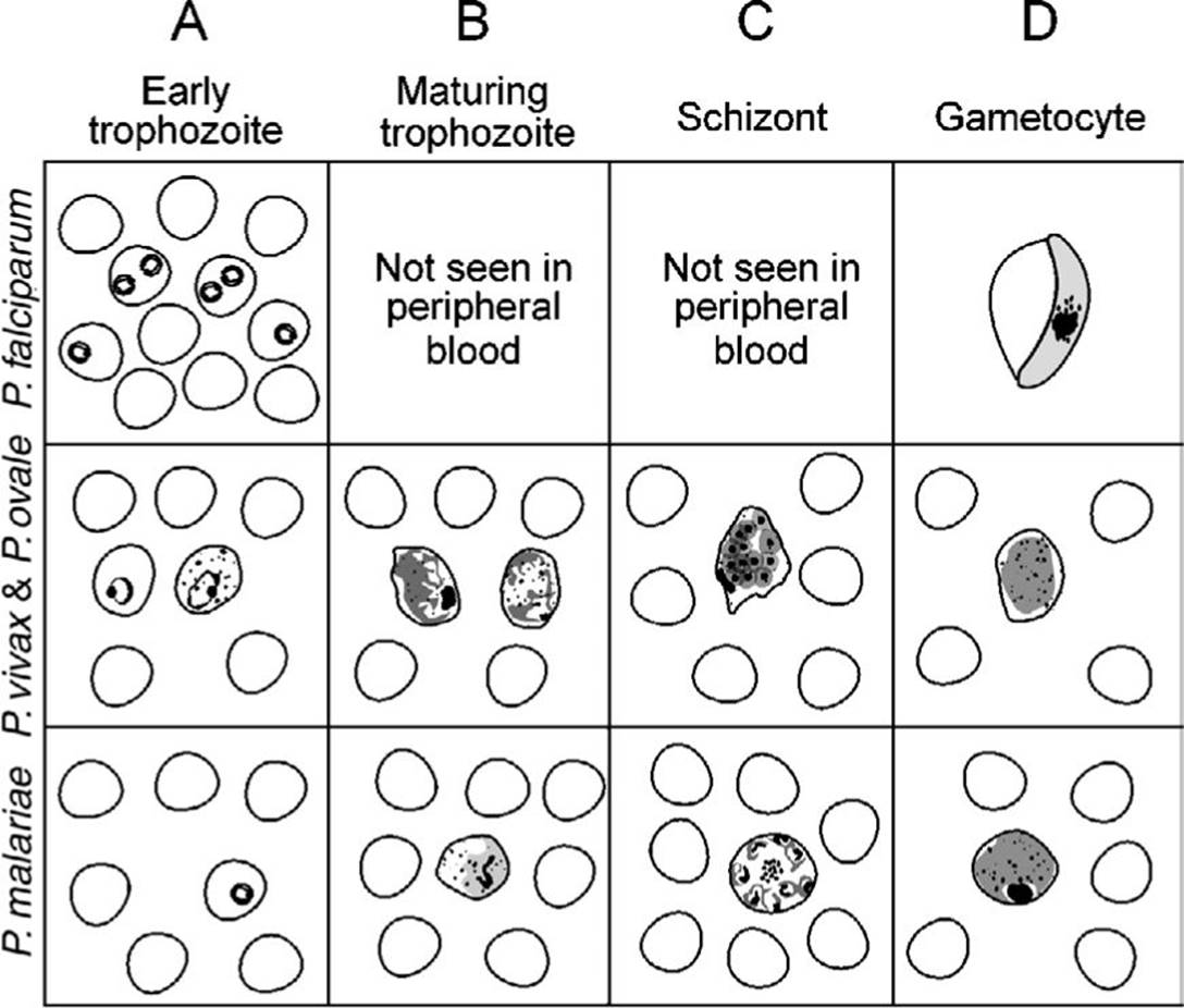
Figure 12.2. Typical blood smear findings for various forms of malaria. (Adapted from Schaechter M, Engleberg NC, Eisenstein BI, Medoff G, eds. Mechanisms of Microbial Disease. 3rd ed. Baltimore, MD: Lippincott Williams & Wilkins; 1999.)
Upon entry into the RBC, some merozoites mature into sexual forms called gametocytes rather than into asexual forms. The male form is smaller and is called a microgametocyte; the larger female form is called a macrogametocyte. Because sexual mating does not occur in the human host, but only in the mosquito, the mosquito is considered the definitive host, and humans are considered the intermediate host.
Once fertilization occurs, a zygote is formed that subsequently develops into an oocyst. The oocyst then forms thousands of infectious sporozoites that gain entry into the mosquito salivary gland, where they are transmitted to the human host.
Life Cycle Differences Between the Various Plasmodium Species
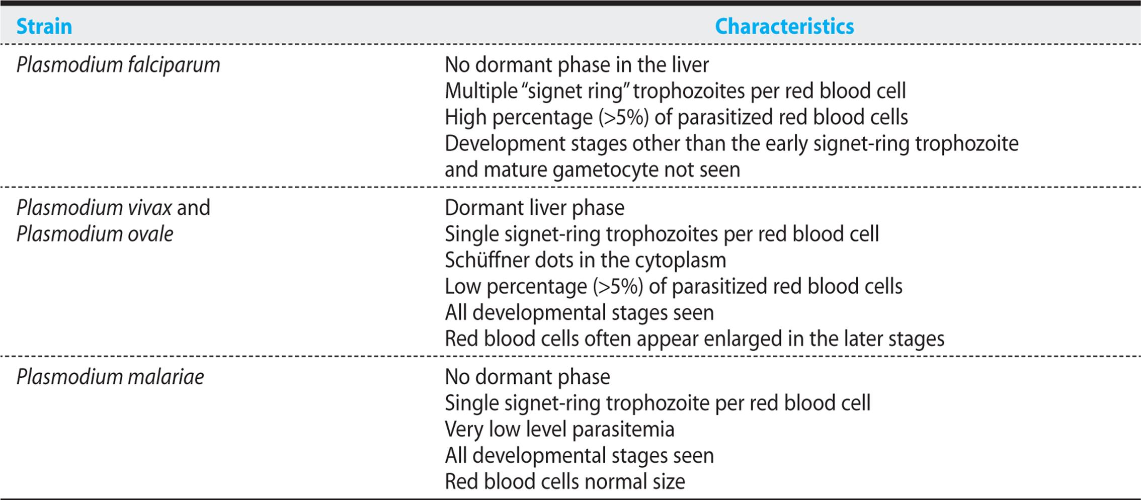
P. falciparum is the most common, and most dangerous, form of malaria. Unlike the sporozoites of other strains, all falciparum sporozoites that enter the liver remain active and develop into tissue schizonts that proceed to form thousands of merozoites. And unlike the merozoites of other strains, P. falciparum merozoites can infect RBCs of all ages, explaining the high level of parasitized RBCs observed in falciparum malaria. Moreover, in the non-falciparum forms of malaria, only a single merozoite gains entry into a given red cell; in falciparum malaria, multiple merozoites can infect and mature within a single RBC.
Once a merozoite has invaded a RBC, it rapidly matures, asexually divides, and, within 48 hours, lyses the host cell. This rapid asexual reproduction produces a rapid rise in the percentage of infected host RBCs, and as the percentage of parasitized RBCs increases, the risk of death or serious complications also increases.
P. falciparum is more harmful to the host because invasion by this strain is uniquely associated with the formation of RBC membrane knobs that tightly adhere to the vascular endothelium through ICAM-1 and CD36 receptors. These knobs express erythrocyte membrane protein 1 on their surface, and this protein binds complement receptor 1 (CR-1) on uninfected red cells, causing red cell clumping (“rosetting”). These adherent RBCs block blood flow in small blood vessels, causing severe hypoxic damage, particularly to the brain and kidneys. Because RBC adherence develops as the merozoite matures beyond the early trophozoite stage, other maturation stages of the parasite (with the exception of the banana-shaped gametocytes) are rarely seen in the peripheral blood (see Table 12.1 and Figure 12.2).
Table 12.1. Differences in Malaria Strains
KEY POINTS
About the Lifecycle of Plasmodium falciparum
1. P. falciparum is the most dangerous form of malaria because it
a) infects red blood cells (RBCs) of all ages and causes high levels of parasitemia;
b) induces the formation of knobs on the RBC surface that adhere to vessel walls and to uninfected RBCs, causing obstruction and local hypoxia;
c) can cause severe hemolysis, renal failure, central nervous system damage, and pulmonary edema.
P. vivax is the next most common form of malaria. P. malariae is less common, and P. ovale is a rare human infection. When a female anopheline mosquito bites an infected human, gametocytes are taken in with the blood. P. vivax and P. ovale can form hypnozoites that can remain dormant within the liver for months before becoming active tissue schizonts. This behavior explains the ability of these strains to relapse 6-11 months after initial treatment. P. malariae has no dormant liver phase, but can persist as a low-level infection for up to 30 years. P. vivax and P. ovale merozoites bind only young RBCs, having the highest affinity for reticulocytes. P. malariae tends to infect older RBCs. The inability of these strains to infect a broad age range of RBCs explains their low level of parasitemia. Furthermore, these three strains do not form knobs and do not obstruct the microcirculation, explaining their milder clinical manifestations.
In Malaysian Borneo and Peninsular Malaysia, P. knowlesi, a form of malaria formerly thought to infect only monkeys, has been identified in humans. Its trophozoite stage is similar in morphology to that of P. falciparum, and its schizont stage is similar to that of P. malariae, Because of the presence of schizonts, P. knowlesi can be mistaken for P. malariae. The level of parasitism can be high, resulting in fatal infections. P. knowlesi should be considered in individuals traveling to forested tropical regions where monkeys are known to be infected.
Genetic and Other Determinants of Susceptibility to Malaria
In areas in which malaria is endemic, the high prevalence of genetic traits that reduce susceptibility to malaria serve as remarkable examples of Darwinian evolution. Specific mutations that affect the surface proteins, cytoskeleton, and hemoglobin of RBCs all interfere with Plasmodium invasion, survival, and spread, and thereby provide a survival advantage to the infected host. Absence of the Duffy blood group antigen blocks invasion by P. vivax. This strain of malaria must bind to this particular blood group antigen to gain entry into RBCs. A significant number of black Africans are Duffy-negative and are resistant to P. vivax. Individuals with mutations in CR-1 demonstrate reduced rosetting in association with P. falciparum and have a decreased propensity to produce cerebral malaria. Individuals with hereditary ovalocytosis, elliptocytosis, and spherocytosis all have defects in specific RBC cytoskeleton proteins, and these defects interfere with entry and release of the malaria parasite.
A broad range of hemoglobinopathies is protective against malaria. The high prevalence of sickle-cell disease and sickle-cell trait in Africa illustrates the frighteningly efficient selective powers of the deadly P. falciparum parasite. Parasite growth is slowed in cells with sickle-cell hemoglobin (HbS). In addition, when parasitized RBCs that contain HbS form membrane knobs and become trapped in small vessels, oxygen tension decreases, and the HbS polymerizes, resulting in sickling of RBCs. The polymerization of HbS kills the P. falciparum parasite, preventing the infection from progressing. As a consequence, people with sickle-cell trait and sickle-cell disease are resistant to severe P. falciparum infection. Because the other strains of malaria do not form knobs and do not become trapped in blood vessels, HbS does not protect against P. vivax, P. ovale, or P. malariae. A number of other hemoglobinopathies including HbC, HbE, α-thalassemia, and to a lesser extent, β-thalassemia reduce the severity of P. falciparum,accounting for their increased prevalence in endemic areas. Neonates are protected from severe malaria as a consequence of fetal hemoglobin, which interferes with the intracellular growth of P. falciparum.
KEY POINTS
About Genetics and Other Factors that Affect Susceptibility to Malaria
1. Surface proteins on red blood cells:
a) Individuals negative for the Duffy blood group antigen are resistant to Plasmodium vivax.
b) Complement receptor 1 mutations reduce the severity of P. falciparum infection.
2. Cytoskeleton defects in red blood cells are protective:
a) Hereditary ovalocytosis
b) Hereditary elliptocytosis
c) Hereditary spherocytosis
3. Hemoglobinopathies confer resistance:
a) Sickle-cell disease and sickle-cell trait are resistant to P. falciparum.
b) Other hemoglobin mutations and fetal hemoglobin are also resistant to P. falciparum.
4. Low-level immunity increases the risk of severe disease:
a) Population immunity wanes in areas with low attack rates.
b) Tourists lack immunity.
c) In pregnant women, the placenta is affected, resulting in low birth weight infants.
In areas that have a high incidence of malaria, the indigenous population is continually exposed to the parasite, resulting in a high level of immunity. In these regions, severe disease is rare. However, because the immune response to malaria is short-lived, immunity wanes in regions in which malaria has been controlled and the attack rate is low. Paradoxically, the percentage of patients developing severe disease increases in these regions. Tourists with no previous exposure to malaria are at highest risk of life-threatening disease (see case 12.1). Pregnant women and their fetuses are also at risk. P. falciparum binds to chondroitin sulfate A in the intervillous space of the placenta, causing hemolytic anemia, which leads to low birth weight infants.
Clinical Presentation
CASE 12.1
A married couple was sailing in the Caribbean near Jamaica with their three children. They lived primarily on their boat, but took several-day trips to a small island off the coast of Jamaica. They noted some mosquito bites and ate some fruit while on the island. The man and the woman both suddenly came down with fever, chills, muscle aches, and loss of appetite. About 3 days into the illness, the man became jaundiced and began passing dark urine. The family sought treatment from a local Jamaican physician, who diagnosed hepatitis secondary to ingestion of a toxic food. Two days later, the man became comatose and died. The woman was referred to the university hospital for possible liver transplant. On further questioning, the medical staff learned that none of the children were sick despite eating the same diet. The family had begun a course of malaria prophylaxis. However, the parents had developed side effects from the chloroquine and had discontinued prophylaxis 2 weeks before the onset of their illness. Thin smears of the woman’s blood revealed many signet-ring trophozoites, with a parasitemia level estimated to be 10%. She was treated with intravenous quinine and rapidly improved. In retrospect, her husband was determined to have died of untreated blackwater fever.
As described in case 12.1, the clinical manifestations of malaria are nonspecific. If the exposure history is not appreciated, the infection can be mistaken for other febrile illnesses. The incubation period is generally 9-40 days, but it may be prolonged in cases of non-falciparum malaria (6-12 months in P. vivax, and years for P. malariae and P. ovale).
The hallmark of all forms of malaria is fever. Fever can occur at regular 2 to 3-day intervals in P. vivax and P. malariae, or in a more irregular pattern with P. falciparum. Fever generally occurs soon after lysis of the RBCs and release of the merozoites. Three classic stages of the febrile paroxysms have been described:
1. The initial “cold stage” occurs 15–60 minutes before the onset of fever. During this period, the patient feels cold and has shaking chills.
2. These symptoms are followed by the “hot stage,” during which body temperature rises between 39°C and 41°C. Fever is associated with lassitude, loss of appetite, and vague pains in the bones and joints. In nonendemic areas, these symptoms are most commonly mistaken for influenza. The clinician must always consider malaria in individuals who develop flu-like symptoms after returning from a developing country. Other symptoms associated with the fever include tachycardia, hypotension, cough, headache, back pain, nausea, abdominal pain, vomiting, diarrhea, and altered consciousness.
3. Usually within 2–hours, symptoms progress to the third “sweating” stage, at which time the patient develops marked diaphoresis, followed by resolution of the fever, profound fatigue, and a desire to sleep.
Other symptoms depend on the strain of malaria. In cases of P. vivax, P. ovale, and P. malariae, there are a few additional symptoms. However, depending on the prior immune status of the host, individuals with P. falciparum can develop a severe fatal illness similar to that described in case 12.1. Because P. falciparum infects RBCs of all ages and induces the formation of knobs on the RBC surface that adhere to endothelial cells and obstruct small vessels, this parasite can cause severe damage, particularly to the kidneys, brain, and lungs. Tourists who have no immunity to P. falciparum and people who have undergone splenectomy can develop very high levels of parasitemia that result in profound hemolysis. The marked release of hemoglobin can exceed the metabolic capacity of the liver. The resulting rise in unconjugated bilirubin in the bloodstream produces jaundice. Hemoglobin also may be excreted into the urine, causing the urine to become dark. The combination of jaundice and hemoglobinuria has been called blackwater fever.
Severe malaria is commonly complicated by renal failure. Heavy infection with P. falciparum also results in obstruction of the small arteries in the central nervous system (CNS), leading to hypoxia. Hypoglycemia may also contribute to CNS dysfunction. Confusion and obtundation can rapidly progress to coma. Grand mal seizures may also develop. Pulmonary edema is a less common complication of P. falciparum infection, being the result of fluid leakage from pulmonary capillaries into the alveoli.
KEY POINTS
About Clinical Presentation in Plasmodium Infection
1. Always consider malaria in the traveler from a developing country who
a) presents with an influenza-like syndrome,
b) presents with jaundice, or
c) presents with confusion or obtundation.
Diagnosis
Microscopic examination of a Giemsa-stained blood smear remains the primary way to identify malaria. In P. falciparum, blood smears are best taken just after the fever peak, when early ring forms are most abundant in peripheral RBCs. At other times, P. falciparum becomes trapped in the capillaries and may not be found in the peripheral blood. In P. vivax, P. malariae, and P. ovale, various stages of the parasite are present at all times, and therefore diagnostic smears can be taken at any time. Because parasites can be absent between attacks, the blood must be examined on 3-4 successive days before malaria can be ruled out. Presence of pigment in peripheral monocytes or neutrophils should encourage a continued search for parasites. Thin smears need to be examined for at least 15 minutes using a high-power oil objective microscope (1000X magnification). Thick smears are the most reliable method for detecting malaria. A 5-minute search will generally yield the diagnosis.
The clinician’s primary goal is to differentiate potentially fatal P. falciparum from other more benign forms of malaria (see Table 12.1). For this purpose, one rapid diagnostic test has been approved by the U.S. Food and Drug Administration (FDA), Binax NOW. This test utilizes immunochromatographic lateral flow technology. Through capillary action, the blood sample is passed over nitrocellulose with immobilized specific antibodies to the malaria antigens HRP-2 and aldolase. The assay was shown to be more sensitive than Giemsa stain and microscopy for the diagnosis of P. falciparum, demonstrating a sensitivity of 95% and specificity of 94%. The test takes only 15 minutes, and allows inexperienced caregivers to rapidly institute appropriate therapy. It is recommended that all rapid tests be confirmed by microscopic examination. A polymerase chain reaction (PCR) amplification of parasite DNA or mRNA has been developed, but is not commercially available.
KEY POINTS
About Laboratory Diagnosis of Malaria
1. The focus must be on differentiating falciparum malaria from other forms of the disease.
2. Blood smear remains the preferred method. In falciparum malaria, signet-ring forms are most abundant on peripheral smear immediately after a fever spike
3. An immunochromatographic lateral flow rapid diagnostic test is now commercially available that detects P. falciparum with high sensitivity and specificity
4. Polymerase chain reaction methods have been developed but are not commercially available.
Anemia, elevated levels of lactic dehydrogenase, and increased reticulocytes are associated with RBC hemolysis. An elevated unconjugated bilirubin level without a significant increase in hepatic enzymes is also observed when hemolysis is severe. A reduced white blood cell (WBC) count is noted in a high percentage of patients, and thrombocytopenia is common. Elevated serum creatinine, proteinuria, and hemoglobinuria are found in severe cases of P. falciparum. Hypoglycemia may also complicate severe cases of P. falciparum, requiring close monitoring of blood sugars during the acute illness.
Prophylaxis and Treatment
Drug treatment exploits unique targets in the parasite not found in host cells. The aminoquinolines, chloroquine, quinine, mefloquine, primaquine, and halofantrine inhibit proteolysis of hemoglobin in the food vacuole and inhibit the heme polymerase that Plasmodium requires for production of malaria pigment. Inhibition of these functions kills the organism. Pyrimethamine, sulfonamides, and dapsone are folate antagonists (see Chapter 1). Atovaquone inhibits parasite mitochondrial transport. Artemisinin derivatives bind iron in the malarial pigment to produce free radicals that damage parasite proteins. These derivatives are faster-acting than quinine, and they have activity against all stages of the intraerythrocytic life cycle.
In recent years, many areas of Africa, northern South America, India, and Southeast Asia have become populated with chloroquine-resistant P. falciparum. These strains contain an energy-dependent chloroquine efflux mechanism that prevents the drug from concentrating in the parasite. Resistance to mefloquine and halofantrine has also developed, being seen primarily in Southeast Asia.
Chemoprophylaxis should start 2 weeks before departure to an endemic area when taking chloroquine and mefloquine, and 1-2 days before travel for atovaquone–proguanil as well as doxycycline. Prophylaxis should be continued for 4 weeks after return if taking chloroquine and mefloquine and for 7 days if taking atovaquone-proguanil or doxycycline. Because of the continual changes in resistance patterns, up-to-date prophylactic and treatment regimens should be reviewed at the CDC’s Web site (www.cdc.gov/travel, accessed January 3, 2013). For areas with chloroquine-susceptible P. falciparum, chloroquine is the drug of choice. The adult dosage is 300 mg base (500 mg of chloroquine phosphate) orally once per week. In areas of chloroquine-resistance, atovaquone–proguanil (atova-quone 250 mg combined with proguanil 100 mg, the combination tablet is called Malarone) orally once per day, mefloquine 250 mg (228 mg base) orally once per week, or doxycycline 100 mg orally once per day. Mefloquine should be avoided in individuals with psychiatric disorders, seizure disorders, or cardiac conduction abnormalities.
KEY POINTS
About Malaria Prophylaxis
1. Determine if the traveler will be visiting areas with chloroquine-resistant strains (check www.cdc.org/travel).
2. Begin prophylaxis 2 weeks or 1-2 days before travel (depending on the medication).
3. Chloroquine is recommended for chloroquine-sensitive areas.
4. Atovaquone-proguanil, doxycycline, or mefloquine recommended for chloroquine-resistant areas.
5. Continue prophylaxis for 1-4 weeks (depending on the medication) after return.
A vaccine, TS, S/AS01 was recently shown to provide protection against both clinical and severe malaria in African children, but is not yet commercially available.
All individuals without previous immunity who contract falciparum malaria should be hospitalized, because their clinical course can be unpredictable. Patients with the P. vivax, P. ovale, and P. malariaestrains can usually be treated as outpatients if follow-up will be reliable. The treatment of these three strains and of chloroquine-susceptible P. falciparum is the same: an initial dose of oral chloroquine 600 mg base (1000 mg chloroquine phosphate), followed 6 hours later by 300 mg base (500 mg phosphate), repeated on days 2 and 3. To prevent relapse of P. vivax or P. ovale, these infections also require treatment with oral primaquine 15.3 mg phosphate base (26.5 mg phosphate salt) daily for 14 days, or 45 mg base (79 mg salt) weekly for 8 weeks. This agent kills dormant hepatic hypnozoites, preventing their subsequent development into infective schizonts. Before the primaquine is administered, the patient should be tested for glucose-6-phosphate dehydrogenase deficiency, because patients with this deficiency are at risk of severe hemolysis during primaquine treatment.
Given the worldwide prevalence of chloroquine resistance, unless absolute assurance can be obtained that travel was only in regions with chloroquine-sensitive P. falciparum, patients should be presumed to have a resistant strain. Treatment of chloroquine-resistant P. falciparum is evolving and has become complex. Artemisinin derivatives have shown superior efficacy for severe chloroquine-resistant P. falciparum infection producing cure rates of 95%. These agents also reduce gametocyte carriage. Their use therefore decreases infectivity after treatment, and can eliminate malaria transmission in endemic areas. Artemether 20 mg/lumefantrine 120 mg (trade name Coartem) has been approved by the FDA and is now the treatment of choice for chloroquine-resistant P. falciparum. Individuals >35 kg should receive 4 tablets per dose by mouth. The first dose should be followed by a second dose 8 hours later, then 1 dose twice a day for the next 2 days. Artesunates are short-acting, and they should always be combined with one or more other classes of antimalarial agents such as lumefantrine, pyronaridine, atovaquoneproguanil or mefloquine to prevent the development of resistance.
KEY POINTS
About Choosing Chemotherapy for Plasmodium Infection
1. Determine whether the traveler came from a chloroquine-resistant area:
a) For chloroquine-sensitive strains, use chloroquine.
b) For chloroquine-resistant strains, use artemether/lumefantrine or alternatively atovaquone–proguanil, quinine, or mefloquine.
2. Determine whether the patient is too ill to take oral medicines (requires intravenous quinidine).
3. Determine whether the patient has Plasmodium vivax or ovale (requires primaquine, if not deficient in glucose-6-phosphate dehydrogenase).
4. Refer to Web sites run by health authorities for the most current antimalarial regimens (Table 12.2).
Table 12.2. Online Sources of Current Guidelines for Antimalarial Therapy

Alternative regimens include atovaquone-proguanil (250 mg/100 mg tablets) four tablets daily for 3 days, which is generally a well-tolerated regimen; quinine 650 mg every 8 hours for 3-7 days, plus doxycycline 100 mg twice daily PO for 7 days; and mefloquine 750 mg PO followed 6-12 hours later by 500 mg. Quinine has a bitter taste and can result in reversible tinnitus and high-frequency hearing loss, hypoglycemia, and cardiac arrhythmias. Mefloquine administration is associated with vertigo (10-20%), gastrointestinal disturbances, seizures, and (less commonly) psychosis. In addition, both quinine- and mefloquine-resistant P. falciparum are increasing in frequency.
KEY POINTS
About Managing Patients with Plasmodium falciparum
1. Levels of parasitemia above 5% constitute a medical emergency and require immediate institution of antimalarial treatment.
2. Hematocrit, blood sugar, volume status, cardiac rhythm, renal function, central nervous system function, and arterial oxygenation must all be closely monitored.
3. In the nonimmune host, the course of P. falciparum infection is not predictable.
4. The severity of organ damage and risk of death correlate with the level of parasitemia.
If a patient is too ill to take oral medicines, intravenous quinidine is the treatment of choice. This drug is three to four times more active than is intravenous quinine, and serum levels can be measured. Furthermore, parenteral quinine is no longer available in the United States. Quinidine gluconate salt 10 mg/kg loading dose (maximum 600 mg) in normal saline should be infused slowly over 1-2 hours, followed by a continuous infusion of 0.02 mg/kg every minute until the patient is able to take oral medication. Given the rapid changes in malaria resistance patterns and newly reported clinical trials, health care providers should refer to excellent Web sites operated by recognized authorities that outline up-to-date treatment regimens (Table 12.2).
The risk of end-organ damage and death increases with the patient’s level of parasitemia. Levels above 5% constitute a medical emergency, and patients with these levels require intensive treatment. Patients with no immunity and levels of P. falciparum parasitemia above 10-15% should be considered for exchange transfusion, a measure that can be life-saving. However, patients with levels of parasitemia of greater than 50% have survived without blood exchange. Volume status, renal function, and serum glucose must be carefully monitored. Respirator support may be required in cases of severe pulmonary edema. Intravenous steroids have been shown to be harmful in cases of cerebral malaria, and those agents should therefore be avoided. Because of the risk of arrhythmias associated with quinine, quinidine, mefloquine, and halofan–trine, cardiac function should be monitored in patients treated with those agents.
TISSUE PROTOZOA
LEISHMANIASIS
GUIDING QUESTIONS
1. How is leishmaniasis contracted, and where is this disease most commonly found?
2. Which form of immunity is most important for protecting against Leishmania, and are patients with HIV or an organ transplant at increased risk of developing leishmaniasis?
3. How do patients with visceral leishmaniasis usually present clinically, and which diseases can this infection mimic?
4. Where are lesions of cutaneous leishmaniasis usually located, and why?
5. What is the therapy approved by the U.S. Food and Drug Administration for visceral leishmaniasis?
POTENTIAL SEVERITY
Visceral leishmaniasis is a chronic disease that can cause severe morbidity and death in debilitated and immunocompromised hosts.
Prevalence, Epidemiology, and Life Cycle
Leishmania has caused major epidemics in eastern India, Bangladesh, and East Africa. Urban outbreaks have been reported in the cities of northeastern Brazil. A small number of American military personnel contracted leishmaniasis during the Persian Gulf War in 1991 and in Afghanistan more recently. Indigenous cases have been reported occasionally in the United States, but most US cases result from travel to a tropical country. Leishmaniasis has emerged as an opportunistic infection in patients with HIV or an organ transplant.
The Leishmania parasite is transmitted by the female phlebotomine sandfly. Sandflies breed in cracks in the walls of dwellings, in rubbish, and in rodent burrows. Because they are weak fliers, sandflies remain close to the ground near their breeding sites, resulting in localized pockets of infectious insects. Humans and other animals infected with Leishmania serve as reservoirs. The sandfly bites the infected host and ingests blood containing the nonflagellated form called an amastigote. In the digestive tract of the insect, the amastigote develops into a flagellated spindle-shaped promastigote. When the infected sandfly takes its blood meal from an uninfected human, the promastigote enters the host’s bloodstream. The promastigote then binds to complement receptors on macrophages and is ingested. Within the phagolysosome, the promastigote differentiates into an amastigote. The amastigote is resistant to lysozyme damage and depends on the low pH of the phagolysosome for the uptake of nutrients. The parasite multiplies by simple division and eventually is released to infect other cells.
KEY POINTS
About the Epidemiology and Life Cycle of Leishmania
1. Contracted in tropical areas where the phlebotomine sandfly is common; rare in the United States Found in South America, India, Bangladesh, the Middle East, and East Africa.
2. Flagellated promastigote introduced by the sandfly is ingested by macrophages.
3. In the macrophage, Leishmania develops into a nonflagellated amastigote that lives happily within the macrophage phagolysosome.
4. This intracellular parasite is controlled by activation of the Th1 cell-mediated immune response that increases levels of interferon-γ.
5. Leishmaniasis can be an opportunistic infection in patients with HIV or an organ transplant.
Cell-mediated immunity plays an important role in controlling leishmaniasis. Interferon-γ activates macrophages to kill the amastigote by inducing the production of nitric oxide. Resolution of leishmanial infection is associated with the expression of CD4+ T cells of the Th1 type, which secrete interferon-γ and interleukin 2. Progression of infection is associated with Leishmania-induced expansion of CD4+ cells of the Th2 type that produce interleukin 4, a cytokine that inhibits the production of Th1 cells and the activation of interferon-γ production.
Clinical Presentation
There are three forms of leishmaniasis: visceral, cutaneous, and mucosal. A single species can produce more than one syndrome, and each syndrome is produced by multiple different species.
Visceral Leishmaniasis (Kala-Azar)
In different areas of the world, certain Leishmania species tend to be most commonly associated with the visceral form of the disease: L. donovani (in India), L. infantum (Middle East), L. infantum (chagasi)(Latin America), and L. amazonensis (Brazil). After inoculation of promastigotes into the skin, a small papule may be noticed. Leishmania amastigotes subsequently silently invade macrophages throughout the reticuloendothelial system. Usually 3-8 months pass before the burden of organisms increases to a level that causes symptoms.
The onset of symptoms can be gradual or sudden. In subacute cases, the patient will experience slow but progressive enlargement of the abdomen as a result of hepatosplenomegaly. Increased abdominal girth is accompanied by intermittent fever, weakness, loss of appetite, and weight loss. This presentation can be mistaken for lymphoma, infectious mononucleosis, brucellosis, chronic malaria, and hepatosplenic schistosomiasis. In acute cases, an abrupt onset of high fever and chills mimics malaria or an acute bacterial infection. On physical examination, the spleen may be massively enlarged, hard, and nontender. Hepatomegaly is also present. The skin tends to be dry and thin, and in light-skinned individuals, it takes on a grayish tint. This characteristic accounts for the Indian name Kala-azar, which means “black fever.” On laboratory examination, anemia, leukopenia, and hypergammaglobulinemia are common.
KEY POINTS
About Visceral Leishmaniasis
1. Incubation period is 3-8 months.
2. Subacute onset presents with increased abdominal swelling (because of massive splenomegaly and hepatomegaly), intermittent fever, and weight loss that can be mistaken for lymphoma or infectious mononucleosis
3. Acute onset presents with persistent high fever mimicking bacteremia or malaria.
4. Anemia, leukopenia, and hypergammaglobulinemia are common.
5. Patients with HIV may have disseminated disease without splenomegaly.
6. Diagnosis is made by biopsy, Giemsa stain showing amastigotes, and rK39 antigen test.
The diagnosis is made when a biopsy of lymphatic tissue or bone marrow demonstrates amastigotes on Wright or Giemsa stain. Enzyme-linked immunosorbent assays (ELISAs) usually demonstrate high antileishmanial antibody titers. However, this test frequently cross-reacts with antibodies to other pathogens. Most recently recombinant kinesin antigen (rK39) ELISA and immunochromatographic strip assays have been developed. The sensitivity of this test varies depending on the region, demonstrating 95% sensitivity and 90% specificity in India, but lower values in East Africa.
Patients with HIV infection frequently fail to develop antibody titers. Splenomegaly may not be present in these patients, and infection may disseminate to the lungs, pleura, gastrointestinal tract, or bone marrow (causing aplastic anemia). In patients with HIV, amastigotes may be identified in macrophages from bronchoalveolar lavage, pleural effusion, bone marrow aspiration, or even buffy coat samples of the peripheral blood.
CUTANEOUS LEISHMANIASIS
The cutaneous form of leishmaniasis is widespread, and it is a problem chiefly for farmers, settlers, troops, and tourists in the Middle East and Central and South America. The species most commonly associated with cutaneous disease are L. major and L. tropica (found in the Middle East, India, Pakistan, and Asia), and L. mexicana, L. braziliensis, L. amazonensis, and L. panamensis (in Central and South America). L. mexicana has been reported in Texas.
After a sandfly bite, significant skin lesions generally take 2 weeks to several months to develop. Lesions usually develop on exposed areas. They are the result of amastigotes multiplying in mononuclear cells within the skin and causing a granulomatous inflammatory reaction. Single or multiple lesions may be found, with varying morphology. Lesions may be crusted and dry, or moist and exudative. Shallow and circular ulcers with sharp, raised borders may develop and progressively increase in size, becoming “pizza-like” in appearance as a result of the beefy red of the ulcer base being combined with a yellow exudate. Lesions may become secondarily infected with staphylococci or streptococci.
The diagnosis is made from a biopsy of the raised border of the skin lesion where Leishmania-infected macrophages are most abundant. Amastigotes are seen on Giemsa stain. PCR tests are also available that are capable of identifying the specific species. These tests are usually highly sensitive, but are not widely available.
MUCOSAL LEISHMANIASIS
Mucosal leishmaniasis is a less common manifestation that is caused primarily by L. braziliensis. Only 2-3% of patients with skin lesions develop this complication. Organisms invade mononuclear cells in the mucosa. The nose is most commonly involved, resulting in nasal stuffiness, discharge, pain, or epistaxis. Later, the nasal septum is destroyed, and the nose collapses. Involvement of the genital mucosa and trachea has also been reported. Diagnosis is made by biopsy.
KEY POINTS
About Cutaneous and Mucosal Leishmaniasis
1. A problem for farmers, settlers, troops, and tourists; incubation period is 2 weeks to 2 months.
2. Found throughout the world; cases have been reported in Texas.
3. Lesions occur primarily on exposed areas.
4. Dry or moist in appearance, ulcers have sharp, raised boarders; “pizza-like” lesions are common.
5. Mucosal disease is rarer, and usually involves the nose.
6. Diagnosis is made by biopsy, always from the border of skin lesions.
Treatment
The only drug approved in the United States for treatment of leishmaniasis is liposomal amphotericin B. For visceral leishmaniasis in immunocompetent patients, administer 3 mg/kg daily on days 1–5, 14, and 21. The course can be repeated if the parasite persists. For the immunocompromised host, the recommended regimen is liposomal amphotericin B 4 mg/kg daily administered on days 1–5, 10, 17, 24, 31, and 38. Relapses are common in HIV-infected hosts (see Table 12.2).
Outside the United States, pentavalent antimony continues be used; however, this treatment is associated with many side effects, including abdominal pain, anorexia, nausea and vomiting, and myalgias. Amylase and lipase levels often rise. Miltefosine, a phosphocholine analog has antileishmanial activity in vitro and in vivo, and acts by interfering with the parasite’s cell-signaling pathways and membrane synthesis. This agent has successfully treated Indian visceral disease. However, it is not available in the United States.
Treatment of cutaneous leishmaniasis depends on the location of the infection. The lesions can heal spontaneously, and so, if there is no mucosal involvement and if the lesions are located in areas of no cosmetic concern, they can be followed without therapy or treated topically with 15% puromycin and 12% methylbenzethonium chloride. Thermotherapy (warming the affected region with radiofrequency waves to 50°C for one treatment of 30 seconds) has proven effective in a high percentage of cases, and that approach compares favorably with 21 days of intralesional administration of pentavalent antimony. Patients with mucosal involvement, progressive lesions, or lesions in cosmetically sensitive areas require treatment with intravenous or intramuscular pentavalent antimony (20 mg/kg daily for 20 days, available through the CDC). Fluconazole (500 mg twice daily for 6 weeks) has been associated with modest response rates. Miltefosine has proved successful against some forms of cutaneous leishmaniasis, but other species are refractory.
KEY POINTS
About the Treatment of Leishmaniasis
1. Visceral disease:
a) Liposomal amphotericin B is the only approved therapy.
b) Miltefosine appears promising, but had not been approved in the United States.
2. Cutaneous:
a) May heal spontaneously.
b) Thermotherapy is safe and effective.
c) In cases of mucosal involvement, infection in a cosmetically sensitive site, or failure to heal, fluconazole or pentavalent antimony is recommended.
d) Miltefosine effective for some Leishmania species, but not others.
TRYPANOSOMA CRUZI
GUIDING QUESTIONS
1. Which insect is responsible for transmitting this disease, and is the disease commonly transmitted to tourists? Why, or why not?
2. How do this insect’s toilet habits affect transmission to the human host?
3. Which organs are most commonly affected by chronic Chagas disease?
POTENTIAL SEVERITY
A chronic disorder that can lead to fatal cardiomyopathy.
Prevalence, Epidemiology, and Life Cycle
Chagas disease caused by Trypanosoma cruzi is found throughout Central and South America. Between 8 and 10 million people are infected with T. cruzi, and 30–40% will suffer cardiomyopathy and/or digestive mega syndromes. The World Health Organization has designed Chagas disease as one of the 13 most neglected tropical diseases. With improvement in substandard housing, the incidence of this disease among young people is decreasing, but this disease remains a major cause of morbidity and mortality.
The parasite is transmitted by triatomine bugs, commonly call kissing bugs, which suck blood from their host. This insect contains trypomastigotes in its gut. At the same time that it bites the host, it also defecates, depositing trypomastigotes on the skin. The human host then scratches the itchy bite, introducing the parasite into the wound and subsequently into the bloodstream. Mucous membranes, the conjunctiva, and breaks in the skin are common sites of entry. Once in the bloodstream, the trypomastigotes enter host cells and differentiate into amastigotes that multiply, filling the cell cytoplasm. They then differentiate again into trypomastigotes, and the cell ruptures, spreading the parasite to adjacent cells and into the bloodstream. Asymptomatic parasitemia is common. In endemic areas, the parasite can be transmitted by blood transfusions. Because the triatomine bugs take up residence in the cracks of primitive homes, this infection occurs almost exclusively among poor rural people. The disease is most commonly transmitted in young children. If one member of a family presents with acute disease, all pediatric family members should be screened for asymptomatic disease.
KEY POINTS
About the Life Cycle of Trypanosoma Cruzi
1. Transmitted by triatomine (kissing) bugs, that carries the trypomastigote in their feces.
2. The host allows the parasite to enter the bloodstream by scratching and rubbing infected insect feces into the skin.
3. The triatomine bugs live in the cracks of substandard housing.
4. The disease affects mainly poor rural people, not tourists.
Chagas disease has not been reported in tourists, because they are unlikely to be exposed to primitive living quarters. Vector control measures and educational programs have helped to reduce the incidence of disease. Insecticide impregnation of bed nets has proven to be an inexpensive and effective control measure.
Clinical Presentation
Acute Chagas disease often causes minimal symptoms. About 1 week after the parasite enters the skin, an area of localized swelling called a chagoma develops, often in association with local lymph node swelling. Entry of the parasite via the conjunctiva causes periorbital edema (Romaña’s sign). Onset of local edema is quickly followed by fever, malaise, anorexia, and edema of the face and legs. Occasionally, myocarditis or encephalitis may develop.
Years to decades after the primary infection, 30-40% of individuals go on to develop chronic Chagas disease. The heart is the organ that is primarily damaged. Severe cardiomyopathy results in thromboembolism, congestive heart failure, and life-threatening arrhythmias. Esophageal involvement can lead to megaesophagus associated with dysphagia, regurgitation, and aspiration pneumonia. Chagasic megacolon is another manifestation of chronic disease causing constipation and bowel obstruction that can lead to perforation and bacterial sepsis. In immunocompromised hosts such as organ transplant patients and patients with AIDS, T. cruzi can reactivate, presenting with manifestations of chronic Chagas disease. Unlike normal hosts, immunocompromised patients are also at risk of developing T. cruzibrain abscesses.
KEY POINTS
About the Clinical Presentation of Chagas Disease
1. Acute disease is associated with localized areas of swelling called chagomas.
2. Chronic disease develops in 30–40% of cases decades after initial infection.
3. Chronic disease affects
a) the heart, causing a cardiomyopathy associated congestive heart failure, emboli, and arrhythmias; and
b) the gastrointestinal tract, causing megaesophagus and megacolon.
Diagnosis
Acute disease can be diagnosed by examining Giemsa-stained blood or buffy coat smears. The trypomastigotes (whose length is approximately twice the diameter of a RBC) can readily be seen by microscopy. In chronic disease, the diagnosis is made by detecting immunoglobulin G (IgG) antibodies. In the United States, two ELISA tests have been approved by the FDA for detecting clinical disease. ELISA demonstrates high sensitivity and specificity, and is being used to screen the blood supply. A quantitative PCR is available through the CDC.
Treatment
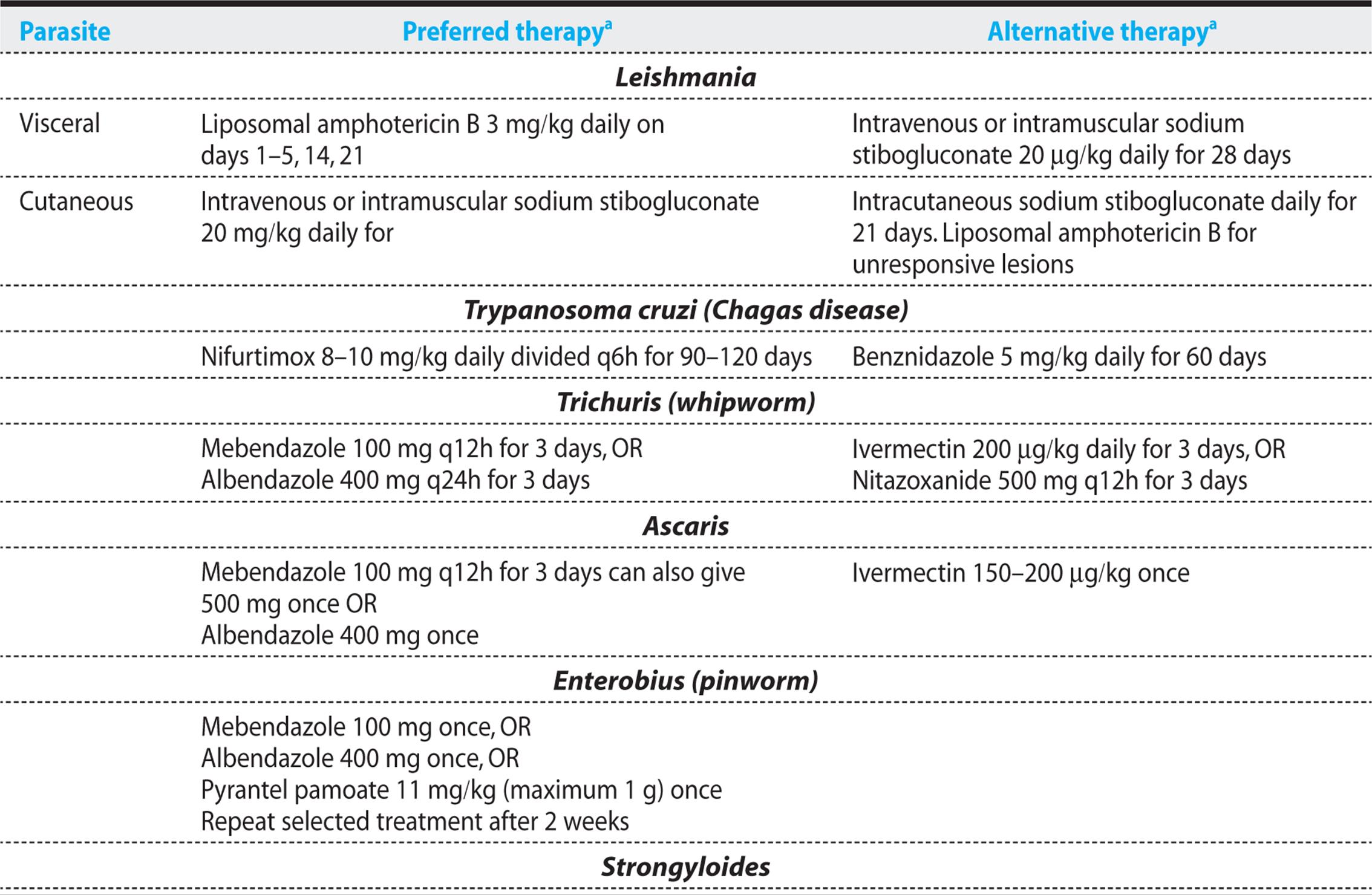
T. cruzi is not sensitive to most antiparasitic drugs (see Table 12.3). Nifurtimox cures about 70% of acute cases. This drug causes gastrointestinal and neurologic side effects in many patients. Benznidazole has a similar cure rate. Peripheral neuropathy, granulocytopenia, and rash are the most common side effects with that agent. Treatment with these two agents is now recommended for chronic Chagas disease in patients under age 50 who do not have advanced cardiac or esophageal disease. Recent studies have shown that treatment slows the progression of heart disease.
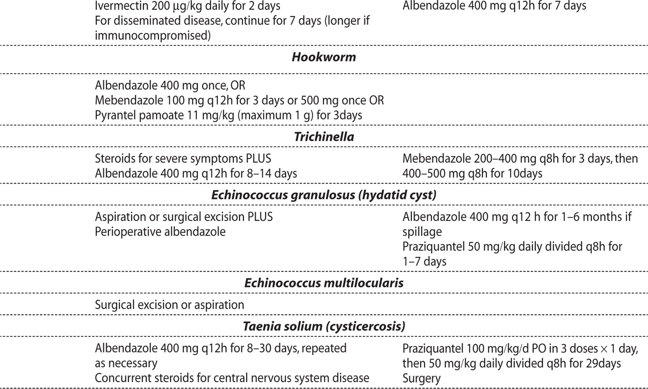
Table 12.3. Antiparasitic Therapy Dosing

KEY POINTS
About the Diagnosis and Treatment of Chagas Disease
1. Acute disease is diagnosed by Giemsa stain of a peripheral blood smear.
2. Chronic disease can be diagnosed by enzyme-linked immunosorbent assay that detects immunoglobulin G antibody to Trypanosoma cruzi or by quantitative PCR.
3. Acute and early chronic disease should both be treated with nifurtimox or benznidazole.
4. Treatment reduces mortality and progression of chronic disease.
TRYPANOSOMA BRUCEI COMPLEX
POTENTIAL SEVERITY
Over weeks to months, this disease can progress to coma, followed by death.
T. brucei complex refers to several Trypanosoma subspecies that are spread by the blood-sucking tsetse fly. Unlike T. cruzi, which takes up residence within cells, T. brucei trypomastigotes multiply within the bloodstream, evading the humoral immune system indefinitely by changing their surface antigens every 5 days. This disease is confined to Africa. No more than a single case per year is imported to the United States. After the initial bite, the infection progresses slowly, with systemic symptoms of fever and lymph node swelling being noted weeks to months later. In the West African form, neurologic manifestations do not develop until months or years after the initial symptoms. In East African trypanosomiasis, systemic complaints may develop days after the insect bite, and CNS complaints may develop within weeks. Symptoms include somnolence, which explains the name “sleeping sickness,” and choreiform movements, tremors, and ataxia mimicking Parkinson disease. Coma and death frequently ensue.
The diagnosis is made by observation of trypomastigotes in Giemsa-stained thick and thin smears of peripheral blood. Trypomastigotes can also be found in the cerebrospinal fluid. The treatment of T. bruceiis complex and depends on the species of the infecting parasite, whether the CNS is involved, and tolerance to the side effects of the treatment regimen. For early infection, pentamidine or suramin is preferred. For late infection, eflornithine combined with nifurtimox or the arsenical melarsoprol is used.
KEY POINTS
About Trypanosoma brucei
1. Transmitted by the blood-sucking tsetse fly.
2. Survives in the bloodstream by continually changing its outer coat antigens.
3. West African form causes lymphadenitis and fever followed months to years later by CNS infection
4. East African form results in somnolence and choreiform movements, tremors and ataxia within weeks. Often, fatal.
5. Treatment is complex and the drugs are associated with significant toxicity.
INTESTINAL HELMINTHS
GUIDING QUESTIONS
1. What are the two ways by which intestinal helminths gain entry to the human host?
2. How does the life cycle of Ascaris differ from that of Trichuris, and how does the difference manifest itself clinically?
3. How is Strongyloides able to persist in the human host for three to four decades?
4. What are the conditions that precipitate Strongyloides hyperinfection syndrome, and why?
5. Which helminth most commonly causes iron deficiency anemia, and why?
POTENTIAL SEVERITY
Infections are often asymptomatic. In the immunocompromised host, Strongyloides can progress to a fatal hyperinfection syndrome.
Helminths include the roundworms (nematodes), flukes (trematodes), and tapeworms (cestodes). These parasites are large, ranging in size from 1 cm to 10 m, and they often live in the human gastrointestinal tract without causing symptoms. Only when the infection is very heavy or the worm migrates to an extraintestinal site, do patients seek medical attention. Transmission to humans results in most cases from contact with human waste. The diagnosis is generally made by examining the stool for eggs, larvae, or adult worms (Figure 12.3).

Figure 12.3. Stool helminths. All eggs drawn to scale. In Strongyloides, only the rhabditiform larvae are usually seen.
INTESTINAL NEMATODES (ROUNDWORMS)
Nematodes can be classified into two groups. Those that gain entry to the host by egg ingestion (Trichuris, Ascaris, and Enterobius) and those that are capable of producing larvae that penetrate the skin of their host (Strongyloidesand hookworm). Roundworm life cycles can also be classified into two groups. One group, Trichuris and Enterobius, attach and grow in the intestine soon after being ingested. The second group, Ascaris, Strongyloides, and hookworm, first penetrate the venous system, enter the lungs, and migrate up the bronchi to the trachea, where they are swallowed. They then take up residence in the gastrointestinal tract (Figure 12.4). These differences in life cycle account for some of the unique clinical characteristics of the various species of nematodes.

Figure 12.4. Comparative life cycles of the intestinal nematodes. (Adapted from Schaechter M, Engleberg NC, Eisenstein BI, Medoff G, eds. Mechanisms of Microbial Disease. 3rd ed. Baltimore, MD: Lippincott Williams & Wilkins; 1999.)
Nematodes Acquired by Ingestion
TRICHURIS TRICHIURA (WHIPWORM)
Trichuris trichiura is one of the most prevalent helminths. More than 2 million people are estimated to be infected in the United States. This parasite is most commonly found in the rural Southeast, particularly Puerto Rico, where the moisture and temperature favor egg maturation. Worldwide, this worm causes infection mainly in poor rural communities with poor sanitation. Humans are the principal host, and infection results from ingestion of embryonated eggs.
KEY POINTS
About Nematodes Acquired by Ingestion
1. Tend to cause minimal symptoms and are not life threatening.
2. Contracted by contact with fecal material.
3. Trichuris trichiura can cause iron-deficiency anemia; excretes lemon-shaped ova.
4. Ascaris passes through the lung and can initially cause respiratory symptoms; can also cause biliary obstruction; excretes round, thick-walled ova.
5. Enterobius is common in children and readily spreads by dust and contaminated linens. Diagnosed when the adhesive cellophane tape test demonstrates worms in the anal area.
6. Mebendazole or albendazole is effective treatment.
Under optimal conditions of shade and moisture, eggs excreted in the stool undergo embryonic development within 2-4 weeks. Then, when ingested by humans, the larvae break out of the eggshell and penetrate the intestinal villi of the small intestine. Over 3-10 days, they migrate down to the cecum, and over 1-3 months, they develop into egg-producing adults.
Most Trichuris trichiura infections are asymptomatic. Heavy infections can result in iron deficiency and abdominal pain and tenderness. Bloody diarrhea, growth retardation, and rectal prolapse are potential complications of a heavy infection.
Diagnosis is made by fecal smear. The ova have a classic lemon shape with plug-like ends (Figure 12.3). Mebendazole is a highly effective treatment and is seldom associated with side effects. Albendazole is also recommended as first-line therapy; ivermectin or nitazoxanide is an efficacious alternative (see Table 12.3).
ASCARIS
Ascaris is the most common helminthic infection of humans, being estimated to infect more than 1 billion humans worldwide. In the United States, infections are found predominantly in the southeast, where weather conditions favor egg embryonation.
Like Trichuris, Ascaris is a parasite of humans, the infection being contracted by ingesting material contaminated with human feces. Eggs can survive in the soil for up to 10 years. Under proper temperature and moisture conditions, eggs develop into infective embryos within 5-10 days. When ingested, the parasites hatch in the small intestine. Embryos then penetrate the intestinal wall and enter the venous bloodstream. On reaching the capillaries of the lung, they break into the alveoli, crawl up through the bronchi and trachea, and then are swallowed, reentering the gastrointestinal tract, where they mature over a period of 2 months. Each mature gravid female can produce 200,000 eggs per day.
As in other roundworm infections, most patients with Ascaris are asymptomatic. However, patients with high worm burdens can experience obstruction of the small intestine, accompanied by vomiting and abdominal pain. Patients may vomit worms during such attacks or may pass them in their stool. Heavy infections may also be associated with malabsorption, steatorrhea, and weight loss. A single Ascarisworm can migrate up the biliary tree and obstruct the common bile duct, precipitating symptoms of cholecystitis, including epigastric abdominal pain, nausea, and vomiting. As the worms migrate into the lungs, some patients experience respiratory symptoms and develop pneumonia visible on chest radiographs, accompanied by peripheral eosinophilia (sometimes called Loeffler syndrome). On occasion, worms can migrate to other sites in the body, causing local symptoms.
Because of the large number of eggs excreted daily, this infection is easily diagnosed by stool smear (Figure 12.3). Ascaris infection is effectively cured with mebendazole. Alternative treatments include pyrantel pamoate, albendazole, and nitazoxanide (Table 12.3). Improved sanitation is critical for controlling this infection. Hand washing and boiling of water have been shown to prevent reinfection. Alternatively, all school-age children in endemic areas can be treated twice or three times per year to reduce the worm burden, although this approach has not been proven to improve their nutritional status or hemoglobin levels.
ENTEROBIUS (PINWORM)
Pinworm is the most common worm infection in countries within the temperate zone. This infection is very common in children of all socioeconomic groups in the United States. Between 20 and 40 million people are estimated to be infected. The eggs of this parasite resist drying and can therefore contaminate bed linens and dust. As a result, infection in one young child can lead to infestation of the entire family. After ingestion, the eggs hatch in the duodenum and jejunum, and the larvae mature in the cecum and large intestine. At night, gravid females migrate to perianal area, where they lay eggs and cause localized itching. When this area is scratched, eggs are trapped under fingernails and are subsequently ingested by the host, resulting in repeated autoinfection.
The major clinical manifestation is nocturnal itching of the perianal area that often interferes with sleep. This parasite rarely causes other symptoms. Because Enterobius rarely migrates through tissue, this infection is not associated with peripheral eosinophilia. Diagnosis is made by pressing adhesive cellophane tape onto the perianal area in the early morning. Small, white, threadlike worms and eggs become attached to the tape and can be easily identified using a low-power (100x) microscope. Two doses of mebendazole or albendazole taken 2 weeks apart are curative. All symptomatic family members should be treated simultaneously.
NEMATODES ACQUIRED BY SKIN PENETRATION
Strongyloides
PREVALENCE, EPIDEMIOLOGY, AND LIFE CYCLE
Strongyloides infection occurs less commonly than do infections involving the other roundworms; however, strongyloidiasis is widely distributed throughout the tropics and commonly infects people in the southern United States. Because Strongyloides can cause a fatal hyperinfection syndrome in the immunocompromised host, clinicians need to be familiar with this parasite.
The filariform larvae excreted in the feces are capable of penetrating the skin. Humans become infected as a result of skin exposure to feces or soil contaminated by feces. Walking barefoot on contaminated soil is the most common way of contracting this infection. After skin penetration, the larvae enter the bloodstream and lymphatics. Subsequently, they become trapped in the lungs, where they enter the alveoli and are coughed up and then swallowed, entering the gastrointestinal tract. The larvae mature in the upper gastrointestinal tract, where females are able to penetrate the bowel mucosa and deposit their eggs. Eggs hatch in the mucosa, releasing rhabditiform larvae that either mature within the intestine, forming filariform larvae capable of penetrating the bowel wall and causing autoinfection, or are passed in the feces. In warm moist soil, the excreted larvae can mature into the infectious form. Because Strongyloides can reinfect the human host, an initial infection can persist for 35-40 years. The intensity of the infection depends not only on the initial inoculum but also on the degree of autoinfection. In the immunocompromised host, autoinfection can be intense and can cause severe disseminated illness.
KEY POINTS
About the Epidemiology and Life Cycle of Strongyloides
1. Endemic in warm areas, including the southeast United States.
2. Larvae in soil contaminated with fecal material penetrate the skin of bare feet.
3. Larvae enter the bloodstream, invade the lung, crawl up the bronchi to the trachea, are swallowed, and mature in the small intestine.
4. Adult worms deposit eggs in the bowel wall where the eggs hatch.
5. Larvae in the bowel can enter the bloodstream, causing autoinfection.
6. Infection can persist for 35-40 years.
CLINICAL PRESENTATION
CASE 12.2
A 60-year-old man was admitted to the hospital for elective cardiac and renal transplantation. He had long-standing diabetes mellitus and had experienced multiple myocardial infarcts leading to severe ischemic cardiomyopathy. He had also developed end-stage diabetic nephropathy. Following transplantation, he received mycophenolate mofetil, tacrolimus, and high doses of methylprednisolone. One month after transplant, he suddenly developed fever and increasing shortness of breath, associated with a cough productive of clear watery sputum. Two days later, he began coughing up bloody sputum.
A social history found that this patient had never smoked. He had never traveled outside of northern Florida, having lived in the area his entire life.
Physical examination showed a blood pressure of 133/72 mmHg, a pulse of 81 per minute, a respiratory rate of 20 per minute, and a temperature of 37.6°C. This patient appeared acutely ill, being short of breath on a Ventimask.
An examination of ears, nose, and throat was unremarkable. The patient’s neck was supple, without lymphadenopathy.
Coarse breath sounds were heard bilaterally in the lungs, and the midline sternal wound was clean and without drainage. The heart exam revealed a normal S1 and S2, with no murmurs, rubs, or gallops. The abdomen was soft and nontender. No organomegaly was noted, and bowel sounds were normal.
Some leg edema was noted (3+ in the left lower leg, and 1+ in the right lower leg), but pedal pulses were intact. A neurologic examination uncovered no focal deficits. The patient was able to follow simple commands.
A laboratory workup showed a white blood cell (WBC) count of 3700/mm3, with 85%, neutrophils. 5.4% lymphocytes, 2% eosinophils, 0.6% basophils, and 4.4% monocytes. Hematocrit was 29%, and platelet count was 301,000 mm3. Serum sodium was 137 mEq/L, and liver function tests were within normal limits. Arterial blood pH was 7.02, with a Paco2 of 59 mmHg, a Pao2 of 51 mmHg, an HCO3 of 15 mEq/L, and oxygen saturation of 66% (Fio2 95%).
A chest radiograph revealed diffuse bilateral parenchymal opacities consistent with pulmonary edema (Figure 12.5A). A computed tomography (CT) scan of the chest showed diffuse reticular interstitial infiltrates consistent with pulmonary edema (Figure 12.5B) and two subsequent bronchoscopy examinations revealed no pathogens. Diffuse alveolar hemorrhage was observed.
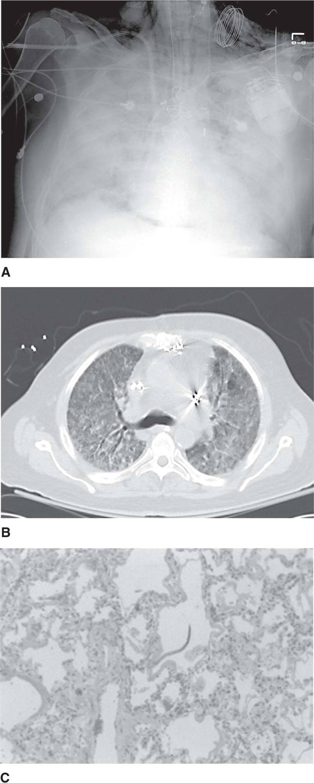
Figure 12.5. Strongyloidiasis and hyperinfection syndrome. A. A chest radiograph demonstrates diffuse opacification of both lung fields. B. A computed tomography scan of the chest shows diffuse interstitial infiltrates consistent with pulmonary edema. C. Lung biopsy with hematoxylin and eosin stain shows inflammatory cells within the alveoli and a rhabditiform larva (middle of the field).
Despite treatment with voriconazole, ganciclovir, and broad-spectrum antibiotics, the patient became hypotensive and remained hypoxic, dying 7 days after the onset of his acute respiratory illness. All blood cultures and sputum culture were negative for pathogens.
At autopsy, numerous Strongyloides stercoralis filari-form larvae were found to be present within the alveolar spaces, alveolar septa, and connective tissue (Figure 12.5C). Occasional filariform larvae were also seen within the sinuses of the hilar lymph nodes and were identified within the myocardial interstitium. Filariform larvae were seen within the walls of the esophagus, stomach, small bowel, and colon, with the heaviest infestation being observed in the colon.
As observed with other roundworm infections, most patients with Strongyloides have no symptoms when they harbor only a small number of worms. Heavier infestations can cause symptoms associated with the parasite’s life cycle. When the filariform larvae first penetrate the skin, they can cause itching and a papular erythematous rash. Migration into the lungs can cause respiratory symptoms, pneumonia, and peripheral eosinophilia (Loeffler syndrome). Once Strongyloides takes up residence in the gastrointestinal tract, the parasite can cause burning abdominal pain that mimics peptic ulcer disease or a colicky abdominal pain that mimics gallbladder disease. Abdominal pain may be associated with diarrhea and the passage of mucus. Malabsorption, nausea, vomiting, and weight loss may also be present. Because the female worm penetrates the bowel mucosa and the filariform larvae can migrate through the bowel wall, the host responds by producing eosinophils, and peripheral eosinophilia is a prominent finding in strongyloidiasis. When larvae penetrate the perianal area, a localized snakelike urticarial rash may be seen. A generalized urticarial rash may also be seen.
As illustrated in case 12.2, when asymptomatic individuals who harbor small numbers of organisms receive immunosuppressants such as high-dose corticosteroids, or develop depressed cell-mediated immunity because of severe malnutrition or AIDS, the level of autoinfection can increase markedly, resulting in a hyperinfection syndrome. Symptoms may include diffuse pulmonary infiltrates, severe abdominal pain, meningitis, and gram-negative sepsis, the latter manifestation being the result of filariform larvae compromising the integrity of the bowel wall. Other clinical manifestations can include hemoptysis and a skin rash. Periumbilical purpura, diffuse nonpalpable purpura, angioedema, and erythroderma mimicking a drug-related allergic eruption have all been described. As in case 12.2, eosinophilia is usually absent in the hyperinfection syndrome. When an immunocompromised patient presents with this clinical constellation and was raised in the rural south or previously lived in a tropical region, hyperinfection with Strongyloides needs to be considered.
KEY POINTS
About the Clinical Presentation of Strongyloides
1. Many patients are asymptomatic.
2. skin penetration can cause an itchy erythematous rash.
3. Lung invasion can produce Loeffler syndrome (cough, wheezing, pneumonia, and eosinophilia).
4. Heavy infection can cause abdominal pain and eosinophilia.
5. Treatment with high-dose steroids can cause a fatal hyperinfection syndrome (accelerated autoinfection).
6. Hyperinfection causes diffuse pneumonia, meningitis, abdominal pain, and gram-negative sepsis, hemoptysis, and skin rashes. Eosinophilia is absent.
DIAGNOSIS AND TREATMENT
Because the eggs usually hatch in the gastrointestinal tract, Strongyloides ova are rarely seen on stool smear. Diagnosis depends on identifying rhabditiform larvae in the feces or duodenal fluid. Diagnosis requires expertise, because hookworm larvae can easily be misdiagnosed as Strongyloides. At least three stools need to be examined under a low-power (100x) microscope; if results are negative, endoscopy should be considered. The ELISA serum test is moderately sensitive and specific, but it cannot differentiate recent from past infection. A more sensitive luciferase immunoprecipitation system (LIPS) has recently been developed. In the Strongyloides-infected immunocompromised host, the ELISA test may be negative. An important clue is the presence of peripheral eosinophilia, which may increase between 10% and 20% of peripheral WBCs. However, lack of eosinophilia, particularly in the hyperinfection syndrome, does not exclude the diagnosis of strongyloidiasis.
Ivermectin for 2 days is curative in most cases. Albendazole can be given as alternative therapy. Because of the potential danger of severe autoinfection, all patients with Strongyloides, even asymptomatic patients, should be treated. Patients who develop the hyperinfection syndrome should be treated for a minimum of 7 days. However, despite treatment, the mortality associated with this syndrome remains high. Patients with a history of Strongyloides or unexplained eosinophilia should therefore be thoroughly examined, tested, and treated before receiving immunosuppressive therapy.
Hookworm
PREVALENCE, EPIDEMIOLOGY, AND LIFE CYCLE
Hookworm (Ancylostoma duodenale and Necator americanus) has been estimated to infect nearly one-quarter of the world’s population, being found throughout the tropical and subtropical zones. Infection is prevalent in areas where untreated human feces are allowed to contaminate the soil, and people walk barefoot. Necator americanus (“New World hookworm”) is found primarily in the Western hemisphere, but also in southern Asia, Indonesia, Australia, and Oceania. Ancylostoma duodenale (“Old World hookworm”) is found predominantly in the Mediterranean region, northern Asia, and the west coast of South America. As a result of sanitary waste disposal policies in the United States, hookworm infection has a low prevalence, being found primarily in the southeast.
The life cycle of hookworm is very similar to that of Strongyloides. Like Strongyloides, the hookworm filariform larvae penetrate the skin, enter the bloodstream and lymphatics, pass into the lung, migrate up the bronchi to the trachea, are swallowed, and finally take up residence in the upper small intestine (Figure 12.5). They attach by means of a buccal capsule that is used to suck blood from the host. A single Necator americanus worm can remove 0.3 mL of blood daily, and a single Ancylostoma duodenale worm 0.5 mL. Worldwide, hookworm is a major cause of iron deficiency anemia. It is responsible for an estimated blood loss of 7 million liters daily—the total blood volume of more than 1 million people!
KEY POINTS
About the Diagnosis and Treatment of Strongyloidiasis
1. Diagnosis is difficult. (stools do not contain ova.)
2. Larvae are found in the stool; duodenal endoscopy may be required.
3. Peripheral eosinophilia may the only finding.
4. Treat asymptomatic infections.
5. Ivermectin is the drug of choice.
The life cycle of the hookworm also differs from that of Strongyloides in several important ways, and the differences account for hookworm’s milder clinical manifestations. The Strongyloides ova mature quickly, hatching in the bowel wall of the host; hookworm ova mature more slowly, requiring several days of incubation in warm, moist, shady soil. As a result, human hookworm infestation is confined to geographic areas with a warm climate. The longer maturation time for hookworm eggs also means that autoinfection does not occur and that infection by fresh feces is not possible.
CLINICAL PRESENTATION
When hookworm larvae penetrate the skin, they can cause intense pruritus, sometimes called “ground itch.” Itching is associated with local erythema and a papular rash at the site of penetration. As is observed with both Ascaris and Strongyloides, respiratory symptoms and patchy pneumonia associated with peripheral eosinophilia (Loeffler syndrome) can develop as the worm penetrates the lung. The abnormalities most commonly associated with hookworm are iron deficiency and protein malnutrition. These abnormalities depend both on the worm burden and on the nutritional status of the patient. Other complaints may include abdominal pain, diarrhea, and weight loss.
KEY POINTS
About Hookworm (Necator americanus and Ancylostoma duodenale)
1. Larvae from the soil penetrate the skin, causing a pruritic rash.
2. Larvae pass through the lung and can cause Loeffler syndrome.
3. Eggs hatch outside of the host in soil (no autoinfection).
4. Adult worms attach to bowel wall and suck blood.
5. Iron deficiency anemia is the most common manifestation.
6. The diagnosis is readily made from observation of ova in the stool.
7. Albendazole is the treatment of choice.
DIAGNOSIS AND TREATMENT
Adult female worms release between 10,000 and 20,000 worms daily, making diagnosis by stool smear simple. The eggs are readily seen using a low-power (100x) microscope (Figure 12.3). Quantitation of the egg count allows for an estimate of the worm burden. A single dose of albendazole is usually curative (see Table 12.3).
TISSUE AND BLOOD HELMINTHS
GUIDING QUESTIONS
1. Which tissues do Trichinella, Echinococcus, and Taenia solium prefer to infect?
2. Why is Trichinella uncommon in the United States?
3. What is a hydatid cyst, and how is it treated?
4. Why does treatment with praziquantel often exacerbate the manifestations of neurocysticercosis?
TRICHINELLA
POTENTIAL SEVERITY
Usually asymptomatic, but heavy infections can lead to severe myocarditis, pneumonia, and encephalitis that can be fatal.
Prevalence, Epidemiology, and Life Cycle
Trichinosis is found worldwide, wherever contaminated meat is undercooked. Trichinella is a roundworm whose larvae are released from cyst walls in contaminated meat by acid–pepsin digestion in the stomach. Upon entering the small intestine, larvae invade the intestinal microvilli and develop into adult worms. Females then release larvae that enter the bloodstream and seed skeletal and cardiac muscle. The larvae grow in individual muscle fibers and eventually become surrounded by a cyst wall. Once encysted, the larvae can remain viable for up to 40 years. If the cyst-containing muscle tissue is ingested, Trichinella is able to take up residence in the new host.
The domestic animal that primarily becomes infected with Trichinella is the pig. In many countries, including the United States, pigs are fed with grain, which explain the low incidence of trichinosis. In the United States, laws were enacted to prevent the feeding of uncooked garbage to pigs, and as a result, fewer than 100 trichinosis cases are reported annually. Most cases of trichinosis result from improperly processed pork, but undercooked bear, walrus, cougar, wild boar, horse meat, and soft-shelled turtle have also been sources of Trichinella infection.
Clinical Presentation
Symptoms correlate with the numbers of worms in tissues. Because the number of cysts ingested is often low, most infections are asymptomatic. Heavier infestations can result in diarrhea, abdominal pain, and vomiting during the intestinal phase, followed in 1-2 weeks by fever, periorbital edema, subconjunctival hemorrhages, and chemosis. Muscle pain, swelling, and weakness are common. The extraocular muscles are frequently involved first, followed by the neck and back, arms, and legs. Occasionally, a macular or petechial diffuse body rash may be seen. These symptoms usually peak within 2-3 weeks, but they may be followed by a prolonged period of muscle weakness. Death is uncommon, but can result from severe myocarditis leading to congestive heart failure. Fatal encephalitis and pneumonia have also been reported.
Diagnosis and Treatment
An elevated peripheral eosinophil count associated with periorbital edema, myositis, and fever strongly suggests the diagnosis. Eosinophil counts are often very high. Serum creatine phosphokinase is also elevated, reflecting muscle damage. A specific diagnosis requires biopsy of a symptomatic muscle to demonstrate Trichinella larvae. Because exposure history and the clinical manifestations are usually distinct, a biopsy is rarely required. Antibody to Trichinella increases within 3 weeks and can be detected by ELISA.
Albendazole is the treatment of choice. Alternatively, mebendazole can be used. When administering meben-dazole myositis may be reduced by using a dosing regimen that starts with a lower dose for 3 days, and then follows with higher doses for 10 days (see Table 12.3). In critically ill patients, corticosteroids (prednisone 50 mg daily for 10-15 days) may be helpful, but no controlled trials have been conducted proving efficacy. Cooking meat above 55°C until all pink flesh is browned kills encysted larvae and prevents trichinosis.
KEY POINTS
About Trichinosis
1. Caused by ingesting larvae cysts, primarily from pork.
2. Uncommon in countries that do not feed pigs uncooked garbage.
3. Larvae infect skeletal and cardiac muscle.
4. Light infections are often asymptomatic.
5. Heavy infection causes abdominal pain and diarrhea, followed by fever, periorbital edema, muscle pain (ocular muscles first), and myocarditis, associated with marked eosinophilia and increased creatine phosphokinase.
6. Diagnosis is made by muscle biopsy, ELISA, or clinical signs.
7. Albendazole is the treatment of choice. Accompany with corticosteroids in severe cases.
ECHINOCOCCOSIS
POTENTIAL SEVERITY
Infections with Echinococcus multilocularis usually lead to symptomatic disease; patients infected with Echinococcus granulosus may remain asymptomatic. Extensive disease causes significant morbidity and mortality.
Prevalence, Epidemiology, and Life Cycle
Echinococcus is member of the cestode (tapeworm) family. Infections with Echinococcus granulosus are found worldwide, including in Africa, the Middle East, southern Europe, Latin America, and the southwestern United States. A second species, Echinococcus multilocularis is found in northern Europe, Asia, the northern United States, and the Arctic. Humans represent an inadvertent intermediate host, the infection being contracted by ingestion of food contaminated with viable parasite eggs. Echinococcus is carried in the feces of sheep, goats, camels, horses, and domestic dogs that live around livestock. In the southwestern United States, most cases are contracted from sheep dogs. The primary host for Echinococcus multilocularis is the fox, and domestic cats and dogs become secondarily infected. An outbreak has been reported, Alaskan Eskimos villages contracted from infected hunting dogs. Because eggs are partially resistant to drying and can remain viable for many weeks, food can become contaminated without coming in direct contact with infected animals.
Ingested eggs hatch in the intestine forming oncospheres that penetrate the bowel wall, enter the bloodstream, and are deposited in various organs—most commonly, the liver and lungs, and less frequently the brain, heart, and bones—where they encyst. The resulting hydatid cysts consist of a germinal membrane that produces multiple tapeworm heads and that also undergoes budding to form multiple, septated daughter cysts within the primary cyst (Figure 12.6). Cysts can survive in the host for decades.

Figure 12.6. A computed tomography scan with both oral and intravenous contrast shows multiple echinococcal hepatic abscesses. (Picture courtesy of Dr. Pat Abbitt, University of Florida College of Medicine)
Clinical Presentation
CASE 12.3
A 33-year-old woman, an immigrant from Jordan, presented with a chief complaint of bloody cough and shortness of breath for a period of 2 weeks. At age 22, she had undergone a computed tomography (CT) scan of the abdomen as part of a workup for polycystic ovaries. She was noted at that time to have a large liver cyst consistent with Echinococcus. Although she was asymptomatic, resection of the left lobe of the liver was performed that year. Despite surgical resection, she experienced recurrent cysts and on three occasions underwent percutaneous aspiration followed by injection of hypertonic saline. One month before admission and 6 years after her last aspiration and injection procedure, she began coughing up blood. At the same time, she noted shortness of breath. She received several courses of oral antibiotics, but failed to improve. Her coughing then became productive of gelatinous, foul-smelling serosanguinous fluid.
Pulmonary examination revealed decreased breath sounds and dullness to percussion at the right base. Bronchial breath sounds and E-to-A changes were noted in the right posterior mid-lung field. The liver was not palpable. A CT scan of the chest and abdomen revealed a fluid collection over the dome of the liver and an 8x5-cm abscess in the right lower lobe that contained an airfluid level.
Most patients with echinococcosis are asymptomatic, the infection being detected incidentally on an imaging study. Symptoms generally develop when the hydatid cyst reaches a size of 8-10 cm and begins compressing vital structures or eroding into the biliary tract or a pulmonary bronchus (as occurred in case 12.3). The cysts can also become superinfected, resulting in a bacterial abscess. Cyst leakage or rupture can result in an anaphylactic reaction, causing fever and hypotension. Cysts can also develop in the brain, heart, kidneys, eyes, and bones. Asymptomatic disease caused by Echinococcus granulosusrarely progresses; however, 90% of cases of asymptomatic Echinococcus multilocularis infection eventually progress to symptomatic disease.
Diagnosis and Treatment
Ultrasonography, CT scan, or magnetic resonance imaging (MRI) reveals a characteristic hydatid cyst with a distinct septated structure representing daughter cysts (Figure 12.6). Often, tapeworm heads can also be visualized. The stage of infection can be classified based on ultrasound findings, but CT scan has been found to be the more effective diagnostic method for delineating the extent of disease. MRI is most sensitive being able to more readily detect liquid collections as compared with CT scan. The diagnosis can be confirmed by ELISA, which is highly sensitive for liver cysts, but less sensitive for cysts in other organs.
Complete surgical resection of the hydatid cyst is often recommended in early symptomatic disease. The cyst should be removed intact, taking great care to avoid a rupture, which will spread the infection by daughter cysts. To reduce the risk of spread, aspiration of the cyst is recommended—a procedure that involves removing a fraction of the contents and instilling a hypertonic saline solution (30% NaCl), iodophor, or 95% ethanol to kill the germinal layer and daughter cysts. Surgical resection should be performed 30 minutes after instillation of the solution. In cases with biliary communication, the foregoing cidal agents are not recommended because of the risk of inducing sclerosing cholangitis.
As compared with medical treatment alone, debulking of cysts does not improve outcome, but it may relieve symptoms in specific cases. Treatment in the perioperative period with three to four cycles of albendazole 400 mg twice daily for 4 weeks, followed by a 2-week rest period, is generally recommended to limit the risk of intraoperative dissemination. The same medical therapy is recommended for patients with inoperable hydatid cyst (see Table 12.3); however, therapy needs to be continued for years. In selected cases, CT or ultrasound has been used to guide percutaneous needle aspiration drainage and instillation of cidal agents (hypertonic saline or ethanol) to sterilize the cyst, followed by reaspiration after 15 minutes to remove the cidal agent (“PAIR,” puncture, aspiration, injection, reaspiration). The PAIR treatment is often curative, and it is becoming the treatment of choice. The efficacy of PAIR has not been confirmed by randomized trials, however. The management of echino-coccosis is complex and requires guidance by an expert. A comprehensive consensus paper describing staging, surgical, and medical management was published in 2010 (see Further Reading).
KEY POINTS
About Echinococcus
1. Spread primarily by domestic dogs, who excrete eggs in their feces. Eggs survive in dust and contaminate food.
2. Eggs hatch in the intestine and oncospheres enter the bloodstream, where they migrate to the liver or lung, or (less commonly) to the brain, where they form hydatid cysts.
3. Hydatid cysts survive and grow over decades, causing symptoms when they reach 8-10 cm in diameter.
4. Diagnosis is made by computed tomography scan or ultrasonography.
5. Treatment involves administration of albendazole, combined with surgical resection preceded by instillation of an agent cidal to the germinal layer. Alternatively, percutaneous needle drainage and cidal agent instillation (“PAIR”) may be curative.
CYSTICERCOSIS
POTENTIAL SEVERITY
Causes neurologic complications in a significant number of infected patients many years after the initial infection.
Prevalence, Epidemiology, and Life Cycle
Taenia solium is another cestode (tapeworm) common in Central and South America, Mexico, the Philippines, Southeast Asia, India, Africa, and southern Europe. It is estimated that 50 million people are infected by the cestode. Like Echinococcus, Taenia can be contracted by ingesting viable eggs. This infection can also be contracted eating raw or undercooked pork containing encysted larvae. Once ingested, the encysted larvae are released into the stomach, where they migrate into the intestine and develop into adult worms that can reach 8 m in length. These individuals become chronic carriers who carry the tapeworm, but do not develop cysticercosis unless they accidentally ingest eggs from their own feces. Chronic carriers spread the disease via the fecaloral route, and person-to-person spread is now thought to be the primary mode of transmission of the disease.
Clinical Presentation
Adult intestinal worms rarely cause symptoms. However, if the eggs released by the worms are ingested, the eggs hatch releasing larvae that penetrate the intestine, enter the bloodstream, and eventually encyst in the brain, causing neurocysticercosis. Cysts may lodge in the cerebral ventricles (causing hydrocephalus), the spinal cord (resulting in cord compression and paraplegia), the subarachnoid space (causing chronic meningitis), or the cerebral cortex (causing seizures). Cysts may remain asymptomatic for many years, becoming clinically apparent only when the larvae die, an event associated with cyst swelling and increased inflammation. Larvae also encyst in other tissues (skin and muscle), but rarely cause symptoms. Eye involvement is also reported.
Diagnosis and Treatment
Computed tomography or nuclear magnetic resonance scan is the preferred diagnostic studies, demonstrating discrete cysts that may enhance following the administration of contrast media depending on the degree of surrounding inflammation. In CNS infection, multiple lesions are generally detected. Older lesions are often calcified (Figure 12.7). In the absence of cerebral edema, lumbar puncture can be performed. Analysis of the cerebrospinal fluid usually reveals lymphocytes or eosinophils accompanied by low glucose and elevated protein. Serologic tests detecting antibody directed against Taenia soliummay be positive, particularly in patients with multiple cysts. Enzyme-linked immunoelectrotransfer blot assay (EITB) is the preferred serological test. The sensitivity of the test depends on the activity of the cysts as well as their number.
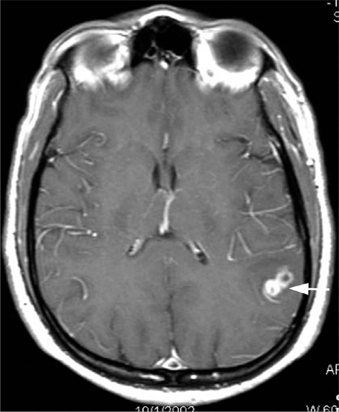
Figure 12.7. Computed tomography scan with contrast of the cerebral cortex, showing two typical ring-enhancing lesions of neurocysticercosis (arrow).
Treatment of neurocysticercosis is complex and controversial. Albendazole and praziquantel may kill living cysts, but larval death results in increased inflammation and edema, and may exacerbate symptoms. A recent randomized trial and a meta-analysis suggested that in symptomatic patients with cortical lesions, albendazole combined with oral dexamethasone (2 mg three times daily) or oral prednisone (40 mg daily) enhances resolution of the lesions and reduces the incidence of seizures. Surgical resection of cysts may be required depending on the symptoms and size and location of the offending cyst. Antiepileptic medications should be used to control seizures. Antiepileptic medications can be safely withdrawn for solitary lesions, if albendazole treatment results in complete resolution without residual calcification.
KEY POINTS
About Cysticercosis (Taenia solium infection)
1. Contracted by ingesting eggs in fecally contaminated food or encysted larvae in undercooked pork.
2. Larvae enter the bloodstream, encysting primarily in the brain.
3. Symptoms develop after many years when the larvae die, causing increased inflammation.
4. Can cause seizures, hydrocephalus, paraplegia, and meningitis.
5. Diagnosis is made by computed tomography scan, magnetic resonance imaging, or serology.
6. Treatment involves administration of albendazole plus corticosteroids for symptomatic disease; surgical resection can be performed in selected patients.
SCHISTOSOMIASIS
GUIDING QUESTIONS
1. Why doesn’t primary schistosomiasis occur in the United States?
2. How is schistosomiasis contracted?
3. Which Schistosoma strain causes swimmer’s itch?
4. What is Katayama fever?
5. In late disease, how does egg deposition cause
POTENTIAL SEVERITY
Usually, a chronic disorder resulting in debilitating complications. Occasionally, fatal during the early stage of infection as a result of a severe serum-sickness syndrome.
Prevalence, Epidemiology, and Life Cycle
Schistosoma mansoni, S. haematobium, and S. japonicum are members of the fluke (trematode) family. Schistosomes are estimated to infect 200 million people worldwide. Primary infection does not occur in the United States because the critical intermediate host—a specific type of freshwater snail—is absent. However, approximately 400,000 imported cases occur in immigrants from Puerto Rico, South America (particularly Brazil), the Middle East, and the Philippines. S. mansoni is found primarily in South America, the Caribbean, Africa, and countries of the Arab Middle East. S. haematobium is found in Africa and the Middle East, and S. japonicum is found primarily in China and the Philippines. Two other strains that have more recently been found to cause disease are S. intercalatum (Western and Central Africa) and S. mekongi (Indochina).
The parasite is contracted by exposure to freshwater containing infectious cercariae. The fork-tailed cercariae are able to swim to and penetrate the skin of people wading in stagnant infested freshwater pools or rice paddies. Once inside the host, cercariae lose their tails and mature into schistosomulae that enter the bloodstream. From the bloodstream, they penetrate the lung and liver, where over a period of 6 weeks, they mature to adult worms. The adult worms then migrate through the venous plexus to various sites, depending on the Schistosoma strain. S. mansoni worms take up residence in the inferior mesenteric veins responsible for venous drainage of the large intestine; S. japonicum, in the superior mesenteric veins that drain the small intestine, and S. haematobium, in the vesicular plexus that drains the urinary bladder.
Once resident in the host, the worms can live for decades, releasing eggs into the bowel or bladder. Improper handling of contaminated stool and urine leads to egg contamination of water. Eggs hatch in freshwater, forming miracidia whose cilia enable them to swim and infect freshwater snails. Each species of schistosome requires a specific freshwater snail intermediate, which explains the geographic distribution of each strain. The miracidia multiply within the snail, and within 4–6 weeks, they release large numbers of cercariae capable of infecting humans.
KEY POINTS
About the Life Cycle of Schistosoma
1. Cercariae swimming in freshwater can penetrate human skin.
2. Cercariae mature into schistosomulae that enter the bloodstream and migrate to the liver and lung, where they mature.
3. Mature worms migrate to the venous system of the small (S. japonicum) or large bowel (S. mansoni) or to the bladder venous plexus (S. haematobium).
4. The worms release eggs into stool or urine for many years, resulting in contamination of freshwater.
5. Freshwater snails are infected by miracidia, a necessary step in the production of cercariae and infection of humans.
Clinical Presentation
CASE 12.4
A 32-year-old man was evaluated for a lesion of the urinary bladder. He had been well until 16 months earlier. Soon after returning from a 1-week vacation in Malawi, he had an episode of perineal pain associated with painful ejaculation and browncolored ejaculate. His condition improved after treatment with ciprofloxacin.
Four months before the evaluation, this patient had begun experiencing urinary frequency, with intermittent passage of small blood clots in the urine. His symptoms failed to improve on ciprofloxacin treatment. An epide-miologic history noted frequent travel outside the United States. Most recently, the man had traveled to Malawi with his wife. While there, he had repeatedly swum in a lake that he was assured was “safe.”
A laboratory workup showed a normal peripheral white blood cell (WBC) count and differential. Urinalysis confirmed hematuria. Cytology found no malignant cells. A urogram and ultrasound demonstrated a round structure, 8x10 mm in diameter, adherent to the bladder wall. Cystoscopic examination disclosed multiple, slightly raised, polypoid lesions that were less than 5 mm in diameter. The lesions were erythematous, with focal yellow areas.
Low-power microscopic examination of material from a bladder biopsy revealed a polypoid inflammatory lesion of the bladder mucosa with dense inflammatory infiltrate surrounding clusters of eggs in the submucosa. At higher magnification, the granulomas were found to contain clusters of helminthic eggs surrounded by epithelioid histiocytes, chronic inflammatory cells, and eosinophils. The eggs were oval and had a terminal spine characteristic of S. haematobium (Figure 12.8). The man’s wife was subsequently examined, and Schistosoma eggs were found in her urine. Both were treated with praziquantel, and the eggs disappeared from both patients’ urine.
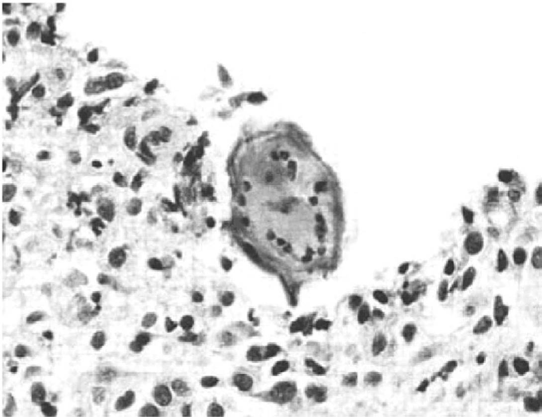
Figure 12.8. Bladder biopsy showing an egg of Schistosoma haematobium. (Picture from the N Engl J Med. 2001;342:1105-1111.)
The three stages of the disease correspond to the life cycle of the parasite in the human host.
The first stage occurs at the time of penetration and is commonly termed “swimmer’s itch.” A very itchy macular papular rash develops within 24 hours of the cercariae penetrating the skin. The lesions spontaneously resolve as the organisms spread to the bloodstream. An avian schistosome is also able to penetrate the skin, but it is not capable of entering the bloodstream. This benign form of swimmer’s itch is common in the Great Lakes of the north-central United States and in freshwater lakes in Europe.
The second stage of clinical disease occurs 4–8 weeks later, when the worms mature and begin releasing eggs. Patients develop a serum-sickness-like syndrome as they react with elevated levels of immunoglobulin E and peripheral eosinophilia to egg antigens. Fever, headache, cough, chills, and sweating are accompanied by lymphadenopathy and hepatosplenomegaly. This clinical constellation has been called “Katayama fever” and is most commonly associated with S. japonicum. The symptoms usually resolve spontaneously, but in heavy infections, this acute reaction can be fatal.
The third, chronic, stage results from granulomatous reactions to egg deposition in the intestine, liver, bladder, and (less commonly) the lung and CNS. Granulomatous reactions in the bowel can lead to chronic diarrhea, abdominal pain, and blood loss. Eggs may enter the portal venous system and gain entry to the liver, where chronic inflammation is followed by fibrosis leading to portal hypertension, splenomegaly, and bleeding esophageal varices. Because the hepatic parenchyma is seldom compromised, liver function tests are usually normal. Peripheral eosinophilia is commonly encountered. Hepatosplenomegaly with normal liver function tests, peripheral eosinophilia, and a history of residence in an endemic area should raise the possibility of chronic hepatic schistosomiasis. The development of collateral venous channels in association with portal hypertension can result in egg deposition in the pulmonary arteries, causing pulmonary hypertension and right-sided congestive heart failure. Deposition of eggs in the CNS is less common and can cause seizures or, if eggs are deposited in the region of the spinal cord, transverse myelitis. In S. haematobium, eggs are deposited in the bladder wall, leading to hematuria, bladder obstruction, hydronephrosis, and recurrent urinary tract infections. Bladder cancer may also complicate chronic S. haematobium infection.
KEY POINTS
About the Clinical Presentation of Schistosomiasis
1. Skin penetration causes “swimmer’s itch.”
2. A serum-sickness syndrome with eosinophilia and high immunoglobulin E levels may follow. This constellation of symptoms is called Katayama fever.
3. Granulomatous reaction to egg deposition leads to chronic diarrhea, portal hypertension and hepatosplenomegaly, and pulmonary hypertension in Schistosoma mansoni and S. japonicum.
4. Eggs deposited in the bladder can lead to hematuria, bladder obstruction, hydronephrosis, recurrent urinary tract infections, and sometimes bladder cancer in cases of S. haematobium.
Diagnosis and Treatment
Demonstration of eggs in the stool or urine allows a specific diagnosis to be made. Quantitative egg counts are helpful in assessing the intensity of the infection. Urine is best collected between noon and 2 PMor following exercise. Passing the urine through a 10-mm filter concentrates the eggs. Eggs may also be identified on tissue biopsies. Rectal biopsy is particularly helpful in diagnosing S. mansoni. The eggs of S. mansoni, S. japonicum, and S. haematobium have distinct morphologies, allowing them to be readily identified using a low-power (100x) microscope (Figure 12.3). In chronic disease, the egg burden may be low, making the diagnosis difficult. Anti-schistosome antibody tests are now available for detecting chronically infected patients; however, the specificity and sensitivity of these tests limit their value. Furthermore, the tests cannot be used in lifelong residents of endemic areas, because serology in these individuals is frequently positive in the absence of active infection.
KEY POINTS
About the Diagnosis and Treatment of Schistosomiasis
1. Characteristic eggs in the stool or urine (check between noon and 2 pm) or on tissue biopsy are diagnostic; consider rectal biopsy in Schistosoma mansoni.
2. Eggs may not be seen in chronic disease, anti-schistosoma antibody may be helpful,
3. Praziquantel is the treatment of choice.
Praziquantel is effective treatment for all forms of schistosomiasis (see Table 12.3). Side effects of treatment are mild and include fever, abdominal discomfort, and headache.
OTHER LESS COMMON TISSUE FLUKES
Other flukes that can infect humans undergo a life cycle similar to that of Schistosoma, requiring snails as the intermediate host. However, rather than gaining entry by penetrating the human skin, the cercariae take up residence in other food sources and become encysted. Infection is contracted when the human host eats cercariae contaminated food.
Clonorchis sinensis (Chinese liver fluke) infections result from the ingestion of raw or undercooked freshwater fish. Infections occur in China, Hong Kong, and Vietnam. Worms gain entry into biliary tract via the ampulla of Vater. Infection can be complicated by cholangitis and, later, by cholangiocarcinoma. Infections are effectively treated with praziquantel (see Table 12.3).
Fasciola hepatica, another liver fluke, is found in sheep-raising areas of the world, including South America, Australia, China, Africa, and Europe. Ingestion of vegetables contaminated with encysted cercariae is the most common route of infection. This fluke is treated with praziquantel or bithionol (see Table 12.3).
Paragonimus westermani (lung fluke) is contracted by eating raw or pickled crawfish or freshwater crabs. This parasite is found in Central and South America, West Africa, India, and East Asia. This parasite first enters the gastrointestinal tract and subsequently penetrates through the diaphragm, entering the pleural cavity and lungs, causing respiratory symptoms. Praziquantel is the treatment of choice (see Table 12.3).
FILARIASIS (WUCHERERIA BANCROFTI AND BRUGIA MALAYI)
GUIDING QUESTIONS
1. How is filariasis transmitted?
2. What is the key characteristic that helps to differentiate inflammatory filariasis from bacterial cellulitis?
3. Is elephantiasis an early or late manifestation of filariasis?
4. When during the day are blood smears most likely to be positive?
POTENTIAL SEVERITY
A chronic debilitating infection that can cause severe disfiguring complications by blocking lymphatic drainage.
Prevalence, Epidemiology, and Life Cycle
Microfilaria is less common than many parasites, being estimated to infect approximately 120 million people. Several strains of worm can cause this disease. Wuchereria bancrofti is found throughout the tropics and is the most common form accounting for 90% of infections. Brugia malayi is restricted to the southern regions of Asia. A third strain Brugia timori is found only in Indonesia.
Infectious larvae are transmitted by the bite of a mosquito. Larvae pass from the skin into the lymphatic system, where, over several months, they mature near the lymph nodes. Adult worms (40–100 mm in length) can survive in the lymphatic system for 5–15 years. During this period, males and females mate, daily producing an average of 10,000 microfilaria (dimensions: 200–300 mm in length, and 10 μm in width). The microfilaria is released into the bloodstream. The time from initial insect bite to appearance of microfilaria in the infected human is usually 12 months. In W. bancrofti, the highest concentration of microfilaria in the blood is generally found in the middle of the night, explaining why midnight blood smears are recommended for diagnosis.
If a mosquito bites an infected human, the microfilaria is ingested and, over 10–14 days, it develops into infective larvae that can be transmitted to a new human host. The percentage of mosquitoes containing infective larvae has been estimated to be just 1% in endemic areas. Repeated mosquito bites are therefore generally required to contract this infection, which may explain why adults—particularly men—more commonly contract this infection.
KEY POINTS
About the Life Cycle of Wuchereria bancrofti and Brugia malayi
1. Transmitted by the bite of an infected mosquito.
2. Repeated mosquito bites are required.
3. Microfilaria lives in the lymphatic system, and worms enter the bloodstream at midnight (except in the South Pacific).
4. Mosquitoes are infected by biting humans.
Clinical Presentation
ASYMPTOMATIC FILARIASIS
Many individuals have asymptomatic infection. Peripheral eosinophilia and palpable lymphadenopathy may be the only clinical manifestations. Children usually experience no symptoms, despite high numbers of microfilaria in their blood.
INFLAMMATORY FILARIASIS
Adults more commonly react with strong allergic reactions to the invasion by worms that begins approximately 1 year after exposure. Fever, chills, vomiting, headache, and malaise may be associated with lymphangitis of an extremity, orchitis, epididymitis, or scrotal swelling. The affected extremity becomes hot, swollen, erythematous, and painful, mimicking cellulitis. These symptoms are associated with peripheral leukocytosis and an increased percentage of eosinophils (6–25%). Unlike cellulitis, which usually begins peripherally and moves up the limb, inflammatory filariasis begins centrally near the lymph nodes and extends peripherally. Attacks may occur monthly and do not respond to antibiotics. The granulomatous response in the lymphatic tissue is thought to be a host inflammatory reaction to dying worms. Death of the worms is associated with release of the rickettsial-like bacteria Wolbachiathat live in a symbiotic relationship within the adult worms.
OBSTRUCTIVE FILARIASIS
Over time, chronic inflammation leads to fibrosis and permanent obstruction of lymphatic flow. This syndrome is the result of continuous microfilaria infection. Persistent lymphatic obstruction and edema lead to marked skin thickening and deposition of collagenous material, eventually causing elephantiasis. Patients suffer from debilitating enlargement of the legs or massive enlargement of the scrotal tissue, making walking difficult. Cellulitis caused by streptococci or Staphylococcus aureus may periodically recur, requiring antibiotic treatment. Rupture of the lymphatics into the kidney or bladder can result in chyluria, and rupture into the peritoneum can cause chylous ascites.
Diagnosis and Treatment
Giemsa- or Wright-stained peripheral smears should be obtained at midnight in all cases except for those from the South Pacific. Identification of adult worms in the blood is definitive; however, in early and late disease, worms often are not seen. Antibody and antigen assays are highly sensitive and specific. An IgG4 antibody titer correlates with active disease. An ELISA for W. bancrofti circulating antigen is now the diagnostic test of choice, and titers correlate with adult worm burden. A PCR test for W. bancrofti has been developed, but it is not widely available. Biopsy of infected lymph nodes is generally not recommended, but when performed may reveal adult worms in addition to granuloma. Ultrasonography of dilated lymphatics in the spermatic cord has revealed motile worms. In early infection and during the inflammatory stage, peripheral eosinophilia is commonly seen. During the chronic stages of disease, eosinophilia is generally not present. If worms cannot be identified, the diagnosis has to be made on clinical grounds.
KEY POINTS
About the Clinical Presentation of Filariasis
1. Many people, particularly children, are asymptomatic.
2. Inflammatory filariasis is associated with periodic erythema, warmth, pain, and swelling that mimic cellulitis (associated with peripheral eosinophilia).
3. Obstructive disease results in chronic limb swelling (elephantiasis) because of lymphatic fibrosis.
4. Obstructive disease can lead to recurrent bacterial cellulitis.
5. Rupture of lymphatics can cause chyluria or chylous ascites.
6. Release of the rickettsial-like bacteria Wolbachia from the adult worms may be the major stimulus for inflammation.
Diethylcarbamazine in a single dose is the recommended therapy, but fails to kill adult worms (see Table 12.3). A reduction in the level of microfilaria in the blood is usually observed. Treatment may increase inflammation and may not halt progression to fibrosis and lymphatic obstruction. Increased inflammation is thought to be due to release of lipopolysaccharide-like proteins from endosymbiotic Wolbachiaorganisms; ivermectin 200–400 mg/kg, combined with albendazole 400 mg, is another effective regimen that may more effectively kill the adult worms.
For more severely infected patients, a 6-week course of doxycycline kills the symbiont Wolbachia, resulting in sterility of the adult worms, and increases the likelihood of cure. This treatment should be followed by diethylcarbamazine or ivermectin plus albendazole. Normally, these agents exacerbate the host’s inflammatory reaction as the microfilaria die, but doxycycline eradication of the Wolbachiaeliminates this complication. Anti-inflammatory agents may be used to reduce the extent of inflammation, and elastic support stockings can be helpful in reducing moderate lymphedema.
In areas where onchocerciasis and Loasis are common, treatment of microfilaria with diethylcarbamazine can precipitate very severe febrile reactions, and is contraindicated. In areas where coinfection is common, pretreatment with doxycycline or treatment with ivermectin plus albendazole is recommended.
KEY POINTS
About the Diagnosis and Treatment of Filariasis
1. Midnight blood smear demonstrating worms yields a definitive diagnosis.
2. In early and late disease, worms may not be seen.
3. Ultrasound of dilated lymphatics may demonstrate worms.
4. Peripheral eosinophilia is common.
5. Enzyme-linked immunosorbent assay is sensitive and specific, and levels correlate with disease activity.
6. Diethylcarbamazine or ivermectin plus albendazole are used for treatment. Treatment can exacerbate symptoms. Pretreatment with doxycycline can reduce febrile reactions
7. Initial treatment with diethylcarbamazine is contraindicated in patients likely to be coinfected with loa loa and Onchocerca.
DIROFILARIASIS (DOG HEARTWORM)
Humans are an accidental host in dirofilariasis. The disease is most commonly found in the southeastern United States and is transmitted by mosquitoes. After developing in the subcutaneous tissue, the young adult filaria migrates. In dogs, it migrates to the right side of the heart and right pulmonary vessels, where it survives. In humans, it migrates to the lung, but fails to develop. Its death produces local granulomatous inflammation. Most human cases present as an asymptomatic pulmonary coin lesion, mimicking an early neoplasm. Microscopic examination of the lung biopsy reveals a dead worm. Treatment of human cases is not necessary.
ONCHOCERCIASIS
The Onchocerca volvulus parasite is found primarily in Africa, where it infects approximately 20 million people. Cases are occasionally seen in Central and South America. The infection is transmitted by a black fly that swarms around the face, often biting around the eyes and depositing Onchocerca larvae onto the skin. These larvae penetrate and crawl through the skin and connective tissue. The worms initially cause an itchy erythematous rash. Later, fibrous skin nodules develop. Worms often migrate into the anterior chamber of the eye, causing inflammation and blindness. Because the offending black fly is commonly found near streams, this disease has been called “river blindness.”
The diagnosis is made by skin snips or by visualizing worms in a slit lamp examination of the eyes. The treatment of choice is a single dose of ivermectin repeated at 3-month intervals until symptoms resolve (see Table 12.3). Fever, itching, and an urticarial rash may develop as result of dying microfilaria.
LOIASIS
The loa loa microfilaria is also transmitted by a fly, and the disease is found in Western and Central Africa. The microfilaria migrates through the skin, causing localized edema called Calabar swellings. Several hours before swelling occurs, local itching and pain are noted. Occasionally, the microfilaria can be seen migrating through the subconjunctiva hemorrhages, causing intense conjunctivitis. Active microfilaria migration is associated with marked peripheral eosinophilia.
The diagnosis is made by daytime blood smear. Diethylcarbamazine or ivermectin is recommended as treatment (see Table 12.3). Diethylcarbamazine can precipitate encephalitis in heavily infected patients.
FURTHER READING
General
Wilson ME, Weld LH, Boggild A, et al. Fever in returned travelers: results from the GeoSentinel Surveillance Network. Clin Infect Dis. 2007;44:1560-1568.
Malaria
Agnandji ST, Lell B, Soulanoudjingar SS, et al. First results of phase 3 trial of RTS, S/AS01 malaria vaccine in African children. N Engl J Med. 2011;365:1863-1875.
Aronson NE, Sanders JW, Moran KA. In harm’s way: infections in deployed American military forces. Clin Infect Dis. 2006;43: 1045-1051.
Baird JK, Hoffman SL. Primaquine therapy for malaria. Clin Infect Dis. 2004;39:1336-1345.
Bruneel F, Gachot B, Wolff M, Regnier B, Danis M, Vachon F. Resurgence of blackwater fever in long-term European expatriates in Africa: report of 21 cases and review. Clin Infect Dis. 2001; 32:1133-1140.
Centers for Disease Control and Prevention. CDC Health Information for International Travel. New York, NY: Oxford University Press; 2012.
Cox-Singh J, Davis TM, Lee KS, et al. Plasmodium knowlesi malaria in humans is widely distributed and potentially life threatening. Clin Infect Dis. 2008;46:165-171.
Crawley J, Sismanidis C, Goodman T, Milligan P. Effect of intermittent preventive treatment for malaria during infancy on serological responses to measles and other vaccines used in the expanded programme on immunization: results from five randomised controlled trials. Lancet.2012;380:1001-1010.
Farcas GA, Soeller R, Zhong K, Zahirieh A, Kain KC. Real-time polymerase chain reaction assay for the rapid detection and characterization of chloroquine-resistant Plasmodium falciparum malaria in returned travelers. Clin Infect Dis. 2006;42: 622-627.
Greenwood BM, Bojang K, Whitty CJ, Targett GA. Malaria. Lancet. 2005;365:1487-1498.
Idro R, Ndiritu M, Ogutu B, et al. Burden, features, and outcome of neurological involvement in acute falciparum malaria in Kenyan children. JAMA. 2007;297:2232-2240.
Kockaerts Y, Vanhees S, Knockaert D, Verhaegen J, Lontie M, Peetermans W. Imported malaria in the 1990s: a review of 101 patients. Eur J Emerg Med. 2001;8:287-290.
Laufer MK, Thesing PC, Eddington ND, et al. Return of chloroquine antimalarial efficacy in Malawi. N Engl J Med. 2006;355:1959-1966.
Marx A, Pewsner D, Egger M, et al. Meta-analysis: accuracy of rapid tests for malaria in travelers returning from endemic areas. Ann Intern Med. 2005;142:836-846.
McKenzie FE, Prudhomme WA, Magill AJ, et al. White blood cell counts and malaria. J Infect Dis. 2005;192:323-330.
Murray CJ, Rosenfeld LC, Lim SS, et al. Global malaria mortality between 1980 and 2010: a systematic analysis. Lancet. 2012;379: 413-431.
Ochola LB, Vounatsou P, Smith T, Mabaso ML, Newton CR. The reliability of diagnostic techniques in the diagnosis and management of malaria in the absence of a gold standard. Lancet Infect Dis. 2006;6:582-588.
Phyo AP, Nkhoma S, Stepniewska K, et al. Emergence of artemisinin-resistant malaria on the western border of Thailand: a longitudinal study. Lancet. 2012;379:1960-1966.
Ponsford MJ, Medana IM, Prapansilp P, et al. Sequestration and microvascular congestion are associated with coma in human cerebral malaria. J Infect Dis. 2012;205:663-671.
Price RN, Uhlemann AC, Brockman A, et al. Mefloquine resistance in Plasmodium falciparum and increased PFMDR1 gene copy number. Lancet. 2004;364:438-447.
Reyburn H, Mbatia R, Drakeley C, et al. Association of transmission intensity and age with clinical manifestations and case fatality of severe Plasmodium falciparum malaria. JAMA. 2005;293:1461-1470.
Shanks GD. Treatment of falciparum malaria in the age of drug resistance. J Postgrad Med. 2006;52:277-280.
Smithuis F, Kyaw MK, Phe O, et al. Effectiveness of five artemisinin combination regimens with or without primaquine in uncomplicated falciparum malaria: an open-label randomised trial. Lancet Infect Dis. 2010;10:673-681.
Stauffer WM, Cartwright CP, Olson DA, et al. Diagnostic performance of rapid diagnostic tests versus blood smears for malaria in US clinical practice. Clin Infect Dis. 2009;49:908-913.
Taylor WR, Hanson J, Turner GD, White NJ, Dondorp AM. Respiratory manifestations of malaria. Chest. 2012;142:492-505.
Williams TN, Mwangi TW, Wambua S, et al. Sickle cell trait and the risk of Plasmodium falciparum malaria and other childhood diseases. J Infect Dis. 2005;192:178-186.
Leishmania
Bern C, Haque R, Chowdhury R, et al. The epidemiology of visceral leishmaniasis and asymptomatic leishmanial infection in a highly endemic Bangladeshi village. Am J Trop Med Hyg. 2007;76:909-914.
Mosleh IM, Geith E, Natsheh L, Schonian G, Abotteen N, Kharabsheh S. Efficacy of a weekly cryotherapy regimen to treat Leishmania major cutaneous leishmaniasis. J Amer Acad Derm. 2008;58:617-624.
Murray HW, Berman JD, Davies CR, Saravia NG. Advances in leishmaniasis. Lancet. 2005;366:1561-1577.
Pintado V, Martin-Rabadan P, Rivera ML, Moreno S, Bouza E. Visceral leishmaniasis in human immunodeficiency virus (HIV)-infected and non-HIV-infected patients. A comparative study. Medicine. 2001;80:54-73.
Reithinger R, Mohsen M, Wahid M, et al. Efficacy of thermotherapy to treat cutaneous leishmaniasis caused by Leishmania tropica in Kabul, Afghanistan: a randomized, controlled trial. Clin Infect Dis. 2005;40:1148-1155.
Schwartz E, Hatz C, Blum J. New world cutaneous leishmaniasis in travellers. Lancet Infect Dis. 2006;6:342-349.
Solomon M, Baum S, Barzilai A, Scope A, Trau H, Schwartz E. Liposomal amphotericin B in comparison to sodium stibogluconate for cutaneous infection due to Leishmania braziliensis. J Amer Acad Derm. 2007;56:612-616.
Sundar S, Maurya R, Singh RK, et al. Rapid, noninvasive diagnosis of visceral leishmaniasis in India: comparison of two immunochromatographic strip tests for detection of anti-K39 antibody. J Clin Micro. 2006;44:251-253.
Trypanosomiasis
Alarcon de Noya B, Diaz-Bello Z, Colmenares C, et al. Large urban outbreak of orally acquired acute Chagas disease at a school in Caracas, Venezuela. J Infect Dis. 2010;201:1308-1315.
Barrett MP, Burchmore RJ, Stich A, et al. The trypanosomiases. Lancet. 2003;362:1469-1480.
Bern C. Antitrypanosomal therapy for chronic Chagas’ disease. N Engl J Med. 2011;364:2527-2534.
Bern C, Montgomery SP, Herwaldt BL, et al. Evaluation and treatment of Chagas disease in the United States: a systematic review. JAMA. 2007;298:2171-2181.
Rassi A, Jr., Rassi A, Marin-Neto JA. Chagas disease. Lancet. 2010;375: 1388-1402.
Reisenman CE, Lawrence G, Guerenstein PG, Gregory T, Dotson E, Hildebrand JG. Infection of kissing bugs with Trypanosoma cruzi, Tucson, Arizona, USA. Emerg Infect Dis. 2010;16:400-405.
Tobler LH, Contestable P, Pitina L, et al. Evaluation of a new enzyme-linked immunosorbent assay for detection of Chagas antibody in US blood donors. Transfusion. 2007;47:90-96.
Intestinal Helminths
Bethony J, Brooker S, Albonico M, et al. Soil-transmitted helminth infections: ascariasis, trichuriasis, and hookworm. Lancet. 2006; 367:1521-1532.
Crompton DW. Ascaris and ascariasis. Adv Parasitol. 2001;48:285-375.
Friis H, Mwaniki D, Omondi B, et al. Effects on haemoglobin of multi-micronutrient supplementation and multi-helminth chemotherapy: a randomized, controlled trial in Kenyan school children. Eur J Clin Nutr. 2003;57:573-579.
Gunawardena GS, Karunaweera ND, Ismail MM. Socio-economic and behavioural factors affecting the prevalence of Ascaris infection in a low-country tea plantation in Sri Lanka. Ann Trop Med Parasitol. 2004;98:615-621.
Juan JO, Lopez Chegne N, Gargala G, Favennec L. Comparative clinical studies of nitazoxanide, albendazole and praziquantel in the treatment of ascariasis, trichuriasis and hymenolepiasis in children from Peru. Trans R Soc Trop Med Hyg. 2002;96:193-196.
Keiser J, Utzinger J. Efficacy of current drugs against soil-transmitted helminth infections: systematic review and meta-analysis. JAMA. 2008;299:1937-1948.
Knopp S, Mohammed KA, Speich B, et al. Albendazole and mebendazole administered alone or in combination with ivermectin against Trichuris trichiura: a randomized controlled trial. Clin Infect Dis. 2010;51(12):1420-1428.
Knopp S, Speich B, Hattendorf J, et al. Diagnostic accuracy of Kato-Katz and FLOTAC for assessing anthelmintic drug efficacy. PLoS Negl Trop Dis. 2011;5:e1036.
Taylor-Robinson DC, Maayan N, Soares-Weiser K, Donegan S, Garner P. Deworming drugs for soil-transmitted intestinal worms in children: effects on nutritional indicators, haemoglobin and school performance. Cochrane Database Syst Rev. 2012;7: CD000371.
Strongyloidiasis
Greiner K, Bettencourt J, Semolic C. Strongyloidiasis: a review and update by case example. Clin Lab Sci. 2008;21:82-88.
Keiser PB, Nutman TB. Strongyloides stercoralis in the immunocompromised population. Clin Microbiol Rev. 2004;17:208-217.
Lam CS, Tong MK, Chan KM, Siu YP. Disseminated strongyloidiasis: a retrospective study of clinical course and outcome. Eur J Clin Micro Infect Dis. 2006;25(1):14-18.
Marty FM, Lowry CM, Rodriguez M, et al. Treatment of human disseminated strongyloidiasis with a parenteral veterinary formulation of ivermectin. Clin Infect Dis. 2005;41:e5-e8.
Newberry AM, Williams DN, Stauffer WM, Boulware DR, Hendel-Paterson BR, Walker PF. Strongyloides hyperinfection presenting as acute respiratory failure and gram-negative sepsis. Chest. 2005;128:3681-3684.
Posey DL, Blackburn BG, Weinberg M, et al. High prevalence and presumptive treatment of schistosomiasis and strongyloidiasis among African refugees. Clin Infect Dis. 2007;45(10):1310-1315.
Ramanathan R, Burbelo PD, Groot S, Iadarola MJ, Neva FA, Nutman TB. A luciferase immunoprecipitation systems assay enhances the sensitivity and specificity of diagnosis of Strongyloides stercoralis infection. J Infect Dis. 2008;198(3):444-451.
Siddiqui AA, Berk SL. Diagnosis of Strongyloides stercoralis infection. Clin Infect Dis. 2001;33:1040-1047.
Hookworm
Diemert DJ, Bethony JM, Hotez PJ. Hookworm vaccines. Clin Infect Dis. 2008;46(2):282-288.
Hotez PJ, Brooker S, Bethony JM, Bottazzi ME, Loukas A, Xiao S. Hookworm infection. N Engl J Med. 2004;351(8):799-807.
Trichinosis
Bruschi F, Murrell KD. New aspects of human trichinellosis: the impact of new Trichinella species. Postgrad Med J. 2002;78:15-22.
Centers for Disease Control and Prevention. Trichinellosis associated with bear meat—New York and Tennessee, 2003. MMWR Morb Mortal Wkly Rep. 2004;53:606-610.
Gelal F, Kumral E, Vidinli BD, Erdogan D, Yucel K, Erdogan N. Diffusion-weighted and conventional MR imaging in neurotrichinosis. Acta Radiol. 2005;46:196-199.
Gottstein B, Pozio E, Nockler K. Epidemiology, diagnosis, treatment, and control of trichinellosis. Clin Micro Rev. 2009;22:127-145.
Marincu I, Neghina AM, Calma CL, Neghina R. New foci of trichinellosis in western Romania, 2011. Acta tropica. 2012;121:47-49.
Echinococcosis
Brunetti E, Kern P, Vuitton DA. Expert consensus for the diagnosis and treatment of cystic and alveolar echinococcosis in humans. Acta tropica. 2010;114(1):1-16.
Dervenis C, Delis S, Avgerinos C, Madariaga J, Milicevic M. Changing concepts in the management of liver hydatid disease. J Gastrointest Surg. 2005;9:869-877.
Jenkins DJ, Romig T, Thompson RC. Emergence/re-emergence of Echinococcus spp.—a global update. Int J Parasitol. 2005;35:1205-1219.
Kadry Z, Renner EC, Bachmann LM, et al. Evaluation of treatment and long-term follow-up in patients with hepatic alveolar echinococcosis. Br J Surg. 2005;92:1110-1116.
Kern P, Bardonnet K, Renner E, et al. European echinococcosis registry: human alveolar echinococcosis, Europe, 1982-2000. Emerg Infect Dis. 2003;9:343-349.
McManus DP, Zhang W, Li J, Bartley PB. Echinococcosis. Lancet. 2003;362:1295-1304.
Schweiger A, Ammann RW, Candinas D, et al. Human alveolar echinococcosis after fox population increase, Switzerland. Emer Infect Dis. 2007;13:878-882.
Stettler M, Fink R, Walker M, et al. In vitro parasiticidal effect of nitazoxanide against Echinococcus multilocularis metacestodes. Antimicrob Agents Chemother. 2003;47:467-474.
Cysticercosis
Abba K, Ramaratnam S, Ranganathan LN. Anthelmintics for people with neurocysticercosis. Cochrane Database Syst Rev. 2010;(3): CD000215.
Del Brutto OH, Roos KL, Coffey CS, Garcia HH. Meta-analysis: Cysticidal drugs for neurocysticercosis: albendazole and praziquantel. Ann Intern Med. 2006;145:43-51.
Garcia HH, Pretell EJ, Gilman RH, et al. A trial of antiparasitic treatment to reduce the rate of seizures due to cerebral cysticercosis. N Engl J Med. 2004;350:249-258.
Garcia HH, Del Brutto OH. Neurocysticercosis: updated concepts about an old disease. Lancet Neurol. 2005;4:653-661.
Singh G, Rajshekhar V, Murthy JM, et al. A diagnostic and therapeutic scheme for a solitary cysticercus granuloma. Neurology. 2010;75:2236-2245.
Schistosomiasis
Friedman JF, Mital P, Kanzaria HK, Olds GR, Kurtis JD. Schistosomiasis and pregnancy. Trends Parasitol. 2007;23:159-164.
Gryseels B, Polman K, Clerinx J, Kestens L. Human schistosomiasis. Lancet. 2006;368:1106-1118.
Ross AG, Bartley PB, Sleigh AC, et al. Schistosomiasis. N Engl J Med. 2002;346:1212-1220.
Talaat M, El-Ayyat A, Sayed HA, Miller FD. Emergence of Schistosoma mansoni infection in upper Egypt: the Giza governorate. Am J Trop Med Hyg. 1999;60:822-826.
Whitty CJ, Mabey DC, Armstrong M, Wright SG, Chiodini P. Presentation and outcome of 1107 cases of schistosomiasis from Africa diagnosed in a non-endemic country. Trans R Soc Trop Med Hyg. 2000;94:531-534.
Filariasis
Babu BV, Swain BK, Rath K. Impact of chronic lymphatic filariasis on quantity and quality of productive work among weavers in an endemic village from India. Trop Med Int Health. 2006;11:712-717.
Dembele B, Coulibaly YI, Dolo H, et al. Use of high-dose, twice-yearly albendazole and ivermectin to suppress Wuchereria bancrofti microfilarial levels. Clin Infect Dis. 2010;51(11):1229-1235.
Fernando SD, Rodrigo C, Rajapakse S. Current evidence on the use of antifilarial agents in the management of bancroftian filariasis. J Trop Med. 2011;2011:175941.
Gass K, Beau de Rochars MV, Boakye D, et al. A multicenter evaluation of diagnostic tools to define endpoints for programs to eliminate bancroftian filariasis. PLoS Negl Trop Dis. 2012;6(1):e1479.
Ramzy RM, El Setouhy M, Helmy H, et al. Effect of yearly mass drug administration with diethylcarbamazine and albendazole on bancroftian filariasis in Egypt: a comprehensive assessment. Lancet. 2006;367:992-999.
Supali T, Djuardi Y, Pfarr KM, et al. Doxycycline treatment of Brugia malayi-infected persons reduces microfilaremia and adverse reactions after diethylcarbamazine and albendazole treatment. Clin Infect Dis. 2008;46(9):1385-1393.
Tisch DJ, Michael E, Kazura JW. Mass chemotherapy options to control lymphatic filariasis: a systematic review. Lancet Infect Dis. 2005;5:514-523.
Onchocerciasis
Allen JE, Adjei O, Bain O, et al. Of mice, cattle, and humans: the immunology and treatment of river blindness. PLoS Negl Trop Dis. 2008;2(4):e217.
Nguyen JC, Murphy ME, Nutman TB, et al. Cutaneous onchocerciasis in an American traveler. Int J Dermatol. 2005;44:125-128.
Udall DN. Recent updates on onchocerciasis: diagnosis and treatment. Clin Infect Dis. 2007;44:53-60.