
Time Recommended to Complete: 3 days
Frederick S. Southwick, M.D.
GUIDING QUESTIONS
1. Why have zoonotic infections increased in frequency?
2. How is Lyme disease contracted and what animal is responsible for spreading this infection?
3. What is the significance of erythema migrans and does this skin lesion require treatment?
4. Should patients with a positive Lyme disease antibody titer and chronic fatigue be treated with antibiotics?
5. What activities are associated with the highest risk of leptospirosis and why?
6. Why is coinfection with Borrelia burgdorferi and Babesia common?
7. How is Rocky Mountain spotted fever treated and how quickly should therapy be instituted?
8. What are morulae and in what disease are they most frequently seen?
9. What organism causes cat scratch fever and how should this infection be treated?
10. Skinning of what animal carries a high risk of developing Brucellosis?
Virtually, all emerging infections are zoonotic. As a consequence of increased outdoor activities, increasing populations of deer in close proximity to urban areas, and the spread of housing to more rural settings, humans are increasingly coming in contact with animals and with disease-spreading insect vectors. In addition, worldwide travel now exposes tourists to native people who live in close proximity to domestic animals that have the potential to carry transmittable diseases. As a consequence of the conditions, the natural spread of infection from lower mammals to humans, termed “zoonotic infection,” has greatly increased since the mid-1970s.
Zoonotic infections represent one of the most important classes of emerging infectious diseases. By combining new understandings of the genomic structures of pathogens with highly sensitive and specific polymerase chain reaction (PCR) detection methods, a number of newly discovered zoonotic diseases have been identified—for example, Bartonella and Ehrlichia.
Several zoonotic pathogens have been engineered for use as bioterrorist weapons: Bacillus anthracis, Yersinia pestis, and Francisella tularensis. These pathogens possess unique characteristics that make them particularly well suited for biological warfare.
SPIROCHETES
POTENTIAL SEVERITY
Can present acutely or result in a chronic disease that is occasionally life-threatening.
LYME DISEASE
Epidemiology
Lyme disease is the most common insect-borne infection in the United States. The incidence of the disease in the United States as well as Europe has been steadily increasing. In 2001, 17,029 cases were reported in the United States, and by 2010 the number of reported cases had increased 77% to 30,158 (13.4 cases per 100,000). Lyme disease is now the sixth most commonly reported disease in the United States. The majority of cases are reported between the months of May and September. Cases are concentrated in two areas of the country: the Northeast and mid-Atlantic region (Maine, New Hampshire, Vermont, Massachusetts, Rhode Island, Connecticut, New York, New Jersey, Pennsylvania, Maryland, Delaware, and Virginia), and the Midwest (primarily Minnesota and Wisconsin). Lyme disease is also found in the temperate regions of Europe, Scandinavia, parts of the former Soviet Union, China, Korea, and Japan. The yearly incidence of Lyme disease is higher in Europe than in the United States, ranging from 69 cases per 100,000 in Sweden to 111 cases per 100,000 in Germany. Children and middle-aged adults are at greatest risk of acquiring this infection. A variant disease called “southern tick-associated rash illness” (STARI) that can cause an erythema migrans-like rash is found in Missouri and regions of the southeastern United States. This disease is caused by Borrelia lonestari.
Pathogenesis
Lyme disease in the United States is primarily caused by the spirochete Borrelia burgdorferi sensu stricto (one of 10 B. burgdorferi spirochetes) and by B. afzelii and B. garinii in Europe and Asia. B. burgdorferi is the longest and narrowest member of the Borrelia species at 20-30 μm in length and 0.2-0.3 μm in width. Like other spirochetes, it is microaerophilic and fastidious, but it can be grown in vitro using Barbour–Stoenner–Kelly medium. B. burgdorferi expresses a number of lipoproteins on its outer surface (called Osp—“outer surface proteins”) that are thought to help the organism survive both within the tick and within mammals and birds. Osp C facilitates invasion into skin and is required for the establishment of infection in the mammalian host. The spirochete alters its surface antigenic properties via a lipoprotein known as variable major protein-like sequence expressed (VlsE). Variation in VIsE antigen allows B. burgdorferi to evade the host’s immune system. This protein is expressed early in human infection. A fibronectin-binding protein, flagellar antigen, and two heat-shock proteins have also been described. The heat-shock proteins cross-react with human proteins and may play a role in the development of the rheumatologic complaints commonly associated with late Lyme disease. This organism does not produce lipid A-containing endotoxin, but does produce lipoproteins that stimulate toll-like receptors on mononuclear blood cells and other cells inducing the release of proinflammatory cytokines similar to endotoxins.
Like Babesia, B. burgdorferi sensu stricto is transmitted by the deer tick Ixodes scapularis. Other Ixodes species are responsible for transmission in the far western United States, Europe, and Asia. The increased incidence of Lyme disease since the end of the 1980s is thought to be the result of the rise in the deer population in suburban areas. Deer and other large mammals are the primary host for the adult tick, but do not play direct role in transmission of the spirochete. The adult Ixodes tick does not transmit Lyme disease to humans. As observed with Babesia (see below), infection is spread to humans by the young Ixodes nymph.
KEY POINTS
About the Epidemiology and Pathogenesis of Lyme Disease
1. The most common insect-borne disease in the United States. Found in
a) the Northeast United States, Wisconsin, California, and Oregon;
b) temperate regions of Europe, Scandinavia, the former Soviet Union, China, Korea, and Japan.
2. Caused by Borrelia burgdorferi, a microaerophilic spirochete, which can be grown on Barbour–Stoenner–Kelly medium.
a) Expresses lipoproteins on its surface that help the organism survive in hosts. OspC required to establish mammalian infection.
b) Expresses a surface protein (VlsE) with variable regions that alter the surface antigens and allow evasion of the immune system.
c) Produces fibronectin-binding protein, flagellar antigen, and two heat-shock proteins that cross-react with human proteins.
3. Transmitted by the nymph of the Ixodes tick. Moves from deer to white-footed mouse to humans.
a) Size of a freckle, commonly missed.
b) Must attach for 36-48 hours to transmit the spirochete.
4. Begins in the skin, and then disseminates.
5. Induces cell-mediated and humoral immunity. Can survive for years in joint fluid, the central nervous system, and skin of untreated humans.
These small ticks survive primarily on the white-footed mouse, but they can also be found on other rodents. Chipmunks in particular are becoming an important reservoir of infected nymphs. These small ticks attach to humans who walk through brush or tall grass. Because the nymph is the size of a small freckle, it often is not detected and is allowed to remain attached for 36-48 hours, the period required to efficiently transmit infection. As the tick feeds, spirochetes escape from the salivary gland of the insect into the skin of human host. As observed primarily with syphilis, B. burgdorferi sensu stricto multiplies locally in the skin and after an incubation period of 3-32 days begins forming a distinct, slowly expanding, circular erythematous lesion called erythema migrans. The organism then disseminates throughout the body.
During the dissemination stage, the organism can be cultured from blood and cerebrospinal fluid (CSF). Initially, the immune response is suppressed; however, over days to weeks, cell-mediated immunity is activated, and macrophages are stimulated to produce the proinflammatory cytokines, tumor necrosis factor, and interleukin 1. During this period, immunoglobulin M (IgM) and G (IgG) antibodies are slowly generated. Levels of IgM usually peak between 3 and 6 weeks after the initial infection; levels of IgG rise gradually over months. Sites of infection are infiltrated by lymphocytes and plasma cells, and evidence of small-vessel vasculitis is often apparent. However, despite these immune responses, B. burgdorferi sensu stricto can survive for years in the synovial fluid, nervous system, and skin of the untreated patient.
Clinical Manifestations
CASE 13.1
A young man sought medical attention because of neck stiffness, shoulder pain, and a rash on his leg. On examination, he was noted to have a macular erythematous circular lesion on one leg. Further examination revealed a wood tick attached to his other leg, indicating recent tick exposure. The tick was subsequently identified as Ixodes pacificus. Western blot assay demonstrated specific IgG and IgM antibodies to B. burgdorferi. He was treated with doxycycline and his symptoms resolved. (Adapted from Murakami EK, Shojania N, Christie S, Internet case report.)
Just as is observed in syphilis (see Chapter 9), Lyme disease has three stages:
1. Early localized infection (“primary Lyme disease”).
Case 13.1 presented with erythema migrans, the hallmark of Lyme disease, noted in 90% of patients (Figure 13.1).
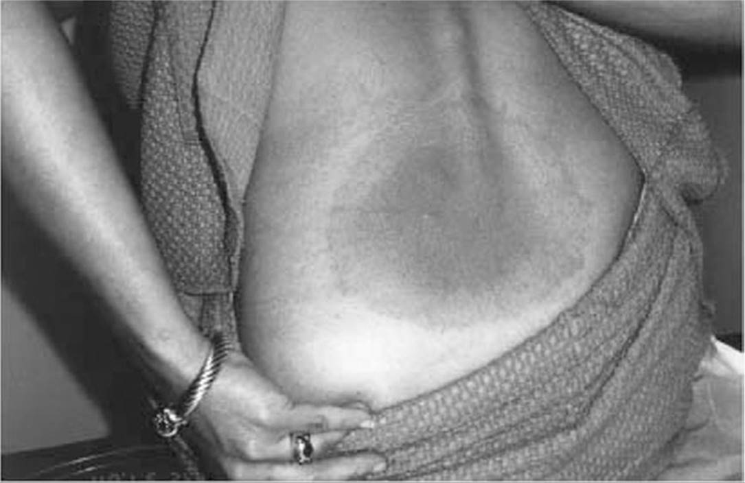
Figure 13.1. Erythema migrans. Note the dark erythematous center. See color image on color plate 2.
The lesion begins within a month of exposure as a red macule or papule at the site of the tick bite. It then expands over days, forming a bright red flat border at the advancing edge. As the lesion expands, central clearing may develop, and in some cases, the site takes on the appearance of a target. However, in many patients, the lesion remains diffusely erythematous. Erythema migrans are usually large, reaching an average size of 15 cm (range: 3-70 cm). They are commonly located in moist, warm areas of the body where ticks prefer to feed (axilla, behind the knees, and at the belt line). Despite their size, warmth, and bright color, the lesions are usually painless, but they can cause burning or itching.
2. Early disseminated disease (“secondary lyme disease”).
Several days after the onset of erythema migrans, small annular satellite lesions may be observed, reflecting early dissemination. Also at this time, patients often experience a viral-like syndrome consisting of malaise, fatigue, myalgias, arthralgias, and headache. They may also develop generalized lymphadenopathy. Migratory joint, tendon, muscle, and bone pain are common complaints. In a significant percentage of patients, symptoms attributable to the nervous system and heart commonly develop at this stage.
Nervous system involvement. The spirochete often initially disseminates to the nervous system, causing a severe generalized headache that waxes and wanes. If the disease is not treated, about 10% of cases develop more serious neurologic manifestations. Frank meningitis can result in neck stiffness, a CSF lymphocytic pleocytosis (usually about 100 cells/mm3), and an elevated CSF protein with normal CSF glucose. Cranial nerve deficits can accompany meningitis, bilateral Bell’s palsy being the most common cranial nerve dysfunction. Lymphocytic infiltration of small vessels supplying axons can lead to axonal degeneration and peripheral neuritis. The triad of meningitis, cranial nerve deficits, and radiculoneuritis has been termed Bannwarth syndrome. This syndrome is more commonly reported in Europe than in the United States.
KEY POINTS
About Primary and Secondary Lyme Disease
1. Hallmark of primary disease is erythema migrans:
a) Macular expanding erythematous lesion, central clearing.
b) Begins 1 month after the tick bite.
c) Mean diameter 15 cm.
d) Painless, can cause itching.
2. Dissemination is associated with small annular lesions and a flu-like illness.
3. Central nervous system involvement can cause waxing and waning headache. Lymphocytosis of the cerebrospinal fluid (100 cells/mm3), cranial nerve deficits (Bell’s palsy), and peripheral neuritis is called Bannwarth syndrome.
4. In cardiovascular involvement, spirochetes infiltrate the myocardium, causing conduction defects.
Cardiovascular involvement. Among untreated patients, 5-8% develop cardiac manifestations within several weeks of the onset of illness. Spirochetes can directly infiltrate the myocardium, causing lymphocytic inflammation. Conduction defects are most common, and an electrocardiogram should be ordered in all patients with symptomatic Lyme disease. First-degree heart block is most common, but second-degree and complete heart block may also develop. However, complete heart block rarely persists for longer than 7 days and does not usually require placement of a pacemaker. More severe myocarditis accompanied by congestive heart failure is rare.
3. Late disease (“tertiary lyme disease”).
Late disease develops months to years after primary infection. Some patients never experience symptoms from the earlier stages. Musculoskeletal complaints are most common at this stage, but neurologic complaints, skin disease, and generalized symptoms may also occur.
Musculoskeletal manifestations. Approximately 60-80% of untreated patients experience musculoskeletal symptoms. Migrating arthralgias or frank arthritis causing joint swelling most commonly involves the knees and other large joints. Less commonly, small joints may be affected. Joint aspiration may reveal white blood cell (WBC) counts of 500 to 110,000/mm3, with a predominance of polymorphonuclear leukocytes (PMNs). The presence of spirochetes in the joint fluid can be detected by PCR in most patients, and arthritis usually resolves after antibiotic therapy.
KEY POINTS
About Late or Tertiary Lyme Disease
1. Symptomatic disease develops months to years after primary disease.
2. Musculoskeletal complaints are most common:
a) Migrating arthritis and arthralgias.
b) Joint fluid contains 500-110,000 cells/mm3, primarily polymorphonuclear leukocytes.
c) Patient usually improves with antibiotics.
3. Central nervous system encephalopathy can cause mood, cognitive, and sleep disorders:
a) Elevated protein and antibody against Borrelia burgdorferi in cerebrospinal fluid.
b) Response to antibiotics variable.
4. Acrodermatitis chronica atrophicans, a chronic skin infection, contains spirochetes.
5. Fibromyalgia-like or chronic fatigue-like syndrome may occur; controversial, antibiotics not helpful.
Neurologic manifestations. Just as is observed in syphilis, B. burgdorferi sensu stricto may invade the cerebral cortex and cause a chronic encephalopathy associated with mood, cognitive, and sleep disorders. Subtle language disturbances have also been observed. The CSF may reveal elevated protein levels and increased titers of antibodies to B. burgdorferi. Patients may also develop peripheral neuropathies leading to paresthesias and radicular pain. Evaluation of these neurologic complaints can be complicated, and the neurocognitive complaints associated with fibromyalgia are often misdiagnosed as central nervous system Lyme disease. The response to antibiotic therapy is variable.
Other manifestations. Acrodermatitis chronica atrophicans can develop years after erythema migrans. It begins as a bright red skin lesion that later becomes atrophic, mimicking localized scleroderma. B. burgdorferi can be cultured from these lesions up to 10 years after their onset. A very difficult management problem arises from the small percentage of patients who experience persistent diffuse aches and pains. Some patients with Lyme disease develop a fibromyalgia-like syndrome; others may experience a chronic fatigue-like syndrome. The contribution of B. burgdorferi infection to these complaints remains controversial, and many patients with these complaints fail to improve after antibiotic therapy.
Diagnosis
Although B. burgdorferi can be grown in vitro, cultures are rarely positive because the number of organisms in skin lesions, blood, and CSF is very low. The diagnosis is based on clinical manifestations and a history of possible tick exposure in an endemic area, combined with serologic testing. In considering the diagnosis, it is important to keep in mind that many patients with confirmed Lyme disease deny being bitten by a tick.
The Lyme disease enzyme-linked immunosorbent assay (ELISA) uses a sonicate of B. burgdorferi as the antigen and detects IgG and IgM antibodies directed against the spirochete. Acute and convalescent titers spaced 2-4 weeks apart should be collected. In early disease, a significant rise in antibody titer is detected in only 60-70% of patients. Negative titers at this stage therefore do not exclude Lyme disease. Also, antibiotic therapy can abort a full antibody response, further complicating serologic diagnosis. For these reasons, ELISA testing is not recommended for patients with classic erythema migrans, because the lesion is pathognomonic for Lyme disease. Titers for IgM begin to rise within 2 weeks, but a significant rise may not be detected for 6-8 weeks. Levels usually peak at 6-8 weeks and decline over 2-3 months. Titers for IgG rise later, being first detected at 6-8 weeks and peaking at 4-6 months. A significant IgG titer usually persists for life. False positive tests occur 3-5% of the time and are more common in patients with syphilis, leptospirosis, malaria, bacterial endocarditis, viral infections, and connective tissue diseases.
Western blot analysis is recommended to verify all positive ELISA tests, commonly called two-tier testing. The Western blot detects serum antibodies directed against specific polypeptide components of B. burgdorferi. Serum from infected patient most commonly contains antibodies directed against the 23 kDa OspC protein and the 41 kDa flagellar antigen, but may also cross-react with the Osp heat-shock proteins. Strict criteria for interpretation of Western blots have been established by the U.S. Centers for Disease Control and Prevention (CDC).
A newer ELISA, VisE-C6, has been developed that detects IgG antibodies directed against the antigenic protein VlsE sixth invariant region (C6). This IgG antibody is detected early in disease and demonstrates higher sensitivity and specificity than the conventional IgM antibody ELISA, and shows comparable sensitivity and specificity to a conventional two-tier test (ELISA followed by Western blot). Addition of the VisE-C6 band to Western blot increases this test’s sensitivity in early disease. It is likely that in the future, the VisE-C6 ELISA will replace the conventional two-tier assay because of its greater simplicity and equivalent sensitivity and specificity.
KEY POINTS
About the Diagnosis of Lyme Disease
1. Cultures are rarely positive and are not recommended.
2. Diagnosis is made by a combination of epidemiology, clinical manifestations, and serology.
3. Many patients with Lyme disease deny a tick bite.
4. Enzyme-linked immunosorbent assay (ELISA) detects immunoglobulin G (IgG) and M (IgM) antibodies:
a) Not recommended in the presence of classic erythema migrans, which is pathognomonic.
b) Titer rise is aborted by early antibiotic treatment.
c) IgM begins to rise at 2 weeks, declines by 2-3 months.
d) IgG rises at 6-8 weeks, persists for life; negative IgG titer excludes late disease.
e) False positive rate is 3-65%.
5. Western blot recommended to confirm all positive ELISA tests (two-tier testing).
a) The 23-kDa OspC protein and the 41-kDa flagellar antigen most commonly cross-react.
b) Strict criteria for a positive Western blot have been established by the U.S. Centers for Disease Control and Prevention.
6. ELISA, VisE-C6 detects IgG antibodies directed against the antigenic protein VlsE sixth invariant region (C6).
a) More sensitive than conventional IgM ELISA for early disease.
b) Equally sensitive and specific as the conventional two-tier test and promises to replace this test in the future.
c) Addition of the VisE-C6 band to Western blot analysis improves sensitivity in early disease.
Serologic tests are best utilized for the patient with suspected early disease who does not have erythema migrans or for the patient with symptoms of late disease. Negative serology in early disease may require follow-up testing because of the delay in the rise of antibody titers in some patients. In patients with suspected late disease, a negative IgG titer virtually excludes the diagnosis.
Treatment
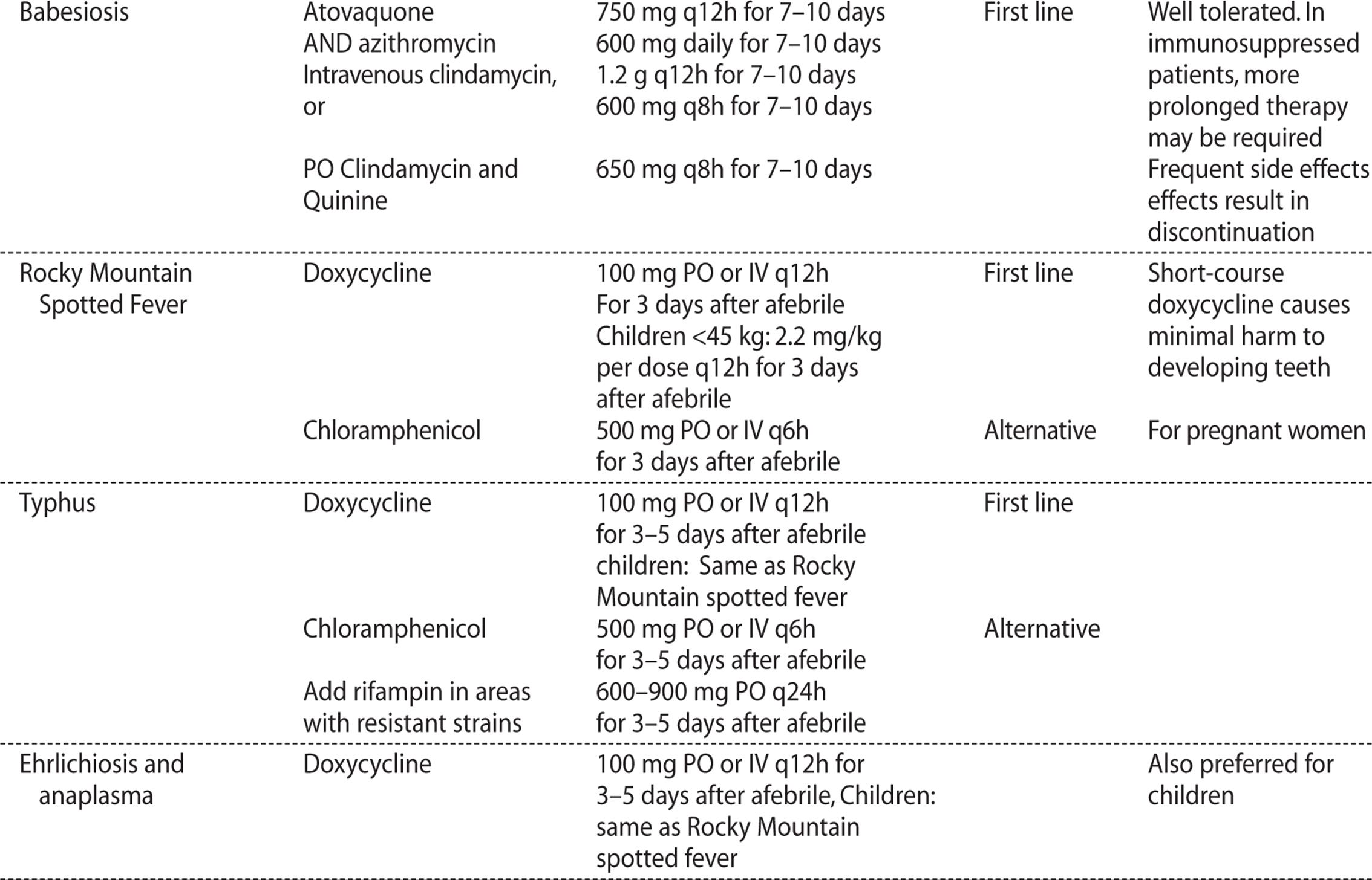
For the treatment of early disease, doxycycline for 10-21 days or amoxicillin for 14-21 days is equally effective (see Table 13.1). The ideal duration of therapy has not been determined, and many physicians opt for the longer course. Cefuroxime axetil is an effective alternative. Oral erythromycin (250 mg every 6 hours) and oral azithromycin (500 mg daily) have proved to be less effective.


For early disseminated disease with isolated palsies of the seventh cranial nerve, multiple erythema migrans lesions, or carditis with first-degree heart block doxycycline for 14-21 days is the treatment of choice. A Jarisch–Herxheimer-like reaction may be observed in up to 15% of patients during the first 24 hours of therapy for disseminated disease. In patients with meningitis or other neurologic abnormalities, and in patients experiencing carditis with high-degree heart block, intravenous ceftriaxone for 10-28 days is preferred. Alternatively, cefotaxime or high-dose penicillin can be administered. In late disease, patients with intermittent or chronic arthritis and no neurological manifestations may be treated with a very prolonged 28-day course of doxycycline or amoxicillin. If arthritis fails to improve, a repeat 28-day course of oral antibiotics can be administered or the patient can be treated with parenteral therapy for 14-28 days. For late central or peripheral neurological disease, parenteral therapy for 14-28 days is recommended.
A rare but difficult management problem arises in the patient who complains of persistent symptoms despite appropriate therapy. Patients must be warned that symptoms can linger for up to 6 months after treatment. Objective evidence for relapse is rarely found in the patient whose symptoms persist for more prolonged periods. Repeat antibiotic therapy has not been proven to be of benefit, and this approach is costly, increases the risk of selecting for resistant organism, and has led to fatal complications. The wisest course of action is reevaluation rather than retreatment, because the most likely explanation for a lack of response to therapy is misdiagnosis. A rheumatologist should be consulted to assist with diagnosis and treatment.
Prevention
Because of the extensive publicity surrounding Lyme disease, people often panic when they sustain a tick bite. In endemic areas, frantic calls to the physician’s office are a frequent occurrence during the summer months. A logical approach to the management of tick bites will reduce unnecessary administration of antibiotics. Assessment of the risk of contracting Lyme disease requires a careful history of the nature of the tick bite. The questioner needs inquire about:
• The size of the tick. Lyme disease is primarily spread by the I. scapularis nymph. This tick is very small, about the size of a small freckle. Larger ticks are unlikely to transmit Lyme disease.
• Attachment. If the tick fails to attach to the skin, it cannot transmit disease. The likelihood of being bitten by a tick can be reduced by wearing long pants and shirts when walking in areas with brush and high grasses. In endemic areas, public health officials recommend that, upon returning from the outdoors, people perform a complete body check for ticks. Removing ticks before they attach is an excellent preventive measure. If an attached tick is discovered, the duration of attachment needs to be estimated. If attachment is less than 24 hours, the risk of disease transmission is low.
• Engorgement. If the tick is engorged with blood, prolonged attachment and an increased risk of disease transmission are suggested.
Prophylactic antibiotics consisting of a single dose of oral doxycycline (200 mg) within 72 hours of the tick bites can prevent the development of Lyme disease. The incidence of Lyme disease is approximately 1 in 100 in areas in which a high percentage of ticks harbor B. burgdorferi. In these locations, prophylaxis should be strongly considered. A more targeted approach of administering prophylactic antibiotics to the person who reports attachment of a small tick for more than 24 hours or who finds an engorged tick may prove more efficacious. In patients who do not fulfill these criteria, a careful explanation of the risk and natural progression of Lyme disease will usually calm the concerned caller.
KEY POINTS
About the Treatment and Prevention of Lyme Disease
1. Treat early disease with doxycycline for 10-21 days or amoxicillin or cefuroxime axetil 14-21 days.
2. Treat disseminated disease characterized by mild carditis (first-degree heart block) or seventh nerve palsy with doxycycline for 14-21 days.
3. Meningitis or carditis with high-degree heart block should be treated with intravenous ceftriaxone, cefotaxime, or penicillin for 10-28 days.
4. Treat chronic arthritis cases with doxycycline or amoxicillin for 28 days. If poor response, repeat the oral course or use the meningitis regimen above.
5. Failure to improve on antibiotics suggests another diagnosis.
6. Prophylactic antibiotics are recommended if a small tick has been attached for more than 24 hours or if an engorged tick is found.
Table 13.1. Antibiotic Treatment of Zoonotic Infections
LEPTOSPIROSIS
POTENTIAL SEVERITY
Can cause a life-threatening systemic illness. Early diagnosis and treatment reduce the severity of the disease.
Epidemiology
Leptospirosis is seldom diagnosed in the United States, except in Hawaii, where annual rates of 128 per 100,000 population have been reported. Leptospirosis is found throughout the world in temperate and tropical climates. Infection often follows hurricanes and flooding in Central and South America and Caribbean islands. In endemic areas, the incidence of leptospirosis is 5-20% annually.
The acute illness often causes nonspecific symptoms that never require medical attention, explaining the low incidence detected by passive surveillance studies. Dogs, livestock, rodents, amphibians, and reptiles can become infected. They often harbor Leptospira in their renal tubules, excrete the pathogen in the urine, and contaminate both soil and water, where the organism can persist for weeks to months. Humans at risk of becoming infected include trappers and hunters, dairy farmers, livestock workers, veterinarians, military personnel, and sewer workers. Infection has also been associated with outdoor activities in freshwater, including wading, swimming, whitewater rafting, kayaking, and canoeing. In cities, humans may be inadvertently exposed to infected rat and dog urine.
KEY POINTS
About the Epidemiology of Leptospirosis
1. Found in temperate and tropical climates:
a) Rare in the United States, except Hawaii.
b) Follows flooding, particularly in Central and South America, Caribbean islands.
2. Dogs, livestock, rodents, amphibians excrete in Leptospira urine, contaminating soil and water.
3. Trappers, hunters, dairy farmers, livestock workers, veterinarians, military, and sewer workers at risk.
4. Outdoor freshwater activities predispose to disease.
Pathogenesis
Leptospirosis is caused by Leptospira interrogans, a tightly spiraled spirochete with 18 or more coils per cell. Like other spirochetes, it is narrow, 0.1 μm in width, and long, 6-12 μm in length, and is best visualized by darkfield microscopy. Leptospira is an obligate aerobe and grows slowly. There are more than 200 serovars of L. interrogans, and different serovars have predilections for different animals.
KEY POINTS
About the Pathogenesis of Leptospirosis
1. Caused by Leptospira interrogans, a tightly coiled spirochete, slow-growing obligate aerobe.
2. Penetrates breaks in skin or softened skin after prolonged water exposure, conjunctiva or mucous membranes; less commonly, it enters the lungs in aerosolized form.
3. Disseminates after traveling to the lymphatics and bloodstream.
4. Outer surface coated with lipopolysaccharide (LPS). Glycoprotein toxin damages endothelial cells.
5. Induces IgM and IgG antibodies directed against LPS; killed by macrophages.
These organisms gain entry to the human host through cuts, abrasions, and skin softened by prolonged water exposure. Mucous membranes and conjunctivae are other portals of entry. Inhalation of aerosolized droplets can lead to pulmonary invasion. Once in the host, the spirochetes spread to the lymphatic system and then enter the bloodstream, disseminating throughout the body. The organisms’ outer wall is coated with lipopolysaccharide (LPS) that serves as a major antigenic stimulus. The spirochete releases a glycolipoprotein toxin that displaces long-chain fatty acids from host vascular endothelial cells, causing breakdown of the vessel walls and fluid leakage, allowing the organisms to escape from the bloodstream to the tissues. The host generates IgM and IgG antibodies directed against the Leptospira LPS. These antibodies are opsonins that enhance phagocytosis by macrophages in the reticuloendothelial system and enhance clearing of the organisms from the bloodstream.
Clinical Manifestations
CASE 13.2
A 25-year-old man presented to the hospital with complaints of fever and headache of 3 days’ duration. His symptoms began 3 days after he completed a 12-day survival race with three teammates, in Sabah State on Borneo Island, Malaysia. The day before his admission, one of his teammates was admitted to the hospital with similar complaints.
Physical examination revealed a body temperature of 37.9°C and a pulse rate of 90 per minute (regular). Conjunctiva was hyperemic, but nonicteric. Lymph nodes were not palpable, and no skin eruptions were seen. A neurologic examination was normal.
A laboratory workup showed a white blood count (WBC) count of 13,100/mm3, with 91% neutrophils; a hemoglobin of 14.8 g/dL; a platelet count of 190,000/mm3; and total bilirubin 0.5 mg/dL. Liver enzymes were 63 IU/L [aspartate aminotransferase (AST)] and 66 IU/L [alanine aminotransferase (ALT)]. Lactate dehydrogenase (LDH) was 420 IU/L; blood urea nitrogen (BUN), 12.5 mg/dL; and creatine, 0.9 mg/dL.
Minocycline was administered intravenously on the third hospital day, and fever subsided over 48 hours. The intravenous minocycline was continued for a week, followed by 2 weeks of oral doxycycline. Acute sera is negative for Leptospira antibody, but convalescent serum 2 weeks later revealed a 1:160 titer of antibody directed against L. interrogans serovar hebdomadis.
Further investigation revealed that 51 of 78 participants had developed symptoms consistent with leptospirosis. Activities had included jungle trekking, canoeing, kayaking, rafting, scuba diving, mountain biking, and cave exploring. Local rivers were flooded at the time of the race. (Adapted from Sakamoto M, Sagara H, Koizumi N, Watanabe H. A case of leptospirosis infection in Borneo Island, Malaysia. Infect Agent Surveill Rep. 2001;22:5-6.)
The incubation period for leptospirosis is usually 5-14 days, but can be up to 30 days. The severity of illness varies greatly, and probably depends on the degree of exposure and the infecting serovar. Certain serovars from cows cause mild disease; others (contracted from rats) are more likely to cause severe disease. Classically, symptomatic disease occurs in two phases: the bacteremic phase and the immunologic phase; however, fewer than half of patients actually experience a biphasic illness. More than 90% of cases are self-limiting, but a small percentage experience a severe—sometimes fatal—illness called Weil disease.
As illustrated in case 13.2, the onset of illness is usually sudden. Symptoms may include fever, rigors, sweating, headache, photophobia, and severe myalgias accompanied by marked tenderness of the calves, thighs, and mid back. Other manifestations can include epistaxis, cough, and sore throat. Severe abdominal pain can mimic an acute abdomen. Nausea, vomiting, and diarrhea may also develop. On examination, the vessels in the conjunctiva are often very prominent because of vascular dilatation. Transient skin rashes may be noted. Capillary fragility can result in macular, maculopapular, purpuric, urticarial lesions, or diffuse skin redness. During the acute phase, Leptospira can be cultured from the blood and CSF.
KEY POINTS
About the Clinical Manifestations of Leptospirosis
1. Incubation period is 5-14 days, and severity depends on inoculum and serovar (rat serovars being more severe).
2. Two phases in fewer than half of patients:
a) Bacteremic phase—Sudden onset; fever, rigors, headache, photophobia, and severe myalgias; dilated conjunctival vessels, marked tenderness calves, thighs, and mid back; macular rash.
b) Immunologic phase (after 4-30 days)—Conjunctivitis, photophobia, retrobulbar pain, neck stiffness, diffuse lymphadenopathy, hepatosplenomegaly, and aseptic meningitis with lymphocytosis in the cerebrospinal fluid.
3. Weil disease is rare, severe; mortality 5-40%:
a) High direct bilirubin, mild elevation in alkaline phosphatase, mild elevation in transaminase values, combined with a high creatine phosphokinase.
b) Renal failure accompanied by thrombocytopenia.
c) Hemorrhagic pneumonia.
Resolution of fever may herald the onset of the second, immune, phase of the illness. This phase can last 4-30 days. Blood cultures turn negative at this time. Prominent conjunctivitis with or without hemorrhage is seen, accompanied by photophobia, retrobulbar pain, neck stiffness, diffuse lymphadenopathy, and hepatosplenomegaly. Aseptic meningitis with or without symptoms is characteristic of this stage and is immune-mediated. Lymphocytes (<500 mm3) are seen in the CSF, together with moderate protein elevation (50-100 mg/mL) and a normal glucose level.
Weil disease can develop after the acute phase and consists of hemorrhage, jaundice, and renal failure. Severe hemorrhagic pneumonitis may also develop. Jaundice is caused by vascular injury to the hepatic capillaries without significant hepatocellular necrosis. Transaminase levels seldom exceed 200 U/L, and an elevated prothrombin time is uncommon. Creatine phosphokinase (CPK, MM fraction) reflecting myositis is often disproportionately high in comparison with the serum transaminase values. Marked elevations in conjugated bilirubin are the hallmark of liver involvement and can reach levels of 80 mg/dL, associated with mild-to-moderate elevations of alkaline phosphatase.
The constellation of a high direct bilirubin, mild elevation in alkaline phosphatase, and mild elevation in transaminase values combined with a high CPK should always raise the possibility of Weil disease. Liver biopsy reveals hypertrophy and hyperplasia of Kupffer cells, accompanied by cholestasis. The hepatic architecture usually remains intact, and little evidence of hepatic necrosis is seen. Acute renal failure is associated with oliguria and usually develops during the second week of illness at the same time that jaundice is noted. Renal biopsy demonstrates acute interstitial nephritis, and immune-complex glomerulonephritis may also be seen. Thrombocytopenia may accompany renal failure in the absence of disseminated intravascular coagulopathy. Pulmonary disease can develop in the absence of hepatic or renal involvement. This hemorrhagic pneumonia is generally associated with a bloody cough, and chest X-ray reveals nodular densities in the lower lobes. Histopathology reveals damage to the capillary endothelium and intra-alveolar hemorrhage. Cardiovascular collapse can develop suddenly. The mortality rate for severe leptospirosis ranges from 5% to 40%.
Diagnosis and Treatment
Even in endemic areas, the early clinical diagnosis of leptospirosis is difficult to make because the clinical manifestations are often nonspecific. Leptospira can be cultured in vitro on special media (Fletcher’s, Ellinghausen’s, or polysorbate 80). Significant growth may be detected after 1-2 weeks, but can take up to 3 months. Blood, CSF, and urine are positive during the first 7-10 days of illness, and urine remains positive during the second and third weeks of the illness.
The sensitivity of culture is low, and therefore the diagnosis must usually be made by measuring acute and convalescent antibody titers. The microscopic agglutination test is the most specific test and allows identification of serum antibodies to specific serovars. Live leptospires are placed on a slide, and the highest serum dilution at which more than 50% of the spirochetes agglutinate on darkfield microscopy is defined as the positive titer. Antibody titers can be detected as early as 3 days into the illness, but usually take 2 weeks, and continue to rise for 3-4 weeks. A rise in titer by a factor of 4 or more is defined as serologic confirmation of leptospirosis. A single titer above 1:800 in combination with appropriate symptoms is considered indicative of active disease, and a single titer of 1:200 or a persistent titer of 1:100 is suggestive evidence. This test is technically demanding and potentially hazardous; it is performed only by CDC reference laboratories. An ELISA test for IgM antibodies is commercially available and has a sensitivity that varies from 100% to 77% and a specificity of 93% to 98%. Methods using PCR have been reported, but are not commercially available.
KEY POINTS
About the Diagnosis and Treatment of Leptospirosis
1. Can be cultured from blood, cerebrospinal fluid, and urine. Low yield.
2. Serology is most helpful.
a) Microscopic agglutination test (only in CDC reference labs): positive at 2 weeks, rises at 3-4 weeks (a rise by a factor of 4 or more is diagnostic), titer above 1:800 plus symptoms indicates active disease, 1:200 is suggestive.
b) Enzyme-linked immunosorbent assay for immunoglobulin M antibodies is commercially available and has good sensitivity and specificity.
3. Treat with intravenous penicillin, ampicillin, or ceftriaxone for severe disease; oral doxycycline or amoxicillin for milder disease.
4. For prophylaxis in endemic areas, use doxycycline.
Penicillin G, ampicillin, or ceftriaxone is recommended for severe disease. In severe disease, penicillin treatment has been shown to reduce the duration of illness. As observed in the treatment of other spirochetes, therapy may be associated with a Jarisch–Herxheimer reaction. For mild leptospirosis, oral doxycycline or amoxicillin may be administered. When exposure in endemic areas is anticipated, prophylaxis with oral doxycycline (200 mg once per week) has been shown to be efficacious (see Table 13.1).
BABESIOSIS
GUIDING QUESTIONS
1. How is babesiosis contracted?
2. Why has the incidence of this infection increased in the United States?
3. How does life cycle of Babesia differ from that of Plasmodium, and how might these differences relate to the differences in clinical manifestations?
4. Which other infection do patients with babesiosis often contract at the same time, and why?
5. Is this blood protozoan treated in the same way as Plasmodium?
POTENTIAL SEVERITY
Usually causes mild disease, but in splenectomized patients can be fatal.
Prevalence, Epidemiology, and Life Cycle
Like malaria, Babesia is a blood protozoan. It has a life cycle similar to that of Plasmodium; however, Babesia is transmitted by the deer tick, I. scapularis. Curiously, Babesia does not infect deer. However, the intermediate host, the white-footed deer mouse, is readily infected by Babesia microti, the primary strain causing human disease in the United States. In endemic areas, the percentage of these rodents infected by Babesia can reach 60%. During its larval and nymph phases, the tick lives on the deer mouse, where it obtains blood meals. The nymph can leave the deer mouse and attach to humans. After attachment, this tiny tick (2 mm in diameter) eats a blood meal and introduces the Babesia sporozoite. The sporozoites enter human red blood cells (RBCs). The mature signet-ring-shaped trophozoite multiplies asexually by binary fission, forming characteristic tetrads. Subsequently, it lyses the host RBC. Because multiplication is asynchronous, massive hemolysis is not seen. Also, unlike Plasmodium, Babesia lacks a hepatic phase.
Babesiosis was once thought to be a disease only of cattle and wild animals. However, in the last 30 years, this organism has been found to increasingly infect humans. Thousands of cases of human babesiosis have been identified, many occurring in Massachusetts on the islands of Nantucket and Martha’s Vineyard. Other cases have been described throughout New England, New York, Maryland, Virginia, Georgia, Wisconsin, Minnesota, Washington State, and California.
KEY POINTS
About the Babesia Life Cycle
1. The small nymph form (2 mm in diameter) of the deer tick, Ixodes scapularis, carries Babesia from white deer mice to humans.
2. In human red blood cells (RBCs), the mature signet-ring trophozoite multiplies by binary fission forming characteristic tetrads.
3. Multiplication is asynchronous, and therefore hemolysis is never massive.
4. Babesiosis has no hepatic phase.
KEY POINTS
About Babesia Epidemiology
1. Endemic in areas where the deer population is abundant.
2. Requires the presence of the white deer mouse, which harbors the infectious deer tick (Ixodes scapularis) nymphs.
3. In Europe, the cattle are the intermediate host harboring adult and nymph ticks.
4. Human infections occur during the period of nymph feeding (May to September).
In addition to tick-borne disease, over 150 cases of transfusion-transmitted disease have been reported primarily due to B. microti. Sporadic cases of babesiosis along the northern Pacific coast were caused by B. duncani and B. duncani-type organisms. In Europe, the primary strain causing disease is B. divergens. Cattle are the natural host for this strain and this parasite primarily causes symptomatic disease in splenectomized hosts. Farmers are at greatest risk, although cases have been reported in foresters and others participating in outdoor activities.
The rise in the incidence of babesiosis has been attributed to the decreased popularity of deer hunting and the associated increase in deer and deer tick populations. Also, migration to the suburbs in the United States has brought humans in closer proximity to the mouse reservoirs harboring the infectious I. scapularis nymph. The infection is contracted by humans during the months of May through September when the nymphs are feeding.
Clinical Presentation
CASE 13.3
A 65-year-old female presented with intermittent fever for the preceding 2 months, associated with intermittent myalgias and fatigue. She had just returned from a 2-month summer vacation in Martha’s Vineyard, Massachusetts. She denied any history of tick bites. One month earlier, she had been diagnosed with Lyme disease. However, despite appropriate treatment, her fevers did not resolve. Aside from a mild anemia, her routine blood tests were normal; however, Giemsa stain of her peripheral blood revealed occasional red blood cells (RBCs) containing ring forms, some in tetrads. Treatment with clindamycin and quinine caused a rapid resolution of her fever.
KEY POINTS
About the Clinical Presentation of Babesiosis
1. Presents as the “summer flu” 1-6 weeks after exposure.
2. History of hiking in tick-infested areas.
3. Often no history of tick bite, because the Ixodes scapularis nymph is mistaken for a small freckle.
4. More serious disease occurs in splenectomized patients, individuals >50 years old and patients receiving immunosuppressants.
5. Patients with babesiosis may also have Lyme disease, because Ixodes scapularis transmits both infections.
The symptoms of babesiosis are nonspecific, making the disease difficult to diagnose clinically. Generally, patients present 1-6 weeks after exposure with a flu-like illness as described in case 13.3. Fever, chills, myalgias, arthralgias, fatigue, and anorexia are most common. The illness presents during the summer months as a “summer flu.” In endemic areas, the clinician should inquire about recent hiking in tick-infested locations, particularly those with tall grasses and brush. Patients often do not give a history of tick bites, having failed to detect the attached nymph because of its small size (the diameter of a small freckle). In the normal host, the disease may cause minimal symptoms and resolve spontaneously. However, in patients over the age of 50 or in those who have undergone splenectomy, infection can be more severe and persistent. Other patients at risk of severe symptomatic disease include patients receiving immunosuppressive drugs for cancer or anticytokine therapy such as infliximab. Cases of adult respiratory distress syndrome and hypotension have been reported, and on rare occasions, patients have died. In Europe, cases primarily involved splenectomized patients, and the clinical presentation has often been fulminant, being associated with severe hemolysis and death.
Patients with babesiosis may also have symptoms suggestive of Lyme disease, particularly the skin rash of erythema migrans. I. scapularis is also the vector for B. burgdorferi, and in one series of cases, 54% of patients with babesiosis also had antibodies against the Lyme spirochete, suggesting that these patients had dual infections as observed in case 13.3.
Diagnosis and Treatment
Giemsa stain of thick and thin smears from the peripheral blood should be examined under an oil-immersion objective. Small ring forms, often grouped in tetrads (Figure 13.2), are the only form seen. Babesiosis is frequently mistaken for P. falciparum. The classic tetrad is not observed in Plasmodium infection, and the banana-shaped gametocytes observed in P. falciparum are never observed in Babesia.An indirect fluorescence antibody (IFA) titer that measures antibody against B. microti, the primary form that causes babesiosis in the United States, is available through the CDC. Significant increases in antibody titer develop 3-4 weeks after the infection is contracted. Most recently a real-time PCR assay has been developed that has greater sensitivity than Giemsa stain, and 100% specificity. This assay also allows accurate speciation of the strain.
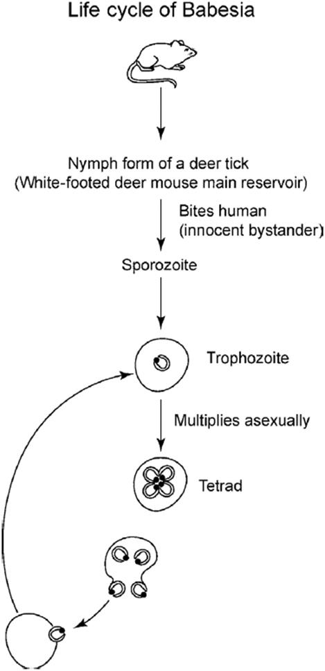
Figure 13.2. Life cycle of Babesia. The white-footed deer mouse is the main reservoir of this pathogen.
Treatment should be initiated in splenectomized patients and in other patients with serious disease. Azithromycin and atovaquone are the preferred regimens because of their lower side effect profile. Clindamycin combined with oral quinine is also an effective regimen but frequently has to be discontinued because of toxicity (see Table 13.1. In serious disease, exchange transfusion has proved life saving.
KEY POINTS
About Diagnosis and Treatment of Babesiosis
1. Giemsa stain of the peripheral blood remains the best way to make the diagnosis.
2. Only ring forms are seen.
3. Frequently mistaken for Plasmodium falciparum.
4. Tetrad ring forms strongly support the diagnosis of babesiosis.
5. Real-time PCR is sensitive and specific.
6. Azithromycin and atovaquone are the treatment of choice.
7. Many malaria regimens, including chloroquine and primaquine, are not effective in babesiosis.
Chloroquine, often initiated when Babesia is mistaken for P. falciparum, is not effective. Similarly, doxycycline, pentamidine, primaquine, and pyrimethamine–sulfadoxine (Fansidar) are not efficacious.
RICKETTSIA AND RELATED INFECTIONS
The Rickettsiaceae family encompasses two genera: Rickettsia and Ehrlichia. These organisms are small gram-negative coccobacilli (coccal forms 0.3 mµ in diameter, bacillary forms 0.3 × 1-2 mµ) whose cell wall consists of a peptidoglycan layer sandwiched between two lipid membranes. They are all obligatory intracellular pathogens.
Rickettsia gains entry by inducing host cells to phagocytose them. Some strains—for example, Rickettsia rickettsii—produce a phospholipase that dissolves the confining phagolysosome membrane, allowing them to escape into the cytoplasm. Other strains multiply and survive within the phagolysosome by blocking the release of toxic enzymes into the phagolysosome (Ehrlichia species, for instance). All rickettsial diseases are spread to humans by arthropods: ticks, mites, lice, and fleas.
Clinically, the rickettsial family of diseases has been classified into four groups:
1. The spotted fever group. Includes R. rickettsii (Rocky Mountain spotted fever), R. conorii (Boutonneuse fever), R. australis (Queensland tick typhus), R. sibirica (North Asian tick typhus), and R. akari(rickettsial pox).
2. The typhus group. Includes R. prowazekii (louse-borne or epidemic typhus and Brill–Zinsser disease), R. typhi (murine typhus), and Orientia tsutsugamushi (scrub typhus).
3. The Ehrlichia group. Consists of E. chaffeensis [human monocytotropic ehrlichiosis (HME)], Anaplasma phagocytophilum [human granulocytotropic anaplasma (HGA)], and the rarer human pathogens E. ewingii, E. muris, and Neorickettsia sennetsu.
ROCKY MOUNTAIN SPOTTED FEVER
POTENTIAL SEVERITY
Untreated Rocky Mountain spotted fever can be fulminant and fatal.
Epidemiology
Rocky Mountain spotted fever (RMSF) is the most severe disease in the spotted fever group of rickettsial diseases. It occurs throughout the United States, Mexico, and Central and South America. Although first recognized in the Rocky Mountains, the disease is most commonly reported in the southeastern and South–Central United States. Small endemic areas are also found in Long Island and Cape Cod. Cases have also been reported in urban parks. The severity of disease tends to vary depending on location, more severe cases being observed in central Oklahoma and southwestern Tennessee. Two areas in north central North Carolina cases tended to be mild. The annual incidence in the United States varies from 16.8 per 1 million in American Indians to 0.5 per million among Asian/Pacific Islanders, and is 4.2 for Caucasians and 2.6 per million for African Americans.
The disease occurs in the late spring and summer, the seasons in which ticks feed. In the south, the dog tick (Dermacentor variabilis) is the primary vector, and in states west of the Mississippi, the wood tick (Dermacentor andersoni) is primarily responsible for transmitting disease. A recent outbreak in Arizona was associated with the common brown dog tick (Rhipicephalus sanguineus).
Pathogenesis
After the tick has attached to the host for between several hours and a day, it injects the rickettsiae into the dermis. Once exposed to the warmer temperature and mammalian blood, R. rickettsii activates and proliferates in the skin. The organism resides in the cytoplasm of host cells, where it divides by binary fission and spreads from cell to cell by a mechanism similar to that used by Listeria monocytogenes. Both organisms induce host cell actin filament assembly to propel them to the periphery of the cell, where they are ingested by adjacent cells, forming plaques of necrotic cells.
KEY POINTS
About the Epidemiology and Pathogenesis of Rocky Mountain Spotted Fever
1. Found throughout the United States, Mexico, and Central and South America.
a) Most common in the southeastern and South-Central United States; also found in the Midwest.
b) Endemic in areas of Cape Cod and Long Island and in some urban parks.
c) Severity varies depending on location, worse central Oklahoma and southwestern Tennessee.
d) Incidence highest among American Indians
2. Injected into the skin by dog and wood ticks in the late spring and summer.
3. Proliferates in the skin, disseminates via the bloodstream.
a) Survives in the host cell cytoplasm; spreads cell to cell, producing plaques of necrotic cells.
b) Causes hemorrhage in skin, intestine, pancreas, liver, skeletal muscle, and kidneys.
R. rickettsii contains outer membrane proteins (“Omps”) and lipoproteins that stimulate cell-mediated immunity, resulting in infiltration of lymphocytes and macrophages. After multiplying in the skin, the organism disseminates via the bloodstream, where it prefers to invade vascular endothelial cells. Damage to endothelial and vascular smooth muscle cells results in a vasculitis that can involve the lungs, heart, and central nervous system. Discrete areas of hemorrhage can be found in these organs and also in the skin, intestine, pancreas, liver, skeletal muscle, and kidneys. Hemorrhage often leads to platelet consumption and thrombocytopenia, but disseminated intravascular coagulopathy is rare. Increased vascular permeability and fluid leakage result in edema, low serum protein levels, hypovolemia, and shock. Decreased intravascular volume can induce antidiuretic hormone secretion and hyponatremia. In severe cases, shock can also precipitate acute tubular necrosis and renal failure.
Clinical Manifestations
CASE 13.4
A 7-year-old girl arrived in the emergency room in Oklahoma with 2-day history of fever (39.3°C), malaise, abdominal pain, nausea, and vomiting. She was discharged with a diagnosis of viral gastroenteritis. Four days later, she was seen at a second emergency room with complaints of persistent fever, anorexia, irritability, photophobia, cough, diffuse myalgias, nausea, and vomiting.
On physical examination, she was noted to have hepatosplenomegaly and an erythematous papular rash with scattered petechiae on the trunk, arms, legs, palms, and soles. Laboratory findings included an elevated white blood cell (WBC) count of 11,400/mm3, a low platelet count of 19,000/mm3, and elevated liver enzymes [aspartate aminotransferase (AST): 279 IU/L; alanine aminotransferase (ALT): 77 IU/L]. Intravenous doxycycline was initiated to treat suspected Rocky Mountain spotted fever (RMSF), and she was placed in intensive care. Her mental status declined, and she developed metabolic acidosis and respiratory failure, dying 6 days after her first visit to the emergency room.
A serum sample drawn 2 days before her death revealed a 1:128 IgG anti-Rickettsia rickettsii antibody titer. Spotted fever group rickettsiae were detected by immunohisto-chemical staining of autopsy specimens from brain, skin, heart, lung, spleen, and kidney. On questioning, the parents reported that their child played frequently in grassy areas near their home. They did not note any recent tick bite, but ticks had been frequently observed on the family’s pet dogs and often were manually removed by members of the household. (Adapted from CDC Fatal cases of Rocky Mountain spotted fever in family clusters—three states, 2003. MMWR Morb Mortal Wkly Rep. 2004;53:407-410.)
As case 13.4 illustrates, the course of unrecognized and untreated RMSF can be fulminant. The incubation period is 2-14 days after a tick bite. The early symptoms and signs of this disease are nonspecific. Patients complain of fever, headache, malaise, myalgias, and nausea. Some patients experience severe abdominal pain, particularly children, suggesting the diagnosis of cholecystitis, appendicitis, or bowel obstruction—or as in case 13.4 with milder abdominal complaints mimicking viral gastroenteritis.
A rash usually develops within 5 days of the onset of illness, and in case 13.4, a rash alerted the physicians to the possibility of RMSF. However, in up to 10% of patients, a rash may never appear. “Spotless” fever occurs more commonly in elderly and in dark-skinned individuals. Patients often seek medical attention before the rash develops, and therefore, as in the above case, the physician may fail to consider the diagnosis. Lesions are nonpruritic. They are usually first noted on the ankles and wrists, subsequently spreading centrally and to the palms and soles. Initially, they are macular or maculopapular, subsequently becoming petechial. The presence of urticarial lesions or a pruritic skin rash makes RMSF unlikely.
KEY POINTS
About the Clinical Manifestations of Rocky Mountain Spotted Fever
1. Incubation period is 2-14 days.
2. Acute onset of nonspecific symptoms: fever, headache, malaise, myalgias, and nausea. Abdominal pain may mimic cholecystitis or appendicitis.
3. Macular, petechial rash begins on ankles and wrists and spreads to trunk 5 days after symptoms begin.
a) “Spotless” infection occurs in 10%—Usually, elderly and dark-skinned individuals.
b) Urticaria or pruritic rash makes the diagnosis unlikely.
4. Other symptoms include aseptic meningitis, conjunctivitis, fundoscopic hemorrhages, and acute respiratory distress syndrome in severe disease.
5. Disease severity worse if <4 or >60 year, male, African American, alcoholic, G-6-PD deficiency
6. Death within 8-15 days if treatment is not initiated within 5 days.
As the disease progresses, headache may become an increasingly prominent complaint. Severe headache can be accompanied by neck stiffness and photophobia suggesting meningitis, and the CSF may contain lymphocytes or PMNs, together with elevated protein; however, low CSF glucose is unusual. Conjunctivitis may be noted, and fundoscopic examination may reveal manifestations of small-vessel vasculitis (flame hemorrhages and arterial occlusion), venous engorgement, and papilledema. Respiratory complaints may become prominent, and chest X-ray may reveal alveolar infiltrates or pulmonary edema, indicating the development of adult respiratory distress syndrome. In severe cases, gangrene of the digits can also develop as a consequence of occlusion of small arterioles.
Risk factors for severe disease include age (<4 years or $60 years), male gender, African American descent, chronic alcohol abuse, and glucose-6-dehydrogenase deficiency.
Laboratory findings tend to be nonspecific. The peripheral WBC count can be normal, elevated, or depressed. Thrombocytopenia is common in more severe cases. Elevations in BUN and serum creatinine may be noted. Hypo-natremia develops in patients with hypotension. Transaminase values and bilirubin levels may be elevated as well. As illustrated in case 13.4, if appropriate therapy is not given within the first 5 days of symptomatic disease, RMSF can progress and cause death within 8-15 days.
Diagnosis
Because of the rapid course of this disease and the inability of most laboratories to culture the organism, the diagnosis of RMSF is usually made based on epidemiology and clinical manifestations. A significant percentage of patients deny a tick bite, making the diagnosis particularly difficult. In the first few days, RMSF is most commonly mistaken for a viral syndrome. If penicillin or a cephalosporin is mistakenly prescribed during this period, the subsequent rash of RMSF may be mistaken for a drug allergy. Severe headache and abnormalities in the CSF may suggest viral meningoencephalitis. The development of petechial skin lesions may raise the possibility of meningococcemia or leptospirosis.
During the spring and summer months, patients in endemic areas must always be treated for RMSF pending culture results. Skin biopsy is helpful in confirming the diagnosis. Immunofluorescence staining using antibodies specifically directed against R. rickettsii can be helpful (70% sensitivity and 100% specificity). If antibiotics for RMSF have been initiated, skin biopsy is not recommended, because the organisms are difficult to identify after treatment has been initiated. Acute and convalescent serum antibody titers can be measured by indirect fluorescent antibody (IFA) test, or complement fixation, and a significant rise in titer allows for a retrospective diagnosis. However, these tests are of no help in managing the acutely ill patient. The Weil–Felix test that detects cross-reactive antibodies to Proteus vulgarisare not only nonspecific but also insensitive, and are no longer recommended.
Treatment
Because of the unpredictable course of RMSF, physicians in endemic areas should have a low threshold for initiating doxycycline or tetracycline therapy in patients who have a nonspecific febrile illness of more than 2 days’ duration during the spring and summer. The disease responds rapidly to antibiotic therapy, and patients usually defervesce within 48-72 hours.
KEY POINTS
About the Diagnosis and Treatment of Rocky Mountain Spotted Fever
1. Presumptive diagnosis must be made based on epidemiology and clinical manifestations.
2. Culture not recommended.
3. Skin biopsy with immunofluorescence staining has high specificity. Not recommended if antibiotics have been given.
4. Serology (IFA) provides a retrospective diagnosis.
5. Can be mistaken for viral syndrome, drug allergy, and meningococcemia.
6. Physicians in endemic areas should have a low threshold for treatment:
a) Doxycycline for adults and children.
b) Chloramphenicol for pregnant women.
7. Mortality has been reported as 22% untreated, 6% with treatment.
Therapy with doxycycline is the treatment of choice for adults and children alike (see Table 13.1). Short courses of doxycycline are reported to cause minimal damage to developing teeth, but the potential benefits of doxycycline far outweigh this potential toxicity. Chloramphenicol is recommended in pregnancy. Antibiotic therapy should be continued for at least 3 days after the patient has defervesced. The mortality in untreated patients varies depending on the strain and inoculum, but in one retrospective series, was 22% in untreated patients and 6% in patients who received treatment within 5 days of the onset of illness.
Other Spotted Fevers
POTENTIAL SEVERITY
Patients can become extremely septic, developing shock and organ failure, and dying.
A number of other rickettsial species cause skin rashes and fever in humans. R. conorii shares 90% DNA homology with R. rickettsii and many of the same proteins; it causes Mediterranean spotted fever (“Boutonneuse fever”). This tick-borne illness is found in southern Europe, Africa, and the Middle East, and is clinically very similar to RMSF. A black eschar called a tache noire may be noted at the site of the tick bite. This lesion is caused by vascular endothelial damage that leads to dermal and epidermal necrosis. A diffuse maculopapular rash develops within 3-5 days of the onset of the febrile illness; however, as observed with RMSF, some patients fail to develop a black eschar or rash.
KEY POINTS
About Other Forms of Spotted Fever
1. Mediterranean spotted fever is caused by Rickettsia conorii, which is clinically similar to Rocky Mountain spotted fever:
a) Forms a black eschar called a tache noire at the site of the tick bite.
b) Found in Europe, Africa, and the Middle East.
2. African tick bite fever is caused by R. africae (previously misidentified as R. conorii).
3. Rickettsialpox, caused by R. akari, is transmitted by a blood-sucking mouse mite.
a) Causes papulovesicular rash, often mistaken for chickenpox.
b) In the United States, the disease is found in Boston, Pittsburgh, and Cleveland, and in Arizona and Utah.
c) Also found in Mexico, South Africa, Ukraine, Croatia, and Korea.
d) Self-limiting disease, responds quickly to tetracycline or doxycycline.
R. africae also results in an eschar at the site of the tick bite, and for 60 years, this infection was mistaken for that caused by R. conorii. This disease, called African tick-bite fever, is found mainly in rural regions of Zimbabwe, South Africa, and the eastern Caribbean. The disease is usually mild, but can be associated with persistent neuropathy.
Rickettsialpox, caused by R. akari, is transmitted by a blood-sucking mite that normally lives on mice; however, on rare occasions, it also bites humans. When mouse populations are reduced by extermination campaigns, the mites are more likely to infest humans and cause disease. Rickettsialpox has been reported in urban areas of the United States, including Boston, Pittsburgh, and Cleveland, and it has also been seen in Arizona and Utah. This disease is not considered by many US physicians, and it is often mistaken for chickenpox. The disease has also been reported in Mexico, where it may be initially mistaken for dengue fever. Rickettsialpox is also found in South Africa, Ukraine, Croatia, and Korea.
The incubation period is 10-14 days and the illness is characterized by development of an eschar at the site of the mite bite and abrupt onset of fever, chills, myalgias, and headache, followed by a rash that initially is maculopapular and later becomes papulovesicular. Lesions then scab over and heal without scars. The number of skin lesions varies, and they can involve the face, mucous membranes, palms, and soles. The disease spontaneously resolves within 2-3 weeks and is never fatal. Treatment with doxycycline or tetracycline is associated with resolution of symptoms within 24-48 hours. The diagnosis can be made by direct immunofluorescence staining of biopsy material from the eschar or by acute and convalescent antibody titers.
TYPHUS
POTENTIAL SEVERITY
Can cause severe, multisystemic disease that is usually not fatal.
This group of diseases received the name “typhus” because the illness caused by species of Rickettsia that clinically mimics typhoid fever (see Chapter 8).
Epidemiology, Pathogenesis, and Clinical Manifestations
R. prowazekii causes the most serious form of typhus. This disease has been called “louse-borne typhus” and “epidemic typhus.” It is spread from person to person by body lice.
The louse harbors high concentrations of Rickettsia in its alimentary canal. When an infected louse bites a human and ingests a blood meal, it also defecates, releasing rickettsial organisms onto the skin. The unwitting host scratches the site and inoculates the infected feces into the wound or onto mucous membranes. This disease is most commonly encountered during periods of war and famine. During World War II, louse-borne typhus was common in eastern European and North African concentration camps. Since the end of the 1980s, infections have been reported most commonly in Africa and less commonly in South and Central America. Rare cases have been reported in the eastern and central United States. Those cases are thought to have been transmitted by lice or fleas from flying squirrels.
The incubation period is approximately 1 week, after which the disease starts with the abrupt onset of high fever, severe headache, and myalgias. The headache is retro-orbital and bifrontal, comes on suddenly, and is unremitting. As observed with severe RMSF, tissue necrosis develops as a result of small-vessel vasculitis, a process that involves multiple organs, including the lungs, liver, gastrointestinal tract, central nervous system, and skin. Skin rash is observed in 60% of patients and begins on the trunk, spreading outward over 24-48 hours. Lesions are initially macular, but quickly progress to a maculopapular form and then to petechiae. Peripheral gangrene can develop as a consequence of small-vessel occlusion. Central nervous system involvement can lead to drowsiness and confusion, and in severe cases, grand mal seizures and focal neurologic deficits can result. Louse-borne typhus has been associated with 30-70% mortality.
KEY POINTS
About the Epidemiology, Pathogenesis, and Clinical Manifestations of Typhus
1. Louse-borne typhus, caused by Rickettsia prowazekii, and is the most serious form.
a) Person-to-person spread by lice, common during World War II.
b) Now found in Africa and, less commonly, in South and Central America.
c) Occasionally found in the eastern and central United States, transmitted by lice or fleas from flying squirrels.
d) Causes small-vessel vasculitis, petechial skin rash on trunk, multiorgan failure, peripheral gangrene, and encephalitis; 30-70% mortality.
2. Brill–Zinsser disease is a reactivation of R. prowazekii, milder, but similar to primary disease.
3. Flea-borne typhus is caused by R. typhi. This milder form of typhus has worldwide distribution.
4. Scrub typhus is caused by R. tsutsugamushi and is transmitted by mite larvae (chiggers).
a) Found in Japan, eastern Asia, Australia, and some Pacific islands.
b) More gradual onset; black eschar at the chigger bite site in half of patients; rash common.
After primary infection, R. prowazekii can remain latent for decades, reactivating after physical or psychological stress, particularly in elderly people. This reactivated form of typhus is called Brill–Zinsser disease, and it is similar in clinical presentation to primary disease, except that the disease is milder. R. typhi, responsible for flea-borne (also called murine or endemic typhus), also causes a milder form of the disease and is found throughout the world. The prognosis for Brill–Zinsser disease and flea-borne typhus is much better than for primary louse-borne typhus, mortality being less than 5% for both diseases.
A third form of typhus called scrub typhus is caused by R. tsutsugamushi. This infection is transmitted by mite larvae (commonly called chiggers). These insects crawl on vegetation and then attach themselves to small mammals and humans as they pass through the brush. This disease is most often contracted by agricultural workers and military personnel in endemic areas. Scrub typhus is found in Japan, eastern Asia, Australia, and in the western and southwestern Pacific islands. The incubation period is similar to that of the other rickettsial diseases (6-21 days); however, the onset is usually gradual rather than sudden. Headache, high fever, chills, and anorexia are the most common symptoms. Diffuse lymphadenopathy, splenomegaly, conjunctivitis, and pharyngitis are common physical findings. Within 1 week of the onset of symptoms, a high percentage of patients develop a maculopapular skin rash. A black eschar may be noted at the site of the chigger bite in approximately half of patients.
Diagnosis and Treatment
The diagnosis of these febrile illnesses is presumptive and based on clinical and epidemiologic findings. Acute and convalescent antibody titers to the specific forms of Rickettsia can be performed, and the specific diagnosis made retrospectively. Immunofluorescence staining of the primary eschar (where available) can yield a more rapid diagnosis. The once-popular Weil–Felix Proteus agglutination test is no longer recommended because of its poor sensitivity and lack of specificity.
The treatment for all forms of typhus is identical to that for the spotted fever group: doxycycline or chloramphenicol (see Table 13.1). Therapy should usually be continued for 3-5 days after fever resolves. Most patients defervesce within 3 days of the initiation of treatment. However, a subgroup of patients without headache, but having jaundice and bradycardia, demonstrate a delay in the resolution of fever, and require more prolonged treatment. In some regions in which antibiotic resistance has developed, oral rifampin (600-900 mg daily) may be more efficacious. Early treatment aborts the antibody response, and as a consequence, relapse may occur after treatment is completed. Patients respond well to retreatment.
KEY POINTS
About the Diagnosis and Treatment of Typhus
1. Presumptive diagnosis must be made by clinical and epidemiologic findings.
2. Antibody titers are available; immunofluorescence staining of primary lesion is helpful.
3. Weil–Felix Proteus agglutination is no longer recommended.
4. Treat with doxycycline or chloramphenicol:
a) Patients may relapse, requiring retreatment.
b) Patients without headache, with jaundice and bradycardia take longer to defervesce.
EHRLICHIA
There are two forms of ehrlichiosis: human monocytic ehrlichia, HME, caused by E. chaffeensis, and human granulocytotropic anaplasmosis (HGA), caused by A. phagocytophilum.
Epidemiology
Both species of Ehrlichia are transmitted to humans by ticks, and the seasonal nature of these diseases is identical to those of other tick-borne illnesses. Most cases of human monocytotropic ehrlichiosis are associated with bites from the lone star tick (Amblyomma americanum). This tick also infests the white-tailed deer, the natural reservoir for E. chaffeensis. This disease is very common in the southeast, and attack rates have been estimated to be 5 per 100,000 population; however, in certain endemic areas, incidences as high as 660 per 100,000 have been reported. In addition to hikers and outdoor workers, golfers are at risk of contracting this disease.
HGA was first reported in 1994, and therefore the understanding of its epidemiology is evolving. To date, cases have been associated with tick bites from I. scapularis, the same tick that transmits the pathogens that cause Lyme disease and babesiosis. Cases have been reported in California, Minnesota, Wisconsin, Massachusetts, Connecticut, New York, and Florida. Nosocomial person-to-person spread of anaplasma was reported in a Chinese hospital following exposure to blood and/or respiratory secretions from a patient with fatal disease.
Two other rarer forms of Ehrlichia have been documented to infect humans; E. ewingii that usually infects dogs reported in Missouri, Oklahoma, and Tennessee; and a variant closely related to E. murinrecently reported in Wisconsin and Minnesota.
Pathogenesis
Once the organism is inoculated into the skin by the tick, it enters the lymphatic system and bloodstream. E. chaffeensis prefers to invade macrophages and monocytes; less commonly, it enters lymphocytes, and occasionally, PMNs. Once phagocytosed by these cells, E. chaffeensis remains in the phagosomes, where it survives by inhibiting fusion of the lysosomes that release the toxic products that normally kill invading pathogens. In addition, this organism blocks the signal transduction pathways that enhance production of interferon-γ and simultaneously upregulates cytokine genes important for generation of the inflammatory response. Finally, it induces clustering of transferrin receptors in the phagolysosome membrane, allowing it to compete effectively for iron, a vital nutrient for bacterial growth. As the bacteria divide by binary fusion, they cluster together, forming intracellular inclusions called morulae. A. phagocytophilum invades primarily PMNs (also called neutrophils or granulocytes) and uses strategies similar to those of E. chaffeensis to survive within those cells. Both pathogens not only invade peripheral leukocytes but also infect the bone marrow, causing disruption of the normal maturation processes and blocking production of leukocytes, RBCs, and platelets.
KEY POINTS
About the Epidemiology and Pathogenesis of Ehrlichiosis
1. Human monocytotropic ehrlichiosis is caused by Ehrlichia chaffeensis.
a) Transmitted by the lone star tick found on the white-tailed deer.
b) Common in the southeast United States; hikers, outdoor workers, and golfers are at risk.
2. Human granulocytotropic anaplasmosis is caused by Anaplasma phagocytophilum.
a) Transmitted by Ixodes, the same tick that transmits Lyme disease and babesiosis.
b) Nosocomial person-to-person spread has been reported in China.
c) Found in California, Minnesota, Wisconsin, Massachusetts, Connecticut, New York, and Florida.
3. Rarer causes include E. ewingii and a variant of E. muris reported in the Midwest.
Clinical Manifestations
CASE 13.5
A 49-year-old man presented to the hospital with a 2-week history of fever and malaise. Fever came on gradually and was associated with generalized headaches. He was given trimethoprim–sulfamethoxazole by his primary physician for presumed sinusitis, but he failed to improve. Fever increased between 39.4°C and 40°C, the generalized headache persisted, and a nonproductive cough developed.
An epidemiologic history indicated that the patient was an avid hunter and had been hunting with his father on several occasions during the last 2 months. He reported extensive tick exposure. His father had died in the hospital from “influenza pneumonia” that had developed at the same time as his current illness.
In the emergency room, the patient was noted to have a fever of 39.4°C, a pulse of 96 beats per minute, a respiratory rate of 22 breaths per minute, and a blood pressure of 144/60 mmHg. He appeared septic and somewhat lethargic and inattentive. Conjunctiva was injected with bilateral hemorrhages. Tender cervical lymphadenopathy was noted, but the neck was supple. A few hyperpigmented macular lesions over the anterior shins were observed, but there was no evidence of tick bites.
A laboratory workup showed a hematocrit of 34%, a platelet count of 61,000/mm3, and a peripheral white blood cell (WBC) count of 3600/mm3, with 66% polymorphonuclear leukocytes (PMNs), 17% lymphocytes, and 16% monocytes. No morulae were noted in a blood smear. Serum sodium was 125 mEq/L; aspartate aminotransferase (AST), 185 IU/L; alanine aminotransferase (ALT), 151 IU/L. Two blood cultures showed no growth. The cerebrospinal fluid (CSF) formula was 205 WBCs (2% PMNs, 78% lymphocytes, 20% monocytes), 0 red blood cells (RBCs), total protein 139 mg/dL, and glucose 153 mg/dL. A chest X-ray was within normal limits.
The patient was treated with doxycycline and defervesced within 48 hours. One week after hospital discharge, his serum IgG and IgM titers came back positive for E. chaffeensis.
Case 13.5 represents a classic presentation of human monocytotropic ehrlichiosis. Both forms of ehrlichiosis have incubation periods of approximately 7 days. Ehrlichia varies in its severity, and fatality rates of approximately 5% have been reported in both diseases. Manifestations tend to be more severe in elderly and immunocompromised patients.
Like rickettsiosis, ehrlichiosis is a multisystem disease. Both forms of Ehrlichia present with the gradual onset of fever, chills, headache, myalgias, anorexia, and malaise. The monocytotropic form can result in respiratory insufficiency, renal insufficiency, and meningoencephalitis. It is possible that the patient’s father in case 13.5 may have died of respiratory complications from ehrlichiosis. Neck stiffness, depressed mental status, coma, and seizures are accompanied by CSF lymphocytosis and elevated CSF protein. Case 13.5 had a depressed mental status and typical CSF findings. The granulocytotropic form can also be associated with respiratory insufficiency. Rhabdomyolysis has also been described. Meningoencephalitis has not been described in granulocytotropic anaplasma. Some patients with the HGA form have developed fatal opportunistic infections in association with neutropenia. Hypotension can develop with either infection and mimic other forms of gram-negative sepsis. A macular, maculopapular, or petechial rash is observed in 30-40% patients with HME, but in only 2-11% of patients with HGA.
Thrombocytopenia is a prominent finding in both diseases, and this finding combined with the epidemiology strongly suggested the diagnosis of ehrlichiosis in case 13.5. The platelet count is depressed (50,000-140,000/mm3) in most patients. Platelet counts can drop below 20,000/mm3 in severe disease and can be associated with gastrointestinal bleeding. Leukopenia (1300-4000/mm3) is also a frequent finding; peripheral neutrophil or lymphocyte counts (or both) being depressed in HME. In the granulocytotropic form, neutropenia predominates and is commonly associated with a left shift and relative lymphocytosis. As observed in case 13.5, elevated transaminase values (aspartate aminotransferase, AST and alanine aminotransferase, ALT) are found in a significant percentage of patients.
Diagnosis and Treatment
If the diagnosis of Ehrlichia is being considered, a Wright stain of the peripheral blood and a buffy coat smear should be carefully examined for the presence of morulae. These intracellular inclusions are seen in the peripheral monocytes of only a small percentage of patients with HME, but in HGA, granulocyte morulae can be identified in 25-80% of patients (Figure 13.3). The percentage of granulocytes containing morulae varies from 1% to 44%, with higher levels of intracellular invasion being seen in elderly patients.
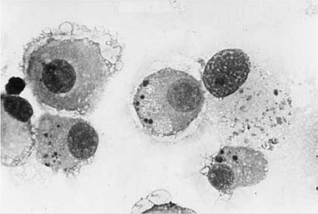
Figure 13.3. Morulae found in human granulocytotropic anaplasma infection caused by Anaplasma phagocytophilum. See color image on color plate 2.
KEY POINTS
About the Clinical Manifestations, Diagnosis, and Treatment of Ehrlichiosis
1. Incubation period is 7 days, and mortality is 5% (mainly elderly and immunocompromised).
a) Gradual onset of fever, chills, headache, myalgias, anorexia, and malaise.
b) Severe monocytic form: respiratory insufficiency, renal insufficiency, and meningoen–cephalitis (with lymphocytosis noted in the cerebrospinal fluid).
c) Severe granulocytic form: respiratory insufficiency, rhabdomyolysis, and neutropenia resulting in gram-negative sepsis.
d) Macular, petechial rash in 30- 40% of cases of the monocytic form, but in 2-11% of cases of the granulocytic form.
2. Diagnosis presumptive in most cases.
a) Thrombocytopenia and leukopenia are common (neutropenia in granulocytic form).
b) Moderate transaminase elevations are seen.
c) Morulae are rare in peripheral blood smears in the monocytic form, common in the granulocytic form.
d) Retrospective serology makes the diagnosis.
3. Treat with doxycycline.
a) Chloramphenicol has no activity in vitro, and therefore doxycycline is also recommended for children.
b) Early treatment reduces the severity of clinical manifestations.
In these diseases, culture techniques are impractical and insensitive, and PCR methods remain experimental. As in rickettsiosis, serologic IFA testing of acute and convalescent serum is the usual method for diagnosis. Antibodies usually take 2-3 weeks to reach detectable levels. Immunofluorescence assays are available through state laboratories and the CDC. Titers above 1:64, combined with a rise of at least a factor of four between acute and convalescent serum, are considered diagnostic.
Doxycycline is the treatment of choice, and in vitro testing confirms that Ehrlichia and Anaplasma are sensitive to tetracyclines. Clinical experience suggests that either oral or intravenous chloramphenicol (500 mg four times daily) is also effective, even though in vitro testing has demonstrated no significant anti–Ehrlichia activity for this drug. Because of these concerns, doxycycline is preferred over chloramphenicol in children (see Table 13.1). Early treatment has the potential to reduce the severity of disease. Delays in therapy increase the risk of transfer to the ICU, as well as the requirement for mechanical ventilation, and greatly prolong the duration of hospitalization.
COXIELLA BURNETII
POTENTIAL SEVERITY
Q fever is usually a self-limiting disease; however, the occasional patient who develops Q fever endocarditis often dies.
Epidemiology
The main reservoirs for C. burnetii, the cause of Q fever, are farm animals: sheep, goats, and cows. Pet cats and dogs may also carry the organism. Mammals shed the pathogen in their urine, feces, and birth products. Transmission occurs most commonly in association with birthing, organisms being aerosolized from the placenta, and inhaled by humans. C. burnetii is resistant to drying and can survive for long periods in the environment, and wind-borne particles can be inhaled weeks after parturition. Individuals at highest risk of contracting Q fever are individuals who frequently come in contact with farm animals, people who live downwind of farms, abattoir workers, and laboratory personnel who work with C. burnetii. Because of the efficiency of infection, this organism has the potential to be used as a biological weapon (see below).
Q fever is rare in the United States, 20-60 cases being reported annually. Outbreaks occur worldwide, but may be missed because of the nonspecific symptoms and signs in this disease. Significant numbers of cases have been reported in Spain, France, England, Australia, and Canada, particularly Nova Scotia. In some areas, the incidence of Q fever has been estimated to be 50 per 100,000 population.
Pathogenesis
C. burnetii is a small pleomorphic rod (0.3–µ mm), whose cell wall has many similarities to gram-negative rods. Although this pathogen was originally classified in the rickettsial family, DNA sequencing has indicated that the organism is more closely related to Legionella and Fran–cisella, and is a proteobacteria. This organism is capable of varying its LPS antigens in response to environmental conditions. In the external environment, the organism usually has phase II LPS antigens; however, on invading the host, a shift to phase I antigens occurs.
KEY POINTS
About the Epidemiology and Pathogenesis of Q fever
1. Disease is rare in the United States, and is more commonly seen in Spain, France, England, Australia, and Canada (Nova Scotia).
2. Most commonly, transmitted by farm animals: sheep, goats, and cows.
a) Organism is excreted in urine, feces, and birth products of the animals.
b) Placenta is highly infectious, and aerosolized organisms survive for prolonged periods.
c) Individuals at risk have extensive exposure to farm animals, live downwind of farms, are abattoir workers or work in a laboratory with Coxiella burnetii.
3. Coxiella burnetii is a small, pleomorphic gram-negative rod that changes its outer lipopolysaccharides:
a) Phase II outer antigens in the environment.
b) Phase I outer antigens when infecting the host.
4. Enters the host through the respiratory tract and survives within phagolysosomes of macrophages (Sodium ion/proton exchangers allow it to survival within this acidic environment.)
a) Induces mononuclear cell infiltration, granuloma formation in the liver.
b) Produces areas of focal necrosis and hemorrhage.
C. burnetii infects the host primary through the respiratory tract. Infectious particles are inhaled and are then phagocytosed by macrophages. Phase I LPS stimulates macrophage membrane ruffling and internalization. The organism is able to survive and grow within the acidic environment of the phagolysome as a consequence of its sodium ion/proton exchangers. The ability to hide within these acidic compartments may be the reason why curing chronic Q fever with antibiotics is so difficult. Pulmonary infection induces infiltration by mononuclear cells and can cause areas of focal necrosis and hemorrhage. Infection can spread to the liver, causing granuloma formation. In patients with damaged heart valves, C. burnetii can survive for prolonged periods and cause chronic endocarditis.
Clinical Manifestations
The incubation period is approximately 3 weeks in most cases of acute disease. Symptoms are often very mild or even absent. When symptoms are reported, most patients develop a self-limiting flu-like illness. Onset of fever is usually abrupt and is associated with headache and myalgias. Some patients complain of a nonproductive cough, and a few rales may be detected on pulmonary examination. Chest X-ray is suggestive of a viral pneumonia with mild bilateral lower lobe infiltrates. Occasionally, patients can develop acute respiratory distress syndrome or pleural effusions. Hepatitis may be asymptomatic or be associated with anorexia and malaise. Transaminase values are elevated, but jaundice is uncommon. Liver biopsy typically reveals doughnut-like granulomas consisting of a lipid vacuole surrounded by a fibrinoid ring. Other, less common manifestations include a maculopapular rash (10% of patients), myocarditis, and pericarditis (1%), and meningitis or encephalitis (1%).
A chronic infection persisting for longer than 6 months develops in about 5% of patients and primarily involves the heart, causing symptoms of subacute bacterial endocarditis. Conventional blood cultures are negative. Most cases of endocarditis develop in patients with valvular damage or a prosthetic valve. Vegetations are seldom seen on cardiac echo, and this negative result often delays the diagnosis. Embolic phenomena and digital clubbing may be observed in late stages of the infection. Valve replacement is commonly required as a consequence of severe valve dysfunction, and mortality in Q fever endocarditis is high (65–45%). Less commonly, chronic infection can develop in an aneurysm, vascular graft, liver, lungs, joints, or bone. If the infection is contracted during pregnancy, the mother may be asymptomatic. However, if untreated, infection is associated with a high rate of spontaneous abortion.
Diagnosis and Treatment
The organism can be readily grown using cell culture techniques; however, cultures are not performed in most facilities because of the danger to lab personnel and the need for a P3 containment facility. The PCR test for this illness has improved in specificity and sensitivity, and it is available in some locations. IFA testing remains the primary method of diagnosis. Anti-phase I and phase II IgG, IgM, and immunoglobulin A (IgA) antibody titers should be tested. Elevated IgG (above 1:200) and IgM (above 1:50) antibody titers against phase II antigens indicate acute disease. Elevated IgG (above 1:800) and IgA (above 1:100) antibody titers against phase I antigens are diagnostic of chronic Q fever.
KEY POINTS
About Clinical Manifestations, Diagnosis, and Treatment of Q Fever
1. Incubation period is 3 weeks, usually causing an abrupt flu-like illness with cough.
2. Less commonly (10% of cases), a maculopapular rash appears. Other, rarer complications include
a) severe respiratory comprise with acute respiratory distress syndrome;
b) hepatitis with elevated transaminases, but minimal elevations in bilirubin;
c) myocarditis and pericarditis;
d) meningitis; and
e) chronic endocarditis (negative echo early in the disease, high mortality).
3. Diagnosis is made by determining immunoglobulin G (IgG) and M (IgM) antibodies against phase I and II antigens (blood cultures negative) by IFA:
a) IgG (titer above 1:200) and IgM (titer above 1:50) antiphase II antigens indicate acute disease.
b) IgG (titer above 1:800) and IgA (titer above 1:100) anti-phase I antigens indicate chronic disease.
c) Polymerase chain reaction is sensitive and specific (available in some locations).
4. Treatment not as effective as for rickettsial infections.
a) Treat with doxycycline for 2 weeks for acute disease; fluoroquinolones may also be helpful.
b) Treat with doxycycline and hydroxychloroquine (alkalinizes phagolysosomes) for 18 months to 4 years or life for chronic endocarditis.
Antibiotics are less effective in Q fever than in rickettsial diseases, and acute disease is usually self-limiting, lasting 2 weeks. Tetracyclines have been shown to shorten the duration of fever in acute disease by 1–2 days. Oral or intravenous doxycycline is the treatment of choice (see Table 13.1), and fluoroquinolones are considered a reasonable alternative. In patients with Q fever endocarditis, cure rates have been improved by combining doxycycline with hydroxychloroquine. The improved cure rates associated with addition of hydrochloroquine are thought to due to this drugs ability to alkalinize the phagolysosomes where the bacteria survive. Therapy for endocarditis must be prolonged—between 18 months and 4 years—to sterilize the valves. In some patients, antibiotics have been continued for life.
CAT SCRATCH DISEASE, BACILLARY ANGIOMATOSIS, AND OTHER DISEASES CAUSED BY BARTONELLA
POTENTIAL SEVERITY
Cat scratch disease and bacillary angiomatosis are usually localized diseases that seldom cause serious illness.
Epidemiology
Cat scratch disease is most commonly contracted by young people under the age of 21 years. This disease is distributed broadly throughout North America and is found worldwide. The incidence in the United States has been estimated to be between 9 and 10 per 100,000 population. Cat scratch disease is more common in warm humid climates.
As the name implies, all epidemiologic data point to the cat as the primary vector for disease. Young cats are most commonly implicated. Kittens have a very high incidence of asymptomatic bacteremia with Bartonella henselae,and they are more likely to scratch humans. In addition to cat scratches, this disease may be transmitted to humans by fleas, and the flea is also responsible for spread from cat to cat.
KEY POINTS
About the Epidemiology of Bartonella Infections
1. Cat scratch disease is caused by Bartonella henselae:
a) Transmitted primarily by young cats and, less commonly, by cat fleas.
b) Common throughout North America; higher incidence in warm, humid areas.
2. Bacillary angiomatosis is caused by B. henselae and B. quintana:
a) B. quintana is transmitted by human body lice.
b) Spreads in areas with poor sanitation, among people with poor personal hygiene.
3. B. bacilliformis is transmitted by the sandfly in the Andes mountains of South America.
B. henselae not only causes cat scratch disease but also causes bacillary angiomatosis. The other species that causes the latter disease, B. quintana, is also globally distributed. It is transmitted by human body lice (Pediculus humanus) and causes disease in areas where sanitation and personal hygiene are poor. A third pathogenic strain, B. bacilliformis, causes Oroya fever and Verruga peruana, diseases found only in the Andes mountains of South America, where the disease is transmitted by the sandfly. Other potentially pathogenic species of Bartonella have been identified; however, their relationship to disease is currently under active investigation.
Pathogenesis
Bartonella is pleomorphic gramnegative bacillus that takes up Gram stain poorly. However, the organism binds silver and can be identified by Warthin–Starry stain. Bartonella enters the host through a break in the skin caused by a cat scratch or insect bite. The bacteria multiply at this site and subsequently spread to the local lymphatic system and adjacent lymph nodes. The bacteria contain flagella that allow them to move within the host. Flagellar and other surface proteins mediate attachment to RBCs and endothelial cells. The attached bacteria can enter red cells, where they can multiply in vacuoles or in the cytoplasm. Bartonella is ingested by endothelial cells and multiply within a vacuole, forming intracellular clusters similar to the morulae of Ehrlichia. Certain species of Bartonella, including B. bacilliformis, B. henselae, and B. quintana, induce the formation of new vessels, and a Bartonella angiogenesis factor has been identified.
KEY POINTS
About the Pathogenesis of Bartonella Infections
1. Pleomorphic gram-negative rods. Takes up Gram stain only weekly; silver stain preferred.
2. Enters via breaks in the skin and spreads to the local lymphatics; rarely disseminates except in patients with AIDS.
3. Survives within host cell intracellular vacuoles and extracellularly.
4. Produces an angiogenesis factor that stimulates the growth of new blood vessels.
5. Induces both a granulomatous and an acute inflammatory reaction that attracts polymor–phonuclear leukocytes and prevents dissemination.
Because Bartonella grows in both the intracellular and extracellular environments of the host, it induces both a granulomatous reaction consisting of macrophages and histiocytes, and an acute inflammatory response consisting primarily of PMNs. This vigorous mixed immune response usually limits the spread of infection, which explains why most Bartonella infections remain localized. In individuals with depressed immunity, such as AIDS patients, the bacteria can cause bacteremia and disseminate throughout the body.
Clinical Manifestations
CASE 13.6
A 21-year-old man presented to the emergency room with a 2-hour history of severe right lower abdominal pain, nausea, vomiting, and loose stools. His temperature was 39.7°C; pulse, 133 per minute; and blood pressure, 101/40 mmHg. His abdomen was soft and nontender; normal bowel sounds were heard. A warm, very tender mass, 1.5 × 1.5 × 6 cm, was palpated in the right inguinal area. Genitalia were normal, without ulcers. A computed tomography scan demonstrated a soft tissue mass.
The patient’s peripheral white blood cell (WBC) count was 12,000/mm3 [54% polymorphonuclear leukocytes (PMNs), 34% bands], and his hematocrit was 43%. Urethral swabs were negative for Chlamydia and gonococcus. Emergency surgical exploration revealed enlarged, matted right inguinal lymph nodes. Histopathology demonstrated an acute inflammatory response, and silver stain identified multiple rods.
Three days following oral administration of ciprofloxacin, the patient defervesced. On further questioning, this college student reported that he had been playing with wild cats near his apartment over the 2 weeks before his admission, but said that he did not recall being scratched.
CAT SCRATCH DISEASE
Cat scratch disease usually presents as a single enlarged, warm, and painful lymph node near the site of skin inoculation. Lymph node swelling usually occurs within 2 weeks of inoculation. Case 13.6developed unusually acute lymph node swelling that caused the sudden onset of severe pain, raising the possibility of a strangulated hernia and precipitating surgical exploration. The node can enlarge between 8 and 10 cm in diameter; however, in most cases, the involved node expands to a diameter of 1-5 cm. Enlargement of a single node is the rule (85% of cases); however, as observed in case 13.6, some patients develop enlargement of a cluster of nodes or, less commonly, experience lymph node enlargement in two distinct anatomic sites. Generalized lymphadenopathy is rare.
The site of lymph node enlargement depends on the site of inoculation. Axillary node involvement is most common. Epitrochlear, supraclavicular, submandibular, and inguinal are other likely sites. In addition to being painful, warm, and erythematous, about 10-15% of the lymph nodes drain pus. The lymphadenopathy usually resolves over a period of 1-4 months, but can persist for several years if not treated with antibiotics.
On careful questioning, the patient may report a skin lesion in the region where the lymph node drains. Within 3-10 days after inoculation, a vesicular lesion develops that becomes erythematous and then papular. The skin lesions usually persist for 1-3 weeks, and by the time the patient seeks medical attention, the site of the scratch may be overlooked. However, if actively searched for, the primary lesion is detected in two-third of patients. A primary lesion was not identified in case 13.6. When questioned, a significant percentage of patients do not recall a cat scratch, but nearly all patients provide a history of contact with a cat or (less commonly) a dog.
Low-grade fever and malaise accompany lymphadenopathy in about half of cases. Conjunctivitis occasionally develops when the eye is the portal of entry, and the combination of conjunctivitis and preauricular lymphadenopathy has been termed Parinaud’s oculoglandular syndrome. Less common manifestations include optic neuritis, encephalopathy that can result in seizures and coma, lytic bone lesions, granulomatous lesions of the liver and spleen, pneumonia, erythema nodosum, and thrombocytopenic purpura.
KEY POINTS
About the Clinical Manifestations of Cat Scratch Disease
1. Presents with a warm, tender, swollen lymph node 2 weeks after the scratch.
a) Axillary node is most common, but the involved node depends on the site of inoculation.
b) The primary scratch can often be identified.
c) Low-grade fever is common.
2. Rarer manifestations include conjunctivitis, encephalopathy, and lesions in the liver and spleen.
KEY POINTS
About the Clinical Manifestations of Bartonella quintana
1. Organism is the major cause of bacillary angiomatosis (B. henselae, less commonly).
a) Seen in indigent patients with AIDS who also have body lice (CD4 count is usually below 100/mm3).
b) Small reddish papules coalesce into nodules, bleed profusely.
c) Histopathology shows multiple small vessels, enlarged endothelial cells, and infiltration by polymorphonuclear leukocytes.
2. Bacteremic illness is rare (seen in some homeless individuals); characterized by recurrent 5-day fever, shin pain, malaise.
BACILLARY ANGIOMATOSIS
Bacillary angiomatosis develops predominantly in indigent patients with AIDS who also have body lice, the primary vector for spread of B. quintana. The disease is also seen in other immunocompromised patients and develops when the CD4 count drops below 100/mm3.
The skin lesions usually begin as cluster of small reddish papules that can enlarge to form nodules. Lesions appear vascular and bleed profusely when traumatized. They can be mistaken for Kaposi sarcoma, pyogenic granuloma, cherry angiomas, or hemangiomas. Skin biopsy reveals multiple small blood vessels, enlarged endothelial cells, and PMN infiltration. B. henselae has also been identified as a cause of bacillary angiomatosis. B. quintana can infect the liver and, less commonly, the spleen, resulting the formation of discrete blood-filled cystic structures. This disease has been called bacillary peliosis.
BACTEREMIC ILLNESS
B. quintana can seed the bloodstream and cause trench fever. This disease was common during World Wars I and II, but is rare today, being seen primarily in homeless individuals with poor hygiene. Cases have been reported in the homeless in Seattle, Washington, and Marseilles, France.
Symptoms of fever, malaise, and bone pain involving the anterior shins usually begin 5-20 days after exposure. Splenomegaly is common, and in some patients, a maculopapular rash may be seen. Recurrent fever every 5 days (quintan fever) is the most common presentation, and it is the basis for the name of the organism. After the primary episode, patients continue to have asymptomatic bacteremia lasting weeks to months. Both B. quintana and B. henselae can cause bacterial endocarditis, and these pathogens should be considered in cases of culture-negative bacterial endocarditis.
Diagnosis
Bartonella grows slowly on fresh blood agar, rabbit-heart infusion agar, and chocolate agar. If Bartonella is suspected, the physician should contact the clinical microbiology laboratory to assure that all cultures are incubated for prolonged periods (at least 21 days) in 5-10% CO2 and high moisture. Because the organism adheres to the sides of glass blood culture flasks, the liquid medium will not appear turbid. The slow rate of growth of this bacterium also impairs recognition by standard CO2 detection methods. Staining of broth with Warthin–Starry stain or acridine orange has been used to overcome these limitations.
Biopsies of lymph nodes and skin lesions are generally not required for diagnosis, and the histopathology of mixed granulomatous and acute inflammatory reaction is not specific. Palisading epithelioid cells are commonly seen, and a positive Warthin–Starry silver stain demonstrating black bacilli provides strong evidence for the diagnosis. However, organisms may be difficult to detect in chronically infected lymph nodes. Bacillary angiomatosis lesions demonstrate characteristic plump endothelial cells, neovascularity, and clusters of bacteria on silver staining.
An IFA and ELISA are available to detect antibodies directed against Bartonella. A IgG titer of 1:64 or below indicates past infection, but does not prove active infection. A titer of 1:64 to 1:256 represents possible Bartonellainfection. When titers are in the low and mid-range they should be retested in 10-14 days to document a rising titer. Titers >1:256 strongly suggest active or recent infection. These tests have now replaced the cat scratch skin test. The skin test was previously considered to be a useful diagnostic tool, but it is no longer recommended. Unlike antibody titers (which have been ineffective at differentiating between species), PCR probes have proven to be more specific and are now commercially available.
Treatment
Azithromycin (standard 5 day course) is effective, and it is the treatment of choice in patients with lymph node disease (see Table 13.1). Oral clarithromycin, oral doxycycline, or oral ciprofloxacin for 10-14 days may also be effective. In severe cases, intravenous azithromycin (500 mg daily) or gentamicin (5 mg/kg daily) combined with oral or intravenous rifampin (600 mg daily) is likely to be the most effective regimen. However, the efficacy of combined therapy has not been proven. In patients with bacteremia attributable to B. quintana, therapy should be continued for 4-6 weeks, and if endocarditis has developed, 6 months of therapy are advisable to reduce the risk of relapse. Patients with bacillary angiomatosis should be treated for 2-4 months, and 4 months of therapy is recommended for patients with bone, hepatic, or splenic lesions. An expert consensus report on the treatment of Bartonellawas published in 2004 (see Further Reading).
KEY POINTS
About the Diagnosis and Treatment of Bartonella Infections
1. Organisms grow on conventional media, but slowly; clinical laboratory must be alerted.
2. Blood cultures frequently yield false negatives, because organisms adhere to the sides of the flask.
3. Biopsies are frequently unnecessary; Warthin–Starry stain shows black rods.
4. Antibody titers by indirect immunofluorescence assay or enzyme immunosorbent assay and PCR are now the tests of choice.
5. Treatment:
a) Azithromycin is the drug of choice, 5 days; alternatives are clarithromycin, doxycycline, or ciprofloxacin. Give for 10-14 days.
b) In severe cases, use intravenous azithromycin or gentamicin plus rifampin (efficacy not proven).
c) Treat B. quintana bacteremia for 4-6 weeks, endocarditis for 6 months.
d) Treat bacillary angiomatosis for 2-4 months; tissue abscesses for 4 months.
Brucellosis
EPIDEMIOLOGY
Brucella is transmitted to humans primarily by infected wild and domestic animals. Direct animal contact, contact with animal products, or ingestion of unpasteurized dairy products is the most common ways in which humans can contract brucellosis. Cattle, buffalo, camels, yaks, goats, and sheep are the domestic animals most commonly responsible for disease transmission. In the wild, swine, fox, caribou, antelope, and elk have been implicated. Bacteria enter the host through abrasions or cuts, the conjunctiva, or the gastrointestinal tract. People at risk are farmers, hunters, and eaters of unpasteurized cheeses or other unpasteurized dairy products. The disease is found worldwide, being most common in the Mediterranean region, Arabian Gulf basin, Indian subcontinent, Mexico, and Central and South America. The highest number of cases are reported in Turkey, Syria, and Iran (14-23,000 cases per year). In the United States, brucellosis is most frequently reported in the south and southwest. As a consequence of a rigorous farm animal screening and vaccination program, and pasteurization of all dairy products, the overall incidence of brucellosis in the United States is low, 0.05 per 100,000 population (100-200 cases per year), with most cases being contracted by travelers who visit endemic areas.
PATHOGENESIS
Brucella is small aerobic gram-negative coccobacillus. The three strains that most commonly cause human disease are B. abortus, B. suis, and B. melitensis. The organism expresses LPS on its surface, and expression of the smooth form enhances intracellular survival, making an important contribution to virulence.
KEY POINTS
About the Epidemiology and Pathogenesis of Brucellosis
1. Transmitted to humans by infected domestic and wild animals:
a) Cattle, buffalo, camels, yaks, goats, and sheep;
b) Swine, fox, caribou, antelope, and elk.
2. Most common in the Mediterranean region, Arabian Gulf basin (Turkey, Iran, and Syria highest number of cases), Indian subcontinent, Mexico, Central and South America. Uncommon in the United States; seen mainly in the south and southwest.
3. Enters via a skin break or ingestion of unpasteurized dairy products (milk, cheeses).
4. Aerobic gram-negative coccobacilli have three pathogenic strains: Brucella abortus, B. suis, and B. melitensis.
5. Survives in phagolysosomes of polymorphonuclear leukocytes and macrophages by producing superoxide dismutase and blocking phagosome–lysosome fusion.
Brucella is a facultative intracellular pathogen. After entering the skin, the bacteria quickly attracts PMNs. These cells ingest the pathogen, where it easily survives within the phagolysosome by producing a superoxide dismutase to neutralize toxic oxygen byproducts. The organism possesses a type IV secretion system allowing it to export bacterial proteins and this system is important for bacterial movement within host cells. The bacteria subsequently invade the lymphatic system and bloodstream, disseminating primarily to organs with rich reticuloendothelial systems (liver, spleen, and bone marrow). Here, the bacteria are ingested by resident macrophages and survive in these cells by blocking phagosome-lysosome fusion, as is observed with Ehrlichia.
Clinical Manifestations
CASE 13.7
A 40-year-old man was seen in the emergency room complaining of right-sided chest pain for 4 days. Pain was sharp and very severe, and was made worse by taking a deep breath. Pain was localized to the right chest, right upper quadrant, but occasionally radiated to the shoulder. The chest pain had been preceded by 2 weeks of a low-grade intermittent fever accompanied by sweating. He noted a mild cough with minimal yellow sputum production.
An epidemiologic history indicated that the patient periodically hunted wild pigs and had been hunting 5 weeks before his hospitalization. Medical history included renal transplant surgery 4 years earlier; patient was on prednisone and azathioprine.
On physical examination, a temperature of 36.7°C, a pulse rate of 102 per minute, a respiratory rate of 24 per minute, and a blood pressure of 126/94 mmHg were recorded. He was ill appearing, breathing shallowly. No lymph nodes were palpable. Bilateral inspiratory rales were heard at the lung bases, with a small area of dullness in the right lower lung field. No abdominal organomegaly or tenderness was noted. Extremities showed 2 + edema. Chest X-ray showed a small right pleural effusion.
Laboratory results showed a hematocrit of 37.5% and a white blood count (WBC) count of 13,700/mm3, with 69% polymorphonuclear leukocytes (PMNs) and 17% bands. Transaminases were 84 IU/L (aspartate aminotransferase, AST) and 32 IU/L (alanine aminotransferase, ALT); alkaline phosphatase, 482 IU/L; total bilirubin, 2.4 mg/dL (1.5 mg/dL direct). Analysis of pleural fluid revealed a WBC count of 250/mm3, with 92% PMNs; lactate dehydrogenase (LDH) 741 IU/L; total protein 3.8 mg/dL; glucose 69 mg/dL; and pH 7.38. Two blood cultures were positive for Brucella suis. The patient was treated with doxycycline and rifampin for 6 weeks and fully recovered.
Fever, chills, malaise, anorexia, headache, and back pain usually develop 2-4 weeks after inoculation or ingestion of Brucella. In case 13.7, the history of intermittent low-grade fever and sweats was typical. These nonspecific symptoms can persist for weeks, making the diagnosis difficult to ascertain. As a result, brucellosis is among the listed infectious causes of fever of undetermined origin (see Chapter 3).
The physical examination is usually unimpressive; often, the only positive findings are lymphadenopathy and splenomegaly. As observed in case 13.7, approximately one-third of patients develop a focal infection. Localized disease is more likely in patients who have had untreated infection for 30 or more days. Immunosuppression probably predisposed case 13.7 to develop a localized pleural infection as well as moderate hepatic involvement.
Septic arthritis is associated with mononuclear cells in the joint fluid; and Brucella can be cultured in half of the cases. Sacroiliitis is particularly common. Osteomyelitis is rare and usually involves the vertebral bodies, mimicking tuberculous osteomyelitis. Granulomas are detected in bone marrow in up to 75% of cases. Infection of the marrow can lead to anemia, leukopenia, and thrombocytopenia.
KEY POINTS
About the Clinical Presentation of Brucellosis
1. Incubation period is 2-4 weeks; symptoms include fever, chills, malaise, anorexia, headache, and back pain.
2. Important cause of fever for unknown origin; lymphadenopathy and splenomegaly are the only positive physical findings.
3. Focal infection is more common if treatment is delayed:
a) Osteomyelitis and arthritis, particularly sac-roiliitis, frequently occur.
b) Hepatic involvement is common.
c) Lymphocytic meningitis is a possibility.
d) Endocarditis usually requires valve replacement.
e) Positive urine culture is common; orchitis occurs in 20% of men.
f) Bone marrow suppression can occur, with granulomas found.
g) Pulmonary disease is rare.
The liver is probably always infected. Mild elevations of liver function tests are noted, and granulomas may be found on liver biopsy, particularly with B. abortus. Purulent abscesses are rare, but may be seen with B. suis and less commonly with B. melitensis. Brucella can often be recovered from the urine, but invasion of the kidney is rare. Orchitis is reported in up to 20% of men with brucellosis, the testes being infiltrated with lymphocytes and plasma cells.
Meningitis is the most frequent complication of the central nervous system and is associated with a CSF lymphocytic pleocytosis, elevated protein, and normal or depressed glucose. Encephalitis and brain abscess are rare. Endocarditis is rare, but can be fatal. Generally, valve replacement must be combined with prolonged antibiotic therapy. Pulmonary involvement is rare, but discrete granulomas can form, and bronchopneumonia occasionally occurs.
Diagnosis
Blood samples for culture should be drawn in all patients who are suspected of having brucellosis. Cultures are positive in up to 70% of patients. However, the organism is slow-growing, taking up to 35 days. However, blood cultures usually take 7-21 days to turn positive.
The clinical microbiology laboratory should be alerted so that cultures are held for beyond 7 days. Bone marrow culture is also a high-yield diagnostic test and should be considered in patients with negative blood cultures. Serology is the most common method for making the diagnosis. Serum agglutination titers measure IgG and IgM antibodies against the three major pathogenic Brucella strains, but do not detect B. canis (a rare cause of disease). A titer above 1:160 in the presence of appropriate symptoms is supportive of the diagnosis, as is a rise in the titer by a factor of four between acute and convalescent sera. ELISA methods for IgG and IgM are also available and demonstrate sensitivity and specificity similar to those of the serum agglutination tests.
Treatment
Because Brucella survives within phagocytes, antibiotics with good intracellular penetration are recommended (see Table 13.1). The treatment of choice is doxycycline and rifampin for 6 weeks. Single-drug therapy is not recommended because of the high likelihood of relapse. Doxy-cycline combined with intramuscular streptomycin (15mg/kg) or gentamicin (5 mg/kg) are useful alternatives. For children, trimethoprim–sulfamethoxazole (10-12 mg/kg of the trimethoprim component daily, divided into two doses) and rifampin (20 mg/kg daily) are recommended. In cases of meningitis or endocarditis, a three-drug regimen consisting of doxycycline, rifampin, and trimethoprim-sulfamethoxazole has been used. Therapy for these diseases must be prolonged (several months to more than 1 year). In patients with endocarditis, replacement of the infected valve is usually required for cure.
KEY POINTS
About the Diagnosis and Treatment of Brucellosis
1. Blood cultures are positive in 70% of cases; hold for 21 days.
2. Bone marrow cultures are often positive.
3. Serologic diagnosis is frequently helpful:
a) Serum agglutination or enzyme-linked immunosorbent assay for immunoglobulin M and G antibody titers.
b) Titers above 1:160 or a rise in titer by a factor of four between acute and convalescent samples is diagnostic.
4. Treatment:
a) Doxycycline plus rifampin, or doxycycline plus gentamicin or streptomycin for 6 weeks.
b) Trimethoprim–sulfamethoxazole plus rifampin is an alternative for children.
c) For cases with meningitis or endocarditis, doxycycline plus rifampin plus trimethoprim–sulfamethoxazole must be continued for months or years.
d) Never use a single drug (high risk of relapse).
Zoonotic Bacterial Infections with the Greatest Potential to Be Used As Bioterrorist Weapons
GUIDING QUESTIONS
1. What are the key characteristics of the ideal bioterrorist agent?
2. What can physicians do to help in the early phases of a bioterrorist attack?
3. What are the clinical clues that should raise the possibility of an anthrax attack?
4. How is bubonic plague normally transmitted, and what are the usual clinical manifestations of plague?
5. Which groups are normally at risk of developing tularemia?
6. How does the clinical presentation of smallpox differ from that of chickenpox?
POTENTIAL SEVERITY
Biologic weapons are intended to kill and terrorize their victims. Treatment must be immediate, and public health measures must be instituted quickly and efficiently to prevent additional casualties.
Bioterrorism was once called biologic warfare, a term that should now be avoided because it suggests that biologic agents are legitimate weapons for defeating a true or perceived enemy. In 1975, biologic weapons were rightfully condemned as inhumane and cowardly, and the civilized world agreed to ban them. Such agents cause great pain and suffering, and have the potential to kill large numbers of innocent bystanders. They subvert science conducted to save lives, to kill, and maim instead.
The term “biologic weapons” is defined as the use of “microbial agents for hostile purposes or in armed conflict.” “Ideal” biologic agents would be expected to
• reliably cause permanently debilitating or fatal disease in a high percentage of victims;
• be capable of being targeted precisely to the enemy, and not cause a worldwide epidemic that could harm friendly soldiers or civilians;
• be capable of being produced in large quantities at reasonable cost;
• be capable of being stored for prolonged periods without losing potency;
• be capable of being readily aerosolized to allow rapid delivery over a broad geographic area.
Only a limited number of biologic pathogens fulfill most of these criteria. Four agents are of particular concern today. However, new “advances” that create super pathogens genetically designed to fit the needs of the bioterrorist are likely to add new organisms to the “most wanted” list. Currently, experts usually list anthrax, plague, tularemia, and smallpox as the top four potential biologic weapons. Other organisms that could be used include Clostridium botulinum (botulinum toxins), Brucella, C. burnetii (Q fever), alpha viruses (Venezuelan equine encephalitis, Eastern and Western encephalitis), and viral hemorrhagic fevers (Ebola virus and Marburg virus).
Medical personnel must be aware of the clinical manifestations, modes of transmission, appropriate diagnostic tests, and available treatment and prophylactic options for managing a biologic attack.
ANTHRAX
Anthrax is a natural infection of animals, primarily herbivores. Humans can contract the disease from infected animals or animal products. With the advent of domestic animal vaccinations, this disease is now seldom encountered in developed countries. As a consequence, most health professionals are unfamiliar with the clinical manifestations of this potentially deadly organism.
The United States, the former Soviet Union, and Iraq have all manufactured anthrax spores capable of being disseminated as aerosols. For the first time in history, anthrax spores were used in 2001 as a biologic weapon against US citizens. That attack underscored the importance of early recognition and treatment of pulmonary and cutaneous anthrax.
Microbiology and Pathogenesis
B. anthracis is a gram-positive rod that can be easily grown on conventional nutrient media. On blood agar plates, the nonhemolytic colonies are gray-white in color with ragged edges. Colonies adhere tightly to the media and cannot easily be displaced by a culture loop. When this bacterium encounters unfavorable environmental conditions, it readily forms endospores. The spores are highly resistant to adverse conditions and are able to survive extreme temperatures, high pH and salinity levels, and disinfectants.
KEY POINTS
About the Pathogenesis and Modes of Spread of Anthrax
1. Bacillus anthracis is an aerobic gram-positive rod, nonhemolytic on blood agar plates.
2. Under poor nutrient conditions, B. anthracis forms spores:
a) Spores resist heat, high salinity, alkaline pH, and many disinfectants.
b) When aerosolized, spores enter the lung, are ingested by macrophages, and are transported to the mediastinum.
3. Spores germinate in the mediastinum, and the bacteria produce three exotoxins:
a) Protective antigen binds to host cell receptors, and allows entry by lethal factor and edema factor.
b) Lethal factor and edema factor paralyze the immune system and cause cell edema and death.
4. Natural transmission of the disease occurs through infected animal products—for example, wool, goat hair, animal hides.
5. Spores can be purposely aerosolized as a bioterror weapon. “Weaponized” anthrax was transmitted by mail in 2001. Postal workers and other mail handlers are at high risk.
When spores are inhaled, their small size allows them to reach small bronchioles and alveoli, where macrophages phagocytose and carry them to the hilar and perihilar lymph nodes. Under the favorable environmental conditions in a host, the spores then germinate, and bacteria begin to quickly multiply.
The bacteria produce three exotoxins: protective antigen, lethal factor, and edema factor. Protective antigen binds to specific receptors on the cell surface and forms a channel that facilitates the entry of edema and lethal factor. These two agents result in cell swelling and death. Lethal factor is a protease that cleaves specific mitogen-activated protein (MAP) kinase kinases, blocking cell signals important for neutrophil chemotaxis, macrophage cell survival, and immune cell cytokine production. Edema factor is an adenyl cyclase that induces excess levels of cyclic adenosine monophosphate (AMP) to alter normal cell signaling, and impair cell motility. As a result of these toxins’ cumulative effects, the host’s innate immune system is paralyzed, and the bacteria continue to grow rapidly and quickly entering the bloodstream to cause overwhelming bacteremia, shock, and meningitis.
Epidemiology
Most cases of anthrax in the United States occur as a result of contact with animal products imported from Asia, the Middle East, and Africa. Anthrax is endemic in areas where domestic animals are not vaccinated. Iran, Iraq, Turkey, Pakistan, and sub-Saharan Africa have the highest number of anthrax cases. Wool, goat hair, and animal hides are the most common sources of infection. Cases of inhalation anthrax as well as a case of gastrointestinal anthrax contracted from contaminated hides have been reported in the United States. Cases have also been traced to shaving-brush bristles, wool coats, yarn, goat-skin bongo drums, and heroin preparations.
A marked increase in the incidence of cutaneous anthrax was observed in Scotland, England, and Germany in 2010 and 2011. Investigations revealed that Turkish heroin was contaminated with anthrax spores and resulted in skin and soft tissue infections among addicts who administered the heroin by injection.
The largest outbreak of anthrax in recent years occurred in Sverdlovsk (now Yekaterinburg), Russia, in 1979. The approximately 96 inhalation cases resulted in 64 deaths. The accidental release of anthrax spores from a germ-warfare facility was suspected, and recent PCR analysis of tissue samples from 11 victims confirmed that suspicion.
The deliberate introduction of anthrax spores into letters sent through the United States Postal Service in 2001 caused 11 cases of inhalation and 11 cases of cutaneous anthrax. Postal workers were at particular risk, because of spores released from sealed envelopes during mail processing. Cross contamination of mail also occurred. As a consequence of those events, all mail recipients have been instructed to avoid opening suspicious mail. If powder is found in an envelope, the letter should be gently set down, the room quickly vacated, and appropriate authorities immediately notified. The events of 2001 emphasize the importance of training public health and law enforcement personnel on the proper handling of potentially contaminated samples and on decontamination and prophylaxis.
Clinical Manifestations
CASE 13.8
A 63-year-old man was taken by his wife to the emergency room with 4-day history of fever, myalgias, and malaise. His wife reported he had no complaints of sore throat, rhinorrhea, and other upper respiratory tract symptoms. He was awoke confused and disoriented on the morning of admission. This man’s prior medical history included mild hypertension and placement of a coronary stent for atherosclerotic heart disease.
An epidemiologic history indicated that the patient was employed as a photo editor for a major tabloid newspaper in Florida, where he spent most of the day reviewing photographs submitted by mail or over the Internet. On physical examination, he was found to be lethargic and disoriented. His temperature was 39°C; blood pressure, 150/80 mmHg; pulse, 110 beats per minute; and respirations, 18 per minute. An ear, nose, and throat examination showed no pharyngeal erythema or exudate, and no nuchal rigidity was noted. Bibasilar rhonchi without rales were heard in the lungs, but no heart murmurs, rubs, or gallops were noted. Abdomen was soft and nontender, with no organo-megaly. The patient’s skin was clear, and a neurologic examination revealed no focal deficits.
The patient’s laboratory workup showed a hematocrit of 46% and a peripheral white blood cell (WBC) count of 9400/mm3, with 77% polymorphonuclear leukocytes (PMNs), 15% lymphocytes, and 8% monocytes. A chest X-ray revealed basilar infiltrates and a widened mediastinum [Figure 13.4(A)]. Cloudy fluid from a lumbar puncture contained red blood cells (RBCs) (1375/mm3), WBCs (4750/mm3, with 81% PMNs and 19% monocytes), 666 mg/dL protein, and 57 mg/dL glucose, A Gram stain of the cerebrospinal fluid (CSF) revealed many PMNs and many large gram-positive bacilli, both single and in chains [Figure 13.4(B)]. Cultures of blood and CSF grew Bacillus anthracis.
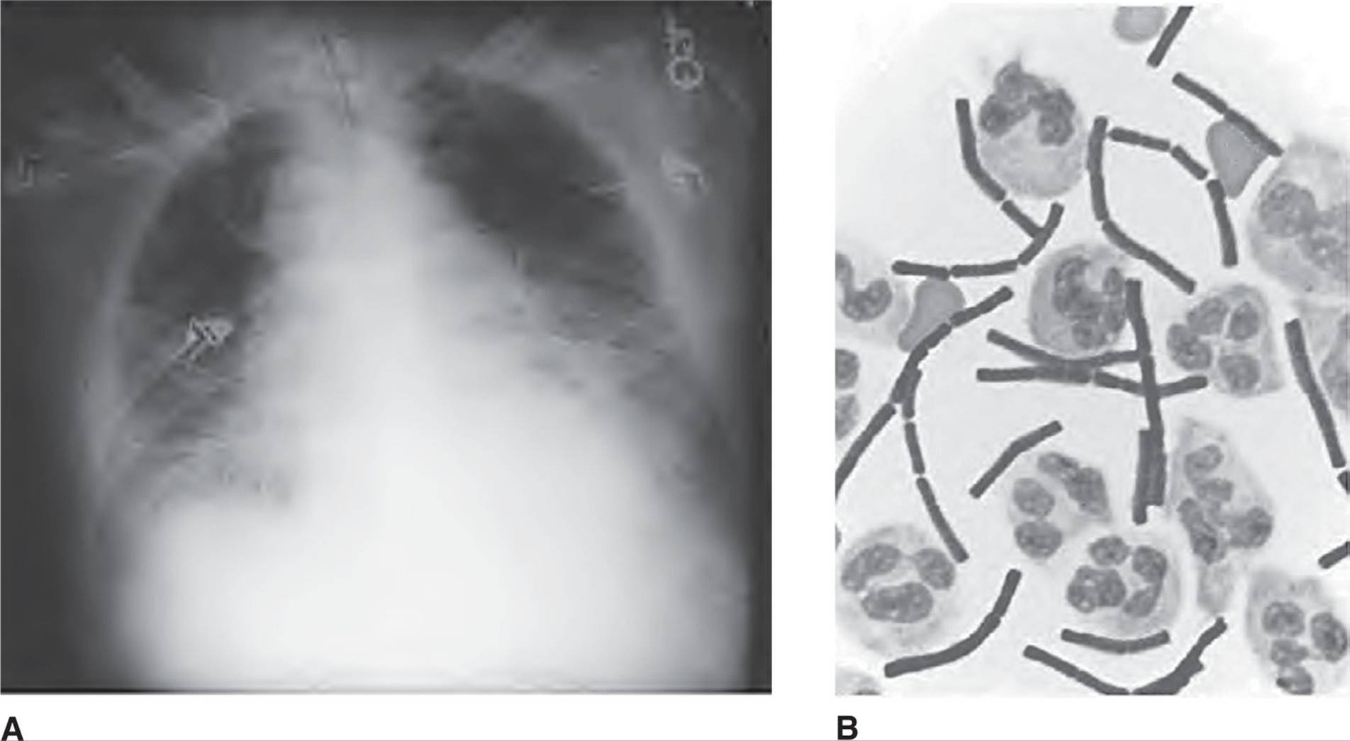
Figure 13.4. Pulmonary anthrax with dissemination to the meninges. A. This chest radiograph shows a widened mediastinum. B. Gram stain of the cerebrospinal fluid demonstrates boxcar-like gram-positive rods.
Despite administration of high-dose penicillin, the patient suffered grand mal seizures, hypotension, acidosis, and renal failure. On the third hospital day, he died of an asystolic cardiopulmonary arrest. Autopsy revealed no pulmonary parenchymal consolidation. Other findings included 50 mL gross blood in the mediastinum and several enlarged lymph nodes (1 cm–2 cm). On cross-sectional examination, the lymph nodes were hemorrhagic. (Adapted from Bush LM, Abrams BH, Beall A, Johnson CC. Index case of fatal inhalational anthrax due to bioterrorism in the United States. N Engl J Med. 2001;345:1607-1610.)
It is critical that health care personnel be familiar with the clinical manifestations of anthrax. In patients with a febrile illness or cutaneous lesions of unclear cause, an exposure and occupational history may be particularly helpful in focusing on the possibility of anthrax. During the 2001 bioterror attack in the United States, early recognition of the index case (case 13.8) in South Florida by an infectious disease specialist led to rapid institution of antibiotic prophylaxis and saved many lives. Unfortunately, several other physicians failed to recognize the early manifestations of inhalation anthrax in postal workers, and those patients were discharged from the emergency room only to return later with full-blown fatal disease. The earlier recognition of several cutaneous anthrax cases could have alerted the authorities in New York in a more timely manner that a bioterror attack had also been launched in that state.
INHALATION ANTHRAX (WOOLSORTER DISEASE)
It is important that clinicians be aware of the biphasic presentation of inhalation anthrax. Recognition and treatment during the first phase can be life saving.
Case 13.8 in all likelihood inhaled spores from a contaminated letter sent to his newspaper, and a flu-like illness was present for 4 days before the onset of fulminant mediastinal involvement, with bacteremia and meningitis. Because the patient failed to seek medical attention during the early phase of his illness, his fatal outcome could not have been prevented.
First Phase
From 1 to 5 days after inhalation of spores, the patient has symptoms suggestive of a viral syndrome: nonproductive cough, malaise, fatigue, myalgia, and mild fever. Occasionally, the sensation of chest heaviness is reported. Rhonchi may be heard on examination, but aside from fever, no other abnormal physical findings are observed. As noted in case 13.8, pharyngitis and rhinitis do not usually accompany inhalation anthrax.
KEY POINTS
About Inhalation Anthrax
1. First phase presents as a viral-like syndrome. No pharyngitis or rhinitis, but chest heaviness may be described. Treatment can abort the second lethal phase.
2. Second phase follows after a brief asymptomatic period and can include:
a) Sudden onset of severe respiratory distress, fever, tachycardia, and tachypnea.
b) Rales on chest examination. Chest radiograph shows a widened mediastinum often with pleural effusions.
c) Thoracentesis reveals hemorrhagic fluid that is positive for Bacillus anthracis on Gram stain and culture.
d) Confusion in half of cases, and cerebrospinal fluid contains polymorphonuclear leukocytes and is positive for B. anthracis on Gram stain and culture.
e) In the terminal stage, blood cultures are positive for anthrax bacilli. Death follows within 24 hours and can occur “in mid-sentence.”
Unless a careful exposure and occupational history are obtained, and inhalation anthrax is included in the differential diagnosis, patients are often sent home with antipyretics for a presumed viral syndrome. It is during this period that spores are being transported by pulmonary macrophages from the lung parenchyma to the mediastinal lymph nodes. At this stage, antibiotic treatment should prevent progression to the second phase.
Second Phase
Within 2-4 days, symptoms temporarily resolve, but are rapidly followed by the second, more severe, stage of the disease. At this time, the spores have germinated in the mediastinal lymph nodes, and protective antigen, lethal factor, and edema factor are being produced by rapidly multiplying anthrax bacilli. Necrosis and hemorrhagic inflammation quickly develop, causing the sudden onset of severe respiratory distress with dyspnea, cyanosis, and diffuse diaphoresis accompanied by fever, tachycardia, and tachypnea. On pulmonary auscultation, moist, crepitating rales are evident, and findings consistent with pleural effusions may be apparent. Chest X-ray demonstrates a widened mediastinum without a definite parenchymal infiltrate. Pleural effusions are often also revealed [Figure 13.4 (A)].
The combination of a widened mediastinum accompanied by pleural effusions should immediately raise the possibility of inhalation anthrax. Thoracentesis reveals hemorrhagic fluid, and Gram stain and culture are both usually positive. As described in case 13.8, confusion followed by lethargy and coma may develop in about half of all cases as a consequence of meningitis. On lumbar puncture, the CSF contains PMNs and large boxcar-like gram-positive rods [Figure 13.4 (B)]. In the terminal stages of the illness, blood cultures are usually positive for B. anthracis. Death usually occurs within 24 hours and may be accompanied by septic shock. Death can be very sudden, and patients have been reported to die “in mid-sentence.”
CUTANEOUS ANTHRAX
Skin disease is the most common manifestation of anthrax. Between 1 and 7 days after spores are inoculated into the skin, a small papule develops. Over the next 3-4 days, the lesion progresses to a vesicle, 1-3 cm in diameter. Erythema and nonpitting edema often surround the vesicle. Initially, the vesicular fluid is serous and contains large numbers of organisms. The vesicle subsequently ruptures, and a black eschar becomes evident at the base of the ulcer (Figure 13.5). The name “anthrax” (Greek for coal) refers to this characteristic black eschar.
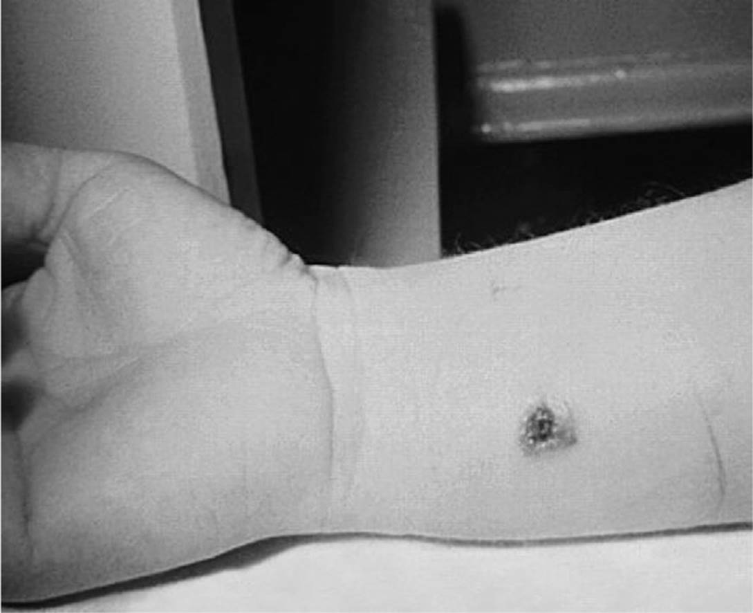
Figure 13.5. Cutaneous anthrax. Note the black eschar and edematous margins of this 7-day-old lesion.
Despite the erythema and swelling, lesions are not painful, but they may be mildly pruritic. Lymphangitis, lymphadenopathy, fever, and malaise may accompany infection of the skin. After several weeks, the skin lesion dries, and a permanent scar is formed. Lesions occur primarily on exposed regions of the body. The arms are the most frequent sites of infection; the face and neck are also commonly involved. A single lesion is usually found, although multiple sites may become infected as a result of simultaneous inoculations.
KEY POINTS
About Cutaneous Anthrax
1. Usually, a single lesion develops on an exposed area of the body, an arm being most common.
2. Develops 1-7 days after inoculation; begins as a papule.
3. Progresses over 3-4 days to a vesicle filled with organisms; margin is edematous.
4. Lesion then ruptures and forms a black eschar.
5. Not painful, but often itches.
6. Spontaneously heals over several weeks, leaving a scar.
GASTROINTESTINAL ANTHRAX
A single case of gastrointestinal infection has been reported in the United States, and it is not an expected clinical consequence of a bioterrorist attack. This disease occurs primarily in developing countries, usually after ingestion of contaminated meat. The incubation period is usually 3-5 days. Patients initially have nausea, vomiting, anorexia, and fever. These symptoms are rapidly followed by acute abdominal pain, hematemesis, and bloody diarrhea. Findings on examination suggest an acute surgical abdomen, and moderate leukocytosis with immature band forms is seen. Rapid progression to toxemia and shock leads to death within 2-5 days after the initial onset of symptoms.
An oropharyngeal form of anthrax has also been described. Inflammatory lesions that resemble the cutaneous lesions develop on the posterior pharynx, hard palate, or tonsils. Tissue necrosis and edema are accompanied by sore throat, dysphagia, fever, regional lymphadenopathy, and toxemia.
Diagnosis
A careful epidemiologic history is the single most important means of reaching the diagnosis. In cases of natural infection, a history of contact with herbivores or products from these animals, particularly if the products come from outside the United States, should raise the possibility of anthrax. In the setting of a possible bioterrorist attack, employment history and a history of being present in a contaminated area are important clues. By the time, Gram stains and cultures of blood and CSF are positive, the illness has progressed to the second fatal phase. Diagnosis must therefore be presumptive, and the threshold for treatment should be low to prevent progression from mildly symptomatic to life-threatening disease.
KEY POINTS
About the Diagnosis of Anthrax
1. Epidemiologic history is important, and the diagnosis is often presumptive.
2. Nasal swabs are helpful for determining the physical parameters of exposure, but not for deciding individual treatment or prophylaxis.
3. Gram stain and culture of skin lesions are often positive.
4. Positive cultures of blood and cerebrospinal fluid usually accompany a fatal outcome.
5. Enzyme-linked immunosorbent assays for antibodies against lethal toxin and edema toxin are available.
For epidemiologic purposes, samples from the nose and face can be obtained using rayon-tipped swabs. Cultures from these sites are specific, but insensitive, and, in the individual patient, cannot be used to decide whether to begin treatment. Nasal samples can be used to determine the physical perimeters of exposure, and the resulting data can used to determine who should receive prophylactic antibiotics. The physical appearance of the skin lesions is characteristic, and Gram stains and cultures of the ulcer base are frequently positive. ELISAs are available that measure antibody titers against lethal and edema toxin. A rise in multiple titers by a factor of four over 4 weeks or in a single titer to 1:32 is considered positive.
Treatment
Although penicillin has been recommended as the treatment of choice for naturally occurring anthrax, penicillin-resistant natural strains have been reported. Penicillin-resistant strains of anthrax have also been genetically engineered as bioterrorist weapons, and the military protocol recommends intravenous ciprofloxacin (400 mg twice daily) or doxycycline (200 mg loading dose, followed by 100 mg twice daily) as first-line therapy (see Table 13.2). Penicillin is recommended as an alternative, once sensitivities have been obtained. Because penicillin treatment induces β-lactamase activity, penicillin should be combined with an additional antibiotic. Two other antibiotics that demonstrate activity against anthrax should be combined with any of the above agents in the seriously ill patient, including rifampin, vancomycin, imipenem, meropenem, and clindamycin. Treatment should be continued for 60 days, with a switch to oral antibiotics as the patient’s clinical condition improves. Excision of skin lesions is contraindicated because of the increased risk of precipitating bacteremia. However, after appropriate antibiotic therapy, excision and skin grafting may be necessary.
Table 13.2. Antibiotic Treatment of Bioterrorist Bacterial Agents
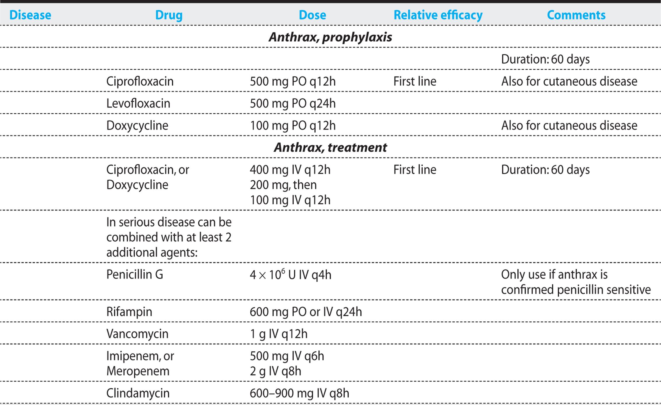
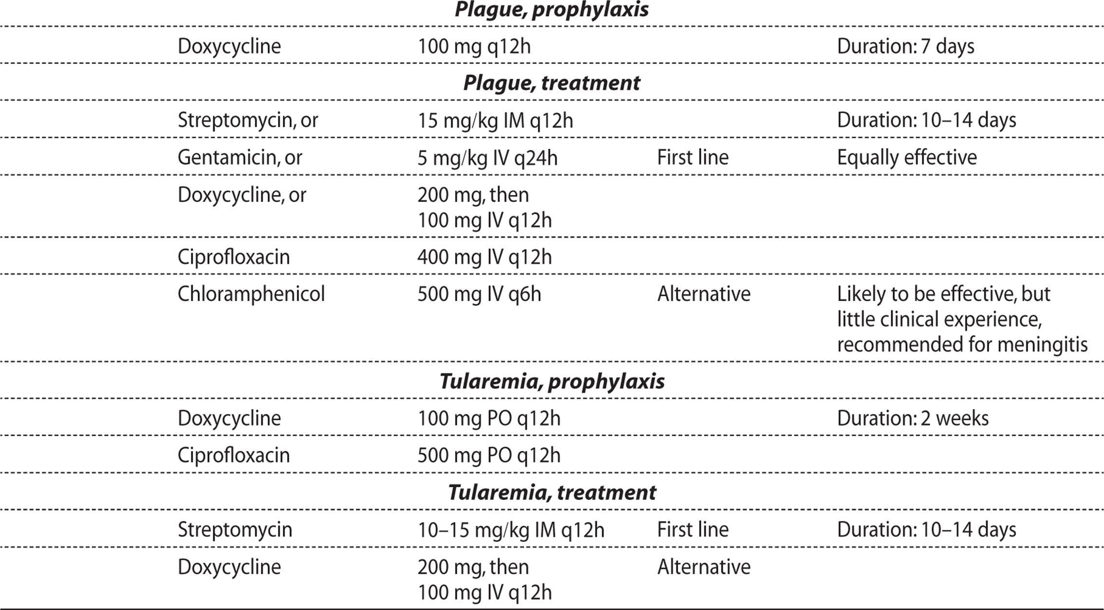
KEY POINTS
About the Treatment and Prevention of Anthrax
1. The treatment threshold must be very low in the setting of a bioterrorist attack.
a) Give intravenous ciprofloxacin, levofloxacin, or doxycycline.
b) Combination therapy is recommended for the seriously ill patient, add two additional drugs: rifampin, vancomycin, imipenem, clindamycin, or clarithromycin to the basic regimen.
c) Avoid excision of skin lesions, which carries a danger of precipitating bacteremia.
d) Continue therapy for 60 days; newly germinating spores can cause relapse.
e) A monoclonal antibody directed against protective antigen, raxibacumab, is now available as adjunctive treatment.
2. All individuals suspected of exposure should receive prophylaxis:
a) Give a fluoroquinolone (ciprofloxacin, levofloxacin, or ofloxacin) or alternatively doxycycline for 60 days.
b) Vaccine based on inactivated exotoxin is given to military personnel and workers at risk of exposure; 6 doses required for immunity, followed by annual booster.
c) Decontaminate exposed areas and personal items with 0.5% hypochlorite.
A human monoclonal antibody, raxibacumab, directed against protective antigen has proved efficacious in multiple animal studies, and in monkeys resulted in a 64% survival rate for inhalation anthrax as compared with 0% for the placebo control group. Raxibacumab is Food and Drug Administration (FDA) approved for inhalation anthrax in humans. A single dose of 40 mg/kg is recommended for children and adults weighing over 50 kg. Patients should be premedicated with diphenhydramine. Raxibacumab is being stockpiled for a potential anthrax bioterrorist attack, and is available through the CDC.
Immunoglobulin derived from the plasma of anthrax-vaccinated individuals is also available through the CDC and its administration was thought to have contributed to the cure of one patient suffering from inhalation anthrax.
Before antibiotics became available, cutaneous disease resulted in a mortality of 10-20%. With appropriate antibiotic treatment, less than 1% of patients die. Despite appropriate antibiotics and respiratory support, inhalation anthrax is frequently fatal. In the 2001 US bioterrorist attack, half of the patients who contracted inhalation anthrax survived, proving that rapid institution of antibiotics can be life saving in early second-phase pulmonary anthrax. Gastrointestinal disease is also associated with high mortality (25-100%).
Prophylaxis
A killed vaccine derived from a component of the anthrax exotoxin is available and is recommended for all industrial workers at risk of exposure to contaminated animal products. As a result of increased concerns about biologic warfare and bioterrorism, military personnel are now vaccinated. To date, surveillance studies have not detected any serious or unexpected adverse reactions. The vaccination (BioThrax), which is available through the CDC (telephone: 770-488-7100; Web site: http://cdc.gov), is administered in six doses at 2-week intervals. A recombinant protective antigen (rPA) vaccine that has demonstrated efficacy in animal models has completed phase I trials; however, problems with the formulation have delayed further progress.
In cases of suspected exposure to B. anthracis, antibiotic prophylaxis and vaccination are recommended. The regimen of choice is an oral fluoroquinolone or, if fluoro-quinolones are contraindicated, doxycycline (see Table 13.2). Prophylaxis should be continued until exposure is excluded. If exposure is confirmed, prophylaxis should be continued for 4 weeks in individuals who have received three or more doses of the vaccine, and for 60 days in the unvaccinated patient. Notably, in the 2001 bioterrorist attack in the United States, only 44% of exposed individuals adhered to the recommended 60-day regimen. Failure to complete the regimen was not accompanied by any adverse outcomes. However, because spores may remain in the body for prolonged periods before germinating, prophylaxis needs to be prolonged, and patients should be closely observed after completion of antibiotics. Within the first several days, exposed skin should be washed extensively with soap and water, and personal items should be decontaminated with 0.5% hypochlorite (one part household bleach to 10 parts water).
PLAGUE
Plague is primarily a disease of animals. The causative organism, Y. pestis, primarily infects rodents. In the United States, the most common reservoirs are squirrels and prairie dogs. An outbreak associated with cats was also reported in the southwestern United States. The disease is transmitted to humans by infected rodent fleas. Approximately 10 human cases are reported annually in the southwestern United States during the late spring, summer, and early fall. Disease outbreaks frequently occur in developing countries throughout the world particularly Africa (Madagascar, Uganda, Mozambique, Malawi, and Zaire), Peru, and India.
Y. pestis was used as a biologic weapon during World War II when the Japanese released plaque-infected fleas in China. However, the spread of the disease proved to be unpredictable and ineffective. Subsequently, both the United States and the former Soviet Union developed reliable and effective methods of aerosolizing this agent.
Microbiology and Pathogenesis
Y. pestis is a gram-negative bacillus that grows aerobically on standard nutrient plates including blood and MacConkey agar. The organism grows slowly, often requiring 48 hours to become apparent, and the colonies are small and grayish.
When an infected flea bites a human, it regurgitates thousands of organisms into the skin, where they are phagocytosed by PMNs and monocytes. Y. pestis is usually killed by PMNs, but is able to survive and replicate within monocytes, evading the host’s immune system. Infected monocytes carry the organism to lymph nodes, where the pathogen actively replicates, causing marked acute inflammation and tissue necrosis. Regional lymph nodes become enlarged, forming buboes. Y. pestis can also quickly enter the bloodstream. Like other gramnegative bacteria, it produces endotoxin and also possesses other virulence factors including a coagulase and a fibrinolysin.
KEY POINTS
About the Epidemiology and Pathogenesis of Plague
1. Usually spread by rodent fleas; cases are occasionally seen in the southwestern United States.
2. The former Soviet Union and the United States developed methods to aerosolize the bacillus.
3. Ingested by PMNs and monocytes, able to replicate in monocytes.
a) Produces acute inflammation and tissue necrosis.
b) Spreads to regional lymph nodes, forming fluctuant buboes.
c) Readily enters the bloodstream.
Clinical Manifestations
Natural infection resulting from flea bites causes bubonic plaque. The incubation period is usually 2-8 days, ending with the abrupt onset of fever, chills, weakness, and headache. Within hours, the patient notes an enlarged, extremely painful cluster of regional lymph nodes termed a “bubo.” Marked swelling is noted, and pain is so severe that the patient avoids moving the infected area. Buboes are usually egg-shaped swellings, 1-10 cm in length. Within 2-4 days, the patient dies of septic shock. Thrombosis of small vessels can develop, causing peripheral tissue necrosis and gangrene that may require amputation. In some patients, no bubo appears, and the patient presents in a moribund state caused by high-grade bacteremia. Meningitis may develop in a small percentage of patients.
If bioterrorists were to aerosolize Y. pestis, the primary clinical presentation would be pneumonic plague. After an incubation period of 2-4 days, fever, chills, and myalgias suddenly begin. Within 24 hours, patients begin coughing up blood as bacterial production of coagulase and fibrinolysin leads to tissue necrosis. Sputum can also be mucopurulent or watery. Chest pain, abdominal pain, nausea, vomiting, and diarrhea are other common symptoms. If antibiotics are not begun within 18 hours, the outcome is fatal. Patients experience increasing dyspnea, stridor, and cyanosis, followed by respiratory arrest and circulatory collapse.
Diagnosis
The possibility of a biologic attack with Y. pestis should be considered if large numbers of patients begin presenting to the emergency room with hemoptysis and severe, rapidly progressive pneumonia. Sputum Gram stain frequently reveals gram-negative rods. A presumptive diagnosis can also be made by finding bacilli on peripheral blood smear. Chest X-ray demonstrates bilateral bronchopneumonia. Definitive diagnosis is made by sputum and blood cultures that often take more than 48 hours because of the organism’s slow growth rate. A rapid ELISA antigen test (takes 15 minutes) has been developed that is highly sensitive and specific. Detection by PCR is under development and, in fleas, is specific and highly sensitive (can detect as few as 11 organisms).
KEY POINTS
About the Clinical Manifestations of Plague
1. In the flea-transmitted form of the disease, incubation of 2-8 days is followed by
a) fever, chills, weakness, and headache; and
b) bubo formation (very painful).
c) Within 2-4 days, septic shock leads to peripheral gangrene and death.
2. Pneumonic form more likely in a bioterrorist attack.
a) Incubation period is 2-4 days, leading to chills, fever, and myalgias.
b) Within 24 hours, bloody sputum production and chest pain begin, followed by dyspnea and cyanosis.
c) Death follows within 18 hours if antibiotic treatment is not started.
Treatment
If pneumonic plague is not considered and if conventional antibiotic treatment for community-acquired pneumonia is mistakenly begun, the infection will quickly progress, resulting in death. Streptomycin, gentamicin, and doxycycline (see Table 13.2 for doses) are the treatments of choice and should be continued for 10-14 days. Levofloxacin has proved to be effective in an African green monkey model, and is likely to be another effective alternative to aminoglycosides for the treatment of human plague. Chloramphenicol is recommended for the treatment of meningitis.
Surgical debridement of buboes should not be performed, because of the risk of spreading the infection to others. Needle aspiration of lymph nodes may provide some relief and also provide material for culture and Gram stain. The lymph nodes usually slowly shrink on antibiotic therapy. The overall mortality for pneumonic plague is 60%; however, if appropriate therapy is delayed for more than 24 hours, then mortality is nearly 100%. The fatality rate for bubonic plague is 14%, but with early therapy, all patients should survive.
Prophylaxis
Person-to-person spread of Y. pestis does occur. Patients with pneumonic plague can cough and aerosolize the organism, leading to secondary cases of pneumonia. Patients with pulmonary disease therefore require strict isolation with droplet precautions for at least 48 hours after the start of antibiotic therapy. People who have had face-to-face contact with patients with plague pneumonia should receive oral doxycycline prophylaxis (100 mg twice daily) for 7 days or for the duration of potential exposure plus 7 days. In patients with bubonic plague, only standard precautions are required, and prophylaxis is unnecessary. Contacts should be observed for 7 days.
KEY POINTS
About the Diagnosis, Treatment, and Prevention of Plague
1. The disease is readily diagnosed by Gram stain of sputum or lymph node aspirate; cultures usually require 48 hours. A sensitive PCR method is under development.
2. Treat with streptomycin, gentamicin, or doxycycline for 14 days; delaying beyond 24 hours can lead to death.
a) Ciprofloxacin and levofloxacin may be effective.
b) Use chloramphenicol for meningitis.
3. Prophylaxis:
a) Take respiratory (droplet) precautions for pneumonic plague for 48 hours after the start of antibiotic treatment.
b) Give doxycycline for 7 days after respiratory exposure.
c) Vaccine is under development.
A vaccine is not currently available. A recombinant plague vaccine (rF1V) is under development, and has been shown to be effective for inhalation disease in animals A previous vaccine no longer being produced, was effective for prevention of the bubonic, but not the inhalation disease.
TULAREMIA
F. tularensis is another zoonotic pathogen that, under natural conditions, incidentally infects humans. Infection is usually contracted following contact with rabbits, musk-rats, beaver, squirrels, and birds. A case was also reported following a pet hamster bite. Hunters develop the disease after skinning, dressing, and eating infected animals. Less commonly, the infection can be spread to humans by ticks, biting flies, and mosquitoes. Aerosol droplets of contaminated water or mud can be produced by lawn mowing and other gardening activities. The organism can survive in contaminated water for prolonged periods by multiplying within Acanthamoeba castellanii.
Tularemia is most commonly encountered in temperate climates during the summer months (insect transmission) and during hunting season. Arkansas, Missouri, Kansas, South Dakota, Oklahoma, and California account for two-third of cases in the United States. European countries (except England), the former Soviet Union, Tunisia, Turkey, Israel, Iran, China, and Japan have endemic tularemia.
The United States (and possibly other countries) has weaponized this agent. Dry and wet forms have both been created. Like B. anthracis and Y. pestis, F. tularensis is most efficiently delivered in lethal doses by aerosol.
Microbiology and Pathogenesis
Francisella is a small aerobic gram-negative coccobacillus that does not routinely grow on standard media; it requires either cysteine or cystine for growth. Glucose-cystine blood agar supports growth; however, a selective medium is often required to isolate this pathogen from normal skin and mouth flora. The cell wall of this bacterium has a capsule with high fatty acid content that resists serum bactericidal activity. Francisella produces no known exotoxins, but it expresses a LPS endotoxin that is one one-thousandth as potent as LPS from E. coli.
Like most natural infections, tularemia begins when F. tularensis bacteria gain entry to the body through a small break in the skin. The organism is phagocytosed by monocytes, where it is able to survive intracellularly by escaping from the phagolysosome into the cytoplasm where it readily multiplies. F. tularensis can also grow in hepatocytes and endothelial cells.
KEY POINTS
About the Mode of Spread and Pathogenesis of Tularemia
1. Francisella tularensis is a gram-negative cocco-bacillus, which usually spreads cutaneously from infected rabbits, muskrats, beaver, squirrels, and birds.
2. An aerosolized form can be manufactured.
3. Growth in culture requires a cystine-supplemented medium.
4. Cell wall has a high fatty-acid content; produces a lipopolysaccharide endotoxin that is considerably less potent than that produced by Escherichia coli.
5. As an intracellular pathogen grows in the cytoplasm of host cells, induces acute inflammation and granuloma formation.
6. A low inoculum (10-50 organisms) can cause disease (very dangerous).
As the organisms grow and lyse cells, they induce an acute inflammatory reaction, and tissue necrosis is followed by granuloma formation. Cell-mediated immunity plays a critical role in controlling this intracellular pathogen. Only 10 to 50 bacteria are required to cause skin and pulmonary infection, making this organism extremely dangerous to laboratory workers.
Clinical Manifestations
The clinical picture of tularemia is very similar to that of plague. The incubation period is usually 3-5 days, ending with the abrupt onset of high fever, chills, malaise, myalgias, chest discomfort, vomiting, abdominal pain, and diarrhea. A severe generalized headache is often a prominent complaint.
Natural disease most commonly takes the ulcero-glandular form. At the site of bacterial entry, a painful ulcer with raised borders develops, associated with painful regional adenopathy. Less commonly, patients develop lymphadenopathy without a skin ulcer, others presents with a febrile illness without lymphadenopathy and may become hypotensive. Watery diarrhea may be a prominent complaint, with the disease being mistaken for Salmonellatyphoid fever.
The pneumonic form is rare under natural circumstances, but can occur in sheep shearers, farmers, and laboratory workers. The pneumonic form would be the expected presentation after an aerosol bioterrorist attack.
KEY POINTS
About the Clinical Manifestations of Tularemia
1. Clinically similar to plague; incubation period of 3-5 days.
a) Abrupt onset of fever, headache, malaise, myalgias, abdominal pain, and diarrhea.
b) Ulceroglandular form presents as a painful ulcer with raised borders and associated regional lymphadenopathy.
c) Glandular form, no skin lesion apparent less common
d) Typhoid fever-like illness without lymphade-nopathy mistaken for Salmonella.
2. Bronchopneumonia would be expected in a bioterrorist attack: similar to plague except that cough is dry, hacking; hemoptysis is rare.
The clinical presentation is identical to that of pneumonic plague, with the exception that the cough is usually dry and hacking rather than productive. Hemoptysis can occur, but is rare. In some patients, respiratory complaints may not be prominent, and primary complaints may mimic typhoid fever.
Diagnosis
Presentation of a large number of patients with severe bronchopneumonia associated with a nonproductive cough should raise the possibility of a bioterror attack involving F. tularensis. Chest X-ray demonstrates changes consistent with a bronchopneumonia in 50% of cases after inhalation. Pleural effusions may be noted in 15% of those with pneumonia. Aspiration of the pleural fluid usually reveals lymphocytes, suggesting tuberculosis. Gram stain of sputum and wounds are usually negative. The organism can be identified in lymph nodes by silver stain. Blood cultures and tissue sample cultures are rarely positive. The organism must be grown using medium containing a sulfhydryl compound. The organism should be handled in a biosafety level 3 containment facility because of the risk to laboratory personnel, and the laboratory should be notified if this tularemia is high on the differential diagnosis.
The diagnosis is usually made by tube agglutination or a microagglutination assays for anti-F tularensis serum antibody. Two weeks are required before significant antibody titers. A single tube dilution titer of 1:160 or higher is supportive of the diagnosis. A fourfold rise in titer on follow-up convalescent titer confirms the diagnosis. PCR is rapid and specific, but is not widely available.
Treatment
Effective treatment regimens include streptomycin and gentamicin (see Table 13.2). In a presumed bioterror attack, gentamicin would be preferred over streptomycin, because a streptomycin-resistant strain was developed in the 1950s and may have been obtained by other countries. (That strain was sensitive to gentamicin.) Doxycycline is another alternative for treatment.
The mortality from tularemia pneumonia is 30%, making weaponized Francisella a less deadly agent than either anthrax or plague.
Prevention
Person-to-person transmission is not reported with tularemia. Standard precautions are therefore sufficient. Prophylaxis should be administered within 24 hours of exposure. Ciprofloxacin or doxycycline for 2 weeks is recommended (see Table 13.2). An investigational live-attenuated vaccine given by scarification is no longer available. Attenuated vaccines are under development.
KEY POINTS
About the Diagnosis, Treatment, and Prevention of Tularemia
1. Gram stain of sputum and skin ulcers is usually negative; culture requires a special medium.
2. May be identified in lymph nodes by silver stain.
3. Diagnosis is usually presumptive; antibody titers rise after 2 weeks 1:160 or higher suggests disease, and a fourfold rise from acute to convalescent serum is diagnostic.
4. Treatment:
a) Gentamicin is the drug of choice; doxycycline and streptomycin are alternatives.
b) Respiratory precautions are not required.
5. Prophylaxis:
a) Treat within 24 hours of exposure with ciprofloxacin or doxycycline for 14 days.
b) A vaccine is under development.
6. Mortality rate is 30% (lower than for pulmonary anthrax or plague).
FURTHER READING
Lyme Disease
Baranton G, De Martino SJ. Borrelia burgdorferi sensu lato diversity and its influence on pathogenicity in humans. Curr Probl Dermatol. 2009;37:1-17.
Feder HM, Jr., Johnson BJ, O’Connell S, et al. A critical appraisal of “chronic Lyme disease”. N Engl J Med. 2007;357:1422-1430.
Hu LT. In the clinic. Lyme disease. Ann Intern Med. 2012;157:ITC2-2-ITC2-16.
Kalish RA, Kaplan RF, Taylor E, Jones-Woodward L, Workman K, Steere AC. Evaluation of study patients with Lyme disease, 10-20-year follow-up. J Infect Dis. 2001;183:453-460.
Nadelman RB, Nowakowski J, Fish D, et al. Prophylaxis with singledose doxycycline for the prevention of Lyme disease after an Ixodes scapularis tick bite. N Engl J Med. 2001;345:79-84.
Smith RP, Schoen RT, Rahn DW, et al. Clinical characteristics and treatment outcome of early Lyme disease in patients with micro-biologically confirmed erythema migrans. Ann Intern Med. 2002;136:421-428.
Stanek G, Strle F. Lyme disease: European perspective. Infect Dis Clin North Am. 2008;22:327-339.
Stanek G, Wormser GP, Gray J, Strle F. Lyme borreliosis. Lancet. 2012;379:461-473.
Wormser GP, Dattwyler RJ, Shapiro ED, et al. The clinical assessment, treatment, and prevention of Lyme disease, human gran-ulocytic anaplasmosis, and babesiosis: clinical practice guidelines by the Infectious Diseases Society of America. Clin Infect Dis. 2006;43:1089-1134.
Leptospirosis
Agampodi SB, Matthias MA, Moreno AC, Vinetz JM. Utility of quantitative polymerase chain reaction in leptospirosis diagnosis: association of level of leptospiremia and clinical manifestations in Sri Lanka. Clin Infect Dis. 2012;54:1249-1255.
Desai S, van Treeck U, Lierz M, et al. Resurgence of field fever in a temperate country: an epidemic of leptospirosis among seasonal strawberry harvesters in Germany in 2007. Clin Infect Dis. 2009; 48:691-697.
Nardone A, Capek I, Baranton G, et al. Risk factors for leptospirosis in metropolitan France: results of a national case-control study, 1999-2000. Clin Infect Dis. 2004;39:751-753.
Panaphut T, Domrongkitchaiporn S, Vibhagool A, Thinkamrop B, Susaengrat W. Ceftriaxone compared with sodium penicillin G for treatment of severe leptospirosis. Clin Infect Dis. 2003;36: 1507-1513.
Segura ER, Ganoza CA, Campos K, et al. Clinical spectrum of pulmonary involvement in leptospirosis in a region of endemicity, with quantification of leptospiral burden. Clin Infect Dis. 2005;40:343-351.
Stern EJ, Galloway R, Shadomy SV, et al. Outbreak of leptospirosis among Adventure Race participants in Florida, 2005. Clin Infect Dis. 2010;50:843-849.
Babesiosis
Hatcher JC, Greenberg PD, Antique J, Jimenez-Lucho VE. Severe babesiosis in Long Island: review of 34 cases and their complications. Clin Infect Dis. 2001;32:1117-1125.
Martinot M, Zadeh MM, Hansmann Y, et al. Babesiosis in immunocompetent patients, Europe. Emerg Infect Dis. 2011;17:114-116.
Teal AE, Habura A, Ennis J, Keithly JS, Madison-Antenucci S. A new real-time PCR assay for improved detection of the parasite Babesia microti. J Clin Microbiol. 2012;50:903-908.
Vannier E, Krause PJ. Human babesiosis. N Engl J Med. 2012; 366:2397-2407.
Rickettsial Diseases
Adjemian JZ, Krebs J, Mandel E, McQuiston J. Spatial clustering by disease severity among reported Rocky Mountain spotted fever cases in the United States, 2001-2005. Am J Trop Med Hyg. 2009; 80:72-77.
Brouqui P, Parola P, Fournier PE, Raoult D. Spotted fever rickettsioses in southern and eastern Europe. FEMS Immunol Med Microbiol. 2007;49:2-12
Dantas-Torres F. Rocky Mountain spotted fever. Lancet Infect Dis. 2007;7:724-732.
Demma LJ, Traeger MS, Nicholson WL, et al. Rocky Mountain spotted fever from an unexpected tick vector in Arizona. N Engl J Med. 2005;353:587-594.
Holman RC, McQuiston JH, Haberling DL, Cheek JE. Increasing incidence of Rocky Mountain spotted fever among the American Indian population in the United States. Am J Trop Med Hyg. 2009;80:601-605.
Jensenius M, Fournier PE, Raoult D. Rickettsioses and the international traveler. Clin Infect Dis. 2004;39:1493-1499.
Kirk JL, Fine DP, Sexton DJ, Muchmore HG. Rocky Mountain spotted fever. A clinical review based on 48 confirmed cases, 1943-1986. Medicine (Baltimore). 1990;69:35-45.
Lai CH, Huang CK, Weng HC, et al. Clinical characteristics of acute Q fever, scrub typhus, and murine typhus with delayed defervescence despite doxycycline treatment. Am J Trop Med Hyg. 2008;79:441-446.
Premaratna R, Chandrasena TG, Dassayake AS, Loftis AD, Dasch GA, de Silva HJ. Acute hearing loss due to scrub typhus: a forgotten complication of a reemerging disease. Clin Infect Dis. 2006;42:e6-e8.
Raoult D, Parola P. Rocky Mountain spotted fever in the USA: a benign disease or a common diagnostic error? Lancet Infect Dis. 2008;8:587-589.
Sonthayanon P, Chierakul W, Wuthiekanun V, et al. Association of high Orientia tsutsugamushi DNA loads with disease of greater severity in adults with scrub typhus. J Clin Microbiol. 2009; 47:430-434.
Ehrlichiosis
Chapman AS, Bakken JS, Folk SM, et al. Diagnosis and management of tickborne rickettsial diseases: Rocky Mountain spotted fever, ehrlichioses, and anaplasmosis—United States: a practical guide for physicians and other health-care and public health professionals. MMWR Recomm Rep. 2006;55:1-27.
Doudier B, Olano J, Parola P, Brouqui P. Factors contributing to emergence of Ehrlichia and Anaplasma spp. as human pathogens. Vet Parasitol. 2010;167:149-154.
Dumler JS, Madigan JE, Pusterla N, Bakken JS. Ehrlichioses in humans: epidemiology, clinical presentation, diagnosis, and treatment. Clin Infect Dis. 2007;45(suppl 1):S45-S51.
Hamburg BJ, Storch GA, Micek ST, Kollef MH. The importance of early treatment with doxycycline in human ehrlichiosis. Medicine (Baltimore). 2008;87:53-60.
Pritt BS, Sloan LM, Johnson DK, et al. Emergence of a new pathogenic Ehrlichia species, Wisconsin and Minnesota, 2009. N Engl J Med. 2011;365:422-429.
Zhang L, Liu Y, Ni D, et al. Nosocomial transmission of human granulocytic anaplasmosis in China. JAMA. 2008;300:2263-2270.
Q Fever
Carcopino X, Raoult D, Bretelle F, Boubli L, Stein A. Managing Q fever during pregnancy: the benefits of long-term cotrimoxazole therapy. Clin Infect Dis. 2007;45:548-555.
Fenollar F, Thuny F, Xeridat B, Lepidi H, Raoult D. Endocarditis after acute Q fever in patients with previously undiagnosed valvu-lopathies. Clin Infect Dis. 2006;42:818-821.
Hartzell JD, Wood-Morris RN, Martinez LJ, Trotta RF. Q fever: epidemiology, diagnosis, and treatment. Mayo Clin Proc. 2008; 83(5):574-579.
Parker NR, Barralet JH, Bell AM. Q fever. Lancet. 2006;367:679-688.
Tissot-Dupont H, Vaillant V, Rey S, Raoult D. Role of sex, age, previous valve lesion, and pregnancy in the clinical expression and outcome of Q fever after a large outbreak. Clin Infect Dis. 2007;44:232-237.
Bartonella Infections
Angelakis E, Pulcini C, Waton J, et al. Scalp eschar and neck lymph-adenopathy caused by Bartonella henselae after Tick Bite. Clin Infect Dis. 2010;50:549-551.
Ben-Ami R, Ephros M, Avidor B, et al. Cat-scratch disease in elderly patients. Clin Infect Dis. 2005;41(7):969-974.
Fournier PE, Lelievre H, Eykyn SJ, et al. Epidemiologic and clinical characteristics of Bartonella quintana and Bartonella henselae endocarditis: a study of 48 patients. Medicine (Baltimore). 2001;80:245-251.
Maman E, Bickels J, Ephros M, et al. Musculoskeletal manifestations of cat scratch disease. Clin Infect Dis. 2007;45(12):1535-1540.
Rolain JM, Lepidi H, Zanaret M, et al. Lymph node biopsy specimens and diagnosis of cat-scratch disease. Emerg Infect Dis. 2006; 12:1338-1344.
Rolain JM, Brouqui P, Koehler JE, Maguina C, Dolan MJ, Raoult D. Recommendations for treatment of human infections caused by Bartonella species. Antimicrob Agents Chemother. 2004;48: 1921-1933.
Brucellosis
Ariza J, Bosilkovski M, Cascio A, et al. Perspectives for the treatment of brucellosis in the 21st century: the Ioannina recommendations. PLoS Med. 2007;4:e317.
Bosilkovski M, Krteva L, Dimzova M, Vidinic I, Sopova Z, Spasovska K. Human brucellosis in Macedonia—10 years of clinical experience in endemic region. Croat Med J. 2010;51:327-336.
Colmenero JD, Reguera JM, Martos F, et al. Complications associated with Brucella melitensis infection: a study of 530 cases. Medicine (Baltimore). 1996;75:195-211.
Colmenero JD, Ruiz-Mesa JD, Plata A, et al. Clinical findings, therapeutic approach, and outcome of brucellar vertebral osteomyelitis. Clin Infect Dis. 2008;46:426-433.
Hasanjani Roushan MR, Mohraz M, Hajiahmadi M, Ramzani A, Valayati AA. Efficacy of gentamicin plus doxycycline versus streptomycin plus doxycycline in the treatment of brucellosis in humans. Clin Infect Dis. 2006;42:1075-1080.
Pappas G, Akritidis N, Bosilkovski M, Tsianos E. Brucellosis. N Engl J Med. 2005;352:2325-2336.
Navarro E, Segura JC, Castano MJ, Solera J. Use of real-time quantitative polymerase chain reaction to monitor the evolution of Brucella melitensis DNA load during therapy and post-therapy follow-up in patients with brucellosis. Clin Infect Dis. 2006; 42:1266-1273.
Solera J, Geijo P, Largo J, et al. A randomized, double-blind study to assess the optimal duration of doxycycline treatment for human brucellosis. Clin Infect Dis. 2004;39:1776-1782.
Troy SB, Rickman LS, Davis CE. Brucellosis in San Diego: epidemiology and species-related differences in acute clinical presentations. Medicine (Baltimore). 2005;84:174-187.
Bioterrorism
United States, U.S. Army Medical Research Institute of Infectious Diseases (USAMRIID). Medical Management of Biological Casualties: Handbook. Frederick, MD: USAMRIID; 2001.
Anthrax
Artenstein AW, Opal SM. Novel approaches to the treatment of systemic anthrax. Clin Infect Dis. 2012;54(8):1148-1161.
Bush LM, Perez MT. The anthrax attacks 10 years later. Ann Intern Med. 2012;156:41-44.
Doolan DL, Freilich DA, Brice GT, et al. The US capitol bioterrorism anthrax exposures: clinical epidemiological and immunological characteristics. J Infect Dis. 2007;195:174-184.
Guarner J, Jernigan JA, Shieh WJ, et al. Pathology and pathogenesis of bioterrorism-related inhalational anthrax. Am J Pathol. 2003;163:701-709.
Holty JE, Bravata DM, Liu H, Olshen RA, McDonald KM, Owens DK. Systematic review: a century of inhalational anthrax cases from 1900 to 2005. Ann Intern Med. 2006;144:270-280.
Jernigan JA, Stephens DS, Ashford DA, et al. Bioterrorism-related inhalational anthrax: the first 10 cases reported in the United States. Emerg Infect Dis. 2001;7:933-944.
Kyriacou DN, Stein AC, Yarnold PR, et al. Clinical predictors of bioterrorism-related inhalational anthrax. Lancet. 2004;364:449-452.
Migone TS, Subramanian GM, Zhong J, et al. Raxibacumab for the treatment of inhalational anthrax. N Engl J Med. 2009;361:135-144.
Palmateer NE, Ramsay CN, Browning L, Goldberg DJ, Hutchinson SJ. Anthrax infection among heroin users in Scotland during 2009-2010: a case-control study by linkage to a national drug treatment database. Clin Infect Dis. 2012;55:706-710.
Sejvar JJ, Tenover FC, Stephens DS. Management of anthrax meningitis. Lancet Infect Dis. 2005;5:287-295.
Vietri NJ, Purcell BK, Lawler JV, et al. Short-course postexposure antibiotic prophylaxis combined with vaccination protects against experimental inhalational anthrax. Proc Natl Acad Sci U S A. 2006;103:7813-7816.
Plague
Layton RC, Mega W, McDonald JD, et al. Levofloxacin cures experimental pneumonic plague in African green monkeys. PLoS Negl Trop Dis. 2011;5(2):e959.
Morris SR. Development of a recombinant vaccine against aerosolized plague. Vaccine. 2007;25:3115-3117.
Prentice MB, Rahalison L. Plague. Lancet. 2007;369(9568):1196-1207.
Tourdjman M, Ibraheem M, Brett M, et al. Misidentification of Yersinia pestis by automated systems, resulting in delayed diagnoses of human plague infections-Oregon and New Mexico, 2010-2011. Clin Infect Dis. 2012;55:e58-e60.
Tularemia
Conlan JW. Tularemia vaccines: recent developments and remaining hurdles. Future Microbiol. 2011;6:391-405.
Eliasson H, Back E. Tularaemia in an emergent area in Sweden: an analysis of 234 cases in five years. Scand J Infect Dis. 2007;39:880-889.
Kugeler KJ, Mead PS, Janusz AM, et al. Molecular epidemiology of Francisella tularensis in the United States. Clin Infect Dis. 2009;48(7):863-870.
Matyas BT, Nieder HS, Telford SR, 3rd. Pneumonic tularemia on Martha’s Vineyard: clinical, epidemiologic, and ecological characteristics. Ann N Y Acad Sci. 2007;1105:351-377.
Santic M, Al-Khodor S, Abu Kwaik Y. Cell biology and molecular ecology of Francisella tularensis. Cell Microbiol. 2010;12:129-139. Thomas LD, Schaffner W. Tularemia pneumonia. Infect Dis Clin North Am. 2010;24:43-55.
Weber IB, Turabelidze G, Patrick S, Griffith KS, Kugeler KJ, Mead PS. Clinical recognition and management of tularemia in Missouri: a retrospective records review of 121 cases. Clin Infect Dis. 2012;55:1283-1290.