
Time Recommended to Complete: 1 day
Frederick S. Southwick, M.D.
GUIDING QUESTIONS
1. How should an immunocompromised host be classified, and why?
2. Which pathogens most commonly infect neutropenic patients?
3. Which pathogens are responsible for infection in patients with defects in cell-mediated immunity?
4. How should bone marrow transplant patients be classified with regard to their host immune deficiencies?
5. Do all immunocompromised hosts with fever require empiric antibiotics?
POTENTIAL SEVERITY
Rapid evaluation and empiric antibiotics are required in the febrile neutropenic patient. High-grade life-threatening bacteremia is common.
DEFINITION OF THE IMMUNOCOMPROMISED HOST
Medical advances in the management of malignancies and organ failure have given rise to a population of patients now commonly called immunocompromised hosts. An immunocompromised host is a patient with leukemia, lymphoma, or solid tumors who is receiving cytotoxic chemotherapy or other chemotherapy, or who has received a bone marrow transplant (including a stem cell transplant), or a solid organ transplant. Additionally, patients in whom immunosuppressive agents and immune modulators are being applied for inflammatory disorders are adding to this expanding population whose major host defense mechanisms are in some way not functioning optimally to combat environmental and endogenous organisms. Immune system failures result in infection not only by normally accepted human pathogens and human saprophytes but also by environmental organisms of low intrinsic virulence.
Another type of immunocompromised host that should be kept in mind is the patient with an immunodeficiency syndrome that has a genetic basis. Most of these patients become apparent in childhood, presenting with histories of recurrent sinopulmonary or skin infections, most often attributable to bacterial agents. The management of these patients is best handled in the pediatric literature.
Thus, in the truest sense, the population under discussion should be called the “medically or iatrogenically compromised host,” because the compromise results mainly from treatment of an underlying disease. Careful attention must also be paid to the splenectomized patient whose ability to clear encapsulated bacteria is compromised in the absence of opsonic antibody and who is susceptible to overwhelming sepsis caused mainly by pneumococcus and Haemophilus influenzae.
CLASSIFICATION OF THE IMMUNOCOMPROMISED HOST
Immunocompromised patients can be divided into three main groups (although overlaps in these populations exist, as will be discussed later in this chapter):
1. Patients whose major defect is caused by cytotoxic therapy or irradiation, or both, with the major defect being neutropenia and mucosal barrier damage. This group can be further divided into low risk and high risk (see below)
2. Patients whose major defect is suppression of cell-mediated immunity resulting from the administration of immunosuppressive agents to control organ rejection or inflammation
3. Patients with both types of major defects
KEY POINTS
About the Classification of Immunocompromised Patients
1. Neutropenia is defined as a neutrophil count below 500 mm3.
a) risk of infection increases as the cell number decreases below this threshold.
b) reduced count is typically caused by cancer chemotherapy that depresses the bone marrow.
2. Cell-mediated immune deficiencies are
a) associated with corticosteroids, and
b) follow immunosuppression for organ transplantation.
3. Mixed defects are seen chiefly in bone marrow transplant patients, who
a) are neutropenic in the early stages, and
b) have depressed cell-mediated immunity after the bone marrow repopulates.
These distinctions should be made at the initial patient encounter, because this assessment guides the clinician’s diagnostic approach, the decision to initiate empiric therapy, and the empiric regimen to be used. Patients suffering from neutropenia as compared with patients receiving more targeted immunosuppressive agents are predisposed to distinctly different types of infections.
Figure 15.1 shows the categorization of common medically compromised patients. Some defects are temporary, until repair mechanisms return to full functionality (e.g., the bone marrow recovers, mucosal regeneration is complete, or immunosuppressive agents are stopped), and some will be lifelong (e.g., immunosuppression may be permanently required to maintain organ function or to control inflammation).
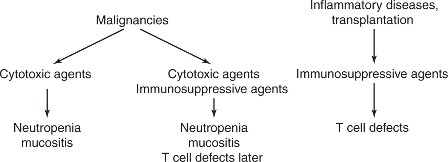
Figure 15.1. Pathways that produce an immunocompromised host.
A full understanding of these classifications and their application to specific populations will provide a firm foundation for managing the immunocompromised host.
NEUTROPENIA AND MUCOSITIS
PATHOGENESIS
Neutropenia is defined as an absolute neutrophil count below 500/mm3. It is often accompanied by mucosal damage. As a result, bacteria from the mouth and lower gastrointestinal (GI) tract are able to pass through the damaged mucosal barrier unchecked by the host’s first line of defense: the neutrophil. Normally, any bacteria passing through the mucosa are phagocytosed and killed by toxic oxygen byproducts, proteases, and small bactericidal cationic proteins within the closed environment of the phagolysosome.
The risk of infection increases as the cell number decreases, with the incidence of infection being inversely related to how far the number of neutrophils falls below the threshold of 500/mm3. The risk of serious infection is considerably higher when the neutrophil count is less than 200/mm3. The duration of neutropenia is also an important determinant of infection risk. The incidence of infections is low if neutropenia lasts only 7-10 days. However, neutropenia that continues for more than 10 days is associated with a high risk of infection.
CASE 15.1
A 51-year-old man received high induction with cytosine arabinoside for relapse of acute lymphocytic leukemia. Two days after completion of his 7-day course of chemotherapy, his absolute neutrophil count was 0/mm3. One day later, he developed a fever and was started on ticarcillin–clavulanate and gentamicin. Over the next 48 hours, he remained febrile, and he developed a black skin lesion (2 × 2 cm) on his right thigh. The lesion was dark black, necrotic in appearance, and mildly painful to touch. Tissue biopsy revealed sheets of gram-negative rods. Four of four blood cultures drawn at the onset of fever were positive for Pseudomonas aeruginosa, Escherichia coli, and Klebsiella pneumoniae. His antibiotic regimen was switched to cefepime and gentamicin. Over the next week, his neutrophil count increased, and he defervesced.
MICROBIOLOGY
The infected organisms usually come from among those found on the skin or in the oral cavity and GI tract, plus any introduced by cross-contamination from environmental sources. For example, environmental cross-infection can occur if organisms are ingested in food at a time when the gut has been denuded by cytotoxic chemotherapy.
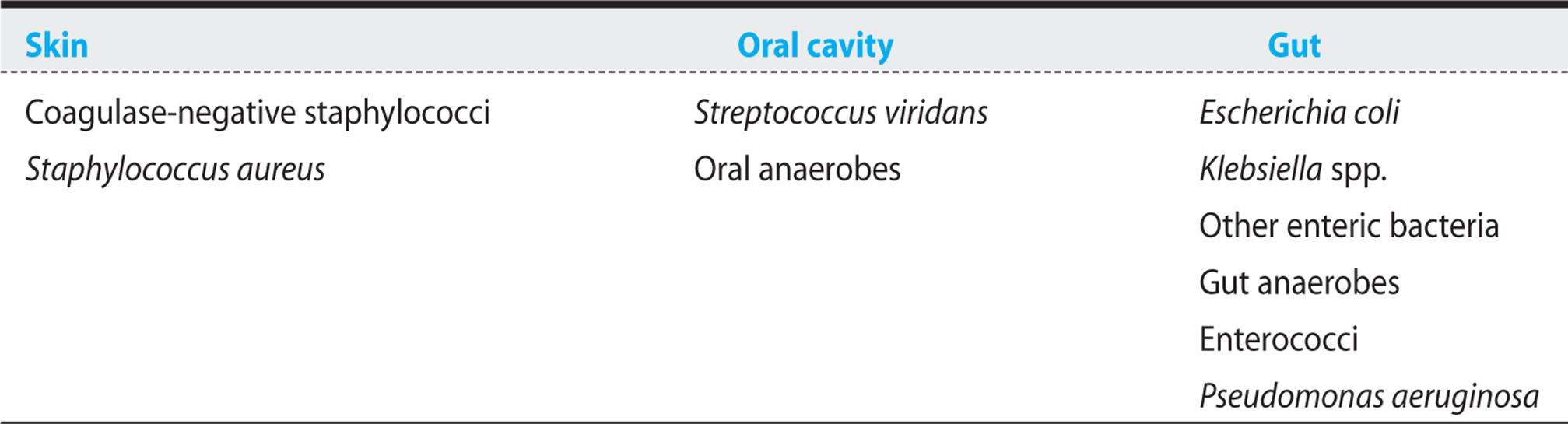
1. Bacteria. Table 15.1 lists the bacteria most commonly reported, with their probable sources. Gram-positive pathogens have increased in frequency in recent series describing neutropenic bacteremia, probably as a consequence of the increased use of long-lasting indwelling venous catheters and the overuse of fluoroquinolones. The most frequent gram-positive bacteria are coagulase-negative staphylococci, Staphylococcus aureus, and Streptococcus viridans. Enterococci and Corynebacterium are increasingly being cultured. Gram-negative pathogens are second in frequency, and as seen in case 15.1, they usually originate from the GI tract. As in that case, severe neutropenia is commonly accompanied by polymicrobial bacteremia. The most frequently encountered organisms are Escherichia coli, Klebsiella species, Pseudomonas aeruginosa, and less commonly, Enterobacter, Proteus, Acinetobacter, Stenotrophomonas, and Citrobacter. Remarkably, despite their presence in large numbers in the GI tract, anaerobic gram-negative rods such as Bacteroides are not frequent causes of bacteremia in neutropenic patients. However, bacteremia with anaerobes is occasionally seen in association with severe mucositis.
Table 15.1. Sources of Bacteria Commonly Infecting Neutropenic Patients
2. Fungi. Bacteria are not the only members of the human or environmental flora that infect the neutropenic patient. Organisms that are present in lower numbers and that are resistant to antibacterial agents—for example, yeasts and moulds—also play a significant role in infections in the neutropenic patient. However, it should be borne in mind that certain fungi are held in check by cell-mediated immunity, and these pathogens infect patients with compromised cell-mediated immunity.
Fungal infections usually develop after broad-spectrum antibiotics have had time to reduce the competing bacterial flora. In patients with no prior history of fungal infection, these pathogens are not usually seen for at least 7 days into a febrile neutropenic episode.
KEY POINTS
About Infections Associated with Neutropenia and Mucositis
1. Risk is inversely related to the number of neutrophils below 500/mm3.
2. Infected organisms primarily come from among those found on the skin and in the oral cavity and gastrointestinal tract.
3. Bacteria include Staphylococcus epidermidis, Staph. aureus, Streptococcus viridans, enterococci, enteric gram-negative organisms, and Pseudomonas; anaerobes are less common.
4. Fungal infections develop after antibiotic therapy has had time to reduce the bacterial flora (usually after 7 days or more); Candida and Aspergillus species are the most common.
Hence, fungal infections are often called “superinfections,” because they occur while patients are receiving antibacterial agents.
Occasionally, when a patient has received antibiotics in the recent past and the level of fungal colonization in the gut is high, fungi may emerge as primary pathogens early in neutropenia before antibiotics are given, or they may infect a central venous catheter from the skin. Some fungi may be acquired early by inhalation from the environment, but may not become symptomatic until much later, after the organism has multiplied sufficiently in the lung and has invaded lung parenchyma and blood vessels, thus appearing to be a superinfection. Fungi that may appear early in neutropenia include Candida species (albicans, tropicalis, krusei, glabrata, and others) and occasionally Aspergillus species. Certain fungi that cause severe infections in other populations—Mucor species, for example—are only infrequently encountered in the neutropenic patient.
Pathogens Encountered in Patients with Suppression of T Cell Functions
The number of patients with suppression of T cell function is progressively increasing. Initially, patients receiving corticosteroids were the major group of patients falling into this category. Increasingly, patients with connective tissue disease including lupus erythematosus and rheumatoid arthritis are being treated with new cytokine antagonists to control their disease. These agents also impair cell-mediated immunity. Most of the patients in this category have undergone organ transplantation. To prevent organ rejection, they receive agents directed against T cells.
Post-transplant infections fall into two groups:
• Infections occurring during the first postoperative month.
• Infections occurring during the subsequent 1-6 months.
During the first month, patients become infected with the same hospital-acquired pathogens as other hosts do. Pathogens of particular concern during this period include Legionella species and other gram-negative bacilli such as P. aeruginosa; gram-positive organisms, particularly antimicrobial-resistant species such as vancomycin-resistant enterococci (VRE) and methicillin-resistant S. aureus; and fungi, such as Aspergillus species and azole-resistant Candida species. Because these patients often receive broad-spectrum antibiotics during their postoperative recovery, they are also at risk of Clostridium difficilecolitis.
During the first month, transplant patients are also at risk of developing infections transmitted by the donor organ or organs. In some instances, the donor had an acute infection (with S. aureus or pneumococci) or gram-negative bacteremia before death. Before organs are harvested, adequate therapy must be assured. Still, bacteria can occasionally survive in a vascular aneurysm or other protected sites. Or the donor may have an asymptomatic low-grade infection that becomes apparent only when the organ is transplanted into the immunocompromised recipient. Recent examples have included West Nile virus, lymphocytic choriomeningitis virus, rabies, leishmaniasis, and Chagas disease.
The period from 1 to 6 months post-transplant is associated with the widest variety of potential opportunistic infections. Immunosuppression is at its highest during this period to prevent acute rejection.
BACTERIA
Mycobacteria are of particular concern. Post-transplant patients harboring latent Mycobacterium tuberculosis can develop miliary tuberculosis (see Chapter 4). Atypical mycobacteria may become more invasive and cause symptomatic infection. Listeria monocytogenes can be contracted by eating contaminated foods, and transplant patients should be instructed how to avoid foods contaminated with this deadly pathogen. Listeria is the third most frequent cause of community-acquired bacterial meningitis, and it almost exclusively infects people with depressed cell-mediated immunity (see Chapter 6). Nocardia species can result in cavitary or nodular pulmonary infections, plus bacteremia and brain abscess. These patients are also at increased risk of L. pneumophila.
FUNGI
Fungal infections in patients with suppressed T cell function are often life-threatening and may be difficult to diagnose. Cryptococcus neoformans is the most common fungal pathogen encountered in the transplant population. The sites primarily infected are the lungs and the meninges. In the meninges, the organism causes a lymphocytic meningitis (see Chapter 6). The filamentous fungi that are most likely to cause disease are Aspergillus species, Fusariumspecies, and the Mucor and Rhizopus groups. Depending on geographic location, Histoplasma capsulatum and Coccidioides immitis are also important pathogens in these patients. Increasingly, the dematiaceous (“black”) fungi are being reported as a cause of infections. However, Candida rarely causes infection in this population, probably because these fungi are controlled primarily by neutrophils.
The role of filamentous fungal infections in organ transplantation cannot be overemphasized. When such infections occur, cure is extremely difficult in the face of continued immunosuppression, and death is a common outcome.
VIRUSES
The most important arms of the host defense mechanisms against viral infections are the T cells and antibodies. In most instances, cell-mediated immunity and humoral immunity function together to prevent and control active viral infections. The patient with T cell defects following immunosuppression is likely to have antibodies against many viruses, unless total ablation of existing T cells and reconstitution with immunologically naive cells has been carried out. Reconstituted donor populations usually contain memory cells to make antibody, but the response may be blunted. Transplant patients therefore tend to be more susceptible to viruses that are latent in the body rather than to infections with new viruses. Loss of cell-mediated immunity allows latent viruses to reactivate. Additionally, patients may acquire reactivation infections from transfused blood components or a transplanted organ. The virus that most commonly reactivates is cytomegalovirus (CMV). A CMV infection can also be acquired from blood transfusion or transplantation with an infected organ. The risk of infection depends on the recipient and donor CMV antibody (Ab) status:
• High risk—CMV Ab (-) recipient, CMV Ab (+) donor
• Intermediate risk—CMV Ab (+) recipient, CMV Ab (+) or Ab (-) donor
• Lowest risk—CMV Ab (-) recipient, CMV Ab (-) donor
The diagnosis of active CMV infection utilizes two tests that are also used to monitor response to therapy: the CMV antigen test always correlates with active replication, and a CMV polymerase chain reaction (PCR) test can detect latent and active infection (a higher copy number is indicative of active invasive infection).
Epstein–Barr virus less commonly causes symptomatic disease. However, the virus actively replicates in 20–30% of transplant recipients and can cause a lymphoproliferative syndrome. Less common viruses that can become active include herpes simplex, herpes zoster, HHV-6, and hepatitis B and C viruses.
OTHER PATHOGENS
Latent parasites can become active as a consequence of reductions in cell-mediated immunity. Pneumocystis jiroveci, a pathogen common in AIDS, can also result in severe, hypoxic pneumonia in transplant patients. Therefore, during the period of peak immunosuppression, these patients should receive prophylactic trimethoprim–sulfamethoxazole (see Chapter 16). Toxoplasmosis is another latent pathogen that can reactivate in the central nervous system causing brain abscesses and encephalitis (see Chapter 16). Patients with low level Strongyloides infection can develop disseminated strongyloidiasis in association with immunosuppression (see Chapter 12). To prevent this often fatal complication, all patients with unexplained eosinophilia should undergo stool sampling and an enzyme-linked immunosorbent assay to exclude Strongyloides before they receive an organ transplant.
KEY POINTS
About Infections in Patients with Defective Cell-Mediated Immunity
1. Can contract the same community-acquired pathogens as normal hosts.
2. Have an increased risk of bacterial infections with Mycobacterium species, Listeria monocytogenes, and Nocardia species.
3. Fungal infections are often life-threatening and may be difficult to diagnose.
a) Cryptococcus is most common.
b) Aspergillus species, Fusarium species, and the Mucor and Rhizopus groups are other common possibilities.
c) Histoplasmosis and coccidioidomycosis should be considered depending on geographic location.
d) Reports of dematiaceous fungi (“black mold”) infection are increasing.
4. Reactivation of old viral infections is a major concern.
a) Cytomegalovirus is most common. Can be the result of reactivation, blood transfusion, or transplantation with an infected organ.
b) Epstein–Barr virus is less common.
5. Other possible pathogens include Pneumocystis, Toxoplasma, and disseminated Strongyloides.
Pathogens Associated with Mixed Deficits Found in Bone Marrow Transplantation
Bone marrow transplantation involves three phases of immunosuppression:
1. Phase I (days 0–30 post-transplant). During this neutropenic phase, patients are managed in a manner similar to that of other neutropenic patients.
2. Phase II (days 30-100 post-transplant). During this phase of (primarily) compromised cell-mediated immunity, the patient is managed in a manner similar to that of other organ transplant patients with compromised cell-mediated immunity. Infection with CMV is particularly common at this stage, and acute graft-versus-host disease is also frequently encountered during this period.
3. Phase III (beyond day 100 post-transplant). Bone marrow transplant patients often continue to have defects in cell-mediated immunity, plus depressed humoral immunity, resulting in continued susceptibility to CMV, herpes zoster virus, and lymphoproliferative disorders related to infection with Epstein–Barr virus. These patients are also at increased risk of infections with encapsulated S. pneumoniae and H. influenzae bacteria. Predisposing factors for these infections include functional hyposplenism after total body irradiation, and chronic graft-versus-host disease. This later disorder renders B cells dysfunctional, resulting in decreased production of immunoglobulin G2 (IgG2) and specific pneumococcal antibodies. Clinicians should have a low threshold for starting coverage for encapsulated organisms when these patients develop a worsening of fever, particularly fever accompanied by rigor (see Chapter 6).
KEY POINTS
About Infections associated with Mixed Deficits Found in bone Marrow Transplantation
1. Three phases of immunosuppression follow transplantation:
a) Phase I (days 0-30): Neutropenia.
b) Phase II (days 30-100): Primarily depressed cell-mediated immunity and graft-versus-host disease.
c) Phase III (beyond day 100): Depressed cell-mediated and humoral immunity, chronic graft-versus-host disease.
2. Major infections seen are the same as are seen with neutropenia (early) and solid organ transplant (later).
3. Problems with encapsulated bacteria (Haemophilus influenzae and Streptococcus pneumoniae) are also a possibility.
DIAGNOSIS AND TREATMENT
Overall Approach in Immunocompromised Hosts
In approaching the febrile compromised host or even a compromised host that has a site of infection, generalizations about the medical urgency required for treatment should be avoided until the patient has been properly classified. The guiding principle is the type of infected organism; hence, empiric therapy and the need for urgency are governed chiefly by the type of host compromise. Not every compromised host requires empiric antibiotic therapy. The questions and algorithm that follow are therefore suggested.
THE FEBRILE NEUTROPENIC PATIENT
If neutropenia is the consequence of recent cytotoxic chemotherapy, then the onset of significant fever (temperature above 38.3°C) warrants emergent diagnostic studies and antibiotic therapy. Antibiotics should be administered within 60 minutes of presentation. The progression of infection in neutropenic patients can be rapid, and infection cannot be readily differentiated from noninfectious causes of fever. The usual manifestations of infection are often absent. Skin infections lack erythema, warmth, and purulence. Conventional chest X-ray may appear normal in bacterial pneumonia, and in bacterial meningitis, the cerebrospinal fluid may contain minimal polymorphonuclear leukocytes.
Initial workup for a fever should include the following:
• Physical examination looking for sites of infection in lungs, skin, mucous membranes, and the perirectal area.
• Biopsy and culture of any skin lesions.
• Blood tests—peripheral WBC with differential, platelet count, BUN, serum creatinine, electrolytes, hepatic transaminases, and serum bilirubin.
• T blood cultures, one drawn from a peripheral vein and the other from the central line.
• Cultures from other sites including the urine, and any other suspicious sites.
• Chest X-ray for patients with respiratory symptoms.
Empiric antibiotic therapy should be initiated emergently. The regimen depends on the severity of disease. Low severity is defined as follows:
• A temperature below 39°C and a nonseptic appearance.
• An absolute neutrophil and monocyte count below 100/mm3, with neutropenia for less than 7 days and recovery expected by less than 10 days.
• A normal chest X-ray.
• Nearly normal liver and renal function.
• No evidence for intravascular device infection.
• Malignancy in remission.
• No neurologic deficits.
• No abdominal pain.
• No comorbid conditions (hypotension, vomiting, diarrhea, evidence for deep organ infection).
• Low-severity score (above or equal to 21—see Table 15.2).
Table 15.2. Scoring Indexa for Identification of Low-Risk Febrile Neutropenic Patients at the Time of Presentation of Fever
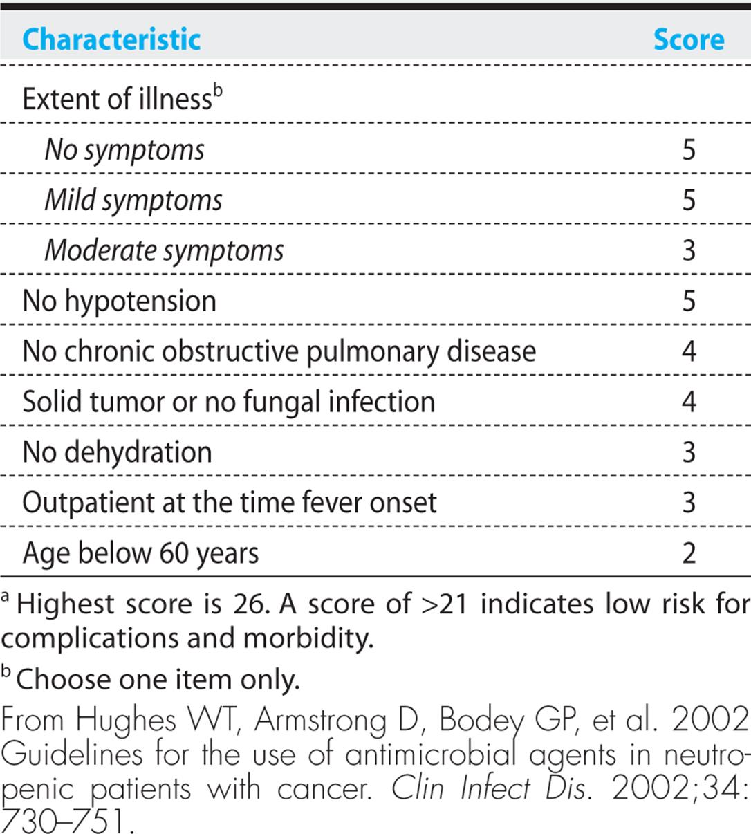
In these patients, oral antibiotics can be administered. Ciprofloxacin (500 mg twice daily) plus amoxicillin–clavulanate (875 mg twice daily) is the recommended regimen.
From Hughes WT, Armstrong D, Bodey GP, et al. 2002 Guidelines for the use of antimicrobial agents in neutropenic patients with cancer. Clin Infect Dis. 2002;34: 730-751.
Intravenous antibiotics should be given to more severely ill patients who do not meet the above criteria. (Intravenous administration can also be used for low-severity cases.) A number of regimens can be used (see Table 15.3), and all have resulted in comparable response rates and reductions in mortality. The specific empiric regimen must take into account the antibiotic resistance patterns of the local institution and the patient’s prior history of infections and antibiotic treatment. Specific doses for each regimen are given in Table 15.3.
Table 15.3. Anti-Infective Therapy for Neutropenic Patients
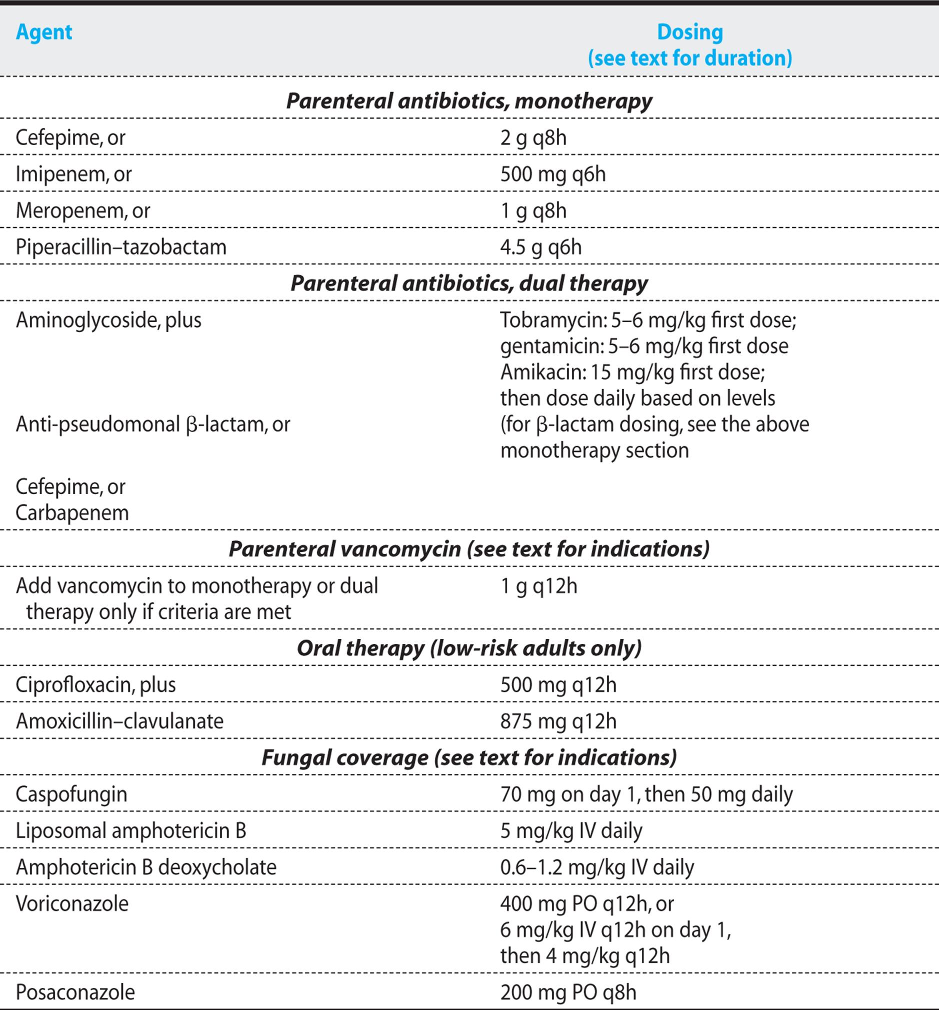
In multiple studies, monotherapy has been shown to be comparable to dual therapy. Monotherapy can be initiated with cefepime, imipenem, or piperacillin–tazobactam. Dual therapy regimens without vancomycin have all proven to be therapeutically equivalent, and they include cefepime combined with gentamicin, tobramycin, or amikacin; ticarcillin–clavulanate or piperacillin–tazobactam combined with an aminoglyco-side; imipenem plus an aminoglycoside; or piperacillin–tazobactam plus ciprofloxacin (see Table 15.3).
Vancomycin should not be administered for routine empiric therapy because of the increased risks of selecting for VRE and of nephrotoxicity. A meta-analysis revealed that the addition of a glycopeptide as part of empiric therapy did not shorten the febrile episode or reduce mortality in neutropenic patients. But a glycopeptide antibiotic should be added if an intravascular device infection is suspected, if colonization with methicillin-resistant S. aureus is known to exist, if blood cultures are positive for gram-positive cocci before final identification and sensitivity testing, or if the patient is hypotensive or has other evidence of cardiovascular compromise. Linezolid has been shown to be therapeutically equivalent to vancomycin in the neutropenic patient. However, in combination with selective serotonin-reuptake inhibitors, linezolid has been associated with severe myelosuppression in bone marrow transplant patients.
In approximately 30% of cases, blood cultures will be positive, and in the patient with positive blood cultures who becomes afebrile in 3-5 days, antibiotic coverage should be adjusted to the least toxic regimen. However, broad-spectrum coverage should be maintained to prevent breakthrough bacteremia. Anti-infective therapy should be continued for a minimum of 7 days. Duration also depends on clinical response and the ability to sterilize the bloodstream. The Infectious Diseases Society of America (IDSA) recommends that antibiotics usually be continued until the neutrophil count rises above 500/mm3.
If the neutropenic patient with a low-risk profile becomes afebrile in 3-5 days, and if cultures are negative, intravenous antibiotics can be switched to oral ciprofloxacin and amoxicillin–clavulanate. For the high-risk patient, intravenous antibiotics should be continued for a minimum of 7 days and until the neutrophil count rises above 500/mm3.
If the patient remains febrile after 3-5 days, all clinical findings must be re-evaluated. Cultures should be thoroughly reviewed, and a complete physical examination repeated, paying careful attention to skin, mucosal surfaces, and intravenous catheter sites. Additional imaging studies should be considered, depending on the physical findings and the patient’s complaints. Computed tomography of the chest may detect infiltrates in half of patients with “normal” conventional radiographs. If an infiltrate is detected, bronchoscopy with lavage should be performed to differentiate among the wide variety of potential pathogens. Antibiotic serum levels (particularly aminoglycosides) should be checked, and cultures should be repeated.
If fever persists for more than 5 days, the patient does not appear septic, no new findings have been uncovered, and the neutrophil count is expected to recover quickly, continue the same antibiotics. However, if clinical worsening or persistent sepsis is noted, the antibiotic regimen should be changed. Switch from monotherapy to dual therapy and consider adding vancomycin if the criteria are appropriate as described earlier. Add antifungal therapy if the neutropenia is expected to persist for more than 5-7 days. Several anti-fungal agents are available: caspofungin or liposomal amphotericin B (amphotericin B deoxycholate is a less expensive, but more toxic alternative) is currently recommended by most experts. Depending on the incidence of aspergillosis and mucormycosis in the specific institution, other agents that can be used are voriconazole and posaconazole (see Table 15.3).
KEY POINTS
About Management of the Neutropenic Patient
1. Conduct a careful physical examination, especially lungs, perirectal area, and skin. Biopsy of new skin lesions.
2. Culture all sites, including blood samples from a peripheral vein and central line.
3. Obtain a chest X-ray. If infiltrate is seen, consider bronchoscopy with lavage.
4. Empiric antibiotics can include
a) monotherapy with cefepime, imipenem, piperacillin–tazobactam; and
b) dual therapy with β-lactam plus an aminoglycoside or a fluoroquinolone.
5. Add vancomycin for catheter-related infection or colonization with methicillin-resistant Staphylococcus aureus.
6. Reassess at 96 hours. If fever persists, add anti-fungal therapy.
7. Outpatient management of fever is increasing in popularity.
8. Fluoroquinolone prophylaxis is recommended by the IDSA for severe neutropenic patients. Danger of selecting for resistant organisms remains a worry.
The duration of antibiotic therapy is an important consideration given the fragile nature of bone marrow transplant patient. This decision must be applied in two major instances:
1. The afebrile patient. If the patient is afebrile after 3–5 days of therapy, and if the neutrophil count has been above 500/mm3 for 2 days, antibiotics can be discontinued after the patient has been afebrile for 48 hours. If the neutrophil count remains below 500/mm3, and if the patient was initially low risk and is not currently septic, then antibiotics can be discontinued when the patient has been afebrile for 5 to 7 days. If the patient was initially at high risk, and if the neutrophil count is below 100/mm3, or if the patient has mucositis or unstable vital signs or other unstable findings, antibiotics should be continued.
2. The persistently febrile patient. If the patient continues to have fever despite empiric antibiotic therapy and if the neutrophil count is above 500/mm3, antibiotics can be discontinued after the neutrophil count has remained at that level for 4-5 days. The patient should then be reassessed. Otherwise, if the neutrophil count is below 500/mm3, the antibiotic therapy should be continued for 2 weeks, with reassessment at that time. Then, if no infection is evident and the patient is clinically stable, antibiotics can be discontinued. Antiviral therapy is not indicated in neutropenic patients unless a specific viral infection is documented.
The IDSA recommends the initiation of fluoroquinolone (ciprofloxacin or levofloxacin) prophylaxis for high-risk patients who are expected to have a prolonged duration of profound neutropenia (ANC <100 cells/mm3 for >7 days). There is concern that routine use of fluoroquinolones will result in widespread antibiotic resistance.
THE FEBRILE NON-NEUTROPENIC PATIENT
The organisms that can cause infection in the febrile non-neutropenic patient population are so large in number that empiric therapy is not recommended unless a specific site of infection is identified or unless, after evaluation, a specific pathogen is thought to be the most likely cause. Even cellulitis may have a nonbacterial origin. However, empiric therapy may be given for central catheter or urinary tract infections, because the usual organisms continue to cause these infections.
Community-acquired infections, both bacterial and viral, are also a problem for susceptible patients, and evaluation of an infectious illness should consider the community context. A thorough history—including details about the onset of the fever, family illnesses, the underlying reason for immunosuppression, and the dose and length of time on immunosuppressive therapy—is therefore important. Patients who have lived in certain geographic areas may experience reactivation of latent infections or succumb to specific infections, such as histoplasmosis in the Ohio River valley or coccidioidomycosis in the Southwest, but none of the specific infections in this patient population require immediate empiric therapy. Emergent anti-infective therapy is usually not life saving in this patient population.
Certain sites of infection do require urgent diagnostic action, however:
1. Patients with a headache or other central nervous system complaint should undergo a lumbar puncture if that procedure can be safely performed. Cryptococcal or Listeria meningitis is the most urgent diagnosis, and these illnesses require immediate treatment.
2. Blood, urine, and any suspicious sites should be cultured. A urinalysis is helpful because these patients are not neutropenic.
3. An inflamed central line may be treated presumptively for gram-positive infection. Central lines should be removed if there is a tunnel infection, or if S. aureus, Pseudomonas, Candida, or atypical mycobacteria are proven to have infected the intravascular device.
4. If a chest radiograph is abnormal, and if the patient is producing sputum, a sample should be obtained for culture and for Gram, acid-fast, and silver staining. If no sputum is being produced, urgent pulmonary and infectious diseases consultations for a diagnostic evaluation should be requested. Because the number of possible causes of pulmonary infection is so large in this population, empiric therapy is not recommended (unless respiratory failure has begun).
5. If the patient is febrile, but none of the foregoing tests has yielded a diagnosis, an infectious diseases consultation should be sought. The possible entities causing fever in the setting of a negative initial evaluation are so diverse that much time and many resources can easily be wasted.
Although the approach outlined above is most applicable to the transplant population, clinicians must not forget that people receiving corticosteroids represent the greatest number of immunosuppressed patients. Much debate has occurred concerning the lowest dosage of corticosteroid that will predispose to infection. A useful general rule is to assume that any dose above physiologic maintenance may be immunosuppressive. Doses as low as 10 mg daily of prednisone have led to invasive pulmonary aspergillosis. Therefore, when faced with a febrile non-transplant patient on corticosteroids for an inflammatory disorder, the diagnostic points discussed in this subsection should be kept in mind. Also, because immunosuppressive agents may blunt an inflammatory response, clinicians should consider that even low-grade fever may indicate the presence of a serious infection.
Prevention
Given the long-term nature of immunosuppression in graft recipients, preventive measures play a critical role in preventing morbidity and mortality. The recommended preventive measures can be categorized by pathogen type:
• Bacteria. In the patient with a documented reduction in IgG level below 400 mg/dL, intravenous IgG may be given to prevent sinus and pulmonary infections resulting from S. pneumoniae infection.
• Viruses. CMV remains one of the most significant concerns, and CMV IgG serum titers should be obtained for all transplant patients and donors. If recipient and donor are both negative, prophylaxis is not required. However, all platelet and blood transfusions should be CMV negative or cleansed of all leukocytes. If the recipient or donor, or both, is CMV positive. then the patient should receive prophylaxis for the first 100 days with oral valganciclovir (900 mg daily). Because of a lower incidence of disease, preemptive therapy is preferred by many experts for the CMV-positive recipient with a CMV-negative donor. If this strategy is chosen, the patient must undergo periodic screening or quantitative PCR for CMV. If these tests are positive, the patient should be treated with intravenous ganciclovir 5 mg/kg twice daily for 14-21 days or oral valganciclovir 900 mg twice during the first 24 hours for induction, and then 900 mg daily.
Another major concern is herpes simplex virus. Antiherpes simplex IgG titers should be measured in all potential recipients before transplantation. Bone marrow transplant patients with a positive titer should receive prophylaxis during the induction phase and during the first 30 days after transplant (phase I) with either oral valacyclovir (1000 mg twice or three times daily) or oral valganciclovir (900 mg twice daily) if the patient also requires CMV prophylaxis.
KEY POINTS
about the Management of Patients with Depressed Cell-Mediated Immunity
1. Empiric antibiotics are generally not recommended.
2. The number of possible organisms is very large.
a) Samples for biopsy and culture are strongly recommended.
b) A thorough epidemiologic history is often helpful.
3. Emergent management is required for
a) central nervous system symptoms such as headache and confusion—consider Cryptococcus and Listeria.
b) infected central lines—institute empiric antibiotics.
c) infiltrate on chest radiographs: seek a pulmonary consultation (bronchoscopy is often required).
4. Even low-grade fever in the patient on corticosteroids is a matter of concern.
Other transplant patients undergoing high-level immunosuppression should also be considered for prophylaxis. Reactivation of varicella virus can lead to serious morbidity and also requires preventive measures. The recipient and all family members should be vaccinated with the live attenuated vaccine at least 4 weeks before the transplant procedure.
• Fungi. Allogeneic bone marrow transplant patients have a high incidence of Candida albicans infection during phase I and should receive oral fluconazole 400 mg daily or posaconazole (200 mg three times daily) for prophylaxis.
• Pneumocystis jiroveci. Throughout the period of immunosuppression, transplant patients are at risk of infection with this organism, and oral trimethoprim–sulfamethoxazole (one double-strength tablet three times weekly, or one single-strength tablet daily) is recommended.
PREVENTIVE MEASURES IN SOLID-ORGAN AND BONE MARROW TRANSPLANT PATIENTS
1. Immunoglobulin G (IgG) should be administered if IgG levels fall below 400 mg/dL.
2. Recipients or donors positive for cytomegalovirus should receive valganciclovir.
3. Bone marrow recipients with positive IgG titer for herpes simplex virus should receive valacyclovir.
4. Vaccine for varicella virus should be given to patients and household contacts before a transplantation procedure.
5. Allogeneic transplant patients should receive fluconazole or posaconazole to prevent fungal infections.
6. All transplant recipients should receive trimethoprim-sulfamethoxazole to prevent Pneumo-cystis infection.
CONCLUSIONS
Continuing advances in the treatment of malignancies and transplantation will maintain a large population of medically compromised hosts. These patients fit into two general categories that predispose them to infections that are usually controlled either by neutrophils or by T cells. Bone marrow or stem cell transplant patients fit into both categories depending on how much time has passed since transplantation. The febrile neutropenic patient can be considered to be a medical emergency requiring empiric antibacterial therapy with one or two broad-spectrum antibiotics. Conversely, the patient with suppression of cell-mediated immunity requires a thorough evaluation, and empiric antibiotic therapy should be avoided unless the cause of the fever is known on presentation. It is advisable that these patients receive care from infectious disease specialists. Outpatient management of these patients can be expected to become increasingly common.
FURTHER READING
Bow EJ. Fluoroquinolones, antimicrobial resistance and neutropenic cancer patients. Curr Opin Infect Dis. 2011;24:545-553.
Bucaneve G, Micozzi A, Menichetti F, et al. Levofloxacin to prevent
bacterial infection in patients with cancer and neutropenia.N Engl J Med. 2005;353:977-987.
Cornely OA, Maertens J, Winston DJ, et al. Posaconazole vs. fluconazole or itraconazole prophylaxis in patients with neutropenia.N Engl J Med. 2007;356:348-359.
Cullen M, Steven N, Billingham L, et al. Antibacterial prophylaxis after chemotherapy for solid tumors and lymphomas. N Engl J Med. 2005;353:988-998.
Denning DW, Kibbler CC, Barnes RA. British Society for Medical Mycology proposed standards of care for patients with invasive fungal infections. Lancet Infect Dis. 2003;3:230-240.
Freifeld AG, Bow EJ, Sepkowitz KA, et al. Clinical practice guideline for the use of antimicrobial agents in neutropenic patients with cancer: 2010 update by the Infectious Diseases Society of America. Clin Infect Dis: An official publication of the Infectious Diseases Society of America. 2011;52(4):e56-e93.
Fukuda T, Boeckh M, Carter RA, et al. Risks and outcomes of invasive fungal infections in recipients of allogeneic hematopoietic stem cell transplants after nonmyeloablative conditioning. Blood. 2003; 102:827-833.
Hachem RY, Hicks K, Huen A, Raad I. Myelosuppression and serotonin syndrome associated with concurrent use of linezolid and selective serotonin reuptake inhibitors in bone marrow transplant recipients. Clin Infect Dis. 2003;37:e8-e11.
Hamour IM, Mittal TK, Bell AD, Banner NR. Reversible sirolimus-associated pneumonitis after heart transplantation. J Heart Lung Transplant. 2006;25:241-244.
Hughes WT, Armstrong D, Bodey GP, et al. 2002 Guidelines for the use of antimicrobial agents in neutropenic patients with cancer.Clin Infect Dis. 2002;34:730-751.
Jaksic B, Martinelli G, Perez-Oteyza J, Hartman CS, Leonard LB, Tack KJ. Efficacy and safety of linezolid compared with vancomycin in a randomized, double-blind study of febrile neutropenic patients with cancer. Clin Infect Dis. 2006;42: 597-607.
Kalil AC, Levitsky J, Lyden E, Stoner J, Freifeld AG. Meta-analysis: the efficacy of strategies to prevent organ disease by cytomegalo-virus in solid organ transplant recipients. Ann Intern Med. 2005; 143:870-880.
Klastersky J. The changing face of febrile neutropenia-from monotherapy to moulds to mucositis. Why empirical therapy? J Antimicrob Chemother. 2009;63(suppl 1):i14-i15.
Legrand M, Max A, Peigne V, et al. Survival in neutropenic patients with severe sepsis or septic shock. Crit Care Med. 2012;40:43-49.
Marty FM, Lee SJ, Fahey MM, et al. Infliximab use in patients with severe graft-versus-host disease and other emerging risk factors of non-Candida invasive fungal infections in allogeneic hematopoietic stem cell transplant recipients: a cohort study. Blood. 2003; 102:2768-2776.
Patterson TF, Boucher HW, Herbrecht R, et al. Strategy of following voriconazole versus amphotericin B therapy with other licensed antifungal therapy for primary treatment of invasive aspergillosis: impact of other therapies on outcome. Clin Infect Dis. 2005; 41:1448-1452.
Raad, II, Escalante C, Hachem RY, et al. Treatment of febrile neutropenic patients with cancer who require hospitalization: a prospective randomized study comparing imipenem and cefepime. Cancer. 2003;98:1039-1047.
Rolston KV, Manzullo EF, Elting LS, et al. Once daily, oral, outpatient quinolone monotherapy for low-risk cancer patients with fever and neutropenia: a pilot study of 40 patients based on validated risk-prediction rules. Cancer. 2006;106:2489-2494.
Segal BH, Almyroudis NG, Battiwalla M, et al. Prevention and early treatment of invasive fungal infection in patients with cancer and neutropenia and in stem cell transplant recipients in the era of newer broad-spectrum antifungal agents and diagnostic adjuncts. Clin Infect Dis.2007;44:402-409.
Vardakas KZ, Samonis G, Chrysanthopoulou SA, Bliziotis IA, Falagas ME. Role of glycopeptides as part of initial empirical treatment of febrile neutropenic patients: a meta-analysis of randomised controlled trials. Lancet Infect Dis. 2005;5:431-439.
Viscoli C, Varnier O, Machetti M. Infections in patients with febrile neutropenia: epidemiology, microbiology, and risk stratification. Clin Infect Dis. 2005;40(suppl 4):S240-S245.
Walsh TJ, Pappas P, Winston DJ, et al. Voriconazole compared with liposomal amphotericin B for empirical antifungal therapy in patients with neutropenia and persistent fever. N Engl J Med. 2002; 346:225–234.
Wingard JR, Eldjerou L, Leather H. Use of antibacterial prophylaxis in patients with chemotherapy-induced neutropenia. Curr Opin Hematol. 2012;19:21-26.
Wisplinghoff H, Seifert H, Wenzel RP, Edmond MB. Current trends in the epidemiology of nosocomial bloodstream infections in patients with hematological malignancies and solid neoplasms in hospitals in the United States. Clin Infect Dis. 2003;36:1103-1110.
Zuckermann J, Moreira LB, Stoll P, Moreira LM, Kuchenbecker RS, Polanczyk CA. Compliance with a critical pathway for the management of febrile neutropenia and impact on clinical outcomes. Ann Hematol. 2008;87:139-145.