
Time Recommended to Complete: 3 days
Bernard Hirschel, M.D.
GUIDING QUESTIONS
1. How is HIV primarily transmitted, and how do genital ulcers increase the risk of HIV transmission?
2. Which cells does the HIV virus primarily infect?
3. What are the symptoms and signs of primary HIV infection?
4. What is the preferred test for diagnosis of HIV, and how is AIDS defined?
5. What is meant by the term “the window period”?
6. How is HIV activity monitored?
7. Below what CD4 count does the host begin to experience opportunistic infections?
8. What are the indications for initiating antiretroviral therapy?
9. What are the goals for therapy, and what are the factors that increase the risk of developing resistance?
POTENTIAL SEVERITY
Management of HIV is challenging and complex. The associated opportunistic infections are often difficult to diagnose and frequently life-threatening.
EPIDEMIOLOGY
Having originated in Eastern Africa between 1910 and 1950, by transmission of a precursor virus from a chimpanzee, HIV infection has now spread across the world. Sub-Saharan Africa remains the epicenter of the epidemic: almost 3 million new infections occur annually, 23 million Africans are living with HIV, and more than 10 million have already died. Uganda alone has lost 2.5 million people due to AIDS: 200-400 deaths daily, every day from 1985 to 2011, in a country with a population of 20 million. In the sub-Saharan countries, transmission occurs predominantly by heterosexual intercourse, with as many women as men being infected. On average, infected women are younger than infected men, but overall, the most productive age strata is where the infection predominates, contributing to the disastrous socioeconomic impact of the AIDS epidemic.
The problems of North America and Western Europe pale in comparison with those of Africa. Nonetheless, the number of HIV-infected people living in the United States has reached more than 1 million. Incidence figures are difficult to determine, because most newly acquired infections are not diagnosed. Judging from the number of first positive tests (which may be the result of an infection acquired years earlier), infection rates declined during the 1990s, reaching a plateau around 1998. Some reports claim that infections have increased slightly since 1998, perhaps because of an increase in sexual-risk taking linked to a false sense of security created by the existence of highly active antiretroviral therapy (HAART). However, by decreasing viremia, HAART may also be having a positive effect on transmission of HIV.
The probability of acquiring an HIV infection varies depending on the type of exposure. Transfusion with a unit of HIV-infected blood is almost certain to infect the recipient. In the absence of treatment, the child of an HIV-positive mother has about a 30% chance of infection. The chance of acquiring an infection after a needle-stick injury involving infected bodily fluids is about 1 in 300. Most infections occur with sexual exposure, the primary determinant of infectivity being the level of virus in genital secretions. This level correlates directly with viremia, which is particularly elevated during primary HIV infection. Local genital factors modulate that risk. Inflammation caused by sexually transmitted diseases attracts lymphocytes. In an infected person, lymphocytes may harbor HIV, and in the recipient, they may provide a reservoir of cells vulnerable to HIV infection. Circumcision reduces a man’s risk of contracting HIV by approximately 60%.
KEY POINTS
About the Epidemiology of HIV Infection
1. Highest incidence is found in Africa where the virus originated:
a) New infections occur at a rate of 2-3 million annually.
b) About 23 million are living with AIDS.
c) Transmission is mainly heterosexual, with the incidence in men and women being about equal.
2. North America and Europe have lower incidences and prevalences:
a) Prevalence in the United States 1.2 million.
3. Risk of HIV infection is
a) very high with a contaminated blood transfusion;
b) about 1 in 300 for a needle stick;
c) about 30% for a child of an untreated infected mother;
d) Less than 0.01-1% for vaginal or anal intercourse. Genital ulcers increase risk by a factor of 10; condoms prevent transmission. Circumcision reduces risk by 60%;
4. Preventive measures are far more cost-effective than treatment.
Depending on the level of viremia, the risk of HIV infection varies from roughly 1% to less than 0.01% for each act of vaginal or anal intercourse. Compared with infection rates for other sexually transmitted diseases (20-40% after exposure to syphilis or gonorrhea), this risk is quite low. Nonetheless, repeated sexual exposure—as occurs in a serodiscordant couple—entails substantial risk, up to 1% per month. The risk is likely higher during the first months of a sexual partnership than later. Indeed, some studies show an HIV-specific, potentially protective cellular immune response in seronegative sexual partners of seropositive individuals.
However, absence of infection in the past is no guarantee for the future: increasing immune deficiency and viremia are part of the natural history of untreated HIV infection, and they carry with them an increased risk of transmission.
Anal and vaginal intercourse is approximately equally effective in transmitting HIV. Some (but not all) studies have found that the risk of transmission from an HIV-infected man to a woman is higher than the reverse. Compared with vaginal or anal intercourse, oral sex is much less risky—specifically, it is too low to be quantified. However, any sizable HIV center encounters examples of probable transmission by oral sex. Condoms are effective in preventing transmission. Transmission has never been observed in a large series of couples who declared “always” to have used condoms. However, perfect compliance with condom use, and avoidance of slippage and breakage is difficult to achieve in practice.
Preventive efforts have had varying success:
• Transmission of HIV infection through infected blood products has almost been eliminated. Rare cases may still occur (less than 1 transmission in 500,000 blood transfusions) if blood is donated during the “window period.”
• Use of ART in the mother has the potential to decrease mother-to-child transmission from more than 30% to less than 1%. Such transmission has now become very rare in Western Europe and the United States, and is almost always the result of some procedural failure.
• Needle exchange programs and methadone or even heroin substitutions have reduced the incidence of HIV infection in intravenous drug users by more than 90%.
• As noted earlier, condoms are effective in decreasing HIV transmission, particularly in stable couples. Incidence rates of HIV have declined in homosexual communities that practice safer sex. The decrease in the prevalence of HIV may also have contributed to a reduced rate of infection among younger homosexual men, even without necessarily perfect adherence to safer-sex guidelines. Among both homosexual and heterosexual communities, some subgroups continue high-risk practices, with a concomitant high incidence of sexually transmitted diseases and HIV. Several instances of small epidemics of syphilis were reported in Dublin, Bristol, Baltimore, Paris, and California. Interestingly, these epidemics were not accompanied by a rise in HIV infections. As noted earlier, the explanation for this seeming paradox may lie in the protective effect of HAART.
• This protective effect was spectacularly shown in 2011, with the publication of a large randomized trial in serodiscordant couples. The infected partners, who had CD4 counts between 350 and 550, were either treated right away (the “immediate” group) or treatment was deferred until CD4 counts fell to approximately 200 (the “deferred” group). In the immediate group, there was only one transmission of HIV, compared with 28 in the deferred group.
When contemplating the use of scarce resources to fight HIV infection, it is important to realize that prevention is much more cost-effective than cure. Even with unrealistically favorable assumptions regarding the efficacy and costs of HAART, costs per life year saved are 20-100 times lower for condoms than for antiretroviral treatment. But it is the sick who are crying for help, not the healthy who are crying for condoms.
PATHOGENESIS
The primary targets of the human immunodeficiency virus are probably the dendritic cells in the mucosa of the genital tract. The virus uses a specific receptor called DC-SIGN to attach to those cells. The dendritic cells then transport HIV into lymph nodes, where the virus infects lymphocytes.
The receptors for HIV are mainly the CD4 molecules on the surface of a subpopulation of T lymphocytes. A co-receptor is also necessary for infection. Viruses that preferentially interact with the co-receptor CCR5 are called “R5 viruses” (or “monocytotropic,” or “nonsyncytium-inducing”), and they predominate in early infection. Later on, HIV often acquires the capacity to interact with the CXCR4 receptor; such viruses are called “X4” (“syncytium-inducing,” or “lymphocytotropic”). The CD4 lymphocytes whose T-cell receptor is specific for HIV proteins proliferate and are preferentially infected. This preferential infection (followed by destruction) may explain the specific deficiency of immunity to HIV as described next.
More than 98% of lymphocytes are localized in the lymph nodes and spleen. Nonetheless, the viruses produced by the newly HIV-infected lymphocytes flood the blood and are transported into all tissues within a matter of days. Viremia reaches high levels—up to millions of HIV genomes per cubic millimeter. During this time, many patients become symptomatic with fever, skin lesions, pharyngitis, and swollen lymph nodes. This self-limiting disease (Figure 16.1), lasting usually a few days to a few weeks, is called “primary HIV infection,” “acute retroviral syndrome,” or “seroconversion syndrome.” Then, the immune response kicks in; antibodies directed against HIV appear in the blood, and cytotoxic T cells specific for HIV-infected cells proliferate. This HIV immune response rapidly achieves partial control of the HIV infection. Viremia levels decrease by several orders of magnitude, stabilizing at a lower level, called “plateau level.” Plateau levels can vary from fewer than 50 to millions of copies of HIV RNA per cubic millimeter, and they correlate closely with further evolution toward immune deficiency: the higher the plateau, the faster the development of AIDS.

Figure 16.1. Acute retroviral syndrome with seroconversion (from www.aids-images.ch). A. Appearance of bands on successive Western blots. Note the negative enzyme-linked immunosorbent assay (ELISA) and early positive p24 antigen (Ag), plus the pleocytosis in the cerebrospinal fluid (CSFf) and elevated serum transaminase level. B. Acneiform lesions can develop. C. Macules on the chest. D.Ulceration in the oral cavity (arrow). See color image on color plate 3.
A progressive reduction in the level of CD4 T cells is the hallmark of HIV-induced immune deficiency. A fall from the normal level of approximately 1000 CD4 cells per cubic millimeter occurs during acute HIV infection. After seroconversion, the level of CD4 lymphocytes again rises, but rarely returns to normal. Later, during the chronic phase of HIV infection, a progressive annual loss of about 70 cells per cubic millimeter occurs. However, the speed at which immune deficiency progresses is extremely variable. In rare individuals, AIDS may appear as early as 1 or 2 years after infection. An incubation period of 10 years is more typical, but occasionally other patients, called “long-term nonprogressors” or “elite controllers,” show no evidence of damage to the immune system at all. Elite controllers survive many years with low viremia and a normal number of CD4 cells.
KEY POINTS
About the Pathogenesis of HIV Infection
1. Dendritic cells in the mucosa transport the virus to CD4 T cells in the lymph nodes.
2. Early infection is caused by monocytotropic (R5) virus; later infection, by R5 or lymphocytotropic (X4) virus.
3. Viral particles enter the bloodstream during primary infection reaching high levels (millions of HIV genomes per cubic millimeter).
4. Anti-HIV antibodies develop and cytotoxic T cells proliferate, controlling the infection. Viral load drops to a plateau level of 30,000 copies per cubic millimeter on average.
5. The CD4 count drops during primary HIV infection, then rises again, but rarely returns to normal. Subsequently, the CD4 count drops annually by 70/μm3.
6. Age and genetic factors affect progression. At a CD4 count below 200/μm3, opportunistic infections begin.
7. Chronic asymptomatic infection is associated with the production of 109-1011 viral particles daily, and destruction of 1011 CD4 cells daily. High risk of virus mutation requires multidrug therapy.
An enormous amount of research has been conducted to find factors that influence the rate of progression. A number of genetic traits are thought to correlate with faster or slower development of immune deficiency. Age at the time of infection also plays a role: the older the individual, the more likely it is that progression will be rapid. (The late fetal and perinatal period is an exception; HIV acquired neonatally may progress very rapidly.) Unfortunately, neither age nor genetic inheritance is easily changed, and no easily influenced factors for progression (“Drink carrot juice, and you’ll never get AIDS”) has been found so far.
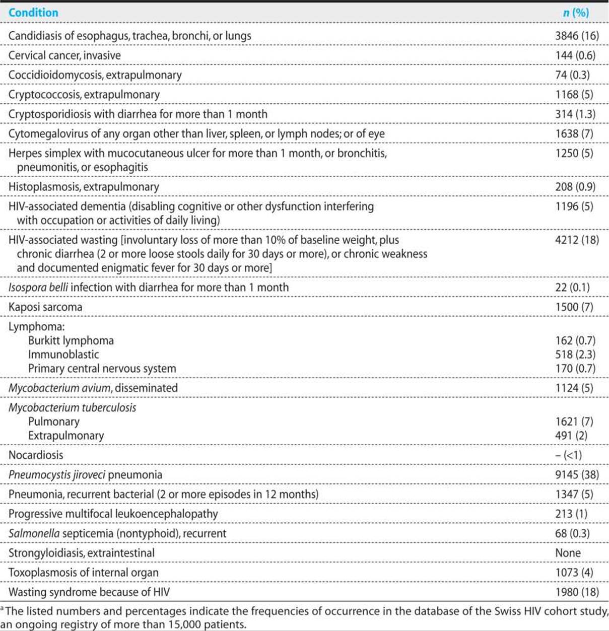
The CD4 cells are the conductors of the immunologic orchestra; they are critical for some of its most important functions, including development of specific CD8 T-cell cytotoxic responses and production of neutralizing antibodies. When the number of CD4 cells declines below a critical level of about 200/μm3, the “AIDS defining diseases” start to appear. The list of these diseases (see Table 16.1) is relatively short: Pneumocystis jiroveci pneumonia (PCP) greater than aspergillosis, Kaposi sarcoma, and lymphoma greater than tumors of other types. Some of these infections—for example, PCP—are highly suggestive of HIV infection; others—pneumococcal pneumonia, Candidastomatitis, and tuberculosis (TB)—also occur in patients with normal immune systems or with immune deficiencies caused by conditions other than AIDS. Most of the opportunistic diseases are caused by reactivation of latent pathogens such as herpes viruses [e.g., cerebral lymphoma resulting from Epstein–Barr virus, or retinitis from cytomegalovirus (CMV)], fungi (PCP), or bacteria (tuberculosis, TB). Other infections—such as salmonellosis or cryptococcosis—may be newly acquired.
Table 16.1. Indicator Conditions in the Case Definition of AIDS in Adultsa
As described earlier, the nascent HIV immune response controls the runaway viral proliferation observed during acute HIV infection. How can the eventual failure of this immune response be explained?
Despite thousands of papers written on the subject, a clear answer is not currently available. The long-term nonprogressors tend to have a vigorous HIV-specific cytotoxic immune response, but overlap with populations showing progression is considerable. The role of antibodies to HIV is also unclear; individual cases show no clear correlation between the existence of neutralizing antibodies and progression. Attention has recently shifted to the nonspecific components of the immune system such as natural killer cells and toll-like receptors. The extreme mutability of HIV leads to the emergence of HIV quasispecies that are no longer recognized by the immune response (the “immune escape phenomenon”). In addition, HIV preferentially infects proliferating lymphocytes, but the lymphocytes that proliferate in response to HIV infection are precisely those whose receptors recognize HIV-derived peptides. Infection of these lymphocytes eventually leads to their destruction.
Effective therapy reverses most of the immune deficiency. Given enough time, recovery of the CD4-cell count occurs even in patients who have practically no cells left when treatment starts. Cell counts continue to increase for several years, finally reaching a plateau of 500-1000/μm3. The immune response to the most important pathogens recovers, as can be seen by the disappearance of opportunistic diseases. But one exception remains: the immune response to HIV itself stays deficient even after successful treatment.
In North America and Western Europe, most patients come to medical attention during the latent or plateau period of chronic HIV infection, when clinical signs and symptoms are rare or absent. Nonetheless, the infection remains active, with the production of 109–1011 viral particles daily. At the same time, several billion CD4 cells are destroyed and replaced each day. Production of 1011 viral particles daily provides the potential for a mutation at every single nucleotide position. Unsurprisingly, under the selective pressure of a partially effective immune response or partially effective therapy, resistant mutations rapidly emerge. To obtain a durable antiviral effect, several drugs must be combined to completely abolish viral production. Once this goal is achieved, emergence of resistance becomes much less likely, and in those circumstances, patients may be treated for many years without viral breakthrough. Nonetheless, the virus persists in reservoirs that are not accessible to current treatment. These reservoirs may include nonproductive infection in pools of long-lived lymphocytes. Sensitive molecular techniques suggest that the half-life of this type of reservoir may reach several years, making eradication by continuous treatment unrealistic.
CLINICAL MANIFESTATIONS OF PRIMARY HIV INFECTION
The incubation period for symptomatic infection is 2–4 weeks, but can be as prolonged as 10 weeks. Onset of fever can be abrupt and is associated with diffuse lymphadenopathy and pharyngitis. The throat is usually erythematous, without exudates or enlarged tonsils. Painful ulcers can develop in the oral and genital mucosa (Figure 16.1). Gastrointestinal complaints are common, with many patients experiencing nausea, anorexia, and diarrhea. A skin rash often begins 2–3 days after the onset of fever and usually involves the face, neck, and upper torso. The lesions are small pink-to-red macules or maculopapules (Figure 16.1). Headache is another prominent symptom, and aseptic meningitis is noted in about one-quarter of patients. Headache is often retro-orbital and worsened by eye movement. Findings in the cerebrospinal fluid (CSF) are consistent with viral meningitis: lymphocytes, normal glucose, and mildly elevated protein. Guillain–Barré syndrome and palsy of the seventh cranial nerve have been reported. Peripheral leukocyte count may be normal or slightly below normal, with a decrease in CD4 lymphocytes and an increase in CD8 lymphocytes (the CD4:CD8 ratio is commonly below 1.0). Liver transaminase values may be moderately elevated. The illness is self-limiting, with severe symptoms usually resolving over 2 weeks. Lethargy and fatigue may persist for several months.
KEY POINTS
About the Clinical Manifestation of Primary HIV Infection
1. Abrupt onset of fever 2-4 weeks after exposure.
2. Accompanied by
a) nonexudative pharyngitis and lymphadenitis.
b) maculopapular skin rash on the head, neck, and upper torso.
c) headache and aseptic meningitis.
d) anorexia, nausea, diarrhea.
3. The acute illness lasts 2-6 weeks. Lethargy and fatigue can persist for several months.
LABORATORY EVALUATION OF HIV INFECTION
DIAGNOSIS
Infection with HIV is diagnosed by the detection of HIV-specific antibodies in plasma or serum. These antibodies appear a few weeks after infection, shortly before or after the symptoms of the acute retroviral syndrome. From studies in which the date of infection is precisely known (e.g., in individuals infected by a blood transfusion), the delay to the appearance of antibodies can be determined: about 5% of patients seroconvert within 7 days, 50% within 20 days, and more than 95% within 90 days. Therefore, a period exists (called the “window period”) during which, although the patient is infected, antibodies cannot be detected in the plasma. For a few days, the HIV-specific p24 antigen is detectable alone, without antibodies [Figure 16.1(A)]. Therefore, screening tests now combine the detection of antigen and antibody. Gene amplification tests [polymerase chain reaction (PCR), as well as other hybridization techniques] for the detection of viral genomes can also be used for early diagnosis, but they are much more expensive than the combined antigen-antibody tests. Antibody tests remain positive for the lifetime of HIV-infected people, except possibly in very rare cases when treatment was started before seroconversion.
Saliva can also be used for diagnosis. A saliva test (OraQuick®) is available for home testing. However, false negatives (infected persons with negative Ora-Quick test) may occur.
Antibody tests for HIV are among the most reliable of all medical tests in chronic HIV infection, with specificity and sensitivity largely exceeding 99% when done in competent laboratories. Nonetheless, in view of the importance of the diagnosis and the possibility of clerical errors (mislabeled tubes and such), confirmation of the diagnosis by a second blood sample is recommended. Confirmation is especially important when the pretest probability is low, raising the proportion of false positive results.
True false-positives are much rarer than “indeterminate” test results. An indeterminate result arises when substances in the patient’s plasma interact with impurities in the HIV antigen preparations. Usually, the color reaction of the enzyme-linked immunosorbent assay is above the threshold for positivity, but much below the results of a routine positive test. To diminish these indeterminate reactions, manufacturers are using recombinant technology to purify the HIV protein. In the presence of an indeterminate test, and particularly in the absence of risk factors for HIV infection, the patient should be reassured that the result is negative, with the negativity confirmed by a second test using a different method. Such a method might involve use of the Western blot. In a Western blot test, the HIV proteins are first separated by electrophoresis and then blotted onto a nitrocellulose membrane (Figure 16.1). This membrane is incubated with a dilution of the patient’s serum. Specific antibodies fix to the respective HIV proteins, producing a colored band after a coloring reaction. The position of the band permits a deduction concerning whether the reaction is nonspecific or the result of an HIV-specific protein.
KEY POINTS
About Diagnosis of HIV Infection
1. Diagnosis of HIV infection is made by measuring anti-HIV antibodies.
2. Following exposure, 5% of people seroconvert within 7 days, 50% within 20 days, and more than 95% within 90 days.
3. The “window” period of viremia with negative serology lasts from a few days to several weeks.
4. Tests based on combined antigen (p24) and antibody measurements are recommended for diagnosis.
5. Tests for HIV antibody are highly specific and sensitive.
6. An “indeterminate” test is usually a false positive; confirm by Western blot analysis.
KEY POINTS
About the Classification of HIV Infection
1. Classification is based on CD4 count and clinical symptoms.
2. The United States and Europe have different definitions of AIDS:
a) The United States uses a CD4 count below 200/μm3 or presence of an AIDS-defining illness.
b) Europe uses the presence of an AIDS-defining illness.
CLASSIFICATION
The stages of HIV infection are defined by clinical events and by CD4 lymphocyte count (Table 16.2). This classification, established in 1992, indicates clearly the immuno-suppression and symptomatic status of the patient.
Table 16.2. Stages of HIV Infection
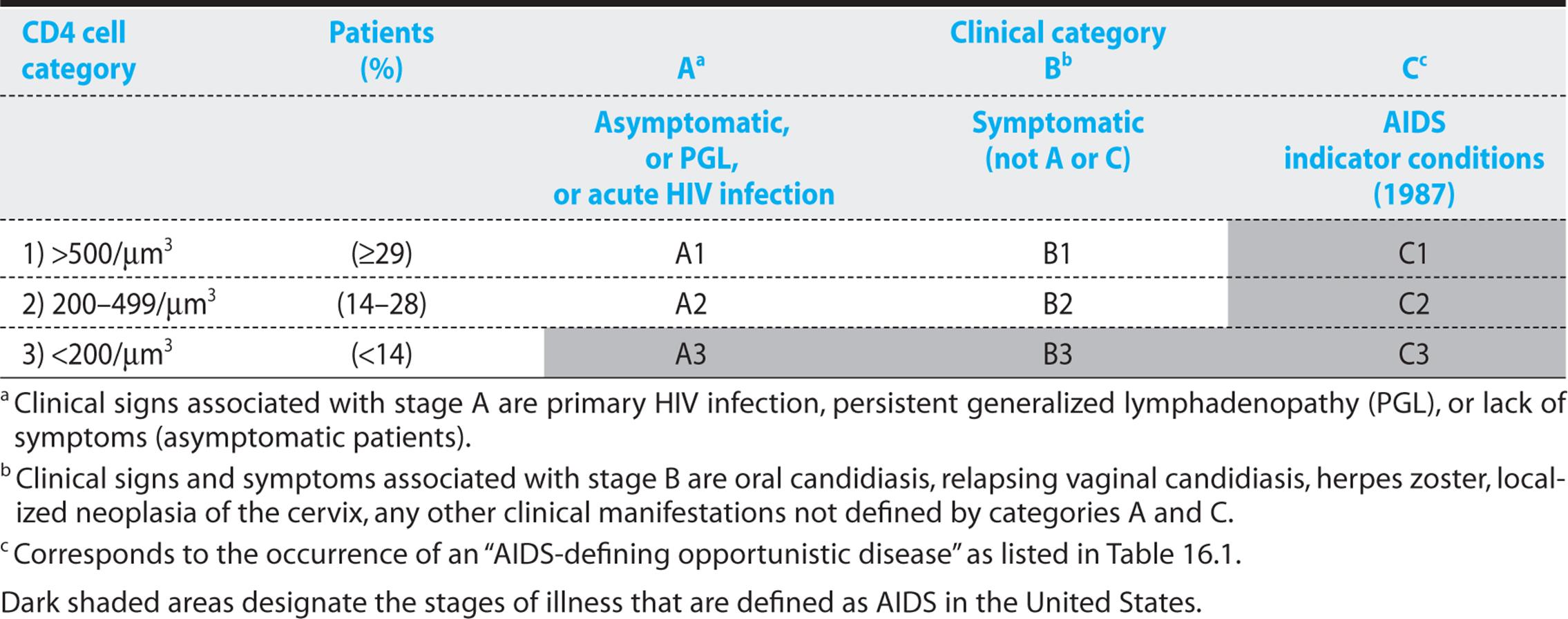
The meaning of the word “AIDS” is not the same on both sides of the Atlantic. In the United States, every person with a CD4 count below 200/μm3 is considered to have AIDS (shaded area in Table 16.2); alternatively, patients may be considered to have AIDS if they have an AIDS-defining opportunistic infection (Table 16.1). In Europe, the CD4 count does not enter into the definition of AIDS, which remains synonymous with the occurrence of an opportunistic disease as those defined in Table 16.1. The stage of HIV infection is defined by the CD4 lymphocyte count (biologic stage 1, 2, or 3) and clinical events (A, B, or C). Occurrence of a type C disease defines AIDS. In addition, in the United States, AIDS is also defined by a CD4 count below 200/μm3 (categories C1, C2, C3, A3, or B3).
TREATMENT AND PROGNOSIS
Monitoring Tests
Infection with HIV has been likened to a train speeding toward a wreck: the speed corresponds to the level of viremia, and the distance to the site of the wreck corresponds to the CD4 count.
To determine viral load, genomic tests are now almost universally used. These tests measure HIV genomes per cubic millimeter of plasma. Because the genomes consist of RNA, the RNA first has to be transcribed into DNA, which is then amplified, most often by the PCR. Patients with untreated HIV infection typically have 10,000–100,000 copies of HIV RNA per cubic millimeter; with treatment, this number declines to undetectability. Depending on the test being used, “undetectability” means fewer than 5–50 copies of HIV RNA. Ideally, after 2–6 months of treatment, all patients on modern ART should have fewer than 50 copies of HIV RNA per cubic millimeter.
Many studies have shown that the long-term prognosis for untreated HIV infection depends on the viremia. However, within this broad correlation, large inter-individual variations occur, with patients remaining in good health for many years despite viremia exceeding 100,000 copies per cubic millimeter.
For short-term prognosis, the CD4 count is more useful. The occurrence of opportunistic infections and tumors is unusual with CD4 counts above 200/μm3. Below this value, the incidence of such infections rises exponentially. It is very unusual for patients to die of AIDS with CD4 counts above 50/um3.
Antiretroviral Resistance Tests
Although antiretroviral combination therapy is effective in most patients, resistance may occur, and treatment may need to be adjusted. To guide the choice of therapy, tests measuring antiretroviral resistance have been developed.
KEY POINTS
About Tests for HIV Drug Resistance
1. Genotype testing detects specific mutations and is used to predict resistance.
2. Phenotype testing inserts viral genes into a standardized viral strain and measures sensitivities. It is time consuming and expensive.
3. Testing may allow ineffective drugs to be discontinued.
Two types of tests are currently in use:
1. Genotypic tests. Determine the sequence of the relevant viral genes: the reverse transcriptase, protease, and integrase genes. The sequence shows the presence or absence of mutations that are associated with antiretroviral resistance. However, with rare exceptions, the occurrence of a specific mutation does not predict a specific resistance phenotype. Rather, the combination of many mutations must be considered. A prediction of resistance from such a combination of mutations has been marketed as a “virtual phenotype.”
2. Phenotypic tests. Excise the relevant gene from amplified patient virus and insert the excised portion into a standard virus of known growth properties. This recombinant virus is then exposed to various drugs and its resistance is ascertained. Phenotypic tests are more expensive than genotypic tests, and they take 1–3 weeks to complete.
The value and use of resistance tests are subjects of continuing controversy. It has been difficult to show that they improve the outcome of treatment, but they allow ineffective drugs to be discontinued, thus sparing side effects and costs. The use of resistance testing is further discussed in the subsection on HIV therapy later in this chapter.
Caveats Regarding Laboratory Tests
Modern antiretroviral treatment would be impossible without the use of laboratory tests. However, physicians and patients need to be aware of the limits of the tests and, in particular, of the need to avoid over-interpretation of small changes. The precision of measurements of viral load is only about 0.3 log (a factor of 2). This means that values of 200 and 400 copies per cubic millimeter may actually be the same. Another problem with the interpretation of HIV viremia is the expression “undetectable” viremia. Detectability depends on the assay used. Experimental assays with sensitivities as low as 1 or 3 copies per cubic millimeter actually show viremia in almost all patients who have started their treatment during chronic HIV infection. Whether viremia that is very low (e.g., fewer than 10 copies per cubic millimeter) is better for the patient than viremia that is detectable but between 10 and 50 copies per cubic millimeter is unknown.
KEY POINTS
About Tests for Monitoring Treatment and Prognosis
1. Level of viremia correlates with speed of progression; copies of HIV RNA per cubic millimeter is usually measured by polymerase chain reaction.
2. The number of copies of HIV RNA per cubic millimeter varies from 500 to 1 million; treatment should reduce that number to below 50 within 6 months.
3. “Undetectable” levels of HIV RNA vary depending on the sensitivity of the test used; individual tests vary by a factor of 2.
4. A CD4 count below 200/μm3 puts the patient at risk of opportunistic infections and tumors.
5. The CD4 count varies by 10-30% between counts.
In patients with viremia that is low on treatment, some values may nonetheless exceed 50 or 100 copies from time to time. These “blips” of viremia are of no great prognostic significance, and they should not prompt a change in treatment. However, values that rise above 500 copies per cubic millimeter are clearly predictive of subsequent resistance and escape.
Similarly, the CD4 count is not a precise measure. It results from the multiplication of two percentages (the percentage of lymphocytes among leukocytes and the percentage of CD4-positive lymphocytes among all lymphocytes). The number of lymphocytes varies during the day, depending on food intake, physical activity, and steroid levels, among other factors. In addition, laboratories and lab technicians vary in their interpretation of the morphology of leukocytes. Therefore, CD4 counts may vary as much as 10–30% when counts are repeated at frequent intervals within the same individual.
MODERN ANTI-HIV THERAPY
INTRODUCTION
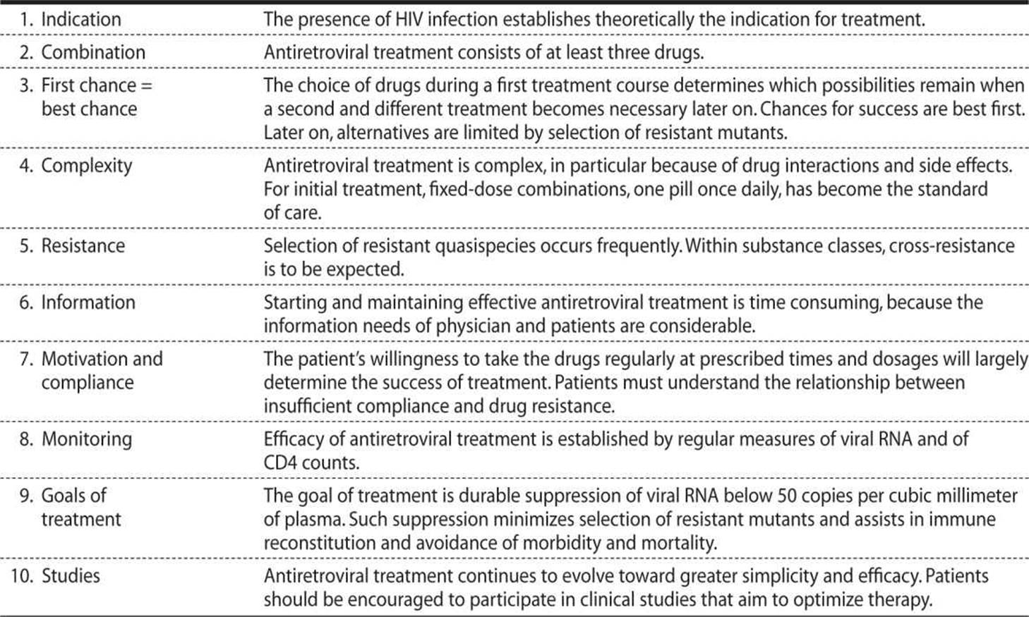
The Ten Principles of Antiretroviral Treatment
Since 1996, treatment with HAART, consisting usually of two nucleoside reverse-transcriptase inhibitors (NRTIs) plus an HIV protease inhibitor (PI), has been widely used. These regimens produced durable suppression of viral replication, with undetectable plasma levels of HIV RNA, in more than half of treated patients. Immunity recovered, and morbidity and mortality fell by more than 80%. Treatment was thought to be particularly effective when started early; HAART was therefore recommended for essentially all HIV-infected people willing to commit themselves to lifelong therapy.
But besides these successes, HAART also produced problems. Present-day drugs do not eradicate HIV, and often, patients cannot comply with long-term combination treatment. Moreover, HAART causes unexpected and ill-understood side effects. The dogma of earliest possible treatment therefore came under attack, and in the early 2000s, treatment indications were restricted. Since 2007, the pendulum has again swung toward more inclusive treatment indications, propelled by the improved tolerability of new drugs, evidence for harm caused by HIV even at high CD4 counts, and for HAART’s effectiveness in preventing new infections.
Table 16.3 summarizes the Ten Principles governing antiretroviral treatment. Starting and maintaining HAART is complex. Within the last few years, the numbers of antiretroviral agents, of their known and potential interactions with each other and with non-HIV drugs, and of their side effects, have all increased exponentially. Usually, a physician specializing in HIV care should be consulted whenever HAART is started or changed. It is this specialist’s job to guarantee that the treatment chosen is optimal for the particular patient. Mismanagement of ART can lead to untoward toxicities and the development of resistant viruses that can no longer be treated.
Table 16.3. Ten Principles for Highly Active Antiretroviral Therapy
CAVEATS ABOUT HAART
A physician specializing in HIV care should be consulted whenever highly active antiretroviral therapy (HAART) is started or changed.
Mismanagement of antiretroviral therapy can lead to untoward toxicities and resistant viruses.
Indications for Starting Treatment
The patient’s CD4 count indicates the degree of immune deficiency and predicts short-term risk of opportunistic disease. Without treatment, that risk is less than 1% per year when the CD4 count is above 500/mm3, but rises to 30% when the CD4 count falls below 100/μm3. In the long term, prognosis is also determined by the viral load—that is, the number of HIV RNA copies per cubic millimeter of plasma. An elevated viral load predicts a more rapid progression toward AIDS in population-based studies, although inter-individual variations are enormous. The destruction by HIV of CD4 cells and lymph node architecture causes progressive immunodeficiency. Antiretroviral treatment suppresses viral replication, prevents further destruction of the immune system, and even allows for considerable repair in patients who start treatment while already immunosuppressed.
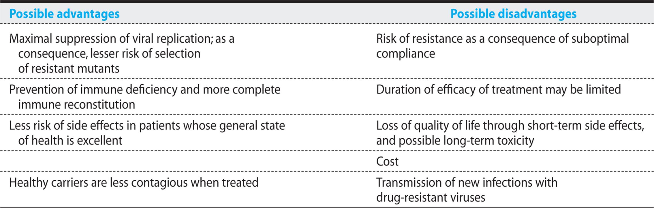
Although ART is recommended for all HIV-infected individuals, the strength of this recommendation varies on the basis of pretreatment CD4 cell count. Treatment must be adapted to the patient, taking into account the speed of progression, acceptance of treatment by the patient, the likelihood of compliance, and possible side effects. The recommendations presented in Table 16.4 are therefore only approximations, because individual factors, although often decisive, do not lend themselves to abstractions in a table. Table 16.5 outlines the advantages and disadvantages of an early start to treatment.
Table 16.4. Initiating Antiretroviral Therapy in Treatment-Naive Patients
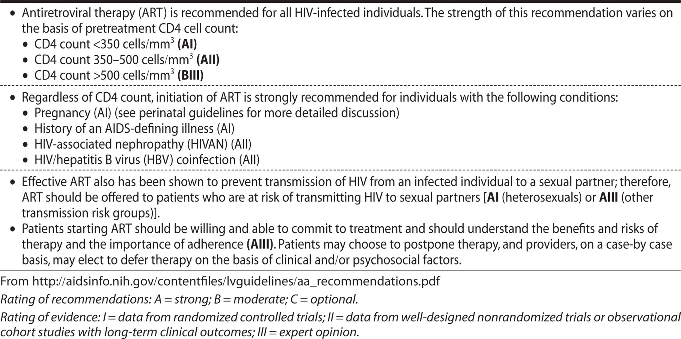
Table 16.5. Potential Advantages and Disadvantages of Early Antiretroviral Treatment
What to Treat With: Choice of an Initial Regimen
Six different classes of drugs are currently available and recommended dosages are provided in Table 16.6:
Table 16.6. Anti-HIV Drugs Available in 2012

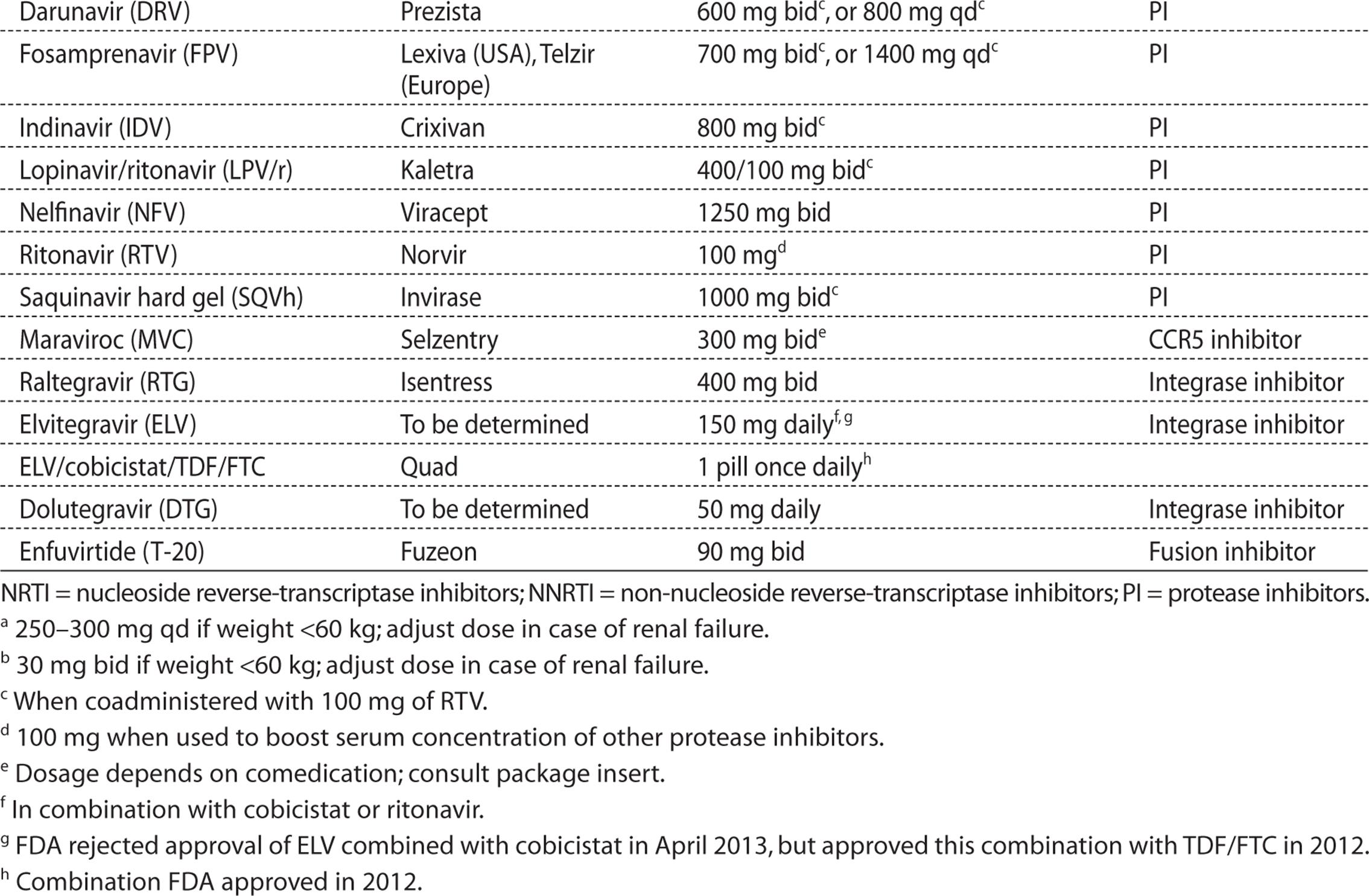
1. The NRTIs, such as abacavir (ABC), didanosine (ddI), emtricitabine (FTC), lamivudine (3TC), stavu-dine (d4T), tenofovir (TDF), and zidovudine (AZT).
2. The non-nucleoside reverse-transcriptase inhibitors (NNRTIs), such as efavirenz (EFV), nevirapine (NVP), etravirine (ETR), and rilpivirine (RPV).
3. The PIs, such as amprenavir (APV), darunavir (DRV), indinavir (IDV), lopinavir/ritonavir (LPV/r), nelfinavir (NFV), ritonavir (RTV), saquinavir (SQV), and tipranavir (TPV).
4. The integrase inhibitors raltegravir and elvitegravir. A third integrase inhibitor, dolutegravir, is expected to receive FDA approval in 2013.
5. The CCR5 inhibitor maraviroc.
6. The parenteral fusion inhibitor enfuvirtide (ENV).
Optimal suppression of viral replication requires a regimen to which HIV can only become resistant with multiple mutations. At the present time, no single drug (with the possible exception of the RTV-boosted PIs) fulfills this requirement. Combination therapy with three drugs is necessary. Choice of drugs is determined by several factors, including drug interactions, dosage intervals (i.e., the need to accommodate professional activity), future therapeutic options, or possible pregnancy.
Currently, no clear criteria are available to assist in making the choice between PIs, NNRTIs, and integrase inhibitors in initial treatment. Table 16.7 describes some advantages and disadvantages of the three classes of drugs.
Table 16.7. PIs Compared with NNRTIs in Initial Treatment, When Combined with NRTIs

The availability of so many drugs provides a bewildering array of possible choices. For antiretroviral naïve patients alone, a table prepared by a NIH consensus panel (http://aidsinfo.nih.gov/contentfiles/lvguidelines/aa_tables.pdf, Table 5a) offers four different “preferred regimens,” and eight “alternative regimens.” In our practice, our first choice is Atripla®, the combination of TDF, FTC, and EFV in one pill. Complera® (RPV/TDF/FTC) is an alternative in patients unable to tolerate EFV, if their viral load is below 100,000. Truvada® (FTC/TDF) plus raltegravir has the advantage of minimal drug interactions. PIs have a higher threshold for resistance development than other drug classes; we use RTV-boosted atazanavir or DRV in combination with Truvada. When Truvada is contraindicated, for example, because of preexisting kidney disease, it can be replaced by the combination of ABC and 3TC (Epzicom®in the USA, Kivexa® in Europe). ABC is liable to cause a dangerous hypersensitivity reaction with fever, rash, upper respiratory symptoms, and hypotension, but only in persons who have the HLA B5701 allele. Prior testing for HLA B5701 is recommended.
The following treatment options are suboptimal:
• Therapy with only one or two drugs.
• Combinations of AZT plus d4T (antagonism), or TDF plus ddI (dosage adjustment necessary because of an increase in the area-under-the-curve for ddI), d4T plus ddI (overlapping toxicity), ABC plus TDF (rapid emergence of mutants with the resistance mutation K65R).
• Use of PIs without concomitant RTV (insufficient drug levels). Atazanavir is an exception to this rule, and can be used without RTV at a dose of 400 mg/day. However, in combination with TDF, boosting with RTV is recommended.
KEY POINTS
About Monitoring Drug Toxicity
1. Follow-up consultations should be scheduled at 1 and 4 weeks after initiation of a new treatment.
2. If all goes well, the interval between visits may then lengthen to every 2-6 months.
3. Tests for surveillance of toxicity should include a complete blood count, liver enzymes, lactates, and serum cholesterol and triglycerides.
Monitoring Treatment
TOLERANCE AND SIDE EFFECTS
NRTIs, in particular the thymidine derivatives AZT and d4T, can be toxic to mitochondria, producing liver damage, lactic acidosis, lipoatrophy, and polyneuropathy. Some PIs cause nausea, vomiting, and diarrhea; elevate plasma cholesterol and triglycerides; induce insulin resistance and glucose intolerance; and contribute, together with AZT or d4T, to the redistribution of fatty tissue (atrophy in the face and extremities, contrasting with fat accumulation in breasts and abdomen). Treatment of dyslipidemia with statins is complicated by potential for drug interactions.
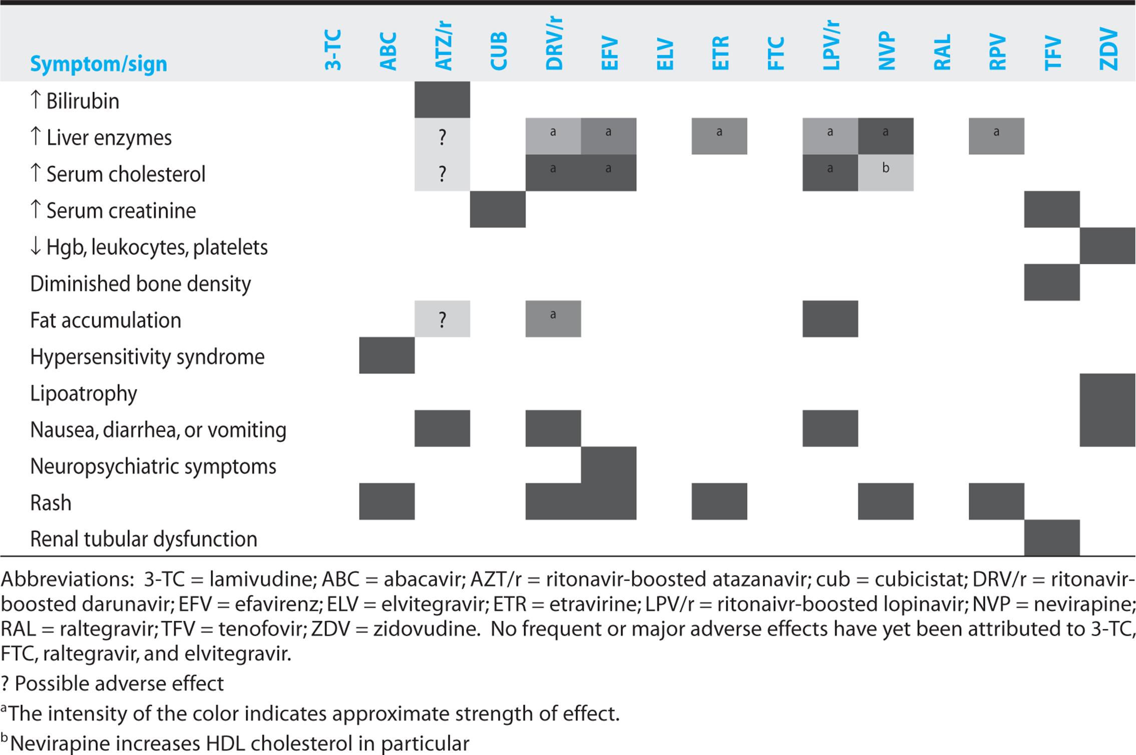
All drugs produce various specific side effects. Table 16.8 shows the main side effects of 15 most commonly used drugs. Because drugs have usually been tested in combination, assignment of a particular side effect to a particular drug is often uncertain; this situation is particularly true of the various aspects of the lipodystrophy syndrome. Lipoatrophy and lactic acidosis are more strongly associated with AZT, than with other NRTIs. The association with d4T was even stronger and has led to the near-disappearance of this drug.
Table 16.8. Major Adverse Effects Associated with Commonly Used ARVs
These potential side effects necessitate regular patient visits. One usual schedule requires a telephone consultation after 3 days and visits after 2 and 4 weeks of treatment; if all goes well, the interval between visits may then lengthen to every 2-6 months. For surveillance of toxicity, a complete blood count, liver enzymes, lactates, and serum cholesterol and triglycerides are useful.
DRUG INTERACTIONS
PIs and NNRTIs are preferentially metabolized by cytochrome P3A. The potential for drug interactions is therefore large. Drugs such as rifampicin or Hypericum (St. John wort) may lower PI and NNRTI concentrations by inducing cytochrome P3A.
RTV and cobicistat are extremely potent inhibitors of cytochrome P4503A. This effect is put to good use in HIV therapy to boost serum concentrations of atazanavir, DRV, LPV, and elvitegravir. However, concentrations of other drugs may be similarly boosted; for instance, in patients with transplants, dosages of tacrolimus and cyclosporin are 10-50 times lower when used with RTV, than when used without RTV. Toxicity may result: Examples include ergot alkaloids (dramatic cases of ergotism with amputation have been published) and many benzodiazepines. Hardly a week goes by without new interactions being reported; consultation of Web resources for up-to-date information is recommended. Among the best of the available sites are those produced by the Liverpool HIV Pharmacology Group of the University of Liverpool (www.hiv-druginteractions.org, also offers an excellent app for smartphones), the electronic journal Medscape (http://medscape.com/hiv), and the NIH (http://aidsinfo.nih.gov/guidelines).
UTILITY OF RITONAVIR
Ritonavir is the most powerful inhibitor of cytochrome P3A known in medical therapeutics. It can be used to boost plasma levels of other protease inhibitors.
COMPLIANCE
Patients must acquire an adequate understanding of HIV pathogenesis, the goals of HIV treatment, and pharmacokinetics. They should be able to recognize the most frequent side effects and know how to manage them.
Aids to improve compliance abound, although few have been tested rigorously. Pillboxes are popular; these contain all the drugs taken during 1 week in separate compartments. The establishment of a detailed written schedule, showing how and when to take prescribed drugs in relation to meals and drinks, is recommended. More elaborate and expensive procedures involve use of electronic pillboxes, involving a device that records each time a bottle cap is unscrewed; the information can be downloaded into a computer and discussed with the patient. Directly observed therapy is possible with once-daily regimens; this approach may be particularly appropriate in combination with methadone maintenance.
EFFICACY
Viral suppression as measured by decline in the viral load, a rise in the CD4 count, and clinical efficacy are all closely related. Above approximately 50 copies per cubic millimeter, the nadir of viral load reached through treatment predicts duration of viral suppression. Time to optimal viral suppression depends on the initial viral load and the sensitivity of the viral load test. Combination treatment must produce a rapid fall in viral load, which should drop to fewer than 400 copies per cubic millimeter after 12 weeks and to fewer than 50 copies after 24 weeks. Measurements of viral load and CD4 count are recommended every 3-6 months.
TREATMENT RESULTS MUST MEET EXPECTATIONS
Viral load should drop to 400 or fewer copies per cubic millimeter after 12 weeks, and 50 or fewer copies after 24 weeks.
RESISTANCE TESTS
Suboptimal treatment, lack of compliance, insufficient bioavailability, or drug interactions can result in prolonged periods of low blood and tissue drug concentrations with continued viral replication and selection of resistant mutants. The presence of resistance genotypes and phenotypes can be detected using commercially available methods. Studies show that these tests are useful mainly for excluding drugs to which the virus is resistant; they are less helpful for finding drugs to which the virus is sensitive. Resistance tests are recommended in patients who are yet untreated, but who have likely been infected since 1997, because they may harbor a primarily resistant HIV variant. Resistance tests are also recommended after early treatment failure.
KEY POINTS
About Resistance Testing
1. Resistance tests are useful mainly for excluding ineffective drugs.
2. Resistance tests should be ordered before treatment commences in patients who are likely to have been infected in 1997 or later.
MEASUREMENT OF PLASMA DRUG CONCENTRATIONS
In prospective studies, trough concentrations of PIs correlated well with degree and duration of viral suppression. However, the utility of these measures in clinical practice is not established. They are recommended in cases of unexpected toxicity, suspected problems with compliance that cannot be otherwise investigated, or when multiple medications may produce unforeseeable pharmacokinetic interactions.
Treatment Modification and Simplification
Once-a-day treatment with one pill has become the standard for initial treatment. Many patients started treatment years ago with more complicated drug regimens. Despite the common suggestion to “never change a winning team,” there is no objection to simplification, provided that there is no evidence for pretreatment resistance to the new drug combination.
Scheduled treatment interruptions have been evaluated in clinical trials, the largest of which (the SMART trial) showed a 1.6% per year increase in AIDS and death among those who interrupted treatment, compared with those who continued therapy. These AIDS/death events were more frequent in those with lower CD4 counts. When treatment is interrupted because of patient preference, it would seem prudent to monitor the CD4 count and start treatment again before the CD4 count falls below 350 per cubic millimeter.
Procedures in Case of Failure
Treatment must often be changed because of intolerance, drug interactions, or side effects. If viremia is below 50 copies per cubic millimeter, a single offending drug can be replaced. In cases of lipodystrophy replacement of d4T, ddI or AZT with TDF or ABC may be helpful: however, patience is necessary, as an increase in limb fat usually takes over 6 months. Virologic failure—that is, viremia—that does not decline to fewer than 50 copies per cubic millimeter after 6 months (9 months if the initial viremia exceeded 1 million copies per cubic millimeter) or that rises to more than 200 copies requires a different approach. In this situation, a new combination should be chosen, containing (if possible) a drug from a class that had not already been used. At least one additional drug should also be replaced by another to which the patient is unlikely to be resistant, given personal medication history and resistance tests.
With the arrival of DRV, ETR, and raltegravir in 2006-2008, patients with intractable resistance have almost disappeared.
KEY POINTS
About Failing Regimens
1. A new combination should be chosen, containing (if possible) a drug from a class that has not already been used.
2. At least one additional drug should also be replaced by another to which the patient is unlikely to be resistant.
Start and End of Prophylaxis for Opportunistic Infections
Efficacious antiretroviral treatment—provided that it is started in time—prevents immune deficiency and obviates the need for prophylaxis of opportunistic infections. Even if started late, effective HAART is followed by immune reconstitution. Prophylaxis of opportunistic infections can be discontinued after the patient’s CD4 count has risen above a given level for at least 3 months. This level is 100/μm3 for stopping prophylaxis of CMV and nontuberculous mycobacteria, and 200/μm3 for stopping prophylaxis of PCP and Toxoplasma encephalitis.
CONCLUSIONS AND OUTLOOK
Once a death sentence, HIV infection has become a chronic condition treated with one pill a day. Chances for success are best in the previously untreated; therefore, every effort must be made to optimize the first treatment given. A specialist should be consulted when starting or changing antiretroviral treatment. Compliance remains essential for treatment success; drugs must be taken as prescribed. In asymptomatic patients with CD4 counts above 350/μm3, it is better to abstain than to risk failure through insufficient treatment. Talking reluctant patients into accepting drugs makes no sense; refusal of HAART must be respected.
Treatments continue to evolve. Three once-a-day one pill combinations (Atripla®, Complera®, and Quad®) are available; a fourth, containing dolutegravir, is presently undergoing expedited FDA review. Further on, replacement of TDF with a prodrug that is effective at much lower doses may address the issues of potential long-term bone and renal toxicity. Ongoing trials will answer the question whether NRTI-free regimens offer any advantages.
Patients often ask whether there is hope for a “cure,” that is, discontinuation of drugs without relapse of viremia. This is a hot topic for scientific meetings, but unfortunately without any solid leads at the present time. Indefinite maintenance treatment, using drugs that are well tolerated and that remain effective, is a more realistic outlook.
RESPECTING PATIENT CHOICE
It makes no sense to talk reluctant patients into accepting drugs; refusal of HAART must be respected.
OPPORTUNISTIC DISEASES
CASE 16.1
A 28-year-old man was admitted to the hospital with a 3-week history of progressive shortness of breath accompanied by a nonproductive cough. Two weeks earlier, he had been seen by his local doctor for the same complaints and had been given an oral antibiotic at that time. He noted no improvement in his symptoms.
An epidemiologic history noted that the patient reported multiple episodes of unprotected homosexual intercourse 3 years earlier, but several months’ abstinence recently. The patient denied intravenous drug use and said that he had never smoked cigarettes. This was an anxious-appearing man who was short of breath.
On physical examination, a temperature of 38.2° C, a pulse of 120 per minute, a blood pressure of 110/60 mmHg, and a respiratory rate of 34 per minute were recorded. No lymphadenopathy was evident, but white plaques consistent with thrush were seen on the posterior pharynx. Breath sounds were clear, with no rales or rhonchi. A II/VI systolic ejection murmur was noted, but no rubs or gallops. No organomegaly was evident on abdominal exam, and the genitalia was within normal limits. Skin was clear, and no edema of the extremities was noted.
On laboratory workup, arterial blood gasses measured pH 7.44, PaCO2 32 mmHg, PaO2 62 mmHg, HCO3 20 mEq/L on room air. Chest X-ray revealed bilateral, interstitial, diffuse, fluffy infiltrates forming a butterfly pattern. Bronchial lavage with Giemsa stain revealed Pneumocystis jiroveci (Figure 16.2).

Figure 16.2. Pneumocystis jiroveci pneumonia (from www.aids-images.ch). A. Chest radiograph shows symmetric infiltrates of the lower lobes similar in appearance to pulmonary edema. B. Repeated chest radiograph after 11 days of treatment. C. Sample of bronchoalveolar lavage stained with toluene blue, showing multiple organisms.
The patient was started on intravenous methylprednisolone and trimethoprim–sulfamethoxazole. His shortness of breath gradually improved over the next 3 days, and he was discharged on oral trimethoprimsulfamethoxazole.
An antibody test for HIV was positive, subsequently confirmed by Western blot. The patient’s CD4 count was 50/mm3.
Case 16.1 represents a typical example of a primary episode of symptomatic AIDS. Opportunistic infections typically represent reactivation of latent infection or acquisition of a new infection, often caused by microorganisms of intrinsically low virulence. Toxoplasma gondii, CMV, viruses of the herpes group, PCP, papovavirus John Cunningham (JC) (the agent of progressive multifocal leukoencephalopathy), and Mycobacterium tuberculosis have usually been acquired years before, but lie dormant as long as the immune response is intact. Once immune deficiency is profound, these microorganisms may start to proliferate. Some of these agents can be isolated long before clinical signs appear. Progressively, however, organ damage and symptoms occur—for example, stomatitis and symptomatic esophagitis from Candida albicans.
In advanced stages of immune suppression, agents that are usually nonpathogenic can have devastating consequences. Examples include destruction of the retina by CMV, or cachexia caused by Mycobacterium avium, present in the blood of 25% of patients with CD4 counts below 10/μm3. Several infections can be present at the same time, greatly complicating diagnosis and treatment.
Even before HAART, the prevention of opportunistic infections by antibiotics and antiretrovirals prolonged survival and improved quality of life. Since 1996, HAART has made an enormous difference. Advanced stages of AIDS with chronic diarrhea, cachexia, and central nervous system (CNS) and pulmonary manifestations have become rare in the United States and Western Europe.
KEY POINTS
About Prophylaxis
1. Latent infections often reactivate as cell-mediated immunity wanes.
2. Use serologic and skin testing to detect latent infections on initial evaluation.
3. Prophylaxis is recommended for a CD4 count below 200/μm3.
4. After treatment of active infections, secondary prophylaxis is often necessary to prevent relapse.
5. Prophylaxis can be discontinued after highly active antiretroviral therapy has been instituted, when the CD4 count is durably above 200/μm3.
PRIMARY AND SECONDARY PROPHYLAXIS
Primary prophylaxis prevents the first occurrence of a disease; secondary prophylaxis prevents relapses after a first episode. In AIDS, many opportunistic infections can be prevented. Patients who should receive prophylaxis are identified by their CD4 lymphocyte count and by serologic tests with evidence of previous exposure to the infectious agent. For instance, the presence of immunoglobulin G (IgG) against T. gondii in a patient whose CD4 count is below 100/μm3 identifies a high risk of cerebral toxoplasmosis. Regular measures of the CD4 count, combined with serologic tests on initial evaluation, are necessary for a timely start to prophylaxis.
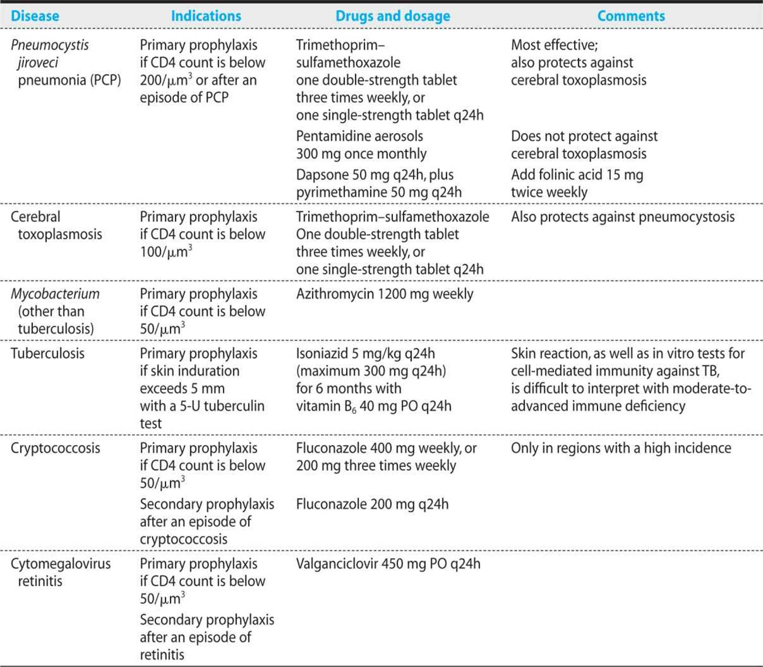
Opportunistic infections have a tendency to relapse. Therefore, as long as the underlying immune deficiency is not corrected, secondary prevention is necessary. Of course, preventive therapy has risks and side effects such as allergies, drug interactions, and development of resistance, but the risk–benefit ratio has been proven to be favorable, especially for prevention of PCP and cerebral toxoplasmosis by trimethoprim–sulfamethoxazole. However, once ART is efficacious and the patient’s CD4 count has risen durably above 200/μm3, these preventive measures can be discontinued. Table 16.9 summarizes the common preventive regimens.
Table 16.9. Prophylaxis of Oopportunistic Infections
PULMONARY INFECTIONS
The differential diagnosis of pulmonary disease in HIV-infected patients depends on the patient’s epidemiologic history (presence of intravenous drug abuse, previous episodes of bacterial pneumonia, exposure to TB), CD4 lymphocyte count, and use of preventive therapy (see Table 16.9).
During the early years of the AIDS epidemic, PCP was the initial opportunistic infection in one-third of cases. The infection remains frequent, but its incidence has greatly decreased because of the use of trimethoprim–sulfamethoxazole and HAART. Bacterial pneumonia, in particular that caused by Streptococcus pneumoniae, is 10-100 times more frequent in HIV-positive than in HIV-negative patients. TB can occur at any degree of immune deficiency, but it is particularly frequent in patients who grew up in developing countries.
With a lobar infiltrate in a patient with a CD4 count above 200/μm3, the presumptive diagnosis is bacterial pneumonia. Empiric treatment should start with amoxicillin–clavulanate, a cephalosporin, or one of the quinolones with activity against gram-positive bacteria. If immune deficiency is more profound (CD4 count is below 200/μm3), PCP is most likely, except if the patient has faithfully taken trimethoprim–sulfamethoxazole prophylaxis. The chest X-ray pattern is helpful in narrowing the diagnostic possibilities (see Table 16.10). However, in all patients, whatever their degree of immune suppression, a definitive diagnosis usually requires bronchoalveolar lavage.
Table 16.10. Lung Diseases Linked to HIV

Pneumocystis jiroveci Pneumonia
DIAGNOSIS AND TREATMENT
As illustrated in case 16.1, PCP is a subacute disease. With rare exceptions, its occurrence is limited to immunosuppressed patients with a CD4 count below 200/μm3. Symptoms originate in the respiratory tract (dry cough, dyspnea) and are accompanied by fever (always), weight loss, and fatigue. A prominent symptom is dyspnea on exertion. Initially, patients experience shortness of breath with exercise, but do not complain of shortness of breath at rest. Alveolar fluid accumulation associated with Pneumocystis infection interferes with oxygen exchange, and patients quickly outstrip the ability of their lungs to supply arterial oxygen.
Lung auscultation is usually normal. Chest radiographs, which can be normal, typically show a reticulonodular bilateral infiltrate that can be asymmetrical (see Table 16.11 and Figure 16.2). Classically, the infiltrates form a butterfly pattern, mimicking pulmonary edema associated with left-sided congestive heart failure. Occasionally, a standard chest X-ray shows cystic lesions or a pneumothorax. When PCP prophylaxis is delivered by pentamidine inhalation, the chest X-ray is often atypical, with asymmetrical infiltrates limited to the lung apex. Tests of the peripheral blood are usually nonspecific, but lactate dehydrogenase (LDH) is found to be elevated in more than 90% of patients with Pneumocystis infection. High values and a persistent elevation despite appropriate therapy are associated with a poor prognosis.
Table 16.11. Chest Radiograph Results and Possible Causes

The diagnosis of PCP is established by special stains of bronchoalveolar lavage fluid or of sputum induced by a 30-minute inhalation of 3% NaCl. If clinical suspicion of PCP is high, starting treatment before confirmation of the diagnosis is recommended, because PCP can still be found in lavage fluid 1-3 days later. In rare cases, the diagnosis may necessitate a transbronchial biopsy—particularly if pentamidine inhalations have been used.
KEY POINTS
About the Clinical Manifestations, Diagnosis, and Treatment of Pneumocystis jiroveci Pneumonia
1. Pneumocystis jiroveci pneumonia (PCP) is a subacute disease that develops in HIV-infected patients with a CD4 count below 200/μm3.
2. Primary symptoms are fever, dyspnea on exertion, dry cough, weight loss, and fatigue.
3. Pulmonary exam is usually normal.
4. Chest X-ray may be normal, but usually demonstrates an interstitial butterfly pattern.
5. Lactate dehydrogenase is usually elevated, and PaO2 depressed.
6. Trimethoprim–sulfamethoxazole is the drug of choice for treatment.
7. If PaO2 is below 70 mmHg, give prednisone before anti-PCP therapy.
Treatment modality will depend on the gravity of PCP. Patients who are very short of breath, with a PaO2 of less than 70 mmHg, particularly if accompanied by nausea or vomiting, will usually be admitted to hospital and treated intravenously. If signs of grave disease are absent, and if the patient is not nauseated, outpatient treatment is possible. The drug of choice is high-dose trimethoprim–sulfamethoxazole, two double-strength tablets (sulfamethoxazole 1600 mg and trimethoprim 320 mg every 8 hours for 21 days), followed by secondary prophylaxis with sulfamethoxazole 400 mg and trimethoprim 80 mg daily until the patient’s CD4 count durably exceeds 200/μm3.
Trimethoprim–sulfamethoxazole has numerous side effects, of which drug rash is the most frequent. If the skin lesions are extensive (and, in particular, if mucosal involvement is evident), if leukopenia and thrombocytopenia are severe, or if renal or hepatic toxicity or serious vomiting occurs, alternative treatment is necessary. In an attempt to reduce the incidence of bone marrow suppression, folinic acid has been added to the treatment regimen; however, it diminishes the efficacy of treatment and is not recommended. Many alternatives to trimethoprim–sulfamethoxazole are available, but their efficacy is, in general, inferior, and many have other serious side effects. Table 16.12summarizes the alternatives.
Table 16.12. Treatment of Pneumocystis jiroveci Pneumonia: trimethoprim–sulfamethoxazole and alternatives
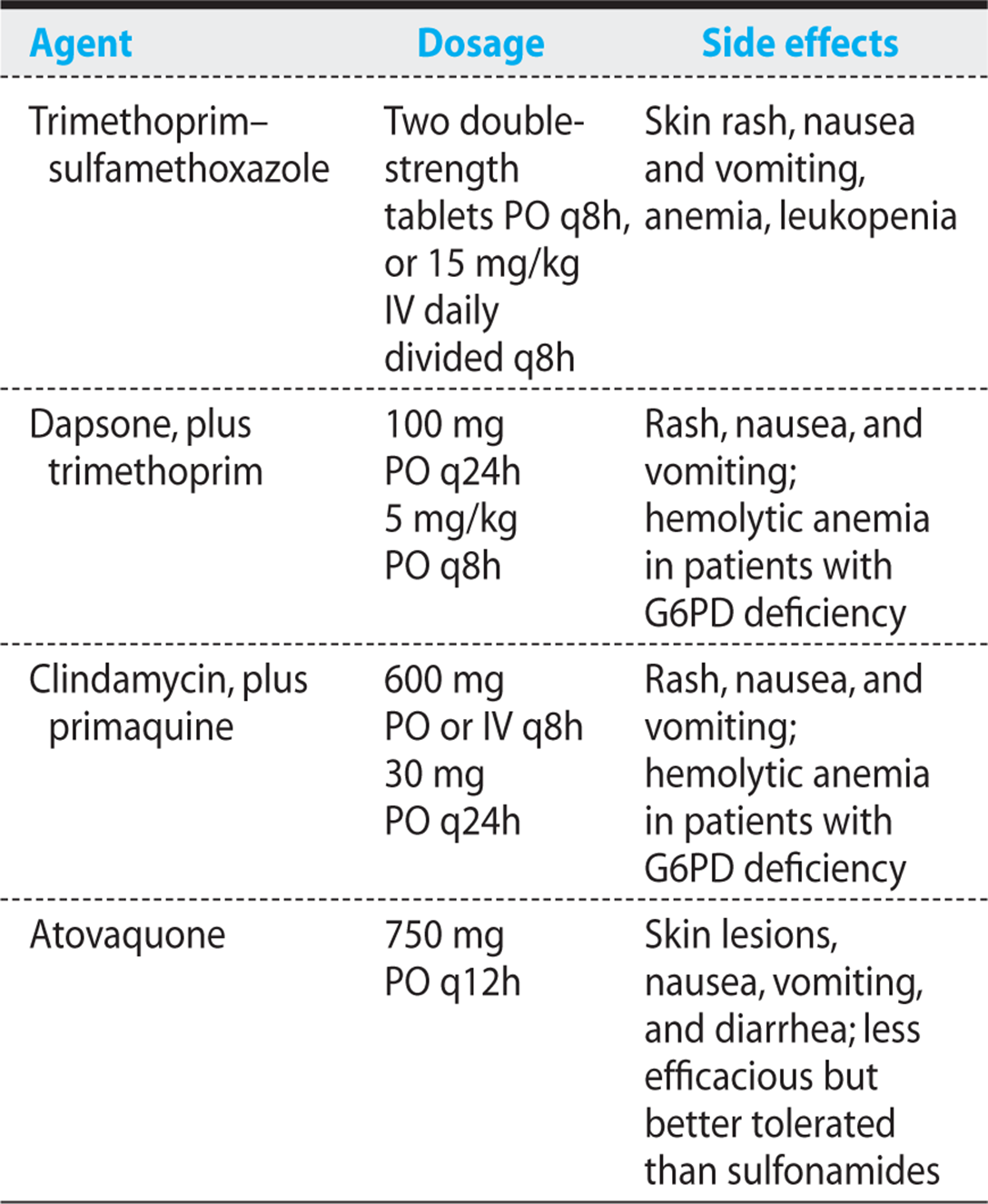
At the start of the AIDS era, patients with Pneumocystis, even if correctly treated, often experienced increased respiratory distress and worsening lung infiltrates during the first few days. In many cases, this initial deterioration necessitated intubation or caused death. Severe respiratory compromise that necessitates intubation can be prevented by giving steroids (prednisone 40 mg q 12 h for 5 days, then 40 mg daily for 5 days, followed by 20 mg daily for 11 days) in cases of severe pneumocystosis with a PaO2 below 70 mmHg. Prednisone should be given before or simultaneously with initiation of anti-Pneumocystis therapy.
KEY POINTS
About Prophylaxis of Pneumocystis jiroveci Pneumonia
1. In HIV patients with a CD4 count below 200/μm3 not on prophylaxis, the annual incidence of Pneumocystis jirovecii pneumonia (PCP) is 20%.
2. Trimethoprim–sulfamethoxazole is the drug of choice: efficacious, inexpensive, and equally active in preventing toxoplasmosis.
3. Alternatives are not as effective:
a) Dapsone does not cover toxoplasmosis; pyrimethamine must be added.
b) Pentamidine is associated with cough and asthma.
c) Atovaquone is expensive.
PREVENTION
In patients with HIV with CD4 counts below 200/μm3, the annual risk of PCP is roughly 20%. The risk of relapse after a first episode is even higher: 40% after 6 months. Primary prophylaxis diminishes the risk of Pneumocystis, but if severe immunosuppression persists without HAART, the risk is still 19% after 3 years of prophylaxis with trimethoprim–sulfamethoxazole, and 33% after 3 years of pentamidine aerosols. Primary and secondary prophylaxis strategies use the same treatment options:
• Trimethoprim–sulfamethoxazole one double-strength tablet three times weekly, or one single–strength tablet daily. Trimethoprim-sulfamethoxazole has the advantages of great efficacy, protection against cerebral toxoplasmosis, and low price. However, almost 50% of patients will develop signs of cutaneous intolerance. Desensitization permits readministration in most cases, but desensitization has been used mostly in cases of treatment, when alternatives to agents are clearly less satisfactory. The mechanisms of trimethoprim–sulfamethoxazole intolerance are not well understood. Dose dependency is one of the features that argues against “allergy.” Another is the observation that up to 60% of patients who have shown cutaneous intolerance do not relapse when reexposed.
• Dapsone, 100 mg daily, does not protect against cerebral toxoplasmosis. If anti-Toxoplasma IgG antibodies are present, add pyrimethamine to dapsone. Daily (dapsone 50 mg, plus pyrimethamine 50 mg) and weekly schedules (dapsone 200 mg, plus pyrimethamine 75 mg) are equivalent.
• Pentamidine by inhalation (Respirgard nebulizer), 300 mg every 4 weeks. Some patients, particularly smokers, cannot tolerate inhaled pentamidine because of cough and asthma. Preventive use of a bronchodilator may be helpful.
• Atovaquone 750 mg divided into two daily doses. Well tolerated, but expensive.
Bacterial Pneumonia
As a complication of HIV infection, bacterial pneumonia produces the same symptoms and signs as pneumonias in HIV-negative patients: sudden onset of fever, chills, cough, and dyspnea. By far, the most frequent cause is S. pneumoniae, but Haemophilus influenzae (particularly in smokers), Staphylococcus aureus, Pseudomonas aeruginosa, and Rhodococcus equi may also be implicated. Bacteremia and relapses are frequent. Empiric treatment consists of amoxicillin–clavulanate, or a second- or third-generation cephalosporin; treatment duration is 10-14 days (see Chapter 4).
Tuberculosis
TB usually presents as a subacute disease with weight loss, cough, fever, night sweats, and lung lesions. However, if immune suppression is very advanced, the chest X-ray may be atypical for the disease. Interstitial infiltrates may predominate, without cavitary lesions; CNS TB becomes more frequent; mediastinal adenopathy is evident on the chest X-ray; and blood cultures are often positive. Diagnosis relies on acid-fast stain of the sputum; however, this test is frequently negative in disseminated (miliary) TB. For culture, liquid media are recommended because results are more rapid: growth is usually evident by 10-14 days, and presumptive identification of Mycobacterium can be made by nucleic acid probes.
In 72% of smear-negative cases of TB, an improved system using PCR (Xpert MTB/RIF) has detected mycobacterial DNA within hours. Xpert can also detect rifampicin resistance and therefore provides early guidance to appropriate treatment.
KEY POINTS
About Mycobacterium tuberculosis in AIDS
1. Tuberculosis (TB) is usually a subacute disease with weight loss, cough, fever, night sweats, and lung lesions.
2. With severe immunosuppression, TB can present as miliary disease:
a) Interstitial involvement
b) Meningitis
c) Negative sputum smears for acid-fast bacilli, but positive blood cultures
3. Susceptibility testing is critical: multiresistant TB is associated with >50% and extensively resistant TB with a near 100% mortality in AIDS.
4. Four-drug therapy: isoniazid, rifampin, pyrazinamide, and ethambutol.
5. Delay HAART only if immunosuppression is not severe (CD4 counts >200).
Susceptibility testing should always be done, because multidrug-resistant tuberculosis (MDR-TB) is a serious threat to an HIV-positive individual, with mortality exceeding 50%, and extensively resistant tuberculosis (XDR-TB) is associated with a near 100% mortality in patients with HIV. Initial treatment should include four drugs: oral isoniazid 300 mg daily (plus vitamin B6), rifampicin 600 mg daily, pyrazinamide 20-30 mg/kg daily, and ethambutol 15 mg/kg daily. This quadruple therapy should be continued during the first 2 months, followed by isoniazid and rifampicin for a further 7 months. Patients respond well to classic antituberculous treatment, but without HAART and reversal of the underlying immune deficiency, a high risk remains of persistent disease and death as a consequence of other complications of AIDS. In cases of isoniazid or rifampicin resistance (or both), consultation with a specialist is advised. Alternative drugs include moxifloxacin, amikacin, linezolid, and the investigational agents TMC-207, OPC-67683 (Delamanid), and SQ 109.
The coadministration of HAART and treatment for TB is a particular problem: on the one hand, PIs and rifampicin mutually modify one another’s plasma levels; on the other hand, concomitant administration of seven or more drugs may be toxic to the liver and gut. In addition, immune reconstitution disease caused by HAART is difficult to distinguish from paradoxical inflammatory reactions that are sometimes observed at the start of anti-TB treatment. If immune suppression is not advanced, it is often more reasonable to postpone HAART for a few months while anti-TB drugs take effect. However, in patients with CD4 counts below 50, early combined treatment against both HIV and TB can be lifesaving.
Mycobacterium Kansasii
In HIV-positive patients, M. kansasii causes a disease resembling classical TB with fever, cough, weight loss, and pulmonary infiltrates predominating at the apex. Very occasionally, apical cavities are observed. Classical antituberculous drugs such as isoniazid, rifampicin, and ethambutol are efficacious.
Mycobacteria Other Than Tuberculosis
Mycobacterium avium intracellulare (and similar mycobacteria) do not usually cause pulmonary disease, but rather a systemic illness with fever, weight loss, night sweats, and liver involvement. However, mycobacteria other than tuberculosis (MOTT) are frequently found in sputum, where their pathogenic significance remains uncertain.
Pulmonary Kaposi Sarcoma
In patients with obvious cutaneous Kaposi sarcoma, involvement of the mucosal surfaces is frequent (30-50% of cases) and, in general, asymptomatic. When lung is involved, the chest X-ray shows reticulonodular infiltrates with a perihilar distribution, hilar lymphadenopathy, and, occasionally, pleural effusions [Figure 16.3(D)]. Treatment with radiotherapy or chemotherapy is indicated for relief of cough or dyspnea. In general, lung lesions, like other manifestations of Kaposi sarcoma, improve on antiretroviral combination therapy.
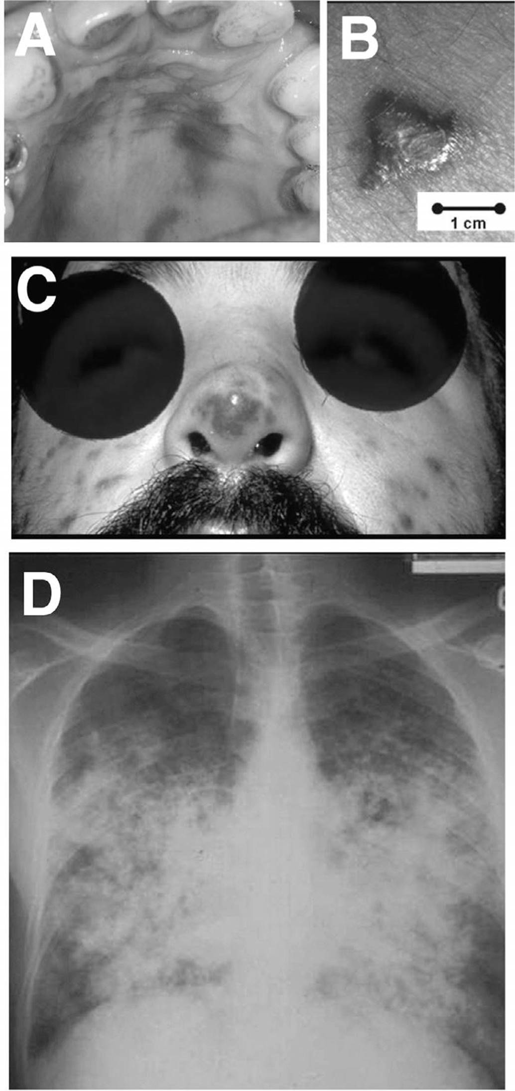
Figure 16.3. Kaposi sarcoma (from www.aids-images.ch). A. Macular lesions on the palate. B. Tumor-like skin lesion. C. Facial lesions including on tip of the nose (picture courtesy of J. Sampson). D. Typical chest radiograph; note the central nodular densities, with peripheral extension. See color image on color plate 4.
Other Rare Pulmonary Diseases
INTERSTITIAL LYMPHOID PNEUMONIA
Interstitial lymphoid pneumonia is usually diagnosed by exclusion. It is particularly frequent in children and presents with fever and dyspnea. The chest X-ray shows reticulonodular infiltrates that may vary and disappear spontaneously. Pathogenesis is not clear; HIV itself may perhaps be implicated. Treatment relies on corticosteroids.
HISTOPLASMOSIS
In contrast to the localized pulmonary disease observed in immunocompetent populations (see Chapter 4), histoplas-mosis in AIDS is often disseminated and accompanied by anemia, enlargement of liver and spleen, and positive blood cultures. Gastrointestinal involvement with ulcers, skin lesions, and lymphadenopathies are also frequent. The diagnosis is established by blood or bone marrow culture. Treatment relies on amphotericin B or fluconazole.
COCCIDIOMYCOSIS
Coccidiomycosis is restricted to the southwestern United States and Central America. Symptoms are fever, cough, and reticulonodular infiltrates. Diagnosis relies on culture of sputum or bronchoalveolar lavage fluid. Treatment is by amphotericin B (0.5-1 mg/kg daily) or fluconazole (400-800 mg daily).
DISSEMINATED TOXOPLASMOSIS
Rarely, and only in the presence of extreme immuno-suppression (CD4 count below 20), T. gondii can cause a devastating disseminated disease, with prominent lung involvement. Typically, the LDH is extremely elevated. Toxoplasma organisms can be seen in the bronchoalveo-lar lavage. This form of toxoplasmosis is treated like cerebral toxoplasmosis.
NOCARDIA ASTEROIDES
N. asteroides is a cause of chronic pneumonia and nodular pulmonary lesions. Other organs than the lung, such as the kidney and the brain, can be involved. The disease is diagnosed by direct stain of the sputum, where delicate, gram-labile, branched filaments are detected. Treatment relies on prolonged administration of high doses of trimethoprim-sulfamethoxazole; alternatives are imipenem and the newer fluoroquinolones.
INVASIVE ASPERGILLOSIS
Aspergillosis is often a terminal complication with a disastrous prognosis in hospitalized patients who have received steroids and are experiencing neutrope-nia. Cardiac and CNS lesions may be associated with pneumonia.
RHODOCOCCUS EQUI
Rhodococcus causes cavitary acute pneumonias that carry a very somber prognosis. Contact with horses is found in about half of patients. Treatment relies on vancomy-cin, which can be combined with ciprofloxacin. Other regimens include imipenem, amikacin, or rifampin.
GASTROINTESTINAL SYSTEM
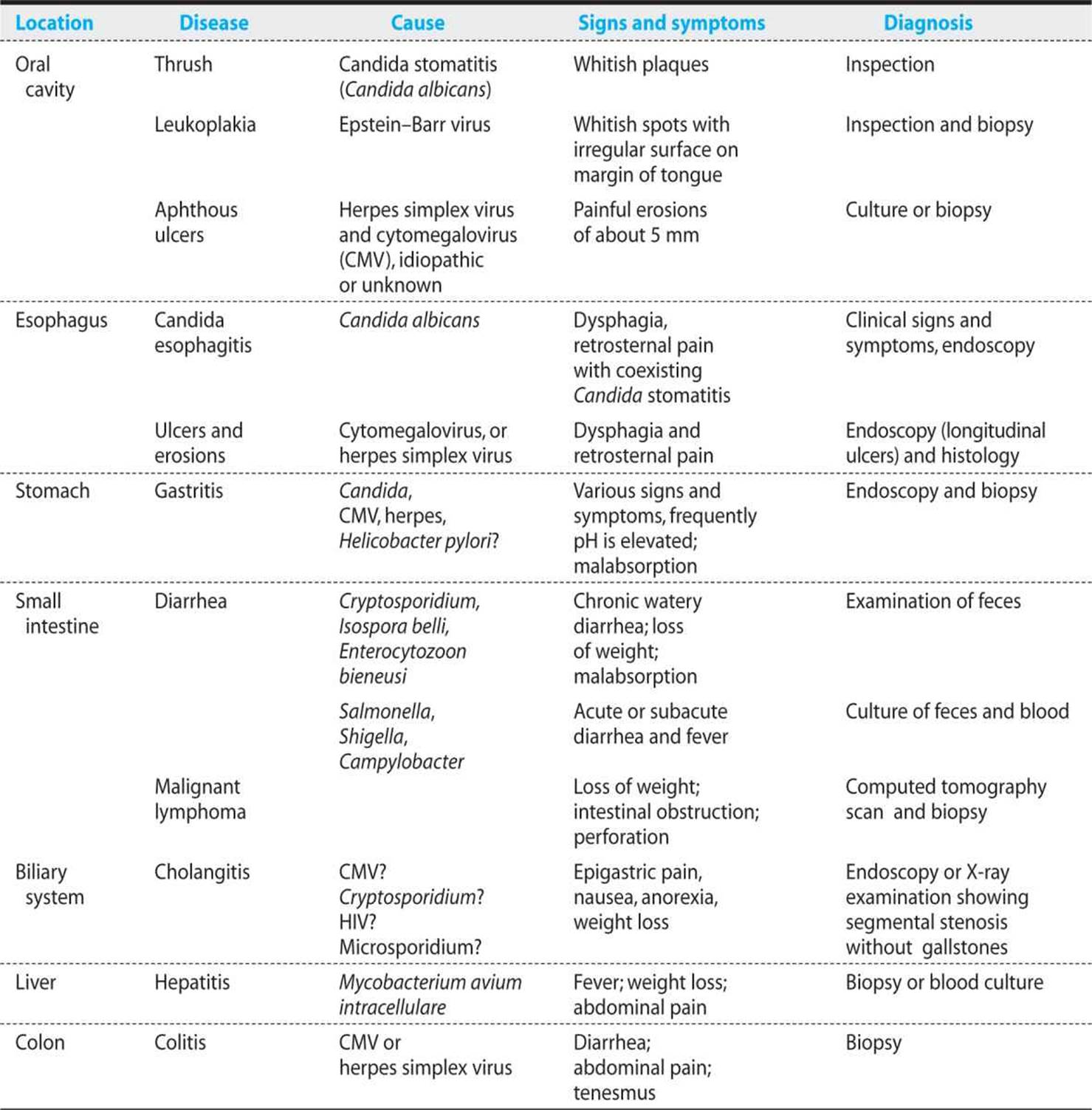
Table 16.13 summarizes the gastrointestinal tract infections associated with HIV infection. Also see Chapter 8 for a discussion of infections that can affect both immu-nocompetent and immunocompromised individuals.
Table 16.13. Gastrointestinal Diseases Associated with HIV Infection
Oral Cavity and Esophagus
CANDIDIASIS
Candidiasis is the most frequent of the opportunistic infections, occurring in virtually all HIV-positive patients with severe immunosuppression. Usually, oral candidiasis presents with yellowish-white plaques on the oral mucosa (“oral thrush”; see Figure 16.4). These plaques detach easily, revealing reddish mucosa beneath. The erythematous form of candidiasis consists of brilliant red spots on the tongue or palate. Candidiasis can also present as angular cheilitis or perleche. The clinical diagnosis is usually evident; cultures are difficult to interpret, because Candida is found in the mouth of many people without stomatitis.
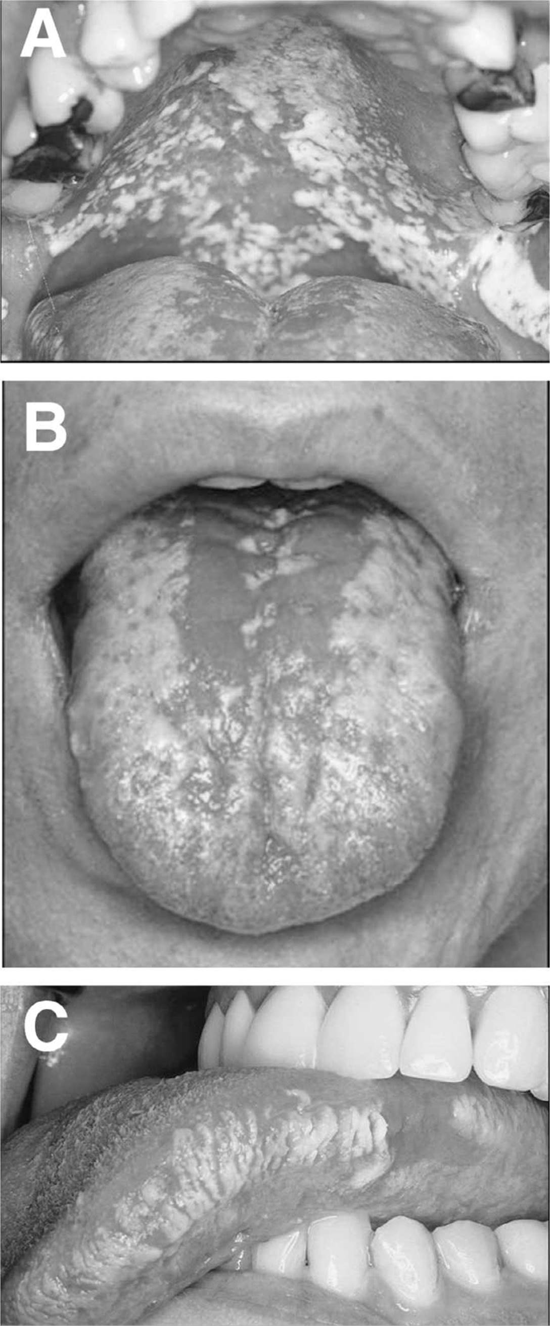
Figure 16.4. Lesions of the oral cavity in AIDS (from www.aids-images.ch). A. Oral thrush involving the soft and hard palate. B. Candidiasis of the tongue (pictures A and B courtesy of J. Sampson). C. Oral leukoplakia. See color image on color plate 4. See color image on color plate 4.
KEY POINTS
About Oral Candidiasis
1. Develops in all HIV-infected patients with serious immunocompromise.
2. Typically seen as white plaques that detach when scraped, or as red spots on the tongue and palate.
3. Often accompanied by esophagitis, an AIDS-defining illness.
4. Fluconazole is the treatment of choice.
5. Recurrent pharyngitis is common; suppression often results in resistance.
Often, Candida stomatitis is associated with esophagitis, which may cause dysphagia and retrosternal pain. Candida esophagitis is one of the designated AIDS-defining opportunistic infection; patients with this complication are stratified into class C. Patients with stomatitis only are stratified into class B.
Oral imidazoles, especially fluconazole, have become the treatment of choice. In previously untreated patients, single doses of 150-400 mg are effective. Options for subsequent management vary. Relapses can be prevented by HAART’s reversal of immune suppression. If achieving reversal is not possible, some physicians prefer to wait for a relapse, which they then retreat; others favor preventive therapy—for instance, fluconazole 50 mg daily or 150 mg weekly.
After years of intermittent treatment or prevention, relapses become more frequent and resistance of Candida is common. Such cases may present difficult problems of management. Other imidazoles—such as itraconazole solution, voriconazole, or ketoconazole—may remain effective. In other cases, intravenous therapy with amphotericin B at doses of 20-30 mg daily is necessary. Newer agents such as the echinocandins (see Chapter 1) are easier to administer, but expensive.
MOUTH ULCERS AND APHTHOUS STOMATITIS
Superficial lesions of the oral and esophageal mucosa can cause pain and dysphagia. Differential diagnoses include herpes simplex, CMV, medication side effects (f), and idiopathic ulcers. If the lesion persists, a biopsy with viral culture or immunofluorescence is often necessary for diagnosis.
ORAL HAIRY LEUKOPLAKIA
Oral hairy leukoplakia, a whitish lesion with an irregular border located along the lateral part of the tongue, is caused by Epstein–Barr virus. Often, the lesion is bilateral. Histology shows epithelial hyperplasia. Usually, treatment is not necessary, but in resistant cases, topical application of podophyllotoxin can be effective. Acyclovir can also be administered, but usually it causes only temporary regression of the lesions.
TUMORS
Kaposi sarcoma frequently involves the oral cavity. It produces painless macules or nodules with characteristic purple coloration on the palate, gingivae, or tongue.
SALIVARY GLANDS
Benign lymphoepithelial lesions and cystic hyperplasia involve mostly the parotid gland. They can be associated with xerostomia. The clinical picture is similar to that in Sjögren syndrome. The parotid lesions are particularly frequent in children; they are attributed to HIV itself.
KEY POINTS
About Esophagitis in HIV
1. Candida albicans is the most common cause.
2. Cytomegalovirus is less common, causing longitudinal ulcers and viral inclusions on biopsy.
3. Herpes simplex virus type 1 is moderately frequent; type 2 and herpes zoster are less common. Diagnosis is made by culture or immunofluorescence.
4. Thalidomide may help idiopathic esophageal ulcers.
DIFFERENTIAL DIAGNOSIS OF ESOPHAGITIS
As noted earlier, the most frequent cause of esophagitis is infection by C. albicans. However, when esophageal symptoms occur in a patient who does not have clear evidence of Candida stomatitis, other causes must be sought.
• CMV causes longitudinal ulcers. The lesion can be diagnosed only by biopsy: characteristic viral inclusions are seen in endothelial, epithelial, or smooth muscle cells.
• Involvement of the esophagus by herpes is most often caused by herpes simplex type 1 and less commonly by herpes type 2 or by herpes zoster. Lesions are typically small. Diagnosis is made by biopsy plus immunofluorescence, or culture, or both.
• Idiopathic ulcer is a diagnosis by exclusion. Treatment with thalidomide may bring relief.
Small and Large Intestine
DIARRHEA
Diarrhea associated with weight loss is one of the hallmarks of AIDS, particularly in Africa, where AIDS, diarrhea, and weight loss are practically synonymous (“slim disease”). Infection with HIV itself, plus many opportunistic pathogens and tumors, can involve the small and large intestine and cause diarrhea. The differential diagnosis is vast. This subsection briefly comments on the most frequent causes (also see Chapter 8).
Drugs. Many of the antiretroviral drugs can cause diarrhea—in particular all PIs, and ddI. Because patients with HIV often receive antibiotics, the possibility of colitis associated with Clostridium difficile must often be considered, and the C. difficile toxin must be sought in feces.
Salmonella, Campylobacter, Shigella. These organisms are frequent causes of acute gastroenteritis both in non-HIV and HIV-infected populations. In HIV infection, bacteremia is extremely frequent, particularly as a result of infection with Salmonella typhimurium or S. enteritidis.
Abdominal Tuberculosis. Abdominal TB presents with fever, pain, weight loss, or obstruction. These symptoms are difficult to distinguish from those of abdominal lymphoma. Often, the diagnosis is made only at laparoscopy.
MOTT. Infections with “mycobacteria other than tuberculosis” are often caused by M. avium, but other mycobacterial species cause similar clinical signs and symptoms, and may be more difficult to diagnose, because they grow poorly in culture (e.g., M. genavense). The MOTT organisms cause a systemic illness with fever, weight loss, and positive blood cultures. In biopsies of the gastrointestinal tract, the submucosa may be filled with characteristic acid-fast microorganisms. Diarrhea and abdominal pain dominate the clinical picture.
KEY POINTS
About HIV-Associated Diarrhea
1. Diarrhea can be caused by HIV infection alone.
2. Antiretroviral drugs and antibiotics can cause diarrhea (with Clostridium difficile, for example).
3. Salmonella gastroenteritis is more commonly associated with bacteremia in patients with HIV.
4. Mycobacterium tuberculosis and atypical myco-bacteria can result in diarrhea.
5. Cytomegalovirus colitis in patients with a CD4 count below 50/μm3 can be diagnosed by biopsy.
6. Infecting protozoa include Cryptosporidium, Microsporidia, and Isospora belli. Search for oocysts, and use trichrome stain for Microsporidia.
Cytomegalovirus Colitis. Diseases caused by CMV are typically the result of reactivation of latent CMV infection—that is, IgG antibodies against CMV were present before symptoms started—in immunosup-pressed patients, usually those with a CD4 count below 50/μm3. Symptoms may be severe, with diarrhea, abdominal pain, tenesmus, and fever. Colonoscopy shows multiple erosions, and biopsies reveal the characteristic intranuclear inclusions. CMV is also implicated in some cases of cholangitis and pancreatitis.
Cryptosporidium. In immunocompetent individuals, C. parvum causes asymptomatic infections and acute diarrhea. In immunosuppressed patients, diarrhea becomes chronic, causing malabsorption. Oocysts can be found in the feces. No treatment has so far proven effective, although oral paromomycin (500-750 mg every 8 hours), macrolides such oral azithromycin (1250 mg daily), oral clarithromycin (500 mg twice daily), and oral albendazole (400 mg daily) can be tried, in addition to symptomatic treatment of diarrhea (loperamide, narcotics).
Microsporidia. Three types of Microsporidia are found in cases of diarrhea:
• Enterocytozoon bieneusi (most frequent)
• Encephalitozoon intestinalis (which can also involve the biliary tract)
• Encephalitozoon cuniculi
Some patients do not exhibit symptoms; however, more often, patients experience profuse diarrhea, abdominal pain, and weight loss. Up to 30% of cases of chronic diarrhea in immunosuppressed HIV-positive patients may be a result of Enterocytozoon bieneusi. A special stain (modified trichrome stain) reveals the parasite in feces. Past treatments were not very effective, and eradication of the organism was usually impossible. Fumagillin (20 mg three times daily for 2 weeks) clears the spores and prevents relapse in most patients (see Chapter 8). Albendazole (400 mg twice daily) is useful in cases of Encephalitozoon intestinalis infection.
Isospora belli. Diarrheas caused by I. belli are frequent in developing countries (in African countries and Haiti, for instance). The treatment of choice is trimethoprim–sulfamethoxazole, which is also effective in primary and secondary prophylaxis.
Rectum and Anus
Many HIV-infected patients are at risk of other sexually transmitted infections such as gonococcal proctitis, syphilis, and venereal warts. Herpes simplex can cause rectitis with tenesmus and bleeding; in addition, in severely immunosuppressed patients, herpes simplex may cause persistent and debilitating ulcerations (see Figure 16.5). Such lesions may necessitate admission to hospital and parenteral therapy with high-dose acyclovir. Resistance to acyclovir may develop; the alternative treatment is foscarnet. Less commonly, such ulcerations can be caused by CMV.
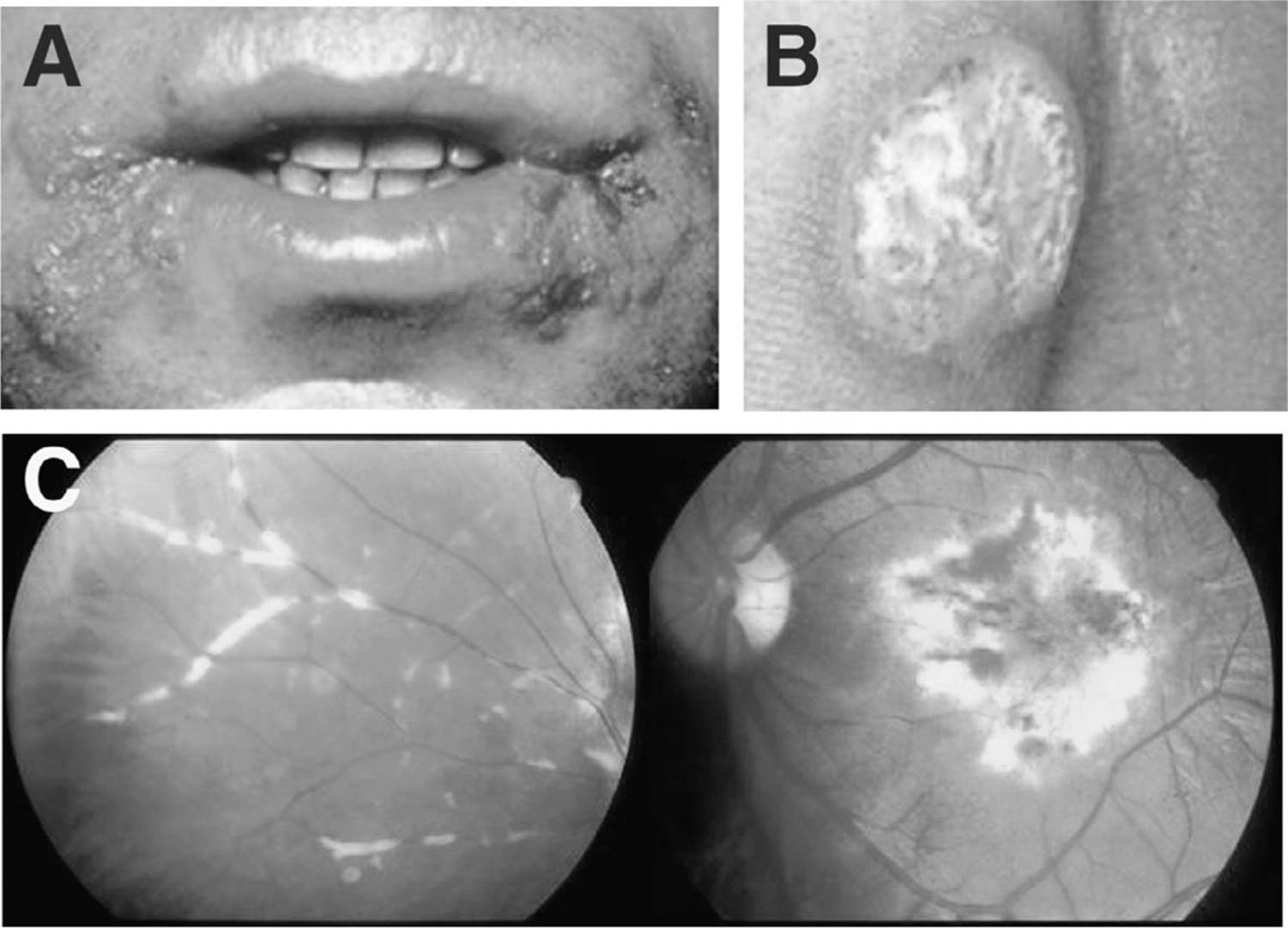
Figure 16.5. Herpesvirus group infections (from www.aids-images.ch) A. Herpes simplex virus 1. These chronic perioral lesions have become resistant to acyclovir. B. Ulcer on the buttocks resulting from infection with herpes simplex virus 2 (diameter: 5 cm). C. Cytomegalovirus retinitis. Left: Initial lesions, showing perivascular sheathing. Right: Later lesions, showing necrosis and hemorrhage. (picture courtesy of E. Baolivo). See color image on color plate 4. See color image on color plate 4.
Anal and rectal carcinomas are particularly frequent in homosexual patients. The development of that tumor is related to the human papilloma virus. Screening programs in homosexual patients for this virus have been considered, analogous to those that screen for cervical cancer, as well as vaccination of adolescents have been considered, but are not yet part of routine clinical practice.
Tumors of the Digestive System
KAPOSI SARCOMA
When patients with cutaneous Kaposi sarcoma undergo endoscopy, gastric or intestinal involvement is found in about one half of cases. However, such involvement is usually asymptomatic, and involvement of the gastrointestinal tract without involvement of skin is rare. Occasional complications include bleeding, obstruction, invagination, and perforations.
LYMPHOMA
The AIDS-associated lymphomas preferentially involve the gastrointestinal tract (and the brain), causing diarrhea, abdominal pain, fever, and weight loss. Symptoms of lymphoma are therefore difficult to distinguish from those of opportunistic infections. Chemotherapy is theoretically effective, but often very difficult to administer to these severely immunosuppressed patients. During chemotherapy, perforation with overwhelming peritonitis and sepsis remains a threat.
Liver
VIRAL HEPATITIS
Transmission of both hepatitis C virus (HCV) and HIV occurs parenterally; which is why HIV–HCV coinfection is particularly frequent in intravenous drug abusers and patients with hemophilia. Transmission of hepatitis B virus (HBV) occurs sexually, and its incidence is increased in men who have sex with men.
In HIV–HCV coinfection, the two viruses influence one another. Coinfected patients tend to have unfavorable prognostic indices for hepatitis C: higher incidence of infection with HCV type 1, cirrhosis, and high levels of HCV viremia. Conversely, HCV influences HIV infection: notably, the CD4 response to HAART is less vigorous in coinfected patients than in those infected with HIV alone. Experience with interferon treatment of HIV–HCV coinfection was previously disappointing. However, as a consequence of HAART for HIV and combination therapy with pegylated interferon and ribavirin for HCV response to therapy has improved.
Nevertheless, treatment of HCV in coinfected patients remains a challenge. Interactions between liver disease and HAART are frequent and unfavorable, and contraindications to the use of interferon (e.g., a history of depression) and ribavirin (anemia) are frequent.
KEY POINTS
About Coinfection with HIV and Hepatitis C Virus
1. Coinfection is frequent in intravenous drug abusers and people with hemophilia.
2. Patients infected with HIV tend to have more severe hepatitis C virus (HCV): they have a higher incidence of HCV type 1 cirrhosis, and higher levels of HCV viremia.
3. Patients infected with HCV have a reduced response to highly active antiretroviral therapy (HAART).
4. The combination of HAART with pegylated interferon and ribavirin is demonstrating increased responsiveness.
5. Direct-acting agents, such as boceprevir and telaprevir, are used in combination with pegylated interferon and ribavirin for the first 12 weeks of treatment.
Two direct-acting anti-HCV drugs, telaprevir and boceprevir, have become available in 2011. They are used in combination with peginterferon and ribavirin for 12 weeks, followed by peginterferon and ribavirin alone for 12-36 weeks. Their efficacy in treating hepatitis C, type 1, has been demonstrated in patients with HCV monoinfection, but early indications are that rates of sustained virologic remissions are similar in HIV/HCV coinfection. Telaprevir causes troublesome skin reactions, while boceprevir’s main side effect is anemia. For many coinfected patients with contraindications to interferon, the best option is waiting for interferon-free therapy using a combination of direct-acting agents. At the time of this writing, early results from small series of monoinfected patients look promising; such treatment may become available by 2015.
3TC, FTC, and TDF are active against both HIV and HBV. After years of therapy, however, the risk of development of 3TC resistance is high. 3TC-resistant HBV is also resistant to FTC. However, TDF remains effective. Therefore, all HBV/HIV coinfected patients should receive TDF. Entecavir is an option for the rare coinfected patient who does not need or want ARV treatment.
LIVER DAMAGE INDUCED BY ANTIRETROVIRAL DRUGS
Almost all antiretroviral agents may cause liver damage. However, the nature of that damage varies with the drug:
• The NRTIs occasionally cause severe steatosis associated with elevated plasma lactate levels. This side effect is more frequent with d4T than with other NRTIs.
• The PIs IDV and atazanavir cause asymptomatic hyper-bilirubinemia (pseudo–Gilbert syndrome). RTV and NFV can occasionally cause cholestasis and hepatitis.
• The NNRTIs are also associated with toxic hepatitis. Severe cases, with death and liver transplantation, have been reported after use of NVP. Risk factors include female sex, pregnancy, obesity, and CD4 counts above 400. Such severe cases are extremely rare with EFV, ETR, or RPV.
CENTRAL NERVOUS SYSTEM
Table 16.14 summarizes the CNS diseases most often seen in HIV infection. See also Chapter 6 for a discussion of infections that can affect both immunocompetent and immunocompromised individuals.
Table 16.14. Central Nervous System Involvement in HIV Infection

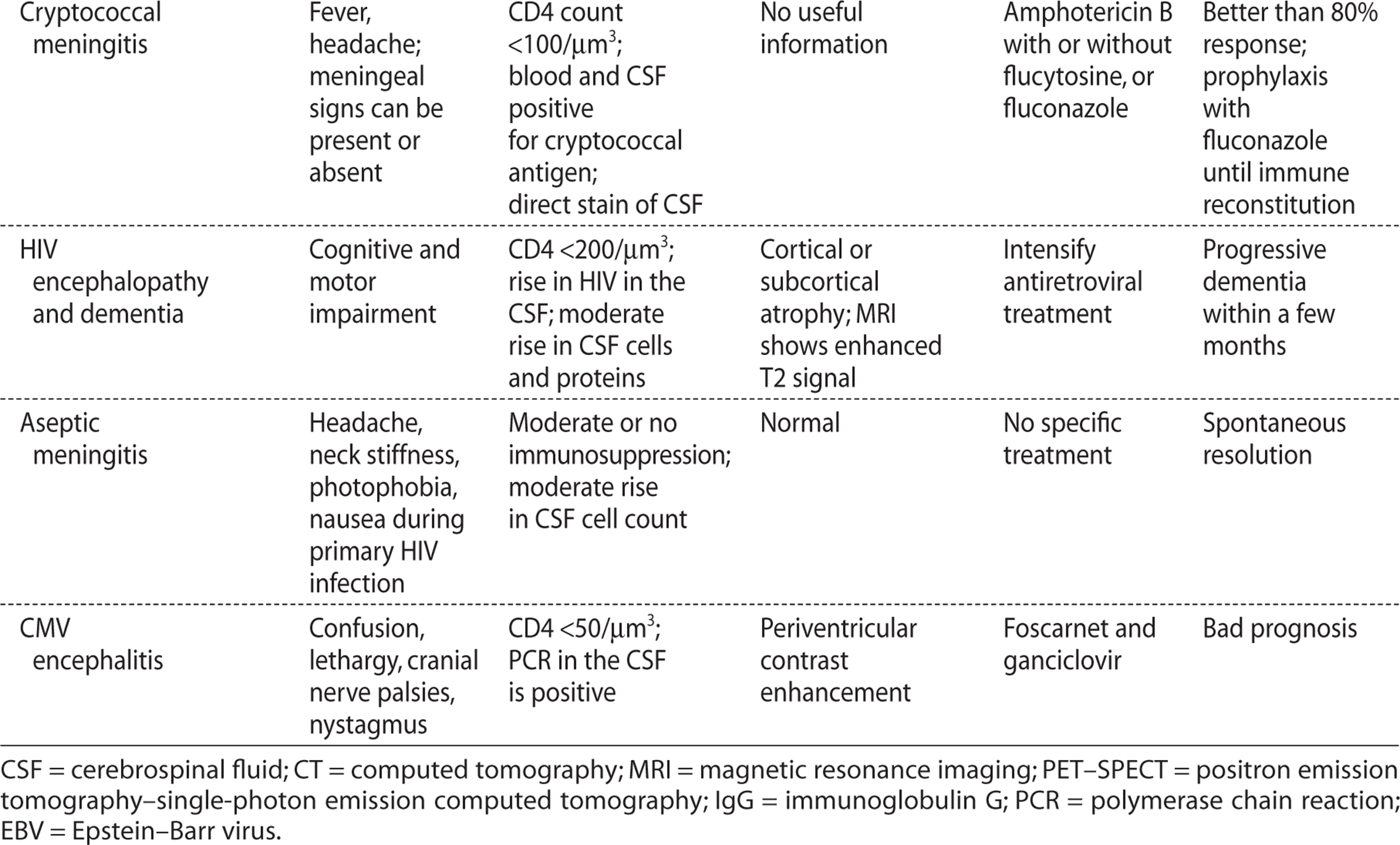
Primary HIV Infection
About half of patients with the acute retroviral syndrome complain of headaches, and in 5-20%, clinical signs of meningitis such as neck stiffness or photophobia are evident. Encephalitis, with symptoms ranging from confusion to coma, is rare. In the CSF, lymphocytes predominate, with a cell count of 5-200/μm3. Cranial nerve involvement may occur. Symptoms usually disappear spontaneously.
HIV Encephalopathy
The disease called HIV encephalopathy is synonymous with HIV dementia or AIDS-related dementia. This syndrome includes cognitive, behavioral, and motor symptoms and signs. The diagnosis is often one of exclusion, after neuroradiologic and CSF examinations have failed to show an opportunistic disease.
The first signs are usually memory problems, mental slowness, and lack of precision. Apathy and withdrawal may be interpreted as a depression. Clinical examination shows difficulties in comprehension and coordination, abnormal gait, nystagmus, and archaic reflexes. Without treatment, dementia progresses within a few months. Convulsions may appear. Neuroradiologic investigation usually shows cerebral atrophy. Magnetic resonance imaging (MRI) scan shows an increased T2 signal in the subcortical white matter, preferentially in the parasagittal regions. The CSF shows a variable increase in protein and mononuclear cells.
Since the introduction of HAART, the incidence of HIV dementia has greatly decreased. In established dementia, the effect of HAART is variable, but spectacular improvements are noted in some patients. Despite HAART, many patients continue to complain of subtle symptoms, such as forgetfulness and difficulties with concentration. This may represent a milder form of HIV-related dementia, perhaps related to the lack of penetration of HARRT into the CNS.
KEY POINTS
About HIV Encephalopathy
1. The diagnosis is made by exclusion.
2. Dementia symptoms are accompanied by apathy and withdrawal that can be mistaken for depression.
3. Magnetic resonance imaging shows an increased T2 signal in the subcortical white matter preferentially in parasagittal regions.
4. Highly active antiretroviral therapy has dramatically decreased the incidence of HIV dementia. However, mild forms of cognitive dysfunction persist.
Focal CNS Lesions
Cerebral toxoplasmosis, primary cerebral lymphoma, and progressive multifocal leukoencephalopathy (Figure 16.6) cause 90% of focal lesions of the CNS in HIV infection. Differential diagnosis relies upon computed tomography (CT) scan, MRI, and PCR amplification of the DNA from the putative infectious agents in the CSF. Cerebral biopsy remains an option in exceptional cases.
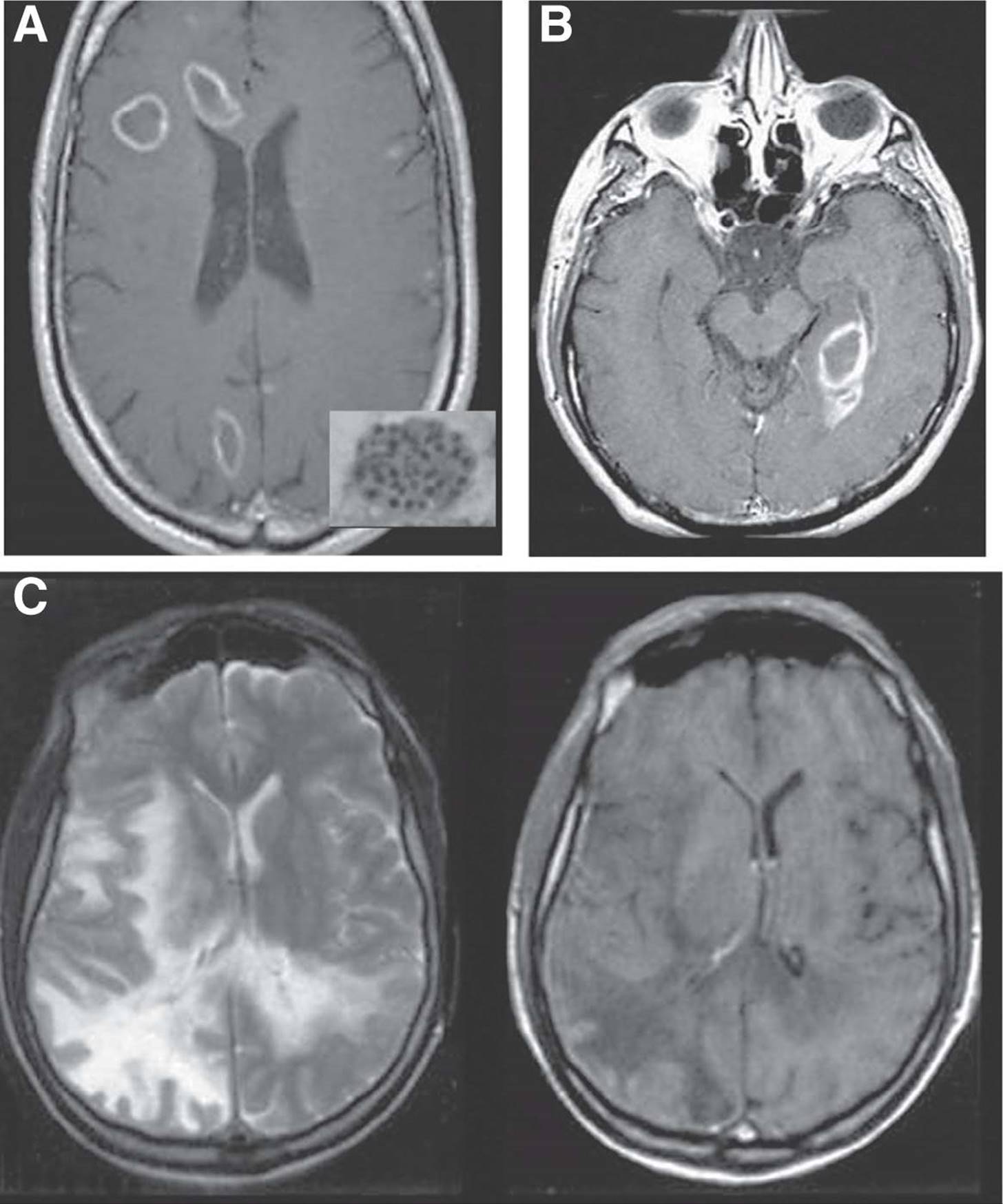
Figure 16.6. Neurologic complication of AIDS (from www.aids-images.ch). Upper left panel: Toxoplasmosis encephalitis. Magnetic resonance imaging (MRI) scan with contrast shows typical ring-enhancing lesions. Insert: Toxoplasma gondii tissue cyst contains thousands of bradyzoites (100–300 mm). Upper right panel: Central nervous system lymphoma. This MRI scan with contrast shows a typical ring-enhancing lesion (picture from Sakaie KE, Gonzalez RG. Imaging of neuroAIDS. NeuroAIDS. 1999;2:online). Bottom panel: Progressive multifocal leukoencephalopathy. Left: MRI T2-weighted image. Right: T1 image without contrast. Note the hypodensities, reflecting loss of myelin (picture courtesy of R. Dupasquier).
TOXOPLASMA ENCEPHALITIS
Toxoplasma encephalitis (Figure 16.6) follows from reactivation of latent Toxoplasma infection. Such latent infection is present in 10% (in the United States) to more than 90% (in developing countries) of HIV-infected people.
Toxoplasma encephalitis usually starts with a focal deficit (hemiplegia, for instance), convulsions, headaches, fever, or confusion. In a preponderance of cases, the CD4 count is below 200/μm3, and if performed, testing for Toxoplasma IgG antibody will be positive. If antibody is absent, or if the patient has taken trimethoprim-sulfamethoxazole prophylaxis, another diagnosis should be considered first. The CT or MRI scan shows abscesses that are usually multiple and preferentially located at the corticomedullary junction and in the basal ganglia. Annular contrast or gadolinium enhancement is typical, as is marked edema.
KEY POINTS
About Central Nervous System Toxoplasmosis
1. Usually presents with focal findings, in the presence of a CD4 count below 200/um3 and a positive test for Toxoplasma immunoglobulin G antibody.
2. Computed tomography (CT) or magnetic resonance imaging (MRI) scan demonstrates multiple contrast-enhancing ring-like lesions.
3. Empiric treatment is indicated if symptoms and MRI findings are typical. Polymerase chain reaction testing of the cerebrospinal fluid is confirmatory.
4. Treat using a combination of sulfadiazine and pyrimethamine, with added folinic acid.
5. Follow-up CT or MRI scan at 2 weeks should demonstrate improvement.
6. After treatment, secondary prophylaxis is required.
If the IgG antibodies are positive and the images are typical, empiric treatment is warranted. If the diagnosis is in doubt, T. gondii DNA can be amplified from the CSF. The rate of DNA positivity decreases when PCR is attempted after treatment has already started. The treatment of choice is a combination of oral sulfadiazine (1–1.5 g every 6 hours) and oral pyrimethamine (200 mg the first day, then 50 mg every 6 hours) combined with folinic acid (10 mg daily) to prevent bone marrow toxicity. Steroids (intravenous dexamethasone 4 mg every 6 hours) may be administered to diminish the cerebral edema. This treatment should be continued for 4–6 weeks; after that, secondary prevention using oral sulfadiazine 2 g daily and oral pyrimethamine 25 mg daily is indicated. The foregoing regimen will also prevent PCP. After 2 weeks, improvement in repeat brain CT or MRI scan is expected.
Often, treatment of toxoplasmosis is not well tolerated because of cutaneous, renal, or hepatic toxicity from sulfadiazine and bone marrow toxicity from both sulfadiazine and pyrimethamine. As an alternative, clindamycin (600 mg every 6 hours, and then 600 mg every 12 hours) can be combined with pyrimethamine; tolerance of that regimen is usually better, but efficacy is reduced. Another alternative is atovaquone suspension (750 mg every 12 or 8 hours) combined with pyrimethamine.
PRIMARY BRAIN LYMPHOMA1
HIV-infected patients can develop highly malignant B-cell brain lymphoma consisting of large immuno-blastic lymphocytes. The tumor always contains the genome of Epstein–Barr virus. Clinical signs usually progress rapidly over a few weeks, with confusion, focal signs, and headache. A CT or MRI scan shows one or several lesions with irregular contrast enhancement and preferential periventricular localization (Figure 16.6). Occasionally, lymphomatous cells can be seen in the CSF, where PCR for Epstein–Barr virus is almost always positive. Newer techniques such as singlephoton emission CT and positron emission tomography show hyperactivity in the lesions and are useful to differentiate lymphoma from cerebral toxoplasmosis and from progressive multifocal leukoencephalopathy. Although these tumors are sensitive to radiation and chemotherapy, the prognosis is poor. Long-term survivors are predominantly those with CD4 counts above 200/um3 at diagnosis.
KEY POINTS
About Central Nervous System Lymphoma in HIV
1. A B-cell lymphoma caused by Epstein–Barr virus (EBV).
2. Headache, focal signs, and confusion progress rapidly.
3. Magnetic resonance imaging or computed tomography scan shows one or two irregular enhancing lesions.
4. Polymerase chain reaction of the cerebrospinal fluid is usually positive for EBV.
5. Positron emission tomography and single-photon emission computed tomography scans are helpful in differentiating lymphoma from toxoplasmosis and progressive multifocal leukoencephalopathy.
6. Sensitive to radiation and chemotherapy, but prognosis poor if the patient’s CD4 count is below 200/um3.
PROGRESSIVE MULTIFOCAL LEUKOENCEPHALOPATHY
Progressive multifocal leukoencephalopathy follows reactivation of papovavirus JC, to which 75% of the population is seropositive. The virus infects oligoden-drocytes, which are localized in the white matter. Their destruction causes demyelinization.
KEY POINTS
About Progressive Multifocal Leukoencephalopathy
1. Caused by reactivated papovavirus John Cunningham (JC), infects oligodendrocytes, and causes demyelinization.
2. Produces dementia, aphasia, and motor deficits.
3. On magnetic resonance imaging (MRI), shows as hyperintense T2 images in subcortical regions.
4. Polymerase chain reaction testing of the cerebrospinal fluid is positive for papovavirus JC.
5. Treat with highly active antiretroviral therapy.
The disease starts insidiously with loss of memory or dysphasia, visual disturbances, aphasia, or motor signs—or, more rarely, with convulsions. A CT or MRI scan shows one or several subcortical lesions without contrast enhancement or edema (Figure 16.6). These lesions are MRI hyperintense in T2 scans. Usually, a PCR test of the CSF is positive for papovavirus JC. No specific treatment is available (cidofovir and cytosine arabinoside have been tried, with inconsistent results). HAART is a double-edged sword; after starting HAART symptoms may worsen; however, over time, stabilization and even clinical improvement may ensue.
Meningitis
CRYPTOCOCCAL MENINGITIS
Cryptococcus neoformans, a yeast, is the most frequent cause of meningitis in HIV-infected patients. Cryptococcosis occurs in profoundly immunosuppressed patients and is particularly frequent in Africa and in the United States.
The disease usually starts with headaches and fever; curiously, meningeal signs can be absent. The diagnosis can be made by direct examination of CSF stained with India ink, by finding of cryptococcal antigen in the CSF or in the blood, or by culture of CSF or blood. The CSF shows moderate pleocytosis and an increase in protein; however, in some cases, the CSF formula is only minimally abnormal. A CT or MRI scan is noncontributory (see Chapter 6 for a complete discussion).
Treatment in severe cases consists of intravenous amphotericin B (0.7 mg/kg/day, or liposomal amphotericin B 3–4 mg/kg/day) for at least 2 weeks. The addition of flucytosine (25 mg/kg every 6 hours) is recommended, but that drug has substantial gastrointestinal and bone marrow toxicity. After 2 weeks, amphotericin and flucytosine are replaced with fluconazole 400 mg daily for 6-10 weeks, and then 200 mg daily until immune function recovers. In less severe cases (without intracranial hypertension, with normal mental status, and with cryptococcal antigen in the CSF at less than 1:1000 dilution), fluconazole (800-1200 mg/day initially) can be used from the start. Itraconazole is not a good choice because it does not penetrate well into the CSF. Voriconazole and posaconazole are effective in vitro, but practical experience in patients is scarce.
KEY POINTS
About Cryptococcal Meningitis in HIV
1. Cryptococcus neoformans is the most common cause of meningitis in HIV-infected patients.
2. Headache and fever are the most common complaints; neck stiffness is absent.
3. Lymphocytosis of the cerebrospinal fluid (CSF) is usual, but the CSF formula may be only minimally abnormal.
a) India ink test positive.
b) Antigen testing of the CSF or blood is positive.
c) Culture of CSF or blood is frequently positive.
4. Treat with amphotericin B with flucytosine for 2 weeks; followed with fluconazole.
5. Immune reconstitution syndrome (IRIS) is common and may be life threatening. Treatment with prednisone, 1.5 mg/kg/day.
Because of the immune reconstitution inflammatory syndrome (IRIS), symptoms may initially worsen. Increased CSF pressure (>250 mmHg) is associated with high mortality, and management of this complication by repeat lumbar puncture or ventriculoperitoneal shunts may be necessary.
CNS Infection by Cytomegalovirus
CMV can cause various nervous system diseases in HIV infection: polyradicular myelitis, peripheral neuropathy, and encephalitis. Patients with encephalitis are usually profoundly immunosuppressed with a CD4 cell count below 50/μm3.
Diagnosis is difficult and is usually made after exclusion of other more frequent causes in patients who are confused and lethargic, and who are showing cranial nerve palsies and nystagmus. The typical finding in an MRI or CT scan is periventricular contrast enhancement. A PCR test of the CSF is more than 80% sensitive, and specific. Although foscarnet and ganciclovir should theoretically be effective, the prognosis is unfavorable.
Cerebrovascular Diseases
Cerebrovascular accidents are much more frequent in the HIV-infected populations than in comparable populations of the same age. The pathogenesis is uncertain, but direct involvement of HIV in vasculitis is suspected. Transient ischemic attacks have also been described.
Other Rare Cerebral Disorders
Rare focal diseases in the HIV-infected population include cryptococcoma (in these cases, the cryptococcal antigen test in CSF and blood can be negative), tuberculoma, varicella virus encephalitis, and secondary or tertiary syphilis. In intravenous drug abusers, septic emboli may be associated with cerebral abscesses and mycotic aneurisms.
Peripheral Neuropathy
DISTAL SYMMETRIC POLYNEUROPATHY
Distal symmetric polyneuropathy may cause painful paresthesia and dysesthesia in hands and feet, associated with diminished reflexes and motor weakness in the legs, and autonomic dysfunction.
These polyneuropathies can be very difficult to manage. Amitriptyline or carbamazepine may be useful. Aggravating circumstances include concomitant vitamin deficiencies, diabetes, alcohol abuse, and use of medications such as dapsone, vincristine, and isoniazid. Among the antiretroviral drugs stavudine causes neuropathy, as do ddI and 3TC less commonly. The foregoing drugs can usually be replaced by other nucleosides if necessary.
INFLAMMATORY DEMYELINATING POLYNEUROPATHY
Inflammatory demyelinating polyneuropathy usually occurs during the early stages of HIV infection. Presentation is similar to that of Guillain–Barré syndrome. With steroids, plasmapheresis, or intravenous immunoglobulins, evolution is usually favorable. In some cases, CMV infection is involved.
MONONEURITIS MULTIPLEX
Sudden palsies of one or several nerves, including cranial and laryngeal nerves, can occur at any stage of HIV infection. Varicella virus can be the cause in cases of advanced immunodeficiency.
MYELOPATHY
Myelopathy presents with gait disturbance, ataxia, spastic paraparesis, and urinary or fecal incontinence. An MRI scan is usually normal, but edema or even enhancing lesions may be seen. Autopsy findings show vacuolization of myelin and an accumulation of macrophages. No specific treatment is available, but potentially reversible causes of myelopathy such as epidural abscess, toxoplasmosis, infection with human T lymphotropic virus type 1, herpes simplex or zoster, CMV, or a vitamin B12 deficit should be excluded.
KEY POINTS
About Peripheral Neuropathies in HIV
1. In distal symmetrical polyneuropathy associated with paresthesias and weakness, drugs that cause neuropathy should be discontinued. Treat with amitriptyline or carbamazepine.
2. Treat inflammatory demyelinating polyneuropathy with plasmapheresis or a cytomegalovirus regimen.
3. Mononeuritis multiplex can be caused by varicella virus.
4. Myelopathy can lead to spastic paraparesis; look for reversible causes.
OPHTHALMOLOGY
Also see Chapter 5 for a discussion of infections that can affect both immunocompetent and immunocompromised individuals.
HIV Retinopathy
HIV retinopathy is frequent and benign; it does not require treatment. “Cotton wool” exudates are characteristically observed; these correspond to focal lesions of ischemia. Besides exudates, intraretinal hemorrhages, telangiectasias, and microaneurysms may occur; these conditions must be distinguished from retinal lesions caused by diabetes or hypertension. HIV retinopathy does not interfere with vision.
Cytomegalovirus Retinitis
Chorioretinitis from CMV occurs in patients with profound immunosuppression (CD4 count below 50/μm3); CMV IgG antibodies are invariably present. Before HAART became available, 25–30% of patients with AIDS developed retinitis before death. All severely immunosuppressed patients with HIV should be repeatedly questioned about changes in vision—blurring of vision, loss of central vision or other blind spots, floaters, or flashing lights.
CMV retinitis is a subacute disease in which visual deficits progress within a few weeks. The diagnosis is easily made by examining the retina, which shows a characteristic mix of exudates, hemorrhages, and atrophy. Exudates often sheath the vessels (see Figure 16.5, C). Without treatment, lesions invariably progress to retinal detachment with progressive loss of vision. Often, both eyes are involved, as are other organs such as the colon, esophagus, or brain.
Treatment starts with high doses of medication, followed by secondary prophylaxis using the same drugs at lower doses. Three drugs are available: ganciclovir, foscarnet, and cidofovir.
• Ganciclovir 5 mg/kg is administered intravenously every 12 hours. Its main side effects are leukopenia and thrombocytopenia. Ganciclovir accumulates in patients with renal failure, and doses have to be adapted. Oral valganciclovir (450 mg BID) has good bioavailability and is as efficacious as intravenous ganciclovir for treatment as well as for maintenance therapy.
• Foscarnet 60 mg/kg is administered every 8 hours. It is nephrotoxic (hydration with 1 L 0.9% NaCl is necessary) and causes numerous electrolyte disturbances (hypocalcemia, hypokalemia, hypophosphatemia, and hypomagnesemia), convulsions, and genital ulcers.
KEY POINTS
About Cytomegalovirus Retinitis
1. Before the advent of highly active antiretroviral therapy, 30-35% of patients with AIDS developed this infection.
2. Visual symptoms—blurred vision, scotomas, floaters, or flashing lights—are subacute in onset.
3. Retinal findings are characteristic: mix of exudates, hemorrhages, and atrophy; vascular sheathing.
4. Treatment is required to prevent progression to retinal detachment and blindness.
a) Ganciclovir is the drug of choice; causes bone marrow toxicity, and dosing must be corrected for renal dysfunction.
b) Foscarnet is associated with renal failure; intravenous NaCl is protective.
c) Cidofovir, a once-weekly therapy, is associated with renal failure in 25% of patients; probenecid and intravenous NaCl are helpful protective measures.
5. Maintenance therapy is required in patients with a CD4 count below 100/μm3; primary prophylaxis reduces the incidence, but is expensive and associated with side effects.
• Cidofovir has the advantage of infrequent administration (5 mg/kg once weekly for 2 weeks, then 5 mg/kg every 2 weeks), but it is also nephrotoxic in 25% of patients and may cause neutropenia. Nephrotoxicity can be diminished, but not eliminated, by administering oral probenecid 2 g before the cidofovir and 1 g at 1 and 8 hours after, in conjunction with intravenous NaCl. Particular care is needed when cidofovir is coadministered with TDF.
After an initial treatment course lasting at least 2 weeks, doses can be lowered: valganciclovir 450 mg daily, foscarnet 100 mg/kg daily 5 days per week, cidofovir 5 mg/kg every 2 weeks. Treatment with intravenous ganciclovir or foscarnet (or both) necessitates use of a permanent catheter.
Secondary prophylaxis of CMV retinitis is onerous. In patients with a good response to HAART and a durable rise in CD4 count above 100/μm3, treatment can be discontinued without risk of relapse.
Patients with persistently low CD4 counts should be regularly examined so as to detect CMV retinitis and institute early treatment to prevent loss of vision. Preventive administration of oral valganciclovir diminishes the incidence of CMV retinitis by at least 50%. However, because of expense, inconvenience, and side effects, such prevention has not commonly been used. Of course, the best prevention of all is correction of the underlying immunodeficiency by effective HAART.
KEY POINTS
About Retinal Necrosis
1. Caused by varicella virus, can follow a bout of herpes zoster.
2. Acute retinal necrosis is accompanied by acute pain and inflammation; hypopyon may be seen.
3. Progressive outer retinal necrosis is painless, but associated with marked visual loss.
4. High-dose intravenous acyclovir must be started emergently—or ganciclovir if cytomegalovirus retinitis is a possibility.
Retinal Necroses
Retinal necrosis is a medical emergency necessitating treatment within hours. This disease is caused by varicella virus. Two clinical presentations can be distinguished:
• Acute Retinal Necrosis. Acute retinal necrosis (ARN) causes orbital pain and inflammation visible in the anterior ocular segment with hypopyon. At the same time, peripheral retinal necrosis with vasculitis occurs. Without treatment, progression to retinal detachment and blindness is rapid.
• Progressive Outer Retinal Necrosis. In contrast to ARN, progressive outer retinal necrosis (PORN) causes no pain. However, the patient notices a marked loss of visual acuity. Often, these patients have recently had herpes zoster. The anterior segment does not show evidence of inflammation; however, peripheral lesions of retinal necrosis occur. Again, there is a major risk of rapid vision loss. For both ARN and PORN, treatment involves high doses of intravenous acyclovir, and ganciclovir if a possibility of CMV retinitis exists.
Other Infectious Eye Diseases
P. jiroveci may occasionally involve the retina. Cryptococcal meningitis may be complicated by papillary edema. Particularly in intravenous drug abusers, C. albicans and other bacteremia may cause retinitis. Uveitis can complicate the administration of rifabutin, particularly when rifabutin levels are boosted by coadministration of macrolides or PIs.
SKIN DISEASES
It is important to recognize skin diseases during HIV infection. The development of a new skin rash often warrants immediate action (see Table 16.15). For instance, new acneiform lesions accompanied by fever suggest primary HIV infection. New onset of a maculopapular total body rash is indicative of a drug reaction. New crops of macular, papular, pustular, or vesicular lesions may represent the first manifestation of an opportunistic infection. Even benign skin diseases may have a major psychological impact when they reveal the patient’s HIV status to the outside world.
Table 16.15. Skin Diseases in HIV
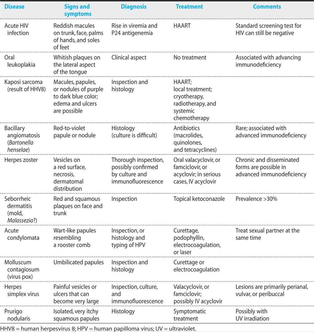
Primary HIV Infection
Primary HIV infection causes erythematous macules or papules with ill-defined borders and symmetrical distribution on the front and back of the trunk, the face, and sometimes on the palms and soles. The skin lesions neither itch nor hurt. They resemble Gilbert pityriasis or the lesions of secondary syphilis, which are the principal differential diagnoses. Other differentials include viral exanthema as a result of Epstein–Barr virus, CMV, rubella, or a toxic or allergic reaction to medication. The lesions persist for a median of 2 weeks, and then fade spontaneously. Less commonly, painful mucosal ulcers occur (Figure 16.1).
Opportunistic Infections with Skin or Mucosal Involvement
CHRONIC HERPES SIMPLEX VIRUS
In severely immunosuppressed patients, herpes simplex type I or II may cause persistent genital, perianal, or perioral ulcerations. Although herpes simplex is by far the most likely causative agent, the differential diagnosis is large, including infections by fungi, mycobacteria, CMV, and varicella virus, and malignant skin tumors. Confirmation is obtained by biopsy and immunofluorescence or by culture of a virus. The preferred treatment is valacyclovir 500 mg or famciclovir 125 mg twice daily. Herpes simplex virus may become resistant to acyclovir and its derivatives, necessitating alternative treatment with foscarnet.
HERPES ZOSTER
Herpes zoster caused by reactivation of varicella virus occurs almost 20 times more frequently in HIV-positive individuals than in HIV-negative individuals of the same age, and the condition can present at any stage of immunosuppression. In the severely immunosuppressed patient, herpes zoster may extend beyond one or two dermatomes, causing atypical, ulcerated, and painful lesions that are difficult to treat. In cases in which the skin lesions are atypical, biopsy with direct immunofluorescence establishes the diagnosis. Particularly in cases in which immune suppression is severe, treatment is indicated: use valacyclovir 1 g every 8 hours or famciclovir 500 mg twice daily. In patients with severe immune suppression, intravenous acyclovir may be preferred.
KAPOSI SARCOMA
Kaposi sarcoma is a very unusual “tumor.” Infection by a virus—human herpesvirus 8 (HHV8)—is a necessary but not sufficient condition. Kaposi sarcoma appears in patients who are HHV8 seropositive and who have a variable degree of immunosuppression. Very often, Kaposi sarcoma is multifocal from the start. Karyotypic anomalies have not been described. Lesions resemble reactive hypoplasia rather than typical malignancies.
In the United States and in Europe, Kaposi sarcoma is essentially a disease of patients who acquired their HIV infection by homosexual contact. Although cases can occur in patients with a nearly normal CD4 count, immune suppression greatly increases the risk.
The lesions of Kaposi sarcoma are macules, papules, or nodules of characteristic purple color. Preferred locations are the extremities, the tip of the nose, and the palate (Figure 16.2). Often, the lesions are only slowly progressive and do not cause pain. In rare cases, Kaposi sarcoma may run an aggressive course with nodular, ulcerated lesions; limb edema; and gastrointestinal and pulmonary involvement. Kaposi sarcoma is easy to recognize; when in doubt, a skin biopsy showing vascular proliferation and fusiform cells will yield the diagnosis.
The incidence and severity of Kaposi sarcoma are favorably influenced by HAART, which has become the mainstay of treatment. If the lesions persist or enlarge, local treatment by cryotherapy or radiotherapy is recommended. Systemic treatment is necessary in cases with edema of extremities, genitalia, or the face, or in cases of massive visceral involvement. Many chemotherapeutic agents produce remissions, but these are rarely of long duration. For reasons of relative lack of side effects and good efficacy, liposomal preparations of doxorubicin, used at a dose of 20-40 mg/m2 every 2-3 weeks were popular, but liposomal doxorubicin is no longer available at the time or writing. The preferred alternative is paclitaxel (100 mg/m2 intravenously every 2 weeks); in resistant cases, bevacizumab may be tried.
KEY POINTS
About Kaposi Sarcoma
1. Associated with human herpesvirus 8; in the United States and Europe, is found mainly in HIV-infected homosexual men.
2. Manifests as macules, papules, or nodules of distinctive purple color, usually on the extremities, the tip of the nose, and the palate.
3. Disease is occasionally aggressive, with limb edema and gastrointestinal and pulmonary involvement.
4. Histopathologic examination shows vascular proliferation and fusiform cells.
5. May be refractory to therapy.
a) HAART usually induces remissions.
b) For local disease, cryotherapy or radiotherapy can be used.
c) In severe disease, liposomal doxorubicin or paclitaxel is favored.
BACILLARY ANGIOMATOSIS
Bacillary angiomatosis is caused by Bartonella henselae, the agent that is responsible for cat scratch disease (see Chapter 13). In HIV infection, B. henselae causes papules and nodules of a red-to-violet color. These are present in variable numbers, are not painful, and may be ulcerated. Patients are usually febrile and extremely immunosuppressed. Liver (“peliosis hepatitis”) and bone may be involved.
A biopsy with silver impregnation stain can show the Bartonella and differentiate the disease from Kaposi sarcoma. A serologic test is also available. Prolonged treatment with clarithromycin 500 mg twice daily, azithromycin 250 mg daily, or ciprofloxacin 500 mg twice daily is necessary.
SEBORRHEIC DERMATITIS
Seborrheic dermatitis is frequent in the general population. However, in HIV-infected patients, the disease may be particularly severe. Reddish plaques covered by small scales appear on the face (nose, between the eyebrows), the scalp, and the sternum. Ketoconazole creams and shampoos are efficacious.
MOLLUSCUM CONTAGIOSUM
The lesions of molluscum contagiosum are caused by poxvirus. The multiple, umbilicated, painless flesh-colored papules or nodules appear particularly on the face and the genitalia. In immunosuppressed patients, they can persist for months and become extremely numerous. The lesions can be destroyed by curettage, electrocoagulation, or cryotherapy. Cidofovir may be effective in extreme cases.
Drug Reactions
Drug rashes are frequent during HIV infection and can constitute an emergency. Conjunctivitis or lesions of the buccal mucosa, generalized erythroderma, and detachment of the skin are alarming; these signs necessitate hospitalization and specialized consultation. However, drug rashes are more often mild and will disappear even if the drug is continued—particularly in the case of early reactions to NNRTIs. Because alternative treatments often have disadvantages of their own, an effort should be made to “treat through” drug eruptions that are not severe.
KEY POINTS
About Drug Rashes in HIV-Infected Patients
1. Conjunctivitis, buccal mucosa lesions, erythroderma, and skin detachment are danger signs.
2. “Treat through” milder drug eruptions.
Skin Diseases Aggravated by HIV
Many common skin diseases—for instance, dryness of the skin, psoriasis, reactions to insect stings, and dermatomycosis—seem to be more severe in patients who also have HIV infection.
SEXUALLY TRANSMITTED DISEASES
The occurrence of sexually transmitted diseases (also see Chapter 9) in an HIV-positive patient is a reminder of unsafe sexual practices and an occasion to reinforce educational messages about the need to prevent transmission of HIV.
Syphilis
Treatment of syphilis in the HIV-infected individual has elicited a great deal of controversy. Contrary to widespread belief, serologic tests for syphilis are as valid in HIV-infected people as in an uninfected population. The recommended treatment regimens are benzathine penicillin 2.4 × 10⁶ U intramuscularly at weeks 0, 1, and 2 in cases of secondary or latent tertiary syphilis, and a prolonged course of high-dose intravenous penicillin or ceftriaxone in cases of suspected neurosyphilis.
FURTHER READING
Some of the best (and certainly the most up-to-date) resources can be accessed via the Internet.
General
Medscape. HIV/AIDS [Web page]. New York, NY 2007: Medscape;n.d.[Available online at: http://medscape.com/hiv; cited]
University of California–San Francisco. HIV InSite [Web page]. San Francisco, California: UCSF; 2007. [Available online at: http://hivinsite.ucsf.edu/InSite; cited]
Drug Interactions
University of Liverpool. Liverpool HIV Pharmacology Group www.hiv-druginteractions.org home page [Web site]. Liverpool, U.K.: University of Liverpool; 2007. [Available online at: http://www.hiv-druginteractions.org; cited]
Epidemiology
United Nations, Joint UN Programme on HIV/AIDS. Home page [Web page]. Geneva, Switzerland: UNAIDS; 2007. [Available online at: http://www.unaids.org; cited]
Up-to-Date Treatment Guidelines
United States, Department of Health and Human Services. AIDSinfo, clinical guidelines. Rockville, MD: DHHS; 2007. [Available online at: http://aidsinfo.nih.gov/Guidelines/Default.aspx?MenuItem?Guidelines; cited]