
Time Recommended to complete: 1 day
Frederick S. Southwick, M.D.
GUIDING QUESTIONS
1. How is sepsis syndrome defined, and what is SIRS?
2. Do all episodes of bacteremia cause sepsis syndrome, and are all sepsis syndromes the result of bacteremia?
3. Which bacterial products can produce sepsis syndrome?
4. What is a “superantigen,” and which bacteria produce them?
5. Which host cells are most important in sepsis syndrome, and how do they mediate it?
6. What are the clinical clues that suggest early septic shock, and why is early septic shock important to recognize?
7. What are the therapeutic measures that need to be instituted in patients with sepsis syndrome?
POTENTIAL SEVERITY
A life-threatening syndrome that must be recognized and treated quickly to prevent progression to irreversible shock.
PREVALENCE
Sepsis—severe infection leading to organ dysfunction—is a problem of increasing magnitude in the United States. The incidence of severe sepsis is estimated to be 240-300 cases per 100,000 individuals, and is associated with 18-50% mortality. In 1995, severe sepsis accounted for 9.3% of all deaths in the United States. About 70-80% of cases occur in patients who are hospitalized for other conditions, and sepsis is one of the leading causes of preventable death in our hospitals.
DEFINITIONS
The sepsis syndrome represents a continuum of clinical manifestations (see Figure 2.1), and in order to reduce mortality all clinicians must be able to recognize this syndrome at its earliest stages to quickly intervene: the earlier appropriate therapy has begun to lower the mortality. When an infection spreads from its primary site into the bloodstream, the patient develops bacteremia. Bacteremia or bacteria in the bloodstream does not necessarily result in sepsis. For example, Streptococcus viridans in most instances causes only a low-grade fever. However, bacteremia due to gram-negative organisms containing lipopolysaccharides (LPS) such as Escherichia coli and Klebsiellaprecipitates a systemic inflammatory response syndrome (SIRS). SIRS is clinically characterized by an elevated heart (>90 beats/min) and respiratory rate (>20 breaths/min), fever (temperature >38°C), or hypothermia (temperature <36°C) accompanied by an increased peripheral white blood cell count (WBC >12,000) or leukopenia (WBC <4000) and/or an increase in immature neutrophils (>10% bands).
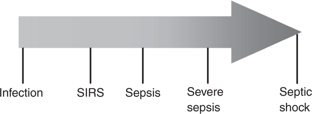
Figure 2.1. Order of progression from infection to septic shock.
Sepsis is defined as SIRS due to bacteremia. This definition clarifies that SIRS can also be precipitated by viral infections such as dengue fever and fungal infections such as candida and also can be caused by extensive tissue necrosis that may accompany severe pancreatitis or trauma. Severe sepsis is defined as sepsis with organ dysfunction and represents the progression of bacteria-induced SIRS. Septic shock is the most severe manifestation of this syndrome and is defined as sepsis with hypotension (systolic BP <90 mmHg) despite fluid resuscitation and pressor therapy. Metabolic derangements such as lactic acidosis, low urine output, altered mental status, and acute lung injury often accompany septic shock.
KEY POINTS
About the Prevalence and Definitions of Sepsis Syndrome
1. Prevalence is 240-300/100,000 cases per year in the United States.
2. Mortality ranges from 18% to 50%.
3. Sepsis is defined as a systemic inflammatory response syndrome (SIRS) accompanied by positive blood cultures for bacteria.
4. Viruses (dengue fever), fungi (Candida), and noninfectious diseases (pancreatitis, severe trauma) can also cause SIRS.
5. Severe sepsis is defined as sepsis associated with organ dysfunction.
6. Septic shock is sepsis associated with hypotension (systolic BP <90 mmHg) initially unresponsive to volume replacement or vasopressors. Elevated serum lactic acid levels often found.
PATHOGENESIS
Bacterial Products Stimulate Cytokine Release
When microbes break through the vascular endothelial barrier, they come in contact with resident macrophages containing toll-like receptors (TLR) that stimulate the host’s innate immune response. For example, LPS from gram-negative bacilli is ushered to TLR4 receptors by binding to the host’s LPS-binding protein. This protein transfers LPS from the bacterial wall to the host receptor CD14 that in turn transfers the bacterial lipid moiety to a signaling complex made up of MD2 and TLR4. Peptidoglycans and lipoteichoic acids on the surface of gram-positive bacteria bind to TLR2. Both TLRs activate the nuclear factor kappa B (NF-κB), a transcription factor that triggers the production of proinflammatory cytokines including tumor necrosis factor α (TNF-α), interleukin 6 (IL-6), and IL-1β causing SIRS (see Figure 2.2). Later neutrophils and macrophages release the inflammatory mediator high-mobility group box-1 (HMGB-1) that is thought to further worsen SIRS and contribute to mortality.
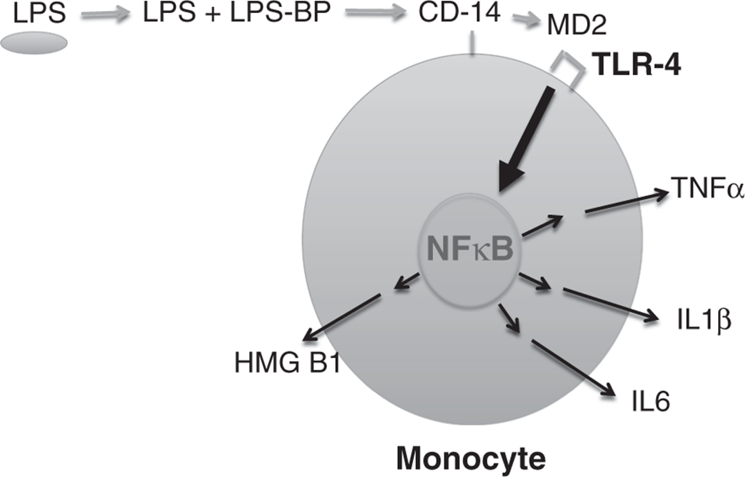
Figure 2.2. Monocyte.
KEY POINTS
About the Bacterial Products that Cause SIRS
1. In gram-negative bacteria, lipopolysaccharide (LPS), also called endotoxin, is transported to macrophage and monocyte receptors.
a) LPS-binding protein to CD14, and CD14 to MD2-TLR4 signaling complex.
2. Gram-positive bacteria produce peptidoglycans and lipoteichoic acid that stimulate TLR2.
3. Both TLR4 and TLR2 activate NF-κB causing release of TNF-α, IL-1 κ, IL-6, and HMGB-1.
4. Gram-positive bacteria also secrete exotoxins.
a) Staphylococcus aureus can secrete toxic shock syndrome toxin 1 (TSST-1).
b) Streptococcus pyogenes secretes streptococcal pyrogenic exotoxin A (SPEA).
c) Called “superantigens,” these exotoxins bypass macrophages and directly stimulate T cells.
Certain strains of Staphylococcus aureus release an exotoxin called toxic shock syndrome toxin 1 (TSST-1) that as its name implies can cause profound shock accompanied by high fever, conjunctival erythema, as well as erythema of the palms and soles followed by desquamation. The majority of toxic shock patients have been young women who had used tampons that promote S. aureus growth and toxin release. TSST-1 toxin producing S. aureusless commonly can be found in wound infections. Similarly, some strains of Streptococcus pyogenes can produce streptococcal pyrogenic exotoxin A (SPEA), and this exotoxin may result in shock as well as a scalded skin syndrome characterized by extreme erythroderma followed by extensive desquamation. These exotoxins have been termed superantigens because these proteins directly stimulate T cells to release cytokines. Unlike the endotoxins described above, superantigens do not require processing by macrophages and dendritic cells.
Sepsis Stimulates Coagulation
Proinflammatory cytokines activate the vascular endothelium and monocytes to release tissue factor (TF), and TF activates the production of thrombin that in turn converts fibrinogen to fibrin. Thrombin generation combined with increased production of von Willebrand factor (a multimeric protein responsible for platelet adhesion and aggregation) results in fibrin deposition and platelet activation, leading to microvascular thrombosis. This extreme activation of the extrinsic coagulation cascade depletes the coagulation factors and also activates secondary fibrinolysis, mediated by protein C, protein S, and plasmin, resulting in increased levels of fibrin degradation products. The excess release of TF leading to a consumption coagulopathy and microvascular thrombosis is called disseminated intravascular coagulopathy (DIC) and often accompanies septic shock, but also can be precipitated by trauma, obstetric complications, hepatic failure, acute pancreatitis, and some forms of cancer.
KEY POINTS
About Disseminated Intravascular Coagulopathy and Sepsis
1. Bacterial products cause the release of tissue factor (TF) by monocytes and endothelial cells.
2. TF activates the extrinsic coagulation cascade: thrombin stimulates fibrinogen conversion to fibrin, fibrinolysis activated.
3. Causes consumption coagulopathy and microvascular thrombosis.
4. DIC also associated with neoplasia, trauma, obstetric complications, and pancreatitis.
Vascular Dysfunction Leads to Shock and Metabolic Acidosis
In the early stages of septic shock, patients experience volume loss as a consequence of reduced oral intake, increased insensible fluid loss, changes in intravascular volume distribution, and capillary leakage. A hallmark of early septic shock is a decrease in intravascular resistance and increased cardiac output. These hemodynamic changes are unique to SIRS, and are not observed in hemorrhagic or cardiogenic shock. The underlying causes of warm shock are multifactorial and include:
1. Decreased sensitivity of vascular catecholamine receptors.
2. Diminished responsiveness to glucocorticoids as well as aldosterone.
3. Production of adrenomedullin that increases renal blood flow, vasodilates, and interferes with aldosterone secretion
4. Release of nitric oxide, a potent vasodilator, from sites of inflammation.
During this early stage of shock, patients can often be readily resuscitated with IV fluids. Administration of glucocorticoids and/or vasopressin during this period can improve endovascular responsiveness to catecholamines (see Treatment).
KEY POINTS
About the Pathogenesis of Septic Shock
1. Intravascular volume loss secondary to poor intake, altered volume distribution, and capillary leakage.
2. Initially present with “warm” shock due to peripheral vasodilation and increased cardiac output (unique to SIRS).
3. Later cold shock accompanied by decreased cardiac output.
a) Less responsive to volume expansion, often requires vasoactive agents.
b) Accompanied by anaerobic glycolysis and lactic acidosis.
c) Endovascular cell–cell junction breakdown and third spacing.
4. Often accompanied by acute respiratory distress syndrome (ARDS).
5. Over time shift from TH-1 to TH-2 immune response causing immunocompromise.
If volume resuscitation and antibiotic therapy are delayed, patients progress to severe septic shock. This condition is characterized by capillary vasoconstriction, tissue hypoxia, and a mitochondrial shift to anaerobic glycolysis causing elevated lactic acid levels and metabolic acidosis. Endovascular cell–cell junctions breakdown results in the extravascular leakage of fluid into the extravascular spaces (third spacing) including the lung parenchyma. This later condition explains the frequent complication of acute respiratory distress syndrome (ARDS) found in many patients with severe septic shock (see below). Cardiac output decreases at this stage further worsening tissue hypoxia and organ dysfunction. Patients become immunocompromised as the immune response shifts from a TH-1 to a TH-2 phenotype associated with increased levels of IL-10 and IL-4. Immune, epithelial, and endothelial cells begin to undergo apoptosis at this stage, and apoptosis of lymphocytes impairs cell-mediated immunity.
CLINICAL MANIFESTATIONS
CASE 2.1
A 66-year-old woman underwent elective thoracoabdominal aneurysm repair. Three days after surgery, she became confused and developed a new fever. She had no cough, no dysuria, and no abdominal pain. A surgical drain was noted to be leaking increasing amounts of serous fluid. She was receiving vancomycin for operative prophylaxis.
On physical examination, her temperature was 39°C, her pulse was 143 per minute, and her blood pressure was 110/70 mmHg. She was intubated and on a respirator. She appeared toxic and somewhat lethargic. No skin lesions were noted. Her respiratory, cardiac, and abdominal examinations were unremarkable. Her extremities were warm to the touch. Chest X-ray revealed no infiltrates.
Laboratory workup showed that the patient’s peripheral white blood cell (WBC) count had dropped to 1400/mm3 from 22,600/mm3 the day before, with 24% polymorphonuclear leukocytes, 37% bands, and 9% metamyelocytes. Her hematocrit was 30%, blood urea nitrogen 41 mg/dL, serum creatinine 1.0 mg/dL, and HCO3 26 mEq/L. Blood cultures and culture of the surgical drain subsequently grew Escherichia coli. Computed tomography scan of the abdomen failed to reveal any abscess. She was initially treated with intravenous cefepime and subsequently switched to ceftriaxone. Except for a brief bout of hypotension requiring intravenous saline and dopamine, she fully recovered and was subsequently discharged from the hospital.
Physical Findings
Many hospitals are now using a modified early warning system (MEWS) to identify hospitalized patients who are becoming critically ill and at risk of progressing to cardiac or respiratory arrest. This system is based on standard parameters periodically obtained by bedside nurses (see Table 2.1).
Table 2.1. Modified Early Warning System (MEWS)

Case 2.1 had a MEWS score of 6, and for any patient with a MEWS score of 4 or greater, the possibility of SIRS should be considered. Case 2.1 had three of the four findings of SIRS. By definition, a patient with SIRS must have one more of the following findings:
1. Fever with a temperature of >38°C or hypothermia with a temperature of <36°C.
2. Heart rate >90 BPM.
3. Respiratory rate >20 BPM or a CO2 blood gas level <32 mmHg.
4. WBC >12,000 cells per liter or <4000 cells per liter or ≥10% band forms on peripheral smear.
On physical examination, rales, rhonchi, and auscultatory findings consistent with lung consolidation may be apparent in patients who have developed SIRS as a consequence of pneumonia (Chapter 4) or whose SIRS has been complicated by ARDS. Abdominal examination may reveal hypoactive or hyperactive bowel sounds, abdominal distension, diffuse or discrete areas of tenderness, and guarding or rebound in the patient whose primary focus of infection lies in the gastrointestinal tract (Chapter 8). Costovertebral angle tenderness combined with a history of dysuria raises the possibility of pyelonephritis as the primary focus of infection (Chapter 9).
The skin should be carefully examined, particularly in areas where intravascular devices have been inserted. Any evidence of purulence, tenderness, or significant erythema should encourage immediate removal of the intravascular device. In patients who have undergone recent surgery such as in Case 2.1, all incisions and drainage sites should be assessed with regard to increased tenderness, erythema, or purulence. In cases of meningococcemia, petechial skin lesions are commonly found, and if not quickly treated with systemic antibiotics, these lesions can rapidly progress to larger areas of skin ecchymosis that reflect similar events taking place in other organs.
The temperature of the extremities and the skin overlying the platella should be assessed. Early septic shock is associated with warm extremities; however, as the disease progresses vasoconstriction will eventually lead to cool and “clammy” extremities and mottling of the skin overlying the platella. These findings suggest hypoperfusion. Other indications of hypoperfusion include delayed capillary refill, oliguria, and altered mentation.
Finally, the examiner should look for evidence of excess bleeding (suggests DIC) at puncture sites, in the gums, and at old wounds. Test for occult blood in the stool and gastrointestinal secretions should be performed.
History
The history should focus on symptoms that may point to the primary infection that has precipitated sepsis. Patients with pulmonary and intra-abdominal infections have the highest incidence of severe sepsis with positive blood culture. Therefore, caregivers need to ask the patient and family members questions designed to explore these two possible diagnoses. For pulmonary infection, caregivers should inquire about cough, sputum production, color of sputum, shortness of breath, pleuritic chest pain, and confusion (see Chapter 4). And for intra-abdominal infections, questions should be focus on abdominal pain, constipation, diarrhea, nausea, and vomiting as well as past history of abdominal surgery and/or bowel problems (see Chapter 8). Less commonly, patients with meningitis present with SIRS; therefore, patients should also be asked about recent headaches, stiff neck, and confusion (see Chapter 6). As illustrated in Case 2.1, soft tissue infections can also be a primary source leading to SIRS; therefore questions should be included about recent cuts, areas of skin erythema, and/or pain (see Chapter 10). In patients with a very abrupt onset of SIRS and no findings to suggest a primary organ site of infection, intravascular device infection should be strongly considered.
KEY POINTS
About the Diagnosis of Sepsis
1. Consider sepsis in any patient with a MEWS score ≤4.
2. Treat immediately for sepsis if the patient has one or more findings of SIRS:
a) Temperature >38°C or <36°C.
b) Heart rate >90 BPM.
c) Respiratory rate >20 BPM.
d) Peripheral WBC >12,000/mL, <4000/mL or >10% band forms.
3. History and examination should focus on identifying the primary focus of infection.
ACUTE MANAGEMENT (WITHIN FIRST HOUR)
When it comes to sepsis, every minute counts, and a leisurely diagnostic workup followed by a treatment plan carries the risk of increasing mortality. Every hour of delay in initiating appropriate treatment increases the mortality of sepsis by 7.6%. To assure an appropriately rapid and effective response, guidelines recommend initiating the sepsis six bundle whenever sepsis is being considered. All elements must be completed within 1 hour and whenever possible should be completed before transfer from the ER to the floor or from the floor to the ICU (see Figure 2.3).
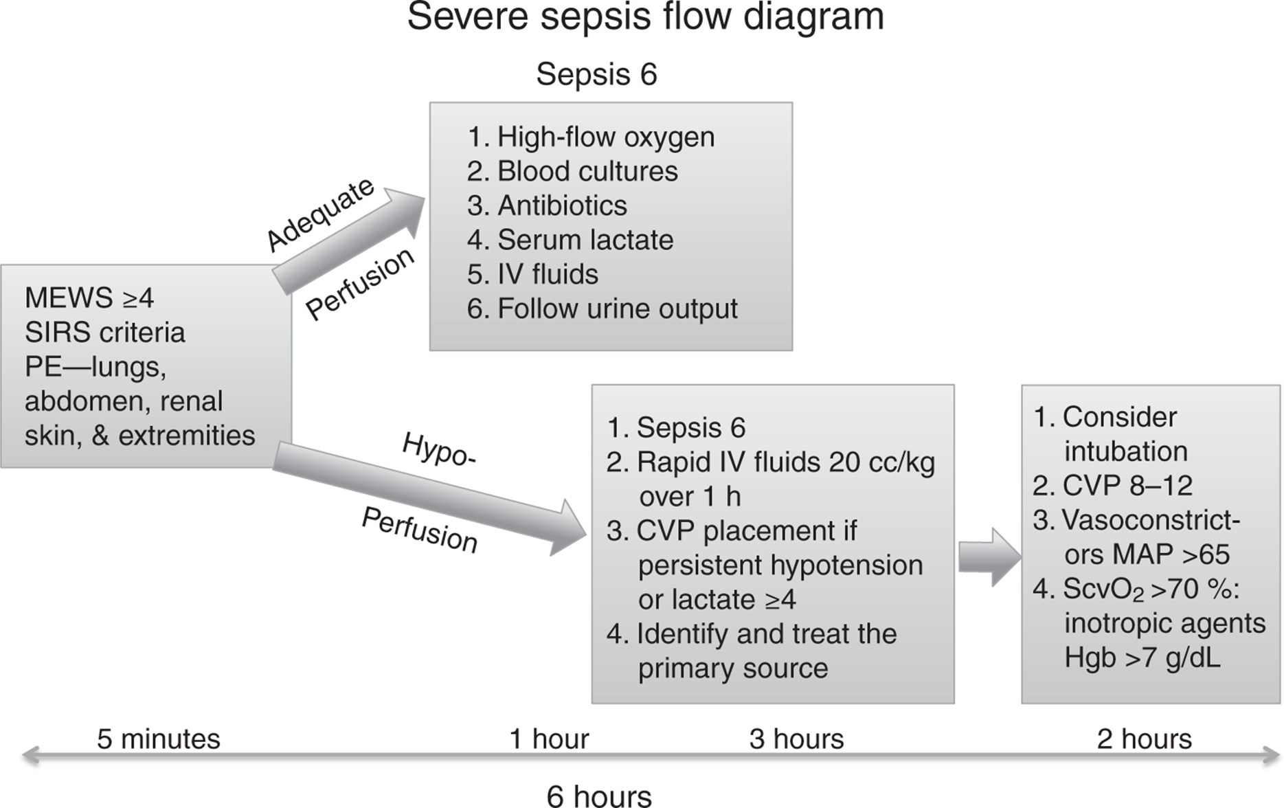
Figure 2.3. Management of sepsis.
1. Deliver high flow oxygen—Oxygen delivery to all organ systems is critical for survival.
2. Draw blood cultures—Assure blood culture volume is 20 cc per sample to maximize sensitivity. With the exception of endocarditis spacing, blood cultures over time have not proven to be helpful, and this approach can delay the initiation of antibiotics. There is no need to exceed four blood cultures because there is no improvement in sensitivity by drawing additional cultures unless there is dramatic change in the patient’s fever pattern. Excess number of blood cultures lowers hemoglobin levels and has the potential to reduce oxygen delivery to the organs.
3. Administer empiric antibiotics within 1 hour (see Table 2.2). Delays in initiating appropriate antibiotics greatly decrease the patient’s likelihood of survival. If appropriate antibiotic therapy is withheld for 36 hours, the mortality is nearly 100%. Whenever possible initiate antibiotics after the blood cultures are drawn. However, logistical problems with blood culture sampling should never be allowed to delay antibiotic administration beyond the first hour.
Table 2.2. Empiric Antibiotic Therapy for Sepsis Syndrome

KEY POINTS
About the Early Management of the Sepsis Syndrome (First Hour)
1. Every hour of delay in treatment increases mortality by 7.6%.
2. Activate the Sepsis 6 bundle and complete within 1 hour; avoid transfer until complete:
a) Deliver high flow oxygen.
b) Draw blood cultures.
c) Begin empiric antibiotics (delay of 36 h = 100% mortality).
d) Draw serum lactate level.
e) Begin rapid IV fluid resuscitation.
f) Begin closely monitoring urine output.
3. Empiric antibiotic therapy must take into account
a) The presumed primary anatomic site of the infection.
b) Local hospital antibiotic sensitivities.
c) Sensitivities of bacteria previously grown from the possible sites of bacteremia.
d) Readjustment based on the blood culture results.
4. Order a serum lactate and a full blood count. A serum lactate level of ≥4 mM/L suggests the patient is progressing from severe sepsis to septic shock.
5. Begin IV fluid resuscitation—If there is evidence of hypoperfusion (cool skin, elevated serum lactate, oliguria), initiate an IV fluid challenge of 20 cc/kg during the first hour. No IV solution has proven to be more beneficial than another (see below).
6. Begin to closely monitor urine output.
Antibiotic Therapy
The initial empiric antibiotic regimen should be chosen based on the suspected primary site of infection (see Table 2.2). Coverage is then chosen to assure treatment of the most common pathogens known to infect that specific site.
Sepsis associated with certain organisms, including Pseudomonas aeruginosa and Candida species, may result in higher mortality rates. Polymicrobial bacteremia also carries an increased mortality risk. When the clinical conditions raise the probability of these organisms, the empiric regimen should include coverage for these pathogens.
The susceptibility of the pathogens within each institution and local unit must also be considered when designing an empiric therapy. When gram-negative bacteria grown from the bloodstream are resistant to the empiric regimen, the risk of death is significantly higher. Therefore, the empiric regimen should always take into account the local antibiotic susceptibility patterns. Furthermore, if a patient has been hospitalized for significant period and develops nosocomial sepsis, coverage should include previously isolated pathogens from the suspicious site.
The regimens suggested in Table 2.2 will treat most pathogens that are isolated at these sites in significant numbers. In 24-48 hours after blood culture results are available, the antibiotic regimen must be adjusted, with narrower spectrum antibiotics utilized whenever possible to reduce the likelihood of selecting for highly resistant pathogens.
CONTINUED MANAGEMENT (AFTER FIRST HOUR)
Hypotension and hypoperfusion are major ongoing concerns in patients with severe sepsis. Therefore, clinicians need to closely monitor mean blood pressure, serum lactate, as well as skin color and temperature of the extremities. When serum lactate levels rise to >4 mM/L, mortality increases to nearly 40% as compared to <15% for patients with serum lactate levels of <2 mM/L. If after institution of the Sepsis 6 the patient continues to demonstrate elevated lactate levels, has physical evidence of hypoperfusion, or continues to be hypotensive (mean pressure <65 mmHg), an intravascular device should placed to monitor central venous pressure (CVP). IV fluids should be aggressively administered to maintain the CVP between 8 and 12 mmHg (see Figure 2.3). Multiple comparisons have failed to demonstrate a significant difference in therapeutic efficacy between IV crystalloid solutions such as normal saline and colloid solutions such as albumin. Given the far greater expense of colloids, crystalloids are considered by most experts to be the treatment of choice. The use of normal saline as the primary IV solution increases the likelihood of developing hyperchloremia; therefore, some experts recommend balanced solutions such as Hartmann solution or Ringer lactate that are not associated with hyperchloremia.
If blood pressure remains low after the aggressive repletion with IV solutions, one or more vasopressors should be added. In the past, dopamine was recommended as the vasopressor of choice; however, a recent meta-analysis has revealed that when compared to norepinephrine, dopamine increases mortality by 1.1–1.2 and increases the risk of arrhythmias. Therefore, the recommended vasopressor of choice is norepinephrine (Levophed). This agent acts on both alpha-1 and beta-1 adrenergic receptors producing potent vasoconstriction as well as a modest increase in cardiac output. The recommended adult dosing is 0.01–3 μg/kg/minute. In the past, lowdose dopamine was recommended to maintain renal output; however, given the increased risk of arrhythmias, this approach has recently been abandoned by most experts.
In critically ill patients, another clinical monitoring system called the Acute Physiologic and Chronic Health Evaluation II (APACHE II) is commonly used. The patient is periodically scored based on vital sign parameters, metabolic studies, and underlying disease. This score closely correlates with the severity and progression of disease as well as mortality; scores theoretically can range from 0 to 63, the higher the score the worse the prognosis. Generally, patients with scores of >20 are considered gravely ill. This score can be readily calculated using the website http://clincalc.com/IcuMortality/APACHEII.aspx.
KEY POINTS
About the Continued Management of Sepsis
1. Hypotension and hypoperfusion need to be closely monitored.
a) Maintain mean BP >65 mmHg.
b) Monitor serum lactate >4 mM/L = mortality of 40%.
c) Monitor extremity temperature.
2. If continued hypotension and/or hypoperfusion after Sepsis 6 bundle, monitor central venous pressure (CVP).
a) IV infusion to maintain CVP of 8-12 mmHg.
b) Use crystalloid solutions, Ringer lactate, or Hartmann solution to avoid hyperchloremia.
3. Vasoconstrictor of choice is norepinephrine (Levophed), and dopamine increases the risk of arrhythmias.
4. Apache II score helpful in monitoring progress.
OTHER CONSIDERATIONS
Disseminated Intravascular Coagulopathy
Consumption coagulopathy commonly accompanies severe sepsis, and the diagnosis of DIC is made clinically with supportive laboratory evidence. The majority of patients demonstrate abnormal thrombosis and/or bleeding. Manifestations of thrombosis can include cyanosis or gangrene of fingers or toes and hemorrhagic necrosis of the skin (most prominent with meningococcemia). Bleeding will first be seen at sites of surgical intervention and at intravascular device placement sites. In more severe cases, widespread bruising is observed as well oozing of blood from all mucosal surfaces.
Laboratory abnormalities include an elevated PT and PTT as a consequence of consumption of host coagulation factors; low fibrinogen and elevated D-dimers are observed as a consequence of fibrinolysis. Thrombocytopenia is observed in up to 98% of cases in some series.
No specific treatment has proven to be efficacious other than clearing the underlying infection. When bleeding is severe and uncontrollable, platelet and fresh frozen plasma transfusions can be administered as temporary supportive measures.
Acute Respiratory Distress Syndrome
Sepsis is the leading cause of ARDS. During sepsis, the high cytokine levels combined with bacterial products precipitate a breakdown of the vascular endothelial junctions within the lung causing leakage of fluid into the alveoli. The high-level concentrations of cytokines in the lung also attract and activate neutrophils. The toxic oxygen products and proteases released by neutrophils damage the endothelial junctions and exacerbate leakage of fluid into the alveoli.
The hallmarks of ARDS are pulmonary edema resulting in extensive opacification of the lungs on chest X-ray (CXR), poor oxygen exchange, and reduced lung compliance. Treatment is supportive and includes supplemental oxygen to maintain arterial oxygen saturation >90%, and mechanical ventilator support utilizing low tidal volume settings and positive end-expiratory pressure (PEEP).
Potentially Efficacious Interventions That Have Failed
Given our extensive understanding of SIRS and sepsis and high mortality of this syndrome, drug companies have invested in clinical trials, but many preliminarily promising approaches have proven to be ineffective including:
KEY POINTS
Other Complications Associated with Sepsis
1. Disseminated intravascular coagulathy (DIC).
a) Evidence of thrombosis and bleeding.
b) Elevated PT and PTT, low fibrinogen, high D-dimers.
c) Thrombocytopenia in almost all cases.
d) Supportive care, platelets, and fresh frozen plasma for uncontrolled bleeding.
2. Acute respiratory distress syndrome (ARDS).
a) Sepsis the most common cause.
b) High cytokine levels and activated neutrophils damage vascular endothelial junctions causing fluid to leak into the alveoli.
c) Hallmarks—Diffuse opacification on CXR, poor O2 exchange, reduced lung compliance.
d) Treatment—Mechanical ventilation with low tidal volumes and positive end expiratory pressure (PEEP).
1. Anti-inflammatory agents such as ibuprofen and even narcotic antagonists have not proven to be of value in large-scale studies.
2. Monoclonal antibody against the core of the endotoxin molecule has not been conclusively shown to be beneficial.
3. Antibody against TNF-α and the TNF-α receptor has failed.
4. Studies utilizing IL-1 receptor antagonists have been inconclusive.
5. Platelet-activating factor antagonists have failed.
Corticosteroids
The potential benefit of corticosteroid administration for sepsis has been studied for over 50 years with continued inconclusive results. Meta-analysis of recent investigations suggests that in patients with severe sepsis, hydrocortisone 100 mg IV Q8h or 50 mg IV Q6H may reduce mortality as compared to placebo. Corticosteroids should not be used in mildly or moderately ill patients. This treatment should be continued for no longer than 7 days.
KEY POINTS
About Adjunctive Therapies for Sepsis Syndrome
1. Multiple clinical trials have failed to document efficacy for
a) Anti-inflammatory agents.
b) Monoclonal antibody against endotoxin.
c) Anti-tumor necrosis factor-α antibodies.
d) Interleukin-1 antagonists.
e) Platelet-activating factor antagonists.
2. Corticosteroids in low doses may be beneficial.
3. Activated protein C (drotrecogin α) of questionable benefit, taken off the market.
Drotrecogin α
Protein C activity is reduced in septic patients, and this protein is known to play a key role in inhibiting coagulation. Animal studies have shown that infusion of activated protein C reduces mortality in lethal E. coli infections. An early clinical trial in humans demonstrated a modest reduction of 28-day mortality in septic shock from 30.8% to 24.7% in patients receiving activated protein C. The U.S. Food and Drug Administration approved the manufactured version of protein C, drotrecogin α, as an adjunct to standard therapy for the treatment of severe sepsis. However, a recent meta-analysis of five studies concluded that drotrecogin α was of no benefit in adult patients with sepsis, and was associated with an increased risk of bleeding. Based on a preliminary analysis of the commercially sponsored PROWESS-SHOCK trial, this product has been withdrawn from the market.
CONCLUSIONS
Despite decades of research, severe sepsis remains a leading cause of preventable death in our hospitals. Experts have recognized that early recognition and treatment are the keys to reducing mortality. When bacteria are allowed to seed the bloodstream, SIRS rapidly progresses to severe sepsis followed by septic shock, organ failure, and death. In all patients with MEWS scores of ≥4, sepsis should be strongly considered as the possible underlying cause, and the finding of hyperthermia or hypothermia combined with an abnormal peripheral WBC should activate the Sepsis 6 protocol that mandates empiric antibiotic coverage within 1 hour. Given the complexity, rapid progression, and eventual irreversibility of the sepsis syndrome, adjunctive therapy is unlikely to provide significant benefit in the foreseeable future. Therefore, the responsibility of curing the sepsis syndrome will continue to lie with frontline caregivers. Early recognition and prompt antibiotic therapy can save hundreds of thousands of lives.
FURTHER READING
Daniels R, Nutbeam T, McNamara G, Galvin C. The sepsis six and the severe sepsis resuscitation bundle: a prospective observational cohort study. Emerg Med J. 2011;28(6):507-512.
De Backer D, Aldecoa C, Njimi H, Vincent JL. Dopamine versus norepinephrine in the treatment of septic shock: a meta-analysis*. Crit Care Med. 2012;40(3):725-730.
Dellinger RP, Levy MM, Carlet JM, et al. Surviving sepsis campaign: international guidelines for management of severe sepsis and septic shock: 2008. Crit Care Med. 2008;36(1):296-327.
Kumar A, Roberts D, Wood KE, et al. Duration of hypotension before initiation of effective antimicrobial therapy is the critical determinant of survival in human septic shock. Crit Care Med. 2006;34(6):1589-1596.
Marti-Carvajal AJ, Sola I, Lathyris D, Cardona AF. Human recombinant activated protein C for severe sepsis. Cochrane Database Syst Rev. 2012;3:CD004388.
Rivers E, Nguyen B, Havstad S, et al. Early goal-directed therapy in the treatment of severe sepsis and septic shock. N Engl J Med. 2001;345(19):1368-1377.
Wiersinga WJ. Current insights in sepsis: from pathogenesis to new treatment targets. Curr Opin Crit Care. 2011;17(5):480-486.