
Time Recommended to Complete: 3 days
Frederick S. Southwick, M.D.
ACUTE PNEUMONIAS
GUIDING QUESTIONS
1. What are the factors that predispose the host to develop pneumonia?
2. What are the symptoms, signs, and diagnostic tests that help to differentiate viral from bacterial pneumonia?
3. How useful is sputum Gram stain, and what are the parameters that are used to assess the adequacy of a sputum sample?
4. How should the clinician interpret the sputum culture, and should sputum cultures be obtained in the absence of sputum Gram stain?
5. What are some of the difficulties encountered in trying to determine the cause of acute pneumonia?
6. How helpful is a chest radiograph in determining the specific cause of pneumonia?
7. How often should chest X-ray be repeated, and how long do the radiologic changes associated with acute pneumonia persist?
8. Which antibiotic regimens are recommended for empiric therapy of community-acquired pneumonia and why?
POTENTIAL SEVERITY
Acute pneumonia is a potentially life-threatening illness requiring rapid diagnosis and treatment. A delay in antibiotic treatment increases the risk of a fatal outcome.
GENERAL CONSIDERATIONS IN ACUTE PNEUMONIA
Prevalence
Annually, 2-3 million cases of pneumonia are reported in the United States. Estimates suggest that pneumonia is responsible for more than 10 million physician visits, 500,000 hospitalizations, and 45,000 deaths annually. Overall, 258 people per 100,000 population require hospitalization for pneumonia, and that number rises to 962 per 100,000 among or nearly 1/100 for those over the age of 65 years. It is estimated that, annually, 1 in 50 people over 65 years of age and 1 in 20 over 85 years will develop a pneumonia. Pneumonia occurs most commonly during the winter months.
Causes
Improved diagnostic techniques have shown that the number of pathogens that cause acute pneumonia is ever expanding (Table 4.1).
Table 4.1. Common Causes of Acute Pneumonia

The leading cause of acute community-acquired pneumonia (CAP) remains Streptococcus pneumoniae, followed by Haemophilus influenzae. Mycoplasma and Chlamydophila pneumoniae also account for a significant percentage of acute pneumonias. Staphylococcus aureus is an unusual community-acquired pathogen, but it can cause ventilator-associated pneumonia (VAP). Gram-negative bacteria other than H. influenzae are also an uncommon cause of CAP except in patients with underlying lung disease or alcoholism. Gram-negative pneumonia most commonly develops in hospitals or nursing homes. Legionellaspecies vary in importance, depending on the season and geographic area. Anaerobes such as anaerobic streptococci and bacteroides can cause acute pneumonia following aspiration of mouth contents. Common viral pathogens include influenza, parainfluenza, and respiratory syncytial virus.
Pathogenesis and Pathology
Under normal conditions, the tracheobronchial tree is sterile. The respiratory tract has a series of protective mechanisms that prevent pathogens from gaining entry [Figure 4.1 (A)].

Figure 4.1. A. Host defense in the respiratory tract. B. Factors that interfere with host defense of the respiratory tract.
1. The nasal passages contain turbinates and hairs that trap foreign particles.
2. The epiglottis covers the trachea and prevents secretions or food from entering the trachea.
3. The tracheobronchial tree contains cells that secrete mucin. Mucin contains a number of antibacterial compounds including immunoglobulin A antibodies, defensins, lysozymes, and lactoferrin. Mucin also is sticky, and it traps bacteria or other foreign particles that manage to pass the epiglottis.
4. Cilia lining the inner walls of the trachea and bronchi beat rapidly, acting as a conveyer belt to move mucin out of the tracheobronchial tree to the larynx.
5. When significant volumes of fluid or large particles gain access to the trachea, the cough reflex is activated, and the unwanted contents are quickly forced out of the tracheobronchial tree.
6. If pathogens are able to bypass all of the above protective mechanisms and gain entry into the alveoli, they encounter a space that, under normal circumstances, is dry and relatively inhospitable. The presence of an invading pathogen induces the entry of neutrophils and alveolar macrophages that ingest and kill infecting organisms. Immunoglobulins and complement are found in this space. Surfactants also have a protective function.
7. The lymphatic channels adjacent to the alveoli serve to drain this space and transport fluid, macrophages, and lymphocytes to the mediastinal lymph nodes.
Bacterial pathogens usually gain entry into the lung by aspiration of mouth flora or by inhalation of small aerosolized droplets (<3 μm in diameter) that can be transported by airflow to the alveoli. Once the pathogen takes hold, a series of inflammatory responses is triggered. These responses have been most carefully studied in pneumonia attributable to S. pneumoniae.
KEY POINTS
About the Protective Mechanisms of the Lung
1. Normally, the tracheobronchial tree is sterile.
2. The nasal turbinates trap foreign particles, and the epiglottis covers the trachea.
3. Mucin has antibacterial activity, and cilia transport mucin out of the lung.
4. Coughing expels foreign material that enters the tracheobronchial tree.
5. Alveoli can deliver polymorphonuclear leukocytes (PMNs), macrophages, immunoglobulins, and complement to destroy invading pathogens.
6. Lymphatics drain macrophages and PMNs to the mediastinal lymph nodes.
First, an outpouring of edema fluid into the alveoli occurs, serving as an excellent culture media for further bacterial growth. As fluid accumulates, it spills over to adjacent alveoli through the pores of Kohn and the terminal bronchioles, resulting in a centrifugal spread of infection. Coughing and the physical motion of respiration further enhance spread.
KEY POINTS
About the Pathogenesis of Pneumonia
1. Pathogens are aspirated or inhaled as small aerosolized droplets.
2. Bacterial invasion of the alveoli induces
a) edema fluid that spreads to other alveoli through the pores of Kohn, and
b) infiltration by polymorphonuclear leukocytes and red blood cells, followed by macrophages.
3. Infection spreads centrifugally:
a) Newer regions in the periphery appear red (“red hepatization”).
b) Older regions are central and appear gray (“gray hepatization”).
4. Streptococcal pneumonia does not cause permanent tissue destruction.
5. Staphylococcus aureus, gram-negative rods, and anaerobes cause permanent damage.
Next, polymorphonuclear leukocytes (PMNs) and some red blood cells begin to accumulate in the alveolar space. Eventually, they fill the region and form a zone of consolidation.
Macrophages then enter the lesions and assist the PMNs in clearing the infection. Histopathology reveals zones of varying age. The most distal regions represent the most recent areas of infection. There, edema fluid, PMNs, and red blood cells predominant. On lower power microscopy, this region has an appearance similar to the architecture of the liver—an effect termed “red hepatization.” Older central regions have more densely packed PMNs and macrophages. This region has a grayer color and forms the zone of “gray hepatization.”
Pulmonary pathogens demonstrate marked differences in their invasiveness and ability to destroy lung parenchyma. S. pneumoniae causes minimal tissue necrosis and is associated with little or no scar formation. Full recovery of pulmonary function is the rule. S. aureus releases a number of proteases that permanently destroy tissue. Gram-negative rods and anaerobic bacteria also cause permanent tissue destruction.
Predisposing Factors
Most bacterial pneumonias are preceded by a viral upper respiratory infection [Figure 4.1 (B)]. Influenza virus is well known to predispose to S. pneumoniae and S. aureus pneumonia. Viral infections of the upper respiratory tract can damage the bronchial epithelium and cilia.
Virus-mediated cell damage also results in the production of serous fluid that can pool in the pulmonary alveoli, serving as an excellent culture media for bacteria. The low viscosity of this fluid, combined with depressed ciliary motility, enables the viral exudate to carry nasopharyngeal bacteria past the epiglottis into the lungs. Smoking also damages the bronchial epithelial cells and impairs mucociliary function. As a consequence, smokers have an increased risk of developing pneumonia. Congenital defects in ciliary function (such as Kartagener syndrome) and diseases resulting in highly viscous mucous (such as cystic fibrosis) predispose patients to recurrent pneumonia.
An active cough and normal epiglottal function usually prevent nasopharyngeal contents from gaining access to the tracheobronchial tree. However, drugs such as alcohol, sedatives, and anesthetics can depress the level of consciousness and impair these functions, predisposing the patient to pneumonia. Elderly individuals, particularly after a cerebrovascular accident, often develop impairments in swallowing that predispose them to aspiration. In addition, elderly people demonstrate reduced humoral and cell-mediated immunity, rendering them more susceptible to viral and bacterial pneumonia.
Patients with impairments in immunoglobulin production, T- and B-cell function, and neutrophil and macrophage function are also at greater risk of developing pneumonia. Organ-transplant patients on immunosuppressive agents and patients with AIDS have a greater likelihood of developing pneumonia. Chronic diseases, including multiple myeloma, diabetes, chronic renal failure, and sickle cell disease, have been associated with an increased risk of pneumonia.
KEY POINTS
About Factors That Predispose to Pneumonia
1. Viral infections damage cilia and produce serous exudate that can transport nasopharyngeal bacteria into the alveoli.
2. Smoking damages bronchial epithelial cells and impairs ciliary function.
3. Alcohol and other drugs depress coughing and epiglottal function.
4. Elderly patients have reduced humoral and cell-mediated immunity, and may have impaired swallowing because of stroke.
5. Patients on immunosuppressive agents and patients with AIDS have depressed humoral and cell-mediated immunity.
6. Patients with chronic diseases are at increased risk of pneumonia.
7. Cold weather dries the mucous membranes and increases person-to-person spread of infection.
Cold weather is thought to contribute to the development of pneumonia. Cold, dry weather can alter the viscosity of mucous and impair bacterial clearance. Cold weather also encourages people to remain indoors, a situation that enhances person-to-person spread of respiratory infections.
Symptoms and Signs
CASE 4.1
A 55-year-old woman was first seen in the emergency room in December complaining of a nonproductive cough, nasal stuffiness, and fever. She also noted diffuse severe muscle aches and joint pains and a generalized headache. In her epidemiologic history, she noted that she had recently seen her grandchildren, who all had high fevers and were complaining of muscle aches.
Physical examination showed these positive findings: temperature, 39°C; throat, erythematous; nasal discharge, clear; muscles, diffusely tender. A chest X-ray (CXR) was within normal limits.
Three days into the clinical course of her illness, the patient noted some improvement in her cough, muscle aches, and joint pains; however, on the fourth day, she developed a high fever (40°C) preceded by a teeth-chattering chill. That day, her cough became productive of rusty-colored opaque sputum, and she began feeling short of breath.
A repeat physical examination showed a temperature of 40.6°C and a respiration rate of 36 per minute. In general, this was a very ill appearing, anxious woman, gasping for air. Lungs were mildly dull to percussion, with E-to-A changes, and rales and rhonchi localized to the right middle lobe area.
A peripheral white blood cell (WBC) count measured 16,000/mm3, with 68% PMNs, 20% immature forms (bands and metamyelocytes), 8% lymphocytes, and 4% monocytes. Sputum Gram stain showed many gram-positive lancet-shaped diplococci, many PMNs (10/high-power field), and no squamous epithelial cells. A CXR revealed a dense right middle lobe infiltrate (Figure 4.2).

Figure 4.2. Pneumococcal pneumonia: A. Chest radiograph demonstrates classical lobar infiltrate (Courtesy of Dr. Pat Abbitt, University of Florida); and B. sputum Gram stain shows Streptococcus pneumoniae. Note that the cocci come to a slight point, explaining the term “lancet-shaped.” See color image on color plate 1
In case 4.1, the patient’s initial symptoms suggested a viral illness involving the upper respiratory tract (rhinitis and nonproductive cough); central nervous system (CNS) or air sinuses, or both (headache); and musculoskeletal system (myalgias and arthralgias). Such symptoms are generally attributed to an influenzalike illness. A number of viruses can explain these symptoms, including influenza, parainfluenza, adenovirus, respiratory syncytial virus (more common in children, but also found in elderly individuals and transplant patients), rhinoviruses (usually less severe), and enteroviruses.
Subsequently, within a 24-hour period, this patient experienced the abrupt onset of a new constellation of symptoms. The onset of the new illness can be classified as acute. An illness is termed “acute” when symptoms and signs develop over 24-48 hours. Symptoms that develop over 3 days to 1 week are generally classified as subacute, and symptoms that progress more slowly (over 3 weeks to several months) are classified as chronic.
In generating a potential list of causative agents, the infectious disease specialist frequently uses the pace of the illness to narrow the possibilities. Pneumonias are generally classified into two groups: acute and chronic. Most bacterial and viral pneumonias develop quickly; fungal and mycobacterial pulmonary infections tend to develop at a slower pace. Acute pneumonia can be further classified as “typical” or “atypical.” Typical pneumonia is characterized by the more rapid onset of symptoms, more severe symptomatology, a productive cough, and dense consolidation on chest X-ray (CXR), as observed in case 4.1. Atypical pneumonia tends to be slower in onset (often subacute), symptoms tend to be less severe, cough is productive of minimal sputum, and CXR usually reveals a patchy or interstitial pattern. Finally, pulmonary infections are separated into community-acquired or nosocomial. “Community-acquired” is defined as an infection developing in a patient who has not recently (>14 days) been hospitalized or resided in a chronic care facility.
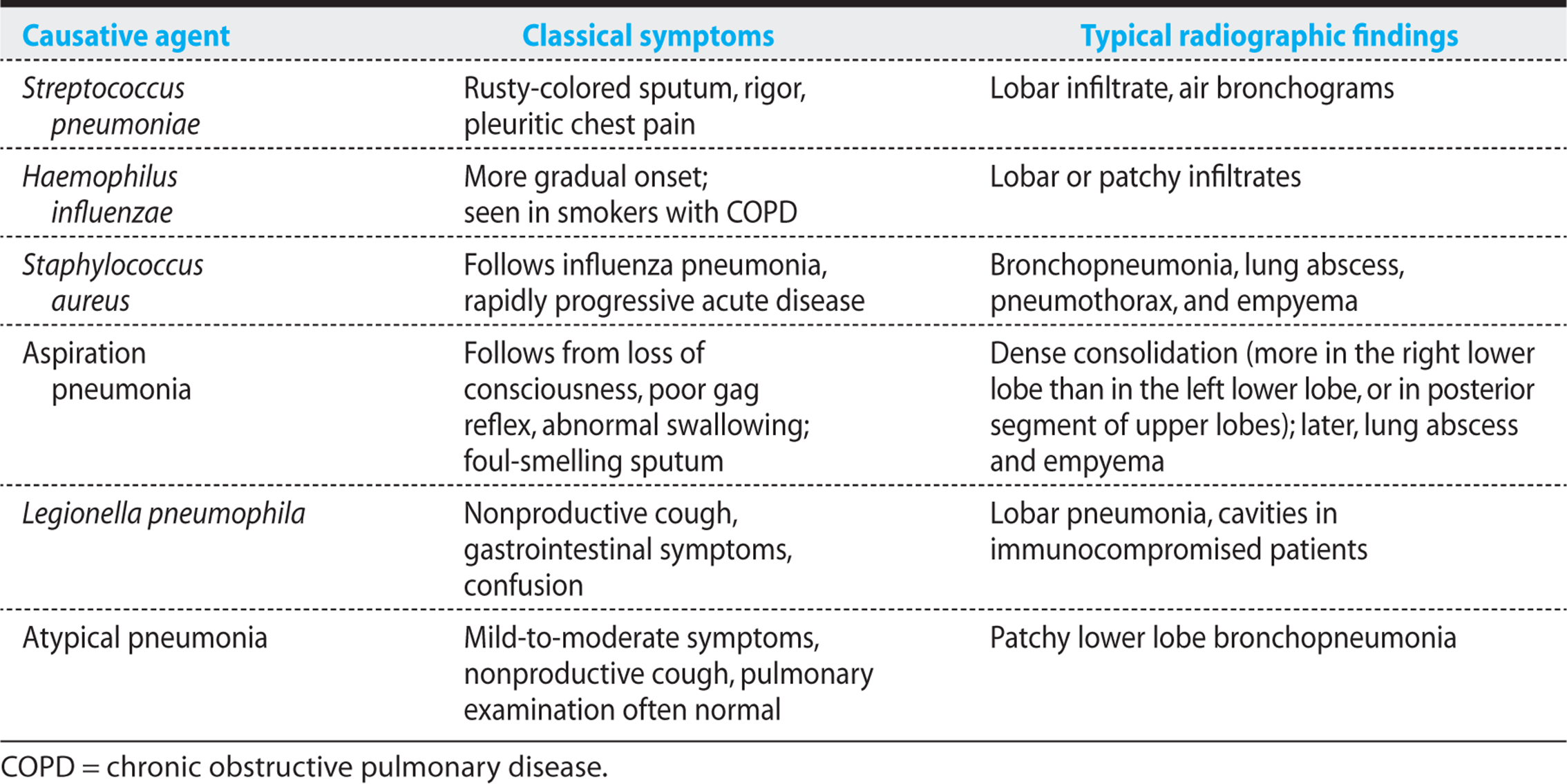
Although considerable overlap in symptoms, signs, and CXR findings are observed in cases of acute CAP, certain key clinical characteristics are helpful in guiding the determination of the most likely causes (Table 4.2). Generation of a logical differential list of potential pathogens guides the choice of diagnostic tests and narrows the possible treatment regimens.
Table 4.2. Clinical Characteristics of Acute Community-Acquired Pneumonia Classified by Cause
KEY POINTS
About the Classification of Pneumonia
Pneumonias are classified by
1. Pace of illness:
a) Acute—symptoms develop over 24-48 hours.
b) Chronic—symptoms progress over 3 weeks or longer.
2. Specific constellations of symptoms:
a) Typical—rapid onset, more severe symptoms, productive cough, dense consolidation on chest X-ray (CXR).
b) Atypical—somewhat slower onset, less severe symptoms, nonproductive cough, patchy interstitial pattern on CXR.
3. Environment in which the pneumonia was acquired:
a) Community acquired—patient not recently (>14 days) in a hospital or chronic care facility.
b) Nosocomial—patient in a hospital when the infection developed.
Important symptoms that need to be reviewed include these:
1. Cough. Frequency of the cough, production of sputum, and color of the sputum should be documented. A nonproductive cough or a cough productive of scanty sputum suggests an atypical pneumonia; a cough productive of rusty-colored sputum raises the possibility of S. pneumoniae. Thick, “red current jelly” sputum has been reported in cases of Klebsiella pneumoniae; green-colored sputum is more frequently encountered in patients with H. influenzae and Pseudomonas aeruginosa pneumonia (typically a nosocomial pathogen, or found in patients cystic fibrosis). Frank hemoptysis is observed in cavitary tuberculosis, lung abscess, and lung carcinoma. It should be emphasized that considerable overlap occurs in the sputum characteristics of the various forms of pneumonia, and these observations cannot be considered specific.
2. Chest discomfort. Pleuritic chest pain (pain associated with deep inspiration) is classically described in patients with S. pneumoniae. Pain is usually sharp and stabbing. Because the pulmonary parenchyma has no pain-sensing nerves, the presence of chest pain indicates inflammation of the parietal pleura. When the diaphragm becomes inflamed, the pain can mimic cholecystitis or appendicitis, and on occasion this type of pain has precipitated exploratory laparotomy. Anaerobes, S. pyogenes, and S. aureus are other pathogens that can also spread to the pleura and cause chest pain. Pleuritic pain is also characteristic of pleurodynia, a pain syndrome caused by the enteroviruses coxsackievirus and echovirus.
3. Shortness of breath. A report of increased shortness of breath suggests poor alveolar oxygen exchange, indicative of severe infection. Some patients experience shortness of breath as a result of pleuritic chest pain that limits the ability to breath deeply. To avoid pain, patients may breath quickly and shallowly, and this breathing pattern may be interpreted as shortness of breath.
4. Epidemiology. A careful epidemiologic history is often helpful. A number of environmental factors predispose to pneumonia. Animal exposure must be carefully reviewed, including contact with wild game, birds, bats, and rodents (see Chapter 13). Exposure to outside air conditioning units or construction sites should be identified (Legionnaires disease). Travel history may be helpful. For example, travel to the Southwest raises concerns about coccidioidomycosis, and travel to the Ohio River valley raises the possibility of histoplasmosis. Because many respiratory illnesses spread from person to person, a history of exposure to family members or friends with illnesses should be ascertained. Occupational and sexual history should also be elicited.
A thorough physical examination should be performed during the initial evaluation of possible pneumonia. Vital signs are helpful in determining the severity of illness. A respiratory rate of more than 30 breaths per minute, a systolic blood pressure under 90 mmHg, a pulse above 125 beats per minute, and a temperature below 35°C (95°F) or above 40°C (104°F) are all bad prognostic signs. Depressed mental status is also associated with a poor prognosis.
KEY POINTS
About the History in Pneumonia
1. Cough. Frequency, production of sputum, color and thickness of sputum.
2. Chest pain. Pain on deep inspiration, usually sharp, suggests pleural involvement. Seen in Streptococcus pneumoniae, Staphylococcus aureus, Streptococcus pyogenes, anaerobes, and coxsackievirus and echovirus.
3. Shortness of breath. A worrisome symptom; may be the result of pleuritic chest pain rather than poor gas exchange.
4. Epidemiology. Travel history, animal exposure, exposure to people with respiratory illnesses, occupational and sexual history.
KEY POINTS
About the Physical Examination of Pneumonia
1. A respiratory rate >30/min, a blood pressure <90 mmHg, a pulse >125/min, and a temperature <35°C or >40°C are bad prognostic findings.
2. Depressed mental status and stiff neck suggest bacterial meningitis.
3. Pulmonary auscultation often underestimates the extent of pneumonia:
a) Bronchial breath sounds and egophony suggest consolidation.
b) Dullness to percussion indicates consolidation or a pleural effusion.
c) Pleural effusion is accompanied by decreased breath sounds and, in some cases, a friction rub.
Ear, nose, and throat examination may reveal vesicular or crusted lesions consistent with Herpes labialis, an infection that may reactivate as a consequence of the stress of the primary illness. Neck stiffness in association with depressed mental status may indicate the development of bacterial meningitis, a potential complication of pneumococcal pneumonia.
Pulmonary auscultation often fails to detect the extent of infection, and when pneumonia is being considered, the physical examination should be followed by a CXR. Asymmetry of chest movements may be observed, movement being diminished on the side with the pneumonia. When infection has progressed to consolidation, as in case 4.1, filling of the lung parenchyma with exudate alters sound conduction. Airflow from the bronchi is conducted through this fluid to the chest wall, resulting in bronchial or tubular breath sounds. When the patient is asked to say “E,” an “A” is heard on auscultation (egophony). Percussion of the chest wall also demonstrates dullness in the areas of consolidation. Dullness to percussion in association with decreased breath sounds suggests the presence of a pleural effusion. A “leathery” friction rub may be heard over the site of consolidation, indicating pleural inflammation.
Laboratory Findings
Radiologic studies Physical examination is unreliable for making the diagnosis of pneumonia. If pneumonia is a potential diagnosis, CXR must be performed to confirm or exclude the disease. The radiologic pattern can serve as a rough guideline to possible causative agents; however, the use of immunosuppressive agents (resulting in neutropenia, decreased cell-mediated immunity, and depressed macrophage function) can greatly alter the typical radiologic appearance of specific pathogens. Patients with AIDS also present with atypical CXR.
Five classical patterns have been described:
1. Lobar pneumonia. “Lobular pneumonia” refers to a homogeneous radiologic density that involves a distinct anatomic segment of the lung (Figure 4.2). Infection originates in the alveoli. As it spreads, this form of infection respects the anatomic boundaries of the lung and does not cross the fissures. Lobar pneumonia is most commonly seen with S. pneumoniae, H. influenzae, and Legionella.
2. Bronchopneumonia. The bronchopneumonia form of pulmonary infection originates in the small airways and spreads to adjacent areas (Figure 4.4). Infiltrates tend to be patchy, to involve multiple areas of the lung, and to extend along bronchi. Infiltrates are not confined by the pulmonary fissures. Bronchopneumonia is commonly observed with S. aureus, gram-negative bacilli, Mycoplasma, Chlamydophila,and respiratory viruses.
3. Interstitial pneumonia. Infections causing inflammation of the lung interstitium result in a fine diffuse granular infiltrate (Figure 16.2). Influenza and cytomegalovirus commonly present with this CXR pattern. In patients with AIDS, Pneumocystis jirovecii infection results in interstitial inflammation combined with increased alveolar fluid that can mimic cardiogenic pulmonary edema. Miliary tuberculosis commonly presents with micronodular interstitial infiltrates.
4. Lung abscess. Anaerobic pulmonary infections often cause extensive tissue necrosis, resulting in loss of lung tissue and formation of cavities filled with inflammatory exudate (Figure 4.5). S. aureus also causes tissue necrosis and can form cavitary lesions.
5. Nodular lesions. Histoplasmosis, coccidioidomycosis, and cryptococcosis can present as nodular lung lesions (multiple or single) on CXR. Hematogenous pneumonia resulting from right-sided endocarditis commonly presents with “cannonball” lesions that can mimic metastatic carcinoma.
The role of high-resolution chest computed tomography (CT) scan is evolving, and this test has proved helpful for more clearly demonstrating interstitial infiltration, pulmonary cavities, nodules, and pleural fluid collections. Given the high cost of this test and the resulting high radiation exposure, this test should be used sparingly.
KEY POINTS
About Chest X-Ray in Pneumonia
1. If pneumonia is being considered, a chest X-ray (CXR) should always be performed.
2. Radiographic patterns may be atypical in patients receiving immunosuppressants and in patients with AIDS.
3. Five typical CXR patterns have been described:
a) Lobar pattern. Streptococcus pneumoniae, Haemophilus influenzae, and Legionella.
b) Bronchopneumonia pattern. Staphylococcus aureus, gram-negative organisms, Mycoplasma, Chlamydophila, and viral.
c) Interstitial pattern. Influenza and cytomegalovirus, Pneumocystis, miliary tuberculosis.
d) Lung abscess. Anaerobes, S. aureus.
e) Nodular lesions. Fungal (histoplasmosis, coccidioidomycosis, cryptococcosis) and right-sided endocarditis.
4. Patterns on chest radiographs are only rough guides. Considerable overlap between the various pathogens has been observed.
Patients with an infiltrate, who are under age 65, have a normal mental status, and normal or only mildly deranged vital signs can be treated as outpatients (Figure 4.5). Sputum Gram stain and culture are optional in these patients, as are any additional tests.
In more severely ill patients who are being considered for hospitalization, additional tests to assess the severity of the illness need to be ordered.
Blood tests: A complete and differential blood cell count should be obtained. Patients with bacterial pneumonia usually have an elevated peripheral white blood cell (WBC) count and a left shift. When pneumococcal pneumonia is accompanied by a low peripheral WBC count (<6000), a fatal outcome is more likely. The finding of anemia (hematocrit <30%), usually indicative of chronic underlying disease, is also associated with a worse prognosis. C-reactive protein (CRP) is another excellent marker for following disease progression and response to antibiotics.
Blood oxygenation also needs to be assessed. The O2 saturation should be determined, and if it is at all depressed, an arterial blood gas should be obtained. Systemic acidosis (pH <7.35) and an arterial partial pressure below 60 mmHg are bad prognostic signs. A significant depression in oxygenation reflects loss of alveolar function and lack of oxygen transfer to alveolar capillaries. Deoxygenated blood passes from the right side of the heart to the left side, creating a physiologic right-to-left shunt.
Other metabolic parameters also need to be assessed. A blood urea nitrogen level above 30 mg/dL reflects hypoperfusion of the kidneys or dehydration (or both) and is a negative prognostic finding. A serum sodium reading below 130 mEq/L reflects increased antidiuretic hormone secretion in response to decreased intravascular volume in addition to severe pulmonary disease. Such a reading is another negative prognostic finding, as is a serum glucose level exceeding 250 mg/dL.
Cultures: Two blood cultures should be drawn before antibiotics are started. Positive blood cultures definitively identify the cause of the disease. Blood cultures are positive in 1-16% of cases of CAP. At the time of bacteremia, some patients may experience a rigor or bed-shaking chill. Patients with pneumococcal pneumonia classically experience a single rigor.
Sputum requires careful analysis and frequently provides helpful clues to the probable diagnosis. Sputum samples often become contaminated with bacteria and cells from the nasopharynx, making interpretation of the cultures difficult. Ideally, the bedside nurse should supervise the acquisition of sputum to ensure that the patient coughs deeply and brings up the sample from the tracheobronchial tree, rather than simply supplying expectorated saliva from the mouth. Use of inhaled saline to induce greater sputum production has proved to be unhelpful, and is not recommended.
KEY POINTS
About Blood Tests in Pneumonia
1. With the exception of patients under the age of 50 years, without underlying disease, and with normal vital signs, multiple blood tests are used to assess the severity of disease.
2. A peripheral white blood cell count below 6000/mm3 in Streptococcus pneumoniae is a bad prognostic finding.
3. Anemia (hematocrit <30%), blood urea nitrogen above 30 mg/dL, serum sodium below 130 mEq/L, and glucose above 250 mg/dL are associated with a worse prognosis.
4. Arterial blood O2 below 60 mmHg and pH below 7.35 worsen prognosis.
5. Two blood samples should be drawn before antibiotics are stated; blood cultures are positive in up to 16% of patients.
The adequacy of the sample should be determined by low-power microscopic analysis of the sputum Gram stain. The presence of more than 10 squamous epithelial cells per low-power field indicates significant contamination from the nasopharynx, and the sample should be discarded. The presence of more than 25 PMNs per low-power field and the presence of bronchial epithelial cells provide strong evidence that the sample originates from the tracheobronchial tree.
Despite originating from deep within the lungs, sputum samples usually become contaminated with some normal throat flora as they pass through the nasopharynx. Gram stain can be helpful in differentiating normal flora (mixed gram-positive and gram-negative rods and cocci) from the offending pathogen. When a single bacterial type predominates, that bacterium is likely to be the primary pathogen. For example, the presence of more than 10 lancet-shaped gram-positive diplococci per high-power field provides strong evidence that S. pneumoniae is the cause of the pneumonia (approximately 85% specificity and 65% sensitivity, Figure 4.2).
In reviewing bacterial morphology, the observer must assess the adequacy of decolorization. In ideally stained regions, the nucleus and cytoplasm should be gram-negative, and a mixture of gram-positive and gram-negative organisms should be seen. A gram-positive nucleus indicates under-decolorization, and the presence of gram-negative bacteria only (including cocci) suggests over-decolorization.
Sputum Gram stain is also helpful for assessing the inflammatory response. The presence of many PMNs suggests a bacterial cause for the disease; a predominance of mononuclear cells is more consistent with Mycoplasma, Chlamydophila, or a viral infection.
Sputum culture is less helpful than Gram stain, because normal flora contaminating the sample frequently overgrows, preventing identification of the true pathogen. To reduce overgrowth, samples should be quickly inoculated onto culture media. Rapid processing has been shown to increase the yield for S. pneumoniae. Sputum cultures are falsely negative approximately half the time. Because of the potential problems with sampling error, and the inability to accurately quantify bacteria by standard culture, sputum should never be cultured in the absence of an accompanying Gram stain.
Culture is most helpful in determining the antibiotic sensitivities of potential pathogens. The combination of sputum Gram stain and antibiotic sensitivity testing may allow the clinician to narrow the spectrum of antibiotic coverage, reducing the likelihood of selecting for highly resistant pathogens. In the intubated patient, sputum culture alone should never be the basis for initiating antibiotic therapy. Sputum culture will almost always be positive, a result that often simply represents colonization and not true infection (see Chapter 1).
Additional diagnostic methods to determine the etiology of pneumonia: Polymerase chain reaction (PCR) is being used to amplify specific strands of DNA from pathogens. This method will be particularly helpful in identifying organisms that are not normally part of the mouth flora and that are difficult to culture: L. pneumophila, Mycoplasma pneumoniae, C. pneumoniae, and P. jirovecii. PCR is also the diagnostic test of choice for identifying Influenza virus.
KEY POINTS
About Sputum Gram Stain and Culture
1. Ideally, the sputum collection should be supervised by a bedside nurse or physician.
2. Adequacy of the sample is assessed by low-power microscopic analysis:
a) More than 10 squamous epithelial cells indicate extensive contamination with mouth flora.
b) More than 25 polymorphonuclear leukocytes (PMNs) or bronchial epithelial cells (or both) per low-power field indicate an adequate sample.
3. Sputum Gram stain should be performed in all seriously ill patients with pneumonia.
a) Decolorization should be assessed for adequacy.
b) Predominance of a single organism suggests that the probable pathogen has been found.
c) Predominance of PMNs suggests bacterial pneumonia.
d) Predominance of mononuclear cells suggests Mycoplasma, Chlamydophila, or a virus.
4. Sputum culture
a) Should never be ordered without an accompanying Gram stain.
b) Should not be the sole basis for antibiotic treatment.
c) Often represents colonization rather than infection when positive in the intubated patient.
d) Is insensitive, because mouth flora can overgrow the pathogen.
e) Is helpful for determining the antibiotic sensitivity of pathogens identified by Gram stain.
When Legionella pneumonia is a consideration (see specific discussion later in this chapter), urinary antigen for L. pneumophila serogroup 1 (the most common pathogenic serogroup) should be performed. This test is moderately sensitive and highly specific. A positive test is therefore diagnostic; a negative test does not exclude the diagnosis, however. A urinary antigen test for S. pneumoniae is also available and is recommended as potentially useful in adults (80% sensitivity for bacteremic patients, 97% specificity). This test is frequently positive in children colonized with S. pneumoniae, and is therefore not recommended for the pediatric population. In elderly patients, pneumococcal carriage is not associated with a positive urinary antigen, making it a useful tool for demonstrating invasive pneumococcal disease.
More invasive procedures are usually not required in CAP, but may be considered in the severely ill patient when an adequate sputum sample cannot be obtained. Invasive procedures such as fiberoptic bronchoscopy with protected brushing or lavage are more commonly required in the immunocompromised patient (see Chapter 16). The sheath surrounding the brush reduces, but does not eliminate, contamination by mouth flora.
Quantitative cultures are required to differentiate infection from contamination in bronchoscopy samples. Growth of more than 103-104 organisms per milliliter usually indicates infection. Lavage of a lung segment with sterile fluid samples a larger volume of lung and is particularly useful for diagnosing P. jirovecii pneumonia in patients with AIDS (see Chapter 16). Bronchoscopy has been shown to be useful in diagnosing not only P. jirovecii but also mycobacterial infections and cytomegalovirus.
The use of bronchial lavage to assist in the diagnosis of VAP is controversial. Contamination of samples by organisms colonizing the endotracheal tube can result in misinterpretation of the quantitative cultures. As compared with samples derived from endotracheal suction, samples obtained by bronchoscopy offer no benefit with regard to morbidity, mortality, or reduction in antibiotic use in VAP.
DECIDING ON HOSPITAL ADMISSION IN ACUTE PNEUMONIA
The Pneumonia Patient Outcome Research Team developed useful criteria called the pneumonia severity index (PSI) for assessing pneumonia severity; however, that index proved to be complex and difficult to use. A simpler index called the CRB-65 (confusion, respiratory rate, blood pressure, age 65 years or older) has been shown to have sensitivity and specificity equivalent to that of the PSI. Both indexes can be used to guide decisions on admission to a hospital ward or intensive care unit (ICU). As shown in Figure 4.3, patients with a CRB-65 score of 0 or 1 can be treated as outpatients; those with a score of 2 or more warrant hospitalization. A patient with a score of 4-5 generally requires placement in an ICU.

Figure 4.3. CRB-65 (confusion, respirations, blood pressure, age 65) criteria for the management of community-acquired pneumonia (30-day mortalities in parenthesis). Similar accuracy has been observed when the urea value is excluded. Adapted from Lim WS, van der Eerden MM, Laing R, et al. Defining community acquired pneumonia severity on presentation to hospital: an international derivation and validation study. Thorax. 2003;58:377–382.
Subspecialty Guidelines for Management of CAP
The Infectious Disease Society of America and the American Thoracic Society convened a panel of experts who reviewed all clinical studies of CAP. Based on the current data they have recommended a standard approach for all patients with CAP:
1. Use CRB-65 or PSI to determine the need for hospitalization as well as placement in the ICU.
2. CXR for all patients suspected of pneumonia
3. Diagnostic tests: If CRB-65 ≥2 obtain
a. 2 BC before initiating antibiotics
b. expectorated sputum sample, NS induction can help
c. throat swab can identify S. pneumonia and H. Influenzae,
d. urinary antigens for Legionella type I and S. pneumoniae
e. sputum PCR respiratory panel is now available that is capable of identifying respiratory viruses, Chlamydophila, and Mycoplasma. This panel is expensive and often fails to alter management.
4. Treatment—Given within 4 hours of arrival (see Table 4.3)
When these guidelines were applied for treatment of CAP in elderly patients, the percentage of patients who stabilize clinically at 7 days significantly increased, hospital length of stay was shorter, and overall mortality decreased as compared with patients who were not treated using the guidelines. To date, many physicians have been unwilling to adopt national guidelines despite their proven efficacy. On average, only 40-50% utilize preorder sets that incorporate the CAP guidelines. Carefully crafted order sets based on CAP guidelines allow even the novice caregiver to manage the patient with pneumonia as if they were experts. Realizing the important benefits to our patients we all need to embrace national and international subspecialty guidelines. Our patients will thank you.
Empiric Treatment
The mainstay of treatment is administration of antibiotics (Table 4.3). Antibiotic treatment should not be delayed because of difficulties with sputum collection. Therapy should be started within 4 hours of diagnosis. Delays beyond this period have been associated with increased mortality.
Table 4.3. Empiric Treatment of Pneumonia
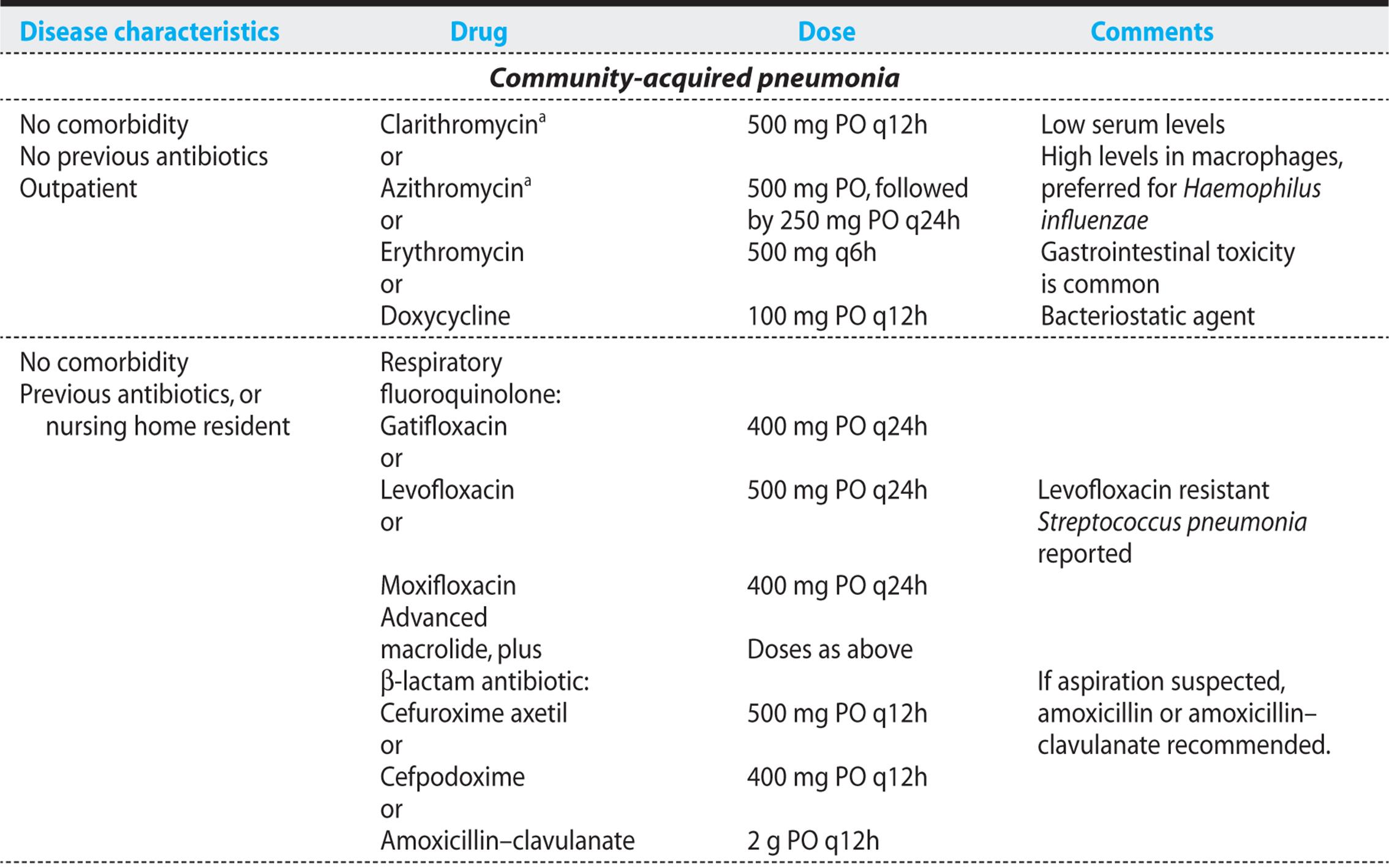


In patients requiring hospitalization for acute CAP, cefotaxime or ceftriaxone (covers S. pneumoniae, H. influnezae, S. aureus, Klebsiella spp, some gramnegative organisms, and aerobic mouth flora), combined with an advanced macrolide [azithromycin or clarithromycin (covers Legionella, Mycoplasma, Chlamydophila)] is recommended for empiric therapy. If aspiration pneumonia is suspected, metronidazole can be added. Ceftaroline has recently been approved for the treatment of CAP; however the specific indications for its use have not been clarified.
In ambulatory patients, either a macrolide in the form of azithromycin or clarithromycin, or a respiratory fluoroquinolone (gatifloxacin, moxifloxacin, or levofloxacin) possessing good gram-positive activity is considered efficacious. Concerns have been raised about the development of resistance to fluoroquinolones, and many experts recommend that this class of antibiotics be reserved for older patients with underlying disease. These patients are not only exposed to the standard causes of CAP but also experience an increased incidence of gram-negative bacilli that will be covered by those agents.
The appropriate duration of treatment has not been systematically studied. For S. pneumoniae, patients are generally treated for 72 hours after they become afebrile. For infections with bacteria that cause necrosis of lung (S. aureus, Klebsiella, and anaerobes) therapy should probably be continued for more than 2 weeks. Treatment for 2 weeks is generally recommended for M. pneumoniae, C. pneumoniae, and Legionella in the immunocompetent patient. Patients on intravenous antibiotics can generally be switched to oral antibiotics when their clinical condition is improving, they are hemodynamically stable, their gastrointestinal tract is functioning normally, and they are capable of taking medications by mouth. In many cases, those criteria are met within 3 days. When possible, the oral antibiotic should be of the same antibiotic class as the intravenous preparation. If staying within the class is not possible, then the oral agent should have a spectrum of activity similar to that of the intravenous agent.
Response to treatment can be assessed by monitoring temperature, respiratory rate, PaO2 and oxygen saturation, peripheral WBC count, and frequency of cough. The changes seen on CXR often persist for several weeks despite clinical improvement. Although CXR is not helpful for assessing improvement, conventional films can be combined with pulmonary CT scan to assess the development of complications such as pneumothorax, cavitation, empyema, and adult respiratory distress syndrome (ARDS), and to document continued progression of infiltrates despite therapy.
Outcome
In the United States, 45,000 deaths are attributed to pneumonia annually. In hospitalized patients, overall mortality ranges from 2% to 30%. Mortality from pneumonia and influenza is particularly high in individuals over the age of 65 years, causing 150-250 deaths per 100,000 population annually. Mortality is also higher in individuals with underlying diseases. Five comorbid illnesses have been identified that result in statistically significant increases in mortality:
• Neoplastic disease
• Liver disease
• Congestive heart failure
• Cerebrovascular disease
• Renal disease
SPECIFIC CAUSES OF ACUTE COMMUNITY-ACQUIRED PNEUMONIA
Great overlap occurs among the clinical manifestations of the pathogens associated with acute CAP. However, constellations of symptoms, signs, and laboratory findings serve to narrow the possibilities. By developing an ability to focus on a few pathogens or to identify a specific pathogen, clinicians can better predict the clinical course of pneumonia and can narrow antibiotic coverage.
Streptococcus Pneumoniae
PATHOGENESIS
Pathogenic strains of S. pneumoniae have a thick capsule that prevents PMN binding and that blocks phagocytosis. Certain capsular types (1, 3, 4, 7, 8, and 12 in adults, and 3, 6, 14, 18, 19, and 23 in children) account for most pneumonia cases. Type 3 has the thickest polysaccharide capsule, and it is the most virulent strain, being associated with the worst prognosis. Immunoglobulins that specifically recognize the capsule are able to link the bacterium to the PMN surface through Fc receptors, enabling PMNs and macrophages (classified as phagocytes) to efficiently ingest and kill the pneumococci. The complement product C3b enhances phagocytosis of the bacteria by the same mechanism. Immunoglobulins and C3b are called “opsonins,” which are products that enhance foreign particle ingestion by phagocytes.
KEY POINTS
About Treatment and Outcome of Pneumonia
1. Treatment must be instituted within 4 hours of diagnosis.
2. Delays are associated with increased mortality.
3. Appropriate triage should be guided by the CRB-65 classification.
4. Empiric therapy depends on the patient and disease characteristics:
a) Outpatient with no comorbidity and no previous antibiotics. Use a macrolide (azithromycin or clarithromycin). If previous antibiotics or elderly nursing home patient, add a β-lactam antibiotic, or use a respiratory fluoroquinolone.
b) Hospitalized patient. Use a third-generation cephalosporin (ceftriaxone or cefotaxime) combined with a macrolide (azithromycin or clarithromycin). If Pseudomonas is a concern, use piperacillin–tazobactam, imipenem, or meropenem.
c) Aspiration outpatient. Use penicillin or clindamycin.
d) Aspiration inpatient. Use a third-generation cephalosporin or a respiratory fluoroquinolone plus metronidazole; or use ticarcillin–clavulanate or piperacillin–tazobactam.
5. Using chest radiographs to monitor improvement is not recommended. (They can take several weeks to clear.) They are useful for documenting worsening of disease or development of complications.
6. Mortality ranges from 2% to 30%. Mortality higher with age more than 65 years, neoplastic disease, liver disease, congestive heart failure, cerebrovascular accident, and renal disease.
KEY POINTS
About the Pathogenesis of Streptococcus pneumoniae
1. The thick outer capsule blocks phagocytosis. Type 3 has the thickest capsule.
2. Immunoglobulins and complement are important opsonins that allow phagocytes to ingest invading pneumococci.
3. Streptococcus pneumoniae does not produce protease and seldom destroys lung parenchyma.
4. It does not cross anatomic barriers such as lung fissures.
5. Disease manifestations are caused primarily by the host’s inflammatory response to the organism.
In addition to its polysaccharide capsule, S. pneumoniae possesses a number of other virulence factors that enhance adherence to epithelial cells, resist phagocytosis, and activate complement. S. pneumoniaedoes not produce significant quantities of proteases, and disease manifestations are primarily the consequence of the host’s inflammatory response. As a result, permanent tissue damage is rare, and spread of the disease across anatomic boundaries, such as lung fissures, is uncommon.
PREVALENCE AND PREDISPOSING FACTORS
S. pneumoniae remains the most common cause of acute CAP; it represents two-thirds of the cases in which a specific pathogen is identified. Because opsonins are required for efficient phagocytosis of the encapsulated organism, patients with hypogammaglobulinemia and multiple myeloma are at increased risk of developing this infection, as are patients with deficiencies in complement (C1, C2, C3, C4). Patients with HIV infection also have defects in antibody production, and they have a higher incidence of pneumococcal infection. Patients with splenic dysfunction have a higher risk of overwhelming S. pneumoniae sepsis because the spleen plays a vital role in clearing these bacteria from the bloodstream, particularly in the absence of specific antipneumococcal capsule antibody. Other chronic diseases, including cirrhosis, nephrotic syndrome, congestive heart failure, chronic obstructive pulmonary disease, and alcoholism, are also associated with greater risk of pneumococcal infection.
KEY POINTS
About Streptococcus pneumoniae Prevalence and Predisposing Factors
1. S. pneumoniae is the most common form of community-acquired bacterial pneumonia.
2. The risk is higher in patients with deficiencies in opsonin production:
a) Hypogammaglobulinemia
b) Complement deficiency
c) HIV infection
3. Splenic dysfunction increases the risk of fatal pneumococcal bacteremia.
4. Risk is increased in patients with chronic diseases:
a) Cirrhosis
b) Alcoholism
c) Nephrotic syndrome
d) Congestive heart failure
e) Chronic obstructive pulmonary disease
UNIQUE CLINICAL CHARACTERISTICS
Classically, pneumococcal pneumonia has a very abrupt onset that begins with a single severe rigor. Because S. pneumoniae invasion of the lung leads to capillary leakage of blood into the alveolar space, sputum can become rusty in color. Furthermore, pneumococcal infection frequently infects the peripheral lung and spreads quickly to the pleura. As a result, pleuritic chest pain is a common complaint.
DIAGNOSIS
Sputum Gram Stain—A careful analysis of the sputum is best performed by a knowledgeable physician. Areas with significant numbers of PMNs per high-power field and a predominance of gram-positive lancet-shaped diplococci suggest the diagnosis [Figure 4.2 (B)]. A finding of pneumococci within the cytoplasm of a PMN strongly supports invasive infection.
KEY POINTS
About Clinical Manifestations and Diagnosis of Pneumococcal Pneumonia
1. Three classic features may be found:
a) Abrupt onset accompanied by a single rigor
b) Rusty-colored sputum
c) Pleuritic chest pain
2. Sputum Gram stain is often helpful: more than 10 gram-positive lancet-shaped diplococci per high-power field indicate pneumococcal pneumonia.
3. Sputum culture is insensitive; specimens (alpha hemolytic, optochin sensitive) should be plated quickly.
4. Blood samples for culture should always be drawn; up to 25% may be positive.
5. A urine pneumococcal antigen test may prove helpful.
6. A chest radiograph shows a classical lobar pattern; small pleural effusions are common, true empyema rare. Abnormalities persist for 4–6 weeks after cure.
Sputum Culture—S. pneumoniae is catalase negative, bile soluble, and, like S. viridans, demonstrates alpha (green) hemolysis on blood agar plates. The propensity of normal mouth flora, in particular S. viridans, to overgrow frequently interferes with the identification of S. pneumoniae. The optochin disk inhibits growth of S. pneumoniae, but not of S. viridans, and this test is used to differentiate the two organisms. Another problem with sputum culture arises from the fact that S. pneumoniae can be present as normal mouth flora in up to 60% of healthy people. A positive sputum culture in the absence of a positive Gram stain or a positive blood culture may therefore simply represent contamination of the sputum with saliva.
Blood Cultures—Some reports have claimed that 25% of patients with pneumococcal pneumonia develop positive blood cultures; however, the denominator required to calculate this percentage is uncertain. Even in the absence of a positive sputum Gram stain, a positive blood culture in combination with the appropriate symptoms and CXR findings is interpreted as true infection. A urine test for pneumococcal polysaccharide antigen is available and is positive in 80% of adults with bacteremia.
Chest X-Ray—The CXR usually reveals a single area of infiltration involving one or more segments of a single lobe. Involvement of the entire lobe is less common. This organism respects the confining fissures of the lung and rarely extends beyond those boundaries, which explains the classical lobar radiologic pattern [Figure 4.2 (A)].
Air bronchograms are found in a few cases. This radiologic finding is the consequence of the alveoli filling with inflammatory fluid and outlining the air-containing bronchi. When found, bronchograms are associated with a higher incidence of bacteremia.
Pleural fluid may be detected in up to 40% of cases. In most instances, the volume of fluid is too small to sample by thoracentesis, and if antibiotic treatment is prompt, only a small percentage go on to develop true empyema.
The radiologic improvement in pneumococcal pneumonia is slow. Despite rapid defervescence and resolution of all symptoms, radiologic changes often persist for 4-6 weeks. If the patient is improving clinically, follow-up CXRs are therefore not recommended during this period.
TREATMENT AND OUTCOME
In the early antibiotic era, S. pneumoniae was highly sensitive to penicillin [minimum inhibitory concentration (MIC) <0.06 μg/mL]. However, since the late 1990s, isolates in the United States have become resistant, with 10% demonstrating intermediate resistance (MIC = 0.12-1 μg/mL), and 5% demonstrate high-level resistance (MIC ≥2 >g/mL). In some areas of Europe and South Africa, higher percentages of resistant strains have been observed. In the Netherlands and Germany, where strictly limited antibiotic use is the standard of care, the prevalence of resistant strains is lower.
Currently, many intermediate strains remain sensitive to the third-generation cephalosporins ceftriaxone and cefotaxime (MIC <1 μg/mL); however, resistance to these antibiotics is increasing. For intermediately resistant strains, amoxicillin is more active than is penicillin VK, and amoxicillin is therefore the preferred oral antibiotic. Because penicillin resistance results from a decrease in the affinity of penicillin-binding proteins, intermediate (but not high-level) resistance can be overcome by raising the concentration of penicillin.
With the exception of the CNS, where the blood–brain barrier limits antibiotic penetration, standard doses of penicillin are effective in curing infections attributable to intermediately resistant pneumococci. Penicillin resistance is usually associated with resistance to many other classes of antibiotics, including the tetracyclines, macrolides, and clindamycin. Imipenem is also inactive against highly resistant strains. The respiratory fluoroquinolones that possess good gram-positive activity (levofloxacin, gatifloxacin, moxifloxacin) and vancomycin usually retain excellent activity against all resistant strains. Several cases of pneumonia attributable to levofloxacin-resistant S. pneumoniae have recently been reported; however, the overall percentage of pneumococcal strains that are resistant to fluoroquinolones remains low. Ceftaroline is the most potent agent for the treatment of all pneumococcal serotypes demonstrating MICs of 0.008-0.5 μg/mL.
TREATMENT RECOMMENDATIONS
For doses of the drugs discussed here, see Table 4.3.
For penicillin-sensitive strains, penicillin G or amoxicillin remains the preferred treatment. Ceftriaxone is also effective. If the patient fails to improve within 48 hours, the possibility of a resistant strain must be considered, and coverage with a respiratory fluoroquinolone is recommended. For cases in which meningitis is suspected, a fluoroquinolone should not be used because of poor penetration of the cerebrospinal fluid (CSF), and the patient should be covered with vancomycin. In the penicillin-allergic patient, a respiratory fluoroquinolone can be used. Ceftaroline has not been approved by the FDA for treatment of penicillin-resistant S. pneumonia; however, this important alternative should be kept in mind in the critically ill patient.
KEY POINTS
About the Treatment, Outcome, and Prevention of Pneumococcal Pneumonia
1. A significant percentage of Streptococcus pneumoniae is resistant to penicillin:
a) 10% are intermediately resistant (MIC = 0.12-1 μg/mL).
b) 5% demonstrate high-level resistance (MIC >2 μg/mL).
2. Penicillin or ampicillin remains the treatment of choice for penicillin-sensitive strains.
3. High-dose parenteral penicillin, a third-generation cephalosporin, or an oral amoxicillin used for intermediate-sensitivity strains, except for meningitis.
4. A respiratory fluoroquinolone (gatifloxacin, moxifloxacin, levofloxacin) is used for strains with high-level resistance. Avoid fluoroquinolones in meningitis, and cover with vancomycin.
5. Mortality is approximately 5%; prognosis is worse for infants and for patients older than 65 years of age, and for those whose treatment is delayed or who have capsular types 2 or 3, multilobar pneumonia, bacteremia or meningitis, or jaundice, or who are pregnant, have an underlying disease, or alcohol intoxication.
6. The 23-valent pneumococcal vaccine is safe and efficacious. It should be given to patients who are over 65 years of age, who have a chronic disease, and who are asplenic, immunocompromised, or alcoholic.
In the pre-antibiotic era, the mortality rate for pneumococcal pneumonia was 20-40%. In the antibiotic era, the mortality rate was reduced to approximately 5%. Prognosis is adversely influenced by
1. Age (patients above 65 years of age and infants have worse outcomes)
2. Delayed treatment
3. Infection with capsular type 2 or 3
4. Involvement of more than one lobe of the lung
5. WBC count less than 6000/mm3
6. Bacteremia, shock, or the development of meningitis
7. Jaundice
8. Pregnancy
9. Presence of other underlying diseases (heart disease, cirrhosis, diabetes)
10. Alcohol intoxication
Prevention
Despite the use of antibiotics, mortality during the first 36 hours of hospitalization has not changed. To prevent early mortality and to reduce the incidence of S. pneumoniae infection—the penicillin-sensitive and penicillin-resistant strains alike—vaccination is strongly recommended for all patients with chronic illnesses or those over the age of 65 years.
Generation of specific antibodies directed against the bacterial cell wall confer, prevent, or reduce the severity of disease. Polyvalent vaccine containing antigens to 23 capsular types is available and is effective (approximately 60% reduction in bacteremia in immunocompetent adults). Efficacy decreases with age and is not measurable in immunocompromised patients. The vaccine has proved to be safe and inexpensive, and should be widely used.
Haemophilus influenzae
Group B and nontypable H. influenzae can both cause CAP. Infection with nontypable H. influenzae is more common in elderly individuals and in smokers with chronic obstructive pulmonary disease. The onset of symptoms tends to be more insidious than that seen with S. pneumoniae, but the clinical pictures are otherwise indistinguishable. A CXR can demonstrate lobar or patchy infiltrates, and sputum Gram stain reveals small gram-negative pleomorphic coccobacillary organisms.
Because of their small size and their color, which is similar to background material, H. influenzae may be missed by an inexperienced diagnostician. For the patient requiring hospitalization, intravenous ceftriaxone or cefotaxime is recommended. For oral antibiotic treatment, amoxicillin–clavulanate is effective. However, a number of other oral antibiotics, including trimethoprim–sulfamethoxazole, the newer macrolides (azithromycin and clarithromycin), the fluoroquinolones, and the extended-spectrum cephalosporins (cefpodoxime, cefixime) are also active against this organism.
KEY POINTS
About Haemophilus influenzae Pneumonia
1. This small, gram-negative, pleomorphic coccobacilli is aerobic. It may be mistaken for the background material on sputum Gram stain.
2. Nontypable strains are more common in elderly people and in smokers with COPD.
3. Clinically, Haemophilus influenzae is similar to Streptococcus pneumoniae, with a somewhat slower onset.
4. Parenteral ceftriaxone or cefotaxime should be used to treat hospitalized patients. Multiple oral regimens–amoxicillin-clavulanate, newer macrolides, fluoroquinolones, and extended-spectrum cephalosporins are useful in outpatients.
Staphylococcus aureus
Fortunately, CAP attributable to S. aureus is rare. The most common predisposing factor is a preceding influenza infection. An increase in the incidence of S. aureus pneumonia is often a marker for the onset of an influenza epidemic. S. aureus pneumonia is also more common in intravenous drug users and in patients with AIDS, in association with P. jirovecii pneumonia.
KEY POINTS
About Staphylococcus aureus Pneumonia
1. These large gram-positive aerobic cocci form tetrads and clusters.
2. The disease is rare and most commonly follows influenza. Also seen in patients with AIDS and in IV drug abusers.
3. Destructive bronchopneumonia is complicated by
a) lung abscesses,
b) pneumothorax, and
c) empyema.
In a few communities, community-acquired methicillin-resistant S. aureus (cMRSA) pneumonia has been described in addition to methicillin-sensitive S. aureus (MSSA). The clinical manifestations of this infection are similar to other forms of bacterial pneumonia. However, the illness is often severe, being associated with high fever and a slow response to conventional therapy. A CXR can demonstrate patchy infiltrates or dense diffuse opacifications. S. aureus produces multiple proteases that allow this bacterium to readily cross the lung fissures and simultaneously involve multiple lung segments. This broader involvement explains the typical bronchopneumonia pattern on CXR [Figure 4.4 (A)]. The rapid spread and aggressive destruction of tissue also explains the greater tendency of S. aureus to form lung abscesses and induce a pneumothorax. Spread of this infection to the pleural space can result in empyema (seen in 10% of patients). Sputum Gram stain reveals sheets of PMNs and an abundance of gram-positive cocci in clusters and tetrads [Figure 4.4 (B)], and culture readily grows S. aureus. Blood cultures may also be positive.

Figure 4.4. Staphylococcus aureus pneumonia: A. Cchest radiograph demonstrates a classic bronchopneumonia (Ccourtesy of Dr. Pat Aabbitt, Uuniversity of Fflorida), and B. sputum Gram stain shows gram-positive cocci in clusters and tetrads.
The treatment of choice for MSSA is high-dose intravenous nafcillin or oxacillin. For MRSA pneumonia, vancomycin is generally recommended. The dose of vancomycin should be adjusted to maintain a trough level of 15-20 μg/mL to assure therapeutic levels in the lung. Linezolid is an expensive alternative that has equivalent efficacy. The newest cephalosporin, ceftaroline, has excellent MICs against nearly all MRSA strains and is a less expensive alternative to linezolid. Ceftaroline is FDA approved for the treatment of CAP (Figure 4.5).

Figure 4.5. Empyema following aspiration pneumonia. CT scan showing a large right pleural effusion as well as discrete rounded cavitary lesions in the lung parenchyma of both the left and right lower lobes.
Legionella Pneumophila
Legionella species are gram-negative bacilli found throughout the environment in standing water and soil. Infection most commonly results from inhalation of water droplets contaminated with Legionella.Cooling towers or shower heads are most often responsible for aerosolizing contaminated water. Less commonly, nosocomial infection has resulted from the use of unsterilized tap water in respiratory therapy devices. Outbreaks of Legionella pneumonia have also been associated with soil excavation. Immunocompromised patients, smokers, and elderly people are more susceptible to this infection.
Clinically, Legionella infection causes symptoms typical of other acute CAPs, including high fever, cough, myalgias, and shortness of breath. As compared with other bacterial pneumonias, cough usually produces only small amounts of sputum. Gastrointestinal symptoms, confusion, and headache are more frequently encountered in patients with Legionella. Laboratory findings are similar to other acute pneumonias. The only distinctive finding may be hyponatremia, which is noted in approximately one-third of patients. A CXR frequently demonstrates lobar pneumonia. In the immunocompromised host, cavitary lesions may be seen. Small pleural effusions are also commonly found.
KEY POINTS
About Legionella Pneumonia
1. These aerobic gram-negative bacteria do not take up Gram stain well.
2. Found in soil and standing water. Aerosolized by cooling towers and showerheads. Also contracted after soil excavation.
3. Elderly people, smokers, and immunocompromised patients are at increased risk.
4. Similar to other acute pneumonias. Somewhat unique characteristics include
a) minimal sputum production,
b) confusion and headache,
c) gastrointestinal symptoms, and
d) hyponatremia.
5. Diagnostic techniques include
a) culture on buffered-charcoal yeast-extract agar,
b) direct fluorescent antibody stain (low sensitivity),
c) polymerase chain reaction, and
d) urinary antigen to serotype I (causes 80% of infections), which is sensitive and specific, and persists for several weeks.
6. Azithromycin or a fluoroquinolone is the treatments of choice. In transplant patients, fluoroquinolones are preferred. Mortality is high: 16-50%.
Diagnosis requires a high index of suspicion, because sputum Gram stain reveals only acute inflammatory cells. The microbiology laboratory must be alerted to the possibility of Legionella species to assure that sputum samples are cultured on buffered-charcoal yeast-extract agar with added suppressive antibiotics. Legionella can also be identified by direct fluorescent antibody staining, although the sensitivity of this technique is low (30-50%). Amplification of Legionella DNA from sputum samples by PCR is available in certain reference laboratories, but not commercially. For L. pneumophila serogroup 1, the most common cause of Legionella pneumonia in the United States (>80% of cases), a highly sensitive and specific urinary antigen test is commercially available. The antigen is excreted early in the illness and persists for several weeks.
For mild disease, an oral macrolide, fluoroquinolone, or tetracycline may be used. However, in more severe disease, high doses of intravenous azithromycin or a fluoroquinolone (ciprofloxacin or levofloxacin) are recommended. In transplant patients, a fluoroquinolone is preferred because the macrolides interfere with cyclosporin or tacrolimus metabolism. In the immunocompetent patient, therapy should be continued for 5-10 days with azithromycin and for 10-14 days with a fluoroquinolone. In the immunocompromised patient, therapy needs to be prolonged for 14-21 days to prevent relapse. Mortality is high in Legionnaires disease: 16-30% in community-acquired disease and up to 50% in hospitalized patients.
Atypical Pneumonia
The atypical forms of pneumonia tend to be subacute in onset, with patients reporting up to 10 days of symptoms before seeking medical attention. Atypical pneumonia is associated with a nonproductive cough, and clinical manifestations tend to be less severe. It is important to keep in mind that significant overlap occurs in the clinical manifestations of this group of infections and the more typical forms of pneumonia associated with purulent sputum production.
M. pneumoniae is one of the most frequent causes of “walking pneumonia.” This infection is seen primarily in patients under age 40 years; it is an uncommon cause of pneumonia in elderly individuals. The disease is seasonal, with the highest incidence of Mycoplasma being seen in the late summer and early fall. Sore throat is usually a prominent symptom, and bullous myringitis is seen in 5% of cases. Presence of this abnormality is highly suggestive of Mycoplasma. Tracheobronchitis results in a hacking cough that is often worse at night and that persists for several weeks. Physical examination may reveal some moist rales, but classically, radiologic abnormalities are more extensive than predicted by the exam. Findings on CXR consist of unilateral or bilateral patchy lower-lobe infiltrates in a bronchial distribution. The clinical course is usually benign. Fever, malaise, and headache usually resolve over 1–2 weeks, but cough can persist for 3–4 weeks. Peripheral WBC is usually less than 10,000. And sputum Gram stain and culture reveal only normal mouth flora and a moderate inflammatory response.
Diagnosis is made by history and clinical manifestations. Epidemiologic history of contact with a person having similar symptoms is particularly helpful. Currently, no definitive test is available. Sputum PCR has been found to be sensitive and specific, and a multiplex PCR has recently been approved for nasal swab samples. Cold agglutinin titers in excess of 1:64 support the diagnosis and correlate with severity of pulmonary symptoms, but are not cost effective. Complement fixation antibody titers begin to rise 7-10 days after the onset of symptoms.
Because a reliable, rapid diagnostic test is not currently available, therapy is usually empiric. With the advent of multiplex PCR, this may change in the near future. A macrolide or tetracycline is the treatment of choice; alternatively, a fluoroquinolone can be administered. Azithromycin is the preferred agent when Mycoplasma is suspected, and a standard 5-day course is effective in most cases.
KEY POINTS
About Atypical Pneumonia
1. These diseases tend to be subacute in onset.
2. Cough is nonproductive.
3. Illness is often less severe than in other community-acquired pneumonias: “walking pneumonia.”
4. Findings in a chest radiograph are usually worse than the physical findings.
5. Three primary causes:
a) Mycoplasma pneumoniae
b) Chlamydophila pneumoniae
c) Respiratory viruses: influenza, adenovirus, parainfluenza, and respiratory syncytial virus.
6. Rapid tests are available for influenza, but not for Mycoplasma or Chlamydophila.
7. Treatment with a macrolide or tetracycline is recommended. If influenza is diagnosed, give amantadine, ranitidine, or a neuraminidase inhibitor within 48 hours of illness onset.
C. pneumoniae (Taiwan acute respiratory agent) is another important cause of atypical pneumonia. This pathogen is a common cause of CAP, representing 5-15% of cases. The disease occurs sporadically and presents in a manner similar to Mycoplasma, with sore throat, hoarseness, and headache in addition to a nonproductive cough. Radiologic findings are also similar to those with Mycoplasma. Multiplex PCR is now available and promises to be highly specific and sensitive. A tetracycline is considered the treatment of choice, but macrolides and fluoroquinolones are also effective.
The final major group of organisms that cause atypical pneumonia is the respiratory viruses: influenza A and B, adenovirus, parainfluenza virus, and respiratory syncytial virus. The respiratory syncytial virus infects primarily young children, elderly people, and the immunocompromised host. These viruses can all present with a nonproductive cough, malaise, and fever. Auscultatory findings are minimal, and lower lobe infiltrates are generally observed on CXR. The clinical virology laboratory can culture each of these viruses from sputum or a nasopharyngeal swab. Rapid commercial tests (10-20 minutes) are available for detection of influenza (Quick View, Flu O1A, and Zstatflu). These tests have a sensitivity of 57-77%, and all three can distinguish between types A and B.
If influenza A virus is diagnosed, early treatment of the virus with amantadine or rimantadine is recommended. Neuraminidase inhibitors are also available, and these agents have activity against both influenza A and B. The influenza vaccine is safe and efficacious, and should be given annually in October through early November to patients over 65 years of age, individuals with serious underlying diseases, nursing home residents, and health care workers (see Chapter 14).
Aspiration Pneumonia
CASE 4.2
A 35-year-old man arrived in the emergency room complaining of left-sided chest pain during the preceding 4 days. He had begun drinking large quantities of alcohol 8 days earlier. He vaguely recalled passing out on at least two occasions. He developed a persistent cough, productive of green sputum, 4 days before admission. At that time, he also began experiencing right-sided chest pain on deep inspiration (pleuritic pain). Initially, these pains were dull; however, over the next few days, they became increasingly sharp.
Physical examination showed a temperature of 38°C and a respiratory rate of 42 per minute. He was a disheveled man, looking older than his stated age, breathing shallowly and rapidly, in obvious pain.
A check of the throat revealed a good gag reflex, extensive dental caries, several loose teeth, severe gingivitis, and foul-smelling breath and sputum. Decreased excursion of the right lung was noted, and the right lower lung field was dull to percussion. Bronchovesicular breath sounds were heard diffusely (inspiratory and expiratory breath sounds of equal duration); moist, medium rales were heard in the right lower and left lower lung fields. Egophony and whispered pectoriloquy were also heard in these areas.
Laboratory workup showed a hematocrit of 50%; a WBC count of 21,400/mm3, with 79% PMNs, 7% bands, 1% lymphocytes, and 13% monocytes. Blood gasses showed a pH of 7.46, PaO2 of 56 mmHg, and a PaCO2 of 36 mmHg. Sputum Gram stain revealed many PMNs and a mixture of gram-positive cocci, gram-positive rods, and gram-negative rods. A CXR demonstrated dense right lower lobe infiltrate.
While on antibiotics, this patient continued to complain of chest pain and developed decreased breath sounds in the right lower lobe associated with dullness to percussion. A repeat CXR and CT scan demonstrated a large right pleural effusion [see Figure 4.5] and thoracentesis revealed more than 100,000 PMNs/mm3, pleural fluid pH of 7.0, and total protein 3.4 mg/mL. Gram stain showed a mixture of gram-positive cocci and gram-positive and gram-negative rods.
Aspiration pneumonia should be suspected in patients with a recent history of depressed consciousness and in patients with a poor gag reflex or an abnormal swallowing reflex. The elderly patient who has suffered a stroke is particularly susceptible to aspiration. In case 4.2, the patient’s heavy consumption of alcohol led to depression in consciousness.
Three major syndromes are associated with aspiration:
1. Chemical burn pneumonitis. Aspiration of the acidic contents of the stomach can lead to a chemical burn of the pulmonary parenchyma. Aspiration of large quantities of fluid can result in the immediate opacification of large volumes of lung. Acid damage causes pulmonary capillaries to leak fluid, release cytokines, and permit infiltration by PMNs. In some patients, noncardiogenic pulmonary edema or ARDS develops. Onset of symptoms occurs immediately after aspiration.
2. Bronchial obstruction resulting from aspiration of food particles. The inhalation of solid particles results in mechanical obstruction and interferes with ventilation. The patient immediately becomes tachypneic.
3. Pneumonia resulting from a mixture of anaerobic and aerobic mouth flora. This form of pneumonia develops several days after aspiration of mouth flora. Patients with severe gingivitis have higher bacterial colony counts in the mouth, and they aspirate a higher inoculum of organisms, increasing the likelihood of a symptomatic pneumonia.
Case 4.2 had poor dental hygiene and severe gingivitis, predisposing him to the latter form of pneumonia. Often, the sputum is putrid smelling as a result of the high number of anaerobes. Necrosis of tissue is common in this infection, resulting in the formation of lung abscesses. Infection often spreads to the pleura, resulting in pleuritic chest pain as experienced in case 4.2. Pleural effusions filled with bacteria and PMNs can develop as observed in this case. Effusions containing bacteria and large numbers of PMNs are called empyemas. Necrosis of the pleural lining and lung parenchyma can result in formation of a fistula tracking from the bronchus to the pleural space. Development of a bronchopleural fistula prolongs hospitalization and may eventually require surgical repair.
DlAGNOSIS
Sputum is often foul-smelling as a result of the high numbers of anaerobic bacteria. Sputum Gram stain reveals many PMNs and a mixture of gram-positive and gram-negative organisms. Sputum culture usually grows normal mouth flora. When aspiration occurs in the hospitalized patient, the mouth often is colonized with more resistant gram-negative organisms plus S. aureus. In these patients, a predominance of gram-negative rods or gram-positive cocci in clusters may be seen on Gram stain, and gram-negative rods or S. aureus may be cultured from the sputum.
A CXR reveals infiltrates in the dependent pulmonary segments. When aspiration occurs in the upright position, the lower lobes are usually involved, more commonly the right lower lobe than the left. This difference has an anatomic explanation. The right bronchus divides from the trachea at a straighter angle than does the left mainstem bronchus, increasing the likelihood that aspirated material will flow to the right lung. When aspiration occurs in the recumbent position, the superior segments of the lower lobes or the posterior segments of the upper lobes usually become opacified.
TREATMENT
Clindamycin and penicillin are effective antibiotic coverage for community-acquired aspiration pneumonia because they kill both aerobic and anaerobic mouth flora (Table 4.3). In cases in which lung abscess has developed, clindamycin has been shown to be slightly superior.
KEY POINTS
About Aspiration Pneumonia
1. Can occur in cases of loss of consciousness, poor gag reflex, or difficulty swallowing.
2. Three forms of aspiration:
a) Aspiration of gastric contents leads to pulmonary burn and noncardiogenic pulmonary edema.
b) Aspiration of an obstructing object causes atelectasis and immediate respiratory distress.
c) Aspiration of mouth flora, when associated with poor dental hygiene and mixed mouth aerobes and anaerobes, can lead to foul-smelling sputum and eventually lung abscess and empyema. Hospital-acquired aspiration causes gram-negative and Staphylococcus aureus pneumonia.
3. Treatment depends on the form of the disease:
a) Penicillin or clindamycin for community-acquired infection.
b) Third-generation cephalosporin for hospital-acquired infection.
c) Bronchoscopy for obstructing foreign bodies.
In nosocomial aspiration, broader coverage with a third-generation cephalosporin is generally recommended. This regimen provides sufficient anaerobic coverage and addition of metronidazole is not required. Alternatively, a semisynthetic penicillin combined with a β-lactamase inhibitor (ticarcillin–clavulanate or piperacillin–tazobactam) or a carbapenem (imipenem or meropenem) can be used.
If aspiration of a foreign body is suspected, bronchoscopy is required to remove the foreign material from the tracheobronchial tree.
Rarer Causes of Community-Acquired Pneumonia
ACTINOMYCOSIS
Actinomyces species are microaerophilic or anaerobic gram-positive rods that can be part of the polymicrobial flora associated with aspiration pneumonia, particularly in patients with poor oral hygiene. Disease is most commonly caused by Actinomyces israelii.
KEY POINTS
About Actinomycosis
1. These branching gram-positive bacteria are microaerophilic or anaerobic, slow growing, modified acid-fast negative.
2. Infection is associated with poor oral hygiene.
3. Slowly progressive infection, breaks through fascial planes, causes pleural effusions and fistula tracks, forms “sulfur granules.”
4. Alert clinical microbiology to hold anaerobic cultures.
5. Treatment must be prolonged: high-dose intravenous penicillin for 2-6 weeks, followed by 6-12 months of oral penicillin.
Actinomycosis pulmonary infection is often indolent and slowly progressive. Lung parenchymal lesions are usually associated with pleural infection, resulting in a thickened pleura and empyema. This organism can break through fascial planes. Spontaneous drainage of an empyema through the chest wall should strongly suggest the possibility of actinomycosis. “Sulfur granules” are often found in purulent exudate; they consist of clusters of branching Actinomyces filaments.
Gram stain reveals branching forms that are weakly gram positive. These forms can be differentiated from Nocardia by modified stain for acid-fast bacilli (AFB), Actinomyces being acid-fast stain negative, and Nocardia being acid-fast stain positive. The organism should be cultured under anaerobic conditions, and grows slowly, with colonies usually requiring a minimum of 5–7 days to be identified. Growth can take up to 4 weeks.
High-dose intravenous penicillin (18 to 24 × 106 U daily) is recommended for 2–6 weeks, followed by oral penicillin therapy for 6–12 months. Therapy must be continued until all symptoms and signs of active infection have resolved. Other antibiotics that have been successfully used to treat actinomycosis include erythromycin, tetracyclines, and clindamycin.
NOCARDIOSIS
Nocardia is an aerobic gram-positive filamentous bacterium that often has to be differentiated from Actinomyces. Nocardia is ubiquitous in the environment, growing in soil, organic matter, and water. Pneumonia occurs as a consequence of inhaling soil particles. The number of species causing human disease is large and includes Nocardia abscessus, N. brevicatena/paucivorans complex, N. nova complex, N. transvalensis complex, N. farcinica, N. asteroides complex, N. brasiliensis, and N. pseudobrasiliensis.
KEY POINTS
About Nocardiosis
1. Nocardia are gram-positive branching bacteria, aerobic, slow growing, modified acid-fast.
2. Ubiquitous organism found in the soil.
3. Inhalation of soil particles leads to pneumonia.
4. The organism infects
a) immunocompromised patients (causing disseminated disease in AIDS),
b) normal hosts, and
c) patients with alveolar proteinosis.
5. Pulmonary infection can lead to bacteremia and brain abscess that can mimic metastatic lung carcinoma.
6. Alert clinical microbiology to use selective media and to hold cultures.
7. Treatment must be prolonged. High-dose parenteral trimethoprim–sulfamethoxazole for at least 6 weeks, followed by oral treatment for 6-12 months.
Infection more commonly develops in patients who are immunocompromised; however, 30% of cases occur in otherwise normal individuals. Patients with AIDS, organ transplant, alcoholism, and diabetes are at increased risk of developing nocardiosis. In addition to pulmonary disease, these patients are at increased risk of developing disseminated infection. Patients with chronic pulmonary disorders, in particular patients with alveolar proteinosis, have an increased incidence of pulmonary Nocardia infection.
Onset of pulmonary disease is highly variable. In some cases, onset is acute; in others, onset is gradual. Symptoms are similar to other forms of pneumonia. A CXR may reveal cavitary lesions, single or multiple nodules, a reticular nodular pattern, interstitial pattern, or a diffuse parenchymal infiltrate. Nocardia pulmonary infection often seeds the bloodstream and forms abscesses in the cerebral cortex. The combination of a lung infiltrate with a CNS lesion or lesions is often mistaken for lung carcinoma with CNS metastasis.
Diagnosis is made by sputum examination or lung or cerebral cortex biopsy. Gram stain demonstrates weakly gram-positive branching filamentous forms that are acid-fast on modified AFB stain. On tissue biopsy, organisms are demonstrated on Brown–Brenn or methenamine silver stain. The organism is slow growing and is frequently overgrown by mouth flora on conventional plates. The clinical laboratory should be alerted to the possibility of Nocardiaso that they can incubate bacteriologic plates for a prolonged period and use selective media.
Most Nocardia are sensitive to sulfonamides and trimethoprim. Trimethoprim–sulfamethoxazole is generally accepted as the treatment of choice, with a daily dose of 2.5-10 mg/kg of the trimethoprim component. High-dose therapy should be continued for at least 6 weeks, followed by lower doses for 6-12 months. Some Nocardia species are resistant to sulfonamides, but they are sensitive to amikacin, imipenem, third-generation cephalosporins, minocycline, dapsone, and linezolid. Whenever possible, culture and antibiotic sensitivities should be used to guide antibiotic therapy.
NOSOCOMIAL (HOSPITAL-ACQUIRED) PNEUMONIA
Pneumonia is the second most common form of nosocomial infection. It accounts for 13-19% of all nosocomial infections. Hospital-acquired pneumonia is defined as a pneumonia that develops 48 hours or longer afterhospitalization and that was not developing at the time of admission. Nosocomial pneumonia is a very serious complication and represents the leading infectious-related cause of death in the hospital, the mortality being roughly one of every three cases. Development of pneumonia in the hospital prolongs hospitalization by more than 1 week.
KEY POINTS
About Nosocomial Pneumonia
1. Pneumonia is one of the most common nosocomial infections.
2. Risk factors include
a) endotracheal intubation (20 times the baseline risk, 1-3% incidence daily),
b) age greater than 70 years,
c) depressed mental status,
d) underlying disease and malnutrition, and
e) metabolic acidosis.
3. Primary causes are gram-negative bacilli and Staphylococcus aureus.
4. Colonization is difficult to differentiate from infection. Bronchoscopy is not helpful. Factors that favor infection include
a) worsening fever and leukocytosis with left shift;
b) sputum Gram stain with increased PMNs, predominance of one organism;
c) decreasing PaO2 indicative of pulmonary shunting; and
d) expanding infiltrate on chest radiographs.
5. Broad-spectrum empiric therapy can be initiated after samples are obtained for culture, but coverage should be adjusted based on culture results and clinical response.
The condition that most dramatically increases the risk of nosocomial pneumonia is endotracheal intubation. Endotracheal tubes bypass the normal protective mechanisms of the lung, and they increase the risk of pneumonia by a factor between 6 and 21. It has been estimated that the risk of pneumonia while on a ventilator is 1-3% daily. Other factors that increase the risk of pneumonia include age greater than 70 years; CNS dysfunction, particularly coma, leading to an increased likelihood of aspiration; other severe underlying diseases; malnutrition; and metabolic acidosis. Patients on sedatives and narcotics have depressed epiglottal function and are also at increased risk of aspiration. Corticosteroids and other immunosuppressants reduce normal host defenses and allow bacteria to more readily invade the lung parenchyma.
Aerobic gram-negative bacteria account for more than half the cases of nosocomial pneumonia. Escherichia coli, Klebsiella, Serratia, Enterobacter, and Pseudomonas species represent the most common gram-negative rods. S. aureus is the most common gram-positive pathogen, causing 13-40% of nosocomial pneumonias. The risk of S. aureus infection is higher in patients with wound infections or burns, and it is also higher in intubated patients with head trauma or neurosurgical wounds. Anaerobes are often isolated in nosocomial pneumonia, but they are thought to be the primary agent in only 5% of cases. S. pneumoniae is seldom the cause of pneumonia in the patient who has been hospitalized for more than 4 days.
Diagnosis of true pneumonia is often difficult in the intubated patient. In elderly patients with chronic bronchitis and congestive heart failure or ARDS, definitively proving that the patient has or does not have an infection is often impossible. Differentiating infection from colonization represents a critical branch point in the appropriate management of antibiotics (see case 1.1). Within 3-5 days of antibiotic initiation, the mouth flora and the flora colonizing the tracheobronchial tree change. A change in the organisms growing from sputum culture is therefore to be expected and does not in itself indicate that the patient has a new infection. The change simply documents colonization of the patient with resistant flora. For example, in a high percentage of patients receiving broad-spectrum antibiotics, Candida albicans begins to grow in sputum cultures because of the reduction in the competing bacterial mouth flora. However, that organism does not invade the lung and almost never causes airborne pneumonia. Antifungal coverage is therefore not required unless the patient develops symptomatic thrush.
Evidence supporting the onset of a new infection includes
• a new fever or a change in fever pattern;
• a rise in the peripheral WBC count, with an increase in the percentage of PMNs and band forms (left shift);
• Gram stain demonstrating increased number of PMNs in association with a predominance of bacteria that are morphologically consistent with the culture results;
• increased purulent sputum production from the endotracheal tube;
• reduced arterial PaO2, indicating interference with alveolar–capillary oxygen exchange; and
• enlarging infiltrate on CXR.
Multiple studies have used bronchoscopy with protected brushings or bronchial lavage and quantitative cultures and Gram stains. A randomized trial found that samples obtained by bronchoscopy provide no advantage over endotracheal suction, and therefore that procedure is not recommended in VAP.
When infection is likely or the patient is extremely ill, and when a new pulmonary infection cannot be convincingly ruled out, antibiotics should be quickly started; or, if the patient is receiving antibiotics, the regimen should be changed to cover for antibiotic-resistant bacteria. In the absence of specific findings indicative of infection, colonization is more likely, and the antibiotic regimen should not be changed.
Indiscriminate modifications of antibiotic therapy eventually select for highly resistant pathogens that are difficult—or in some cases impossible—to treat. Switches to broader-spectrum, more powerful antibiotics should be undertaken cautiously, and should be initiated only when convincing evidence for a new infection is present. In the patient who is deteriorating clinically, broader-spectrum coverage can be temporarily instituted once blood, urine, and sputum samples for culture and Gram stain have been obtained. The 3-day rule should then be applied (see Chapter 1), with the antibiotic regimen being modified within 3 days, based on the culture results, so as to prevent colonization with even more highly resistant bacteria.
These regimens (see Table 4.3) are recommended for nosocomial pneumonia:
1. Third-generation cephalosporin (ceftriaxone, cefotaxime, ceftizoxime, or ceftazidime)
2. Cefepime
3. Ticarcillin–clavulanate or piperacillin–tazobactam
4. Imipenem or meropenem
An aminoglycoside (gentamicin, tobramycin, or amikacin) may or may not be added. If P. aeruginosa is suspected, ciprofloxacin, piperacillin–tazobactam, ticarcillin–clavulanate, cefepime, aztreonam, imipenem, or meropenem should be used. Many experts recommend administration of two agents from different classes to prevent development of resistance. Aminoglycosides should never be used alone to treat P. aeruginosa because the antibiotic levels achievable in the lung are low. Aerosolized tobramycin (80 mg twice daily) has proven to be useful adjunctive therapy. If Staph. aureus is suspected, vancomycin should be added pending culture and sensitivity results. Specific anaerobic coverage is usually not required in the absence of clear aspiration.
Empyema
CAUSATION
Infection of the pleural space is most commonly the consequence of spread of pneumonia to the parietal pleura. More than half of empyema cases are associated with pneumonia. The most common pathogens in this setting are S. pneumoniae, S. aureus, S. pyogenes, and anaerobic mouth flora. Empyema is also a complication of trauma and surgery, and when those are the inciting factors, S. aureus and aerobic gram-negative bacilli predominate. In the immunocompromised patient, fungi and gram-negative bacilli are most commonly encountered.
KEY POINTS
About Empyema
1. Suspect empyema if fever persists despite appropriate antibiotic treatment of pneumonia.
2. The condition is most common with Streptococcus pneumoniae, Staphylococcus aureus, S. pyogenes, and mouth anaerobes.
3. A chest radiograph with lateral decubitus is sensitive; computed tomography scan is also helpful.
4. If empyema is being considered, an ultrasound-guided thoracentesis should be performed.
5. When pH is less than 7.2, glucose is less than 40 mg/dL, and lactate dehydrogenase exceeds 1000 IU/L, empyema is strongly suggested.
6. Use tube drainage initially; if loculation continues, urokinase can be given. May require surgical intervention.
7. Early diagnosis and drainage prevent lung and pleural compromise.
8. Mortality associated with empyema is high: 8-15% in young patients, and 40-70% in elderly ones.
PATHOPHYSIOLOGY
Pleural effusions occur in approximately half of all pneumonias; however, only 5% of pneumonias develop true empyema. Because pleural fluid is deficient in the opsonins, immunoglobulin G (IgG), and complement, bacteria that find a way to this culture medium are only ineffectively phagocytosed by PMNs. As PMNs break down in the closed space, they release lysozyme, bacterial permeability-increasing protein, and cationic proteins. These products slow the growth of bacteria, lengthening doubling times by a factor of 20-70. The slow growth of the bacteria renders them less sensitive to the cidal effects of antibiotics. In the empyema cavity, pH is low, impairing WBC function and inactivating some antibiotics—in particular, the aminoglycosides.
CLINICAL MANIFESTATIONS
Persistent fever despite appropriate antibiotic treatment for pneumonia should always raise the possibility of an enclosed pleural infection. Fever is often accompanied by chills and night sweats. Pleuritic chest pain is a common complaint, as is shortness of breath. Physical examination is helpful in detecting large effusions. As noted in case 4.2, the area in which fluid is collecting is dull to percussion, and breath sounds are decreased. At the margin between fluid and aerated lung, egophony and bronchial breath sounds are commonly heard, reflecting areas of pulmonary consolidation or atelectasis.
On CXR, fluid collections as small as 25 mL can alter the appearance of the hemidiaphragm on posterior–anterior view, and on lateral views, 200 mL of fluid is generally required to blunt the posterior costophrenic angle. A lateral decubitus view with the pleural effusion side down can demonstrate layering of 5-10 mL of free fluid. Contrast-enhanced chest CT is particularly helpful in differentiating lung abscess from empyema, and it demonstrates the full extent of the effusion and the degree of pleural thickening.
Ultrasound is very useful in determining the dimensions of the effusion, and it is the most effective method for guiding thoracentesis. Septations are readily visualized by this technique and indicate the development of a loculated collection that requires drainage. Ultrasound guidance of thoracentesis is strongly recommended because of the associated decreased incidence of complicating pneumothorax. The fluid should be analyzed for cellular content, and Gram stain, fungal stain, AFB stain, and aerobic and anaerobic cultures should be obtained. If the fluid is frankly purulent, the pleural space should be completely drained. If the fluid is not overtly purulent, the fluid should also be analyzed for pH, glucose, lactate dehydrogenase, and total protein. A pleural fluid pH below 7.2, a glucose level below 40 mg/dL, and a lactate dehydrogenase level above 1000 IU/L are consistent with empyema and justify pleural fluid drainage to prevent loculation, pleural scarring, and restrictive lung disease.
TREATMENT
Antibiotic therapy for the offending pathogen is of primary importance, and antibiotic coverage depends on the pathogen identified by sputum or pleural fluid Gram stain and culture. When a significant pleural fluid collection is apparent, a more prolonged course of antibiotics (2-4 weeks) is generally required.
Parapneumonic effusions that move freely and that are less than 1 cm in width on lateral decubitus film can be managed medically; thoracentesis is not required. If the collection is larger or does not flow freely, thoracentesis should be performed. If biochemical evidence for empyema is present, drainage by chest tube is recommended. Repeated thoracentesis is rarely successful in completely draining the pleural fluid collection unless the fluid has a thin viscosity and is present in small volumes. Drainage by closed chest tube is usually successful with smaller effusions occupying up to 20% of the hemithorax, but it is often ineffective when the volume of fluid occupies more than 40% of the hemithorax. Interventional radiology is required to precisely place French catheters at sites of loculation and to break up areas of adhesion under CT guidance. If tube drainage proves ineffective after 24 hours, intrathoracic urokinase (125,000 U diluted in 50-100 mL sterile normal saline) should be instilled to break down intrapleural fibrin and encourage free drainage of infected fluid. If thoracentesis and urokinase are unsuccessful, operative intervention is required.
Empyema is a serious complication, with an associated 8-15% mortality in young, previously healthy patients and 40-70% mortality in patients who are elderly or have significant underlying disease. Patients with nosocomial pathogens and polymicrobial infection also have a worse prognosis. Delay in diagnosis and appropriate drainage increases the need for surgical resection of the pleura and manual re-expansion of the lung.
CHRONIC PNEUMONIAS
GUIDING QUESTIONS
1. How is tuberculosis contracted, and how can this disease be prevented?
2. What is primary tuberculosis?
3. What is reactivation tuberculosis?
4. Why are the apices of the lung the most common location for tuberculosis?
5. What are the typical symptoms and findings in military tuberculosis?
6. How is tuberculosis diagnosed?
7. Why should combination antituberculous therapy always be prescribed in active tuberculosis?
8. What does having a positive PPD or interferon gamma test mean, and how should an individual with a positive test be treated?
9. In which areas of the country is histoplasmosis most commonly encountered, and why?
10. In which areas of the country is coccidioidomycosis most commonly encountered, and why?
TUBERCULOSIS
POTENTIAL SEVERITY
The miliary form of the tuberculosis can be fatal. Clinicians must maintain a high index of suspicion for tuberculosis in immigrants, indigent and elderly patients, and patients with AIDS.
CASE 4.3
A 73-year-old black man, a retired bartender, came to the emergency room complaining of increasing shortness of breath and worsening cough over the preceding 3 weeks. About 5 months earlier, he had begun to notice night sweats that drenched his pajamas. That symptom was followed by development of a nonproductive cough. He began bringing up small quantities of yellow sputum 1 month before presentation at the emergency room. At that time he noticed the sputum production, and he began experiencing increased shortness of breath, even after mild exertion (walking two blocks to the grocery store). During the past few months, he felt very tired, and he has lost 10 pounds despite a “good” diet.
Epidemiologic history indicated city residence and visits with a number of old drinking buddies. The patient denied exposure to anyone with tuberculosis, and he had no family history of tuberculosis.
Medical history revealed an abnormal CXR 20 years earlier and treatment at New York City’s Bellevue Hospital with isoniazid (INH) and para-aminosalicylic acid for 1 year.
Social history indicated that the patient had recently retired after 35 years of tending bar. He lives alone in a one-bedroom apartment and supports himself on social security. He is a former smoker (half a pack daily for 28 years) and drinks half a pint daily.
On physical examination, his temperature was 38°C and his respiratory rate was 18 per minute, presenting a picture of a thin male breathing comfortably. Aside from mild clubbing of his nail beds, the physical findings (including lung examination) were within normal limits.
The laboratory workup showed a hematocrit of 39% and a WBC count of 6000/mm3, with 55% PMNs, 30% lymphocytes, and 15% monocytes.
Sputum Gram stain revealed many PMNs, few gram-positive cocci, and rare gram-negative rods. Bilateral upper lobe cavitary lesions were observed on CXR [see Figure 4.6 (A)]. Acid-fast stain of the sputum revealed multiple acid-fast bacilli per high-power field [see Figure 4.6 (B)].
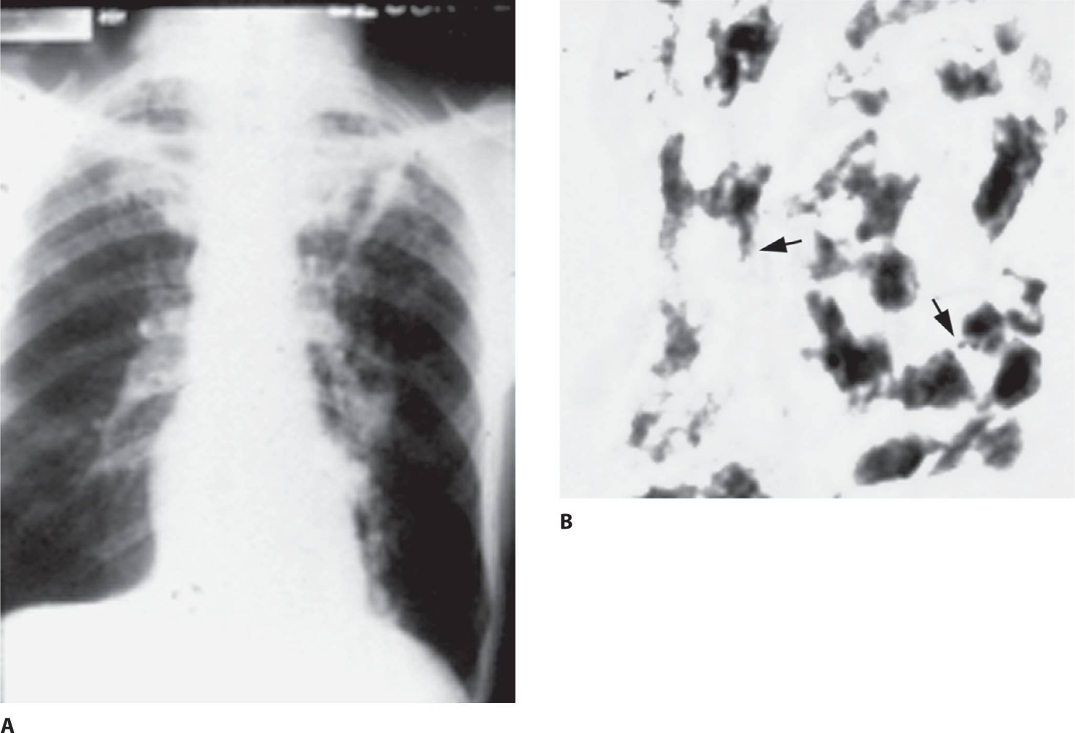
Figure 4.6. Cavitary pulmonary tuberculosis: A. Chest radiograph demonstrates bilateral upper lobe cavitary lesions, and B. sputum smear for acid-fast bacilli confirms the presence of those organisms. See color image on color plate 1
Pathogenesis
Mycobacterium tuberculosis is an aerobic, nonmotile bacillus with a waxy lipid-rich outer wall containing high concentrations of mycolic acid. This waxy outer wall fails to take up Gram stain. Visualization of mycobacteria requires heating to melt the outer wall, which allows for penetration and binding of the red dye fuchsin. The lipids in the cell wall bind this dye with high affinity and resist acid–alcohol decolorization. This acid-fast bacillus is small in size and appears beaded [Figure 4.6 (B)]. Genomic analysis reveals that, as compared with other bacteria, M. tuberculosis has a large number of genes encoding for enzymes that regulate lipogenesis and lipolysis. The resulting high lipid content of this pathogen accounts for many of its unique clinical characteristics, including its ability to resist killing by macrophages and PMNs and to survive for many years within the body. Rate of growth in M. tuberculosis is very slow, being about 1/20th the growth rate of most conventional bacteria. The slow rate of growth may also be explained by the waxy cell wall, which limits access to nutrients.
Mycobacteria survive and grow in macrophages, and they therefore induce a profound chronic inflammatory response. On gaining entry to the lungs, these organisms are ingested by alveolar macrophages and transported to the hilar lymph nodes. Here macrophages and dendritic cells present tubercular antigens to T cells, inducing a cell-mediated immune response. Helper T cells (CD4+) then activate macrophages to kill the mycobacteria and control the infection. Accumulation of one of the cell wall waxes, cord factor, stimulates the formation of granulomas that contain clusters of epithelioid cells, giant cells, and lymphocytes. Over time, the centers of the granulomas become necrotic, forming cheesy debris called caseous necrosis. Caseating granulomas are the hallmark lesion of tuberculosis. This pathologic finding is only rarely found in other diseases. If intracellular growth of M. tuberculosis continues, increasing numbers of macrophages are activated to produce multiple cytokines. Interleukin 1 stimulates the hypothalamus to raise core body temperature, causing fever. Tumor necrosis factor interferes with lipid metabolism and causes severe weight loss. These cytokines are primarily responsible for the symptoms of fever, night sweats, and weight loss described in case 4.3.
Epidemiology
Humans are the only reservoir for M. tuberculosis. Person-to-person spread of infection is almost exclusively caused by inhalation of droplet nuclei that have been aerosolized by coughs or sneezes. The likelihood of inhaling infectious droplets is greatly increased in a closed, crowded environment. A single cough has been estimated to form 3000 infectious droplets, with a sneeze producing even higher numbers.
The infectiousness of an individual patient can be estimated by AFB smears. The higher the number of organisms per microscopic field, the greater the infectious potential. Patients with laryngeal tuberculosis are particularly infectious and can release large numbers of organisms while speaking. Patients with AIDS and tuberculosis often harbor a large organism burden. Patients with large pulmonary cavities tend to intermittently release large numbers of infectious particles.
KEY POINTS
About the Pathogenesis of Tuberculosis
1. Slow-growing aerobic rod, not seen on Gram stain. The lipid-rich outer wall binds the red dye fuchsin, which is not removed by acid, making the bacterium acid-fast.
2. The lipid wall also allows the bacterium to resist drying and many disinfectants. It further allows the bacterium to survive within macrophages for years.
3. Macrophages carry the mycobacterium to the lymph nodes, where a cell-mediated immune response is generated.
4. Caseating granulomas are formed as a consequence of the cell-mediated immune response and the accumulation of lipid-rich bacteria.
5. Increased levels of interleukin 1 cause fever, and increased levels of tumor necrosis factor cause weight loss.
Repeated exposure and close contact are generally required to contract this disease. Respiratory isolation and rapid treatment of infected individuals are the primary ways to prevent spread of infection.
Despite the availability of antituberculous agents, tuberculosis remains a leading cause of death worldwide. It has been estimated that one-third of the world’s population is infected with M. tuberculosis and that 1.4-1.8 million die each year as a consequence of this infection. The incidence of tuberculosis infections is as high as 100/100,000 in regions of sub-Saharan Africa, India, China, and South East Asia. The living conditions and the existence of immunologically naive populations continue to allow rapid person-to-person spread, particularly in underdeveloped countries.
After a surge in cases in the United States during the mid-1980s because of the AIDS epidemic, the case rate has steadily declined. In 2010, it reached the lowest level ever recorded: 3.6 cases per 100,000. This steady decline among permanent U.S. residents contrasts with the steady increase in the percentage of tuberculosis cases among people immigrating to the United States. Immigrants now account for over half of all reported cases in the United States, the majority of cases being identified in those emigrating from underdeveloped countries. Immigrants from established market economies such as those of Western Europe have rates similar to those in the United States.
KEY POINTS
About the Epidemiology of Tuberculosis
1. Humans are the only reservoir of this disease.
2. Person-to-person spread occurs via aerosolized infectious droplets from sneezes or coughs.
a) Laryngeal tuberculosis is highly infectious.
b) Patients with HIV release large numbers of organisms.
c) Large cavitary lesions are also highly infectious.
3. People with these characteristics are at increased risk:
a) Immigrants from developing countries
b) Alcoholics
c) Urban poor
d) Single men
e) Intravenous drug abusers
f) Migrant farm workers
g) Prison inmates
h) People infected with HIV
i) Elderly people
4. A genetic predisposition is found in people who are black, Hispanic, Asia-Pacific Islanders, and Native Americans (5-10 times the incidence seen in Caucasians)
Tuberculosis also occurs more frequently in single men, alcoholics, intravenous drug abusers, the urban poor (particularly homeless people), migrant farm workers, and prison inmates. Elderly people are more likely to develop reactivation tuberculosis because cell-mediated immunity wanes with age.
A genetic predisposition to the development of active tuberculosis is known. People with European heritage tend to be more resistant, probably as a consequence of the devastating effects of the tuberculosis epidemic during the Industrial Revolution. At that time, tuberculosis was responsible for one-fourth of the deaths in Europe, killing off a significant percentage of the population that had a reduced immune response to mycobacteria. As compared with white people, people who are black or Hispanic, or who are Asia-Pacific Islanders or Native Americans experience a 5-10 times higher incidence of tuberculosis. Patients with AIDS are particularly susceptible to tuberculosis, and this population has spread the infection to others. Areas and demographic groups in which AIDS is more prevalent therefore have a higher incidence of tuberculosis.
KEY POINTS
About Primary Tuberculosis
1. Represents the first exposure to inhaled infectious particles.
2. Followed by a flu-like illness.
3. Spread is controlled over 4-8 weeks by the development of cell-mediated immunity.
4. Ghon foci are calcified lung lesions at the site of the primary infection.
5. Bacteremia develops and seeds the kidneys, epiphyses of the long bones, and vertebral bodies (areas with high oxygen content). The infection can later reactivate.
The patient in case 4.3 has a number of epidemiologic characteristics that increase his risk of tuberculosis. He is a single male, black, possibly alcoholic, and elderly.
Clinical Manifestations
There are two forms of human tuberculosis infection: primary tuberculosis and reactivation tuberculosis.
PRIMARY TUBERCULOSIS
Primary disease occurs when a patient inhales infectious M. tuberculosis droplets for the first time. A flu-like illness usually follows; however, some people experience no symptoms. Within 4-8 weeks of exposure, the human host usually mounts a cell-mediated immune response. Activated macrophages control the spread and growth of the organism. Pulmonary lesions heal spontaneously and form areas of fibrosis or calcification called Ghon lesions or foci. A Ghon lesion in combination with hilar adenopathy is called a Ranke complex.
In addition to transporting organisms to the hilum and mediastinum, infected macrophages may gain access to the thoracic duct, enter the bloodstream, and spread throughout the body. M. tuberculosis grows best in regions with high oxygen tension, including the kidneys, long-bone epiphyses, and vertebral bodies. It most commonly infects the apices of the lung, the regions with the highest oxygen content and reduced lymphatic flow.
Although the infection is brought under control, the bacilli are not usually completely eradicated. Organisms can survive for decades, being held in check by the host immune response. But any condition that subsequently depresses cell-mediated immunity can free M. tuberculosis to grow and cause symptomatic tuberculosis.
MILIARY TUBERCULOSIS
In some individuals, initial exposure to M. tuberculosis fails to induce cell-mediated immunity, or the immune response is not robust enough to control the infection. Under these conditions, the mycobacteria continue to multiply and disseminate, causing miliary tuberculosis. Very young and very old patients are at higher risk of developing disseminated disease, as are patients receiving immunosuppressants and those with HIV infection. Underlying medical conditions often associated with miliary tuberculosis include alcoholism, malignancy, connective tissue diseases, renal failure, and pregnancy. However, it must be emphasized that absence of an underlying disease does not exclude the possibility of miliary tuberculosis.
KEY POINTS
About Miliary Tuberculosis
1. The disease develops in very young, very old, and HIV-infected patients.
2. It is also associated with alcoholism, malignancy, connective tissue diseases, renal failure, and pregnancy.
3. In children, it presents with high fever, night sweats, weight loss, hepatosplenomegaly, and lymphadenopathy.
4. Adults usually show moderate- to low-grade fever, night sweats, malaise, anorexia, weakness, and weight loss.
5. Look for choroid tubercles in the fundi (present in up to 50% of cases).
6. Provokes leukemoid reaction, anemia, hyponatremia, and abnormal liver function tests. May also produce adrenal insufficiency.
7. Micronodular interstitial pattern on chest radiographs; may be negative in elderly and HIV-infected patients.
8. Blood samples, transbronchial biopsy, bone marrow samples, and liver biopsy may all yield positive cultures.
9. Provide early treatment for all suspected cases, using isoniazid, rifampin, ethambutol, and pyrazinamide.
Children usually present to the physician with high fever, night sweats, weight loss, hepatosplenomegaly, and lymphadenopathy. However, in adults, particularly elderly people, the clinical manifestations may be subtle. Patients usually have nonspecific complaints of fever, malaise, anorexia, weakness, and weight loss. Night sweats are also common.
Physical examination usually reveals a chronically ill patient with no specific findings. In some patients, lymphadenopathy may be detected. In all patients, fundoscopic examination should be carefully performed following pupillary dilation and may reveal choroid tubercles in up to 50% of cases. The diagnosis is often missed, and in up to 20% cases, it is made postmortem.
The peripheral WBC count is usually normal; however, some patients develop extremely high WBC counts (30,000-40,000/mm3), also termed a “leukemoid reaction,” that can be mistaken for leukemia. Pancytopenia can also develop. Liver function abnormalities are common. Elevated alkaline phosphatase and moderate increases in transaminase values are found in most patients. Serum sodium may be low as a consequence of adrenal insufficiency (a well-known complication of miliary tuberculosis) or inappropriate antidiuretic hormone secretion. Morning and evening serum cortisol levels should be measured to exclude adrenal insufficiency. In approximately two-thirds of patients, CXR reveals small nodules (0.05–1 mm in diameter) that resemble millet seeds (the basis for the designation “miliary”); however, a negative CXR in elderly patients and in patients with HIV does not exclude this diagnosis. In a few patients, ARDS may develop, causing complete opacification of the lungs.
The key to the diagnosis of miliary tuberculosis is a high index of suspicion. Sputum smears are positive in only a few patients. Samples from enlarged lymph nodes, liver biopsy, and bone marrow should therefore be sought for histopathology (seeking granulomas and AFB) and culture. Transbronchial biopsy can yield the diagnosis in many patients. Blood samples for culture should be drawn; in patients with AIDS, cultures are commonly positive. If CNS symptoms are noted, a lumbar puncture should also be performed, although the resulting smears are usually negative.
A delay in treatment can have fatal consequences. Therefore, if miliary tuberculosis is high on the differential diagnosis, empiric antituberculous therapy should be initiated as soon as samples for culture have been obtained. A four-drug combination consisting of isoniazid (INH), rifampin, pyrazinamide, and ethambutol is the preferred regimen. Patients usually defervesce within 7-14 days.
REACTIVATION TUBERCULOSIS
Reactivation of tuberculosis after primary disease occurs in 10-15% of patients. In half of these cases, the infection reactivates within 2 years of exposure. In past decades, reactivation occurred most commonly in elderly patients, but in the United States today, most reactivation cases are now reported in middle-aged adults (30-50 years of age). Early in the course of reactivation, patients are often asymptomatic, and evidence of reactivation is found only on CXR. However, if the infection is not detected, symptoms slowly develop and worsen over several months. The gradual nature of symptom onset often causes patients to delay seeing a physician. The patient in case 4.3 has the typical symptoms of reactivation tuberculosis: a progressively worsening cough with sputum production, low-grade fever, night sweats, fatigue, and weight loss. Symptoms that suggest more advanced disease are hemoptysis (indicating erosion of a tuberculous cavity into an arteriole) and pleuritic chest pain (suggesting pleural involvement and probable tuberculous pleural effusion).
Physical examination is often unrevealing, as observed in case 4.3. Despite the presence of extensive pulmonary disease, auscultation may be normal. Fine rales may be heard in the apices after a short cough and quick inspiration or after full expiration followed by a cough and rapid inspiration (post-tussive rales).
KEY POINTS
About Reactivation Tuberculosis
1. Reactivation occurs in 10-15% of patients, half within 2 years of primary disease.
2. Reactivation is most common in men 30-50 years of age.
3. Apical infection is most common. The high oxygen content and reduced lymphatic flow favor Mycobacterium tuberculosis survival in this region.
4. Symptoms progress slowly over several months: worsening cough with sputum production, low-grade fever, night sweats, fatigue, and weight loss.
5. Hemoptysis or pleuritic pain indicates severe disease.
6. Physical examination usually produces minimal findings; post-tussive rales may be seen.
7. Chest radiograph shows apical cavities (without fluid); order apical lordotic. A computed tomography scan is often helpful.
8. Cavitary disease is highly infectious; cavities contain between 109 and 1010 organisms. Isolate all patients. In HIV infection, the chest radiograph often does not show cavities. All pneumonias in patients with AIDS are considered to involve tuberculosis until proven otherwise.
The hallmark of reactivation pulmonary disease is the presence of apical cavitary lesions on CXR. Lesions usually develop in posterior segments of the upper lobes just below the clavicle. Less frequently, infiltrates are noted in the apex of the lower lobe (usually obscured by the heart shadow). In addition to routine posterior–anterior and lateral chest films, an apical lordotic view is often helpful in visualizing upper lobe apical lesions. A chest CT scan can be helpful for assessing the extent of disease and for defining the size of the cavities. Unlike conventional lung abscesses, tuberculous cavities rarely have air fluid levels. In patients with AIDS, infiltrates may be in any region of the lung and may not cavitate. Any HIV-infected patient with a new pulmonary infiltrate should therefore be considered to have tuberculosis until proven otherwise. In fact, in some instances, HIV-infected patients with active respiratory tuberculosis may have a negative CXR.
Individuals with cavitary disease are potentially highly infectious. Cavities may contain between 109 and 1010 organisms. Patients should be placed in respiratory isolation while sputum AFB smears and cultures are obtained. The number of organisms seen on smear directly correlates with infectiousness—that is, the higher the number of organisms per microscopic field, the higher the likelihood of disease spread.
Diagnosis
The classic test for making the diagnosis of pulmonary tuberculosis is the Ziehl–Neelsen acid-fast sputum smear. Morning sputum samples tend to have the highest yield. A single negative smear should not delude the clinician into a false sense of security. Three sputum smears are recommended, because in cavitary disease, the release of infectious droplets is intermittent. Only after three smears are negative should the patient be declared to be at low risk of spreading infection. Negative smears do not definitively exclude tuberculosis.
To be positive, the sputum smear must contain 104 organisms per milliliter. A fluorochrome stain using auramine–rhodamine is more sensitive and allows sputum to be examined at low magnification (20× or 40× magnification) as compared with conventional AFB smears that must be examined at high magnification (100–). Sputum smear has only a 60% sensitivity as compared with sputum culture. The PCR technique can effectively detect as few as 10 organisms in a clinical specimen. Two assays, one using mycobacteria RNA as its initial template, and the other using mycobacterial DNA, are commercially available. Sensitivity and specificity are greater than 95% in smear-positive cases, and specificity in smear-negative cases is high. False negative and false positive results are common in less experienced laboratories, and nucleic acid amplification assays complement traditional methods. In patients on antituberculous therapy, PCR cannot differentiate killed from actively growing organisms. GeneXpert MTB/RIF is a nucleic acid based amplification assay that detects MTB and rifampin-resistant with 98% specificity and sensitivity in smear-positive patients. This assay can be useful in guiding initial antituberculous therapy.
KEY POINTS
About the Diagnosis of Tuberculosis
1. Ziehl–Neelsen acid-fast stain can detect 104 organisms per milliliter with 60% sensitivity.
2. Release of acid-fast bacilli from cavitary lesions is intermittent. To ensure low infectivity, three negative smears are needed.
3. Culture remains the most sensitive and specific test.
a) Mycobacterium tuberculosis grows at 1/20th the rate of conventional bacteria.
b) Automated techniques can detect bacteria within 9-16 days.
c) Growth in conventional Lowenstein–Jensen medium takes 3-6 weeks.
4. Polymerase chain reaction is available, but should be performed only by experienced laboratories.
Culture remains the most accurate method for diagnosing M. tuberculosis. In patients who fail to produce sputum, aspiration of the gastric contents in the morning before the patient arises from bed is useful for obtaining samples for culture. In patients with suspected disseminated disease, blood samples in which all cells are lysed to release intracellular mycobacteria should be collected. The bacterium grows at about 1/20th the rate of more conventional bacteria, taking 3-6 weeks to grow on Lowenstein–Jensen medium. Living mycobacteria can be more quickly detected in blood, sputum, pleural fluid, or CSF using the Bactec radiometric or fluorometric culture system, which is designed to detect mycobacteria metabolism within 9-16 days. Drug susceptibilities can also be reliably tested using this method.
Treatment
The principal strategies for treating mycobacteria differ somewhat from more conventional bacteria. Because mycobacteria are intracellular and grow very slowly, and because dormant tuberculous organisms found in necrotic cavitary lesions are difficult to kill, antituberculous therapy must be prolonged—for a period of months.
Second, because the number of mycobacterial organisms in the host is usually high, the potential for selecting for resistant mycobacteria is high. To reduce this risk, treatment with two or more antimycobacterial medications is recommended. Generally, 1 in 106 organisms is naturally resistant to INH. Cavitary lesions often contain between 109 and 1010 organisms, assuring the survival and replication of resistant organisms. Administration of two drugs reduces the probability of selecting for a resistant organism because only 1 in 1012 organisms (106 × 106) would be expected to be resistant to both antimicrobial agents.
A third major consideration is advent of multidrug-resistant M. tuberculosis (MDR-TB). These mycobacteria are resistant to INH and rifampin, and they must be treated with three or more other antimycobacterial agents. In the early 1990s in the United States, MDR-TB was major concern; however, with improved infection control measures, the use of four-drug regimens, directly observed therapy (DOT), and the incidence of MDR-TB have been reduced to less than 2%, and resistance to INH alone is approximately 8%.
Resistance is classified as either secondary or primary. Primary resistance is defined as infection with a resistant strain in a patient who has never received antituberculous drugs. When a resistant strain is cultured from a patient who was previously treated for drug-sensitive tuberculosis, the infection is said to be secondarily resistant. Secondary resistance is a major problem among homeless people, illicit drug users, and patients with AIDS. To reduce the development of secondary resistance, DOT is the recommended approach for all patients with active pulmonary TB.
Outside of the United States, the percentages of MDR-TB and INH-resistant strains vary widely. The worldwide median frequency of primary INH resistance is estimated to be 7.3%, with higher levels being observed in Asia, Africa, and Latin America, and lower levels in Europe and Oceania. The number of MDR-TB cases worldwide has been estimated to be 500,000 annually. Extensively drug-resistant tuberculosis (XDR) was initially reported in South Africa and has subsequently been discovered in 68 other countries. The worldwide incidence is estimated to be 40,000 cases annually. XDR TB is defined as resistance to both INH and rifampin with additional resistance to at least one fluoroquinolone and one injectable agent. Patients with XDR TB fail to respond to nearly all drugs and the mortality can exceed 90%.
The various antituberculous agents have been classified as first-line and second-line drugs. First-line medications include INH, rifampin, pyrazinamide, streptomycin, and ethambutol. These agents are more efficacious and less toxic than the second-line drugs. With the exception of ethambutol, first-line agents are also bactericidal. Whenever possible, first-line drugs should be employed for the treatment of M. tuberculosis.Tables 4.4 and 4.5 summarize the toxicities and recommended doses of each of these agents.
Table 4.4. Toxicities of Antituberculosis Medications

Table 4.5. Antituberculous Medications: Half-Life, Dosing, Renal Dosing, and Cost

KEY POINTS
About Antituberculous Therapy
1. A four-drug regimen (pending sensitivity testing) is recommended.
a) Of every 106 organisms, 1 is naturally resistant to one drug.
b) Cavitary lesions contain between 109 and 1010 organisms.
c) A minimum of two effective drugs is needed to prevent resistance (106 × 106 = 1012).
d) Primary isoniazid (INH) resistance is common; to reliably prevent resistance, treat with INH, rifampin, pyrazinamide, and ethambutol (pending sensitivities).
2. INH-resistance is 8% in the United States, 7.3% worldwide. Higher in Asia, Africa, and Latin America.
3. Multidrug resistance is below 2% in the United States, worldwide incidence 500,000 per year.
4. Secondary resistance occurs in patients who do not reliably take their medications.
5. Directly observed therapy (DOT) is now recommended for all patients.
The recommended treatment of presumed drug-sensitive pulmonary tuberculosis (pending sensitivity tests) is a four-drug regimen: INH, rifampin, pyrazinamide, and ethambutol or streptomycin. This regimen is recommended for 2 months, to be followed by INH, rifampin, and pyrazinamide for 4 months. The CDC guidelines list four alternative regimens that can be used depending on the patient’s social situation. If MDR-TB is suspected, extensive susceptibility testing should be performed, and expert advice sought to design an appropriate regimen. Treatment should consist of at least three drugs to which the organism is proven to be susceptible. Fluoroquinolones combined with aminoglycosides are particularly useful for treating MDR-TB.
In all patients, CDC guidelines recommend DOT. Poor adherence greatly increases the risk of secondary MDR-TB, and with the institution of DOT in these patients, the emergence of resistance is minimized. One commonly accepted DOT regimen is outlined in Table 4.6.
Table 4.6. Typical Course of Direct Observed Therapy for Tuberculosis

Prevention
Tuberculosis is spread strictly from person to person. Identifying and preventing individuals who have been exposed to tuberculosis from developing active disease is a major public health goal. The focus for testing should be individuals with potentially latent disease who are at increased risk of developing active disease. Testing of the general population is no longer recommended. Risk groups include patients with HIV, patients receiving immunosuppressants, including transplant patients, and those at high risk of recent infection: recent immigrants, health care workers, and tuberculosis contacts.
The purified protein derivative (PPD) test is a very helpful skin test that assesses exposure to tuberculosis. The test is produced by acid precipitation of tubercle bacilli proteins, and the 5-tuberculin unit dose has been standardized and is administered as a 0.1-mL subcutaneous injection on the volar aspect of the forearm. Deeper injection is ineffective because tuberculous proteins can be removed by blood flow, producing a false negative result.
The injection should produce a discrete raised blanched wheal. The test is read 48-72 hours after injection; however, the reaction usually persists for 1 week. The diameter of induration is measured, and a diameter of more than 10 mm is defined as positive. A positive test indicates high risk of contracting tuberculosis. Of people with a PPD reaction 10 mm in diameter, 90% are infected with tuberculosis. If the reaction measures more than 15 mm, 100% are infected. The 15-mm diameter is defined as a positive reaction in individuals with no risk factors for tuberculosis. In individuals who are immunocompromised (HIV-positive, organ-transplant patients receiving more than 15 mg prednisone daily) or who are recent household contacts of a patient with active tuberculosis, more than 5 mm is considered a positive reaction.
A major logistics problem for skin testing is the requirement that patients return at 48 hours to measure the extent of induration. Second, patients who have received Bacille Calmette–Guérin (BCG) vaccine often cross-react with the PPD antigens. To address these issues, a whole blood tests are now commercially available that measure T-cell release of interferon gamma in response to specific M. tuberculosisantigens: early secreted antigenic target 6 (ESAT-6) and culture filtrate protein 10 (CFP-10). These antigens are more specific than PPD, do not cross-react with BCG, and with the exception of M. marinumand M. kansansii, do not cross-react with atypical mycobacteria. QuantiFERON-TB Gold In-Tube utilizes an ELISA assay to quantify gamma interferon release, while the T-SPOT.TB uses an immunospot method that measures the percentage of peripheral mononuclear cells releasing gamma interferon. Extensive studies reveal that the sensitivity of QuantiFERON-TB Gold In-Tube is equivalent to the PPD skin test, and the T-SPOT.TB assay has greater sensitivity for detecting latent disease.
A positive skin or whole blood test simply indicates that, sometime in the past, the individual was exposed to active tuberculosis; however, this findingdoes not indicate active disease. The conversion from negative to positive in an individual who is tested annually indicates exposure to tuberculosis during the time interval between tests. These tests cannot be relied upon to determine exposure in patients with HIV with low CD4 counts, in patients receiving immunosuppressants, or in patients with severe malnutrition.
Individuals with a positive PPD or interferon gamma release assay should have a CXR, and if pulmonary lesions are noted, three sputum samples should be obtained for culture and smear. Prophylaxis should be given only if all sputum samples prove negative for tuberculosis. Because the risk of developing active disease is highest within 2 years of exposure, all individuals who have converted from a negative to a positive test within 2 years should receive INH prophylaxis. Preventive therapy is also warranted when a positive test is associated with other specific risk factors (HIV infection, known recent exposure to tuberculosis, abnormal CXR, intravenous drug abuse, and certain underlying diseases). In other individuals with a positive PPD, the risk of INH hepatotoxicity must be balanced against the likelihood of preventing the development of active disease.
KEY POINTS
About Tuberculosis Testing and Prophylaxis
1. The purified protein derivative (PPD) test is carefully standardized, and induration at 48 hours is considered positive at
a) more than 5 mm in people who are HIV-positive or immunocompromised, or who have had recent household exposure;
b) more than 10 mm in people at overall risk of exposure; and
c) more than 15 mm in people with no risk factors.
2. Interferon gamma release assay QuantiFERON-TB Gold equivalent sensitivity to PPD, but does not cross-react with BCG. T-SPOT.TB greater sensitivity.
3. A positive result indicates exposure sometime in the past; negative to positive conversion indicates exposure during the period between tests.
4. Prophylaxis with isoniazid (300 mg daily) for 9 months or INH (15-25 mg/kg maximum 900 mg) and rifapentine (900 mg, adjust if weight <50 kg) weekly for 3 months directly observed therapy if
a) conversion within the last 2 years and negative chest radiograph (CXR).
b) positive PPD and negative CXR (recommendation of the Centers for Disease Control and Prevention).
c) abnormal CXR, and three follow-up sputum smears for acid-fast bacilli are negative.
5. If prophylaxis recipient is older than 35 years, a consumer of alcohol or other hepatotoxic drugs, pregnant, or HIV-positive, risk of INH hepatitis requires monthly monitoring of hepatic enzymes.
In individuals 35 years of age or older, the risk of hepatotoxicity may outweigh the potential benefit of INH prophylaxis. Hepatic enzymes should be monitored at monthly intervals in HIV-positive patients, pregnant women, patients with underlying liver disease, and in those receiving other potentially hepatotoxic drugs or drinking alcohol daily. Prophylaxis should be discontinued if transaminase levels rise exceed three times the normal values in association with symptoms consistent with hepatitis.
The recommended prophylactic regimen is INH 300 mg daily for 9 months or 3 months of weekly INH and rifapentin (15-25 mg/kg INH and 900 mg rifapentin, adjusted if <50 kg weight) given by direct observation. Alternative regimens include daily INH for 6 months, daily rifampin for 4 months, or daily INH and rifampin for 3 months. For HIV-infected patients, 12 months of INH prophylaxis is recommended.
Atypical Mycobacteria
Atypical mycobacteria are found throughout the environment in soil and water. These organisms have a low virulence, and they do not usually cause pulmonary disease in otherwise healthy individuals. In patients with underlying pulmonary disease, these organisms can be inhaled and cause pulmonary infection.
M. avium complex is the most common of the atypical mycobacteria to infect the lung. A cavitary upper lobe lesion is the usual manifestation of this disease. The cavities tend to be somewhat smaller and thinner walled than those with M. tuberculosis. Pulmonary infection with M. avium complex is seen primarily in male smokers in their early 50s who abuse alcohol. Infection of the lungs is also seen in women 60 years of age or older with no apparent underlying disease, most commonly involving the right middle lobe or lingula.
M. kansasii, M. fortuitum, and M. abscessus can also infect the lungs, causing chronic cavitary disease. Because these organisms are found throughout the environment and may colonize as well as infect patients with chronic lung diseases, elaborate criteria for differentiating colonization from infection have been established. Therapy for atypical mycobacterial infection must be prolonged and is based on sensitivity testing. Often, these organisms respond poorly to therapy, and resection of the infected lung segment may be required for cure. Management of these patients is complex and requires the supervision of an experienced pulmonary or infectious disease specialist.
KEY POINTS
About Atypical Mycobacteria Pulmonary Infection
1. Atypical mycobacteria are found in soil and water.
2. Infects males over the age of 50 years, who are also alcoholic, smokers with chronic lung disease. Often presents as upper lobe cavitary disease.
3. Infects women over the age of 60 years without apparent underlying disease. Presents as right middle lobe or lingular disease.
4. Mycobacterium avium is the most common pathogen; M. kansasii, M. fortuitum, and M. abscessus are rarer.
5. Management is complex and requires a pulmonary or infectious disease specialist.
Fungal Pneumonias
The most common forms of fungal pneumonia in the normal host are histoplasmosis and coccidioidomycosis. In the immunocompromised host, Cryptococcus and Aspergillus can also cause pneumonia (see Chapter 15).
HISTOPLASMOSIS
Epidemiology. Histoplasma capsulatum is one of the more common causes of chronic pneumonia in the Midwestern and Southeastern United States. This organism survives in moist soil in temperate climates and is concentrated in decayed trees, on riverbanks, old chicken coops, starling roosts, and caves contaminated with bat guano. The development of histoplasmosis is generally associated with construction or excavation of soil contaminated with H. capsulatum. Infection is also reported in spelunkers, who contract the infection by disturbing dried bat guano containing high concentrations of infectious particles. Exposure to infectious particles can also occur after the renovation of old buildings previously inhabited by birds or bats.
KEY POINTS
About the Epidemiology and Pathogenesis of Histoplasmosis
1. Found primarily in the Midwest and Southeast United States.
2. Grows in moist soil in temperate zones, mainly Ohio and Mississippi River valleys.
3. Found in caves and old buildings; bat guano is a concentrated source.
4. Mycelial form in soil, as macro- and microconidia. Microconidia readily aerosolized.
5. Inhaled microconidia ingested by macrophages and neutrophils convert to yeast forms and upregulate many genes, including a gene for calcium binding.
6. Yeast forms are transported to hilar nodes, where cell-mediated immunity is induced.
Pathogenesis. H. capsulatum is a fungus and exists in two forms: mycelia or yeast. In the moist soil of temperate climates, the organism exists in the mycelial form as macroconidia (8-15 μm in size) and microconidia (2–5 μm in size). When infected soil is disturbed, microconidia float in the air and can be inhaled into the lung. Once in the lung, microconidia are ingested by alveolar macrophages and neutrophils. In the intracellular environment of these phagocytes, the mycelia transform to rounded, encapsulated yeast cells. During this transformation, multiple genes are upregulated, including a gene that increases production of a calcium-binding protein important for acquiring calcium (an essential ion for yeast survival) from the intracellular environment. The expression of this calcium-binding protein may explain the frequent finding of calcifications in infected tissues.
As is observed in tuberculosis, infected macrophages transport the yeast forms to the hilar lymph nodes where Histoplasma antigens are presented to T cells. Within several weeks, cell-mediated immunity develops, and CD4 T cells activate macrophages to produce fungicidal products.
Clinical Manifestations. In more than 90% of patients, infection is controlled. In many patients, primary exposure is asymptomatic or results in a mild influenza-like illness. Very young people, elderly people, and patients with compromised immune systems are more likely to develop active disease. Symptoms usually develop within 14 days of exposure and may include high fever, headache, nonproductive cough, and dull nonpleuritic chest pain. This form of chest pain is thought to be the result of mediastinal node enlargement. In other patients, chest pain may be sharper and may worsen upon lying down, reflecting the development of pericarditis (observed in approximately 6% of cases).
On CXR, patchy infiltrates may be seen during acute disease, which subsequently calcify producing a “buckshot” appearance. Healed histoplasmosis is also the most common cause of calcified lesions in the liver and spleen. In acute disease, mediastinal lymphadenopathy may be prominent and may mimic lymphoma or sarcoidosis. A history of exposure to a site where soil was excavated is particularly important in trying to differentiate between these various possibilities. Occasionally, mediastinal nodes can become massively enlarged, reaching diameters of 8-10 cm. Severe mediastinal fibrosis is rare, but it can lead to impingement and obstruction of the superior vena cava, bronchi, and esophagus.
KEY POINTS
About the Clinical Manifestations of Histoplasmosis
1. In 90% of cases, a brief self-limiting flu-like illness occurs or the person remains asymptomatic.
2. Disease can develop in elderly, very young, and immunocompromised individuals.
3. At 14 days postexposure, the individual may have
a) high fever, headache, nonproductive cough, and dull, nonpleuritic chest pain.
b) a CXR with patchy infiltrates that later convert to “buckshot” calcifications.
c) mediastinal lymphadenopathy that may mimic lymphoma or sarcoidosis.
d) progressive mediastinal fibrosis (a rare complication).
4. Cavitary disease is clinically similar, with men older than 50 years who have chronic obstructive pulmonary disease at higher risk.
5. Disseminated disease occurs in 10% of symptomatic primary disease.
a) Likelihood of dissemination increases in people who are very old, very young, or immunosuppressed (because of AIDS or transplantation).
b) Meningitis with lymphocytosis and low glucose may develop.
c) Reticulonodular pattern on CXR in most cases, but CXR normal in one-third.
Chronic cavitary histoplasmosis develops in about 8% of patients. This complication is more common in men over the age of 50 years who have chronic obstructive pulmonary disease. The symptoms and CXR findings associated with chronic pulmonary histoplasmosis are indistinguishable from cavitary tuberculosis. In fact, in the past, patients in the Midwestern and Southeastern United States with chronic pulmonary histoplasmosis were frequently misdiagnosed as having pulmonary tuberculosis and were mistakenly confined to tuberculosis sanatoriums. Spontaneous resolution of cavitary disease occurs in 10-60% of cases.
Progressive disseminate histoplasmosis occurs in about 10% of symptomatic primary infections. Progressive dissemination also develops as a consequence of reactivation of old disease. In the immunosuppressed individual, reactivation is the most likely pathway for disseminated disease. Onset of symptoms is usually abrupt. Fever and malaise are followed by nonproductive cough, weight loss, and diarrhea. Hepatosplenomegaly usually develops, and lymphadenopathy may be detected. Anemia, thrombocytopenia, and leukopenia are observed in a high proportion of patients. Meningitis may develop, resulting in lymphocytosis and low glucose in the CSF. A CXR may show a reticulonodular pattern or scattered nodular opacities; however, the CXR is normal in nearly one-third of cases. Mortality is high if treatment is not initiated.
Diagnosis. H. capsulatum can be readily grown from tissue samples and body fluids using brain–heart infusion media containing antibiotics and cycloheximide (inhibits the growth of saprophytic fungi). Mycelial growth can usually be detected within 7 days and confirmed using a DNA probe. The clinical microbiology lab must be notified that H. capsulatum is the possible pathogen, because the necessary culture methods are not employed on routine samples.
A single sputum culture has only a 10-15% yield; collection of multiple sputum cultures increases the yield. Bronchoscopy has proved useful for providing good sputum samples yielding positive cultures in 90% of patients HIV with pulmonary histoplasmosis. Bone marrow and blood cultures should also be obtained and are positive in up to 50% of cases.
The most effective method for detecting progressive disseminated histoplasmosis is the urine and serum polysaccharide antigen test. Antigen is detected in up to 90% of patients with disseminated disease and sensitivity of urine and serum is equivalent for disseminated disease; however, in acute pulmonary disease a subpopulation of patients have only a positive serum antigen. The urine antigen test is also positive in 40% of patients with cavitary pulmonary diseases and 20% with acute pulmonary histoplasmosis. Pulmonary lavage fluid can also be tested for antigen and one series of immunocompromised hosts demonstrated 94% sensitivity. Real-time PCR has proved to be highly sensitive and moderately specific when applied to cultures, as well as tissue and fluid samples.
KEY POINTS
About the Diagnosis of Histoplasmosis
1. Sputum culture is often positive.
a) Requires selective media (brain–heart infusion with antibiotics and cycloheximide).
b) Not a routine method; clinical microbiology must be notified.
c) Bronchoscopy improves yield (90% in HIV patients).
2. Bone marrow positive in 50% of cases.
3. Lysis–centrifugation method positive in up to 50% of blood samples.
4. Polysaccharide urine and serum antigen test is the most sensitive, being positive for
a) 90% of disseminated disease,
b) 40% cavitary disease, and
c) 20% acute pulmonary disease.
5. Method can also be used to test bronchoscopic lavage fluid.
6. Histopathology shows noncaseating or caseating granulomas. Silver stain best for identifying the yeast forms. Hematoxylin–eosin is not useful; periodic acid Schiff may help with identification.
7. Urine antigen test positive in 90% of disseminated histoplasmosis.
Histopathologic examination of infected tissue also allows for rapid diagnosis. Noncaseating or caseating granulomas may be seen. An excessive fibrotic reaction may be seen in some patients. Silver stains are most effective for identifying the typical yeast forms in tissue biopsies. Organisms are poorly visualized by hematoxylin–eosin staining, but can often be seen on periodic acid Schiff stain.
Treatment. IDSA guidelines are available for the management of histoplasmosis, and these guidelines should be followed to assure the most efficacious approach. In patients with moderately severe to severe acute pulmonary histoplasmosis, lipid formulation of amphotericin B 3-5 mg/kg/day should be administered intravenously for 1-2 weeks, followed by itraconazole (200 mg three times daily for 3 days and then 200 mg twice daily) for a total of 12 weeks. For mild-to-moderate acute pulmonary disease, treatment is unnecessary. If symptoms persist for over 1 month, itraconazole can be administered at the doses described above for 6-12 weeks.
KEY POINTS
About the Treatment of Histoplasmosis
1. Itraconazole the oral agent of choice. Recommended for
a) acute pulmonary disease that fails to improve over 7 days
b) extensive mediastinal involvement with progression requiring steroids
c) progressive cavitary disease.
2. Liposomal Amphotericin B recommended for moderate to severe and severe disseminated disease × 1-2 weeks followed by oral itraconazole × 12 months.
For chronic cavitary pulmonary disease, itraconazole (200 mg three times daily for 3 days and then once or twice daily) for at least 1 year is recommended, but some prefer 18-24 months because of the risk of relapse.
In patients with extensive mediastinal involvement treatment is generally not recommended. For more progressive disease requiring prednisone (0.5-1 mg/kg per day), itraconazole, 200 mg three times daily for 3 days followed by 200 mg one to two times daily for 6-12 weeks can be initiated. Patients with chronic mediastinal fibrosis may also require surgical intervention to correct vascular and airway obstruction.
In moderately severe to severe disseminated histoplasmosis, liposomal amphotericin B 3 mg/kg/day should be administered for 1-2 weeks followed by itraconazole 200 mg three times daily for 3 days and then 200 mg twice daily for a minimum of 12 months.
Other azoles have proved helpful in selected cases of histoplasmosis. Posaconazole has excellent activity against this organism and has been used for salvage therapy in patients who have failed to respond to other regimens. Voriconazole also has activity against this Histoplasma and has been administered in a small number of patients.
COCCIDIOIDOMYCOSIS
Epidemiology. Like H. capsulatum, Coccidioides immitis survives and grows in soil. The ideal conditions for survival of C. immitis are dry, alkaline soil, hot summers, and winters with few freezes. These conditions exist in central California’s San Joaquin Valley and in the southern regions of Arizona, New Mexico, and Texas. C. immitis is also found in Mexico, Central America, and South America. Infections are most commonly reported in the summer months when dry soil more readily forms dust particles. Epidemics have been associated with disruption of soil by archeological excavation, earthquakes, and dust storms. In recent years, the incidence of coccidioidomycosis has increased as a consequence of the increased numbers of people living in endemic areas.
KEY POINTS
About the Epidemiology and Pathogenesis of Coccidioidomycosis
1. Grows in soil; prefers dry, alkaline soil, hot summers, and winters with few freezes.
2. Primarily found in central California, southern Arizona, New Mexico, and Texas. Also found in Mexico, Central America, and South America.
3. Contracted during the summer, often through dust storms, excavation, earthquakes.
4. Mycelial form of this dimorphic fungus is called arthroconidia.
5. Inhaled arthroconidia transform to spherules (yeast forms) that release endospores.
6. Endospores ingested by macrophages are transported to hilar lymph nodes, lymphatics, and bloodstream.
7. Cell-mediated immunity critical for control of infection.
Pathogenesis. Also like H. capsulatum, C. immitis is a dimorphic fungus. It exists in soil as mycelia that can form small arthroconidia (5-μm barrel-shaped structures). Arthroconidia can become airborne, whereupon they are inhaled by humans and become lodged in the terminal bronchioles. In the warm moist environment of the lung, the arthroconidia transform into spherules. As the spherules mature, their outer walls thin, and they release endospores that are ingested by macrophages. As is observed in histoplasmosis and tuberculosis, macrophages transport the infectious particles to the hilar lymph nodes, the lymphatic system, and the bloodstream, resulting in dissemination. Cell-mediated immunity is critical for control of the infection.
Clinical Manifestations. Approximately two-thirds of patients exposed to arthroconidia experience minimal symptoms. When symptoms are noted, they usually develop 7-21 days after exposure. Nonproductive cough and fever are the most frequent symptoms. Pleuritic chest pain, shortness of breath, headache, and fatigue are also commonly reported. Skin manifestations may include erythema nodosum (red, painful nodules on the anterior shins), erythema multiforme (target-like lesions involving the entire body, including the palms and soles), or a nonpruritic papular rash. Arthralgias may develop in association with erythema nodosum. Eosinophilia is commonly observed on peripheral blood smear.
KEY POINTS
About the Clinical Manifestations of Coccidioidomycosis
1. Symptoms (nonproductive cough, fever, pleuritic chest pain, shortness of breath, headache, and fatigue) occur in about one-third of exposed individuals 7-21 days after inhalation.
2. Skin manifestations are common: erythema nodosum, erythema multiforme, nonpruritic papular rash.
3. Eosinophilia may be noted on peripheral blood smear.
4. Abnormal CXR findings are frequent: unilateral infiltrates, pleural effusions, and hilar adenopathy.
5. In patients with AIDS whose CD4 counts fall below 100/mm3, the disease can disseminate, causing diffuse lung opacification, meningitis, bone infection, and arthritis.
6. Chronic lung disease can lead to fibrosis, nodules, or cavities.
7. Isolated pulmonary nodules are not calcified, can be differentiated from neoplasm by biopsy.
8. Chronic pleural effusions most commonly develop in young, healthy, athletic males.
In about half of patients, a CXR is abnormal, most commonly demonstrating unilateral infiltrates, pleural effusions, and hilar adenopathy. In patients with depressed cell-mediated immunity (primarily patients with AIDS and CD4 counts below 100/mm3), the infection can disseminate, causing diffuse opacification of the lungs and severe respiratory failure. Meningitis, skin lesions, bone infection, and arthritis may also develop as a consequence of dissemination.
In some patients, pulmonary infection can persist, causing progressive destruction of lung parenchyma associated with a productive cough, chest pain, and weight loss. A CXR may demonstrate areas of fibrosis, nodules, cavitary lesions, or a combination. An isolated nodule can persist in approximately 4% of pulmonary cases and can be differentiated from neoplasm only by biopsy. These lesions seldom calcify as the lesions of histoplasmosis do. A chronic pleural effusion can result from the rupture of a peripheral cavitary lesion into the pleural space. This complication is most commonly reported in young, otherwise healthy, athletic males.
Diagnosis. Travel to, or past residence in, an endemic area should alert the clinician to the possibility of coccidioidomycosis. Examination of induced sputum or sputum obtained by bronchoscopy may reveal spherules. The fungus is not seen on Gram stain, but can be detected by silver stain. Biopsies of infected tissue should be obtained; they usually reveal caseating or noncaseating granulomas and spherules. The organism grows readily as a white mold on routine mycology media and on bacterial media under aerobic conditions.
Multiple serologic tests are available. These tests are often required to make the diagnosis, because of unavailability of sputum and biopsy specimens. IgM serum titers against C. immitis are usually positive within the first week of disease and highly specific. IgG levels are most commonly tested by complement fixation or immunodiffusion. Levels of IgG increase after IgM and often persist for years. A correlation has been observed between the IgG serum titer and severity of disease. A rising titer that exceeds 1:32 may signal disseminated disease; a falling titer indicates a favorable prognosis. Patients with no detectable lesions can have titers below 1:8 for many years after exposure. Urine Histoplasma antigen is frequently positive, and a highly specific antibody directed against Coccidioides galactomannan has been developed for urine antigen detection, but is not commercially available.
KEY POINTS
About the Diagnosis of Coccidioidomycosis
1. Spherules may be seen on induced sputum or after bronchoscopy.
2. The organisms are readily cultured on routine bacterial and mycology culture plates.
3. Histopathology shows noncaseating and caseating granulomas; Gram stain is not useful, silver stain is best.
4. Multiple serology tests are available to measure immunoglobulin G (IgG) and M (IgM) antibody titers.
a) The IgM titer is elevated in acute disease.
b) The IgG level often persists for years. A rising titer exceeding 1:32 signals dissemination; a falling titer is indicative of a favorable prognosis.
5. Urine antigen test under development.
Treatment. Most infections with this organism spontaneously resolve. Treatment is reserved for patients with disseminated disease and patients with persistent or progressive coccidioidal pneumonia with hypoxia. Treatment needs to be also considered in patients with pulmonary disease who are at increased risk of dissemination, including patients who are African American, Philippino, pregnant, diabetic, and immunosuppressed (including patients with AIDS).
In patients who warrant treatment, oral azoles are generally effective: fluconazole (400 mg daily) or itraconazole (200 mg twice daily) are the treatments of choice. These agents are preferred because of their low toxicity and suitability for prolonged therapy. Treatment should be continued until symptoms and signs of infection have resolved. Generally, 3-6 months of therapy is recommended. In patients with meningeal involvement, triazole therapy should be continued indefinitely. Treatment with lipid preparations of Amphotericin B is reserved for extensive reticulonodular pulmonary and disseminated disease.
Surgical debridement of large purulent collections is recommended. Resection of rapidly expanding pulmonary cavities should be performed to prevent rupture into the pleural space. Surgical resection is also recommended to prevent bronchopleural fistula formation and to correct life-threatening pulmonary hemorrhage.
KEY POINTS
About the Treatment of Coccidioidomycosis
1. Treatment usually reserved for disseminated disease.
2. Lipid preparation Amphotericin B reserved for disseminated and extensive reticulonodular pulmonary disease.
3. Fluconazole or itraconazole the treatment of choice.
4. Treatment continues for a minimum of 6 months.
5. Complement fixation immunoglobulin G titers should decline to a stable low tier.
6. For meningitis, triazole therapy should be continued indefinitely.
7. Surgical resection can be used to expand lung lesions.
FURTHER READING
General
Arnold FW, LaJoie AS, Brock GN, et al. Improving outcomes in elderly patients with community-acquired pneumonia by adhering to national guidelines: Community-Acquired Pneumonia Organization International cohort study results. Arch Intern Med. 2009;169:1515-1524.
File TM. Community-acquired pneumonia. Lancet. 2003;362:1991-2001.
Fowkes CT, Gee C, Bluemink T, Cole D, Falkner BL, Hamour AA. Audit of physicians’ adherence to a preprinted order set for community-acquired pneumonia. Can J Hosp Pharm. 2010;63:289-294.
Hageman JC, Uyeki TM, Francis JS, et al. Severe community-acquired pneumonia due to Staphylococcus aureus, 2003-04 influenza season. Emerg Infect Dis. 2006;12:894-899.
Heyland D, Dodek P, Muscedere J, Day A. A randomized trial of diagnostic techniques for ventilator-associated pneumonia. N Engl J Med. 2006;355:2619-2630.
Jackson ML, Neuzil KM, Thompson WW, et al. The burden of community-acquired pneumonia in seniors: results of a population-based study. Clin Infect Dis. 2004;39:1642-1650.
Mandell LA, Wunderink RG, Anzueto A, et al. Infectious Diseases Society of America/American Thoracic Society consensus guidelines on the management of community-acquired pneumonia in adults. Clin Infect Dis. 2007;44(suppl 2):S27-S72.
Lim WS, van der Eerden MM, Laing R, et al. Defining community acquired pneumonia severity on presentation to hospital: an international derivation and validation study. Thorax. 2003;58:377-382.
Oosterheert JJ, van Loon AM, Schuurman R, et al. Impact of rapid detection of viral and atypical bacterial pathogens by real-time polymerase chain reaction for patients with lower respiratory tract infection. Clin Infect Dis. 2005;41:1438-1444.
Spindler C, Stralin K, Eriksson L, et al. Swedish guidelines on the management of community-acquired pneumonia in immunocompetent adults-Swedish Society of Infectious Diseases 2012. Scand J Infect Dis. 2012.
Pneumococcal Pneumonia
Centers for Disease Control and Prevention. Effects of new penicillin susceptibility breakpoints for Streptococcus pneumoniae–Unhed States, 2006-2007. MMWR Morb Mortal Wkly Rep. 2008;57:1353-1355.
Jacobs MR, Good CE, Windau AR, et al. Activity of ceftaroline against recent emerging serotypes of Streptococcus pneumoniae in the United States. Antimicrob Agents Chemother. 2010;54:2716-2719.
Marrie TJ. Pneumococcal pneumonia: epidemiology and clinical features. Semin Respir Infect. 1999;14:227-236.
Musher DM, Alexandraki I, Graviss EA, et al. Bacteremic and nonbacteremic pneumococcal pneumonia. A prospective study. Medicine (Baltimore). 2000;79:210-221.
Musher DM, Montoya R, Wanahita A. Diagnostic value of microscopic examination of Gram-stained sputum and sputum cultures in patients with bacteremic pneumococcal pneumonia. Clin Infect Dis. 2004;39:165-169.
Haemophilus influenzae Pneumonia
Sarangi J, Cartwright K, Stuart J, Brookes S, Morris R, Slack M. Invasive Haemophilus influenzae disease in adults. Epidemiol Infect. 2000;124:441-447.
Aspiration Pneumonia
Smith Hammond C. Cough and aspiration of food and liquids due to oral pharyngeal dysphagia. Lung. 2008;186 Suppl 1:S35-S40.
Marik PE. Aspiration pneumonitis and aspiration pneumonia. N Engl J Med. 2001;344:665-671.
Legionnaires Disease
Murdoch DR. Diagnosis of Legionella infection. Clin Infect Dis. 2003;36:64-69.
Waterer GW, Baselski VS, Wunderink RG. Legionella and community-acquired pneumonia: a review of current diagnostic tests from a clinician’s viewpoint. Am J Med. 2001;110:41-48.
Von Baum H, Ewig S, Marre R, et al. Community-acquired Legionella pneumonia: new insights from the German competence network for community acquired pneumonia. Clin Infect Dis. 2008;46:1356-1364.
Atypical Pneumonia
Eliakim-Raz N, Robenshtok E, Shefet D, et al. Empiric antibiotic coverage of atypical pathogens for community-acquired pneumonia in hospitalized adults. Cochrane Database Syst Rev. 2012;9:CD004418.
Schneeberger PM, Dorigo-Zetsma JW, Van der Zee A, Van Bon M, Van Opstal JL. Diagnosis of atypical pathogens in patients hospitalized with community-acquired respiratory infection. Scand J Infect Dis. 2004;36:269-273.
Actinomycosis and Nocardiosis
Heffner JE. Pleuropulmonary manifestations of actinomycosis and nocardiosis. Semin Respir Infect. 1988;3:352-361.
Peleg AY, Husain S, Qureshi ZA, et al. Risk factors, clinical characteristics, and outcome of Nocardia infection in organ transplant recipients: a matched case-control study. Clin Infect Dis. 2007;44:1307.
Yildiz O, Doganay M. Actinomycoses and Nocardia pulmonary infections. Curr Opin Pulm Med. 2006;12:228-234.
Tuberculosis
Blumberg HM, Burman WJ, Chaisson RE, et al. American Thoracic Society/Centers for Disease Control and Prevention/Infectious Diseases Society of America: treatment of tuberculosis. Amer J Resp and Crit Care Med. 2003;167:603.
Campbell IA, Bah-Sow O. Pulmonary tuberculosis: diagnosis and treatment. BMJ. 2006;332:1194-1197.
Mazurek GH, Jereb J, Vernon A, et al. Updated guidelines for using interferon gamma release assays to detect Mycobacterium tuberculosis infection—United States, 2010. MMWR. Recommendations and reports: Morbidity and mortality weekly report. Recommendations and reports/Centers for Disease Control. 2010;59:1.
Packham S. Tuberculosis in the elderly. Gerontology. 2001;47:175-179.
Palomino JC. Newer diagnostics for tuberculosis and multi-drug resistant tuberculosis. Curr Opin Pulm Med. 2006;12:172-178.
Sterling TR, Villarino ME, Borisov AS, et al. Three months of rifapentine and isoniazid for latent tuberculosis infection. N Engl J Med. 2011;365:2155.
Histoplasmosis
Hage CA, Davis TE, Fuller D, et al. Diagnosis of histoplasmosis by antigen detection in BAL fluid. Chest. 2010;137:623-628.
Hage CA, Ribes JA, Wengenack NL, et al. A multicenter evaluation of tests for diagnosis of histoplasmosis. Clin Infect Dis. 2011; 53:448-454.
Wheat LJ, Freifeld AG, Kleiman MB, et al. Clinical practice guidelines for the management of patients with histoplasmosis: 2007 update by the Infectious Diseases Society of America. Clin Infect Dis. 2007;45:807-825.
Coccidioidomycosis
Crum-Cianflone NF, Truett AA, Teneza-Mora N, et al. Unusual presentations of coccidioidomycosis: a case series and review of the literature. Medicine (Baltimore). 2006;85:263-277.
Durkin M, Connolly P, Kuberski T, et al. Diagnosis of coccidioidomycosis with use of the Coccidioides antigen enzyme immunoassay. Clin Infect Dis. 2008;47:e69-e67.
Valdivia L, Nix D, Wright M, et al. Coccidioidomycosis as a common cause of community-acquired pneumonia. Emerg Infect Dis. 2006;12:958-962.