
Time Recommended to Complete: 1 day
Frederick S. Southwick, M.D.
EYE INFECTIONS
GUIDING QUESTIONS
1. What is the most common cause of conjunctivitis?
2. What is the greatest risk factor for development of keratitis?
3. Which symptom is most helpful for differentiating conjunctivitis from keratitis?
4. Which infection is associated with unsterilized tap water?
5. What is the most likely diagnosis in the patient with a recurrent history of a red eye?
6. What are the three most common ways in which patients develop endophthalmitis?
Many eye infections are managed by the ophthalmologist, who possesses the specialized equipment and skills required for optimal diagnosis and treatment. However, infectious disease consultants and primary care physicians need to be familiar with these forms of infection to be able to initiate preliminary empiric therapy pending referral.
CONJUNCTIVITIS
POTENTIAL SEVERITY
Usually responds rapidly to therapy and does not threaten vision.
Predisposing Factors
The conjunctiva is a mucous membrane that covers the globe of the eye up to the cornea and the lid of the eye. The surface of this transparent membrane is normally protected from infection by tears, which contain numerous antibacterial agents, including lysozyme and immunoglobulins A and G. Patients with decreased tear production—for example, those with scleroderma with infiltration of the lacrimal duct—often experience recurrent conjunctivitis and also keratitis.
Causes and Clinical Manifestations
Inflammation of the conjunctiva is called conjunctivitis. It is accompanied by dilatation of vessels within the membrane, causing the underlying white sclera to appear red. In addition to redness, pus formation accompanies conjunctivitis. Purulent discharge is commonly associated with swelling of the eyelids, pain, and itching. Upon awakening in the morning, the patient may find that dried exudate has glued the eyelid shut. Vision is usually unimpaired, and the cornea and pupil appear normal.
Bacteria, viruses, Chlamydia, fungi, and parasites can all cause conjunctivitis (Table 5.1). Allergic reactions and toxic substances can also produce inflammation of the conjunctiva. The specific findings on eye examination vary depending on the particular cause:
Table 5.1. Infectious Causes of Conjunctivitis

1. Bacterial. Bacterial conjunctivitis is highly contagious, particularly among children. Copious quantities of pus usually exude from the eye, and when pus is removed, it is quickly replaced by new exudate. The discharge is usually thick and globular.
2. Viral. Viral infection is the most common cause of conjunctivitis, representing approximately 14% of diagnosed cases. The exudate in viral infection is less purulent and more serous in nature. In viral, chlamydial, and toxic conjunctivitis, the lymphatic tissue in the conjunctiva can become hypertrophied, forming small, smooth bumps called follicles. Viral conjunctivitis is highly contagious; the second eye commonly becomes involved within 24-48 hours. Unilateral involvement does not exclude the diagnosis, however. The infection is self-limiting, resolving over a period of 1-3 weeks.
3. Chlamydial. Chlamydia trachomatis conjunctivitis is a leading cause of blindness worldwide. In the United States, this infection is most commonly seen in indigent Native Americans. Another form of C. trachomatis infection, including conjunctivitis, is transmitted to adults by genital secretions from an infected sexual partner. This form of conjunctivitis is also common in neonates who pass through an infected birth canal.
4. Fungal. Fungal conjunctivitis is rare. Candida conjunctivitis is usually associated with prolonged use of corticosteroid eye drops.
5. Parasitic. The parasites listed in Table 5.1 have all been associated with conjunctivitis.
6. Allergic and toxic. Pollens can induce allergic conjunctivitis that usually involves both eyes and is accompanied by itching. Almost any topical solution applied to the eye can also result in an allergic conjunctivitis. Hard and soft contact lenses and cosmetics are also frequent offenders. This form of conjunctivitis is usually accompanied by itching.
7. Other. Other clinical conditions in which conjunctivitis is a component of the disease include Reiter syndrome, keratoconjunctivitis sicca, graft-versus-host disease, and pemphigoid.
Diagnosis
Cultures are not usually obtained in routine cases of conjunctivitis. In more severe cases, conjunctival scrapings are obtained for culture and Gram stain. An abundance of polymorphonuclear leukocytes (PMNs) are found in bacterial and chlamydial conjunctivitis. Viral conjunctivitis usually results in a mononuclear cell exudate, and allergic conjunctivitis is associated with a predominance of eosinophils. Follicular inflammation combined with an exudate containing PMNs strongly suggests chlamydial infection. A red eye can also be associated with narrow angle glaucoma; however, crusting exudate should not accompany this disease.
KEY POINTS
About Conjunctivitis
1. Tears contain antibacterial agents that protect against conjunctivitis.
2. Bacterial conjunctivitis causes a thick purulent discharge. Most common causes are Staphylococcus aureus, Streptococcus pneumoniae, Haemophilus influenzae, and Moraxella. Neisseria gonorrhoeaecauses a very severe conjunctivitis that can progress to keratitis. Fluoroquinolone eye drops are preferred.
3. Viruses are the most common cause of conjunctivitis. Bilateral involvement is the rule. Results in serous exudate and follicle formation. The disease is self-limiting.
4. Allergic conjunctivitis is usually bilateral and is accompanied by itching.
Treatment
Systemic treatment is generally not recommended except for extremely severe cases of bacterial conjunctivitis. One helpful approach to assist on deciding whether or not to initiate topical antibiotics is to ask 3 questions: does your eyelid stuck closed in the morning (+5), does your eye itch (–1), and have you had recurrent conjunctivitis (–2). A score of 5 increases the probability of bacterial conjunctivitis from 33% to 77% while a score of –3 lowered the probability to 4%. Meta-analysis has revealed that although bacterial conjunctivitis is self-limiting, topical antibiotics are of clinical benefit with regard to shortening the clinical manifestations of the infection, and reducing the possibility of person to person spread.
A fourth-generation fluoroquinolone eye-drop preparation is now preferred by many ophthalmologists (e.g., moxifloxacin 0.5% solution TID × 7 days) because of the accompanying rapid clinical improvement in cases of bacterial infection. This regimen treats both gram-positive and gram-negative pathogens (see “Corneal infections,” later in this chapter, for dosing). Alternative topical agents include gentamicin or tobramycin for gram-negative infections, and polymyxin B/bacitracin, neomycin/polymyxin, polymyxin B–trimethoprim, or erythromycin for gram-positive infections.
CORNEAL INFECTIONS, KERATITIS
POTENTIAL SEVERITY
Can cause blindness, and requires rapid treatment. Often requires management by an experienced ophthalmologist.
Corneal infections cause inflammation of the cornea, which is also termed keratitis. Any corneal inflammation must be considered sight-threatening and should be treated promptly. Corneal perforation can lead to blindness. Because of the potential subtleties of diagnosis and treatment, and the potential consequences of misdiagnosis, all patients with significant corneal lesions should be provided with a same day referral to an ophthalmologist experienced in the management of keratitis.
Predisposing Conditions
A small break in the cornea is usually required for bacteria and fungi to gain entry into the cornea. Trauma to the eye, contact lens abrasions, eye surgery, and defective tear production can all result in damage to corneal epithelium. Defective eye closure in comatose patients receiving respiratory support puts those patients at increased risk of keratitis. Immunosuppression and diabetes mellitus also increase the risk of keratitis.
CASE 5.1
A 28-year-old man had been spending long hours at work and was somewhat sleep deprived. Three days earlier, he had gone to the beach for the afternoon. The night before seeing the doctor, he noted a sensation of a foreign body in his left eye. Every time he blinked, he noted pain. When he awoke in the morning, his left eye was glued shut with yellow exudate. On prying the lid open, he noted that the eye was extremely red and sensitive to light. His vision in that eye was blurred and images were outlined by halos. In the ophthalmologist’s office later that day, a slit-lamp examination revealed a large dendritic lesion that stained with fluorescein, indicative of herpes simplex keratitis (Figure 5.1).

Figure 5.1. Herpes keratitis. Fluorescein stain shows the typical dendritic corneal lesions of herpes simplex. Picture courtesy of Dr. William Driebe, University of Florida College of Medicine. See color image on color plate 1.
Causes and Clinical Manifestation.s
The primary symptom of keratitis is eye pain. The rich enervation of the corneal surface transmits the pain sensation each time the eyelid migrates across the corneal ulcer. As described in case 5.1, patients often complain of a foreign body sensation in their eye. Unlike conjunctivitis, corneal edema usually impairs vision. Photophobia and reflex tearing are also common. Slit-lamp examination can identify the corneal break and the degree of inflammation. Loss of corneal substance (which can lead to perforation or corneal scar formation) may be apparent. Intraocular inflammation is commonly seen. Severe inflammation can lead to the collection of inflammatory cells in the anterior chamber. These cells then settle by gravity at the bottom of the chamber, forming a hypopyon (Figure 5.2).

Figure 5.2. Pseudomonas aeruginosa keratitis. Note the large hypopyon that accompanies the severe corneal opacification in this patient who used tap water to wash hard contact lenses. Picture courtesy of Dr. William Driebe, University of Florida College of Medicine.
Clinical manifestations in keratitis, including the eye findings, vary with the cause of the condition:
1. Bacterial. Bacterial infection (Table 5.2) is the leading cause of keratitis, accounting for 65-90% of cases. Several bacteria produce toxins and enzymes that allow them to penetrate intact corneal epithelium; most other bacteria require a break in the epithelial lining to invade the cornea. Gram-positive organisms are most frequently cultured, Staphylococcus aureus being the most common pathogen in this group. However, a number of other gram-positive cocci and bacilli have also been associated keratitis. One of the most destructive bacteria is Pseudomonas aeruginosa. Infection with this gram-negative rod is commonly associated with hard contact lenses. Pain is severe, and the corneal ulcer spreads rapidly as a consequence of the production of bacterial proteases. Development of a large hypopyon is the rule (Figure 5.2). Perforation can occur quickly. The exudate is often greenish in color, and the infiltrate appears soupy. Other gram-negative rods also produce a soupy infiltrate. In addition to Neisseria, other gram-negative coccobacilli can also cause bacterial keratitis.
Table 5.2. Infectious Causes of Keratitis
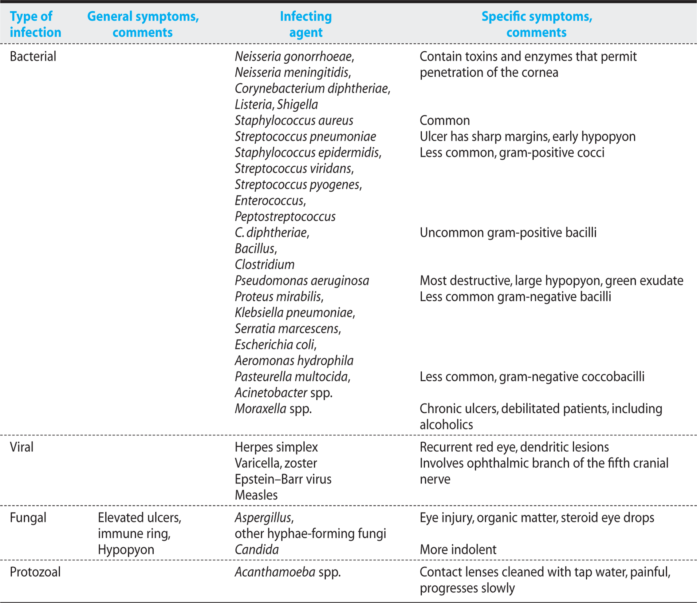
2. Viral. Patients with a history of a recurrent red eye most commonly have recurrent herpes simplex keratitis. Latent virus in the Vth cranial nerve reactivates and migrates down the nerve to the corneal surface. Ultraviolet light exposure, menstruation, fever, and other acute stresses can induce viral reactivation. In the hospitalized patient with unilateral red eye, herpes simplex keratitis should always be considered. Corneal anesthesia may develop initially, minimizing pain. Erythema and a foreign body sensation associated with tearing are frequently noted. A classic dendritic lesion that stains with fluorescein dye is readily seen on slit-lamp examination (Figure 5.1). Other forms of viral keratitis are less common (Table 5.2).
3. Fungal. Corneal ulcers caused by hyphae-forming fungi such as Aspergillus most commonly follow an eye injury from organic material (such as, a tree branch). Use of chronic glucocorticoid eye drops also increases the risk of fungal keratitis. Ulcers tend to be superficial and are often elevated above the corneal surface. The infiltrate tends to be irregular, and an immune ring is often apparent. Smaller satellite lesions are commonly seen surrounding the main infiltrate. A severe anterior-chamber reaction associated with a hypopyon is commonly observed. Yeast-like fungi such as Candida can also cause corneal ulcers. These infections tend to be more indolent, but they can have all the characteristics described for hyphae-forming fungi.
4. Protozoal. Protozoa are a rare but very serious cause of corneal ulcers. Acanthamoeba species most commonly develop in contact lens wearers, particularly those that use unsterilized tap water in their cleaning solutions. Acanthamoeba ulcers are painful, progress slowly, and fail to respond to topical antibiotics.
Diagnosis and Treatment
Slit-lamp examination is helpful in identifying the potential cause of an eye infection. If a bacterial or fungal cause is suspected, corneal scrapings for culture, Gram stain, Giemsa stain, and methenamine silver stain should be performed. A surgical blade is gently scraped across the surface of the ulcer, and the resulting samples are inoculated onto solid media. Aerobic bacteria grow readily on standard media within 48 hours. Special processing may be required if Acanthamoeba, a fungus, Mycobacteria, or Chlamydia is the suspected pathogen. Viral keratitis can usually be diagnosed by appearance and generally does not require culturing.
Treatment must be instituted emergently. Because of the potential risk of perforation and visual loss, patients with bacterial keratitis and significant ulceration are often hospitalized for close observation.
Initially, therapy can be based on Gram stain in 75% of patients. In cases in which a cause is not clearly identified or the patient has already received antibiotic therapy, broader antibiotic coverage is warranted. Antibiotics are commonly given topically and, in some instances, also subconjunctivally. Systemic therapy in addition to topical therapy is recommended for patients with imminent perforation.
Topical regimens include bacitracin 5000 U/mL and gentamicin (13 mg/mL) for Streptococcus pneumoniae; cephalothin (50 mg/mL), plus bacitracin for other gram-positive cocci such as S. aureus;tobramycin (13.6–15 mg/mL) or gentamicin for Pseudomonas species; gentamicin for other gram-negative bacilli; amphotericin B (1.5-3 mg/mL), plus flucytosine (1%) for yeast-like fungi; natamycin (5%) for hyphal fungi; and neomycin (5-8 mg/mL), plus pentamidine isethionate (0.15%) for Acanthamoeba species. Topical fluoroquinolones are also efficacious and have been recommended as empiric therapy for non–sight-threatening bacterial keratitis. Moxifloxacin 0.5% is effective, and it is the least toxic regimen. This fluoroquinolone is often combined with topical cephalothin.
Eye drops need to be administered every half an hour during the day and hourly during sleep for 7-10 days. Subconjunctival injections should be repeated every 12-24 hours for a total of 3-6 doses.
For herpes simplex keratitis, topical ganciclovir (Zirgan) 0.15% gel is now the treatment of choice; 5× daily is recommended until the corneal epithelial heals and then 3x per day for 1 week. This regimen is convenient and is not toxic the corneal epithelium. Previously, Trifluorothymidine 1% (trifluridine, Viroptic) was the preferred regimen; however, this drug causes significant epithelial cell toxicity and requires administration 8-9x per day. Oral acyclovir 400 mg orally 5x per day is nearly as effective as topical therapy. Recognizing the high likelihood of recurrence, many clinicians maintain their patients on oral acyclovir, 400 mg twice daily for several months or, in some cases, for years.
KEY POINTS
About Keratitis (Corneal Infection)
1. Condition needs to be treated quickly to prevent blindness.
2. Usually preceded by a break in the cornea (Neisseria spp., Corynebacteria diphtheriae, Listeria, and Shigella can invade without a break first occurring).
3. Streptococcus pneumoniae causes a well-circumscribed ulcer with sharp margins.
4. Pseudomonas aeruginosa is associated with hard contact lenses. It is very destructive and causes severe eye pain.
5. Herpes simplex causes distinct dendritic lesions that take up fluorescein. Consider this diagnosis in the hospitalized patient who develops unilateral red eye.
6. Aspergillus usually follows eye injury from organic matter (tree branch, for example).
7. Acanthamoeba occurs in contact lens wearers who use tap water with their cleaning solutions.
ENDOPHTHALMITIS
POTENTIAL SEVERITY
An ocular emergency. A very serious infection that often leads to permanent visual impairment or blindness. Should be managed by experienced ophthalmologist.
Endophthalmitis is an inflammatory disease involving the ocular chamber and adjacent structures. When the inflammation involves all of the ocular tissue layers and chambers, the disease is called panophthalmitis. This infection must be managed by an experienced ophthalmologist.
Predisposing Conditions and Causes
Endophthalmitis has four major causes, each associated with distinctive pathogens. In order of frequency, they are
1. Posttraumatic endophthalmitis. Mixed infections are common. Staphylococcus epidermidis and S. aureus, Streptococcus species, and Bacillus species are the most frequently cultured. Although Bacillus cereus is usually a minimally invasive organism, this bacteria causes rapidly progressive endophthalmitis when it gains entry into the eye. Fungi are encountered in penetrating injuries caused by organic matter. The likelihood of infection is increased when a foreign body is retained in the eye.
2. Hematogenous endophthalmitis. Any source of bacteremia can seed the choroid, with subsequent spread to the retina and vitreous humor. Two-thirds of blood-borne infections arise in the right eye, and one-quarter involve both eyes. The most common blood-borne pathogens to cause endophthalmitis are fungi, in particular, Candida albicans. B. cereus is the most common cause of hematogenous endophthalmitis in intravenous drug abusers. Patients with bacterial meningitis caused by S. pneumoniae, Neisseria meningitidis, and Haemophilus influenzae may also develop endophthalmitis. In neonates, group B streptococcus is most common, and in elderly patients, group G streptococcus. In the immunocompromised host with pulmonary infiltrates, Nocardia asteroides can gain entry into both the eyes and the cerebral cortex. If the primary source of bacteremia is not apparent, subacute bacterial endocarditis should be considered.
3. Endophthalmitis resulting from the contiguous spread of uncontrolled bacterial or fungal keratitis. Delays in treatment of bacterial or fungal keratitis can result in spread of infection to the aqueous and eventually the vitreous humor. The same organisms that cause severe keratitis cause this form of endophthalmitis.
4. Endophthalmitis associated with ocular surgical procedures. Acute postoperative endophthalmitis generally originates from endogenous flora in the eye. The most common pathogens are gram-positive cocci (S. epidermidisbeing most common), followed by S. aureus, and Streptococcus species. Infection most frequently develops within 24 hours after surgery but can develop up to 5 days postoperatively. Delayed postoperative endophthalmitis usually arise weeks to months after surgery and is caused by opportunistic pathogens. Endophthalmitis can also develop after creation of a filtering bleb. This surgical procedure allows bacteria to gain entry into the chamber of the eye; it is frequently preceded by conjunctivitis.
Clinical Manifestations
Eye pain, photophobia, reduced vision, and redness are the primary symptoms of bacterial endophthalmitis. In cases of hematogenous spread, sudden onset of blurred vision without pain, photophobia, or redness is the most common complaint. On examination, eyelid edema, chemosis of conjunctiva, and moderate-to-severe anterior or chamber inflammation with a hypopyon are often seen. Retinal hemorrhages, venous sheathing, and loss of the red reflex are noted on retinal examination. In fungal endophthalmitis, the symptoms and signs tend to be less severe. The patient often complains only of blurry vision or spots in the visual field. In the comatose patient, Candida endophthalmitis is commonly missed unless frequent funduscopic examinations are performed. Monitoring of the fundi is recommended in all patients who have developed candidemia. Findings of focal areas of inflammation, particularly gray-white fluffy exudates in the retina, chorioretina, or inferior vitreous strongly suggest Candidaendophthalmitis (Figure 5.3).

Figure 5.3. Candida retinitis. The typical rounded white exudates are caused by seeding from the bloodstream. Picture courtesy of Dr. William Driebe, University of Florida College of Medicine. See color image on color plate 1.
Diagnosis and Treatment
Cultures and smears must be obtained promptly from the aqueous and vitreous, the vitreous giving the highest positive yield. Specimens of exudate from the conjunctiva are often misleading. Cultures of the site of foreign body penetration should be obtained in cases of traumatic endophthalmitis. In patients suspected of hematogenous endo-phthalmitis, cultures of blood, urine, and cerebrospinal fluid often reveal the causative agent. In patients with vision better than light perception, vitreous sampling should be followed by intravitreal antibiotic injection. In patients with only light perception, vision is improved by immediate vitrectomy, followed by intravitreal antimicrobials.
KEY POINTS
About Endophthalmitis
1. Endophthalmitis is an ocular emergency; 1 in 10 patients requires enucleation.
2. Posttraumatic form results in mixed infections. Bacillus cereus is very aggressive. Fungal infections follow from injuries with organic material.
3. Hematogenous form more commonly involves the right eye. Candida is the most common cause, with B. cereus in intravenous drug abusers.
4. Contiguous form spreads from severe keratitis.
5. Acute postoperative form is caused by endogenous flora, Staphylococcus epidermidis, Staphylococcus aureus, Streptococcus species.
6. Intravitreal antibiotics are required. If patient has light perception only, vitrectomy is recommended.
Initially, broad-spectrum antibiotic intravitreal injection is recommended—for example, vancomycin 0.1 mL of a 10 mg/mL solution, plus gentamicin 0.1 mL of a 1 mg/mL solution. Systemic therapy must be instituted in cases of hematogenous endo-phthalmitis but is not of benefit in other forms of the disease. For Candida endophthalmitis, intravenous amphotericin B is recommended, and in more severe cases, intravitreous amphotericin B (5–10 μg) is also administered. About half of all patients with endophthalmitis retain 20/400 visual acuity or better. One in 10 patients requires enucleation.
THROAT INFECTIONS
GUIDING QUESTIONS
1. What is the most common cause of pharyngitis?
2. Which disease is suggested by the presence of a gray pseudomembrane?
3. What complication should be considered when unilateral tonsillar swelling develops?
4. Which infection should be considered in the patient with inspiratory stridor and sore throat?
PHARYNGITIS
POTENTIAL SEVERITY
Usually a self-limiting disease. One exception is the rare, life-threatening complication of peritonsillar abscess.
Causes and Clinical Manifestations
Pharyngitis is one of the most common infectious diseases that presents to the primary physician, and it has many causes. Many physicians assume their patient has group A streptococci (GAS) and unnecessarily initiate antibiotics despite the observation that viruses are the most common cause of pharyngitis: rhinoviruses and coronaviruses (common cold viruses), adenoviruses, herpes simplex, parainfluenza viruses, influenza viruses, coxsackievirus A, Epstein–Barr virus, cytomegalovirus, and HIV. The most common bacterial cause is GAS, also called Streptococcus pyogenes. GAS accounts for more than 50% of all cases of pharyngitis in children, but only about 10% of the cases in adults. Other forms of streptococci, groups B and G, have also been associated with pharyngitis in adults. Mixed anaerobic flora can cause a severe form of pharyngitis called Vincent angina that extends under the tongue and into the neck.
In recent decades, Corynebacterium diphtheriae has become an occasional cause of pharyngitis in the United States. With the waning immunity of the elderly population, recrudescence of this dangerous infection is now an increasing risk. Classically, a grayish pseudomembrane that tightly adheres to the pharyngeal wall develops. This finding should alert the clinician to the possibility of diphtheria.
Neisseria gonorrhoeae and Treponema pallidum are two rarer causes of pharyngitis that need to be included as part of the differential diagnosis in sexually promiscuous patients. And when pharyngitis is accompanied by pneumonia, Mycoplasma and Chlamydia are the most likely causes.
Fusobacterium necrophorum has become a major problem in Denmark, where this organism has populated the normal mouth flora of adolescents. Infection with this organism is associated with septic jugular vein thrombosis and septic emboli, a complication called Lemierre syndrome.
Diagnosis and Treatment
Antibiotics are overutilized in the management of pharyngitis and criteria for differentiating group A streptococcal pharyngitis from other forms have been established. The Centor clinical criteria are the most widely accepted:
1. Tonsillar exudates
2. Tender anterior cervical adenopathy
3. Fever by history
4. Absence of cough
If three to four of these criteria are met, the positive predictive value is only 40-60%, but the absence of three to four of the criteria has a negative predictive value of 80%. Patients with positive criteria should receive a rapid antigen test for GAS. The tonsillar area should be extensively swabbed to assure diagnostic accuracy, with the sample being acid- or enzyme-extracted for rapid antigen testing. Several different tests are available. All have a better than 90% specificity, but their sensitivity is variable (35-95% depending on the study). The recommended practice is therefore to perform a throat swab for culture in patients with positive Centor criteria and a negative rapid antigen test. Most clinicians use a dual-tip pharyngeal swab and send the second tip for culture if the antigen test is negative.
KEY POINTS
About Pharyngitis
1. Viruses are the most common cause. With severe prolonged pharyngitis, keep in mind primary HIV and Epstein–Barr virus.
2. Streptococcus pyogenes is also common (50% in children, 10% adults).
3. A grayish pseudomembrane should suggest Corynebacterium diphtheriae.
4. Keep Neisseria gonorrhoeae in mind in the sexually promiscuous patient.
5. If asymmetric tonsillar swelling is seen, consider a peritonsillar abscess.
6. Centor criteria (tonsillar exudates, cervical adenopathy, fever, lack of cough) suggest but do not prove a bacterial cause.
7. The rapid antigen test for S. pyogenes is specific but varies in sensitivity. A negative rapid antigen test should be followed by a throat culture.
8. Avoid antibiotics in viral pharyngitis. Penicillin remains the drug of choice for S. pyogenes, and its use reduces the risk of poststreptococcal glomerulonephritis and rheumatic heart disease.
If medial displacement of one or both tonsils is observed, the possibility of a peritonsillar abscess must always be considered. In the antibiotic era, this complication is rare, and it can be readily diagnosed by computed tomography (CT) of the neck with contrast. This study clearly delineates the location and size of the abscess. Delay in appropriate surgical intervention can result in spread of the infection to the retropharyngeal and pretracheal spaces. Entry into the retropharyngeal area can result in spread to the danger space, which extends to the posterior mediastinum. The result can be the development of potentially fatal purulent pericarditis (see Chapter 7).
Treatment depends on the cause of the illness (Table 5.3). Antibiotics should not be administered to patients who lack between three and four of the Centor criteria, nor to patients with three or four positive criteria who have a negative rapid antigen test and a negative throat culture. In patients with positive Centor criteria and a negative antigen test, 2 days of antibiotics may be prescribed while awaiting throat culture results. In cases of proven S. pyogenes,the treatment of choice continues to be penicillin: for adults, oral penicillin VK or a single injection of long-acting benzathine penicillin (1.2 × 10⁶ U intramuscularly). For penicillin-allergic patients, a 10-day course of erythromycin is recommended. Although antibiotic treatment of S. pyogenes shortens the symptomatic period by only 24-48 hours, eradication of the organism from the pharynx markedly reduces the incidence of poststreptococcal glomerulonephritis and rheumatic heart disease.
EPIGLOTTITIS
POTENTIAL SEVERITY
An infectious disease emergency because of the risk of a fatal respiratory arrest.
In the past, epiglottitis occurred most commonly in children, but with the advent of the H. influenzae B vaccine (HIB), adults now constitute a higher proportion of the cases seen. Patients present with a sore throat that subsequently results in drooling and difficulty swallowing, followed by difficulty breathing. Patients often sit in an upright position leaning forward and may or may not have inspiratory stridor. Indirect laryngoscopy reveals a swollen, cherry-red epiglottis. Swelling at this site can be confirmed by lateral neck radiography. The risk of respiratory arrest secondary to airway obstruction is high, and in children, this event is associated with 80% mortality. Therefore, in pediatric cases, a tentative diagnosis should be made based on clinical presentation, and emergent laryngoscopy and nasotracheal intubation performed under anesthesia. Adult patients can be closely observed in an intensive care setting until respiratory distress resolves. An endotracheal tube should be placed at the bedside in those cases.
The primary cause of this infection is H. Influenzae. However, Streptococcus pneumoniae, other Streptococcus species and S. aureus are increasing in frequency in children and adults alike. Treatment with intravenous cefotaxime or ceftriaxone for 7-10 days is recommended (see Table 5.3).
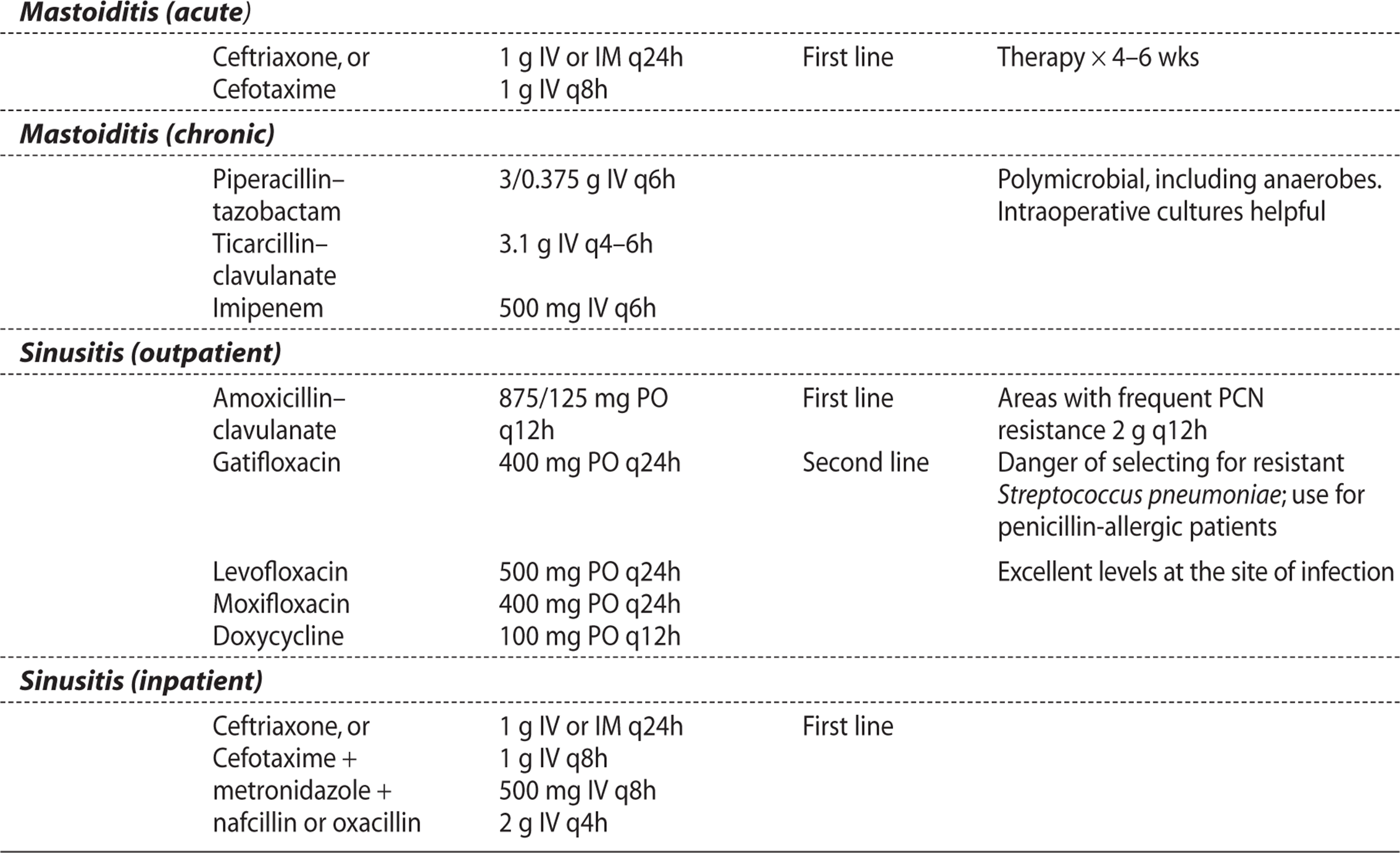
Table 5.3. Antibiotic Therapy for Ear, Nose, and Throat Infections

KEY POINTS
About Epiglottitis
1. Usually a disease of children, but increasingly common in adults.
2. Sore throat combined with drooling and inspiratory stridor suggests the diagnosis.
3. Indirect laryngoscopy demonstrates a cherry-red epiglottis.
4. Respiratory arrest is a danger, and pediatric patients should be electively intubated.
5. Haemophilus influenzae is the most common cause, but streptococcal and staphylococcal cases are increasing in frequency.
6. Ceftriaxone or cefotaxime are the treatments of choice.
EAR INFECTIONS
GUIDING QUESTIONS
1. Which organism is responsible for malignant otitis externa?
2. Why do children develop otitis media more commonly than adults do?
3. What are the two most common pathogens that cause otitis media?
4. Untreated mastoiditis can result in which two complications?
OTITIS EXTERNA
POTENTIAL SEVERITY
In the normal host, usually an annoying, but not serious disease; however, in the diabetic or immunocompromised host, can be life-threatening.
Otitis externa is also called “swimmer’s ear,” and it originates with water trapped in the external auditory canal, producing irritation, maceration, and infection. This infection can follow swimming, but it also follows irrigation of the ear to remove cerumen. This infection has also been associated with devices that occlude the ear such as ear phones and hearing aids. Symptoms include local itching and pain. Physical findings may include redness and swelling of the external canal. Tenderness of the pinna is often noted.
KEY POINTS
About Otitis Externa
1. Results when water is trapped in the external ear.
2. Caused by gram-negative bacilli, Pseudomonas aeruginosa being the most common.
3. Malignant otitis externa can occur in diabetics and immunocompromised patients; can infect the base of the skull; can be fatal; and requires prolonged antipseudomonal antibiotic therapy.
Gram-negative bacilli are most commonly cultured, with P. aeruginosa being the major pathogen. Coagulase negative staphylococcus and S. aureus as well as anaerobes can also cause this infection. Rarely, the canal can become superinfected with Candida or Aspergillus, and this complication is called otomycosis.
The recommended treatment of bacterial otitis externa is polymyxin neomycin otic drops combined with hydrocortisone (Cortisporin Otic), or topical ciprofloxacin combined with hydrocortisone (Cipro HC). For otomycosis, topical clotrimazole or miconazole accompanied by aggressive debridement of the ear canal are the recommended treatments.
A more invasive form of otitis externa called malignant otitis externa can develop in diabetics and immunocompromised patients. In this disease, pain tends to be more severe and can spread to the temporomandibular joint. Granulation tissue is often found in the external canal. This necrotizing infection can spread to the cartilage, blood vessels, and bone. Infection can involve the base of the skull, meninges, and brain, resulting in death. Multiple cranial nerves can be damaged, including cranial nerves VII, IX, X, and XII.
This infection is usually accompanied by an elevated erythrocyte sedimentation rate. Diagnosis is made by CT scan or magnetic resonance imaging (MRI) and can be confirmed by gallium scan. P. aeruginosais almost always the cause. Systemic therapy for Pseudomonas must be instituted for a minimum of 6 weeks, and necrotic tissue should be surgically debrided (see Table 5.3). As a consequence of the overuse of fluoroquinolones, infections with ciprofloxacin-resistant Pseudomonas are now being reported, necessitating prolonged treatment with intravenous ceftazidime or cefepime.
OTITIS MEDIA
POTENTIAL SEVERITY
Rapid treatment and close follow-up reduce the risk of serious complications. Delay in therapy can lead to potentially fatal complications.
Otitis media occurs most commonly in childhood, and by the age of 3 years, two-thirds of children have had at least one attack. Otitis media with effusion is the consequence of obstruction of the eustachian tube. In younger children, the eustachian tube tends to be smaller and more susceptible to obstruction. Loss of drainage results in accumulation of serous fluid and resorption of air in the middle ear.
The initial precipitating event is usually a viral upper respiratory infection. Five to 10 days later, the sterile fluid collection becomes infected with mouth flora, resulting in ear pain, ear drainage, and occasionally, hearing loss. Fever, vertigo, nystagmus, and tinnitus are other associated symptoms. In infants, other accompanying symptoms include irritability and loose stools.
The finding of redness of the tympanic membrane is consistent with, but not proof of, otitis media. It can be the result of diffuse inflammation of the upper respiratory tract. Presence of fluid in the middle ear should be determined by pneumatic otoscopy. More recently, acoustic reflectometry has become available as a method for monitoring ear effusions.
The American Academy of Pediatrics recommends these criteria for a diagnosis of otitis media:
1. Recent, usually abrupt onset of signs and symptoms of middle-ear inflammation and effusion; AND
2. Presence of middle-ear effusion that is indicated by any of the following:
a. bulging of the tympanic membrane,
b. limited or absent mobility of the tympanic membrane,
c. air–fluid level behind the tympanic membrane, OR
d. otorrhea; AND
3. Signs or symptoms of middle-ear inflammation as indicated by either
a. distinct erythema of the tympanic membrane, OR
b. distinct otalgia (discomfort clearly referable to either or both ear) that interferes with or precludes normal activity or sleep.
KEY POINTS
About Otitis Media
1. Results from obstruction of the eustachian tube in association with a viral upper respiratory tract infection. More common in children who have narrow eustachian tubes.
2. Infants may present with irritability and diarrhea.
3. Diagnosis is made by demonstrating the presence of fluid behind the tympanic membrane and inflammation of that membrane.
4. Streptococcus pneumoniae, Haemophilus influenzae, and Moraxella catarrhalis are the most common causes.
5. Amoxicillin to start; follow with amoxicillin–clavulanate or cefuroxime if no response within 72 hours.
Patients more than 2 years of age who do not meet the foregoing criteria should be observed for at least 24 hours before antibiotic therapy is considered. One exception is the patient with conjunctivitis and symptoms suggestive of otitis media. These patients have a high likelihood of infection with H. influenzae and should receive antibiotic therapy.
The cause of otitis media can be determined by needle aspiration of the tympanic membrane; however, this procedure is generally recommended only for immunocompromised patients. Culture of the nasopharynx is not helpful in predicting the bacterial flora in the middle ear. The pathogens that primarily cause otitis media are S. pneumoniae, H. influenzae (usually nontypable strains not covered by HIB vaccine), Moraxella catarrhalis, less commonly S. pyogenes, and S. aureus.
Amoxicillin is inexpensive and covers most cases of bacterial otitis media. Many experts recommend starting with amoxicillin, recognizing that patients with β-lactamase-producing organisms (some strains of H. influenzae and Moraxella catarrhalis) will not respond. If improvement is not seen within 72 hours, the patient should be switched to amoxicillin–clavulanate and cefuroxime. Treatment for 10 days is recommended (see Table 5.3)
MASTOIDITIS
POTENTIAL SEVERITY
A rare consequence of otitis media that can lead to fatal complications.
CASE 5.2
Five months before presenting to the emergency room, a 44-year-old man had noted purulent drainage from his right ear. The drainage was associated with fever and a shaking chill. He received no medical treatment at that time, and the symptoms spontaneously resolved. Three weeks before presentation, he again noted increased purulent drainage from the same ear, associated with earache and dizziness. One week before presentation, he developed a severe right-sided headache, and he experienced difficulty walking because of dizziness. Dizziness was accompanied by nausea and vomiting.
His medical history revealed chronic right otitis media since the age of 13 years. Physical examination found a temperature of 38.9°C (102°F), foul-smelling purulent discharge from a perforated right tympanic membrane, and tenderness behind the right ear, with localized erythema and swelling. Laboratory workup showed a white blood cell (WBC) count of 8900, with 68% PMNs. Analysis of fluid from a lumbar puncture found a WBC count of 950 (with 92% mononuclear cells), protein 275 mg/dL, and glucose 45 mg/dL. The sample was culture-negative. Mastoid radiographs uncovered extensive destruction of the right mastoid air cells, the attic, and the aditus. Mastoidectomy was performed, and infection of the temporal bone, epidural space, and mastoid were noted. An intraoperative culture found Proteus mirabilis.
With the advent of antibiotics, mastoiditis is now a rare complication of otitis media. However, as described in case 5.2, infection can occasionally spread to the mastoid air cells. Swelling, redness, and tenderness can develop directly behind the ear in the area of the mastoid bone. Chronic mastoid disease can spread to the temporal bone and cause temporal lobe brain abscess. The infection can also spread by epiploic veins to the lateral and sigmoid venous sinuses, causing septic thrombosis. As seen in case 5.2, radiographs of the mastoid area may show increased density with loss of mastoid trabeculae, bony sclerosis, and lytic lesions of the temporal and parietal bones (Figure 5.4). Treatment is similar to that given for otitis media (see Table 5.3); however, therapy must be prolonged for 3-4 weeks. Chronic mastoid infections can be associated with gram-negative aerobic bacteria (as in case 5.2).

Figure 5.4. Computed tomography scan with contrast of mastoiditis. This axial view shows marked soft-tissue swelling in the area of the mastoid, surrounded by an enhancing ring (arrowheads). The arrow points to the otic canal. (Picture courtesy of Dr. Ilona Schamalfus, University of Florida College of Medicine.)
If an abscess has formed within the mastoid, or if temporal lobe abscess or septic lateral sinus thrombosis has developed, surgical drainage and mastoidectomy need to be performed. Case 5.2 had headache, and the severe destruction demonstrated by the mastoid radiographs warranted mastoidectomy and surgical exploration of the temporal region.
KEY POINTS
About Mastoiditis
1. A rare complication of otitis media.
2. Readily diagnosed by mastoid radiographs.
3. Requires prolonged antibiotic therapy for 3-4 weeks.
4. Can lead to brain abscess or septic lateral sinus thrombosis.
SINUS INFECTIONS
GUIDING QUESTIONS
1. Infection of which air sinus is the most difficult to evaluate by physical examination?
2. Which physical findings are helpful in evaluating bacterial sinusitis?
3. What is the most common complication associated with ethmoid sinusitis?
4. What are the complications associated with frontal sinusitis?
5. What are the complications associated with sphenoid sinusitis?
6. How can orbital cellulitis be differentiated from septic cavernous sinus thrombosis?
POTENTIAL SEVERITY
Delays in therapy can result in spread of infection outside of the air sinus, with possibly fatal complications.
SINUSITIS
Predisposing Factors
Viral upper respiratory infections caused by rhinoviruses, influenza viruses, parainfluenza viruses, and adenoviruses cause inflammation of the sinuses and production of serous exudate. About 0.5-1% of viral upper respiratory infections progress to bacterial sinusitis.
Anatomic obstruction increases the likelihood of bacterial sinusitis. Causes of obstruction include septal deformities, nasal polyps, foreign bodies, chronic adenoiditis, intranasal neoplasms, and indwelling nasal tubes. Patients undergoing nasotracheal intubation or those who have a large-bore nasogastric tube are at increased risk of developing bacterial sinusitis. These tubes interfere with normal drainage of the sinus ostia. Nasal allergies are associated with edema, obstruction, and the accumulation of serous fluid; they are another predisposing factor for bacterial sinusitis. Dental abscesses of the upper teeth can spread to the maxillary sinuses and can result in recurrent bacterial sinusitis. Two genetic disorders, cystic fibrosis (associated with abnormally viscous mucous) and Kartagener syndrome (which causes defective mucous cell ciliary function) are rarer predisposing factors for bacterial sinusitis.
Clinical Manifestations
The critical decision point for clinician is differentiating viral from bacterial sinusitis. Experts suggest that bacterial sinusitis should be strongly considered if any one of three events occur:
1. Persistent symptoms or signs of acute sinusitis from the onset that last for 10 days without clinical improvement. Viral infections are self-limited and progressively resolve over time.
2. Onset is accompanied by severe symptoms or high fever (39°C or 102°F]) and purulent nasal discharge or facial pain lasting for at least 3-4 consecutive days at the beginning of illness. Irritation of the cranial nerves, a purulent discharge, and high fever are more commonly associated with bacterial as compared with viral infections.
3. The sudden worsening of a typical viral upper respiratory infection that had lasted 5-6 days and was beginning to improve (sometimes termed double-sickening). New symptoms and signs may include: new onset of fever, headache, or increase in nasal discharge.
CASE 5.3
A 15-year-old female developed an upper respiratory infection 3 weeks before admission to hospital. Nasal discharge was clear, but after 10 days, she developed a severe left retro-orbital and left occipital headache, associated with left-eye tearing. She saw her physician 3 days later, complaining of persistent headache and nausea. Tenderness over the left maxillary sinus was noted. She was treated with Neo-Synephrine nose drops and Gantrisin (a sulfa antibiotic). She failed to improve, and 2 days later, she developed swelling of both eyes. Tetracycline was started, but she became confused and uncooperative.
Physical examination showed a temperature of 39.4°C, with a pulse of 140 per minute, and a respiratory rate of 40 per minute. The patient was toxic, disoriented, and lethargic.
An ear, nose, throat examination revealed dry, crusted purulent secretions in the left middle turbinate. Proptosis, chemosis, and complete ocular paralysis of the left eye was noted. Proptosis and chemosis of the right eye was less severe, with lateral gaze palsy (deficit of the VIth cranial nerve). Left disc blurred margin indicated papilledema. Tenderness was elicited over the left maxillary and frontal sinuses. Sensation on the left side of the face in the ophthalmic and maxillary branches of the Vth cranial nerve was decreased. The patient’s neck was very stiff. The remainder of the examination was unremarkable.
A laboratory workup showed a WBC count of 18,700/mm3, with 78% PMNs and 10% bands. An analysis of the cerebrospinal fluid showed a WBC count of 18,000/mm3, with 95% PMNs, protein 400 mg/dL, and glucose 25 mg/dL. Sinus radiographs revealed opacification of the left frontal, ethmoid, maxillary, and sphenoid sinuses. Six days after admission, the patient died. Autopsy revealed pansinusitis (including the left sphenoid sinus), bilateral cavernous sinus thrombosis, and bacterial meningitis. Culture of the meninges grew group H. streptococci.
Although case 5.3 is unusually severe, it does illustrate many of the potential clinical manifestations of bacterial sinusitis. Severe headache strongly suggests a bacterial infection, and the headache is often localized to the area of the infected sinus. Infection of the sphenoid sinus, which is located deep within the skull, does not cause an easily recognizable pain syndrome. As described in case 5.3, sphenoid sinusitis is associated with retro-orbital pain and/or severe pain extending to the frontal, temporal, and occipital regions. Pain is frequently unilateral and severe; it interferes with sleep and is not relieved by aspirin. Sphenoid sinus pain is often misdiagnosed as a migraine headache, resulting in delayed treatment.
In addition to pain, patients with bacterial sinusitis often note drainage of thick, discolored, purulent material. However, some deny nasal drainage and instead complain of a foul taste or smell. As a consequence of chronic postnasal drainage, recurrent coughing is another frequent complaint, particularly in the nighttime, when the patient is lying in a recumbent position. Surprisingly, despite extensive inflammation in the sinuses, the minority of adults experience fever. However, fever almost always accompanies bacterial sinusitis in children.
As noted in case 5.3, physical examination can readily elicit localized sinus tenderness over the maxillary and frontal sinuses. In maxillary sinus infection, tooth tenderness may appear. Infection of the sphenoid sinus is not associated with tenderness. Transillumination can be performed in a darkened room using a flashlight tightly sealed to the skin. Marked reduction in light transmission correlates with active purulent infection in maxillary sinusitis. Light reduction may also be helpful for diagnosing frontal sinusitis; however, accurate performance of the examination requires experience. Examination of the nose reveals edema and erythema of the nasal mucosa, and if the ostia are not completely obstructed, a purulent discharge may be seen in the nasal passage and posterior pharynx. Inflammation of the Vth cranial nerve is often associated with sphenoid sinusitis, posterior ethmoid sinus infection, cavernous sinus thrombosis, and, less commonly, with maxillary sinusitis. Hypo- or hyperesthesia in the regions enervated by the ophthalmic and maxillary branches may be detected on sensory examination. That finding was noted in case 5.3, and in combination with oculomotor paralysis, proptosis, papilledema of the left eye, and meningitis, it indicated that the patient’s bacterial sinusitis was complicated by cavernous sinus thrombosis (see “Complications,” later in this section).
KEY POINTS
About the Clinical Manifestations of Sinusitis
1. Bacterial sinusitis is more likely in case of one of the following three presentations:
• Persistent symptoms or signs of acute sinusitis from the onset that last for 10 days without clinical improvement.
• Onset is accompanied by severe symptoms or high fever (39°C or 102°F]) and purulent nasal discharge or facial pain.
• The sudden worsening of a typical viral upper respiratory infection that had lasted 5-6 days and was beginning to improve (sometimes termed double-sickening). New symptoms and signs may include new onset of fever, headache, or increase in nasal discharge.
2. Symptoms in bacterial sinusitis as compared with viral disease:
• More severe pain, often localized to a cranial nerve dermatome.
• Purulent discharge and/or foul-smelling breath.
• Imaging cannot differentiate bacterial from viral in most cases.
Diagnosis
Despite extensive inflammation of the sinuses, the peripheral WBC count is often within normal limits. Case 5.3 had meningitis, which explains her peripheral leukocytosis. Cultures of the nasopharynx correlate poorly with intrasinus cultures and are not recommended. Direct sampling of the infected sinus is required for accurate microbiologic assessment. Fiberoptic cannulation can be performed, but such cultures are often contaminated by normal mouth flora. In children, needle aspiration of the infected maxillary sinuses has produced accurate sampling, but this procedure is not recommended in routine cases.
KEY POINTS
About the Diagnosis of Sinusitis
1. Nasopharyngeal cultures are not helpful.
2. Imaging is not recommended unless a complication is suspected.
a) A limited computed tomography scan of the sinuses is preferred over routine sinus radiographs or MRI.
b) CT allows for assessment of bony erosions and extension of infection beyond the sinuses.
In the absence of suspected complications imaging studies, sinus X-rays, CT scan, and MRI are not helpful for differentiating viral from bacterial sinusitis. Both diseases are commonly accompanied by abnormalities of the maxillary sinusitis. Therefore, routine sinus imaging is not recommended.
If there are focal neurological deficits or failure of symptoms to improve after appropriate antibiotic therapy, the imaging study of choice is CT scan, and a limited CT scan of the sinuses is a cost-effective alternative to conventional sinus films (Figure 5.5). The integrity of the bony sinus walls can be assessed in more detail with a CT scan. Such a study can readily detect extension of the infection from the ethmoid sinuses to the orbit and development of an orbital abscess (Figure 5.6). CT scan is also useful for assessing extension of frontal sinus infection to the epidural or subdural space, and for diagnosing frontal brain abscess, a rare complication of frontal sinusitis. In sphenoid sinusitis, CT with contrast injection is the study of choice for detecting early extension to the cavernous sinuses. It can also readily detect development of sinus mucocele.
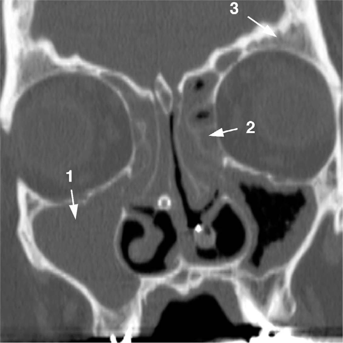
Figure 5.5. Computed tomography scan of pansinusitis, coronal view of the air sinuses. 1. Maxillary sinus; 2. Ethmoid sinus; 3. Frontal sinus. Note the marked opacification of the right maxillary sinus and the marked mucosal thickening of the left maxillary sinus. Both ethmoid sinuses are opaque, as are the frontal sinuses. (Picture courtesy of Dr. Ilona Schamalfus, University of Florida School of Medicine.)
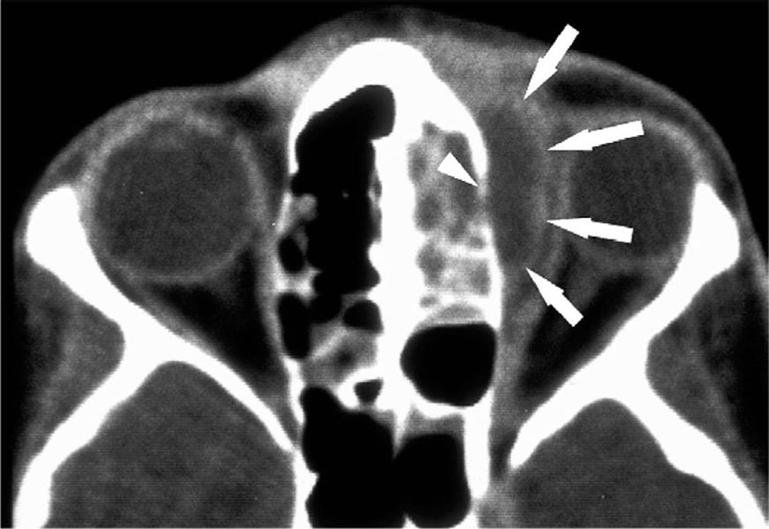
Figure 5.6. Computed tomography scan with contrast of orbital cellulitis with accompanying orbital abscess. This axial view shows the break in the ethmoid sinus wall (arrowhead) and the ring enhancing orbital abscess (arrows) that is pushing the eye laterally. (Picture courtesy of Dr. Ilona Schamalfus, University of Florida College of Medicine.)
Complications
Distinct complications are associated with ethmoid, frontal, and sphenoid sinusitis. To be able to make the proper diagnostic evaluation and begin prompt therapy, the primary care physician and the infectious disease specialist must both be able to recognize the early clinical manifestations associated with spread of infection beyond the sinuses. Complicated air sinus infection can be life-threatening and frequently leads to permanent neurologic deficits.
The ethmoid sinus is separated from the orbit by the lamina papyracea. This thin layer can easily be breached by infection, particularly in children. Infection in the ethmoid sinus can also spread to the orbit via the ethmoid veins. The extent of orbital involvement varies and can cause four different syndromes:
1. Periorbital cellulitis. Infection of the skin in the periorbital area results in swollen eyelids, but eye movements are normal and no displacement of the eye is seen.
2. Orbital cellulitis. When infection spreads to the orbital tissue, not only are the eyelids swollen, but the eye becomes tender to palpation. Ophthalmoplegia with reduction of all eye movements occurs as a consequence of inflammation of the extraocular muscles. Chemosis (marked swelling and erythema of the conjunctiva) develops—a reflection of the intense inflammation within the orbit. Finally, proptosis (outward displacement of the eye) is usually seen as a consequence of edematous tissue within the orbit pushing the eye out of its socket. This infection is usually unilateral.
3. Orbital abscess. A discrete abscess can develop in the periosteum or soft tissue of the orbit. This complication cannot be detected by examination, the diagnosis being made by CT scan of the orbit. Abscess formation usually warrants surgical drainage (Figure 5.6).
KEY POINTS
About the Complications of Ethmoid Sinusitis
1. Ethmoid sinusitis can easily spread medially through the lamina papyracea to cause periorbital cellulitis, orbital cellulitis, orbital abscess, or septic cavernous sinus thrombosis (rare).
2. Orbital cellulitis is usually unilateral; cavernous sinus thrombosis is bilateral. Papilledema, deficits of the Vth cranial nerve, and pleocytosis of the cerebrospinal fluid are also found with septic cavernous sinus thrombosis.
3. Orbital computed tomography scan with contrast delineates the extent of infection.
4. Surgical drainage of the sinus is recommended if loss of visual acuity, proptosis, or ophthalmoplegia develop
4. Cavernous sinus thrombosis and meningitis. Orbital infection can spread via the superior ophthalmic veins to the cavernous sinus. Because the cavernous sinuses are connected by the intercavernous sinuses, and because the superior ophthalmic veins have no valves, infection usually spreads quickly from one cavernous sinus to the other. As a consequence, bilateral eye involvement is the rule. The finding of bilateral eye involvement makes orbital cellulitis less likely. Other findings that favor a diagnosis of cavernous sinus thrombosis are abnormal sensation in the Vth cranial nerve, development of papilledema, and inflammatory cells in the cerebrospinal fluid. High-resolution CT scan with contrast is now the diagnostic study of choice.
Surgical intervention should be considered if progression on antibiotics, loss of visual acuity below 20/60, proptosis, or ophthalmoplegia occurs. The ethmoid sinuses should be drained, further debridement being guided by the findings of a CT scan.
Frontal sinusitis can also be life-threatening if not properly managed. Infection can spread anteriorly into the frontal bone, causing a subperiosteal abscess that can result in pitting edema of the forehead. This complication has been termed “Pott’s puffy tumor.”
Infection can also spread posteriorly. Particularly in teenage males, the posterior wall of the frontal sinus may be thin, allowing infection to spread to the epidural or subdural space. Infection can also reach the cerebral cortex, forming a brain abscess. These complications are usually associated with a severe frontal headache that interferes with sleep and that is not relieved by aspirin. In some cases, seizures may develop, but in most instances, frontal brain abscess is neurologically silent. Abscess formation in the subdural or epidural space and brain abscess are readily diagnosed by contrast-enhanced CT scan.
KEY POINTS
About the Complications of Frontal Sinusitis
1. Infection can spread anteriorly, causing Pott’s puffy tumor.
2. Infection can spread posteriorly and cause epidural, subdural, or brain abscess.
3. Posterior spread leads to severe headache, but frontal cerebral cortex lesions are usually neurologically silent.
4. Contrast enhanced computed tomography scan is recommended in cases of severe frontal sinusitis.
KEY POINTS
About the Complications of Sphenoid Sinusitis
1. Sphenoid sinusitis is the most dangerous form of sinusitis.
2. Most patients require hospitalization and intravenous antibiotics.
3. The sphenoid is close to many vital neurologic structures.
4. The major complication is septic cavernous sinus thrombosis.
5. Computed tomography scan with contrast defines the sites of involvement, including cavernous sinus thrombosis.
6. Surgical drainage of the sinus is often required to prevent spread outside its walls.
Sphenoid sinusitis is the most dangerous sinus infection. If a patient with sphenoid sinusitis does not respond rapidly to oral antibiotics and decongestants, intravenous antibiotics should be initiated. Nafcillin and a third-generation cephalosporin are generally adequate coverage (see the “Treatment” subsection).
A low threshold should be set for surgical drainage. The sphenoid sinus lies deep in the skull. Its walls are adjacent to the pituitary gland, optic canals, dura mater, and cavernous sinuses. The thickness of the lateral walls of the sphenoid sinuses varies. If infection extends beyond these walls, patients can present with cavernous sinus infection that causes impairment of function in the IIIrd, IVth, and VIth cranial nerves, causing ophthalmoplegia, Vth nerve dysfunction [ophthalmic and maxillary branches (hypo or hyperesthesia)], proptosis, and chemosis. Case 5.3 had all of those characteristics and had sphenoid sinusitis and septic cavernous sinus thrombosis. The intercavernous sinuses allow infection to spread from one sinus to the other, usually within 24 hours. Diagnosis is most readily made by contrast-enhanced CT scan. The early venous phase following administration of contrast demonstrates regions of reduced or irregular enhancement, thickening of the lateral walls, and bulging of the sinus. Anticoagulation with heparin in the very early stages of infection may be helpful, although intravenous antibiotics (covering S. aureus,other gram-positive organisms, and gram-negative organisms as indicated) are the mainstay of treatment.
Microbiology
These major pathogens are associated with bacterial sinusitis:
1. S. pneumoniae and H. influenzae (50-70% in maxillary sinusitis)
2. Other gram-positive aerobic mouth flora (S. pyogenes, Streptococcus viridans)
3. Occasionally, other gram-negative aerobic mouth flora (Moraxella catarrhalis)
4. S. aureus more frequent in ethmoid and sphenoid disease
5. Anaerobic mouth flora (Bacteroides melanogenics and anaerobic streptococci) more frequent in adults and in patients with chronic sinusitis
6. Gram-negative organisms rare in the normal host, most frequent in chronic sinusitis
7. Pseudomonas aeruginosa frequent in patients with AIDS
8. Fungal sinusitis, particularly Aspergillus, becoming an increasing problem in immunocompromised patients, being most frequently associated with neutropenia
KEY POINTS
About the Microbiology and Treatment of Sinusitis
1. Streptococcus pneumoniae and Haemophilus influenzae are most common. Anaerobes are seen in adults and in chronic disease. Staphylococcus aureus is most frequent in sphenoid disease.
2. Gram-negative organisms (Pseudomonas aeruginosa) are seen in patients with AIDS.
3. Fungi (Aspergillus) often infect neutropenic patients.
4. If presents with one of the three conditions (see clinical manifestations), begin antibiotics immediately.
a) Amoxicillin–clavulanate treatment of choice
b) Fluoroquinolones (concerns about resistance)
c) Doxycycline
d) Azithromycin and amoxicillin is no longer recommended.
5. Patients with frontal, ethmoid, or sphenoid sinus infection often require hospitalization and intravenous antibiotics (oxacillin plus a third-generation cephalosporin plus metronidazole).
Treatment
ANTIBIOTICS
As soon as a patient fulfills one of the three conditions outlined above that suggest bacterial sinusitis, empiric antibiotics should be initiated. No single antibiotic will treat all possible pathogens. Treatment of uncomplicated disease should be continued for 5-7 days in adults and 10-14 days for children. Table 5.3 lists the recommended oral regimens.
1. Amoxicillin plus clavulanic acid (Augmentin) is the drug of choice. It covers S. pneumoniae, H. Influenzae (including ampicillin-resistant strains), Moraxella catarrhalis, and S. aureus. In areas where PCN-resistant S. pneumoniae is prevalent, high-dose Augmentin should be administered: 2 g twice per day or 90 mg/kg twice per day.
2. The fluoroquinolones—levofloxacin, gatifloxacin, or moxifloxacin—cover all of the major pathogens that cause acute bacterial sinusitis. The development of fluoroquinolone-resistant S. pneumoniae is a major concern. These antibiotics should therefore be reserved for the penicillin-allergic patients.
3. Doxycycline is a reasonable alternative for adults. Excellent antibiotic levels are achieved in the sinuses, and this antibiotic covers all the major pathogens associated with sinusitis.
4. Trimethoprim–sulfamethoxazole is not recommended because of high levels of resistance.
5. Second- and third-generation cephalosporins are no longer recommended because S. pneumoniae strains are becoming increasingly resistant to these antibiotics.
6. Amoxicillin is a cheaper alternative, but it has a narrower spectrum. This antibiotic was previously considered the drug of choice for initial therapy, but more recent bacteriologic studies have revealed a high percentage of β-lactamase–producing organisms capable of degrading amoxicillin.
7. Azithromycin and other macrolides are no more efficacious than amoxicillin and are no longer recommended.
Patients with frontal, ethmoid, and sphenoid sinusitis frequently require hospitalization and intravenous antibiotic therapy to prevent spread of the infection to vital organs beyond the sinus walls. High-dose intravenous antibiotics directed at the probable organisms (see the “Microbiology” subsection) should be instituted emergently. Empiric therapy should include a penicillinase-resistant penicillin (either nafcillin or oxacillin) at maximal doses, plus a third-generation cephalosporin (either ceftriaxone or cefotaxime).
Anaerobic coverage should also be instituted with intravenous metronidazole (see Table 5.3).
OTHER TREATMENT MODALITIES
In patients with suspected bacterial sinusitis-nasal decongestants such as Neo-Synephrine nose drops or pseudoephedrine are not recommended because these treatments may unduly dry out the nasal passages and increase the viscosity of the nasal discharge.
Saline irrigation of the sinuses may be of benefit and is recommended. Also, intranasal corticosteroids are recommended in patients whose illness may have been precipitated by allergic sinusitis.
FURTHER READING
Eye Infections
Everitt HA, Little PS, Smith PW. A randomised controlled trial of management strategies for acute infective conjunctivitis in general practice. BMJ. 2006;333:321.
Leibowitz HM. The red eye. New Engl J Med. 2000;343:345-351.
Rietveld RP, ter Riet G, Bindels PJ, Schellevis FG, van Weert HC. Do general practitioners adhere to the guideline on infectious conjunctivitis? Results of the Second Dutch National Survey of General Practice. BMC Fam Pract. 2007;8:54.
Seal D, Reischl U, Behr A, et al. Laboratory diagnosis of endophthalmitis: comparison of microbiology and molecular methods in the European Society of Cataract & Refractive Surgeons multicenter study and susceptibility testing. J Cataract Refract Surg. 2008;34:1439-1450.
Pharyngitis
Alcaide ML, Bisno AL. Pharyngitis and epiglottitis. Infect Dis Clin North Am. 2007;21(2):449-469.
Fine AM, Nizet V, Mandl KD. Large-scale validation of the Centor and McIsaac scores to predict group A streptococcal pharyngitis. Archives of internal medicine. 2012;172(11):847-852.
Humair JP, Revaz SA, Bovier P, Stalder H. Management of acute pharyngitis in adults: reliability of rapid streptococcal tests and clinical findings. Arch Intern Med. 2006;166:640-644.
Epiglottitis
Berger G, Landau T, Berger S, Finkelstein Y, Bernheim J, Ophir D. The rising incidence of adult acute epiglottitis and epiglottic abscess. Am J Otolaryngol. 2003;24:374-383.
Faden H. The dramatic change in the epidemiology of pediatric epiglottitis. Pediatr Emerg Care. 2006;22:443-444.
Malignant Otitis Externa
Ismail H, Hellier WP, Batty V. Use of magnetic resonance imaging as the primary imaging modality in the diagnosis and follow-up of malignant external otitis. J Laryngol Otol. 2004;118:576-579.
Rubin Grandis J, Branstetter BF 4th, Yu VL. The changing face of malignant (necrotising) external otitis: clinical, radiological, and anatomic correlations. Lancet Infect Dis. 2004;4:34-39.
Otitis Media
American Academy of Pediatrics Subcommittee on Management of Acute Otitis Media. Diagnosis and management of acute otitis media. Pediatrics. 2004;113:1451-1465.
Coco A, Vernacchio L, Horst M, Anderson A. Management of acute otitis media after publication of the 2004 AAP and AAFP clinical practice guideline. Pediatrics. 2010;125(2):214-220.
Rovers MM, Schilder AG, Zielhuis GA, Rosenfeld RM. Otitis media. Lancet. 2004;363:465-473.
Schwartz LE, Brown RB. Purulent otitis media in adults. Arch Intern Med. 1992;152:2301–2304.
Sinusitis
Chow AW, Benninger MS, Brook I, et al. IDSA clinical practice guideline for acute bacterial rhinosinusitis in children and adults. Clin Infect Dis. 2012;54:e72-e112.
Gwaltney JM, Wiesinger BA, Patrie JT. Acute community-acquired bacterial sinusitis: the value of antimicrobial treatment and the natural history. Clin Infect Dis. 2004;38:227-233.
Lew D, Southwick FS, Montgomery WW, Weber AL, Baker AS. Sphenoid sinusitis. A review of 30 cases. N Engl J Med. 1983;309:1149-1154.
Piccirillo JF. Clinical practice. Acute bacterial sinusitis. N Engl J Med. 2004;351:902-910.
Complications of Ear, Nose, and Throat Infections
Ramsey PG, Weymuller EA. Complications of bacterial infection of the ears, paranasal sinuses, and oropharynx in adults. Emerg Med Clin North Am. 1985;3:143-160.
Southwick FS. Septic thrombophlebitis of major dural venous sinuses. Curr Clin Top Infect Dis. 1995;15:179-203.