
Edited by Anthony Brown
OUTLINE
2.1 Airway and ventilation management 14
2.2 Oxygen therapy 21
2.3 Haemodynamic monitoring 32
2.4 Shock overview 38
2.5 Sepsis and septic shock 48
2.6 Arterial blood gases 52
2.7 Cerebral resuscitation after cardiac arrest 59
2.8 Anaphylaxis 62
2.1 Airway and ventilation management
Stephen A Bernard
Essentials
1 Respiratory failure is a common presentation to the emergency department and ventilatory support may be required.
2 Non-invasive ventilation is appropriate for many patients with respiratory failure; endotracheal intubation and mechanical ventilation are used for cases where non-invasive ventilation is unsuccessful or contraindicated.
3 Endotracheal intubation performed in the emergency department almost always requires the use of sedative plus muscle relaxant drugs to facilitate endotracheal tube placement.
4 If visualization of the vocal cords at laryngoscopy is difficult, a ‘Failed Intubation Drill’ should be initiated immediately to avoid patient hypoxaemia.
5 Clinical checks to confirm correct endotracheal tube position are unreliable. Waveform or similar capnography must be used to confirm tracheal placement.
6 In patients with acute lung injury and decreased pulmonary compliance, mechanical ventilation should use low tidal volumes to avoid barotrauma.
Introduction
Assessment and management of the airway is the first step in the resuscitation of a critically ill patient in the emergency department (ED). Once the airway has been assessed and managed, evaluation of adequacy of ventilation follows. This chapter outlines the initial management of airway and ventilation in the ED.
Evaluation of the airway
Evaluation of the airway commences with a ‘look, listen, feel’ approach to detect partial or complete airway obstruction. If airway compromise is suspected, initial basic airway manoeuvres include the jaw thrust, chin lift and head tilt (providing with the latter that there is no suspicion of cervical spine injury); and placement of an oropharyngeal airway (OPA) (see Chapter 1.1 on Basic Life Support).
Gentle direct inspection of the upper airway using a laryngoscope may be necessary to detect a foreign body, which can be removed using a Yankauer suction catheter for liquids/secretions and/or Magill’s forceps for solid material. Once the airway is cleared, supplemental oxygen by face-mask is commenced as consideration is given to the breathing status.
Evaluation of breathing
Evaluation of breathing also uses a ‘look, listen, feel’ approach. Confirmation of adequate oxygenation initially uses a pulse oximeter. The adequacy of breathing may be confirmed with an arterial or venous blood gas analysis to confirm that the PCO2 is in the normal range.
Conscious patients with a patent airway but who have hypoxia and/or hypercapnoea should then be considered for non-invasive ventilation (NIV) or endotracheal intubation (ETI) and mechanical ventilation.
Non-invasive ventilation
Many patients in respiratory failure with hypoxaemia and/or hypercapnoea may benefit from a trial of NIV [1]. The use of NIV involves administration of a controlled mixture of oxygen and air delivered at a set positive pressure via a tightly sealed face-mask. The pressure is generally maintained between 5 and 10 cm H2O during both inspiration and expiration. This continuous positive airway pressure (CPAP) recruits lung alveoli that were previously closed, improving the ventilation/perfusion ratio and thus helping to correct hypoxaemia. There may also be a reduction in the work of breathing as a result of an increase in pulmonary compliance.
Inspiratory support (i.e. 5–20 cm H2O above the baseline pressure) during NIV is known as bi-level NIV. This additional inspiratory support is thought to further reduce the work of breathing when there is poor lung compliance or increased airway resistance.
Contraindications to NIV include comatose or combative patients, poor tolerance of a tight-fitting face-mask, poor seal of the face-mask due to facial hair, and/or the lack of trained medical or nursing staff to institute and monitor the NIV.
Clinical indications for non-invasive ventilation in the ED
Patients who present with severe acute pulmonary oedema (APO) should receive CPAP to improve cardiac and pulmonary function, while medical therapy with nitrates and diuretics is initiated [2]. On the other hand, patients who present with an exacerbation of chronic obstructive pulmonary disease (COPD) may benefit from bi-level NIV rather than CPAP alone [3].
There is also some evidence to support the use of NIV in patients with respiratory failure due to other common ED conditions, such as community-acquired pneumonia [1], although the role of NIV in the management of asthma remains uncertain [4]. Thus, it is common ED practice to administer a trial of NIV in most awake patients with respiratory distress or respiratory failure, prior to any consideration of a requirement for ETI and mechanical ventilation.
Endotracheal intubation
Endotracheal intubation (ETI) is performed for any one or more of the following four major reasons: to create an airway; to maintain an airway; to protect an airway; and/or to provide for mechanical ventilation.
Thus, a patient in respiratory arrest requires immediate bag/valve/mask (BVM) ventilation with supplemental oxygen while preparation is made for ETI and mechanical ventilation to protect and maintain the airway and provide mechanical ventilation. Alternatively, a patient with a reduced conscious state and/or depression of the cough reflex requires ETI for airway maintenance and protection. Also, ETI may be indicated as part of general anaesthesia in the combative patient who needs imaging and/or a practical procedure. Finally, ETI will be required for mechanical ventilation in a patient with respiratory failure in whom NIV has been unsuccessful or is contraindicated.
Challenges to ETI in the emergency department
There are additional challenges to ETI in the ED compared to ETI in the operating theatre. There is often inadequate time for consultation with the patient and/or family and details of current medications, previous anaesthetics and/or allergies may not be available. Also, the status of the cervical spine in a patient with an altered conscious state following trauma is unknown, even if initial plain imaging appears normal. Finally, patients who present to the ED are generally not fasted and thus at a higher risk of aspiration of stomach contents during ETI.
Accreditation in ETI
Given these risks, accreditation by the hospital for a medical practitioner to undertake ETI in the ED is essential. Such accreditation should be based on gaining appropriate qualifications as well as considerable experience in both the operating theatre or in the emergency department, under the supervision of an experienced anaesthetist or airway operator. In addition to initial accreditation, on-going skills maintenance in airway techniques using simulation training should be required [5].
A number of possible techniques for ETI in the ED are reviewed below. The selection of the appropriate technique depends upon physician preference and the clinical setting. A checklist approach is now becoming standard.
Rapid sequence intubation
Unless the patient is deeply comatose but not in cardiac arrest, upper airway reflexes will generally be present and ETI will require the use of sedative and neuromuscular blocking drugs to facilitate laryngoscopy and the placement of the endotracheal tube. Rapid sequence intubation (RSI) involves the simultaneous administration of sedative drugs and a rapid-onset muscle relaxant at a predetermined dose and is the technique of choice when intubation is required in the ED.
Precautions and relative contraindications to RSI
Precautions and relative contraindications to the performance of RSI aim to avoid the technique in those judged to be difficult or impossible to intubate, including a patient with upper airway obstruction, distorted facial anatomy, micrognathia or an ankylosed neck. An alternative elective intubation technique in such cases may be awake intubation under local anaesthesia or an awake surgical airway (see later) to avoid at all costs creating the situation of ‘can’t intubate, can’t ventilate’.
Preparation for RSI
Careful preparation is essential prior to RSI. If time and patient status allow, seek a history of current medications, allergies and time of the last meal. Make a careful examination of the upper airway looking for anatomical features that may predict difficult intubation. A ‘4×4’ checklist to ensure adequate preparation prior to RSI is shown in Box 2.1.1.
Box 2.1.1
‘4×4’ check list for rapid sequence intubation
Monitoring
Blood pressure
Electrocardiogram
Pulse oximetry
Waveform end-tidal CO2 detector
Drugs
Sedation (usually 2 drugs, i.e. fentanyl/ propofol)
Muscle relaxant (suxamethonium or rocuronium)
Atropine
Preparation
Pre-oxygenation
IV running (fluid preload)
Laryngoscope×2
Suction
Failed intubation equipment
Video-laryngoscope
Bougie
Intubation laryngeal mask airway
Cricothyroidotomy kit
Pre-oxygenation
The conscious patient should receive explanation and reassurance during the preparatory phase. Pre-oxygenation with 100% oxygen is essential to prevent oxygen desaturation during the procedure. Ideally, NIV with 100% oxygen for a 3-minute period should be administered. If this is not possible, then spontaneous breathing through a tight-fitting bag/valve/mask using 15 L/min oxygen is an alternative way to pre-oxygenate the patient. In addition, the application of nasal prongs with continuous oxygen flowing at 15 L/min can decrease the incidence of hypoxia during the RSI process [6].
If the patient has suspected spinal column injury, the neck must be immobilized in the anatomically neutral position. Reliable intravenous access as well as equipment for suctioning the airway must be available as well as a tipping trolley.
Monitoring during RSI
Monitoring required during RSI must include a continuous ECG trace and pulse oximetry. The blood pressure should be measured either non-invasively using an automated monitoring device each minute or invasively using an intra-arterial catheter. Waveform capnography for end-tidal carbon dioxide (ETCO2) measurement following RSI must be calibrated and ready to use. In addition, a disposable calorimetric capnograph should be available in case the waveform capnograph fails.
Drugs used in RSI
The drugs required will depend on physician preference and the clinical situation. Common choices for induction include propofol at 1–2 mg/kg, a narcotic, such as fentanyl 1.0 μg/kg, a benzodiazepine, such as midazolam 0.05–0.1 mg/kg, followed by a rapid-onset depolarizing neuromuscular blocking drug, such as suxamethonium 1.5 mg/kg.
Contraindications to suxamethonium include known allergy, hyperkalaemia, burns, crush injury, spinal cord injury (not in the acute setting) or a history of malignant hyperthermia. Therefore, an alternative when suxamethonium is contraindicated is the rapid acting non- depolarizing drug rocuronium 1 mg/kg [7]. Details of the indications, dosages and side effects of all the commonly used drugs for RSI intubation are shown in Table 2.1.1.
Table 2.1.1
Common intravenous drugs for rapid sequence intubation

ICP: intracranial pressure; MR: muscle relaxant.
Preparation of equipment and personnel prior to RSI
All drugs must be drawn up and checked in advance and the syringes clearly labelled. A spare laryngoscope must be available in case of failure of the first and the appropriate size of endotracheal tube (ETT) opened, lubricated and the cuff checked. Another ETT (one size smaller) should be immediately available. Finally, a bougie must be ready to hand. An ETT introducer (stylet) is preferred by some to provide a ‘hockey-stick’ J-shape to the end of the ETT.
At least two assistants will be required; one to assist the operator with the drugs and equipment and another to provide cricoid pressure following the administration of sedation and muscle relaxation drugs. If cricoid pressure is utilized, it must not allow distortion or impediment to the visualization of the vocal cords; if this occurs, it may be abandoned to improve and optimize the laryngoscopic view. A fourth person is required to provide in-line manual immobilization in the case of RSI for the trauma patient with possible spinal column injury. Additional equipment in case of difficult or failed intubation should be readily available, ideally kept together in the ‘Airway Trolley’ containing the items necessary for a failed intubation protocol as shown in Figure 2.1.1.

FIG. 2.1.1 Algorithm for failed intubation. ETT: endotracheal tube; BVM: bag/valve/mask; ILMA: intubating laryngeal mask airway; ETCO2: end-tidal carbon dioxide.
Endotracheal tube insertion
When all preparations are complete, including pre-oxygenation, the sedative drugs are administered as a bolus with gentle cricoid pressure applied via the cricoid ring cartilage. As consciousness is lost, the muscle relaxant is administered. Following fasciculations and the loss of muscle tone, firm cricoid pressure is applied and laryngoscopy performed. The patient is positioned in the ‘sniffing the morning air’ position with the neck flexed and the head extended, using a pillow under the head.
The laryngoscope is inserted and the vocal cords visualized. If the larynx is sighted, the endotracheal tube is placed directly through the vocal cords into the trachea, the cuff inflated and the ETT secured with tapes. Many brands of ETT now include a line marked proximal to the cuff which should be visible at the conclusion of laryngoscopy, thus avoiding placement of the ETT in the right main bronchus. Cricoid pressure should be maintained until the position of the tube is checked with the waveform capnograph and the ETT secured. The operator then indicates that the ETT is correctly placed and cricoid pressure may be released.
Ensuring optimal tracheal position
Clinical methods of ensuring optimal tracheal position include sighting the passage of the ETT through the vocal cords, misting of the ETT during exhalation and auscultation of breath sounds in both the lung fields. However, these clinical tests may be misleading and, in all cases, the ETT must be confirmed as placed in the trachea using waveform capnography, which is the gold standard for confirmation of tracheal placement in patients with a palpable pulse. However, during cardiac arrest there may be inadequate delivery of carbon dioxide to the lungs and hence a false-negative reading. In this setting, any doubt about correct airway placement should include removal of the ETT and ventilation using a supraglottic airway [8].
After placement of the ETI is confirmed, an orogastric or nasogastric tube should be inserted and a chest X-ray (CXR) performed to confirm correct positioning of the tip of the ETT in the trachea at least 1 cm proximal to the tracheal carina. The CXR also allows confirmation of correct placement of the orogastric or nasogastric tube in the stomach.
Maintenance of sedation and paralysis
As the drugs used for sedation and muscle relaxation wear off, further drugs for the maintenance of sedation and paralysis will be required. Appropriate monitoring of vital signs, pulse oximetry and waveform capnography with visual and audible alarms must be maintained at all times. Humidification of the inspired oxygen is desirable using a disposable heat and moisture exchange filter. When the patient is placed on mechanical ventilation, the PaCO2 should be checked to ensure adequate ventilation and to confirm correlation with the ETCO2. The unconscious patient also requires eye care, pressure area care, temperature control and catheterization of the urinary bladder.
Complications of RSI
Hypotension following endotracheal intubation is common and must be addressed promptly. The causes include the vasodilator and/or negative inotropic effects of the sedative drug(s) given and/or the reduction in preload from positive-pressure ventilation decreasing venous return and cardiac output. Treatment consists of administration of a 10–20 mL/kg fluid bolus of crystalloid fluid, such as saline or Hartmann’s and/or infusion of a vasopressor/inotrope, de- pending on the clinical setting.
Alternatively, in the setting of bronchospasm, hypotension may be due to gas trapping and dynamic hyperinflation from excessive ventilation, with the development of auto-PEEP (positive end-expiratory pressure), which is improved by immediate reduction in ventilation and allowing increased time for expiration. Importantly, hypotension can be due to the development of a tension pneumothorax occurring after the commencement of positive-pressure ventilation. On the other hand, hypertension usually indicates inadequate sedation and should be treated with supplemental sedation.
Intubation in a patient with severe head injury
The following additional measures need to be considered during intubation in patients with severe head injury. An assistant must hold the head in the neutral position due to the possibility of cervical spine instability, which increases the difficulty of visualizing the larynx. Laryngoscopy may raise intracranial pressure, although the benefit of pretreatment with lignocaine 1.5 mg/kg is uncertain in this setting [9]. In addition, thiopentone or propofol must be used cautiously in patients with severe head injury as profound hypotension due to unrecognized hypovolaemia may occur. In this setting, ketamine may be the preferred induction agent since this agent is more likely to maintain blood pressure compared with other sedating agents [10].
The technique of RSI is not recommended for a patient with a grossly abnormal upper airway and/or impending upper airway obstruction. In this setting, the larynx may then not be able to be visualized and ventilation of the now apnoeic patient may become impossible, leading to the ‘can’t intubate, can’t ventilate’ situation. An awake technique using local anaesthesia and/or a fibreoptic-assisted intubation should be performed in these patients (see later). Alternatively, an inhalational anaesthetic agent or a short-acting intravenous agent, such as propofol, can be used, as the sedative effects will rapidly reverse and spontaneous respirations resume if intubation and ventilation prove impossible.
Difficult intubation
Endotracheal intubation under direct vision may be easy or difficult, depending on the view of the larynx during laryngoscopy. This laryngeal view has been classified by Cormack and Lehane into grades 1–4 [11].
A Cormack and Lehane grade 1 laryngoscopy is a clear view of the entire laryngeal aperture. A grade 2 laryngoscopy is a view of only the posterior part of the larynx. In a grade 3 laryngoscopy, only the epiglottis is visualized and in grade 4 only the soft palate is seen. A difficult intubation is defined as a Cormack and Lehane grade 3 or 4 view at laryngoscopy.
Difficult intubation may be anticipated in the presence of pathological facial and upper airway disorders that may be congenital or acquired, such as maxillofacial and airway trauma, airway tumour or abscess or cervical spine immobility. There may also be anatomical reasons for a Cormack and Lehane grade 3–4 laryngoscopy, such as micrognathia or microstomia, poor mouth opening and/or a large tongue. A range of clinical tests have been proposed that help predict difficulty in visualization of the larynx, including relative size of the tongue to the pharynx, atlanto- occipital joint mobility and a thyromental distance<6 cm. However, these are not always clinically useful in the emergency setting. More recently, sonographic measurements of anterior neck soft tissue thickness at the level of hyoid bone and thyrohyoid membrane have been used to distinguish difficult and easy laryngoscopy [12].
Failed intubation drill
Attempts at blind placement of the ETT into the trachea when the larynx is not visualized are unlikely to be successful and may result in pharyngeal or laryngeal trauma making the situation even more difficult, with hypoxaemia. In this situation, a failed intubation drill must be immediately initiated [13]. A failed intubation algorithm suitable for use in the ED is shown in Figure 2.1.1.
The first step is to ensure that all medical and nursing staff present are made aware that the intubation is difficult and that a failed intubation drill is being initiated [14]. Second, depending on hospital resources, an urgent call for assistance from another physician with additional experience should be made.
Simple initial manoeuvres to improve visualization of the larynx include adding a second pillow to flex the neck further (unless cervical spine injury is suspected), the use of a straight Mackintosh laryngoscope blade and ‘backward/upward/rightward external pressure’ (BURP) on the thyroid cartilage with abandonment of cricoid pressure if needed.
If the larynx still cannot be visualized, blind placement of a bougie and subsequent railroading of a well lubricated size 7.0 mm ETT over the bougie should be attempted [15]. Correct bougie airway placement is suggested by feeling the tracheal rings ‘clicks’ and by hold-up at around 30–40 cm as the bougie reaches distally in a mainstem bronchus. Absence of hold-up indicates likely oesophageal placement. Rotating the ETT through 90° in an anticlockwise direction may be helpful to facilitate passage along the bougie through the larynx.
If this initial step at ETI is unsuccessful, adequate oxygenation must be re-established and or maintained using a bag/mask with an OPA or a laryngeal mask airway (LMA) between attempts at intubation. If oxygenation is able to be maintained using bag/valve/mask ventilation, alternative approaches suitable for use in the ED should be considered. A summary of these approaches for a failed intubation is given in Figure 2.1.1. However, if oxygenation cannot be maintained during the drill, immediate cricothyroidotomy is indicated.
Laryngeal mask airway
The LMA is commonly used for airway management during elective general anaesthesia. During a failed intubation drill, the LMA may be superior to a bag/mask and oral airway for oxygenation and ventilation, even though there is still the potential risk of aspiration of the contents of the stomach into the airway as this remains unprotected. This risk may be decreased with the use of an LMA with a distal drainage tube (LMA ProSeal™) or an LMA incorporating an oesophageal vent that allows placement of an orogastric tube to provide access for suctioning the upper oesophagus (LMA Supreme™) [16].
Modified laryngeal mask airways
In addition to the provision of oxygenation and ventilation, there are a number of modified LMAs that may be useful to facilitate intubation during a failed intubation drill in the ED. The intubating LMA is a modification of the standard LMA that incorporates a rigid curved outer airway tube with a metal handle and a special modified endotracheal tube specifically made to pass blindly through the LMA into the trachea (LMA Fastrach™). An LMA incorporating a video image of the larynx has been developed (LMA CTrach™), which may facilitate passage of a bougie through the LMA into the trachea. The LMA can then be removed and an ETT placed over the bougie. The latter has a higher success rate for first-time intubation compared with the LMA Fastrach [17].
Video-laryngoscope assisted intubation
There are an increasing number of laryngoscopes available that include a video image projected from the tip of the laryngoscope and displayed on a separate small screen or laryngoscopes with the screen fitted to the handle. These have the advantage of giving a superior view of the larynx and, although the first-pass success rate appears to be similar to traditional direct laryngoscopy [18]. Increasing familiarity may see this become the preferred method of laryngoscopy.
Fibreoptic bronchoscope-assisted intubation
A fibreoptic bronchoscope may assist in the intubation of the patient when RSI fails or is contraindicated. In particular, fibreoptic bronchoscope-assisted intubation (FBI) is the technique of choice in suspected traumatic injury to the larynx and in the obstructed airway, particularly with distorted anatomy, such as with an upper airway burn or tumour. The FBI may diagnose the severity of the laryngeal injury or pathology and the possible requirement for surgery. However, it requires considerable training and should only be performed by an experienced operator, usually in theatre. Equipment sterilization, maintenance and checking procedures must also be in place.
Technique of fibreoptic bronchoscope-assisted intubation
Topical anaesthetic is applied to the nasal passage using gauze soaked in 5 mL of lignocaine 2% with adrenaline 1:100 000 if the patient is aware. In addition, the upper airway is anaesthetized with lignocaine 10% spray. A well-lubricated 7.0 mm ETT is introduced nasally and passed to the posterior pharynx. Then the bronchoscope is inserted through the ETT to visualize the vocal cords. The suction port of the bronchoscope is used to clear any secretions and also to administer further local anaesthesia into the airway.
The bronchoscope is advanced through the larynx and the ETT then railroaded over the bronchoscope and down the trachea. Further sedation to decrease the cough reflex will be required at this time. The bronchoscope is removed and the patient is then ventilated with oxygen and given additional sedation as needed.
If an LMA has been used during a failed intubation drill and is in place to provide ventilation, this may be utilized to guide the bronchoscope into the larynx. A guidewire may be passed via the bronchoscope into the trachea. The LMA is then removed and an ETT is replaced over the guidewire [19].
Limitations of fibreoptic bronchoscope-assisted intubation
The use of a fibreoptic bronchoscope in the ED is limited by several factors. The bronchoscope and light source must be immediately available for a failed intubation drill. The technique requires considerable additional practice for skills maintenance, yet its use is rare in routine ED practice. The larynx may be difficult to visualize in the presence of blood, vomitus or secretions. Finally, the equipment is expensive to purchase and maintain.
Other airway management techniques
Retrograde intubation
If the patient is able to be adequately oxygenated with a supraglottic airway, the technique of retrograde intubation may be used in the ED [20]. The cricothyroid membrane is punctured by a needle and a guidewire is passed and directed cephalad. The wire is then brought out through the mouth using Magill’s forceps. The ETT may be passed over the wire and back into the larynx using the introducer of a cricothyroidotomy kit [21]. Alternatively, the wire may be passed inside the end of the ETT and then out through the ‘Murphy eye’. Resistance may be felt when the ETT reaches the larynx and some anticlockwise rotation may be required to facilitate passage into the larynx. When the level of the cricothyroid is reached, the guidewire is removed and the ETT passed further down the trachea. The technique of retrograde intubation takes time and experience to perform and is usually unsuitable in a critical airway emergency.
Blind nasotracheal intubation
Blind nasotracheal intubation (BNTI) is a traditional technique that may still occasionally be useful in the ED, either as the initial technique of choice or as part of a failed intubation drill once spontaneous respirations have resumed [22]. Contraindications include a fractured base of skull or maxillary fracture, a suspected laryngeal injury, coagulopathy and/or upper airway obstruction.
Technique of blind nasotracheal intubation
High-flow oxygen is administered by mask and the nasal passages are inspected to assess patency. The larger nasal passage is prepared as per the nasal fibreoptic intubation described above. Local anaesthetic may also be sprayed into the upper airway and intravenous sedation may be administered if required and clinically appropriate.
An ETT one size smaller than the predicted oral size is passed via the nose to the pharynx and advanced slowly towards the larynx with the operator listening for breath sounds. The head may need to be flexed, extended or rotated to facilitate entry into the larynx, the ETT rotated clockwise through 90° and/or a suction catheter used to guide the ETT. When the tube passes into the trachea, louder spontaneous respirations heard from the ETT or the onset of coughing down the tube, confirm successful placement. However, there are significant complications with BNTI including epistaxis, injuries to the turbinates, perforation of the posterior pharynx, laryngospasm and injury to the larynx.
Cricothyroidotomy
Cricothyroidotomy is an essential skill for all emergency physicians and must be considered immediately in the situation of ‘can’t intubate, can’t ventilate’. There are several possible techniques for emergency cricothyroidotomy described below.
Guidewire cricothyroidotomy
Proprietary kits allow a cricothyroidotomy tube to be placed using a Seldinger technique. In this approach, the cricothyroid membrane is punctured with a needle mounted on a syringe; free aspiration of air confirms placement in the airway. A guidewire is passed through the needle caudad down the trachea. The needle is then removed and a dilator passed along the wire, then a 4.5–6 mm cricothyroidotomy tube is mounted on a guide and passed along the wire and into the trachea. The position of the cricothyroidotomy tube must be carefully checked as it is possible to misplace it anterior to the trachea. Note, if the cricothyroidotomy tube is uncuffed, interpretation of a capnograph waveform can be misleading as much of the exhaled gas passes into the upper airway and not out through the cricothyroidotomy tube during exhalation, resulting in a potentially false-negative end-tidal CO2 trace.
Surgical cricothyroidotomy
Alternatively, a surgical cricothyroidotomy may be rapidly performed by making a small vertical incision over the cricothyroid membrane. Artery forceps are then used for blunt dissection to the cricothyroid membrane which is opened horizontally with the artery forceps. A bougie is passed through the opening into the trachea, then a size 6 mm cuffed ETT is ‘railroaded’ over the bougie. The ETT cuff is inflated, the bougie removed and bag/valve ventilation with oxygen commenced. This technique is faster to perform than a guide-wire technique, although physicians with limited surgical experience may prefer the guidewire approach [23].
Longer-term placement of a larger (>6 mm) ETT through the cricothyroid membrane is unsatisfactory because of the possibility of stricture occurring at the level of the cricoid ring. Therefore, the cricothyroidotomy is subsequently converted to either oral endotracheal intubation or a tracheostomy when it is safe and convenient to do so.
Tracheostomy
Compared with cricothyroidotomy, a surgical tracheostomy is time-consuming and difficult to perform in the ED [24], but may be indicated in suspected direct laryngeal injury. Pre-tracheal dissection requires adequate lighting, instruments and diathermy, with distorted anatomy and bleeding making the technique more complex. Percutaneous dilatational tracheostomy is commonly performed in the ICU and can be rapidly performed by an experienced operator in the ED [25].
Mechanical ventilation
Once intubation has been achieved, the patient is connected to a mechanical ventilator to provide continued ventilatory support. Because ventilated patients can initially be managed for some time in the ED, it is important that recommendations for optimal mechanical ventilation are implemented in the ED.
Optimal mechanical ventilation
A tidal volume of 8 mL/kg and a respiratory rate of 10–14 breaths per minute are considered safe for most patients. In general, 5 cm of positive end-expiratory pressure (PEEP) is provided.
However, patients with acute lung injury may have reduced pulmonary compliance and elevated peak inspiratory and plateau pressures. These patients should receive a ‘protective lung ventilation strategy’ [26]. This involves limiting the tidal volume to 6 mL/kg, with the respiratory rate setting increased to 16–20 breaths per minute to prevent excessive hypercapnoea. If hypoxia persists (PaO2<60 mmHg), then additional PEEP is indicated. This may be titrated in steps of 2.5 mmHg towards a maximum of 22 mmHg [27]. It is important to measure peak and plateau pressures in such a patient to avoid excessive intrinsic lung pressures. The latter is undertaken by pausing ventilation at end-inspiration for 10 seconds and observing the pressure trace on the screen of the ventilator.
Permissive hypercapnoea
Patients with severe airways obstruction, such as asthma or COPD, should receive a standard tidal volume of 8 mL/kg, but at a decreased respiratory rate of 4 to 8 breaths per minute to allow sufficient time for adequate passive exhalation [28]. This slow respiratory rate reduces the risk of dynamic pulmonary hyperinflation and development of auto-PEEP leading to hypotension. Using this strategy, the PaCO2 level will rise (‘permissive hypercapnoea’) as oxygenation is maintained.
Deliberate hyperventilation
In complete contrast, deliberate hyperventilation using a respiratory rate of 16–20 breaths per minute may be indicated to provide hypocapnoea in a patient who has been intubated and who has a severe metabolic acidosis, such as diabetic ketoacidosis. Also, hyperventilation in a patient with raised intracranial pressure to normocapnoea or slight hypocapnoea temporarily reduces the intracranial pressure while other treatments are being implemented.
Extubation in the emergency department
Increasingly, patients who are intubated pre-hospital by paramedics or by a physician in the ED may be considered for planned extubation in the ED, after investigation and treatment have excluded the requirement for mechanical ventilation in ICU. Examples include a patient with a drug overdose or those requiring brief general anaesthesia for a procedure.
In general, patients should be lightly sedated with a short-acting sedative, such as propofol, able to follow commands and able to cough adequately to tracheal suction. Ideally, a trial of spontaneous breathing with the ventilator set to a CPAP of 5 cm H2O, with minimal inspiratory pressure support (i.e. 5–10 cm H2O) with modest supplemental oxygen (i.e.<50% oxygen) is necessary. Also, the stomach should be emptied via an orogastric or nasogastric tube prior to extubation.
Controversies
 Training and skills maintenance of airway management techniques in the ED, including the use of standardized airway algorithms or pathways.
The increasing role of video-laryngoscopy.
Practising the failed intubation drill in the ED, including the role of simulation.
The optimal surgical airway technique in the case of ‘can’t intubate–can’t ventilate’.
References
1. Boldrini R, Fasano L, Nava S. Noninvasive mechanical ventilation. Curr Opin Crit Care. 2012;18:48–53.
2. Nouira S, Boukef R, Bouida W, et al. Non-invasive pressure support ventilation and CPAP in cardiogenic pulmonary edema: a multicenter randomized study in the emergency department. Intens Care Med. 2011;37:249–256.
3. Brulotte CA, Lang ES. Acute exacerbations of chronic obstructive pulmonary disease in the emergency department. Emerg Med Clin N Am. 2012;30:223–247.
4. Lim WJ, Mohammed Akram R, Carson KV, et al. Non-invasive positive pressure ventilation for treatment of respiratory failure due to severe acute exacerbations of asthma. Cochrane Database Syst Rev. 2012;12:CD004360.
5. Okuda Y, Bond W, Bonfante G, et al. National growth in simulation training within emergency medicine residency programs, 2003–2008. Acad Emerg Med. 2008;15:1113–1116.
6. Weingart SD, Levitan RM. Preoxygenation and prevention of desaturation during emergency airway management. Ann Emerg Med. 2012;59:165–175.
7. Herbstritt A, Amarakone K. Towards evidence-based emergency medicine: best BETs from the Manchester Royal Infirmary BET 3: Is rocuronium as effective as succinylcholine at facilitating laryngoscopy during rapid sequence intubation? Emerg Med J. 2012;29:256–258.
8. Neumar RW, Otto CW, Link MS, et al. Part 8: Adult Advanced Life Support 2010 American Heart Association Guidelines for Cardiopulmonary Resuscitation and Emergency Cardiac Care. Circulation. 2010;122:S729–S767.
9. Robinson N, Clancy M. In patients with head injury undergoing rapid sequence intubation, does pretreatment with intravenous lignocaine/lidocaine lead to an improved neurological outcome? A review of the literature. Emerg Med J. 2001;18:453–457.
10. Sih K, Campbell SG, Tallon JM. Ketamine in adult emergency medicine: controversies and recent advances. Ann Pharmacother. 2011;45:1525–1534.
11. Cormack RS, Lehane J. Difficult intubation in obstetrics. Anaesthesia. 1984;39:1105–1111.
12. Shiga T, Wajima Z, Inoue T, Sakamoto A. Predicting difficult intubation in apparently normal patients: a meta-analysis of bedside screening test performance. Anesthesiology. 2005;103:429–437.
13. Adhikari S, Zeger W, Schmier C, et al. Pilot study to determine the utility of point-of-care ultrasound in the assessment of difficult laryngoscopy. Acad Emerg Med. 2011;18:754–758.
14. Henderson JJ, Popat MT, Latto IP, et al. Difficult Airway Society Difficult Airway Society guidelines for management of the unanticipated difficult intubation. Anaesthesia. 2004;59:675–694.
15. Heard AM, Green RJ, Eakins P. The formulation and introduction of a ‘can’t intubate, can’t ventilate’ algorithm into clinical practice. Anaesthesia. 2009;64:601–608.
16. Eschertzhuber S, Brimacombe J, Hohlrieder M, Keller C. The laryngeal mask airway Supreme – a single use laryngeal mask airway with an oesophageal vent A randomised, cross-over study with the laryngeal mask airway ProSeal in paralysed, anaesthetised patients. Anaesthesia. 2009;64:79–83.
17. Liu EH, Goy RW, Lim Y, Chen FG. Success of tracheal intubation with intubating laryngeal mask airways: a randomized trial of the LMA Fastrach and LMA CTrach. Anesthesiology. 2008;108:621–626.
18. Mosier J, Chiu S, Patanwala AE, Sakles JC. A comparison of the GlideScope Video Laryngoscope to the C-MAC Video Laryngoscope for intubation in the emergency department. Ann Emerg Med. 2013;61:414–420.
19. Joffe AM, Arndt G, Willmann K. Wire-guided catheter exchange after failed direct laryngoscopy in critically ill adults. J Clin Anesth. 2010;22:93–96.
20. Dhara SS. Retrograde tracheal intubation. Anaesthesia. 2009;64:1094–1104.
21. Slots P, Vegger PB, Bettger H, et al. Retrograde intubation with a Mini-Trach II kit. Acta Anaesthesiol Scand. 2003;47:274–277.
22. Dauphinee K. Nasotracheal intubation. Emerg Med Clin North Am. 1988;6:715–723.
23. Kanji H, Thirsk W, Dong S, et al. Emergency cricothyroidotomy: a randomized crossover trial comparing percutaneous techniques: classic needle first versus “incision first”. Acad Emerg Med. 2012;19:E1061–E1067.
24. Dillon JK, Christensen B, Fairbanks T. The emergent surgical airway: cricothyrotomy vs tracheotomy. Int J Oral MaxillofacSurg. 2013;42:204–208.
25. Hamaekers AE, Henderson JJ. Equipment and strategies for emergency tracheal access in the adult patient. Anaesthesia. 2011;66(Suppl 2):65–80.
26. Petrucci N, De Feo C. Lung protective ventilation strategy for the acute respiratory distress syndrome. Cochrane Database Syst Rev. 2013;2:CD003844.
27. Briel M, Meade M, Mercat A, et al. Higher vs lower positive end-expiratory pressure in patients with acute lung injury and acute respiratory distress syndrome: systematic review and meta-analysis. J Am Med Assoc. 2010;303:865–873.
28. Oddo M, Feihl F, Schaller MD, Perret C. Management of mechanical ventilation in acute severe asthma: practical aspects. Intensive Care Med. 2006;32:501–510.
2.2 Oxygen therapy
David R Smart
Essentials
1 Oxygen is the most commonly used drug in emergency medicine.
2 Oxygen-delivery systems may be divided into variable performance (delivering a variable concentration of oxygen) and fixed performance (delivering a fixed concentration of oxygen, including systems that deliver 100% oxygen).
3 Fixed-performance systems are essential where precise titration of oxygen dose is required, such as with chronic obstructive pulmonary disease (COPD) or where 100% oxygen is indicated.
4 Free-flowing circuits are least efficient at attempting to deliver 100% oxygen. A reservoir or demand system improves efficiency and a closed-circuit delivery system is most efficient.
5 There is increasing evidence for goal-directed oxygen therapy, which should be regarded as a drug prescribed in therapeutic doses titrated to SaO2, rather than applied in a variable manner.
6 Titrated-dose oxygen therapy is required when treating patients with COPD, commencing with 24–28%. Response to therapy in these patients should be monitored with blood gases measurements.
7 Oxygen should never be abruptly withdrawn from patients in circumstances of suspected CO2 narcosis.
8 Pulse oximetry provides valuable feedback regarding the appropriateness of oxygen dose provided to individual patients, but should not be used as a measure of ventilatory adequacy – this should be monitored by end-tidal CO2 and conscious state.
Introduction
Oxygen was first discovered by Priestley in 1772 and was first used therapeutically by Beddoes in 1794. It now forms one of the cornerstones of medical therapy.
Oxygen (O2) constitutes 21% of dry air by volume. It is essential to life. Cellular hypoxia results from a deficiency of oxygen, regardless of aetiology. Hypoxaemia is a state of reduced oxygen carriage in the blood. Hypoxia leads to anaerobic metabolism that is inefficient and may lead to death if not corrected. A major priority in acute medical management is correction of hypoxia, hence oxygen is the most frequently administered and important drug in emergency medicine. There are sound physiological reasons for the use of supplemental oxygen in the management of acutely ill and injured patients.
Uses of supplemental oxygen
To correct defects in the delivery of inspired gas to the lungs. A clear airway is essential.
Where there is inadequate oxygenation of blood due to defects in pulmonary gas exchange.
To maximize oxygen saturation of the arterial blood (SaO2) where there is inadequate oxygen transport by the cardiovascular system.
To maximize oxygen partial pressure and content in the blood in circumstances of increased or inefficient tissue oxygen demand.
To provide 100% oxygen where clinically indicated.
To titrate oxygen dose in patients with impaired ventilatory response to carbon dioxide.
Physiology of oxygen
Oxygen transport chain
Oxygen proceeds from inspired air to the mitochondria via a number of steps known as the oxygen transport chain. These steps include:
ventilation
pulmonary gas exchange
oxygen carriage in the blood
local tissue perfusion
diffusion at tissue level
tissue utilization of oxygen.
Ventilation
The normal partial pressure of inspired air oxygen (PIO2) is approximately 20 kPa (150 mmHg) at sea level. If there is a reduction in the fraction of inspired oxygen (FIO2), as occurs at altitude, hypoxia results. This is relevant in the transport of patients at 2400 m in commercial ‘pressurized’ aircraft, where ambient cabin pressures of 74.8 kPa (562 mmHg) results in a PIO2 of 14.4 kPa (108 mmHg).
Hypoxia can result from inadequate delivery of inspired gas to the lung. The many causes include airway obstruction, respiratory muscle weakness, neurological disorders interfering with respiratory drive (seizures, head injury), disruption to chest mechanics (chest injury) or extrinsic disease interfering with ventilation (intra-abdominal pathology). These processes interfere with the maintenance of an adequate alveolar oxygen partial pressure (PAO2), which is approximately 13.7 kPa (103 mmHg) in a healthy individual.
Alveolar gas equation
An approximation of the alveolar gas equation permits rapid calculation of the alveolar oxygen partial pressures:

Pulmonary gas exchange
Oxygen diffuses across the alveoli and into pulmonary capillaries and carbon dioxide diffuses in the opposite direction. The process is passive, occurring down concentration gradients. Fick’s law summarizes the process of diffusion of gases through tissues:

where  , ∝=proportional to, A=area of tissue, T=tissue thickness, Sol=solubility of the gas, MW=molecular weight, PA=alveolar partial pressure, and Ppa=pulmonary artery partial pressure.
In healthy persons, oxygen rapidly passes from the alveoli to the blood and, after 0.25 seconds, pulmonary capillary blood is almost fully saturated with oxygen, resulting in a systemic arterial oxygen partial pressure (PaO2) of approximately 13.3 kPa (100 mmHg). The difference between the PAO2 and the PaO2 is known as the alveolar to arterial oxygen gradient (A−a gradient). It is usually small and increases with age.
Expected A−a gradient
The expected A−a gradient when breathing air approximates to: Age (years)÷4+4.
An approximation of the actual value can be calculated as follows:

There is a defect in pulmonary gas exchange if the calculated value exceeds the expected value. The A−a O2 gradient is increased if there is a barrier to diffusion, such as pulmonary fibrosis or oedema or a deficit in perfusion, such as a pulmonary embolism. An increased A−a gradient also reflects widespread ventilation–perfusion mismatch.
In circumstances of impaired diffusion in the lung, raising the FIO2 assists oxygen transfer by creating a greater pressure gradient from the alveoli to the pulmonary capillary. The increase in FIO2 may not be as helpful when lung perfusion is impaired as a result of increased intrapulmonary shunting.
Oxygen carriage in the blood
Four steps are required to deliver oxygen to the periphery:
uptake of oxygen by haemoglobin (Hb)
generation of a cardiac output to carry the oxygenated haemoglobin to the peripheral tissues
dissociation of oxygen from haemoglobin into dissolved oxygen in plasma
diffusion from blood to cells via plasma, extracellular fluid (ECF) and, finally, intracellular fluid (ICF).
Haemoglobin–oxygen (Hb–O2) dissociation curve
The haemoglobin–oxygen (Hb–O2) dissociation curve is depicted in Figure 2.2.1, which also summarizes the factors that influence the position of the curve. If the curve is shifted to the left, this favours the affinity of haemoglobin for oxygen. These conditions are encountered when deoxygenated blood returns to the lung. A shift of the curve to the right favours unloading of oxygen and subsequent delivery to the tissues.
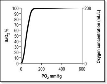
FIG. 2.2.1 The haemoglobin–oxygen dissociation curve.
A number of advantages are conferred by the shape of the Hb–O2 dissociation curve that favour uptake of oxygen in the lung and delivery to the tissues:
Flat upper portion of the curve allows some reserve in the PAO2 required to keep the haemoglobin fully saturated; a reduction in PAO2 of 20% will have minimal effect on the oxygen loading of Hb
Flat upper portion of the curve also ensures that a large difference remains between PAO2 and the pulmonary capillary oxygen partial pressure (PpcO2), even when much of the haemoglobin has been loaded with oxygen. This pressure difference favours maximal Hb–O2 loading
Lower part of the curve is steeper, which favours offloading of oxygen in peripheral tissues with only small falls in capillary PO2. This maintains a higher driving pressure of oxygen, facilitating diffusion into cells
Right shift of the Hb–O2 curve in circumstances of increased temperature, fall in pH, increased PCO2 and increased erythrocyte 2,3 diphosphoglycerate (2,3-DPG) assists in further offloading of oxygen, even when the driving pressure has fallen and PO2 has reached 5.3 kPa (40 mmHg), i.e. venous blood which is still 75% saturated with oxygen.
Oxygen is carried in the blood as dissolved gas and in combination with haemoglobin. At sea level (101.3 kPa), breathing air (FIO2=0.21), the amount of oxygen dissolved in plasma is small (0.03 mL oxygen per litre of blood for each 1 mmHg PaO2). Hence at PaO2=100 mmHg, 3 mL of oxygen are dissolved in each litre of plasma. Dissolved oxygen is important because it is the first available oxygen to diffuse into the tissues. The dissolved component assumes greater significance in the hyperbaric environment, where at 284 kPa and FIO2=1.0 up to 60 mL oxygen can be carried dissolved per litre of blood.
Haemoglobin carries 1.34–1.39 mL oxygen per gram when fully saturated. Blood with a haemoglobin concentration of 150 g/L carries approximately 200 mL oxygen per litre.
Oxygen flux
The total amount of oxygen delivered to the body per minute is known as oxygen flux.
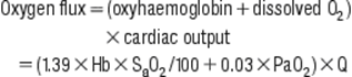
where Hb=haemoglobin concentration g/L; SaO2=arterial oxygen saturation (percentage); PaO2=partial pressure of arterial oxygen (mmHg); Q=cardiac output (L/min).
A healthy individual breathing air transports approximately 1000 mL of oxygen per minute to the tissues, based on a cardiac output of 5 L/min; 30% or 300 mL/min of this oxygen is not available, because at least 2.7 kPa (20 mmHg) driving pressure is required to allow oxygen to enter the mitochondria. Therefore, approximately 700 mL/min are available for use by peripheral tissues. This provides a considerable reserve above the 250 mL/min consumed by a healthy resting adult.
In illness or injury, this reserve may be considerably eroded. Factors that reduce oxygen flux include a fall in cardiac output of any aetiology (including shock states), anaemia or a reduction in functional haemoglobin (carbon monoxide poisoning) and a drop in the SaO2. These situations are frequently encountered in the emergency department. Supplemental oxygen is required in addition to specific therapy, such as volume replacement, transfusion, and measures to improve cardiac output.
Local tissue perfusion and diffusion
Cellular hypoxia results if there is impairment of perfusion to local tissues. Oedema associated with medical illness or local injury increases the diffusion distance between blood and the cell, thus mandating a higher PaO2 to ensure adequate tissue oxygen delivery.
Tissue utilization of oxygen
Increased oxygen flux is required if:
tissue demands for oxygen are higher than normal or
tissue utilization of oxygen is impaired.
Elevation of cardiac output increases oxygen flux in these circumstances but, frequently, this too is significantly impaired by the disease state.
Tissue demands for oxygen increase by 7% for each degree Celsius elevation in body temperature and considerably greater increases in demand occur in seizures, sepsis, severe dyspnoea, restlessness and shivering.
Tissue extraction of oxygen is impaired in sepsis and by poisons, such as carbon monoxide or cyanide. In all cases, oxygen therapy must be combined with general measures, such as reduction of fever and specific treatment of the primary disease process.
Oxygen delivery systems
Oxygen delivery systems are classified into three groups (Box 2.2.1):
variable-performance systems
fixed-performance systems
100% oxygen systems.
Box 2.2.1
Oxygen delivery systems
Variable-performance systems
Nasal cannulae
Hudson mask±reservoir
T pieces and Y connectors
Fixed-performance systems
Venturi mask
Oxygen blenders
100% Oxygen systems
Non-rebreathing circuits
Free-flowing circuits
Self-refilling circuits
Soft reservoir bags
Oxygen-powered resuscitators
Partial-rebreathing circuits
Closed circuit systems
Definitions
Variable-performance oxygen delivery systems
These systems deliver a variable FIO2 to the patient which is altered by the inspiratory flow rate, the minute volume of the patient and the physical characteristics of the delivery system.
Fixed-performance oxygen delivery systems
These systems deliver a specified FIO2 to the patient that is not altered by changes in ventilatory pattern, volume or inspiratory flow rate.
One hundred per cent (100%) oxygen systems
This is a subgroup of fixed-performance systems wherein 100% oxygen is delivered to the patient.
General principles
The oxygen source in most Australasian emergency departments consists of a wall-mounted flow meter capable of delivering oxygen up to 15 L/min, with most available oxygen delivery systems connecting to this apparatus. A 15 L/min flow rate limits the delivery of high FIO2 to adults for the following reasons:
The quietly breathing adult has a peak inspiratory flow rate (PIFR) of approximately 30–40 L/min, which exceeds the oxygen supply. Hence a free flowing system, such as a Hudson mask, must entrain air into the system in order to match the patient’s PIFR, with a resultant reduction in FIO2 to a maximum of 0.6
The quietly breathing adult has a respiratory minute volume of 4–8 L; in a child, this value is approximately 150 mL/kg. Oxygen is stored during expiration by incorporating a reservoir into the circuit for use during inspiration, with a considerable improvement in the economy of oxygen use. This system is limited by the patient’s minute volume. If the minute volume exceeds 15 L, there is a danger of the patient asphyxiating due to insufficient gas supply and, if safety valves allow air into the system, the FIO2 falls.
Multiple-port oxygen supply outlets can overcome the above limitations of inspiratory flow rate and minute volume. The use of ‘Y’ connectors and ‘T’ pieces enable 30, 45 or 60 L per minute to be delivered to the patient to achieve an FIO2 of nearly 1.0, however, these systems can be untidy, using multiple hoses.
More efficient control of flow is achieved via higher output or dial-up flow meters. Extra source oxygen flow may cause variable-performance systems such as the Hudson mask to become fixed-performance systems. Hence the terms ‘variable performance’ and ‘fixed performance’ are loosely applied and are largely dependent on whether or not the gas flow delivered is sufficient to match the patient’s ventilatory requirements.
An example of this is in paediatric oxygen delivery. A high FIO2 can be delivered using a standard 15 L/min oxygen source because the child’s ventilatory requirements are smaller in proportion to the available oxygen supply.
The oxygen delivery systems available for use in emergency medicine, summarized in Box 2.2.1, can be further subdivided according to economy of oxygen use and whether or not the system can be used to ventilate the patient manually.
Variable-performance systems
The FIO2 delivered by these systems is summarized in Table 2.2.1. Options available for use in emergency medicine include:
nasal cannulae
face-masks with air inlets, with or without reservoirs
T pieces and Y connectors.
Table 2.2.1
Variable-performance oxygen delivery systems
|
Apparatus |
Oxygen flow (L/min) |
Oxygen concentration (%) |
|
Nasal catheters |
1–4 |
24–40 |
|
Semi-rigid mask |
6–15 |
35–60 |
|
Semi-rigid mask+double O2 supply |
15–30 |
Up to 80 |
|
Semi-rigid mask+reservoir bag |
12–15 |
60–90 |
Nasal cannulae
The system must be used at flow rates of 4 L/min or less to avoid painful drying of the nasal mucosa, although a flow rate of 2 L/min or less is insufficient to create a nasopharyngeal reservoir during the expiration pause, ready for inspiration with the next breath.
The inspired oxygen concentration is a function of the patient’s inspiratory flow rate and is usually in the vicinity of 22–28%. At flow rates of 2–4 L/min, the nasopharynx acts as a partial reservoir during the expiratory pause, resulting in an increased FIO2. The delivered FIO2 is then influenced by the pattern of breathing (mouth or nose) and the positioning of the nasal cannula.
Nasal cannulae provide a higher FIO2 in paediatric patients and nose breathers. They are less effective in dyspnoeic patients because of the greater amounts of air inspired through the mouth. They are frequently used in patients with stable COPD because of the absence of dead space that prevents CO2 rebreathing. However, fluctuations in FIO2 make nasal cannulae less than ideal in the management of patients who rely on hypoxic respiratory drive and they are second choice after Venturi masks in the emergency management of these patients.
If nasal cannulae are used, there should be strict titration of flow rates to a target SaO2. Advantages for ward or home therapy include the ability to eat and drink, less noise than masks, and economy of oxygen use.
Face-masks (e.g. Hudson, Edinburgh, Medishield)
A small reservoir of oxygen is provided by these masks, but this has little effect on FIO2. The small increase in dead space created by the mask necessitates a flow rate greater than 6 L/min to prevent rebreathing of CO2. Two factors influence the FIO2 provided by this system:
patient’s inspiratory flow rate
source oxygen supply flow rate.
At flow rates of 6–14 L/min, the delivered FIO2 varies from 0.35 to 0.6. This will be less in a dyspnoeic patient because of the higher inspiratory flow rate and greater in a child as the converse applies. If the PIFR increases, greater amounts of air will be entrained into the mask, diluting the oxygen. During expiration, the exhaled gas and excess oxygen are vented through the side perforations.
Attaching a reservoir bag to this mask improves the economy of oxygen use by storing these vented gases during the expiratory phase. This increases the delivered FIO2, but this may be at the expense of increased CO2 rebreathing. Commercially available reservoir bags have a volume of 750 mL to 1 L, which is inadequate for a dyspnoeic patient. The author recommends a minimum flow rate of 12 L/min to avoid CO2retention.
Using a source oxygen supply of 15 L/min, the maximum FIO2 delivered via a Hudson mask to a quietly breathing adult is 0.6. By attaching another source of oxygen using a T piece or Y connector, the resultant flow rate of 30 L/min can deliver an FIO2 up to 0.8. With even greater flow rates, the mask may be converted into a fixed-performance system delivering an FIO2 of almost 1.0. Then the ability to deliver 100% oxygen is limited by the mask’s ‘fit’.
The Medishield mask is stated to be more efficient than the Hudson because dead space is reduced by bringing the oxygen supply closer to the mouth, allowing more effective entrainment during inspiration. An FIO2 of 0.75 may be obtained with a gas flow rate of 15 L/min.
T pieces and Y connectors
The term ‘T piece’ has been used to describe a number of different oxygen delivery systems, including the T piece for supplying humidified oxygen to patients with a tracheostomy and the ‘Ayre’s T piece’ which is a Mapleson E circuit. The use of T pieces or Y connectors in emergency medicine is to supplement an existing oxygen supply with:
extra oxygen
nebulized medication
humidification.
The disadvantage of the system is that several oxygen ports are necessary, which is untidy and may restrict the patient’s mobility. There is loss of economy of oxygen use because of higher flow rates. T pieces allow a higher FIO2 to be delivered to severely dyspnoeic patients.
Fixed-performance systems
Two systems are available for use in emergency departments:
high-flow Venturi masks
oxygen blenders.
High-flow Venturi mask
Oxygen flow through a Venturi system results in air entrainment with delivery of a fixed concentration of oxygen to the patient. The masks deliver FIO2 values from 0.24, 0.28, 0.35, 0.40 and 0.50 to 0.60, using different colour-coded adaptors or by varying the position of a dial on the mask connector. Many studies have assessed their accuracy. The patient receives the stated FIO2 provided the total flow rate exceeds 60 L/min or is 30% higher than the patient’s PIFR. As the patient’s PIFR increases, the system’s performance becomes variable.
In supplying an FIO2 of 0.24 using 6 L/min oxygen flow rate, the total flow rate delivered to the patient is 120 L/min. This falls to 30 L/min total flow for FIO2=0.6 using 15 L/min oxygen supply. This is just equal to the PIFR of a quietly breathing adult and unlikely to be sufficient to provide consistent performance in delivery of the stated FIO2. In severe dyspnoea, these masks may therefore not deliver the stated FIO2.
Increasing the oxygen flow rate above the manufacturer’s recommendations will increase the total gas flow to the mask, while maintaining the stipulated FIO2. At very high-flow rates, however, turbulence is likely to reduce the performance of the system.
Venturi masks provide the best means of managing a patient with chronic obstructive pulmonary disease in the ED because they provide a predictable FIO2 and the air entrained is more humid than fresh oxygen (see below). The entrained gas mixture can be further heated and humidified to assist with sputum clearance. High gas flows minimize rebreathing of CO2 and claustrophobia, but cause problems with sleeping due to noise.
Oxygen blenders
Air is blended with oxygen from a number of inlet ports to supply a fixed FIO2 to the patient. It is a high-flow system and fine-tuning of FIO2 from 0.21 to 1.0 is possible. The resultant mixture can then be channelled to the patient through systems such as continuous positive airways pressure or humidifiers. Lack of portability and high cost are disadvantages. Oxygen blenders are best suited to the resuscitation room and critical-care setting.
100% oxygen delivery systems
These systems vary in their economy of oxygen use and are summarized in Table 2.2.2. The least economical is the free-flowing system as it can only deliver 100% oxygen if the flow rate exceeds the patient’s PIFR. Incorporating a reservoir and unidirectional valves into the circuit enables greater economy of oxygen use by storing oxygen during expiration ready for the inspiratory phase.
Table 2.2.2
Classification of 100% oxygen systems

Devices incorporating a reservoir into the circuit are capable of delivering 100% oxygen only when the total oxygen flow equals or exceeds the patient’s respiratory minute volume (RMV), plus there are no leaks in the system. The reservoir volume must exceed the patient’s tidal volume, otherwise storage of oxygen is inefficient, fresh gas loss occurs when the reservoir is full, and there is the risk of asphyxia during inspiration.
A demand valve system delivers precisely the patient’s minute volume without the added bulk and problems of a reservoir. It is able to cope with changes in RMV provided fresh gas flow always exceeds the patient’s PIFR. Closed-circuit systems are the most economical in oxygen consumption. Carbon dioxide is absorbed by soda lime and low-flow fresh oxygen replaces that consumed during metabolism, which is approximately 250–1000 mL/min, which is considerably less than the patient’s RMV.
Classification
One hundred per cent oxygen-delivery systems available for use in emergency medicine are summarized in Table 2.2.2.
Free-flowing circuits
Flow rates in excess of the patient’s PIFR are required to provide 100% oxygen using a free-flowing system, which necessitates the use of multiple oxygen ports. The system may not deliver 100% oxygen, is wasteful of oxygen and may be untidy, restricting patient mobility for investigations. Sophisticated free-flowing systems using oxygen blenders and humidification are available, but restrict the ability to move the patient.
Soft reservoir circuits
These are non-rebreathing systems incorporating unidirectional valves to channel fresh oxygen to the patient and exhaled gas to the atmosphere. With one oxygen supply port the system delivers 100% oxygen, provided the patient’s minute volume is less than 15 L/min. Higher flow first stage regulators or two oxygen supply ports enable delivery of up to 30 L/min. Fresh gas flow is titrated to the patient’s minute volume by watching the reservoir bag, which should be fully distended at the start of inspiration and more than one-third full when inspiration is complete.
The reservoir bag has a minimum volume of 3 L and, for optimal performance, the patient’s tidal volume should not exceed 2 L. A soft silicone mask is strapped to the head to ensure a firm but comfortable fit without leaks. The system cannot be used to ventilate patients manually and may be hazardous if the patient has an impaired conscious state owing to the risk of aspiration if they vomit and asphyxiation if there is a fall in fresh gas flow or a sudden rise in minute volume. Complications are avoided with clinical vigilance and the use of safety valves to entrain air if the oxygen supply ceases.
Self-refilling, non-rebreathing resuscitators (Air Viva and Laerdal systems)
Most Australasian emergency departments possess at least one type of self-refilling system. They can be used to ventilate a patient manually as well as allowing spontaneous ventilation. The Laerdal system has three sizes for adults, children and infants, whereas the Air Viva system has one size for adults only (Table 2.2.3).
Table 2.2.3
Self-refilling, non-rebreathing resuscitators
|
Self-refilling bag volume (mL) |
Reservoir bag volume |
|
|
Air Viva |
1700 |
2300 |
|
Laerdal (Adult) |
1600 |
2600 |
|
Laerdal (Child) |
500 |
2600 |
|
Laerdal (Infant) |
240 |
600 |
Advantages
Self-inflation and hence the ability to ventilate patients with air if oxygen supply is exhausted.
Low-resistance unidirectional valves prevent rebreathing of CO2.
Use in spontaneously ventilating patients and for manual ventilation.
System is capable of delivering FIO2=1.0 provided fresh gas flow exceeds minute volume and the reservoir bag is attached. Without the reservoir bag, a maximum FIO2 of 0.6 is obtainable.
A safety valve entrains air into the system to prevent asphyxiation if there is a sudden rise in minute volume, but this is at the expense of FIO2.
Over-pressure relief valves are incorporated into the Laerdal paediatric and infant apparatus to prevent barotrauma in these patients.
Addition of positive end-expiratory pressure (PEEP) to the system is possible by attaching a PEEP valve to the expiratory limb. Close apposition of the mask to the face or endotracheal intubation is required for this to be effective.
Disadvantages
Reduction in FIO2 occurs when minute volume exceeds fresh gas flow. Dual oxygen supply ports can minimize this problem, especially in an extremely dyspnoeic patient.
Unit is bulky and disconnections sometimes occur.
There is less ‘feel’ during manual ventilation than with soft bag circuits. Inflation of the stomach is more likely during bag/mask ventilation, especially if there is airway obstruction or reduced pulmonary compliance.
Oxygen-powered resuscitators
Examples of this type of system include the Oxy Viva, Laerdal and DAN demand valve systems. High-pressure oxygen is fed to a demand valve which delivers high-flow oxygen to the patient. The system can be used in a spontaneously breathing patient and, for manual ventilation, by depressing a manual override button. Spontaneously ventilating patients initiate an oxygen flow of up to 120 L/min by generating a negative pressure of 0.3 kPa (2.25 mmHg) at the start of inspiration. Fresh gas flow is delivered at a pressure of up to 5.3 kPa (40 mmHg).
Advantages
Portability, as it is easy to attach to an oxygen cylinder and take to the field. There are no bulky reservoir bags.
Economy of oxygen use as the patient’s minute volume is precisely delivered at sufficient flow rates to match the PIFR. Provided there are no leaks, the system delivers FIO2=1.0.
Disadvantages
Increased work of breathing for spontaneous ventilation as negative pressure must be generated to initiate oxygen flow.
System cannot function when fresh gas supply is exhausted.
During manual ventilation, it is almost impossible to judge ventilatory volume except by observing the patient’s chest. The safety over-pressure relief valve may not prevent barotrauma, especially in children.
Lack of ‘feel’ during manual ventilation may lead to over-inflation of the stomach if there is airway obstruction or reduced pulmonary compliance.
Mapleson circuits
Mapleson circuits are still used in some emergency departments. Partial rebreathing of gases occurs with all of the circuits but CO2 retention can be avoided if fresh gas flow exceeds minute volume by a ratio of 2–2.5:1.
The most commonly used versions are the Mapleson B and the Mapleson F, which are covered under paediatric considerations. Mapleson A, C, D and E circuits are not discussed further.
Advantages of the Mapleson B circuit
Used for both spontaneous and manual ventilation. Its performance is similar in both circumstances.
Soft bag has excellent ‘feel’ for manual ventilation and it is easy to monitor spontaneous ventilation by observing the filling and emptying of the reservoir bag.
Disadvantages
Carbon dioxide build-up with lower oxygen flow rates. This can be avoided with higher flow rates or by intermittently purging the reservoir bag.
System cannot function without a fresh gas supply.
May be difficult to use when ventilating a patient manually using a mask.
Valve assembly may occasionally ‘stick’.
Closed-circuit systems
An example is the MD Oxyresuscitator. The circuit is the same as the Boyle’s anaesthetic circle system. A soda lime canister absorbs exhaled CO2 and a low-flow oxygen supply replaces oxygen consumed by metabolism at approximately 0.5–2 L/min. Considerable economy of oxygen use is thus achieved by rebreathing from the circuit.
Advantages
Economy of oxygen use. More than 6 hours of oxygen can be provided by a ‘C’-sized oxygen cylinder at 1 L/min. This markedly exceeds the endurance of the cylinder using other systems.
Can be used for spontaneous or manual ventilation. A soft reservoir bag provides excellent ‘feel’ for ventilation.
Pressure on the system is controlled by the operator during manual ventilation. This minimizes gastric distension.
Portable and can easily be taken to the field.
Disadvantages
Circuit ceases to function when fresh gas flow is exhausted.
Exhaled nitrogen from the patient’s early breaths may enter the circuit and reduce FIO2 below 1.0. This is prevented by intermittent purging of the reservoir.
CO2 may accumulate if the soda lime canister is old or stops functioning.
Incorrect packing of the soda lime canister may result in inhalation of soda lime dust (which is extremely rare).
Reservoir bag is remote from the patient mask and the system may be cumbersome to operate.
Helium and oxygen mixtures
Over the last decade, there has been interest in adding helium to oxygen (maximum 30% oxygen, also known as ‘Heliox’). Heliox has a lower density than air with the potential to reduce airway resistance and hence the work of breathing when treating disease processes such as COPD and asthma.
Helium (He, MW=4) is much lighter than nitrogen and therefore significantly lowers the density of the gas mix when combined with oxygen in the range of FIO2=0.2–0.4. This advantage is lost when FIO2>0.4. Despite lower density, the viscosity of Heliox is not significantly lower than that of air. Its main theoretical advantage is if there is turbulent gas flow that is density dependent. This may occur with COPD where there is a combination of small and medium airways disease. Early studies also suggested that Heliox may enhance nebulizer particles in the lung; however, greater flow rates may be required to drive the nebulizer. Despite the potential advantages, the clinical evidence for use in COPD is not strong.
Cochrane reviews of the topic concluded that there is insufficient evidence to support the routine use of Heliox to treat COPD exacerbations or exacerbations of asthma. However, the review of adults and children with asthma did conclude that Heliox may improve pulmonary function when there is more severe obstruction. Most of the studies of Heliox for asthma have assessed it as a driver of nebulizer therapy rather than for continuous administration.
Of the two studies of Heliox therapy for COPD assessed in the Cochrane review, only one study included acutely decompensated patients in the ED. This study failed to show a benefit from Heliox when it was used to drive nebulized β-agonist therapy. Further randomized studies using Heliox are needed in asthma and COPD, both continuously and as a driver for nebulizer therapy, with hard endpoints such as physiological parameters, response to nebulized β-agonists, need for non-invasive ventilation or intubation and admission rates.
Measurement of oxygenation
Clinical assessment of oxygenation is unreliable and the time-honoured sign of cyanosis varies with the level of haemoglobin, skin pigmentation, perfusion and external light. Arterial blood gases and pulse oximetry provide an objective measurement of oxygenation and enable precise titration of oxygen therapy to the clinical situation.
Pulse oximetry
Pulse oximetry is the most frequently used indicator of oxygenation in emergency medicine as it is non-invasive. It is regarded as the ‘fifth vital sign’ and provides continuous real-time assessment of a patient’s oxygenation and response to therapy. It has a proven role in emergency medicine and is an excellent clinical tool, provided the limitations are understood.
It is important to recognize that SaO2 is not an adequate marker of ventilatory function and will not detect rising PaCO2 in respiratory failure or a sedated patient until late, when conscious state becomes depressed or there is respiratory arrest. Hence all patients with potential respiratory compromise due to disease or sedative medication require careful monitoring of vital signs, conscious state and CO2 via end-tidal CO2monitoring or arterial blood gases. Recent literature suggests a fall in SaO2 may precede CO2 accumulation during procedural sedation and analgesia in children.
A detailed knowledge of the haemoglobin–oxygen dissociation curve is required to interpret pulse oximetry, as well as the factors that influence readings obtained by this equipment. These factors are summarized in Table 2.2.4.
Table 2.2.4
Factors that influence pulse oximetry readings

Paediatric considerations in oxygen therapy
The general principles of oxygen therapy and its indications apply equally well for children as for adults, but there are a number of important differences in relation to body size, psychology and oxygen toxicity.
Body size
Children are smaller than adults both anatomically and physiologically, so that any increase in equipment dead space will significantly increase CO2 retention. Children are less able to tolerate increased resistance to ventilation, particularly if negative pressure must be generated to open valves in the apparatus.
Peak inspiratory flow rate and respiratory minute volume are lower; hence, a given oxygen supply flow rate will produce a higher FIO2 in a child than in an adult. A Hudson mask at 8 L/min may supply an FIO2 of 0.8 in a young child. Reservoir bags are not required to deliver FIO2 values near 1.0 to children weighing less than 15 kg as available supply flow rates (maximum 15 L/min) exceed the child’s PIFR.
Appropriately sized equipment is essential: a range of sizes of oxygen masks, oximeter probes, laryngoscopes and endotracheal tubes must be available to manage children of different ages as serious barotrauma may result from the use of excessive volume during manual ventilation. Resuscitator bags are available with paediatric-sized reservoirs. The Laerdal system has both paediatric and infant sizes. These units also have a pressure relief valve designed to prevent barotrauma. Pressure rapidly rises as the child’s lung reaches full inflation.
Jackson–Rees (Mapleson F) circuit
A smaller Mapleson circuit, the Jackson–Rees (Mapleson F) circuit, is available to ventilate children, which can be used for both spontaneous and manual ventilation. Rebreathing of carbon dioxide does not occur provided the fresh gas flow is 2–3 times minute volume and the bag is separated from the patient by a tube of internal volume greater than the patient’s tidal volume. The overall relationship between fresh gas flow, minute volume and PaCO2 is complex.
The principal advantages over the Laerdal system are that the operator can observe bag movement in spontaneous respiration and has a better ‘feel’ for airway obstruction in manual ventilation. However, considerable skill and experience are required to use the system safely.
Psychological considerations
Gaining the trust and confidence of an ill child is an art learnt with experience. They frequently respond with fear when oxygen therapy is administered, so it is helpful to ask a parent to nurse the child during treatment. A tight-fitting mask is less important in a child because source flow rate more closely approximates PIFR. Parents may assist by holding the oxygen mask close to the child’s face or by directing high-flow oxygen straight at the child’s mouth using a tube only. A cupped hand with the oxygen tube held between middle and ring fingers can serve as a surrogate oxygen ‘mask’.
Oxygen toxicity
Prolonged administration of oxygen at FIO2>0.6 for longer than 24 hours may be toxic to infants. This toxicity may not become apparent during their acute stay in the emergency department, but the oxygen dose received there contributes to the cumulative toxicity. Appropriate monitoring using pulse oximetry ensures administration of the correct dose and minimizes the risk of toxicity. However, supplemental oxygen should never be withheld because of fear of toxicity.
Transfer of patients on oxygen therapy
Supplemental oxygen therapy is a vital part of transporting the ill patient and is especially important for air travel where lower ambient PIO2 may exacerbate hypoxia already present as a result of the patient’s disease process. Patients with decompression illness or arterial gas embolism should not be transported at cabin pressures lower than 101.3 kPa (1 atmosphere absolute, ATA) because lower ambient pressure exacerbates their disease process by increasing bubble size. A number of factors must be considered for successful oxygen therapy during transport of a patient.
Knowledge of the oxygen delivery apparatus and its maximum rate of delivery are essential for estimating transport oxygen requirements. These estimates must take into account current oxygen consumption, duration of transport (including delays), oxygen required in the event of deterioration and a safety factor of at least 50%.
The sizes of oxygen cylinders available in Australasia, their filling pressures and approximate endurances are summarized in Table 2.2.5. The most economical circuit for prolonged transport with FIO2=1.0 is a closed circuit with a CO2 absorber and the least economical is a free-flowing circuit.
Table 2.2.5
Oxygen cylinder sizes for patient transport

Monitoring during transport should be of the same standard as that initiated in the emergency department. Pulse oximetry is an essential tool to detect hypoxia during transport and should include audible and visual alarms. Oxygen therapy can be titrated against SaO2, which is particularly important in air travel where PIO2 varies with ascent and descent. All the usual clinical parameters must also be monitored.
Oxygen therapy in specific circumstances
Asthma
Hypoxia in asthma results from ventilation–perfusion mismatch created by bronchospasm, secretions and airway inflammation and oedema. Supplemental oxygen should be titrated to provide an SaO2>90% (preferably 94%) and must be continued during the interval between doses of inhaled bronchodilators.
Initial management should include a Hudson mask at 8 L/min flow rate, with SaO2 monitored continuously by pulse oximetry. The oxygen dose should be rapidly increased up to 100% if the patient remains hypoxic. Bronchodilator therapy is administered proportionate to the severity of the attack, using oxygen to drive the nebulizer. Oxygen should not be withheld or administered in low doses for fear of respiratory depression. Hypercapnia is an indication of extreme airway obstruction and its presence mandates aggressive therapy and/or mechanical ventilation.
Mechanical ventilation in asthma
Mechanical ventilation requires an FIO2=1.0, high inspiratory flow rate (100 L/min), low tidal volume (6–8 mL/kg), a prolonged I:E ratio of at least 1:3 and a low ventilation rate (6–10 breaths/min or less), to reduce the risks of progressive dynamic hyperinflation with the development of auto-PEEP (iPEEP) reducing venous return and hence preload, and of barotrauma with the development of a pneumothorax. Permissive hypercapnia is accepted with mechanical ventilation.
Occasionally, patients with asthma become hypoxic during nebulizer therapy because the oxygen flow rates driving the nebulizer (6–8 L/min) are lower than the flow rate required to maintain SaO2>90%. In these circumstances, extra oxygen is supplied to maintain SaO2 via a T piece or Y connector during nebulizer therapy.
Chronic obstructive pulmonary disease
Most ED patients with COPD have a degree of acute respiratory failure that caused their emergency presentation. This may be due to infection, bronchospasm, retention of secretions, coexistent left ventricular failure, worsening right heart failure, pulmonary embolism, pneumothorax, sedation or reduction of regular therapy, such as inhaled or oral steroids. Clues to the degree of severity and chronicity of the COPD are obtained from the patient’s history, past clinical records, emergency department blood gases and the response to initial oxygen therapy.
Clinical indicators of patients at risk of CO2 retention include a housebound patient, FEV1<1 L, polycythaemia, a warm vasodilated periphery and cor pulmonale. In the acutely unwell patient, treatment may be required before the history can be obtained.
COPD groups
Patients with COPD fall into two groups as regards management, although this classification is still debated.
Normal ventilatory response to CO2 (‘can’t breathe’ – the most common). Gas exchange and air flow into the lungs are impaired, but ventilatory drive is normal.
Impaired ventilatory response to CO2 (‘won’t breathe’ – less common). Ventilation does not increase in response to hypercapnia and acidosis.
There is overlap between the advanced stages of illness. The aims of oxygen therapy are targeted to produce SaO2 of 88–90% and to identify the second group of patients such that the oxygen dose can be titrated to achieve an acceptable clinical response without excessive elevation of PaCO2. Serial arterial blood gas analysis is essential in their management.
The majority will have a normal ventilatory response to CO2. Hypercapnia indicates that ventilatory failure is developing, with a danger of respiratory arrest if the patient’s disease is severe and progressive. This can also result from uncontrolled oxygen therapy with failure to monitor the patient’s clinical status and arterial blood gases. Any patient with impaired consciousness due to respiratory failure should be manually ventilated while being clinically assessed and treated.
Controlled titration of oxygen dose in COPD
Successful management of the cooperative patient with COPD necessitates controlled titration of oxygen dose. Variable-performance oxygen masks do not have a role in the emergency management of COPD, unless there is careful monitoring of SaO2 and ETCO2. A consistent initial approach to oxygen therapy for a conscious patient with advanced COPD is used as, at the time of presentation, their ventilatory response to CO2 is unknown.
In most patients, the administration of 24–28% oxygen by Venturi mask will improve oxygenation, with a target SaO2 of about 88–92%. It is also acceptable initially to titrate oxygen therapy to a target SaO2range. A recent Australian pre-hospital randomized clinical trial (RCT) showed lower mortality for patients with COPD if they received oxygen by nasal prongs titrated to SaO2 range ≈88–92%. All nebulizer therapy was administered by air.
Below 90% saturation, the Hb–O2 dissociation curve falls steeply and, unless a pulmonary shunt is present, even small increments in oxygen will make a positive difference. The patient’s response to initial oxygen therapy (FIO2=0.24–0.28) will direct further oxygen dose changes and identify any patients not already known to be suffering chronic hypercapnia.
A repeat blood gas sample should be taken after 10 minutes of breathing FIO2=0.24–0.28. The PaCO2 may rise slightly because of the ‘Haldane effect’. If this rise is excessive (>1–1.3 kPa [8–10 mmHg]), it is consistent with an impaired ventilatory response to CO2. The FIO2 should then be adjusted downwards in steps to achieve a satisfactory pulse oximetry reading that is compatible with an acceptable CO2 level. In a patient with COPD, an acceptable range for SaO2 is 88–92%.
Blood gas sampling in COPD
Blood gas samples taken during the initial assessment of these patients (breathing air or controlled oxygen) assists management. Venous samples are acceptable, provided they are used consistently to monitor trends. If the bicarbonate level is>30 mmol/L or is elevated by more than 4 mmol/L for each 1.3 kPa (10 mmHg) rise in PaCO2 above normal (5.3 kPa, 40 mmHg), this provides strong evidence of chronic hypercapnia, provided there is no cause of a metabolic alkalosis.
Management in COPD
Patients with a normal ventilatory response to CO2 will not exhibit a significant elevation of PaCO2 in response to oxygen therapy. If hypoxaemia persists and the PaCO2 remains stable, then the oxygen dose may be increased incrementally until the desired oxygen saturation is achieved. A lower than normal SaO2 (≈88%) and PaO2 (≈56 mmHg) may be acceptable provided the patient remains conscious and cooperative.
Non-invasive positive-pressure ventilation (NIPPV) is indicated if the patient remains hypoxic or becomes progressively more hypoxic and the elevation of PaCO2 persists or worsens or their conscious state deteriorates. Intubation and ventilation may be required, which should be regarded as a last resort (and will not be covered). Supplemental oxygen should never be abruptly withdrawn from a patient with COPD as a catastrophic fall in PaO2 will occur. All reductions in controlled oxygen dose should be in a stepwise manner, similar to incremental increases.
In the majority of cases, an acceptable balance between PaO2 and PaCO2 can be achieved, where both hypoxia and hypercarbia are reversed by specific therapy. Treating the cause of the ventilatory failure is a priority.
A pilot study showed that short-term administration of bronchodilators using oxygen-driven nebulizers in the acute management of chronically hypercapnic patients may be safe. Caution is advised, as some authors suggest that COPD is still poorly managed in the emergency department with respect to oxygen dose. Interestingly, the authors of that paper offered only limited practical advice on the titrated use of oxygen in the acute management of COPD and did not differentiate between COPD patients with an acute elevation of CO2 and those with chronic elevation. The authors of the pre-hospital RCT were more specific, titrating to a target SaO2.
Goal-directed oxygen therapy
The oxygen dose in the initial management of many medical conditions including myocardial infarction, asthma and even pneumonia has been questioned. A recent Cochrane review of oxygen therapy for myocardial infarction claimed oxygen may be harmful. This conclusion was based on a non-significant analysis, dominated by a 1976 trial (86% of the weighting). That trial contained many clinical deficiencies not reflective of modern practice (e.g. diagnostic criteria, no intravenous glyceryl trinitrate (GTN) or reperfusion therapy, not monitoring oxygen saturations and no blood gases and use of gas flow rates known to cause CO2 accumulation). The authors correctly suggested that oxygen treatment for myocardial infarction should be further evaluated in a larger trial.
One recent trial suggested that high concentration oxygen therapy caused a greater rise in CO2 in adults presenting with community-acquired pneumonia, compared with titrated dose oxygen. The study did not examine clinical outcomes. Another area of interest is oxygen dose for asthma. Perrins et al. using transcutaneous (tc) CO2 monitoring demonstrated that high concentration oxygen therapy led to clinically significant increases in PtcCO2, compared to the titrated dose oxygen therapy. The high concentration group had 4.5 times greater gradient of increasing PtcCO2 and over twice the odds of requiring admission. Current trends in clinical evidence suggest that oxygen should be treated in the same way as any other drug, that is to provide the optimum dosage appropriately titrated to the clinical needs of the patient. Goal-directed oxygen therapy is no different in principle with available clinical monitoring systems to many other therapies for physiological disturbance.
Special delivery systems
Oxygen humidification
Humidification may be desirable when prolonged use (>6 h) of supplemental oxygen is required as oxygen is totally dry, possessing no water vapour. Humidification is particularly important in a patient ventilated with an endotracheal tube as the natural humidification that occurs in the nose, mouth and nasopharynx is bypassed. Patients with COPD and retained secretions benefit from humidification.
Additional heat is required to provide effective humidification by vaporization of water. Various systems are available to humidify inspired gas and, ideally, they should be able to deliver inspired gas to the trachea at 32–36°C with low resistance and at greater than 90% humidity. These devices should be simple to use and able to maintain temperature and humidity at varying gas flows and FIO2. There should also be safety alarms monitoring temperature and humidity.
Humidification of warmed inspired gas also enables heat transfer to hypothermic patients and is essential in treating the pulmonary complications of near drowning. Dry oxygen will exacerbate hypothermia. There are a number of commercial humidifiers available that provide humidification by heating coils with a large surface area for contact with inspired gas.
Continuous positive airways pressure
Continuous positive airways pressure (CPAP) has a role in the management of pulmonary oedema, pneumonia, bronchiolitis, respiratory tract burns and acute respiratory failure. Benefit to the patient is achieved as a result of increasing functional residual capacity and reduced pulmonary compliance. Hypoxaemia is reversed by reduction in intrapulmonary shunting and the work of breathing is reduced.
Circuit designs for CPAP
Circuit designs usually consist of a reservoir based on the Mapleson D circuit or a high-flow turbine system. Humidification can be added to the system and is considered essential for long-term use (>6 h). Use of an oxygen blender enables variable FIO2 to be administered. CPAP has a proven role in the emergency department in the acute management of cardiogenic pulmonary oedema. Reduced requirements for endotracheal intubation have been demonstrated when CPAP is used for severely ill patients. Complications of CPAP include aspiration and pulmonary barotrauma. It may elevate intracranial pressure and precipitate hypotension by reducing venous return to the thorax.
Hyperbaric oxygen treatment
Hyperbaric oxygen (HBO) treatment consists of administering oxygen at pressures greater than 1 ATA, usually in the range of 2.0–2.8 ATA. This requires a hyperbaric chamber which is pressurized with air while the patient breathes an FIO2=1.0 from various delivery systems for periods of 2–7 hours. The high PIO2 results in PaO2 of up to 267 kPa (2000 mmHg) if 2.8 ATA treatment pressure is used. This is beneficial, as there is increased dissolved oxygen in the plasma (up to 300 mL oxygen may be carried to the periphery each minute in the dissolved form), which maintains oxygen flux even if haemoglobin is non-functional, for instance in carbon monoxide poisoning. Increased PIO2 enables more rapid elimination of toxic gases from the body, for example carbon monoxide or hydrogen sulphide.
Uses of hyperbaric oxygen
HBO treatment has a number of benefits in treating gas embolism and decompression illness (DCI). It provides extra oxygen to tissues rendered ischaemic by nitrogen bubbles and the increased pressure reduces bubble size and enhances nitrogen removal from the body. The increased PO2 also creates a greater driving pressure of oxygen into ischaemic tissues in problem wounds and reduces swelling by vasoconstriction in crush injury. In addition, HBO treatment is used in anaerobic infections by virtue of being bacteriostatic to anaerobes, inhibiting clostridial α toxin and stimulating host defences via granulocyte function. Recognized indications for acute referral to a hyperbaric facility for HBO treatment are summarized in Table 2.2.6 (see Chapter 28.3).
Table 2.2.6
Indications for acute treatment with hyperbaric oxygen
Decompression illness
Air or gas embolism
Carbon monoxide poisoning
Gas gangrene and anaerobic fasciitis
Necrotizing soft tissue infections
Acute crush injury with compartment syndrome
Acutely compromised skin flaps or grafts, due to injury or post-surgery
Complications of oxygen therapy
These can be classified into three categories:
equipment-related complications
carbon dioxide narcosis
oxygen toxicity.
Equipment-related complications
These are entirely preventable with careful supervision and monitoring. Tight-fitting masks may cause asphyxia if there is insufficient oxygen reservoir or flow and aspiration of vomitus may occur if the patient has depressed airway reflexes. Use of appropriate oxygen flow rates with rebreathing circuits prevents CO2 accumulation.
Barotrauma can be prevented during mechanical ventilation by the use of appropriate volumes and pressures, although it can be difficult to avoid when there is reduced lung compliance, as in the moribund asthmatic. Knowledge of potential equipment complications enables prompt intervention should they arise. When investigating a sudden deterioration in the patient’s condition, a thorough check of the equipment in use is mandatory.
Carbon dioxide narcosis
This can be prevented by controlled oxygen therapy titrating the FIO2 against SaO2, arterial blood gases and conscious state (see above). An unconscious patient should be intubated and manually ventilated using high FIO2, preferably 100% oxygen. A patient with a deteriorating conscious state and respiration due to CO2 narcosis should be vigorously stimulated and encouraged to breathe while FIO2 is reduced in a stepwise manner. Oxygen should never be suddenly withdrawn as this precipitates severe hypoxia. Reversible causes of respiratory failure should be treated and non-invasive ventilation instituted.
Oxygen toxicity
Oxygen is toxic in high doses which is a function of PIO2 and duration of exposure. Toxicity is thought to occur by the formation of free radicals and toxic lipid peroxides, inhibition of enzyme systems and direct toxic effects on cerebral metabolism. Toxicity is mainly restricted to the respiratory system and central nervous system (CNS), although it can affect other regions such as the eye. Premature infants develop retrolental fibroplasia after prolonged exposure to high FIO2. CNS oxygen toxicity manifested by neuromuscular irritability and seizures (Paul Bert effect) is restricted to hyperbaric exposures.
Pulmonary oxygen toxicity (Lorrain Smith effect) is of relevance to emergency medicine, although exposures of 0.6–1 ATA for more than 24 hours are required to produce demonstrable evidence of lung injury. Acute changes such as pulmonary oedema, haemorrhage and proteinaceous exudates are reversible on withdrawal of oxygen. Longer durations of high PIO2 may lead to permanent pulmonary fibrosis and emphysema. Physicians should be alert to acute symptoms of cough, dyspnoea and retrosternal pain, although these are non-specific symptoms of oxygen toxicity. A progressive reduction in vital capacity may be demonstrated. As with all drugs, oxygen dose should be monitored and carefully titrated against SaO2 and clinical effect. However, oxygen therapy should never be withheld acutely because of fear of toxicity.
Controversies/emerging issues
Is oxygen a drug with a therapeutic window?
Is oxygen harmful in myocardial infarction?
Should we monitor all patients with COPD and asthma using CO2 measuring devices?
What is the best device for monitoring CO2 during emergency treatment?
Goal-directed oxygen therapy titrated to target SaO2 for specific disease processes will be the future best practice for oxygen therapy.
Further reading
1. Austin MA, Wills KE, Blizzard L. Effect of high flow oxygen on mortality in chronic obstructive pulmonary disease patients in prehospital setting: randomised controlled trial. Br Med J. 2010;341:c5462.
2. Cabello JB, Burls A, Emparanza JI. Oxygen therapy for acute myocardial infarction. Cochrane Database Syst Rev. 2010;6:CD007160.
3. Fracchia G, Torda TA. Performance of Venturi oxygen delivery devices. Anaes Intens Care. 1980;8:426–430.
4. Gesell L. Hyperbaric oxygen therapy indications. USA Undersea Hyperbaric Med Soc 2011. Kensington, Maryland.
5. McKenzie DK, Frith PA, Burdon JGW, et al. The COPDX Plan: Australian and New Zealand Guidelines for the management of chronic obstructive pulmonary disease. Med J Austral. 2003;178:S1–S39.
6. O’Driscoll R. Emergency oxygen use. Br Med J. 2012;345:e6856.
7. Perrin K, Wijesinghe M, Healy B, et al. Randomised controlled trial of high concentration versus titrated oxygen therapy in severe exacerbations of asthma. Thorax. 2011;66:937–941.
8. Rodrigo GJ, Pollack CV, Rodrigo C, Rowe BH. Heliox for non-intubated acute asthma patients. Cochrane Database Syst Rev. 2006;4:CD002884.
9. Rodrigo G, Pollack C, Rodrigo C, et al. Heliox for treatment of exacerbations of chronic obstructive pulmonary disease. Cochrane Database Syst Rev. 2001;1:CD003571.
10. Sivilotti M, Messenger D, van Vlymen J, et al. A comparative evaluation of capnometry versus pulse oximetry during procedural sedation and analgesia on room air. Can J Emerg Med. 2010;12:397–404.
11. Smart DR, Mark PD. oxygen therapy in emergency medicine. Physiology and delivery systems Emerg Med (Fremantle). 1992;4:163–178.
12. West JB. Respiratory physiology – the essentials 9th ed. Baltimore: Lippincott, Williams & Wilkins; 2012.
2.3 Haemodynamic monitoring
Liz Hickson and Craig Hore
Essentials
1 Haemodynamic monitoring includes observation of the complex physiology of blood flow, with the aim of providing data that can be used to improve patient management and outcomes.
2 Numerous methods are available that should be considered in a stepwise fashion, from simple clinical assessment to highly technical, invasive procedures, such as the pulmonary artery catheter.
3 Effective use of haemodynamic monitoring devices requires an understanding of cardiovascular physiology.
4 Currently, there is a move away from simple blood pressure measurements towards targeting end-organ perfusion and the adequacy of cardiac output.
5 Use of any monitoring technology in the emergency department (ED) must consider the time associated with its introduction, the skill levels required and the clinical benefits that are provided.
6 No monitoring modality improves outcome unless it is linked to a treatment protocol.
7 The pulmonary artery (Swan–Ganz) catheter was for many years considered a ‘gold standard’ for haemodynamic monitoring, but evidence suggests no improvements in patient outcome. It should therefore not be used in the ED.
8 Less invasive devices have been developed in recent years. Their role in the ED is yet to be fully elucidated.
9 Further developments will likely result in greater use of less invasive methods for haemodynamic monitoring, with an increased ability to monitor at the microcirculation and/or cellular level, and better correlation between observed events and final diagnosis.
Introduction
Haemodynamics is concerned with the physiology of blood flow and the forces involved within the circulation. Haemodynamic monitoring involves studying this complex physiology using various forms of technology to understand these forces and put them into a clinical context that can be used to direct therapy. The utility of basic monitoring is universally accepted. However, the maxim that ‘not everything that counts can be counted and not everything that can be counted counts’ (Albert Einstein, 1879–1955) should be borne in mind.
This is particularly salient in the emergency department (ED) where the pressure of work and the diversity of patients do not allow the unlimited use of complex and expensive monitoring systems.
This chapter provides an outline of current approaches to the various technologies available for haemodynamic monitoring and their applicability in the ED. Many methods are available which should be thought of in a stepwise progression from simple clinical assessment to invasive, highly technical methods using sophisticated devices.
Historical background
As recently as 100 years ago, only temperature, pulse and respirations were measured and used to manage patients. The technology for auscultatory blood pressure measurement was available, but did not come into regular use until the 1920s.
Intensive care as a medical/nursing specialty evolved in tandem with the electronic revolution of the 1960s. At the same time, increasingly sophisticated haemodynamic and laboratory techniques vastly improved diagnosis and provided a way further to evaluate therapy. Despite these major advances in the ability to monitor multiple physiological variables, there is little evidence to suggest that they have resulted in tangible improvements in patient outcome.
Practical use of monitoring
The practical use of any monitoring device must be appropriate to the individual clinical environment. Thus, it may be reasonable to insert a pulmonary artery Swan–Ganz catheter in the intensive care unit (ICU) where the necessary time can be taken, yet impractical and potentially unsafe in a busy ED. Another consideration is that haemodynamic monitoring should only be used when the clinical outcome may be influenced and potentially improved. Once irreversible cellular damage has occurred, no benefit occurs no matter how far therapy is maximized. Further, haemodynamic monitoring may not improve patient outcome unless linked to a clinical protocol or ‘goal-directed therapy’.
Clinicians should only introduce monitoring equipment that will have a direct influence on their choice of therapy, as the use of invasive monitoring carries potential risks of harm to the patient. The injudicious use of physiologically based treatment protocols may lead to worse outcomes. All monitored variables must be evaluated and applied in a manner proven to lead to benefit, in terms of both the diagnosis and the management.
Overview of cardiovascular physiology
One possible reason that haemodynamic monitoring has not been associated with improvements in outcome is the inability to understand and manipulate patients’ physiology effectively.
Circulatory model
Haemodynamic data are traditionally considered in the context of a circulatory model. This model varies, but usually consists of a non-pulsatile pump and a hydraulic circuit with discrete sites of flow resistance, alongside the Frank–Starling mechanism with its concepts of preload, contractility and afterload.
Cardiac output
Cardiac output (CO) is the volume of blood pumped by the heart per unit of time, usually expressed in litres per minute (L/min). The heart operates as a pump and ejects a bolus of blood known as the stroke volume(SV) with each cardiac cycle. CO is the product of SV and heart rate (HR).
A complex set of interrelated physiological variables determines the magnitude of CO, including the volume of blood in the heart (preload), the downstream resistance to the ejection of this blood (afterload) and the contractility of the heart muscle. However, it is the metabolic requirements of the body that are the most potent determinant of cardiac output.
Regulation of CO
The regulation of CO is therefore complex. A single measurement represents the summation of many interacting physiological processes. Basal CO is related to body size and varies from 4 to 7 L/min in adults. This value divided by the body surface area enables comparison between patients with different body sizes, giving the cardiac index (CI).
Bedside methods do not measure CO directly, meaning that the values obtained are only estimates. Assessment of CO is therefore not done routinely. Indeed, misuse of CO data may worsen outcomes. The International Consensus Conference on Haemodynamic Monitoring in Shock (2007) suggested that monitoring of CO is only of value if it guides therapies to improve outcome.
Cardiac index (CI)
CI measurement is valued over simple blood pressure recording as it describes the total volume of blood flow in the circulation per unit of time and hence serves as an indicator of oxygen delivery to the tissues. The CI is also useful for understanding and manipulating the pump activity of the heart.
Role of haemodynamic monitoring in the emergency department
The role of haemodynamic monitoring in the ED is even less well defined. Given the plethora of devices but the lack of a ‘gold standard’, there are insufficient data to recommend any one method over another.
Recent advances in the management of sepsis include haemodynamic optimization with early goal-directed therapy (EGDT) during the pre-intensive care period, especially in the ED. The latest Surviving Sepsis Campaign guidelines published in 2013 emphasized that resuscitation of a patient with severe sepsis should begin as soon as the diagnosis is made and not be delayed until ICU admission. The use of such an approach based on strict treatment protocols has been shown to reduce morbidity and mortality (see Chapter 2.5).
Early goal-directed therapy
Although widely accepted, the application of this strategy of early goal-directed therapy in clinical practice is far from common. Obstacles include a lack of skill to perform the initial procedures and difficulty in providing the required higher level of care due to ED staffing and patient flow constraints. However, with a potential patient stay in ED of up to 24 hours with finite critical care resources, approximately 15% of critical care is being provided in this setting already.
Clinical assessment
Current guidelines on haemodynamic monitoring recommend frequent measurement of blood pressure and physical examination variables, including signs of hypoperfusion, such as reduced urine output and abnormal mental status. Clinical examination is ‘low risk’ yet may yield much important information, but the sensitivity and specificity are low, even when individual elements are interpreted in isolation. Also, clinical assessment of the circulatory state may be misleading.
Nevertheless, clinical assessment still has an important role in the initial assessment of a critically ill patient. Paradoxically, the development of haemodynamic measuring devices was driven by the poor ability to assess the critically ill patient clinically, yet those patients managed simply by clinical assessment may do better than those managed with invasive, complex devices.
Key properties of an ‘ideal’ haemodynamic monitoring system include:
measurement of variables that are clinically relevant
measurements that are accurate and reproducible
measurements that are continuous
generation of data that are clinically interpretable and useful for guiding therapy
operation that is simple and user-independent
operation and utility that result in clinical benefit to the patient
operation and utility that cause no harm to the patient
operation and utility that are cost-effective.
Clinical markers of cardiac output
The underlying issue is not what a patient’s CO is, but rather whether this CO is effective for that particular patient. Trends are more important than specific, single-point values in guiding therapy. An effective CO should need no compensation and therefore a patient should have warm toes simultaneously with a normal BP and HR. One of the advantages of clinical endpoints is that they remain the same whatever the phase of the illness.
Clinical endpoints
Clinical endpoints that are important in the management of septic shock were set out by the American College of Critical Care Medicine (ACCM) in 1999, and again in 2007 and 2012 by an International Consensus Conference. These are essentially markers of perfusion and include skin temperature, urine output and cerebral function.
In patients with heart failure, simple clinical assessment of perfusion and congestion can define profiles (‘dry–warm’; ‘wet–warm’; ‘wet–cold’; ‘dry–cold’) that may be used to guide therapy and investigations. Further, in advanced heart failure, orthopnoea (≥2 pillows), jugular vein pulse (JVP) and a global assessment of perfusion (‘cold’ profile) help detect a reduced CI.
Physiological measurements and clinical endpoints should be viewed as complementary. Physiological measurements combined with clinical examination may provide a numeric target for a management strategy. Measurements also provide a universal language for information exchange.
Sound clinical evaluation in the ED in terms of markers of effective CO aid the early diagnosis and implementation of EGDT. Abnormal findings also suggest the need for more invasive haemodynamic monitoring and the need to involve the ICU team early in the patient’s management.
Blood pressure monitoring
The pressure under which blood flows is related to the force generated by the heart and the resistance to flow in arteries. Measurement of mean arterial pressure (MAP) is a more reliable measure of blood pressure than either the systolic or diastolic pressures. It is least dependent on the site or method of measurement, least affected by measurement damping and it determines the actual tissue blood flow.
Traditionally, low blood pressure was used to reflect shock and haemodynamic instability. This approach is being challenged as more reliance is placed on concepts of global tissue hypoxia and the estimation of CO and its adequacy. While Ohm’s law predicts a relationship between MAP and CO, MAP is a physiologically regulated variable and therefore can be a weak predictor of acute increases and decreases in CO.
Non-invasive blood pressure measurement
Non-invasive blood pressure (NIBP) measurements using a sphygmomanometer and palpation were first proposed in the late 1800s before Korotkoff introduced the auscultatory method in 1905. Originally, routine blood pressure measurements were not a regular part of clinical patient assessment. Today, non-invasive or indirect blood pressure measurement is the most common method used in the initial assessment of cardiovascular status.
Although there are significant differences between direct (i.e. invasive) and indirect measurements, non-invasive measurements should rightly form part of every patient’s assessment and management in the ED.
Non-invasive blood pressure devices
Non-invasive measurement techniques use blood flow within a limb to measure pressure. Automated oscillometric devices are now the standard, with manual methods (using either palpation or auscultation) becoming increasingly obsolete in clinical ED practice.
The cuff width should be about 40% of the mid-circumference of the limb. Failure to use the appropriate size of cuff leads to inaccurate and misleading measurements. The cuff is inflated until all oscillations in cuff pressure cease, then the occluding pressure is gradually reduced and proprietary algorithms compute mean, systolic and diastolic pressures.
The 95% confidence limits in the normotensive range are±15 mmHg but, in states of hypotension and hypertension, oscillometry tends to over- and underestimate, respectively, the pressures. Complications are unusual, although repeated measurements could cause skin bruising, oedema and even ulceration.
Other non-invasive monitoring methods for cardiac output
The ideal device has yet to be developed for the non-invasive measurement of CO and other related variables in the ED. Devices that are available do not compare reliably with invasive methods and are not suited to all patient cohorts and/or may be too elaborate or time-consuming for a busy ED.
Ultrasonic cardiac output monitor (USCOM)
This device was developed in Australia and introduced for clinical use in 2001. It provides non-invasive transcutaneous estimation of CO based on continuous-wave Doppler ultrasound. An ultrasound transducer is used to obtain a Doppler flow profile (velocity–time graph) from either the aortic (suprasternal notch) or the pulmonary (left of sternum, below the second intercostal space) window. The transducer is manipulated to get the best flow profile and audible feedback. CO is calculated from the product of the velocity–time integral (VTI) and the cross-sectional area of the target valve.
The device performs well in terms of the time taken to become competent and the reproducibility of its readings. It appears to be a rapid and safe estimate of CO and may assist in the prompt starting of EGDT by emergency physicians, including pre-hospital and retrievals.
The correlation of USCOM with standard estimates of CO, such as by thermodilution using a pulmonary artery catheter, has been reported as good, although studies are conflicting. Concerns have also been raised that reliability is affected by patient pathology and the severity of illness.
More is needed to define clearly the utility of USCOM in the ED. The device can be used as part of the overall clinical assessment, but should not be used in isolation. It may be best at looking at responses to treatment, such as changes in CO associated with a fluid bolus.
Oesophageal Doppler
Estimation of CO using various Doppler-based techniques has been extensively studied. The main difficulties are an inability to obtain acceptable flow signals with the transthoracic approach and problems in the measurement of the cross-sectional area using flow. The transoesophageal approach (TOE) is more reliable than the transthoracic.
The oesophageal Doppler device requires minimal training and volume challenge protocols can be developed such that nursing staff may use them at the bedside. However, this technique is not well tolerated in the awake patient and thus has limited application in the ED.
Transthoracic echocardiography
Transthoracic echocardiography (TTE) is used to determine left ventricular size, thickness and performance. It can also help identify a patient who requires fluids. The use of TTE has increased as the technology and familiarity have improved, with a move towards training in TTE for most intensive care specialists. This would also seem a natural progression for ED physicians, given the uptake of ultrasonography in ED for other indications.
Treatment decisions
Effective treatment decisions can be based on the TTE screen and on subsequent assessment of left ventricular function. One widely used parameter is respiratory variation of the vena cava diameter for assessment of intravascular volume and fluid responsiveness in shock. This is quantified by measuring the decrease in the inferior vena cava (IVC) diameter with inspiration compared with expiration, expressed as the IVC caval index or the IVC collapsibility index (IVC–CI). Higher IVC–CI values have been correlated with lower right atrial (RA) filling pressures and lower values have been correlated with higher RA filling pressures.
The utility of the IVC caval index in the ED for assessement of volume responsiveness and for fluid management is unclear. One issue is its applicability in the spontaneously breathing patient, as using the IVC caval index for fluid responsiveness in mechanically ventilated ICU patients appears to be of more value. Further, other factors can affect IVC diameter and collapsibility including left and right ventricular function, pulmonary hypertension and tricuspid valve dysfunction, which need to be considered when interpreting the findings.
Left ventricular systolic function
The most common initial technique in the ICU for TTE assessment of left ventricular systolic function is simply looking at the amount of endocardial border excursion toward the centre of the left ventricle and at increasing wall thickness during contraction. Using these, at a minimum the systolic function of the left ventricle may be described as being normal, hyperdynamic or having moderate or severe dysfunction.
Other useful TTE information with signicant haemodynamic and therapeutic value includes valvular function, right ventricular function and evidence of pericardial tamponade, regional left ventricular hypokinesis, transient apical ballooning and left ventricular outflow tract (LVOT) obstruction. One major criticism as regards TTE for haemodynamic monitoring is that it cannot be done continuously. Other problems include the skill base needed and having to reassess variables after changes in patient management. Knowledge and skills are rapidly increasing and, with the development of hand-held and compact portable devices, TTE is becoming more common despite its usefulness for general haemodynamic monitoring in the ED remaining unclear.
Invasive devices
Invasive blood pressure measurement
Arterial cannulation allows continuous blood pressure measurement, beat-to-beat waveform display and repeated blood sampling. A cannula inserted into an artery is connected via fluid-filled, non-compliant tubing<1 m in length to a linearly responsive pressure transducer. The system is then zeroed with reference to the phlebostatic axis (the midaxillary line in the fourth intercostal space). Modern transducers are pre-calibrated and therefore no further calibration is needed.
Sites and safety of arterial cannulation
The most common site for cannulation is the radial artery, as it is easy to access during placement and subsequent manipulations, the wrist has a dual arterial supply and there is a low complication rate. Temporary occlusion of the artery may occur and, in a small number of cases, this may be permanent. Other complications can include haematoma formation, bleeding, cellulitis and those associated with the catheter itself.
Alternative arterial cannulation sites are femoral, axillary and brachial, but all have similar complications. Arterial cannulation is a safe procedure if the optimal site for insertion is selected carefully for each patient. The preference in the ED is for the radial and femoral sites.
Use of invasive blood pressure monitoring
Invasive blood pressure monitoring should be used in all haemodynamically unstable patients and when vasopressor or vasodilator therapy is used. Relying on external NIBP monitoring to guide diagnosis and therapy does not provide sufficient diagnostic data, particularly in sepsis. Additional methods of haemodynamic monitoring may be considered in these patients, with early involvement of the intensive care department.
The remainder of this chapter discusses some of the supplementary methods available to assess important physiological measures that guide the management of the haemodynamically unstable patient.
Central venous pressure monitoring
Central venous access was first performed in Germany in the late 1920s, but the utility of the process was not really appreciated until the 1950s. This led to the development of cardiac angiography, central blood oxygenation determination and pressure recordings. The technique and clinical relevance of continuous central venous pressure (CVP) monitoring were first described in 1962, as it allowed direct determination of right heart function and assessment of intravascular volume status.
However, correlation with left heart function was found to be unpredictable and unreliable in the critically ill. Thus, the physiological meaning of the values obtained and their role in patient management are not clear. Problems result from errors in measurement and failure to understand the underlying pathophysiology involved.
Central venous access
Central venous access is obtained in the ED by inserting a catheter into a peripheral or central vein and is defined by the position of the catheter tip which should be positioned at the junction of the proximal superior vena cava and right atrium.
There is no ideal insertion site. Selection depends on the experience of the operator and patient factors, such as body habitus, disease or injury sustained and coagulation profile. The main routes used are the internal jugular, subclavian and femoral veins.
Indications for central venous access
Indications include fluid and electrolyte replacement; drug therapy where peripheral use is contraindicated, such as vasopressors; monitoring of the CVP to guide management; sampling of central venous blood to monitor central venous oxygen saturation (ScvO2); venous access for insertion of a pulmonary artery catheter or transvenous pacemaker; and a lack of an accessible peripheral vein.
Complications of central venous access
Complications related to insertion are divided into early and late. Relevant early complications in the ED include pneumothorax, haemothorax, dysrhythmias and injury to surrounding structures, including arterial puncture, nerve and tracheal injury. Late complications include catheter-related sepsis, superior vena cava erosion with cardiac tamponade and venous thrombosis.
The CVP is often used as a marker of preload and is considered an estimate of right atrial pressure (RAP). The normal CVP in the spontaneously breathing supine patient is 0–5 mmHg, with 10 mmHg considered an upper limit of normal in those being mechanically ventilated. The CVP also correlates with left ventricular end-diastolic pressure (LVEDP) in a patient with normal heart and lungs. However, in disease states this relationship is frequently abnormal. Thus, the CVP can only be a rough guide to right ventricular preload, with emphasis on dynamic changes rather than absolute values.
Central venous oxygen saturation
Rivers et al. reported in 2001 that, in septic shock, early aggressive resuscitation guided by CVP, MAP and continuous ScvO2 monitoring reduced 28-day mortality rates from 46.5% to 30.5%. ScvO2 is measured in blood taken via the central venous catheter and reflects the balance between oxygen delivery and oxygen consumption. Oxygen extraction in health is normally about 25–30% and an ScvO2>65% reflects an optimal balance. ScvO2 correlates well with mixed venous saturations (SvO2) obtained via a pulmonary artery catheter.
Current guidelines recommend instituting goal-directed therapy in septic shock, especially when the ScvO2 is below 70%. The ScvO2 is also significant in postoperative surgical patients in the ICU, with levels<70% independently associated with a higher rate of complications and increased length of hospital stay.
Australasian Resuscitation in Sepsis Evaluation (ARISE)
Continuous measurement of ScvO2 is possible in the ED where central venous catheterization is commonly performed and when the alternative of pulmonary artery insertion is impractical. The role of ScvO2 and EGDT is being studied further in the current Australasian Resuscitation in Sepsis Evaluation (ARISE), a multicentre, unblinded, randomized, controlled trial of EGDT versus standard care in patients with severe sepsis presenting to the EDs of hospitals in Australia, New Zealand, Finland and Hong Kong. It will test the hypothesis that EGDT, compared to standard Australasian resuscitation practice, reduces 90-day all-cause mortality in patients presenting to the ED with severe sepsis.
Pulse contour techniques for cardiac output
The use of pulse contour techniques to obtain a continuous estimation of CO by analysis of the arterial waveform dates back over 100 years. Erlanger and Hooker first proposed a correlation between stroke volume and changes in arterial pressure and suggested there was a correlation between CO and the arterial pulse contour. Advances in computer technology have since led to the development of complex algorithms relating the arterial pulse contour and CO.
The appeal of arterial waveform monitoring is that it can now be performed using a minimally invasive technique, with several companies producing devices that take measurements from an arterial line. The PiCCO (Pulsion Medical Systems, Munich, Germany) is discussed as it is one of the most established of the commercially available systems.
PiCCO system of arterial waveform monitoring
The PiCCO system uses pulse contour analysis to provide a continuous display of CO according to a modified version of Wesseling’s algorithm. The patient requires a central line, preferably sited in either the internal jugular or the subclavian vein so that the venous injectate port is placed in the central cardiopulmonary cicrculation. If the femoral vein is accessed the intrathoracic volumetric measurements may be overestimated, although the transpulmonary thermodilution CO measurement may still be reliable. An arterial catheter with a thermistor is also required, which needs to be placed in one of the larger arteries, such as the femoral or brachial-axillary access.
The PiCCO system combines the pulse contour method for continuous CO measurement and a transpulmonary thermodilution technique to offer complete haemodynamic monitoring. Transpulmonary thermodilution works on the principle that a known volume of thermal indicator (cold 0.9% NaCl) is injected into a central vein. The injectate rapidly disperses both volumetrically and thermally within the pulmonary and cardiac volumes. This volume of distribution is termed the intrathoracic volume. When the temperature signal reaches the arterial thermistor, a temperature difference is detected and a dissipation curve is generated. The Stewart–Hamilton equation is applied to this curve and the CO is calculated.
Transpulmonary thermodilution
This transpulmonary thermodilution also gives measures of preload and volume responsiveness in terms of global end-diastolic blood volume (GEDV) as well as intrathoracic blood volume (ITBV). The extravascular lung water (EVLW) provides a measure of water content outside the pulmonary vasculature, including the interstitium and any alveolar fluid and may be useful as an indicator of pulmonary oedema. The technique of transpulmonary thermodilution appears to be comparable in accuracy to pulmonary artery thermodilution. Following calibration by thermodilution, the PiCCO then continually quantifies parameters such as:
pulse-induced contour cardiac output (CO)
arterial blood pressure
heart rate
stroke volume (SV)
systemic vascular resistance (SVR)
intrathoracic blood volume (ITBV)
extravascular lung water (EVLW)
cardiac function index (CFI).
Decision trees to guide the use of the last three parameters in the clinical setting have been devised. ITBV is used as an indicator of cardiac preload and may be helpful in guiding fluid therapy. It is derived from the GEDV and its clinical utility is likely to be equivalent. GEDV and ITBV may be of greatest clinical value when dynamic measures of volume responsiveness, such as stroke volume variation (SVV) and systolic pressure variation (SPV), cannot be used.
EVLW correlates with extravascular thermal volume in the lungs. The EVLW may also be used to guide fluid management, especially in those already known to have pulmonary oedema.
The cardiac function index is the ratio of CO to GEDV. It aids in evaluation of the contractile state of the heart and hence overall cardiac performance. It is a preload-independent variable and reflects the inotropic state of the heart. The CFI has the potential to become a routine parameter of cardiac performance, but it is a derived variable and its benefit over the individual components that comprise it (CO and GEDV) is not clear.
Advantages of the PiCCO system
The main advantage of the PiCCO system is that it is less invasive than a pulmonary artery catheter, requiring only a central line and an arterial line which most critically ill patients already have. This, in turn, leads to fewer complications. The data collected are extensive and allow manipulation of haemodynamics using reliable parameters.
Contraindications to using the PiCCO include when access to the femoral artery is restricted, such as in burns. The PiCCO may also give inaccurate thermodilution measurements in the presence of intracardiac shunts, an aortic aneurysm, aortic stenosis, pneumonectomy, rapid changes in body temperature and during extracorporeal circulation. There can also be drift in measured values when there is a major change in vascular compliance.
The use of the PiCCO system in the ED is plausible. The technique is relatively non-invasive and uses access lines that are already used in the management of the critically ill. The device can aid both diagnosis and provide a monitoring tool for clinical decision making regarding fluid replacement. Further validation studies and technological advances will consolidate its potential.
Pulmonary artery Swan–Ganz catheter
The pulmonary artery catheter (PAC) or Swan–Ganz catheter was long considered the ‘gold standard’ method of monitoring unstable circulation, for example in patients with advanced heart failure. Since its introduction in the 1970s, it was assumed that the information provided improved patient outcome. However, various observational studies have now shown that its use does not improve outcomes and may even be associated with a worse outcome. Hence, the use of the PAC without targeting specific endpoints confers no benefit to the patient.
Disadvantages of pulmonary artery catheters
The insertion of a PAC is time-consuming and requires skill and experience. The technique also has significant complications (e.g. haematoma, arterial puncture, infection, pulmonary infarction, pulmonary artery perforation, arrhythmias, catheter knotting) and the data generated can be difficult to interpret. Current guidelines recommend that the PAC is not used routinely in the management of shock and, therefore, its use in the ED should not be considered.
Conclusion
The challenge in emergency medicine is to select those haemodynamic monitoring methods and technologies that are best suited to the clinical environment and which are able to influence positively both the diagnosis and the subsequent management to improve patient outcome. Currently, the best approach is to begin with sound clinical assessment and then to increase the invasiveness of monitoring in tandem with the patient’s suspected diagnosis and response.
Future developments
Interest in the microcirculation and metabolic assessment at a cellular level. Methods to assess these include near infrared spectroscopy (NIRS) and NADPH fluorescence, a novel method using fluorescence microscopy for real-time assessment of ATP release from individual cells. Both may have a role in the management of shock.
NIRS appears the most advanced and promising modality with reported use in a number of settings including military, trauma, congestive cardiac failure and sepsis. It continuously and non-invasively measures peripheral tissue oxygen saturation (StO2) utilizing oxygenation variables, such as deoxyhaemoglobin (HHb), oxyhaemoglobin (HbO2) and total haemoglobin (HbT). How best to use this technology and the threshold StO2 that should prompt intervention are not yet clear.
Direct assessment of the microcirculation (e.g. sublingual) using orthogonal polarization spectral (OPS) or sidestream dark field (SDF) videomicroscopy.
Non-invasive tonometry to reconstruct central aortic pressures from radial artery pressure waveforms.
Controversies
Whether the PAC data are in fact of value, but interpretation of them is lacking, or whether the detailed haemodynamic data cannot ultimately be translated to the benefit of the patient.
Whether any monitoring technology taken in isolation, rather than in an evidence-based protocol, influences patient outcome.
The best haemodynamic monitoring devices to use and what physiological variables are important to measure.
Further reading
1. Antonelli M, Levy M, Andrews PJD, et al. Haemodynamic monitoring in shock and implications for management. International Consensus Conference, Paris, France, April 2006. Intens Care Med 2007; 33:575–90.
2. Connors AF, Speroff T, Dawson NV, et al. The effectiveness of right heart catheterization in the initial care of critically ill patients SUPPORT Investigators. J Am Med Assoc. 1996;276:889–897.
3. Corl K, Napoli AM, Gardiner F. Bedside sonographic measurement of the inferior vena cava caval index is a poor predictor of fluid responsiveness in emergency department patients. Emerg Med Australas. 2012;24:534–539.
4. Darovic GO, ed. Haemodynamic monitoring, invasive and noninvasive: clinical application. 3rd ed. St Louis: WB Saunders; 2002.
5. Dellinger R, Levy M, Rhodes A, et al. Surviving Sepsis Campaign: International Guidelines for Management of Severe Sepsis and Septic Shock: 2012. Crit Care Med. 2013;41:580–637.
6. Drazner MH, Hellkamp AS, Leier CV, et al. Value of clinician assessment of hemodynamics in advanced heart failure. The ESCAPE trial Circulation: Heart Failure. 2008;1:170–177.
7. Harvey S, Harrison DA, Singer M, et al. Assessment of the clinical effectiveness of pulmonary artery catheters in management of patients in intensive care (PAC-Man): a randomized controlled trial. Lancet. 2005;366:472–477.
8. Litton E, Morgan M. The PiCCO monitor: a review. Anaes Intens Care. 2012;40:393–409.
9. Morgan TJ. Haemodynamic monitoring. In: Bersten AD, Soni N, eds. Oh’s intensive care manual. 6th ed. Oxford: Butterworth–Heinemann; 2009.
10. Nelson M, Waldrop RD, Jones J, et al. Critical care provided in an urban emergency department. Am J Emerg Med. 1998;16:56–59.
11. Nguyen HB, Rivers EP. The clinical practice of early goal-directed therapy in severe sepsis and septic shock. Adv Sepsis. 2005;4:126–131.
12. Nohria A, Tsang SW, Fang JC, et al. Clinical assessment identifies hemodynamic profiles that predict outcomes in patients admitted with heart failure. J Am Coll Cardiol. 2003;41:1797–1804.
13. Pearse R, Dawson D, Fawcett J, et al. Changes in central venous saturation after major surgery, and association with outcome. Crit Care. 2005;9:R694–R699.
14. Rivers E, Nguyen B, Havstad S, et al. Early goal-directed therapy in the treatment of severe sepsis and septic shock. N Engl J Med. 2001;345:1368–1377.
15. Task Force of the American College of Critical Care Medicine SoCCM. Practice parameters for haemodynamic support of sepsis in adult patients with sepsis. Crit Care Med 1999; 27:639–60.
16. Vincent J-L, Rhodes A, Perel A, et al. Clinical review: update on hemodynamic monitoring – a consensus of 16. Crit Care. 2011;15:229.
2.4 Shock overview
Peter Garrett
Essentials
1 Broad categories of shock include disorders of intravascular volume, vascular resistance, cardiac filling and the myocardial pump. Overlapping aetiologies are commonly encountered.
2 Hypotension is only one characteristic of shock, which should be considered a late and concerning finding.
3 Hypovolaemia and hence the need for volume resuscitation is a concern in every patient with shock.
4 Interventions in any form of shock are initially directed at the physiological deficit and act as a test of the underlying clinical hypothesis. Continuous reassessment is required.
5 Common errors in management are late diagnosis; inadequate control of the primary problem; inadequate fluid loading; delayed ventilatory assistance; and excessive reliance on inappropriate adjuncts. There is not sufficient evidence that any one of the investigated vasopressors is clearly superior over others.
6 Mortality following cardiogenic shock is improved by revascularization strategies and cardiothoracic surgical intervention. Thrombolysis alone is unproven, as is intra-aortic balloon counterpulsation.
7 Currently no adjunctive therapies are of benefit in septic shock over adequate fluid resuscitation, vasopressors and inotropes, timely and appropriate antibiotics and/or source control.
Introduction
Shock is a clinical syndrome where tissue perfusion, and hence oxygenation, is inadequate to maintain normal metabolic function of the cells and organ. Although the effects of inadequate perfusion are initially reversible, prolonged oxygen deprivation leads to generalized cellular hypoxia with disruption of critical biochemical processes, eventually resulting in cell membrane ion pump dysfunction, inadequate regulation of intracellular pH, intracellular oedema and cell death.
Shock is traditionally classified and managed according to the presumed aetiology, but a common approach in practice is to attend urgently to the cardiorespiratory physiological abnormalities, assess the response to adjust the working diagnosis with later attention to the underlying diagnosis.
Recognizing shock may be difficult, particularly at the extremes of age. Pre-existing disease and the use of medications modify the compensatory mechanisms that safeguard perfusion of vital organs. Consider the possibility of inadequate tissue perfusion (‘shock’) in any emergency presentation with symptoms, signs or laboratory findings of abnormal end-organ function. Early, aggressive and targeted treatment of shock is associated with an improved outcome.
Aetiology and epidemiology
Shock is due to malfunction of components of the cardiovascular system, not uncommonly with more than one contributing mechanism. If the aetiology is apparent, classification based on the mechanism, such as hypovolaemic, cardiogenic, septic, neurogenic or anaphylactic shock, will guide therapy.
When the aetiology is unclear or the shock fails to respond to therapy, the following physiologically based classification assists in decision making (see Boxes 2.4.1–2.4.4).
Box 2.4.1
Volume loss contributing to shock: ‘hypovolaemic shock’
INTRAVASCULAR COMPARTMENT:
Blood Loss:
External bleeding
trauma
gastrointestinal tract bleeding
Internal (concealed) bleeding
haemothorax
haemoperitoneum (ruptured abdominal aortic aneurysm, ruptured ectopic pregnancy)
retroperitoneum (ruptured abdominal aortic aneurysm, pelvic trauma)
Plasma Loss:
Burns
Sweating/dehydration
Pancreatitis
Ascites (peritonitis, liver disease)
Toxic epidermal necrolysis (TEN), erythroderma, pemphigus
EXTRAVASCULAR LOSS:
Gastrointestinal tract
Vomiting
Diarrhoea
Bowel obstruction
Renal tract
Adrenal insufficiency (aldosterone deficiency)
Diabetes mellitus (polyuria)
Diabetes insipidus (polyuria)
Diuretics
Polyuric intrinsic renal disease
Box 2.4.2
Shock resulting from altered venous capacitance and/or reduced vascular tone: ‘distributive shock’
Septic shock
Anaphylactic shock
Neurogenic shock
Vasoactive drugs (vasodilators, sedatives or toxins)
Adrenal insufficiency (cortisol deficiency)
Thyrotoxicosis/thyroid storm
Liver failure
Systemic inflammatory response features, e.g. pancreatitis, trauma, burns
Prolonged shock from any cause – ‘decompensated shock’
Box 2.4.3
Inadequate filling due to extrinsic obstruction: ‘obstructive shock’
Tension pneumothorax
Pericardial tamponade/other pericardial disease
Pulmonary hypertension (large pulmonary embolus, chronic pulmonary hypertension)
Atrial myxoma and left atrial mural thrombus
Box 2.4.4
Myocardial dysfunction resulting in shock: ‘cardiogenic shock’
Reduced contractility (systolic dysfunction)
Ischaemia (acute myocardial infarction)
Myocarditis (infectious, hypersensitivity)
Myocardial contusion
Cardiomyopathy
Toxins/drugs (calcium channel blockers, doxorubicin)
Inadequate filling (due to intrinsic problem)
Diastolic dysfunction
Right ventricular infarction
Arrhythmia
Ventricular tachycardia
Atrial fibrillation (when cardiac output is dependent on atrial priming)
Bradycardia (heart block, drugs)
Failure of forward flow
Ruptured ventricular septum or free wall
Chordae tendinae rupture or papillary muscle dysfunction (post-MI)
Critical mitral or aortic stenosis
Mitral or aortic regurgitation
Prosthetic valve thrombus/dysfunction
Reduced return to the heart – reduced preload – hypovolaemia (Box 2.4.1)
Intravascular compartment
Extravascular loss.
Reduced total peripheral resistance – reduced afterload – distributive (Box 2.4.2)
Arterial vasodilatation
Altered venous capacitance.
Obstruction to filling – obstructive (Box 2.4.3)
Tension pneumothorax
Pericardial tamponade
Large pulmonary embolism/pulmonary hypertension
Atrial myxoma.
Pump dysfunction – cardiogenic (Box 2.4.4)
Reduced contractility – systolic dysfunction
Impaired relaxation – diastolic dysfunction / RV infarction
Abnormal cardiac rate or rhythm
Forward flow failure – valvular dysfunction.
No classification is exhaustive, and contributory causes may feature in more than one category.
Pathophysiology
Most organs and tissues are able to autoregulate or adjust their flow according to metabolic demand, as long as flow is adequate. This flow is dependent on a gradient between an area of higher pressure (mean arterial pressure, MAP) and the lower-pressure side of the venous system (represented by a central venous pressure, CVP).
The mean arterial pressure may fall if the cardiac output (CO) is reduced or if the total peripheral resistance (TPR) in the arterial tree falls:

Cardiac output is determined by the stroke volume (SV) and heart rate (HR). The heart is a relatively simple pump and hence preload (the volume of blood in the left ventricle at the end of filling or the amount of stretch of the left ventricle) determines SV until disease states intervene:

Relaxation of the arterial and venous tone by vasoactive mediators or lack of vasotonic mediators results in reduced resistance and increased capacitance and lower pressures in both the arterial and venous systems. Any injury to the endothelium will result in loss of volume, as well as failure of vascular autoregulation. Additionally, if there is a defective valve causing regurgitation of blood and repumping or a fixed narrow orifice, there is a failure in forward flow.
Compensatory mechanisms
Compensatory mechanisms are provoked by the combination of lowered pressure and inadequate perfusion of tissues and contribute to the symptoms and signs of shock. Neurohumeral stimulation produces increased circulating catecholamines, angiotensin, aldosterone and vasopressin manifesting clinically with anxiety, thirst, restlessness, tachycardia, diversion of blood from the skin bed and a reduction in urinary output and urinary sodium. Blood flow to the brain and heart is maintained at the expense of renal, splanchnic, skin and muscle blood flow [1]. Significant fluid shifts occur from the interstitium to the intravascular compartment, which may falsely maintain haematocrit.
Decompensated shock
The ultimate consequence of shock, if tissue perfusion is not returned by compensatory measures or resuscitation, is inadequate regeneration of adenosine triphosphate (ATP) causing failure of membrane ion pumps to maintain the function and structural integrity of the cell.
This cellular dysfunction manifests in the myocardium as systolic contractile dysfunction (also due in part to reduction in sensitivity to catecholamines and circulating myocardial depressant factors) and impaired ventricular relaxation (lusiotropy). This myocardial failure, along with failure of vascular beds despite the increased circulating catecholamines, contributes to what is described as ‘decompensated shock’.
Clinical features
The clinical features in the initial diagnosis of shock are due to inadequate perfusion of tissues and resulting multiorgan dysfunction of the body’s compensatory mechanisms. Clinicians should not wait for physical observations to trigger a preconceived BP limit before considering shock, but should actively look for signs of inadequate perfusion in any patient presenting with abnormal organ function:
Mental state reflects reduced cerebral perfusion and may range from anxiety or confusion to coma.
Patients may describe thirst, coldness or impending doom and have presyncopal symptoms including nausea, yawning and preferring to lie down.
Retrospectively, the patient may have been a challenge to assess, the vital signs hard to elicit or variable and venepuncture or IV access difficult.
Peripheral circulation reveals venoconstriction, with decreased peripheral temperature, pallor and mottling. Capillary return may be prolonged beyond 4 seconds, although peripheral mottling or central cyanosis are late signs. However, with vascular tone failure, such as spinal, anaphylactic, neurogenic shock and sepsis, the skin may initially be warm and dry and capillary refill indeterminate as a consequence of vasodilatation.
Hypotension is a cardinal clinical sign, defined as a systolic blood pressure<90 mmHg or a reduction of>30 mmHg in a previously hypertensive patient. Shock can be present despite an elevated blood pressure and a low systolic blood pressure may not be associated with other signs of shock, or be physiological in young thin females.
A low systolic blood pressure should be considered a highly significant, if not late, finding in shock. Mean arterial pressure is increasingly considered more relevant and accurate as a measured parameter [2]. Tachycardia is frequently present, but may be masked by drugs or advanced age.
The trend with serial observations is more significant than absolute values. Bradycardia can occur in younger patients or following an inferior myocardial infarction (MI).
Tachypnoea is regarded as a sensitive but non-specific predictor of deterioration and is part of the shock syndrome [3].
Core temperature may be low, normal or elevated and will be affected by age, environment, volume status, coexisting disease, drug therapy and pre-hospital interventions.
Oliguria.
Initial management of shock
A structured framework, such as that advocated by early management of severe trauma (EMST) (advanced trauma life support (ATLS)) or advanced cardiac life support (ACLS), promotes both a systematic survey and effective therapy to occur simultaneously. Treatments based on an initial working diagnosis are modified by the observed responses to therapy and/or the results of investigations. Frequent reassessment of status and adequacy of response is vital. Once shock is recognized as present this implies a high chance of death, thus urgent escalation to management by a multidisciplinary team in a monitored resuscitation area is indicated, with a designated team leader and communication being vital [4,5].
Primary survey
Assess and support airway and ventilation. Give supplemental high-flow oxygen to ensure maximal arterial oxygen saturation. Consider tracheal intubation and mechanical ventilation in the significantly shocked patient for reasons over and above the standard indications of airway protection and intractable hypoxaemia: to divert needed cardiac output to other hypoperfused organs; reduce oxygen consumption from respiratory musculature; maximize arterial oxygenation; manage respiratory acidosis; facilitate invasive monitoring procedures; and guard against sudden catastrophic respiratory decompensation. The role of non-invasive ventilation is unproven in this setting. Positive-pressure ventilation and anaesthesia will have a significant effect in the setting of inadequate preload, so prior fluid resuscitation is vital (see Chapter 2.1).
Circulation with haemorrhage control. Obtain and secure intravenous access in more than one site with short, large-bore peripheral cannulae, within the skill level of the operator. Central venous access is rarely required in an emergency and may increase delay and morbidity. Consider a supine/head-up position and elevation of the legs if tolerated [6].
Draw blood for investigations, including immediate bedside glucose level and venous or arterial blood gases.
Infuse fluid as the initial correction of shock with hypotension. Hypovolaemia and hence the need for volume resuscitation should be assumed in every patient with shock, until proven otherwise. Close observation of the response to fluid boluses will guide further boluses.
The usual initial fluid is isotonic normal saline or Hartmann’s (lactated Ringer’s) solution.
Use immediately available blood products (O-negative or group specific) warmed by a cartridge-warming device for haemorrhagic shock or where haemoglobin may fall to a point where oxygen carriage is compromised (7–9 g/dL, except in patients with acute haemorrhage or significant coronary artery disease).
Add an effective vasopressor/inotrope, such as epinephrine (adrenaline) by infusion if, despite ongoing rapid fluid volume resuscitation, hypotension and inadequate perfusion persist (see ‘Goal-directed’ resuscitation). However, this may simply achieve an adequate blood pressure but at the expense of correct fluid volume replenishment.
Secondary survey
Review vital signs and any available history obtained, followed by a directed physical examination. Cardiac rhythm and pulse oximetry (SaO2) are monitored continuously. All observations, including temperature, are recorded regularly.
Perform a chest X-ray, ECG and other bedside emergency investigations, such as ultrasound, at this point, which may point to the aetiology (e.g. a ruptured aortic aneurysm or ectopic pregnancy, cardiac tamponade or right ventricular failure).
Place an indwelling urinary catheter in all shocked patients.
Anticipate complications and interventions and organize definitive care and disposal. Liaise with surgeons, radiologists and other specialists early. The complications of hypothermia, coagulopathy, hypoglycaemia, hypokalaemia and respiratory failure should be actively sought, prepared for and prevented. The need to move the patient to imaging or theatre should be anticipated and communicated to team members to allow for early preparation of the patient and monitoring set up.
Guidance for interventions and treatments
A key goal in the treatment of shock during and after the initial resuscitation is correction of the underlying problem. Methods used to guide resuscitation are discussed below.
Emergency department (ED) observations
The presence and progress of shock is detected in the ED by careful recording of vital signs and frequent and repeated clinical assessment.
‘Vital signs’ – pulse, respirations, blood pressure and temperature – are measured regularly and observed for absolute values, trend and adequacy of response to therapy. Accuracy and frequency of temperature measurement is facilitated by an indwelling catheter with temperature probe.
ECG monitoring provides an assessment of heart rate and ST segment changes suggesting inadequate myocardial perfusion if calibrated.
Continuous pulse oximetry provides assessment of hypoxaemia as the management of shock necessitates the adequate delivery of oxygen to tissues.
Non-invasive oscillometric blood pressure (NIBP) measurement is convenient and set to frequent automated measurement. Accuracy is affected by cuff size, age, movement, some disease states and extremes of hypotension or hypertension. Mean arterial pressure is more accurately and reliably measured than systolic [2,7].
Urine output is the most apparent bedside monitor of the adequacy of end-organ perfusion. Levels below 0.5 mL/kg/h suggest underperfusion of the renal bed. Diuretics can both confuse and exacerbate the shock state.
ED investigations
Bedside tests should include blood sugar to exclude hypoglycaemia, which will compromise resuscitation efforts.
Arterial or venous blood gas measurements are available rapidly and contain information to assess the cause (e.g. haemoglobin level), guide the interventions required (hyperkalaemia correction, assisted ventilation) and monitor the adequacy of tissue perfusion by tracking lactate or base deficit changes with resuscitation.
Lactate measurements are an objective marker of the presence and severity of shock. Normal levels are<2 mmol/L, with levels of>4 mmol/L associated with increased mortality. Lactate and base deficit (BD) are used to assess the adequacy of resuscitation and have been used to predict mortality, transfusion requirements, the need for ICU and the length of stay [8,9].
Full blood count, coagulation profile, electrolytes, liver function tests and troponin, with a chest X-ray and electrocardiogram is usually enough to commence a working diagnosis of the aetiology of the shock.
Bedside focused ultrasound (FAST) or formal trans-thoracic echocardiogram (TTE), if available, are now incorporated in many resuscitation algorithms. FAST is used to assess for free abdominal fluid and exclude pericardial tamponade. More advanced ultrasound allows assessment for intrathoracic free fluid, aortic or vena caval diameter and assessment of cardiac function: ventricular cavity dimensions (adequate filling); ventricular ejection fraction; regional wall motion abnormalities indicating ischaemia; and valvular dysfunction.
Invasive monitoring
Invasive monitoring in the ED may include:
Intra-arterial blood pressure monitoring which gives more reliable arterial pressures and detects hypotension earlier than intermittent non-invasive means [2].
Systolic pressure variation or ‘swing’ of the arterial waveform baseline during respiration (usually with mechanical ventilation) is at least as sensitive as CVP or pulmonary artery (PA) wedge pressure as a marker of the need for more fluid [10].
Stroke volume variation is the measured difference between the maximal and minimal systolic blood pressure values during one (mechanical) breath and ‘delta down’ is the component of this variation from apnoea to minimal SBP. A variation of greater than 5 mmHg suggests fluid responsiveness [11].
Response and trends in CVP are followed after volume loading.
End-tidal CO2 in a ventilated patient may be compared to arterial PaCO2. A difference of more than a few mmHg may suggest a shunt due to inadequate lung perfusion and has been used to track the adequacy of resuscitation [12].
Central venous oxygen saturation can be measured using a specific central venous line (CVL) or aspirating blood. Lower levels than 65–75% suggest imbalance between oxygen delivery and consumption.
Pulse contour analysis devices (e.g. Flowtrac, Edwards Lifesciences) use the arterial pulse wave contour and an algorithm to present cardiac output and other derived parameters which may be used to track responses in resuscitation.
Pulmonary artery catheterization, peripheral invasive cardiac output monitors (PiCCO), gastric tonometry, sublingual capnometry, transoesophageal echocardiography, Doppler cardiac output studies and other more sophisticated investigations are best performed in an intensive care environment (see Chapter 2.3).
‘Goal-directed’ resuscitation
The concept of acheiving specific levels of cardiac output by manipulating haemoglobin concentration, inotropes, vasopressors, vasodilators and fluid volumes is promoted. Early hypotheses suffered from mathematical linkage error and balanced studies showed that ‘supranormal’ cardiac outputs or oxygen delivery was not beneficial in undefined groups or trauma [13,14]. A seminal single centre study by Rivers in 2001 proposed that, in severe sepsis in the ED, a resuscitation algorithm guided by CVP, MAP and central venous saturation ‘goals’ led to an improvement in survival. The commonest intervention change was an increase in fluid resuscitation volume [15].
Having clear clinical goals communicated during resuscitation does allow the team to focus together. These targets can be physiological, time or intervention based.
Interventions in shock
1. Fluid therapy
Choice of fluid
A sensible maxim is: ‘Replace that which is lost, at the rate at which it is lost’.
There is no evidence that any one fluid type is superior in undifferentiated shock, thus the commonest choice in the emergency situation remains ‘isotonic’ 0.9% normal saline. There is retrospective evidence that hypotonic fluids and glucose-containing fluid are detrimental in the critically ill [16]. Hartmann’s (or similar strong ion ‘balanced’) solution reduces the risk of hyperchloraemic acidosis from normal saline use, but this appears to be clinically irrelevant [17].
The SAFE study investigators demonstrated that there was no difference in outcome, or any clinically significant measure, between those resuscitated with saline versus human albumin solution [18,19]. Hydroxyethyl starch has no clinical advantage over saline and has more complications [20]. The theoretical advantages of hypertonic saline have not been demonstrated [20].
When blood is lost or diluted by large volumes of fluid, both oxygen carriage and coagulation activity must be maintained. Retrospective and prospective studies on transfusion triggers suggest that Hb levels of 70–90 g/L are appropriate in most patients and levels of 100 g/L are tolerated by patients with ischaemic heart disease [21,22]. Aiming for a higher target Hb (>100 g/L or HCt>0.4) may be sensible in those who are shocked due to active bleeding.
Dilutional coagulopathy during resuscitation is sought for or proactively avoided by administering fresh frozen plasma (FFP). Clinical coagulopathy may be present before laboratory parameters alter.
Fluid administration
Aliquots of between 10 and 40 mL/kg (averaging 20 mL/kg) at free flow or ‘stat’ over minutes are recommended. If the heart is suspected of having abnormal compliance or being too ‘full’, a smaller bolus is given equally rapidly and the clinical response closely observed.
Cannulae sized 16 and 20 gauge may achieve flow rates of 1 L over 5 and 10 minutes, respectively [23]. In the emergency situation, hand-pump infusion lines or gravity or pressure bag-driven infusion will deliver volumes effectively. Volumetric infusion pumps or lines should not be used in resuscitation, as the maximum rate of infusion is inadequate and alarm features may delay infusion. Pressure infusion pumps can achieve high rates but at a significant risk of complications [24].
Route of fluid therapy
Large volumes can be delivered by any route, but central lines, smaller peripheral inserted catheters and intraosseous needles may require a driving pressure. The latter may fail unless carefully supervised. The antecubital, saphenous and femoral veins are reliably accessed with few complications. Consider ultrasound-guided access in difficult situations.
Titration targets
Defining a target for ‘how much is enough’ is problematic, as each shock scenario has a different aetiology, clinical features and monitoring requirements. Traditionally, the return of physiological variables towards normal and set perfusion targets are used (Table 2.4.1). Decisions are made using multiple inputs preferably using the technique of fluid challenge and review of response to each challenge.
Table 2.4.1
Target physiological, perfusion and invasive parameters in the management of shock
|
Traditional physiological targets |
Perfusion targets |
Invasive measurement targets |
|
Return of systolic BP to>90 mmHg or to normal for that person |
Urine output of>0.5 mL/kg/h |
Stroke volume variation<5 mmHg |
|
MAP>65 mmHg |
Lactate<2 mmol/L |
Cardiac index of>2.5 L/min/m2 |
|
Pulse rate<100/min |
Resolving base deficit |
Pulmonary artery occlusion pressure>15 mmHg |
|
CVP>10 mmHg |
Central venous oximetry levels of 70–80% |
Echocardiogram assessment of left ventricular end-diastolic volume and cardiac output |
|
Sustained rise of CVP>7 mmHg in response to fluid |
Capillary refill times<4 s |
Mixed venous oximetry of 70–75% |
|
Reduction in pressor/inotrope requirement |
Clinical impression of improved skin perfusion and peripheral pulses |
Complications of fluid therapy
Hypothermia is likely after infusing large volumes of fluid and should be monitored for. Each ED should be proactive in including a warmed environment, warmed fluid and blanket stores and active warming devices. Consider using a commercial warming cartridge for all resuscitations anticipated above a certain volume and/or when a massive transfusion protocol is instituted.
Coagulopathy may be due to dilution, sepsis, hypothermia or acidosis. Fresh frozen plasma will not resolve the latter causes. Hypocalcaemia is rarely an issue.
Tissue oedema is common and usually clinically irrelevant, but may exacerbate limb and abdominal compartment syndromes.
Pulmonary oedema is just as likely to be due to the inflammatory process accompanying significant shock as to excessive preload and is managed either by positive-pressure ventilation and/or diuresis if appropriate. Respiratory failure or the requirement for ventilation does not influence mortality in most ICU outcome studies. Conversely, renal failure and infarction of the myocardium, brain and gut are all major risk factors for death.
Failure to recognize that ongoing fluid requirements are due to an unresolved primary process may cause later deterioration.
Dilutional or ‘hyperchloraemic’ acidosis is common but clinically insignificant.
Anaphylaxis to synthetic colloids or blood products does occur and will complicate the management of shock.
2. Inotropes and vasopressors
Choice of inotrope
Drugs described as vasopressors and inotropes overlap considerably in activity, thus traditional descriptions using receptor-based categories may confuse. Familiarity, institutional preference and awareness of both the clinical and side effects, desired and undesired, should influence individual choice (Table 2.4.2).
A ‘vasopressor’ affects the venous or arterial vascular tone and should raise total peripheral resistance and hence mean arterial driving pressures as well as reducing venous capacitance and increasing preload/filling. Other vasoregulatory drugs affect the responsiveness of the vasculature to endogenous and infused vasopressors, including vasopressin and steroids.
An ‘inotrope’ increases the velocity and force of myocardial muscle fibres and should result in increased contractility. This increased contractility, if combined with adequate preload/filling, will increase the stroke volume and hence cardiac output and raise the blood pressure. This will increase oxygen consumption, which may not be desirable, such as in myocardial ischaemia.
Expert opinion-based recommendations guide the choice of vasopressor/inotrope in septic shock [25], neurogenic shock and anaphylactic shock (see below). There is not sufficient evidence that any one of the investigated vasopressors is clearly superior over others [26,27].
Dopamine appears inferior to other catecholamines in shock as it appears to increase the risk for arrhythmia [28]. It does not prevent or ameliorate the development of renal failure [29].
Dobutamine was frequently recommended, but its deleterious effect on blood pressure means it is avoided in hypotension or used in combination with norepinephrine, or as guided by invasive monitoring.
Dopexamine, levosimendan and the older phosphodiesterase inhibitors are rarely used in regular ED practice. They have not been convincingly shown to improve outcome in either undifferentiated or cardiogenic shock [30]. They can be used in specific and carefully monitored situations, such as shock with right ventricular (RV) failure or shock with excessive β-blockade.
Table 2.4.2
Clinical effects of inotropes and vasopressors
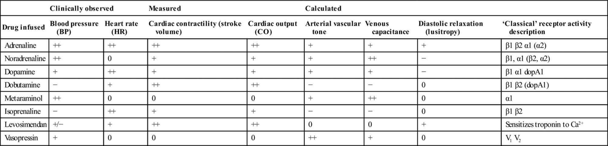
Administration
Norepinephrine, epinephrine (or salbutamol and isoprenaline) can be made up as 6 mg in 100 mL (or 3 mg in 50 mL) given by infusion pump into a central vein. The advantage of this particular dose dilution is that an infusion rate of 1 mL/h equates to 1 μg/min.
Dobutamine and dopamine are presented as 250 mg and 200 mg ampoules and may be made up as weight (kg)×6 mg in 100 mL or weight (kg)×3 mg in 50 mL to give a dose dilution where an infusion rate of 1 mL/h equates to 1 μg/kg/min.
Route
Vasopressors/inotropes may be administered into a large peripheral vein with fast-flowing crystalloid in an emergency. The clinical effect may be variable and thrombophlebitis can occur.
Dedicated lines and lumina without side injection ports should be used to avoid inadvertent boluses.
Placement of central venous lines (CVLs) or peripherally inserted central catheters (PICC) is performed under strict asepsis in an appropriate setting and may be required early in the ED for vasopressor/inotrope infusion.
Titration targets
The use of vasopressors/inotropes without adequate preload is associated with a worse outcome [31,32], so volume infusion should always precede their use, unless there is unequivocal evidence that the heart is ‘too full’. Even in cardiogenic shock, judicious boluses of fluid with close monitoring may result in improved cardiac output.
Add an effective inotrope if, despite ongoing rapid fluid volume resuscitation, cardiac output markers such as MAP are low (see ‘Goal-directed’ resuscitation) and titrate rapidly upwards until an effect is noted. Wean the inotrope/vasopressor as further volume infusion allows or evidence develops that the heart is over-full. Reassess frequently to judge whether further fluid is needed.
The upper level of the infusion is titrated to effect and only limited by the development of undesired side effects or a lack of therapeutic effect. Thus, any infusion is simply ‘titrated to desired effect and monitored for undesired effect’.
Complications
Side effects include excessive tachycardia, hypertension, tremor, anxiety and raised intracranial pressure (if monitored). Conversely, watch for disconnection or failure to infuse, when parameters unexpectedly fall.
Epinephrine causes metabolic effects including hyperglycaemia, hypokalaemia and lactic acidosis (usually clinically irrelevant).
Increased myocardial oxygen consumption can worsen myocardial ischaemia and precipitate cardiac arrhythmias.
Peripheral digit and skin infarction described in the past is likely due to endothelial injury from prolonged shock or the underlying primary cause (e.g. meningococcus), with no evidence that it was due to a vasoconstrictor effect.
Splanchnic or myocardial infarction also described is more likely to be due to inadequate resuscitation and hypotension rather than vasoconstriction, as these vessels are poorly reactive.
‘Too large, too concentrated or too rapid’ a bolus will cause severe hypertension and risks sequelae such as intracranial haemorrhage and myocardial damage.
3. Other interventions
Corticosteroids in shock should be reserved for adrenal insufficiency or if the patient is already receiving corticosteroids. There is no evidence to support their routine use in anaphylactic shock (see Chapter 2.8).
Corticosteroids such as hydrocortisone 200 mg per day may improve haemodynamic parameters in unresponsive septic shock, but two controlled multicentre trials found that corticosteroids have no effect on mortality [33,34]. Steroids are still recommended in patients with bacterial meningitis in high-income countries to reduce hearing loss and neurological sequelae, but they do not reduce overall mortality [35]. Some spinal injury centres recommend high-dose methylprednisolone started within 8 h of injury given for 24–48 hours in spinal cord injury, but data are unconvincing [36].
Military anti-shock trousers (MAST) or pneumatic anti-shock garments (PASG) increase morbidity and mortality and are no longer used [37].
Effects of shock on other interventions
Hypoperfusion of tissues will affect the delivery of drugs, particularly orally and subcutaneously administered drugs, and affects the pharmacokinetics with a reduced clearance of drugs. Unpredictable delivery and efficacy may require dose changes or use of alternate routes. Carefully titrated intravenous doses given centrally are advisable.
Sedative, analgesic and anaesthetic drugs, particularly thiopentone, midazolam, propofol and even ketamine (when the sympathetic ganglia are exhausted of catecholamine) have adverse effects on vascular tone and may worsen shock. These drugs also have a delayed circulation time and can appear not to be working, prompting inappropriate repeat dosing.
Catecholamines are less effective in severe acidosis states, hence the theoretical but unproven use for sodium bicarbonate in severe, resistant metabolic acidosis.
Endotracheal intubation and positive-pressure ventilation reduce venous return that may further reduce cardiac output and systolic blood pressure. Minimal initial tidal volume and positive end expiratory pressure (PEEP) settings ameliorate this effect. Physiological dead space may be increased by positive-pressure ventilation reducing lung perfusion, thus the arterial PaCO2 may rise. ‘Normalization’ of PaCO2may then lead to an apparent worsening of compensated metabolic acidosis.
Inotropes are arrhythmogenic and this complication is increased in the setting of hypokalaemia, acidosis and poorly perfused myocardium.
The stress response and some inotropes cause or exacerbate hyperglycaemia.
Infused fluids will eventually redistribute to all tissues and produce widespread oedema. An example is the burns victim who may have minimal airway burns, but after many litres of crystalloid may have a compromised oedematous airway.
Management of specific shock syndromes
The following shock syndromes are discussed briefly here and in other chapters:
Hypovolaemia (absolute)
Hypovolaemia (relative)
Neurogenic shock (see Chapter 3.3)
Anaphylactic shock (see Chapter 2.8)
Hypoadrenal shock (see Chapter 11.3)
Cardiogenic shock (see Chapter 5.2)
Septic shock (see Chapter 2.5).
Absolute hypovolaemia
Clinical features
The history and examination may point to fluid loss from vessels, gut, kidneys or evaporation. Bleeding needs to be excluded in all hypovolaemia (see Box 2.4.1). In addition to those described previously, clinical features will include signs of reduced preload, with flat neck veins as a consequence of a low central venous pressure.
Relevant investigations
If hypovolaemia is due to bleeding, haemostasis is the most effective intervention by direct surgical or specialist intervention and may parallel resuscitation and precede investigations. If initial resuscitation allows, investigations, such as formal ultrasound, computed tomography (CT) with contrast angiography, may identify the site of bleeding. Radiographic intervention, such as angiography with embolization, may be life saving in severe pelvic trauma.
Therapy
Initial resuscitation as described previously and ensure all efforts are made to avoid hypothermia.
Passive leg elevation is more effective in hypovolaemic shock than the Trendelenburg (head lower than the pelvis body position) in increasing left ventricular end-diastolic volume, stroke volume and cardiac output, but these effects are transient [6].
External haemorrhage is controlled with firm, direct manual pressure. Tourniquets are associated with morbidity, but may be useful in the short term [38].
Surgical consultation is required urgently. Efforts to return the systolic blood pressure to ‘normal’ in bleeding trauma patients may be counterproductive and occasionally harmful, particularly in penetrating truncal trauma. Surgical haemostasis must take priority and over-resuscitation should be avoided, adopting a ‘minimal-volume’ approach. Thus, patients with uncontrolled haemorrhage following penetrating truncal trauma, who are in close proximity to facilities capable of definitive care, should undergo minimal-volume or ‘hypotense’ fluid resuscitation pending prompt surgical intervention [39]. ‘Minimal volume’ is interpreted variously as fluid sufficient to keep the line open (TKVO) or small (250 mL) boluses titrated to a palpable radial pulse or conscious level, with the aim to ‘keep the brain and heart perfused’. Any minimal-volume approach is contraindicated when traumatic brain injury is associated with hypotension, as cerebral perfusion pressure is dependent on maintaining the MAP.
Infuse packed red cells in major blood loss where oxygen delivery is known to be impaired or Hb is less than 70 g/L. Recognition or anticipation of coagulopathy will need fresh frozen plasma and platelets as in a major transfusion protocol. Patients with lesser amounts of blood loss or controlled bleeding, or non-haemorrhagic hypovolaemic shock can be managed with warmed crystalloid [19].
Hypertonic saline 3% or 7% was considered to improve outcome in a subgroup of patients with shock and traumatic brain injury, but this remains unproven. Despite this, hypertonic saline has been recommended as the initial fluid of choice in haemorrhaging battlefield casualties [40].
There are no current definitive recommendations concerning the use of blood substitutes, such as modified haemoglobin or non-blood perfluorocarbons.
Other causes of impaired preload or contractility, such as tension pneumothorax, cardiac tamponade and myocardial contusion, must be considered in the hypotensive trauma patient. Increasing preload is still beneficial in these settings and all trauma patients should be assumed to be hypovolaemic until proven otherwise. Urgent bedside ultrasound is essential.
Relative hypovolaemia
This may be due to anaphylaxis, Addisonian crisis, neurogenic shock, septic shock or a drug or toxin effect.
Anaphylaxis
The mainstay of treatment in shock is the physiological antagonist epinephrine (adrenaline) plus oxygen and fluid, with the patient supine and the legs raised (see Chapter 2.8).
Adrenal shock
Hypotension due to hypoadrenalism is uncommon, but should be suspected in the acutely unwell patient with past or current steroid use or when hypotension occurs with relative polyuria or a relatively high urinary sodium>20 mmol/L (see Chapter 11.3).
Neurogenic shock
Neurogenic shock is manifested by the triad of hypotension, bradycardia and hypothermia in the setting of an acute spinal cord injury, related to the loss of sympathetic nerve tone. One in four patients with a complete cervical-cord injury may require haemodynamic support for their hypotension [41]. Other causes of hypovolaemia or shock in the trauma patient should still be actively sought, such as concealed bleeding, tension pneumothorax and cardiac tamponade (see Chapter 3.3).
Septic shock
Septic shock is sepsis accompanied by hypotension or hypoperfusion and can be underappreciated as the patient may have few signs of inadequate perfusion. Persistent hypotension and/or signs of organ hypoperfusion, despite ongoing rapid fluid resuscitation, are indications for early vasopressor/inotrope support. Vasopressin 0.04 units/min has no greater effect on survival [42]. A post-hoc analysis of the SAFE study suggests that albumin may have a survival advantage [19]. Hydrocortisone may improve unresponsive shock, as can high volume haemofiltration, but trials have found no overall effect on mortality [32–34](see Chapter 2.5).
Drug effects
Multiple drugs or toxins cause hypotension by impairing vascular or cardiac muscle contractility or permeability. Intervention with increased fluid or inotropes/pressors will manage hypotension. If the drug affects an inotrope/vasopressor receptor, either physiological antagonism or alternative receptor stimulation can overcome this effect. When the toxin is a metabolic or mitochondrial poison, the general principals of removal and support are used (see Chapter 29.2).
Cardiac causes of shock: cardiogenic shock
Cardiogenic shock is the inability of the heart to deliver sufficient blood to the tissues to meet resting metabolic demands and is clinically defined as a systolic blood pressure of<90 mmHg or MAP>30 mmHg below baseline for at least 30 minutes.
An alternative definition is a significant arteriovenous oxygen difference and a cardiac index of<2.2 L/min/m2 where pulmonary capillary wedge pressure is>15 mmHg. Failure to respond to correction of hypoxaemia, hypovolaemia, arrhythmias and acidosis is a requirement for the diagnosis [43]. There is clinical evidence of poor tissue perfusion in the form of oliguria, cyanosis and altered mentation.
Aetiology
The most common cause of cardiogenic shock is myocardial infarction (MI) or ischaemia. Cardiogenic shock complicates 5–8% of patients with acute myocardial infarction and has a mortality as high as 56–74%. It is the commonest cause of in-hospital death post- infarction [43,44]. Only 10% of these patients develop cardiogenic shock in the ED, but this subgroup has a higher mortality [44].
Other cardiac causes of shock include valvular rupture or degeneration, critical stenosis, septal or free wall rupture and atrial myxoma. Cardiac tamponade or a large pulmonary embolus are better considered as obstructive causes of shock, as the myocardial pump is unaffected initially.
Older patients with anterior MI, previous MI, diabetes, angina or congestive heart failure are at greatest risk of cardiogenic shock. There is a higher prevalence in patients with multivessel disease (e.g. diabetes) and involving the left main coronary artery [45]. Patients with persistent occlusion of the left anterior descending artery are at the highest risk of developing shock [46]. Only aggressive revascularization within 12 hours of symptoms improves outcome in these patients.
Pathophysiology
Activation of the sympathetic nervous and renin–angiotensin systems contributes to an increase in myocardial oxygen demand which causes an increase in infarct size and further decreases contractility, cardiac output and coronary perfusion pressure. Systolic dysfunction results in an increase in end-systolic volumes and reductions in ejection fraction, stroke volume and cardiac output. Diastolic dysfunction is also present. Pulmonary oedema exacerbates hypoxia and systemic tissue hypoperfusion and selective vascular redistribution leads to organ failure and metabolic acidosis.
Clinical features
Clinical signs in cardiogenic shock in addition to those described previously include:
Signs of excessive catecholamine outflow, such as tachycardia, pallor, poor capillary refill and evidence of low cardiac output with a decreased urine output and raised lactate.
Blood pressure that may initially remain within normal limits as a result of compensatory mechanisms, which also produce tachycardia and narrowed pulse pressure.
Classic signs of left heart failure with a third heart sound gallop rhythm and basal crackles from pulmonary oedema. A ‘gallop’ or additional heart sound suggests reduced ventricular compliance (fourth heart sound) and increased ventricular diastolic pressure (third heart sound).
Raised jugular venous pressure (JVP), hepatic congestion and peripheral oedema of right ventricular failure may occur secondary to left heart failure or alone in right ventricular infarction usually associated with an inferior myocardial infarction. This can be inferred by ST elevation in a right-sided V4 chest lead (V4R).
Loud murmur or thrill in systole may be due to mitral regurgitation or critical aortic stenosis and, rarely, rupture of the ventricular septum.
Investigations in cardiogenic shock
Twelve-lead ECG may define territory and need for reperfusion therapy. Adding leads V4R and V7–9 are indicated to rule out right ventricular and posterior myocardial infarction, respectively.
Troponin I (or T) levels.
Chest X-ray to show pulmonary oedema and an enlarged cardiac silhouette.
Bedside echocardiography (TTE) should be performed in any patient who remains with undiagnosed shock, as an extension of the physical examination. Pericardial effusion or cardiac tamponade are excluded and global systolic function, filling and regional wall motion abnormalities assessed.
Transoesophageal echocardiography (TOE) may additionally diagnose loculated cardiac tamponade, a haemodynamically significant pulmonary embolus and obscure valvular lesions.
ED therapy
Initial care and monitoring should be provided as described earlier, with management of the myocardial infarct according to local reperfusion policy. When cardiogenic shock is recognized, immediate discussion regarding revascularization should be made with a referral centre, particularly if TTE does not show a mechanical cause of shock [47].
Tracheal intubation and ventilation should be considered early for cardiac ‘respite’ and continuous positive airway pressure (CPAP) or non-invasive ventilation with bi-level positive airway pressure (BIPAP) in selected patients. Invasive blood pressure monitoring is recommended [47].
Arrhythmias considered contributory to the presence of cardiogenic shock should be treated according to standard ACLS principles.
Hypovolaemia must be sought and corrected in all patients with 250 mL aliquots of fluid given as a bolus and the response assessed. Volume loading to maintain higher right atrial filling pressures is important in inferior MI with right ventricular involvement, plus avoidance of drugs that reduce preload including nitrates, diuretics and excess opiates.
Relevant targets should include evidence of perfusion, such as urine output, lactate and clinical signs of improved skin perfusion and resolution of pulmonary oedema. Coronary autoregulation occurs at a MAP of 60 mmHg.
Persistence of the shock state following adequate fluid challenge in the presence of end-organ dysfunction is an indication for urgent revascularization. Intra-aortic balloon pump (IABP), extracorporeal membrane oxygenation (ECMO) and/or inotropic support may be considered as a bridge to this [45].
Early revascularization by either percutaneous coronary intervention (PCI) or coronary artery bypass graft (CABG) is recommended for patients less than 75 years old with ST elevation or new left bundle branch block (LBBB) who develop cardiogenic shock within 36 hours of acute MI and who are suitable for revascularization that can be performed within 18 hours of shock onset [47]. Early transfer and revascularization confers a survival advantage in patients with MI plus cardiogenic shock [43–45,47].
Initial therapy for patients who present to a facility without early primary PCI capability can include thrombolysis followed by urgent transfer [45,46].
IABP does not appear to reduce 12 month all-cause mortality in patients undergoing early revascularisation for myocardial infarction complicated by cardiogenic shock [48]. Intra-aortic balloon counterpulsation increases aortic root diastolic pressure (and hence coronary perfusion) and duration of apparent systole (and hence MAP), with no increase in oxygen demand. Complications include leg ischaemia, arterial dissection, thromboembolism and thrombocytopenia [49,50].
Use of inotropes and vasopressors in cardiogenic shock has not been shown to improve survival, but may be needed to maintain perfusion [47–49]. Dobutamine and levosimendan have inotropic and vasodilator effects, but are not recommended in hypotension. Norepinephrine (noradrenaline) is now the recommended inotrope/pressor [47], allowing the later introduction of a vasodilator. Dopamine was previously used, but the tachycardia limits its efficacy by increasing myocardial oxygen demand. There is no evidence for a reduction in mortality with the use of any of the newer inodilators, such as dopexamine, milrinone or levosimendan [30,49].
ECMO is being increasingly used as support for refractory cardiogenic shock with reasonable outcomes described, but no definitive trial exists [49,51].
Vasodilators can be considered when blood pressure has been restored but fails to improve peripheral end-organ perfusion. Glyceryl trinitrate or angiotensin-converting enzyme inhibitors (ACEI) can be given if titrated carefully, although precipitate hypotension may occur.
Consider referral for emergency cardiac transplantation in the younger patient.
Overall, those patients with large infarctions, a resting tachycardia and signs of poor tissue perfusion should be identified early and managed aggressively with cardiology advice. There should be early discussion with a cardiac referral centre and, if the patient is unstable or unsuitable for transfer, an IABP can be considered with a lower threshold for thrombolysis if not contraindicated. Inotropes are a temporizing measure.
Pericardial tamponade
Pericardial (cardiac) tamponade causes a failure of filling of the right atrium as a result of increased pericardial pressure. The right ventricle, and subsequently the left ventricle, has limited stroke volume and cardiac output, so tachycardia and raised peripheral resistance are compensatory mechanisms (see Chapter 5.6).
The presence of pericardial tamponade should also be suspected when there is unexplained shock with blunt or penetrating chest trauma, pericarditis, anticoagulant use or iatrogenic misadventure, e.g. CVP insertion.
Volume loading will raise right-sided filling pressures and volumes and tachycardia should be preserved. Vasopressor support will maintain MAP until surgical pericardiotomy (traumatic cause) or pericardiocentesis under echo guidance are performed.
Conclusion
The aetiology of shock in patients presenting to the ED is varied. Interventions in all forms of shock are simple and initially directed at the physiological deficit and should be seen as a test of the clinical hypothesis. Continuous reappraisal is required. Hypovolaemia should be sought in all cases, although further specific management will depend on the underlying cause(s).
References
1. Dutton RP. Current concepts in hemorrhagic shock. Anesthesiol Clin. 2007;25:23–34 viii.
2. Pickering TG, Hall JE, Appel LJ, et al. Recommendations for blood pressure measurement in humans and experimental animals Part 1: blood pressure measurement in humans: a statement for professionals from the Subcommittee of Professional and Public Education of the American Heart Association Council on High Blood Pressure Research. Circulation. 2005;111:697–716.
3. Fieselmann JF, Hendryx MS, Helms CM, Wakefield DS. Respiratory rate predicts cardiopulmonary arrest for internal medicine inpatients. J Gen Intern Med. 1993;8:354–360.
4. American College of Surgeons Committee on Trauma. Advanced Trauma Life Support Student Course Manual 9th ed. Chicago: ACS; 2012.
5. Jones AE, Aborn LS, Kline JA. Severity of emergency department hypotension predicts adverse hospital outcome. Shock. 2004;22:410–414.
6. Terai C, Anada H, Matsushima S, et al. Effects of Trendelenburg versus passive leg-raising autotransfusion in humans. Intens Care Med. 1996;22:613–614.
7. Bur A, Herkner H, Vlcek M, et al. Factors influencing the accuracy of oscillometric blood pressure measurement in critically ill patients. Crit Care Med. 2003;31:793–799.
8. Bakker J, Coffernils M, Leon M, et al. Blood lactate levels are superior to oxygen derived variables in predicting outcome in human septic shock. Chest. 1991;99:956–962.
9. Davis JW, Parks JN, Kaups KL, et al. Admission base deficit predicts transfusion requirements and risk of complication. J Trauma. 1996;41:769–774.
10. Lamia B, ChemLa D, Richard C, Teboul JL. Clinical review: interpretation of arterial pressure wave in shock states. Crit Care. 2005;9:601–606.
11. Tavernier B, Makhotine O, Lebuffe G, et al. Pressure variation as a guide to fluid therapy in patients with sepsis-induced hypotension. Anesthesiology. 1998;89:1313–1321.
12. Jin X, Weil MH, Tang W, et al. End-tidal carbon dioxide as a noninvasive indicator of cardiac index during circulatory shock. Crit Care Med. 2000;28:2415–2419.
13. McKinley BA, Kozar RA, Cocanour CS, et al. Normal versus supranormal oxygen delivery goals in shock resuscitation: the response is the same. J Trauma. 2002;53:825–832.
14. Kern JW, Shoemaker WC. Meta-analysis of hemodynamic optimization in high-risk patients. Crit Care Med. 2002;30:1686–1692.
15. Rivers E, Nguyen B, Havstad S, et al, the Early Goal-Directed Therapy Collaborative Group. Early goal-directed therapy in the treatment of severe sepsis and septic shock. N Engl J Med. 2001;345:1368–1377.
16. American Heart Association Guidelines for Cardiopulmonary Resuscitation and Emergency Cardiovascular Care 2005. Circulation 2005; 112.
17. Waters JH, Gottlieb A, Schoenwald P, et al. Normal saline versus lactated Ringer’s solution for intraoperative fluid management in patients undergoing abdominal aortic aneurysm repair: an outcome study. Anesth Analges. 2001;93:817–822.
18. Finfer S, Bellomo R, Boyce N, et al. A comparison of albumin and saline for fluid resuscitation in the intensive care unit. N Engl J Med. 2004;350:2247.
19. Bunn F, Trivedi D. Colloid solutions for fluid resuscitation. Cochrane Database Syst Rev. 2012;7:CD001319.
20. Myburgh JA, Finfer S, Bellomo R, et al. CHEST Investigators; Australian and New Zealand Intensive Care Society Clinical Trials Group Hydroxyethyl starch or saline for fluid resuscitation in intensive care. N Engl J Med. 2012;367:1901–1911.
21. Hebert PC, Wells G, Blajchman MA, et al. A multicenter, randomized, controlled clinical trial of transfusion requirements in critical care. N Engl J Med. 1999;340:409–417.
22. Australian and New Zealand Society of Blood Transfusion. Guidelines for the administration of blood component products, 2nd ed., 2011.<http://www.anzsbt.org.au/publications/documents/ANZSBT_Guidelines_Administration_Blood_Products_2ndEd_Dec_2011_Hyperlinks.pdf>(Accessed Mar. 2013).
23. Becton Dickinson product information. Becton Dickinson Pty Ltd, Eight Mile Plains, QLD 4113.
24. Mendenhall ML, Spain DA. Venous air embolism and pressure infusion devices. J Trauma. 2007;63:246.
25. Dellinger R, Levy M, Rhodes A, et al. Surviving sepsis campaign: international guidelines for management of severe sepsis and septic shock. Crit Care Med. 2013;41:580–637.
26. Myburgh JA. An appraisal of selection and use of catecholamines in septic shock – old becomes new again. Crit Care Resus. 2006;3:353–360.
27. Havel C, Arrich J, Losert H, et al. Vasopressors for hypotensive shock. Cochrane Database Syst Rev. 2011;5:CD003709.
28. Sakr Y, Reinhart K, Vincent JL, et al. Does dopamine administration in shock influence outcome? Results of the Sepsis Occurrence in Acutely Ill Patients (SOAP) study. Crit Care Med. 2006;34:589.
29. Bellomo R, Chapman M, Finfer S, et al. Low-dose dopamine in patients with early renal dysfunction: a placebo-controlled randomized trial Australian and New Zealand Intensive Care Society (ANZICS) Clinical Trials Group. Lancet. 2000;356:2139–2143.
30. Mebazaa A, Nieminen M, Packer M, et al. Levosimendan vs dobutamine for patients with acute decompensated heart failure: the SURVIVE Randomised Trial. J Am Med Assoc. 2007;297:1883–1891.
31. Beale RJ, Hollenberg SM, Vincent JL, et al. Vasopressor and inotropic support in septic shock: an evidence-based review. Crit Care Med. 2004;32:S455–S465.
32. Nordin AJ, Makisalo H, Hockerstedt KA. Failure of dobutamine to improve liver oxygenation during resuscitation with a crystalloid solution after experimental haemorrhagic shock. Eur J Surg. 1996;162:973.
33. Annane D, Sebille V, Charpenteir C, et al. Effect of treatment with low doses of hydrocortisone and fludrocortisone on mortality in patients with septic shock. J Am Med Assoc. 2002;288:862–871.
34. Lipiner-Friedman D, Sprung CL, Laterre PF, et al, Corticus Study Group. Adrenal function in sepsis: the retrospective Corticus cohort study. Crit Care Med. 2007;35:1012–1018.
35. Brouwer MC, McIntyre P, de Gans J, et al. Corticosteroids for acute bacterial meningitis. Cochrane Database Syst Rev. 2010;9:CD004405.
36. Bracken MB. Steroids for acute spinal cord injury. Cochrane Database Syst Rev. 2012;1:CD001046.
37. Roberts I, Blackhall K, Dickinson K. Medical anti-shock trousers (pneumatic anti-shock garments) for circulatory support in patients with trauma. Cochrane Database Syst Rev. 1999;4:CD001856.
38. Lee C, Porter KM, Hodgetts TJ. Tourniquet use in the civilian prehospital setting. Emerg Med J. 2007;24:584–587.
39. Bickell WH, Wall Jr MJ, Pepe PE, Martin RR. Immediate versus delayed fluid resuscitation for hypotensive patients with penetrating torso injuries. N Engl J Med. 1994;331:1105–1109.
40. Alam HB, Rhee P. New developments in fluid resuscitation. Surg Clin N Am. 2007;87:55–72 vi.
41. Guly HR, Bouamra O, Lecky FE. on behalf of the Trauma Audit and Research Network The incidence of neurogenic shock in patients with isolated spinal cord injury in the emergency department. Resuscitation. 2007;76:57–62.
42. Levy MM, Fink MP, Marshall JC, et al. 2001 SCCM/ESICM/ACCP/ATS/SIS International Sepsis Definitions Conference. Crit Care Med 2003; 31:1250–1256.
43. Reynolds H, Hochman J. Cardiogenic shock: current concepts and improving outcomes. Circulation. 2008;117:686–697.
44. Webb JG, Sleeper LA, Buller CE, et al. Implications of the timing of onset of cardiogenic shock after acute myocardial infarction: a report from the SHOCK Trial Registry Should we emergently revascularize occluded coronaries for cardiogenic shock? J Am Coll Cardiol. 2000;36:1084.
45. Goldberg RJ, Gore JM, Thompson CA, et al. Recent magnitude of and temporal trends (1994–1997) in the incidence and hospital death rates of cardiogenic shock complicating acute myocardial infarction: The second National Registry of Myocardial Infarction. Am Heart J. 2001;141:65.
46. Wong SC, Sanborn T, Sleeper LA, et al. Angiographic findings and clinical correlates in patients with cardiogenic shock complicating acute myocardial infarction: a report from the SHOCK Trial Registry SHould we emergently revascularize Occluded Coronaries for cardiogenic shocK? J Am Coll Cardiol. 2000;36:1077.
47. Antman EM, et al. ACC/AHA Guidelines for management of patients with ST elevation MI: a report of the American College of Cardiology/American Heart Association Task Force on Practice Guidelines. Circulation. 2004;110:e82–e29251.
48. Thiele H, Zeymer U, Neumann F, et al. Intra-aortic balloon counterpulsation in acute myocardial infarction complicated by cardiogenic shock (IABP-SHOCK II). Lancet. 2013;382:1638–1645.
49. Sayer GT, Baker JN, Parks KA. Heart rescue: the role of mechanical circulatory support in the management of severe refractory cardiogenic shock. Curr Opin Crit Care. 2012;18:409–416.
50. Prieto A, Eisenberg J, Thakar RK. Non-arrhythmic complications of acute myocardial infarction. Emerg Med Clin N Am. 2001;19:397–415.
51. Tsao NW, Shih CM, Yeh JS, et al. Extracorporeal membrane oxygenation assisted primary percutaneous coronary intervention may improve survival of acute myocardial infarction complicated by profound cardiogenic shock. J Crit Care. 2012;27(530):e1–11.
2.5 Sepsis and septic shock
Anna Holdgate
Essentials
1 Early recognition and intervention in the emergency department (ED) reduces mortality in patients with sepsis and septic shock.
2 Appropriate broad-spectrum antibiotics should be administered within 1 hour of the recognition of sepsis.
3 Aggressive haemodynamic resuscitation with fluids, vasopressors and inotropes should begin as soon as possible.
4 Systemic blood pressure, serum lactate levels and urine output should be monitored closely to determine the effectiveness of treatment.
Introduction
Septic shock is the extreme end of the spectrum of septic syndromes. Globally, septic shock is associated with a mortality rate of up to 46%. In Australia and New Zealand, the reported mortality is substantially lower (27.6%) for septic patients admitted from the emergency department (ED) to intensive care (ICU) [1]. Each year more than 1500 septic patients are admitted to Australasian ICUs from the ED and this incidence has been steadily rising over the past decade [1]. Optimal, time-critical care of the septic patient in ED is crucial, as early intervention in several areas has been shown to reduce mortality.
Aetiology and pathophysiology
Approximately 95% of identified causative organisms are bacterial, with Gram-positive organisms (mostly Staphylococcus aureus, coagulase-negative staphylococci, enterococci and streptococci) now slightly more common than Gram-negative species (particularly Escherichia coli, Klebsiella pneumoniae and Pseudomonas aeruginosa). The remaining 5% are caused by fungi, mostly Candida, with the incidence of fungal sepsis increasing threefold in the last 20 years [2].
Pathogens are identified from blood or other tissue cultures in approximately 70% of patients. The primary sources of infection are respiratory, genitourinary, intra-abdominal, skin/other soft tissue and primary bacteraemia [3].
Pathogenic mechanisms
The pathogenic mechanisms in sepsis are initiated by a variety of host responses to the infecting organism. Pattern recognition receptors in the cell membrane (transmembrane toll-like receptors, TLRs) and in the cytoplasm (NOD-like receptors, NLRs) are responsible for initiating the immune response following recognition of an invading pathogen.
Inflammatory mediators
Inflammatory mediators, such as tumour necrosis factor α (TNF-α) and the interleukins, are produced by the host, resulting in activation of neutrophils, direct injury to the endothelium with increased vascular permeability and release of nitric oxide (NO) that results in vasodilatation. Modification of the coagulation cascade causes an increase in procoagulant factors and lower levels of the anticoagulant factors protein C, protein S and antithrombin III. These proinflammatory and procoagulant responses lead to reduced vascular resistance, relative hypovolaemia, loss of vasoregulatory control in microvascular beds, reduced myocardial contractility, acute lung injury and renal dysfunction.
These changes further impair oxygen delivery and consumption at a tissue level, resulting in tissue hypoxia and worsening organ dysfunction. Anaerobic metabolism causes a rising lactate when oxygen delivery cannot meet tissue oxygen demands. Central venous oxygen saturations (SCVO2) will generally be low (<70%) as the peripheral tissues extract a higher percentage of oxygen, resulting in less oxygen in venous blood returning to the central circulation [4,5].
The progression of sepsis to septic shock is associated with an inability to contain the infection, owing to (either or both) compromised patient immunity or characteristics of the infection itself, such as highly virulent organisms, a high burden of infection and antibiotic resistance.
Clinical features
Infection associated with systemic illness results in a spectrum of clinical syndromes based on clinical signs. Much broader definitions released in 2013 are now in use [6].
Clinical syndrome definitions
Systemic manifestations of infection
The systemic manifestations of infection have been vastly expanded and replace those features that were previously used to define the systemic inflammatory response syndrome (SIRS). These originally included two or more of the following:
abnormal body temperature (>38°C or<36°C)
tachycardia>90 bpm
tachypnoea (respiratory rate>20/min or PaCO2<32 mmHg)
abnormal white cell count (>12 000/μL or<4000/μL or>10% immature (band) cells).
Sepsis
Sepsis is defined as the presence (probable or documented) of infection together with systemic manifestations of infection (blood cultures do not need to be positive).
Severe sepsis
Severe sepsis is defined as sepsis plus sepsis-induced organ dysfunction or tissue hypoperfusion. Sepsis-induced tissue hypoperfusion is defined as infection-induced hypotension, elevated lactate, or oliguria. Sepsis-induced hypotension is defined as a systolic blood pressure (SBP)<90 mmHg or mean arterial pressure (MAP)<70 mmHg or a SBP decrease>40 mmHg, or less than two standard deviations below normal for age in the absence of other causes of hypotension.
Septic shock
Septic shock is defined as sepsis-induced hypotension persisting despite adequate fluid resuscitation.
History
Important components of the clinical history include the patient’s immune status, prior infections and/or antibiotic use, co-morbid disease, such as diabetes or malignancy, travel or exposure history and an assessment of acute respiratory, abdominal or urinary symptoms, plus identification of potential sources for infection, such as recent procedures or prosthetic devices including stents and indwelling catheters.
Physical examination
Look for the systemic manifestations of infection, as well as for sites of infection, plus evidence of organ dysfunction, or hypoperfusion, such as an altered mental status, an episode of hypotension, tachycardia, tachypnoea from metabolic acidosis, hypoxia, pneumonia or acute respiratory distress syndrome (ARDS) or oliguria.
Include a top-to-toe assessment including the head and neck, oropharynx, skin, chest, abdomen, pelvis and perineum, back, limbs and joints.
A septic patient is relatively hypovolaemic due to peripheral vasodilatation in addition to fluid depletion from vomiting and third-space sequestration. The patient with severe sepsis may have warm peripheries and a bounding pulse early due to mediator-driven vasodilatation though, in the later stages, they are more usually hypothermic and peripherally shut down as a result of cardiovascular collapse and/or vasoconstriction.
Clinical investigations
Investigations are important in determining the nature of the condition as SIRS features occur in non-infectious conditions, such as post-trauma or surgery, burns, pancreatitis and other shock states, as well as confirming the aetiology and severity of the underlying infection.
Laboratory
Basic blood pathology helps identify potential causes, such as biliary obstruction, and will quantify end-organ dysfunction, such as renal failure, hypo-/hyperglycaemia and coagulopathy. Arterial blood gases should be measured early to assess both the adequacy of ventilation and the degree of lactic acidosis. A venous blood gas is equally useful for lactate estimation.
Source
A search for the underlying source should include two sets of paired blood cultures peripherally and a set from any indwelling line(s), urine microscopy and culture, chest X-ray, culture of any open wound and aspiration of any superficial collections.
In the absence of a clearly identified focus, abdominal computed tomography (CT) scanning and, particularly if there is an altered mental state, CT brain and lumbar puncture are usual, unless contraindicated by the patient’s clinical status.
Risk stratification
Factors associated with an increased mortality include the initial lactate level, age and multiple co-morbidities. Elevated lactate levels are associated with a higher mortality independent of other signs of shock and organ dysfunction and are therefore useful in early risk stratification [7,8]. The elderly have a higher mortality, in part related to greater co-morbidity. However, age alone is an independent risk factor, as the elderly are more likely to present with seriously deranged physiological parameters. While fever is more commonly associated with sepsis, hypothermia is a worrying sign associated with a higher morbidity, particularly in the elderly [9].
Treatment
The principles of treatment in sepsis are haemodynamic resuscitation, supportive measures to maximize tissue oxygen delivery, early antibiotic therapy and source control (see also Chapter 2.4). The International Surviving Sepsis Campaign Consensus Guidelines were developed to promote a more uniform ‘bundle of care’ for the acute management of patients with sepsis, aimed at reducing mortality [10]. These guidelines incorporate the concept of early goal-directed therapy (EGDT), with specific targeted endpoints to guide sequential treatment.
The use of standardized ED guidelines focused on haemodynamic resuscitation and early, appropriate antibiotic therapy improves compliance with recommended treatment and reduces mortality [11,12].
Haemodynamic resuscitation and supportive care
Early goal-directed therapy
The components of EGDT are adequate volume replacement followed by vasopressor and inotropic therapy aimed at maintaining MAP≥65 mmHg, urine output≥0.5 mL/kg/h and, in some settings, SCVO2≥70% [10]. Whereas measurement of MAP and urine output is straightforward, measurement of SCVO2 requires either frequent blood gas sampling from a standard central venous catheter or continuous measurement using a commercial central venous catheter with a specialized fibreoptic module.
Fluid resuscitation
Fluid resuscitation begins with a minimum of 30 mL/kg of crystalloid, such as normal saline. Patients who remain hypotensive, acidotic or oliguric usually warrant central venous pressure (CVP) and invasive arterial pressure monitoring to guide further therapy [10]. Fluid resuscitation should continue to a CVP of 8–12 mmHg in the absence of pulmonary oedema.
Vasopressor therapy
Vasopressor therapy is indicated concurrently with fluid resuscitation in the presence of profound hypotension or if fluid resuscitation fails to restore tissue perfusion (as indicated by normalization of MAP to at least 65 mmHg, lactate levels and urine output). Norepinephrine (noradrenaline) is commonly used first choice due to its potent α effects that increase blood pressure predominantly by direct vasoconstriction and more moderate β effects associated with some increase in heart rate and stroke volume. Thus, it is a preferred agent in patients with profound hypotension [10].
Epinephrine (adrenaline) can be used as an alternative vasopressor, although it may be associated with a higher incidence of arrhythmias, splanchnic ischaemia and metabolic effects than norepinephrine [13,14]. All vasopressor agents need to be administered via a central venous catheter and the infusion rate titrated to MAP measured by an intra-arterial catheter, urine output and cerebral perfusion.
Measurement of SCVO2
The measurement of SCVO2 is advocated as a further endpoint to assess tissue perfusion and guide ongoing therapy. In the seminal 2001 EGDT study by Rivers et al., red cell transfusion to a haemocrit≥30% and dobutamine infusion to improve cardiac output were used in patients who failed to achieve SCVO2≥70% with fluids, vasopressors and ventilatory support [12].
Patients who received EGDT had a lower mortality than patients receiving ‘standard’ therapy. An alternative method of measuring improvement in tissue perfusion is to monitor the lactate level, aiming for a reduction of at least 10% which achieves a similar in-hospital mortality and does not require the insertion of an SCVO2-monitoring catheter [15].
Reported mortality rates for sepsis in Australasian patients are substantially lower than in the Rivers’ study, hence the applicability of Rivers’ EGDT in the Australasian setting is unclear [1]. International guidelines currently recommend the use of adjuvant dobutamine in patients with a low cardiac output despite fluids and vasopressors. Blood transfusion is recommended only to a target haemoglobin level of 7–9 g/dL, except in patients with acute haemorrhage or significant coronary artery disease [10].
Maximizing oxygen delivery
As sepsis is associated with increased oxygen consumption, oxygen delivery must be optimized at the beginning of resuscitation. Endotracheal intubation and mechanical ventilation with appropriate sedation and paralysis minimize oxygen consumption and should be considered early in patients with respiratory acidosis, hypoxia or persistent haemodynamic compromise. Low tidal volume ventilation (6 mL/kg) with peak inspiratory pressures maintained≤30 cm H2O are recommended to minimize further acute lung injury [10].
Antibiotic therapy
The early administration of appropriate antibiotic therapy in the ED is essential to the effective management of the septic patient. Time to appropriate antibiotic administration is one of the prime determinants of survival in severe sepsis. Patients with severe sepsis or septic shock who do not receive antibiotics within 1 hour have an increase in mortality of approximately 7.6% for each additional hour of delay [3,16]. Thus, a patient with severe sepsis should have appropriate cultures collected and antibiotics started within 1 hour of arrival. Collection of cultures or imaging studies must not delay the administration of antibiotics [10].
Choice of antibiotic
The choice of antibiotic depends on a number of criteria, including the likely source of infection, local bacterial sensitivities and patient factors, such as allergy, immunocompetence and renal function. The initial choice should be broad enough to cover a range of potential pathogens, as treatment with ineffective antibiotics is also associated with increased mortality [17,18]. Antibiotic therapy can be more specifically targeted once the causative organism and its sensitivities are known, but this is rarely appropriate in the ED [19]. Table 2.5.1 outlines one approach to initial antibiotic choice.
Table 2.5.1
Empirical initial intravenous antibiotic recommendations based on likely source of infection in a patient with severe sepsis
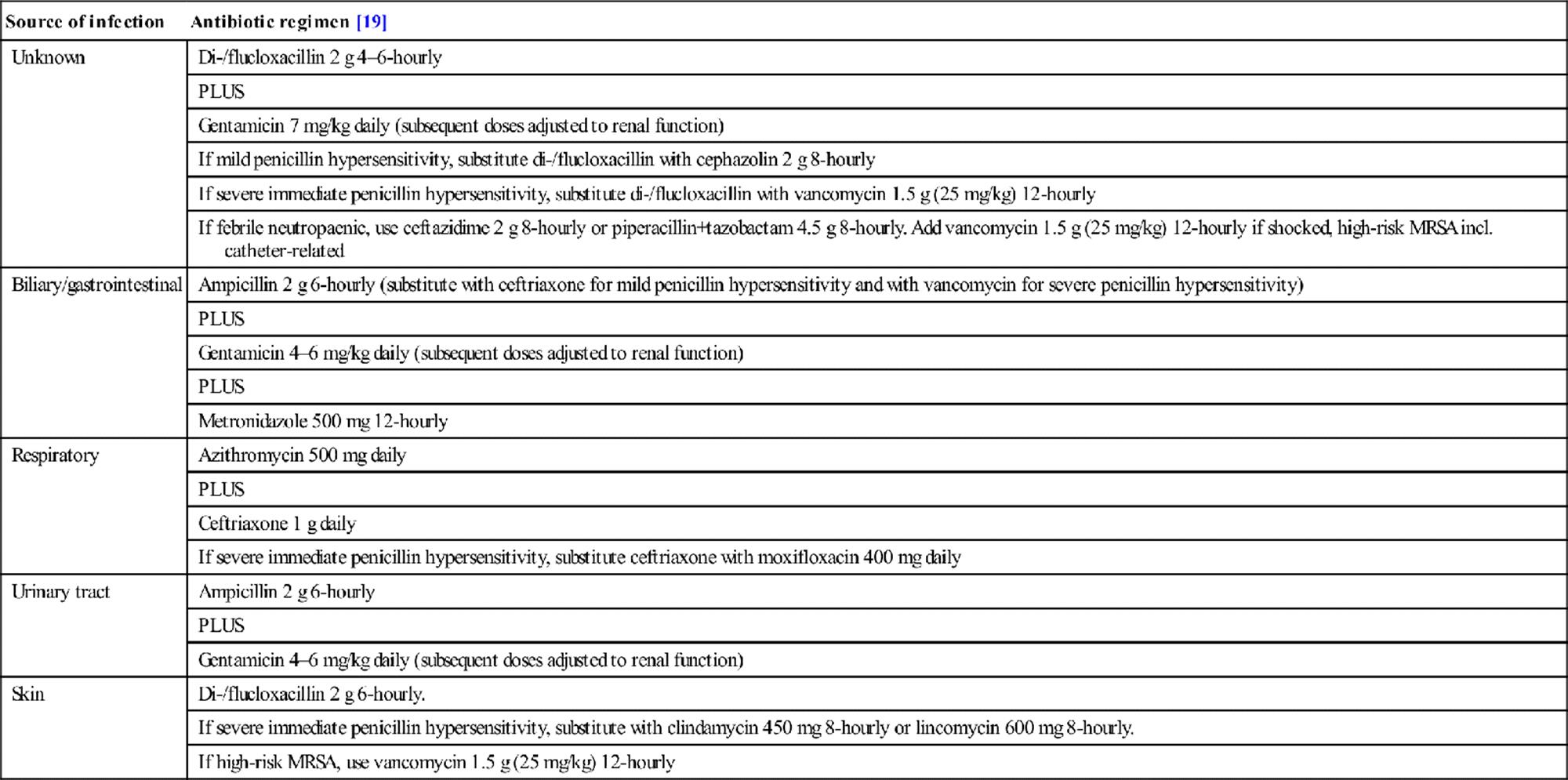
Empiric monotherapy with a third- or fourth-generation cephalosporin has been shown to be as effective as dual therapy with a β-lactam and an aminoglycoside. However, most of the evidence is derived from patients with febrile neutropaenia and only a few small studies have been conducted in immunocompetent patients with severe sepsis or septic shock. Many practitioners still prefer dual therapy on the basis that this may confer synergistic effects that enhance antibacterial activity and reduce the incidence of bacterial resistance [18]. These potential benefits must be weighed against the increased risk of nephrotoxicity and ototoxicity associated with the use of aminoglycosides [2].
Empirical treatment with glycopeptides, such as vancomycin, is recommended in patients with known methicillin resistant S. aureus (MRSA) colonization, severe penicillin hypersensitivity, high prevalence of community-associated MRSA (CA-MRSA), suspected line sepsis or in institutions with high levels of MRSA [2,18].
Empirical antifungal treatment with amphotericin or fluconazole is recommended in patients at high risk for invasive candidiasis, such as those who have been treated with prolonged broad-spectrum antibiotics, are immunosuppressed and have had Candida isolated from multiple sites [20].
Source control
Source control refers to physical measures to control or contain the focus of infection by drainage, debridement or anatomical repair. In principle, the removal of an infected nidus will help minimize the inflammatory response, but the size and site of the infective source will determine the feasibility and timing of source control. The focus in the ED is on identifying the likely source of infection and determining, in consultation with radiological, surgical and other specialties, the best method of drainage or containment.
Percutaneous drainage
Localized collections may be amenable to percutaneous drainage with or without radiological guidance. This is appropriate for renal, hepatic and other intra-abdominal abscesses and soft tissue collections. Immediate debridement of infected and necrotic tissue is mandatory for soft tissue infections, such as necrotizing fasciitis, but more deep-seated necrosis, such as pancreatitis, may require delayed debridement as early operative intervention in difficult-to-access areas is associated with significant morbidity [21,22].
Biliary tract obstruction with associated infection requires early decompression with percutaneous cholecystostomy or endoscopic retrograde cholangiopancreatography (ERCP). Urinary sepsis with shock in association with ureteric obstruction should be managed by urgent percutaneous nephrostomy. Gastrointestinal perforation with leakage of luminal contents usually requires early surgical repair, except for contained perforations in diverticulitis.
Indwelling devices
The best approach for sepsis associated with indwelling devices is removal of the device, but this needs to be balanced against the risks of removal and the ongoing medical need [9,22]. An infected intravascular device can be exchanged by ‘rewiring’ over a guidewire, provided there are no signs of infection at the insertion site [23]. Alternatively, a combination of antibiotics and thrombolysis may be effective [22].
Other therapies
Patients who are on long-term steroids should receive 100 mg hydrocortisone IV as soon as possible in their ED care. Otherwise, the role of steroids remains controversial; intravenous hydrocortisone 200 mg per day is suggested in patients with unresponsive hypotension [10,24].
There is no evidence to support using activated protein C (APC) for treating patients with severe sepsis or septic shock. Additionally, APC is associated with a higher risk of bleeding [10].
Following initial stabilization, glycaemic control is indicated if the blood sugar level (BSL) is greater than 10 mmol/L, aiming for a BSL of 8–10 mmol/L. Aiming for tighter BSL targets has been shown to increase the risk of hypoglycaemia with no mortality benefit [25].
Pragmatically, most of these interventions are usually administered within the first 24 hours of care in an intensive care unit, rather than in the first few hours of ED management [10,26].
Controversies
Efficacy of early goal-directed therapy has not been established in the Australasian environment where mortality rates are relatively lower than international figures. This is currently under investigation in the Australasian Resuscitation in Sepsis Evaluation Randomized Controlled Trial (ARISE-RCT).
Routine measurement of central venous oxygen saturations as an endpoint for goal-directed therapy is currently not widely used in Australia or New Zealand. Measurement of lactate clearance may be an acceptable alternative.
Use of dobutamine and liberal blood transfusion to attain designated endpoints of goal-directed therapy are not universally accepted. There is evidence in some critically ill patients that these strategies might be potentially harmful.
References
1. ARISE: ANZICS APD management committee. The outcome of patients with sepsis and septic shock presenting to emergency departments in Australia and New Zealand. Crit Care Resus. 2007;9:8–18.
2. Bochud P-Y, Bonten M, Marchetti O, et al. Antimicrobial therapy for patients with severe sepsis and septic shock: An evidence-based review. Crit Care Med. 2004;32:S495–S512.
3. Gaieski DF, Pines JM, Band RA, et al. Impact of time to antibiotics on survival in patients with severe sepsis or septic shock in whom early goal-directed therapy was initiated in the emergency department. Crit Care Med. 2010;38:1045–1053.
4. Wiersinga WJ. Current insights in sepsis: from pathogenesis to new treatment targets. Curr Opin Crit Care. 2011;17:480–486.
5. Rivers E, McIntyre L, Morro D, Rivers K. Early and innovative interventions for severe sepsis and septic shock: taking advantage of a window of opportunity. Can Med Ass J. 2005;173:1054–1065.
6. Dellinger R, Levy M, Rhodes A et al. Surviving sepsis campaign: international guidelines for management of severe sepsis and septic shock: 2012. Crit Care Med 2013;41:580–637.
7. Mikkelsen ME, Miltiades AN, Gaieski DF, et al. Serum lactate is associated with mortality in severe sepsis independent of organ failure and shock. Crit Care Med. 2009;37:1670–1677.
8. Shapiro N, Howell M, Talmor D, et al. Serum lactate as a predictor of mortality in emergency department patients with infection. Ann Emerg Med. 2005;45:524–528.
9. Tiruvoipati R, Ong K, Gangopadhyay H, et al. Hypothermia predicts mortality in critically ill elderly patients with sepsis. BMC Geriatrics. 2010;10:70.
10. Levy MM, Dellinger RP, Townsend SR, et al. The surviving sepsis campaign: results of an international guideline-based performance improvement program targeting severe sepsis. Intens Care Med. 2010;36:222–231.
11. Marti-Carvajal AJ, Sola I, Lathyris D, Cardona AF. Human recombinant activated protein C for severe sepsis. Cochrane Database Syst Rev. 2012;3:CD004388.
12. Rivers E, Nguyen B, Havstad S, et al. Early goal directed therapy in the treatment of severe sepsis and septic shock. N Engl J Med. 2001;345:1368–1377.
13. Myburgh JA, Higgins A, Jovanoska A, et al. A comparison of epinephrine and norepinephrine in critically ill patients. Intens Care Med. 2008;34:2226–2234.
14. De Backer D, Biston P, Devriendt J, et al. Comparison of dopamine and norepinephrine in the treatment of shock. N Engl J Med. 2010;362:779–789.
15. Jones AE, Shapiro NI, Trzeciak, et al. Lactate clearance vs central venous oxygen saturation as goals of early sepsis therapy. J Am Med Assoc. 2010;303:739–746.
16. Kumar A, Roberts D, Wood K, et al. Duration of hypotension before initiation of effective antimicrobial therapy is the critical determinant of survival in human septic shock. Crit Care Med. 2006;34:1589–1596.
17. MacArthur R, Miller M, Albertson T, et al. Adequacy of early empiric antibiotic treatment and survival in severe sepsis: experience from the MONARCS trial. Clin Infect Dis. 2004;38:284–288.
18. Kumar A. Optimizing antimicrobial therapy in sepsis and septic shock. Crit Care Clin. 2009;25:733–751.
19. Antibiotic guidelines. In: eTG complete [Internet]. Melbourne: Therapeutic Guidelines Limited; 2012.<http://www.tg.com.au>(Accessed Mar. 2013).
20. Rex J, Walsh T, Sobel J, et al. Practice guidelines for the treatment of candidiasis. Clin Infect Dis. 2000;30:662–678.
21. Hsaio G, Chang C, Hsaio C, et al. Necrotizing soft tissue infections Surgical or conservative treatment? Dermatol Surg. 1998;24:247–248.
22. Marshall J, Maier R, Jiminez M, Dellinger E. Source control in the management of severe sepsis and septic shock: an evidence-based review. Crit Care Med. 2004;32:S513–S526.
23. Cook D, Randolph A, Kernerman P, et al. Central venous catheter replacement strategies: a systematic review of the literature. Crit Care Med. 1997;25:1417–1424.
24. Annane D, Bellissant E, Bollaert P-E, et al. Corticosteroids in the treatment of severe sepsis and septic shock in adults: a systematic review. J Am Med Assoc. 2009;301:236–237.
25. Griesdale DEG, de Souza RJ, van Dam RM, et al. Intensive insulin therapy and mortality among critically ill patients: a meta-analysis including NICE-SUGAR data. Can Med Assoc J. 2009;180:821–827.
26. Osborn T, Nguyen H, Rivers E. Emergency medicine and the Surviving Sepsis Campaign: an internation approach to managing severe sepsis and septic shock. Ann Emerg Med. 2005;46:228–231.
2.6 Arterial blood gases
Anthony D Holley
Essentials
1 The value of arterial blood gas analysis is almost entirely dependent on understanding and correctly interpreting the results in the clinical context.
2 When abnormalities are detected with arterial blood gas analysis, ensure the sample was obtained, transported and analysed appropriately.
3 An arterial blood gas result assists in the assessment of a patient’s gas exchange, ventilatory control and acid–base balance.
4 Common sampling sites include the radial, femoral, brachial, dorsalis pedis or axillary artery. There is no evidence for superiority of any particular site.
5 The alveolar gas equation allows comparison of arterial and alveolar partial pressures of oxygen (PaO2). A higher than expected value indicates a ventilation–perfusion defect (high A–a gradient).
6 Isolated hypoxaemia is referred to as type I respiratory failure; type II respiratory failure is characterized by a partial pressure carbon dioxide (PaCO2) higher than 50 mmHg.
7 There are five potential pathophysiological mechanisms responsible for hypoxaemia, which include decreased inspired fractional oxygen, impaired diffusion, shunting, ventilation–perfusion (V/Q) mismatch and hypoventilaton.
8 Type II respiratory failure (hypercapnia) is due to inadequate alveolar ventilation, commonly secondary to poor central drive, neuromuscular disease or profound mechanical derangement of lungs or chest wall.
9 The primary acid–base disturbance is established by assessing the relationship between the direction of change in the pH and the direction of change in the PaCO2.
10 When a metabolic acidosis is diagnosed, calculate the anion gap and delta ratio to narrow the differential diagnosis.
11 In the presence of a metabolic alkalosis, establish both an initiating and maintaining factor.
12 Venous pH, bicarbonate and base excess have sufficient agreement to be clinically interchangeable with arterial values in patients who are not shocked.
Introduction
Arterial blood gas analysis is an essential tool for diagnosing and managing the critically ill emergency department (ED) patient’s respiratory status and acid–base balance (Table 2.6.1). Its usefulness is almost entirely dependent on the ability to understand and interpret the results correctly.
Table 2.6.1
Indications for arterial blood gas analysis

Technical aspects of arterial blood gas analysis
The technology for arterial blood gas analysis became available more than 50 years ago with the development of electrodes that allowed the measurement of partial pressures of oxygen (PaO2) and carbon dioxide (PaCO2) in arterial blood samples taken directly from the patient [1].
Blood gas analysers and applied physiology
Clarke’s oxygen electrode constitutes a platinum probe suspended in an electrolyte solution and separated from the blood sample by a membrane permeable to oxygen. Oxygen molecules diffuse from blood through the membrane to the electrode where they are reduced to hydroxyl ions. The partial pressure of oxygen is directly proportional to the current measured from this reduction reaction [2].
A pH-sensitive glass probe maintained in a bicarbonate solution and protected by a carbon dioxide permeable membrane constitutes the Severinghaus carbon dioxide electrode. In this model, the measured PaCO2is proportional to the hydrogen ions produced, as CO2 reacts with water to form hydrogen and bicarbonate ions [1].
The pH of arterial blood is measured directly by an electrode which then allows the blood gas analyser software to calculate the base excess (BE) and bicarbonate concentration.
The arterial oxygen saturation (SaO2) may be calculated from the PaO2. However, it may still be unreliable even if the haemoglobin–oxygen affinity shifts secondary to acid–base disturbances are accounted for in calculating SaO2 from PaO2. Most modern blood gas analysers now include a co-oximeter that is capable of measuring concentrations of saturated haemoglobin, reduced haemoglobin, carboxyhaemoglobin, and methaemoglobin [3]. Wavelengths of light corresponding to unique absorption spectra for each haemoglobin species allow for these measurements to be determined.
Temperature formulae and nomograms were previously applied to pH, PaCO2 and PaO2. However, modern automated blood gas analysers can report the pH, PaO2 and PaCO2 at either 37°C (the temperature at which the values are measured by the blood gas analyser) or at the patient’s body temperature. Most machines report the values of pH, PCO2 and PaO2 at 37°C, regardless of the patient’s actual temperature. The corrections are generally minimal and corrected values are no more clinically useful than 37°C values.
Collection and handling
Accurate results for arterial blood gases are dependent on appropriate collection and handling. Prepared syringes are pretreated with sodium or lithium heparin to prevent coagulation of the specimen. The presence of air bubbles in the sample syringe that exceed 1–2% of the blood volume can spuriously elevate PaO2, but has little effect on pH and PaCO2. Delaying the processing of a specimen beyond 20 minutes may result in a reduction in PaO2 and pH, with a concomitant elevation in PaCO2.
These changes reflect ongoing cellular metabolism which is more pronounced in the presence of a leucocytosis or thrombocytosis [4]. Erythrocytes in the arterial blood sample continue to undergo anaerobic glycolysis, generating lactic acid and thereby lowering the pH of the sample. Placing the specimen on ice immediately after drawing will improve its stability [5].
Arterial puncture technique
The most commonly accessed artery is the radial; other potential sites include the femoral, brachial, dorsalis pedis or axillary arteries [6]. Brachial artery puncture when performed properly is safe and reliable, with a minor complication rate of approximately 2% [7,8]. The radial artery is the most frequently accessed as it is convenient, accessible, and well tolerated. There is no evidence that any single site is superior.
Modified Allen test
The Allen test or modified Allen test may be performed in patients undergoing radial artery puncture to ensure that there is reliable collateral flow [9]. While the modified Allen test has been the most frequently used method clinically to assess the adequacy of ulnar artery collateral flow, it is controversial as to whether it can reliably predict ischaemic complications [10]. Its use is recommended as it is easily performed and, acknowledging its limitations, may be useful.
The patient’s clenched fist is elevated with both the ulnar and radial arteries occluded. Subsequently, the hand is lowered, the fist released and occlusion of the ulna artery removed. Rapid colour return to the hand confirms that both the ulnar artery is patent and the superficial palmar arch is functional. The test is considered abnormal if there is a delay (>6 s) before the colour returns to the hand, in which case an alternate puncture site should be considered [11]. Ideally the non-dominant hand is selected when cannulating the radial artery.
Indwelling arterial catheter
An indwelling arterial catheter may be required in the ED for continuous blood pressure monitoring and/or regular blood gas sampling. A meticulous aseptic technique is needed during insertion and with catheter maintenance to decrease the risk of catheter-related infection. Complications of radial artery cannulation/puncture include haematoma (14.4%), local infection (0.72%), bleeding (0.5%), sepsis (0.13%), pseudoaneurysm formation (0.09%) and permanent ischaemic injury secondary to embolization or thrombosis (0.09%) [12].
Interpretation
An arterial blood gas sample is useful in the evaluation of a patient’s gas exchange, assessment of ventilatory function and determination of acid–base status. It is important that all measurements are evaluated in the context of the clinical history, the patient’s examination findings and the normal values (Table 2.6.2).
Table 2.6.2
Normal arterial blood gas values on room air (FiO2=0.21)
|
Parameter |
Reference range |
|
pH |
7.35–7.45 |
|
PaO2 |
80–100 mmHg |
|
PaCO2 |
35–45 mmHg |
|
HCO3− |
22–26 mmol/L |
|
Base excess |
−2 to+2 mmol/L |
FiO2: fractional inspired oxygen.
Gas exchange
Respiration is the process whereby oxygen is delivered to metabolically active tissues and the carbon dioxide (CO2) produced from this metabolism is subsequently removed. Respiratory failure occurs when the system can no longer effectively maintain this gas exchange resulting in organ dysfunction or death. If oxygenation is principally affected, this results in hypoxaemia; if ventilation is impaired, hypercapnia and respiratory acidosis may supervene. It is important to recognize that both processes frequently occur together.
Oxygenation of tissues is dependent on arterial oxygen content, delivery and consumption. Oxygen content may be expressed by the following equation:

1.39 in the formula represents the amount of oxygen (mL) carried by haemoglobin (g) although some measurements give 1.34 or 1.36 mL as, under normal conditions, small amounts of Hb are in forms such as methaemoglobin that cannot combine with O2. Thus, it is apparent that it is the oxygen saturation and not PaO2 which is most important as regards oxygen delivery to the tissues.
Hypoxaemic respiratory failure is defined as a clinically significant decrease in PaO2, usually considered to be a PaO2 less than 60 mmHg. Although a definition based solely on an absolute PaO2 value maybe an over simplification, it is useful when considered in the context of the oxygen–haemoglobin dissociation curve (Fig. 2.6.1). Importantly, when the PaO2 declines below 60 mmHg, the haemoglobin oxygen saturation falls precipitously with any further decrease in PaO2.
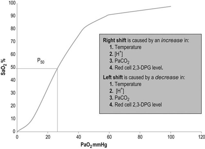
FIG. 2.6.1 Oxygen dissociation curve.
The position of the oxyhaemoglobin dissociation curve is modified by alterations in the partial pressure of carbon dioxide (PaCO2), temperature and the presence of acidosis or red blood cell 2,3-diphosphoglycerate (2,3-DPG). The curve is displaced to the right by an increase in PaCO2, a temperature rise, acidosis or an increase in 2,3-DPG concentration. This right shift facilitates more effective delivery of oxygen to peripheral tissues, which is beneficial in the presence of hypoxia. The P50 is used to reference the oxygen dissociation curve (see Fig. 2.6.1) [13]. It is the partial pressure of oxygen at which 50% of the haemoglobin is saturated with oxygen and specifies the position of the oxygen dissociation curve. Normally, a PaO2 of 26.6 mmHg corresponds to 50% haemoglobin saturation. Modern arterial blood gas machines routinely report this value.
Alveolar–arterial oxygen gradient
The alveolar–arterial PO2 gradient (A–a) PO2 is determined to evaluate a patient’s oxygenation. This gradient is defined by the difference between the partial pressure of O2 in the alveoli (PAO2) and the partial pressure of O2 dissolved in the arterial blood plasma (PAO2−PaO2). The partial pressure of O2 in the alveoli (PAO2) is established by using the alveolar gas equation:

where FiO2=fraction of inspired oxygen, Patm=atmospheric pressure (760 mmHg at sea level, decreasing progressively with increasing altitude), PH2O=partial pressure of saturated vapour (47 mmHg at 37°C) and R=the respiratory quotient (≈0.8).
The normal reference range for the alveolar–arterial gradient is 5–15 mmHg, with increases encountered from cigarette smoking, increasing FiO2 and advancing age. An approximate expected P(A–a)O2 can be determined using the following formula:

Pathophysiology of hypoxaemic respiratory failure
There are five potential mechanisms responsible for hypoxaemia when determining the aetiology of a patient’s respiratory failure which include decreased inspired fractional oxygen, impaired diffusion, shunting, ventilation–perfusion (V/Q) mismatch and hypoventilaton [14]. Isolated hypoxaemia is often referred to as type I respiratory failure, with type II respiratory failure characterized by a partial pressure of carbon dioxide (PaCO2) higher than 50 mmHg [15].
Mechanisms responsible for hypoxaemia include:
Decreased inspired fraction of oxygen
This is not common in clinical practice, but may be encountered at high altitude or in environments where the gas mixture is oxygen depleted.
Impaired diffusion
Diffusion impairment secondary to thickening of the membrane between the capillary and alveolus is now accepted as a rare mechanism. Thickening of the blood–gas barrier is found in diseases such as diffuse interstitial fibrosis. Under normal resting conditions, oxygen diffusion at the alveolar–capillary barrier requires only a third of the circulatory time available for equilibration to be complete. Therefore, even in the presence of a moderate diffusion impairment, there is sufficient ‘diffusion time’ to compensate.
Severe impairments of diffusion become clinically significant, particularly during exercise, when blood flow rates are increased and the time for diffusion equilibration is restricted. Shunting or a V/Q mismatch are almost always found to coexist with a diffusion defect and are likely, quantitatively, to be far more significant as a cause of hypoxemia.
Shunt
Hypoxaemic respiratory failure may result from either extrapulmonary or intrapulmonary shunts, with a shunt defined as the movement of blood from the venous to arterial circulation without transiting ventilated lung tissue and thus not afforded the opportunity to be oxygenated.
The clinical feature suggestive of the presence of a shunt is the failure of partial pressure of oxygen (PaO2) to rise despite inhalation of 100% oxygen (FiO2 1.0). Extrapulmonary shunts are encountered in the setting of acquired or congenital cardiac abnormalities, where the ventricles or the atria communicate secondary to septal defects. When the pressure gradient favours blood bypassing the pulmonary circulation, a shunt is established.
Intrapulmonary shunts occur in severe pneumonia, atelectasis, pulmonary arteriovenous malformations or the hepatopulmonary syndrome (regional dilation of pulmonary capillaries). The situation where there is alveolar consolidation or collapse such that there is unventilated but perfused lung may also be considered an extreme V/Q mismatch [16]. This is distinguished from a shunt by virtue of the correction of PaO2 in response to enhanced O2 administration. The shunt fraction can be calculated with the following equation:

where QS=shunt flow, QT=total blood flow, Cc’O2=end capillary oxygen content derived from PAO2, CaO2=arterial oxygen content, CvO2=mixed venous oxygen content.
A calculated shunt of less than 20% seldom requires support, whereas a calculated shunt greater than 30% usually needs significant cardiopulmonary intervention. However, the shunt equation is limited in clinical practice by the need for sampling mixed-venous blood and hence the presence of a pulmonary artery catheter.
Ventilation–perfusion (V/Q) mismatch
All the blood circulating through the lungs must perfuse individual ventilated lung units in order for effective gas exchange to occur. Theoretically, the ideal ratio of perfusion to ventilation (V/Q ratio) should numerically be one [17]. Under normal physiological conditions, perfusion is more pronounced at the lung bases as compared to the apices, while the converse is true for ventilation. Therefore, the usual overall V/Q ratio is approximately 0.8.
V/Q ratios demonstrate a wide spectrum of abnormalities: in some situations where alveolar units receive no ventilation, but are fully perfused, this results in a V/Q ratio of 0. This constitutes a shunt (as discussed above) and may be either cardiac or pulmonary in aetiology. Alternatively, the alveolar units may be fully ventilated, but receive no perfusion producing a V/Q ratio which trends to infinity [16]. A clinical example of this is a massive pulmonary embolus. Non-perfused alveolar units are referred to as physiological ‘dead space’.
Clinically, derangements of V/Q matching are the most common cause of gas exchange impairment and are characterized by hypoxaemia, hypercapnia or a combination of both. The presence of an increased alveolar–arterial PO2 gradient helps the emergency physician identify a V/Q abnormality.
Hypoventilation
The partial pressure of carbon dioxide in arterial blood (PaCO2) is determined by the production of CO2 (VCO2) and alveolar ventilation (VA):

where alveolar ventilation (VA)=(tidal volume (Tv) – dead space (VD))×respiratory rate.
Type II respiratory failure is characterized by a PaCO2 greater than 50 mmHg and is commonly encountered in clinical practice (Table 2.6.3).
Table 2.6.3
Causes of hypoventilation resulting in type II respiratory failure

The alveolar gas equation demonstrates how significant alveolar hypoventilation can result in a proportional decrease in alveolar oxygen pressure PAO2. When the respiratory quotient (R=0.8) remains constant, an increase in PaCO2 will be associated with a concomitant reduction in PAO2 which, in profound hypercapnia, may then result in hypoxaemia. If hypoventilation is the presumed mechanism of the hypoxaemia, then the alveolar–arterial oxygen tension gradient (A–a) PO2 should be calculated. This gradient will be normal when the hypoxaemia is entirely secondary to hypercapnia, but will be increased if there are other mechanisms, such as an impaired V/Q ratio or the presence of a shunt contributing to the hypoxaemia.
It is important when interpreting any blood gas that a standardized structured evaluation is undertaken to determine the aetiology of the respiratory failure (Table 2.6.4 and Fig. 2.6.2).
Table 2.6.4
Six-step approach to evaluate respiratory failure using arterial blood gases
|
Step |
Parameter |
|
1 |
Confirm the presence of hypoxaemia by determining the PaO2<60 mmHg or SaO2<90% |
|
2 |
Establish if there is an increased alveolar–arterial oxygen tension gradient ↑(A–a) PO2 |
|
3 |
Determine if there is evidence for hypoventilation. If the PaCO2>50 mmHg, then alveolar hypoventilation is present |
|
4 |
If there is hypoxaemia, a normal alveolar–arterial oxygen tension gradient (A–a) PO2 and the PaCO2 is not elevated, the patient is in a hypoxic environment, e.g. altitude |
|
5 |
Determine if the hypoxaemia is entirely accounted for by hypoventilation, established by calculating the alveolar–arterial oxygen tension gradient (A–a) PO2. If the gradient is normal (<15), hypoventilation alone is the cause, such as from central nervous system depression or respiratory muscular failure. Conversely, if the alveolar–arterial oxygen tension gradient (A–a) PO2 is elevated, other conditions including pneumonia or acute respiratory distress syndrome are likely responsible |
|
6 |
If the PaCO2 is normal, hypoxaemia is present and there is an increased alveolar–arterial oxygen tension gradient (A–a) PO2, the response to breathing an enhanced O2 mixture discriminates between a ventilation/perfusion mismatch and a shunt |

FIG. 2.6.2 Evaluating respiratory failure using arterial blood gases. CNS: central nervous system; AVM: arteriovenous malformation; ASD: atrial septal defect; VSD: ventricular septal defect; PFO: patent foramen ovale.
Acid–base balance
While this chapter principally addresses the respiratory aspects of arterial blood gas analysis, it is impossible to separate clinically the component information provided by arterial blood sampling (see Chapter 21.1). Under normal physiological conditions, humans maintain closely regulated acid–base homeostasis required for normal cellular activity. This homeostasis results from a complex series of interactions between the lungs and kidneys, moderated by a range of physiological buffers. The resultant blood hydrogen ion concentration (pH) is a function of the ratio of bicarbonate concentration and the partial pressure of CO2 in arterial blood.
Acid–base disorders
Primary metabolic acid–base disorders and the secondary metabolic compensation for primary respiratory disturbances are reflected by changes in the serum bicarbonate concentration. Primary respiratory acid–base disorders and the secondary respiratory compensation for primary metabolic disturbances result in changes in the measured PaCO2 (Table 2.6.5). Similarly to the evaluation of respiratory failure, evaluation of an acid–base abnormality requires a systematic approach:
Table 2.6.5
Determining the primary acid–base disorder

Step 1: First determine if there is alkalaemia or acidaemia present, simply defined by the following: pH<7.35=acidaemia, or pH>7.45=alkalaemia.
Step 2: Then determine if the primary disturbance is respiratory or metabolic in origin, established by assessing the relationship between the direction of change in the pH and the direction of change in the PaCO2.
Step 3: Then assess whether the primary disturbance has been compensated (Table 2.6.6), recognizing that compensation does not return the pH to normal. If the expected compensation is not present, it is likely that a mixed acid–base disorder exists [18,19].
Table 2.6.6
Predicting compensation for primary acid–base disorders
|
Primary disorder |
Expected compensation |
|
Metabolic acidosis |
PaCO2=(1.5×HCO3−)+8 |
|
Acute respiratory acidosis |
For every 10 mmHg increase in PCO2 the HCO3− should increase by 1 mmol/L |
|
Chronic respiratory acidosis (3–5 days) |
For every 10 mmHg increase in PCO2 the HCO3− should increase by 3–4 mmol/L over 4 days |
|
Metabolic alkalosis |
PaCO2=0.8×HCO3−+20 |
|
Acute respiratory alkalosis |
For every 10 mmHg decrease in PCO2 the HCO3− should decrease by 1 mmol/L |
|
Chronic respiratory alkalosis |
For every 10 mmHg decrease in PCO2 the HCO3− should decrease by 2 mmol/L |
Step 4: Calculate the anion gap in the presence of a metabolic acidosis, to allow for formulation of the differential diagnosis (high or normal anion gap):

A high anion gap acidosis is most commonly secondary to lactate or ketones (Table 2.6.7). Lactic acidosis was classified in 1976 into type A or B, based on the absence or presence of adequate tissue oxygenation [20]. Lactic acidosis is a common life- threatening form of metabolic acidosis in the critically ill.
Table 2.6.7
Causes of a high anion gap metabolic acidosis

The differential diagnosis of a non-anion gap metabolic acidosis includes conditions characterized by bicarbonate loss, excess chloride or ingestions (Table 2.6.8).
Table 2.6.8
Causes of normal anion gap metabolic acidosis

If the anion gap is elevated (>12) and not explained by an obvious aetiology then, under appropriate clinical circumstances, a toxic ingestion, such as methanol or ethylene glycol, is considered. This is associated with a high osmolal gap, the difference between the measured serum osmolality and the calculated osmolality which, under normal physiological conditions, should be<10 mmol/L:

Step 5: If an increased anion gap is present, then determine the delta ratio to establish whether a mixed acid–base disorder is present. This is deduced from assessing the relationship between the increased anion gap and the decrease in bicarbonate [21].
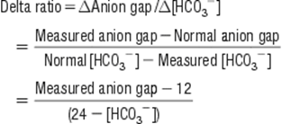
The magnitude of the delta ratio can range from<0.4 to>2 and allows for refinement of the differential diagnosis (Table 2.6.9).
Table 2.6.9
Delta ratio interpretation
|
Delta ratio |
Interpretation |
|
<0.4 |
Normal anion gap hyperchloraemic metabolic acidosis |
|
<1 |
Combined high and normal anion gap acidosis |
|
1–2 |
Isolated high anion gap metabolic acidosis |
|
>2 |
Mixed high anion gap metabolic acidosis and metabolic alkalosis |
Step 6: Identify the ‘initiating factor’ if the arterial blood gas reflects a primary metabolic alkalosis. This may include loss of hydrogen ions from the gastrointestinal system, transcellular hydrogen shifts, mineralocorticoid excess or addition of alkali. Furthermore, a ‘maintenance’ factor is also needed to preserve the metabolic alkalosis as, in the presence of an elevated serum bicarbonate, a patient with intact renal function will rapidly excrete excess bicarbonate in the urine. Therefore, for a metabolic alkalosis to persist, there must be a reduction in the renal ability to lose excess bicarbonate. In practice, this is usually secondary to hypovolaemia or reduced effective arterial blood volume (including heart failure and cirrhosis), chloride depletion, hypokalaemia, renal impairment or a combination of these factors (Table 2.6.10).
Table 2.6.10
Causes of a metabolic alkalosis
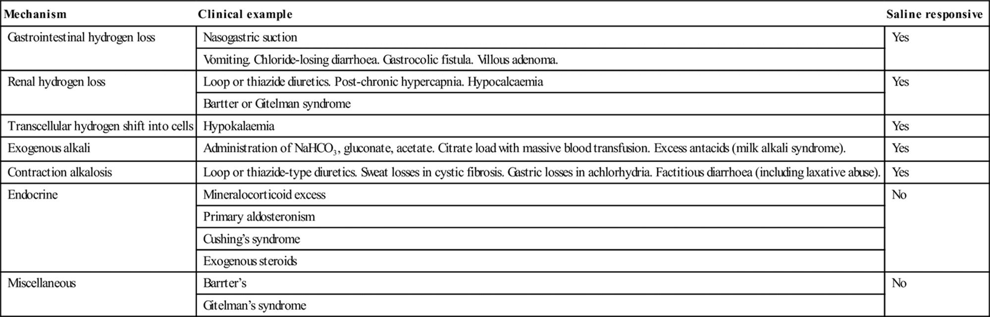
Venous blood gases
It is technically much easier and quicker to obtain a venous blood sample than an arterial one and, therefore, there is substantial interest in using venous blood as a surrogate for arterial blood gas analysis. In some situations, analysis of venous blood provides sufficiently reliable correlation with arterial blood to assist in clinical decision making.
The peripheral venous pH is approximately 0.02–0.04 pH units lower than the arterial pH, the venous serum HCO3− concentration is approximately 1–2 meq/L higher and the venous PCO2 is approximately 3–8 mmHg higher [22]. There are currently insufficient data to determine if these relationships persist in a shocked patient or those with mixed acid–base disorders. In a patient who is not shocked, venous pH, bicarbonate and base excess have sufficient agreement to be clinically interchangeable for arterial values [23]. Agreement between arterial and venous PCO2 is too unpredictable to be clinically reliable.
References
1. Severinghaus JW. First electrodes for blood PO2 and PCO2 determination. J Appl Physiol. 2004;97:1599–1600.
2. Clark Jr LC, Wolf R, Granger D, et al. Continuous recording of blood oxygen tensions by polarography. J Appl Physiol. 1953;6:189–193.
3. Barker SJ, Curry J, Redford D, Morgan S. Measurement of carboxyhemoglobin and methemoglobin by pulse oximetry: a human volunteer study. Anesthesiology. 2006;105:892–897.
4. Schmidt C, Muller-Plathe O. Stability of pO2, pCO2 and pH in heparinized whole blood samples: influence of storage temperature with regard to leukocyte count and syringe material. Eur J Clin Chem Clin Biochem. 1992;30:767–773.
5. Harsten A, Berg B, Inerot S, Muth L. Importance of correct handling of samples for the results of blood gas analysis. Acta Anaesthesiol Scand. 1988;32:365–368.
6. Scheer B, Perel A, Pfeiffer UJ. Clinical review: complications and risk factors of peripheral arterial catheters used for haemodynamic monitoring in anaesthesia and intensive care medicine. Crit Care. 2002;6:199–204.
7. Okeson GC, Wulbrecht PH. The safety of brachial artery puncture for arterial blood sampling. Chest. 1998;114:748–751.
8. Williams AJ. ABC of oxygen: assessing and interpreting arterial blood gases and acid–base balance. Br Med J. 1998;317:1213–1216.
9. Kohonen M, Teerenhovi O, Terho T, et al. Is the Allen test reliable enough? Eur J Cardiothorac Surg. 2007;32:902–905.
10. Barone JE, Madlinger RV. Should an Allen test be performed before radial artery cannulation? J Trauma. 2006;61:468–470.
11. Puttarajappa C, Rajan DS. Images in clinical medicine Allen’s test. N Engl J Med. 2010;363:e20.
12. Clark VL, Kruse JA. Arterial catheterization. Crit Care Clin. 1992;8:687–697.
13. Morgan TJ. The oxyhaemoglobin dissociation curve in critical illness. Crit Care Resus. 1999;1:93–100.
14. West JB. Pulmonary pathophysiology: the essentials 7th ed. Philadelphia: Lippincott Williams & Wilkins; 2008.
15. Hanley ME, Bone RC. Acute respiratory failure Pathophysiology, causes, and clinical manifestations. Postgrad Med. 1986;79(166-9):72–76.
16. Jones JG, Jones SE. Discriminating between the effect of shunt and reduced VA/Q on arterial oxygen saturation is particularly useful in clinical practice. J Clin Monit Comput. 2000;16:337–350.
17. West JB, Wagner PD. Pulmonary gas exchange. Am J Respir Crit Care Med. 1998;157:S82–S87.
18. Carmody JB, Norwood VF. A clinical approach to paediatric acid–base disorders. Postgrad Med J. 2012;88:143–151.
19. Narins RG, Emmett M. Simple and mixed acid-base disorders: a practical approach. Medicine (Baltimore). 1980;59:161–187.
20. Cohen RD, Woods HF. Lactic acidosis revisited. Diabetes. 1983;32:181–191.
21. Reddy P, Mooradian AD. Clinical utility of anion gap in deciphering acid–base disorders. Int J Clin Pract. 2009;63:1516–1525.
22. Malatesha G, Singh NK, Bharija A, Rehani B, et al. Comparison of arterial and venous pH, bicarbonate, PCO2 and PO2 in initial emergency department assessment. Emerg Med J. 2007;24:569–571.
23. Kelly AM. Review article: can venous blood gas analysis replace arterial in emergency medical care. Emerg Med Australas. 2010;22:493–498.
2.7 Cerebral resuscitation after cardiac arrest
Stephen A Bernard
Essentials
1 Anoxic neurological injury is common following out-of-hospital cardiac arrest (OHCA) and carries a high rate of morbidity and mortality.
2 Reperfusion of the ischaemic brain results in biochemical cascades that lead to further cell death, largely mediated by calcium influx into cells.
3 Therapeutic hypothermia (32–34°C) after resuscitation from cardiac arrest was considered an effective treatment for anoxic neurological injury and is recommended by the Australian Resuscitation Council (ARC) and New Zealand Resuscitation Council (NZRC). However the largest trial to date has shown no improvement in outcome in any measurable way, and has cast doubt on its efficacy.
4 There are currently no proven pharmacological interventions that improve neurological outcome after global cerebral ischaemia.
5 Hypotension is deleterious to the injured brain and should be promptly treated.
6 Recent laboratory and observational clinical evidence suggests that hyperoxia following resuscitation may be associated with worse outcomes.
Introduction
Out-of-hospital cardiac arrest (OHCA) is common and is a leading cause of death in patients with heart disease [1]. Prolonged OHCA causing global cerebral ischaemia may lead to permanent neurological injury, despite effective cardiopulmonary resuscitation with return of spontaneous circulation (ROSC). Many patients who are initially successfully resuscitated from OHCA remain comatose in the emergency department (ED) because of the anoxic neurological injury, which results in considerable morbidity and mortality following hospital admission. This chapter details the pathophysiology of anoxic neurological injury and current cerebral resuscitation therapies.
Definition
Cerebral resuscitation involves the use of pharmacological or other strategies to minimize injury to the brain following a prolonged ischaemic insult [2].
Pathophysiology of cerebral ischaemia
The brain is highly dependent on an adequate supply of oxygen and glucose for metabolism. When cerebral oxygen delivery falls below 20 mL/100 g brain tissue/minute, aerobic metabolism changes to anaerobic glycolysis, with a marked decrease in the generation of adenosine triphosphate (ATP) [3].
After several minutes of cerebral ischaemia, the supply of ATP is exhausted and cellular metabolism ceases. The failure of the sodium/potassium transmembrane pump leads to a shift of sodium into the cell with subsequent cell swelling. In addition, hydrogen ions are generated with lactate ions and the resulting intracellular metabolic acidosis is toxic to intracellular enzyme systems. This acidosis is partly dependent on the concentration of glucose, with hyperglycaemia contributing to an increase in the intracellular acidosis.
Reperfusion injury
Additional injury occurs following resuscitation with reperfusion of the brain with oxygenated blood. The intracellular levels of glutamate, an excitatory neurotransmitter released from presynaptic terminals, increase dramatically during reperfusion. Glutamate activates calcium ion channel complexes which shift calcium from the extracellular fluid to the intracellular fluid [4]. The calcium influx into cells initiates multiple biochemical cascades, leading to the production of so-called ‘free-radicals’ and the activation of degradative enzymes.
Free-radical production
Intracellular iron also plays an important role in free-radical production. Iron is usually maintained in the ferric state and is sequestered to intracellular proteins. During ischaemia, iron is reduced to the soluble ferrous form and reacts with peroxide, generating damaging hydroxyl free radicals.
There are also effects on leucocytes, endothelium and platelets. The generation of free radicals activates an upregulation of molecules that mediate leucocyte adhesion and extravasation of these into brain parenchyma. Also, microvessel occlusion with leucocyte–platelet complexes leads to increased cerebral ischaemia.
Ischaemia and reperfusion
Ischaemia and reperfusion are also a stimulus for nitric oxide (NO) synthase activation that generates NO, a potent mediator of injury. The NO may combine with superoxide to form peroxy-nitrite radicals, which are potent activators of lipid peroxidation. Other proposed actions of NO include DNA damage, increased glutamate release and microvascular vasodilatation.
Finally, some neurons that survive the initial anoxic insult proceed to ‘programmed’ cell death, known as apoptosis [5]. After reperfusion, this delayed neuronal death may occur at different rates, varying from 6 hours for neurons in the striatum to 7 days for hippocampal CA1 neurons. Apoptosis is characterized by cellular and nuclear shrinkage, chromatin condensation and DNA fragmentation.
Cerebral haemodynamics after reperfusion
Cerebral haemodynamics may remain abnormal for some hours after resuscitation and restoration of a spontaneous circulation [6]. In animal models, there is an initial hyperaemia after resuscitation, followed by decreased cerebral blood flow despite normal mean arterial blood pressure (MAP). Due to the inflammatory processes described above, cerebral metabolic rate for oxygen increases slightly. Thus, there may be a mismatch of cerebral oxygen delivery and demand for some hours following resuscitation from prolonged cardiac arrest. Cerebral oxygen delivery and/or demand are also adversely affected by arterial hypotension, hypoxaemia, raised intracranial pressure, fever and/or seizure activity.
Pharmacological interventions
There is considerable interest and research into pharmacological interventions that might decrease reperfusion injury, as much of the neurological injury seen following an ischaemic injury actually occurs after reperfusion. A number of drugs that showed promise in animal models of global cerebral ischaemia underwent randomized, controlled human trials. These included thiopentone [7], a corticosteroid [8], lidoflazine [9], nimodipine [10], magnesium [11], diazepam [11] and co-enzyme Q10 [12]. However, none of these have shown an improved neurological or overall outcome in clinical trials.
Therapeutic hypothermia
Therapeutic hypothermia (TH) was demonstrated to benefit patients who remain comatose after resuscitation from OHCA. It was thought that the TH decreased cerebral oxygen demand without decreasing cerebral oxygen supply. Also, that TH decreased the reperfusion injury by reducing the production of oxygen free radicals after reperfusion.
Two prospective, controlled, human studies suggested improved outcome using moderate TH in comatose survivors of pre-hospital cardiac arrest [13,14]. In an Australian study, 43 patients were randomized to TH (33°C for 12 hours) and 34 patients were maintained at normothermia [13]. Hypothermia was induced in the ED using surface cooling with ice-packs. At hospital discharge, 21/43 (49%) of the TH group had a good outcome compared with 9/34 (26%) in the control group (p=0.046). Following multivariate analysis for differences at baseline, the odds ratio (OR) for good outcome in the hypothermic group was 5.25 (95% confidence intervals [CI] 1.47 to 18.76: p=0.011). There were no adverse effects of TH apparent, such as sepsis, lactic acidosis or coagulopathy.
A second clinical trial of TH after OHCA was conducted in Europe [14]. At 6 months post-arrest, 55% of the TH patients had a good outcome, compared with 39% of the normothermic controls (OR 1.4, 95% CI 1.08 to 1.81). The complication rate also did not differ between the two groups.
However, a much larger study of 939 unconscious survivors of OHCA of presumed cardiac origin in Europe and Australia failed to show a benefit of a targeted temperature of 33 °C, as compared with a targeted temperature of 36 °C [15].
Australian Resuscitation Council (ARC)/New Zealand Resuscitation Council (NZRC) recommendations
The original two trials form the basis of the recommendation by the Australian Resuscitation Council and New Zealand Resuscitation Council [16] that therapeutic hypothermia (32–34°C for 12–24 hours) should be induced in patients who remain comatose after OHCA, when the initial cardiac arrest rhythm is ventricular fibrillation (VF).
Techniques and timing of therapeutic hypothermia
Current research is focused on the techniques and timing of therapeutic hypothermia. Surface cooling in the clinical trials cited above had the significant limitation of a slow decrease in core temperature, with 0.9°C/h decrease in core temperature using ice packs and 0.5°C/h using forced cold air cooling.
Surface cooling
Proprietary cooling jackets have become available which circulate temperature-controlled water through a jacket applied to the patient. The core temperature is monitored and the machine adjusts the temperature of the water circulating through the jacket, thus ‘overshooting’ with inadvertent cooling should be avoidable. As surface cooling still has the drawback of a relatively slow heat exchange due to the poor perfusion of the skin in many post-arrest patients, other techniques have been tested.
Intravascular cooling
Intravascular cooling is now feasible using a catheter inserted into the femoral vein that includes a saline-filled balloon for heating/cooling. A catheter balloon contains saline that is pumped via the balloon and back to the machine. This allows heating/cooling of blood in the inferior vena cava. This approach has been shown to be more effective at core temperature control compared with surface cooling, however, there is a significant cost to the catheter [17].
Another technique for rapid induction of TH is to administer rapidly a large volume (30–40 mL/kg) of ice-cold (4°C) intravenous fluid [18]. An infusion of 30 mL/kg lactated Ringer’s solution at 4°C was studied in an ED in 22 patients resuscitated from OHCA and was found to be an effective and safe technique to induce mild hypothermia, with a decrease in the core temperature of 1.7°C over 25 minutes. In addition, there were improvements in mean arterial blood pressure, acid–base and renal function with no apparent complications, such as pulmonary oedema.
Pre-hospital cooling trials
A number of pre-hospital trials of large-volume, ice-cold crystalloid for the rapid induction of TH in patients resuscitated from OHCA have now been undertaken. Kim et al. randomized 125 patients who were comatose following resuscitation from OHCA to receive either standard care or intravenous cooling by paramedics using 2000 mL ice-cold (4°) saline [19]. Sixty-three patients received an infusion of 4°C normal saline before hospital arrival which resulted in a decrease in temperature of 1.2°C, whereas the 62 patients having standard care had an increase in temperature of 0.10°C.
Two randomized, controlled, clinical trials of paramedic cooling have now also been undertaken in Melbourne, Australia. In one, post-VF arrest patients were cooled using 2 L ice-cold crystalloid fluid immediately after resuscitation [20]. A total of 234 patients were assigned to either paramedic cooling (118 patients) or hospital cooling (116 patients). Patients allocated to paramedic cooling received a median of 1900 mL of ice-cold Hartmann’s solution, which resulted in a mean decrease in core temperature of 0.8°C. In the paramedic-cooled group, 47.5% patients had a non-statistically favourable outcome at hospital discharge compared with 52.6% in the hospital-cooled group (RR 0.90, 95% confidence interval 0.70 to 1.17, p=0.43).
In a parallel trial, 163 patients who had been resuscitated from cardiac arrest with an initial cardiac rhythm of asystole or pulseless electrical activity were randomized to either pre-hospital cooling using a rapid infusion of 2 L ice-cold Hartmann’s solution (82 patients) or cooling after hospital admission (81 patients) [21]. Patients allocated to pre-hospital cooling received a median of 1500 mL of ice-cold fluid which resulted in a mean decrease in core temperature of 1.4°C compared with 0.2°C in hospital cooled patients (p<0.001). The time to therapeutic hypothermia (<34°C) was 3.2 h in the pre-hospital cooled group, compared with 4.8 h in the hospital cooled group (p=0.0328). Overall, there was no statistical difference in outcome at hospital discharge with a favourable outcome (discharge from hospital to home or rehabilitation) in 12% in the pre-hospital cooled patients, compared with 9% in the hospital-cooled patients (p=0.50). In the patients with a cardiac cause of the arrest, there was a trend towards improved outcome in the paramedic-treated patients, with 8 of 47 patients (17%) who received pre-hospital cooling with a favourable outcome at hospital discharge, compared with 3 of 43 (7%) in the hospital-cooled group (p=0.146).
Currently, there is a clinical trial comparing TH inducted by paramedics during CPR using a bolus of 30 mL/kg ice-saline [22]. It is hoped that an earlier bolus of cold fluid during CPR may lead to earlier TH and thus improved patient outcomes.
The negative findings of the European/ Australian TH trial now cast doubt on this ubiquitous modality of care.
Hyperoxia compared with normoxia after resuscitation
A number of laboratory studies suggest that post-OHCA, the administration of 100% oxygen may be associated with increased neurological injury compared with an oxygen/air mix titrated to provide normoxia [23]. It is proposed that the excessive oxygen increases the degree of free-radical production in injured neurons. Clinical studies in neonates with hypoxic–ischaemic encephalopathy also support the proposal that hyperoxia causes additional neurological injury [24].
In a large, multicentre observational study, the association between the highest PaO2 measured on the first day of admission to the intensive care unit and mortality was determined in 4459 patients following resuscitation from OHCA [25]. After multivariable analysis, each 100 mmHg increase in PaO2 was associated with a 24% increase in in-hospital mortality risk (OR 1.24: 95% confidence interval 1.18 to 1.31).
While observational, these data support the concept that there may be a dose-dependent association between supranormal oxygen tension and risk of in-hospital mortality, suggesting that hyperoxia in the early post-resuscitation period may be harmful. Therefore, decreasing the fraction of inspired oxygen while carefully monitoring the patient with continuous pulse oximetry to avoid hypoxia should be considered. However, prospective, controlled trials are required to validate this finding.
Other interventions
Another strategy to improve neurological outcome following resuscitation from OHCA includes maintenance of adequate blood pressure using fluid therapy and/or vasopressors [15]. Although human studies have not established optimal targets for blood pressure or blood oxygenation, one study showed benefit in OHCA patients with goal-directed therapy that included aiming for a mean arterial pressure target>65 mmHg and a mixed venous oxygen target>70% [26].
Outcome prediction
The early prediction of outcome is important after a severe anoxic neurological injury. Once a poor prognosis is reliably established, then decisions concerning limitations of costly intensive care treatments may be made. Previously, the clinical examination at day three was regarded as an accurate predictor of expected outcome [27]. However, this time frame preceded the widespread use of TH and the possibility that decreased metabolism of sedative agents might mask neurological improvement. More recent recommendations suggest that prognostication should now occur at a minimum of 72 hours after rewarming from TH [28].
Investigations such as brain computed tomography (CT), magnetic resonance imaging (MRI) and/or electroencephalogram (EEG) are relatively insensitive and/or non-specific for the early prediction of neurological outcome. While absent somatosensory responses bilaterally reliably predict a poor outcome after anoxic brain injury in unsedated patients, this investigation is not available in most hospitals.
References
1. Sasson C, Rogers MA, Dahl J, Kellermann AL. Predictors of survival from out-of-hospital cardiac arrest: a systematic review and meta-analysis. Circ Cardiovasc Qual Outcomes. 2010;3:63–81.
2. Schneider A, Böttiger BW, Popp E. Cerebral resuscitation after cardiocirculatory arrest. Anesth Analg. 2009;108:971–979.
3. Pundik S, Xu K, Sundararajan S. Reperfusion brain injury: focus on cellular bioenergetics. Neurology. 2012;79:S44–S51.
4. Takata K, Takeda Y, Sato T, et al. Effects of hypothermia for a short period on histologic outcome and extracellular glutamate concentration during and after cardiac arrest in rats. Crit Care Med. 2005;33:1340–1345.
5. Ferrer I. Apoptosis: future targets for neuroprotective strategies. Cerebrovasc Dis. 2006;2:9–20.
6. Oku K, Kuboyama K, Safar P, et al. Cerebral and systemic arteriovenous oxygen monitoring after cardiac arrest: inadequate cerebral oxygen delivery. Resuscitation. 1994;27:141–152.
7. The Brain Resuscitation Clinical Trial Study Group. Randomized clinical study of thiopentone loading in comatose survivors of cardiac arrest. N Eng J Med. 1986;314:397–410.
8. The Brain Resuscitation Clinical Trial Study Group. Glucocorticoid treatment does not improve neurologic recovery following cardiac arrest. J Am Med Assoc. 1989;262:3427–3430.
9. Brain Resuscitation Clinical Trial II Study Group. A randomized clinical study of a calcium-entry blocker (lidoflazine) in the treatment of comatose survivors of cardiac arrest. N Eng J Med. 1991;324:1225–1231.
10. Roine RO, Kaste M, Kinnamen A, et al. Nimodipine after resuscitation from out-of-hospital ventricular fibrillation: a placebo-controlled double-blind randomized trial. J Am Med Assoc. 1990;264:3171–3177.
11. Longstreth Jr WT, Fahrenbruch CE, Olsufka M, et al. Randomized clinical trial of magnesium, diazepam, or both after out-of-hospital cardiac arrest. Neurology. 2002;59:506–514.
12. Damian MS, Ellenberg D, Gildemeister R, et al. Coenzyme Q10 combined with mild hypothermia after cardiac arrest: a preliminary study. Circulation. 2004;110:3011–3016.
13. Bernard SA, Gray TW, Buist MD, et al. A randomised, controlled trial of induced hypothermia in comatose survivors of prehospital cardiac arrest. N Eng J Med. 2002;346:557–563.
14. The Hypothermia after Cardiac Arrest Study Group. Mild therapeutic hypothermia to improve the neurological outcome after cardiac arrest. N Eng J Med. 2002;346:549–556.
15. Nielsen N, Wetterslev J, Cronberg T, et al. Targeted temperature management at 33 ° C versus 36 ° C after cardiac arrest. NEJM. 2013;369:2197–2206.
16. Australian Resuscitation Council; New Zealand Resuscitation Council. Post-resuscitation therapy in adult advanced life support. ARC and NZRC Guidelines 2010. Emerg Med Australas 2011; 23:292–296.
17. Hinz J, Rosmus M, Popov A, et al. Effectiveness of an intravascular cooling method compared with a conventional cooling technique in neurologic patients. J Neurosurg Anesthesiol. 2007;19:130–135.
18. Bernard SA, Buist M, Monteiro O, Smith K. Induced hypothermia using large volume, ice-cold intravenous fluid in comatose survivors of out-of-hospital cardiac arrest: a preliminary report. Resuscitation. 2003;56:9–13.
19. Kim F, Olsufka M, Longstreth Jr WT, et al. Pilot randomized clinical trial of prehospital induction of mild hypothermia in out-of-hospital cardiac arrest patients with a rapid infusion of 4°C normal saline. Circulation. 2007;115:3064–3070.
20. Bernard SA, Smith K, Cameron P, et al. Induction of therapeutic hypothermia by paramedics after resuscitation from out-of-hospital ventricular fibrillation cardiac arrest A randomized controlled trial. Circulation. 2010;122:737–742.
21. Bernard SA, Smith K, Cameron P, et al. Induction of therapeutic hypothermia by paramedics after resuscitation from out-of-hospital non-ventricular fibrillation cardiac arrest. Crit Care Med. 2012;40:747–753.
22. Deasy C, Bernard SA, Cameron P, et al. Design of the RINSE Trial: The rapid infusion of cold normal saline by paramedics during CPR. BMC Emerg Med. 2011;11:17.
23. Pilcher J, Weatherall M, Shirtcliffe P, et al. The effect of hyperoxia following cardiac arrest – A systematic review and meta-analysis of animal trials. Resuscitation. 2012;83:417–422.
24. Saugstad OD. Resuscitation of newborn infants: from oxygen to room air. Lancet. 2010;376:1970–1971.
25. Kilgannon JH, Jones AE, Parrillo JE, et al. Relationship between supranormal oxygen tension and outcome after resuscitation from cardiac arrest. Circulation. 2011;123:2717–2722.
26. Gaieski DF, Band RA, Abella BS, et al. Early goal-directed hemodynamic optimization combined with therapeutic hypothermia in comatose survivors of out-of-hospital cardiac arrest. Resuscitation. 2009;80:418–424.
27. Wijdicks EF, Hijdra A, Young GB, et al. Quality Standards Subcommittee of the American Academy of Neurology Practice parameter: prediction of outcome in comatose survivors after cardiopulmonary resuscitation (an evidence-based review): report of the Quality Standards Subcommittee of the American Academy of Neurology. Neurology. 2006;67:203–210.
28. Rossetti AO, Oddo M, Logroscino G, Kaplan PW. Prognostication after cardiac arrest and hypothermia: a prospective study. Ann Neurol. 2010;67:301–307.
2.8 Anaphylaxis
Anthony Brown
Essentials
1 Anaphylaxis describes both IgE, immune-mediated reactions and non-allergic, non-immunologically triggered events. Co-morbidities, such as asthma, infection, exercise, alcohol or stress, and concurrent medications, such as β-blockers, angiotensin converting enzyme inhibitors and aspirin, increase the risk (‘summation anaphylaxis’).
2 Deaths occur by hypoxia from upper airway asphyxia or severe bronchospasm or by profound shock from vasodilatation and extravascular fluid shift.
3 Parenteral penicillin, hymenopteran stings and foods are the most common causes of IgE, immune-mediated fatalities. Radiocontrast media, aspirin and other non-steroidal anti-inflammatory drugs are the most common causes of non-allergic fatalities. Older patients with ischaemic heart disease or on treatment, such as a β-blockers, are at increased risk of death.
4 Oxygen, adrenaline (epinephrine) and fluids are first-line treatment.
5 The role of H1 and H2 antihistamines, steroids, glucagon and salbutamol is unclear and unproven. They should only be considered once cardiovascular stability has been achieved with first-line agents.
6 Discharge follows a period of observation from 4 to 6 h after full recovery. A written discharge plan with adrenaline autoinjector and referral to an allergist for all significant, recurrent, unavoidable or unknown stimulus reactions are essential. Patient education is important to successful, long-term care.
7 Two comprehensive practice guidelines recently released include the Joint Task Force on Practice Parameters (2010) in the USA and the World Allergy Organization (2011).
Introduction
Anaphylaxis represents the most catastrophic of the immediate-type generalized hypersensitivity reactions and remains the quintessential medical emergency. It usually occurs unheralded in otherwise healthy people following exposure to a trigger. It presents as a dynamic continuum from mild to severe, gradual in onset to fulminant and may involve multiple organ systems or cause isolated shock or wheeze. Prompt clinical recognition and treatment with oxygen, adrenaline and fluids to restore cardiorespiratory stability are essential to ensure a favourable outcome. Careful discharge planning, including allergy referral where appropriate, protects against further attacks of anaphylaxis.
Definition
The term ‘anaphylaxis’ was introduced by Richet and Portier in 1902, literally meaning ‘against protection’. It is currently used to describe the rapid, generalized and often unheralded immunologically-mediated events that follow exposure to certain foreign substances in previously sensitized persons known as antigen-induced, or immune-mediated, allergic anaphylaxis.
An identical clinical syndrome known as non-allergic anaphylaxis follows non-immunological mechanisms, with the release of identical inflammatory mediators. Non-allergic anaphylaxis may occur on first exposure to an agent and does not require a period of sensitization. The term ‘non-allergic anaphylaxis’ is preferred by the World Allergy Organization (WAO) to the older one of an ‘anaphylactoid reaction’ [1]. This chapter will use the clinical term ‘anaphylaxis’ to describe both of these syndromes, despite their important aetiological differences.
Classification of anaphylaxis
Surprisingly, there is still no international agreement on the classification, diagnosis or severity grading of anaphylaxis [2]. One simple definition is that anaphylaxis is ‘a serious, life-threatening generalized or systemic hypersensitivity reaction’ [1]. Similarly, the National Institute of Allergy and Infectious Disease (NIAID) and the Food Allergy and Anaphylaxis Network (FAAN) recommend an equally brief, broad definition: ‘Anaphylaxis is a serious allergic reaction that is rapid in onset and may cause death’. Their full definition is considerably longer yet more complete, aiming to capture over 95% of clinical cases within the three diagnostic criteria [3]. Criterion 1 aims to identify at least 80% of anaphylaxis cases, even if the allergic status of the patient and potential cause of the reaction may be unknown, as the majority of anaphylactic reactions include skin symptoms. Criterion 2 is to identify anaphylaxis in the absence of cutaneous features, such as in children with food allergy or insect sting allergy, but requires a known allergic history and possible exposure. Gastrointestinal symptoms are included. Criterion 3 aims to capture the rare patient with an acute hypotensive episode after exposure to a known allergen (Box 2.8.1) [3]. These inclusive definitions for anaphylaxis should be used by researchers unless and until refined by future prospective data [4].
Box 2.8.1
Definition of anaphylaxis: clinical criteria for diagnosis
Anaphylaxis is highly likely when any one of the following three criteria are fulfilled:
1. Acute onset of an illness (minutes to several hours) with involvement of the skin, mucosal tissue or both (e.g. generalized hives, pruritus or flushing, swollen lips–tongue–uvula) and at least one of the following:
Respiratory compromise (e.g. dyspnoea, wheeze–bronchospasm, stridor, reduced PEF, hypoxaemia)
Reduced BP or associated symptoms of end-organ dysfunction (e.g. hypotonia (collapse), syncope, incontinence)
2. Two or more of the following that occur rapidly after exposure to a likely allergen for that patient (minutes to several hours):
Involvement of the skin-mucosal tissue (e.g. generalized hives, itch–flush, swollen lips–tongue–uvula)
Respiratory compromise (e.g. dyspnoea, wheeze–bronchospasm, stridor, reduced PEF, hypoxaemia)
Reduced BP or associated symptoms (e.g. hypotonia (collapse), syncope, incontinence)
Persistent gastrointestinal symptoms (e.g. crampy abdominal pain, vomiting)
3. Reduced BP after exposure to known allergen for that patient (minutes to several hours):
Infants and children: low systolic BP (age-specific) or greater than 30% decrease in systolic BP*
Adults: systolic BP of less than 90 mmHg or greater than 30% decrease from that person’s baseline
PEF: peak expiratory flow; BP: blood pressure.
*Low systolic blood pressure for children is defined as less than 70 mmHg from 1 month to 1 year; less than (70 mmHg+[2×age]) from 1 to 10 years; and less than 90 mmHg from 11 to 17 years. Reproduced with permission from Sampson HA, Munoz-Furlong A, Campbell RL, et al. Second symposium on the definition and management of anaphylaxis: summary report – Second National Institute of Allergy and Infectious Disease/Food Allergy and Anaphylaxis Network symposium. Journal of Allergy and Clinical Immunology 2006;117:391–7.
Severity grading
No validated grading system exists that prospectively links the clinical features of anaphylaxis with severity, urgency, treatment or outcome. One system based on a retrospective multivariate analysis of over 1000 clinically diagnosed generalized hypersensitivity reactions defined three grades (Table 2.8.1) [5]. Generalized allergic reactions confined to the skin and subcutaneous tissues were considered as mild grade, but the moderate and severe grades with multisystem involvement that correlated with the need for adrenaline represent true anaphylaxis according to the NIAID/FAAN criteria. Again, this grading system should be used as a starting point by researchers for descriptive purposes, until prospective data in the future refine the criteria.
Table 2.8.1
Severity grading system for generalized hypersensitivity reactions
|
Grade |
Defined by |
|
1: Mild1 (skin and subcutaneous tissues only) |
Generalized erythema, urticaria, periorbital oedema or angio-oedema |
|
2: Moderate2 (features suggesting respiratory, cardiovascular or gastrointestinal involvement) |
Dyspnoea, stridor, wheeze, nausea, vomiting, dizziness (presyncope), diaphoresis, chest or throat tightness, or abdominal pain |
|
3: Severe2 (hypoxia, hypotension, or neurological compromise) |
Cyanosis or SpO2≤92% at any stage, hypotension (SBP<90 mmHg in adults), confusion, collapse, LOC, or incontinence |
SBP: systolic blood pressure; LOC: loss of consciousness; SpO2: oxygen saturation on pulse oximetry.
1Mild reactions can be further subclassified into those with and those without angio-oedema.
2Only grades 2 and 3 constitute true anaphylaxis. Reproduced with permission from Brown SGA. Clinical features and severity grading of anaphylaxis. Journal of Allergy and Clinical Immunology 2004;114:371–6.
Aetiology
Important clinical categories of anaphylaxis include anaphylaxis related to medications, biologics and vaccines, as well as to insect stings, foods, anaesthesia, natural rubber latex exposure, exercise and idiopathic anaphylaxis (Box 2.8.2) [1,6]. Geographic variations are reported, such as sesame anaphylaxis in the Middle East and chickpea and rice reactions in Asia.
Box 2.8.2
Causes of anaphylaxis
IgE-DEPENDENT MECHANISMS:
Drugs, chemicals and biologic agents
penicillins, cephalosporins, sulphonamides, muscle relaxants, vaccines, insulin, thiamine, protamine, gamma globulin, cis-/carboplatinum and doxorubicin, monoclonal antibodies cetuximab/rituximab, antivenoms, formaldehyde, ethylene oxide, chlorhexidine, semen
Foods
peanuts, tree nuts, shellfish, fin fish, milk, egg, fruits, vegetables, sesame, flour
Hymenopteran sting venom, insect saliva, other venoms
bees, wasps, ants, hornets, ticks, triatomid ‘kissing bugs’, snakes, scorpions, jellyfish
Natural rubber latex
Environmental:
pollen, horse dander, hydatid cyst rupture
NON-IgE-DEPENDENT MECHANISMS:
Physical factors
exercise, cold, heat, sunlight
Medications and biologic agents
opiates, aspirin and NSAID, ACEI, vancomycin, radiocontrast media, N-acetylcysteine, fluorescein
Food additives
metabisulphite, tartrazine
IDIOPATHIC
exclusion of all known causes including mastocytosis
NSAID: non-steroidal, anti-inflammatory drugs; ACEI: angiotensin converting enzyme inhibitors.
Note: Cross-reactivity is seen; both IgE-dependent and non-IgE-dependent reactions may occur with the same agent.
Several mechanisms may coexist such as exercise-induced following food.
Non-IgE-dependent mechanisms include complement activation, kinin production or potentiation and direct mediator release.
ACEI use is an important cause of unexplained angioedema, occurring in up to 1:200 patients on these drugs, and may develop at any interval after starting (most commonly early on).
Drug-induced anaphylaxis
Penicillin is the most common cause of drug-induced anaphylaxis. Around 1:500 patient courses have an apparent allergic reaction, mostly urticaria alone [7]. True allergic cross-reactivity to cephalosporins only occurs in around 1–2% and is largely with the first-generation cephalosporins.
Aspirin and non-steroidal anti-inflammatory drugs (NSAIDs) are the next most common cause of drug-induced anaphylaxis. Reactions appear to be medication-specific, as there is no clinical cross-reactivity with structurally unrelated NSAIDs [6].
Valid tests for IgE-mediated reactions are unavailable for most drugs or biologics, with the exception of the penicillins.
Insect sting anaphylaxis
Reactions to stings from bees, wasps and ants of the order Hymenoptera are second only to drug-induced anaphylaxis and occur in up to 3% of the population (<1% of children). Fatalities are more common in adults often from shock. Non-anaphylactic toxic, large local or late serum sickness-like reactions also occur following a sting.
Food-induced anaphylaxis
This is most common in the young, particularly following peanuts, tree nuts, such as walnuts and pecans, shellfish, fin fish, cow’s milk, soy and egg ingestion. Cross-reactivity with other foods is unpredictable or reactions may occur to additives, such as carmine, metabisulphite and tartrazine. Mislabelling and contamination during manufacturing or at home cause inadvertent exposure and associated factors such as exercise after food must be recognized (see later).
Although fatalities are rare and usually associated with pre-existing asthma, biphasic reactions are seen which, similar to all the other causes, means that the symptoms subside only to recur several hours later. Patient and carer education is paramount, with schools in particular prepared to respond with auto-injector adrenaline (epinephrine) in an emergency, such as the EpiPen or EpiPen Jr, or the Anapen or Anapen Jr.
Anaesthesia-related anaphylaxis
Perioperative anaphylaxis
Neuromuscular blocking agents and latex cause most cases, followed by antibiotics and induction drugs, but opioids, NSAIDs, colloids, blood products, radiocontrast dye, isosulphan or methylene blue, methylmethacrylate, chlorhexidine and protamine may be responsible for ‘perioperative anaphylaxis’. The overall median annual incidence in France was 100:1 000 000 anaesthetics, of which 73% were IgE-mediated [8]. Other estimates range from 1:4000 to 1:25 000 anaesthetics, with up to 4% of reactions fatal.
General anaesthesia reactions are due to muscle relaxants in 60% of cases, with suxamethonium and rocuronium in the highest-risk group. Reactions to suxamethonium and other relaxants can occur in the absence of prior use suggesting cross-reactivity and rendering large-scale preoperative testing unfeasible.
Latex-induced anaphylaxis
Healthcare workers, children with spina bifida and genitourinary abnormalities who undergo multiple surgical procedures and occupational exposure are the highest-risk groups for natural rubber latex (NRL) allergy. Atopy and cross-reacting fruit allergy are also associated with an increased risk. Reactions may follow direct contact, parenteral contamination or aerosol transmission.
Patients at known high risk require treatment in a latex-free environment with special syringes and non-latex containing gloves, stethoscope, breathing-system, blood pressure cuff, intravenous tubing and administration ports. Every emergency department (ED) must have the capacity to support an unexpected case of latex allergy, perhaps by sharing with the anaesthesia department access to a ‘latex allergy resuscitation cart’ containing relevant latex-free equipment. A clear patient warning should be posted in the ED at the time and staff access limited.
Exercise-induced anaphylaxis
Anaphylaxis occurs with a variety of physical activities, although up to 50% of exercise-induced reactions occur following the ingestion of a food or are associated with prior aspirin or NSAID use or high pollen levels. Mast cell degranulation appears to be triggered by cross-linking of allergen-specific IgE combined with neuropeptide release by adjacent nerve endings.
The severity of symptoms is generally influenced by the amount of food ingested, the vigour of the exercise and the lapse of time between the two, with more severe reactions occurring with exercise soon after food ingestion. Prophylactic medication is ineffective, unlike prophylactic salbutamol or sodium cromoglycate that prevent exercise-induced asthma.
Idiopathic anaphylaxis
This is defined as anaphylaxis in which no discernible causative allergen, inciting physical factor or disease state can be identified. The diagnosis is by exclusion, with the majority of cases seen in adults of whom 50% are atopic, although it does occur in children. Among other conditions, indolent systemic mastocytosis and hereditary or acquired angio-oedema require exclusion.
Co-factors ‘summation anaphylaxis’
Many co-factors and co-morbidities or concurrent medications increase the risk of anaphylaxis, giving rise to the concept of ‘summation anaphylaxis’ [9]. These include asthma, severe atopy (which predisposes to some types of anaphylaxis, such as latex or exercise-induced), intercurrent infection, cardiac disease, exercise, alcohol, psychological stress, premenstrual status and drugs [1].
Summation anaphylaxis may also explain the unpredictable response of some individuals to recurrent antigen exposure.
Predisposing drugs
Drugs that predispose to or worsen anaphylactic reactions include β-adrenergic blockers, NSAIDs and the angiotensin converting enzyme inhibitors (ACEIs). ACEIs, plus to a much lesser extent, angiotensin II receptor blockers (ARBs) together with the gliptins are associated in particular with non-histaminergic, bradykinin-related angioedema.
Epidemiology
The true incidence of anaphylaxis is unknown. Data are unreliable with the lack of a standard definition and are mostly derived from retrospective case collections from sources as diverse as the ED, perioperatively or the allergist–immunologist’s office. Under-reporting is common as the diagnosis may have been missed or when there is spontaneous recovery, pre-hospital treatment or a fatality. However, despite this, all anaphylaxis data from Western countries show that the incidence is increasing [1,10].
Emergency department anaphylaxis
ED anaphylaxis presentations in adults have an annual incidence from 1:439 to 1:1100 ED cases, representing up to one adult presentation per 3400 population per year [11]. The annual incidence of paediatric anaphylaxis is around 1:1000 ED presentations, although generalized allergic reactions in children (that is, without multisystem involvement) are nearly 10 times more common than this [12].
The causative agent is suspected in 75% of ED anaphylaxis cases, recognized from a previous reaction or by close temporal association with symptom onset. The most frequent causes in adults are drug-related and hymenopteran stings whereas, in children, food-induced or drug-related predominate. Respiratory features appear more common in paediatric anaphylaxis and cardiovascular features in adults [12].
Fatal anaphylaxis
Deaths follow hypoxia from upper airway swelling with asphyxia, bronchospasm and mucus plugging and from shock related to vasodilatation, extravascular fluid shift and direct myocardial depression. Tachycardia is usual in shock, but bradycardia related to a neurocardiogenic, vagally-mediated mechanism (Bezold–Jarisch reflex) has occasionally been observed. This may respond to atropine if adrenaline fails (see under Management, second-line agents).
Fatalities are rare at less than one (0.33–0.64) per million population per year [10,13]. When they do happen, fatal reactions are rapid with a median time to cardiorespiratory arrest of just 5 min if iatrogenic, 15 min for venom and 30 min following foods, with no death occurring greater than 6 hours after contact with a trigger. Adrenaline was given in only 14% of cases prior to arrest and not at all in 38% of fatalities [13].
Fatal food-induced anaphylaxis in the UK included 43 of 48 patients having associated asthma usually with suboptimal daily inhaled steroid use, of which over half had only ever had a mild previous food reaction. This suggests that the severity of subsequent reactions cannot be predicted from the reaction history and that sound professional advice was often inadequate or absent [14].
Pathophysiology
Triggering events
Most cases of immune-mediated, allergic anaphylaxis are IgE- or occasionally IgG4-mediated. Reaginic antibodies are released into the circulation by plasma cells derived from B lymphocytes, under the influence of helper T cells following previous exposure to an antigen (quite why this happens is unclear). These antibodies then bind to glycoprotein receptors on blood-borne basophils or tissue mast cells, sensitizing them. A huge variety of substances induce IgE antibody formation ranging from drugs, chemicals and biologic agents, foods, hymenopteran sting venom, insect saliva and other venoms, to latex and environmental allergens (see Box 2.8.2).
Non-IgE-dependent, non-allergic anaphylaxis
Non-IgE-dependent, non-allergic anaphylactic reactions are caused by mediator release triggered independently of reaginic antibodies, leading to complement activation, the direct pharmacological release of mediators or clotting/fibrinolysis system activation. Physical factors, medications, biologic agents and food additives may trigger these non-IgE reactions (see Box 2.8.2).
Cellular events
Tissue-based mast cells and circulating basophils release inflammatory mediators following the binding of multivalent allergen cross-linking the surface, high-affinity IgE Fc receptors (FcεRI) or from cell membrane perturbation. This, coupled with the mobilization of Ca2+ in the endoplasmic reticulum, leads to release of preformed granule-associated mediators by exocytosis, or to the de novo synthesis of eicosanoid lipid mediators from endogenous membrane arachidonic acid stores, and the activation of genes for various cytokines and chemokines [15,16].
Mast cell and basophil inflammatory mediators
The preformed mediators include histamine, serine proteases, such as tryptase, chymase and carboxypeptidase A, and proteoglycans, such as heparin and chondroitin sulphate E. Newly synthesized lipid mediators include prostaglandin D2 and thromboxane A2 via the cyclo-oxygenase pathway and the leukotrienes LTC4, LTD4 and LTE4 via the 5-lipoxygenase pathway. The cytokines released include tumour necrosis factor alpha (TNF-α), various interleukins, such as IL-3, IL-5, IL-6, IL-10, IL-13 and IL-16, and granulocyte macrophage colony-stimulating factor (GM-CSF). Finally, chemokines include platelet activating factor, neutrophil chemotactic factor (IL-8) and eosinophil chemotactic factor, plus macrophage inflammatory protein 1α [16].
Modulation of mediator release
At the cellular level, mediator release is modulated by the steady-state resting intracellular cyclic AMP (cAMP) levels. Substances that elevate cAMP, such as adrenaline, inhibit mediator release, partly explaining adrenaline’s essential role in treatment. Also, from knowledge of the complex array of mediators involved, it is self-evident why antihistamines cannot form the first line of therapy.
Mediator pharmacology
Mediators act to induce vasodilatation, increase capillary permeability and glandular secretion, cause smooth muscle spasm, particularly bronchoconstriction and to attract new cells such as eosinophils, leucocytes and platelets. Positive feedback mechanisms amplify and perpetuate reactions recruiting further effector cells to release increasing amounts of mediators in a ‘mast cell–leucocyte cytokine cascade’ effect [17]. In addition, it appears that severe and/or fatal reactions also relate not only to the amount of mediators released, but also to the speed of their degradation, for instance in the case of reduced platelet activating factor (PAF) catabolism from lower levels of PAF acetylhydrolase [1,6].
Conversely, other anaphylactic reactions self-limit, with spontaneous recovery related to endogenous compensatory mechanisms including increased adrenaline, angiotensin II or endothelin 1 secretion [18].
Clinical features
Anaphylaxis is characteristically a disease of fit patients and is rarely seen or described in critically ill or shocked patients, other than asthmatics. The speed of onset relates to the mechanism of exposure and the severity of the reaction. Parenteral antigen exposure may cause life-threatening anaphylaxis within minutes, whereas symptoms can be delayed for some hours following oral or topical exposure.
Cutaneous and generalized allergic reactions
A premonitory aura, tingling or warm sensation, anxiety and feeling of impending doom precede generalized erythema, urticaria with pruritus and angioedema of the neck, face, lips and tongue. Rhinorrhoea, conjunctival injection and tearing are seen.
Eighty to 90% or more of patients with anaphylaxis have cutaneous features which assist in the prompt, early diagnosis [11,12,18]. However, alerting cutaneous features may be absent because of pre-hospital treatment or their spontaneous resolution, be subtle clinically and missed or the onset of other life-threatening systemic complications, such as laryngeal oedema or shock, may precede them.
Systemic reactions
The hallmark of anaphylaxis is the precipitate onset of multisystem involvement with respiratory, cardiovascular, gastrointestinal and or neurological system dysfunction (Box 2.8.3).
Box 2.8.3
Clinical features of anaphylaxis
Cutaneous
Tingling or warmth, erythema (flushing), urticaria, pruritus (itch), angioedema
Rhinorrhoea, conjunctival injection, lacrimation
Respiratory
Throat tightness, cough, dyspnoea, hoarseness, stridor, aphonia
Tachypnoea, wheeze, SpO2<92%,* cyanosis*
Cardiovascular/neurological
Tachycardia (rarely bradycardia), hypotension,* chest pain,# arrhythmia,# cardiac arrest*
Light-headedness, sweating, incontinence,* syncope,* confusion,* coma*
Gastrointestinal
Odynophagia (difficult or painful swallowing), abdominal cramps, nausea, vomiting, diarrhoea
Miscellaneous
Premonitory aura, anxiety, feeling of impending doom
Pelvic cramps
SpO2: oxygen saturation on pulse oximetry.
*: Indicative of a severe reaction (see Table 2.8.1 for grading system).
#: From cardiac mast cell-mediated coronary artery spasm (or adrenaline-related following treatment).
Respiratory manifestations
Throat tightness and cough precede mild to critical respiratory distress due to oropharyngeal or laryngeal oedema with dyspnoea, hoarseness, stridor even aphonia; or related to bronchospasm with tachypnoea and wheeze. Hypoxia with an oxygen saturation less than 92% on pulse oximetry and central cyanosis indicate severe anaphylaxis and the need for immediate treatment (see severity grading Table 2.8.1).
Cardiovascular and neurological manifestations
Light-headedness, sweating, incontinence, syncope or coma may precede or accompany cardiovascular collapse with tachycardia, hypotension and cardiac arrhythmias, which again, herald severe anaphylaxis. These arrhythmias can appear seemingly benign supraventricular rhythms, particularly in children, but may progress to an impalpable pulse requiring external cardiac massage (see severity grading Table 2.8.1).
Cardiac chest pain
Chest pain may occur due to coronary artery spasm from cardiac mast cell release of histamine, leukotrienes and platelet activating factor even in the absence of coronary artery disease, or exacerbating this when present or subclinical in the older patient [1,6,18].
Gastrointestinal manifestations
Difficult or painful swallowing, nausea, vomiting, diarrhoea and abdominal cramps occur in up to one-third of cases, but are usually overshadowed by the more immediately life-threatening features.
Differential diagnosis
The protean manifestations of anaphylaxis have a potentially vast differential diagnosis, although the rapidity of onset, accompanying cutaneous features and their relationship to a likely or known trigger suggests the true diagnosis in most cases. The following differential diagnoses should be considered.
Wheeze and difficulty breathing
Bronchial asthma, cardiogenic pulmonary oedema, foreign body inhalation, irritant chemical exposure and tension pneumothorax are distinguished by the history, co-morbidity and associated presenting features.
Light-headedness and syncope
An anxiety or vasovagal reaction should be considered where there is a history of exaggerated fear of an impending reaction or in the context of a painful procedure, such as an injection or local anaesthetic infiltration with collapse. Bradycardia, sweating and pallor without urticaria, erythema or itch, associated with a brief prodrome and rapid response to the recumbent position favour a vasovagal reaction over anaphylactic shock.
Other forms of shock
Other types of distributive shock, such as septicaemia, spinal denervation, epidural or spinal block; hypovolaemic shock from haemorrhage or fluid loss; cardiogenic shock from primary myocardial dysfunction; and obstructive shock from cardiac tamponade or tension pneumothorax should all be apparent from the history and examination. Cutaneous and respiratory features other than tachypnoea are absent in these non-anaphylactic causes of shock.
Flushing
Scombroid poisoning following spoiled-fish ingestion, carcinoid syndrome, alcohol and systemic mastocytosis all produce flushing and require a careful history and investigation to differentiate.
Facial swelling or angioedema
Bacterial or viral infections usually have fever and/or pain and traumatic or anticoagulant- related bleeding causes recognizable bruising.
Angioedema in the absence of urticaria or itch can be caused by actual or functional C1 esterase inhibitor deficiency or be drug related.
C1 esterase inhibitor deficiency
This may be hereditary autosomal dominant, with a positive family history, the absence of pruritus or urticaria, prominent abdominal symptoms and a history of recurrent attacks related to minor stress. Alternatively, C1 esterase inhibitor deficiency may be acquired in lymphoproliferative and some connective tissue disorders. A rapid, inexpensive screening test for serum C4 should be performed and, if low, be followed by the more specific C1 esterase inhibitor assay to confirm the diagnosis. Management of a serious attack is with 20 units/kg C1 esterase inhibitor concentrate intravenously or with icatibant 30 mg subcutaneously, a bradykinin 2 receptor (B2R) antagonist.
Angiotensin converting enzyme inhibitor (ACEI) use
ACEI are the single most common cause of drug-related angioedema, usually within weeks of commencing the drug, but angioedema may occur months or years later and even after recently stopping them. The angioedema is non-histaminergic related to localized bradykinin effects, again without associated pruritus or urticaria. It does not respond to conventional treatment with adrenaline or antihistamines. A similar reaction is also seen with the gliptins.
Clinical investigations
The diagnosis of anaphylaxis is clinical. No immediate laboratory or radiological test confirms the process and must never delay immediate management. The measurement of electrolytes and renal function, blood glucose, chest X-ray and an ECG are indicated only if there is a slow response to treatment or when there is doubt about the diagnosis.
Disease progress may be monitored by pulse oximetry, haematocrit level, which may rise with fluid extravasation, and arterial blood gases to look for a respiratory or metabolic acidosis.
Laboratory testing
This is rarely performed or of immediate clinical relevance and should never delay management.
Mast cell tryptase
Ideally, three samples should be taken for mast cell tryptase (MCT) in liaison with the hospital laboratory. The first as soon as possible after resuscitation has commenced, the next at 1–2 h after the start of symptoms (but no longer than 6 h) and one at 24 h or in convalescence (for baseline tryptase levels) [19].
Despite initial promise, a serum MCT taken from 1 to 6 h after a suspected episode cannot solely be relied upon to diagnose anaphylaxis, as it is not consistently elevated above the reference range of 1–11.4 ng/mL, particularly following food allergy. Conversely, a mast cell tryptase assay may be elevated post-mortem in a non-anaphylactic death [6,19].
However, measuring change in levels ‘delta tryptase’, specific allelic subtypes such as mature b tryptase or using a multimarker approach to include PAF may improve the value of laboratory testing, providing this does not interfere with acute mangement.
Histamine
Histamine levels are impractical to measure as they are unstable and evanescent, only remaining elevated for 30–60 min maximum.
Management
Initial approach
Immediately stop any potential causative agent, such as an intravenous drug or infusion. Manage the patient in a monitored resuscitation area, including at least a pulse oximeter, non-invasive blood pressure device and ECG tracing. Call for immediate senior help.
Obtain a brief history of possible allergen exposure and perform a rapid assessment of the extent and severity of the reaction. Look particularly for signs of upper airway swelling, bronchospasm or circulatory shock.
The primary objective is to achieve stabilization of cardiorespiratory status by administration of oxygen, adrenaline (epinephrine) and fluids to the supine/recumbent patient. Antihistamines and steroids play no role until after this has been achieved and, even then, their value is debatable (Box 2.8.4) [3,6].
Box 2.8.4
Treatment of anaphylaxis
Initial treatment
Stop delivery of any potential causative agent
Call for senior help
Give adrenaline (epinephrine) 0.01 mg/kg intramuscular (IM) into upper lateral thigh, to maximum 0.5 mg, e.g. 0.3–0.5 mL of 1:1000 adrenaline (epinephrine) IM
may be repeated every 5–15 min
or use patient’s EpiPen or Anapen if readily available – may be given through clothing
Lay supine (or elevate legs) for shock
Give high-flow oxygen
Insert large-bore IV cannula (14 g or 16 g) and give crystalloid fluid bolus of 10–20 mL/kg
Deteriorating rapidly or failure to respond
Start 1:100 000 adrenaline (epinephrine) infusion with 1 mL (1 mg) of 1:1000 adrenaline in 100 mL normal saline at 60–120 mL/h (10–20 μg/min) titrated to response:
must be on ECG monitor
give faster in cardiopulmonary collapse/arrest
Consider assisted ventilation and endotracheal intubation by a skilled emergency doctor (may be technically challenging)
Oxygen and airway patency
Give oxygen by face-mask to all patients, aiming for an oxygen saturation above 92%. Place the patient supine, preferably with the legs elevated to optimize venous return. Elevate the head and torso if respiratory distress is prominent or worsened. Prepare for active airway intervention, including opening the difficult airway kit, if there are signs of impending airway obstruction or rapidly progressive respiratory failure.
Cyanosis and exhaustion indicate imminent respiratory arrest. Never give a sedative or muscle relaxant drug unless well trained in the management of the difficult airway, as endotracheal intubation and mechanical ventilation can be extremely challenging. Perform a surgical airway via the cricothyroid membrane as a last resort, before hypoxic cardiac arrest occurs.
Adrenaline (epinephrine)
Adrenaline is the drug of choice for acute anaphylaxis, whether allergic IgE-mediated or non-allergic. Give adrenaline in all but the most trivial cases and certainly if there is progressive airway swelling, bronchospasm or hypotension. Adrenaline has α-, β1- and β2-adrenergic effects to counteract the profound vasodilatation, mucosal oedema and bronchospasm. Equally important is that adrenaline triggers a rise in intracellular cyclic AMP inhibiting further mast cell and basophil mediator release [1,6].
Adrenaline (epinephrine) dose
The dose of adrenaline is 0.01 mg/kg up to a maximum of 0.5 mg intramuscularly, repeated every 5–15 min as necessary. Give this as 0.01 mL/kg of 1:1000 aqueous adrenaline, or 0.3–0.5 mL (0.3–0.5 mg) into the upper outer thigh.
The adrenaline may be injected through clothing in an emergency, including when self-administered pre-hospital using an EpiPen or EpiPen Jr containing 300 μg and 150 μg respectively, or Anapen or Anapen Jr (same respective adrenaline doses).
Adrenaline (epinephrine) route
Intramuscular adrenaline
Intramuscular adrenaline is recommended when anaphylaxis is treated early, progressing slowly, venous access is difficult or delayed or in the unmonitored patient. The intramuscular route is superior to subcutaneous and the vastus lateralis muscle in the thigh is preferred to the deltoid muscle in the arm. Adrenaline IM is successful in the large majority of cases, particularly if given promptly [1].
Intravenous adrenaline
Intravenous adrenaline is only necessary if there is rapidly progressive vascular collapse with shock, imminent airway obstruction or critical bronchospasm and/or impending cardiac arrest. The patient must have ECG monitoring and an experienced emergency physician in charge. Administer the intravenous adrenaline slowly with extreme care, suitably diluted and titrated to response to avoid potentially lethal complications, such as myocardial ischaemia, cardiac arrhythmias and cerebrovascular accident [13,20,21].
Adrenaline (epinephrine) infusion
Although 1:10 000 adrenaline containing 100 μg/mL is readily available, for instance as 10 mL prefilled syringes, it is impossible to give this slowly enough at 10 μg/min, in the small initial quantities of 0.75–1.5 μg/kg (i.e. 50–100 μg) necessary.
Therefore, make up an infusion of adrenaline by putting 1 mg in 100 mL normal saline (that is 1:100 000 adrenaline with 10 μg/mL) and start at 60–120 mL/h via an infusion device, to deliver 10–20 μg/min and titrate to response. Be prepared to continue the infusion for anything up to 60 min after resolution of all the symptoms and signs of anaphylaxis, then wean over the next 30 min and stop, watching closely for any recurrence [22]. Patients with persistent symptoms (protracted anaphylaxis) require a maintenance infusion of 5–10 μg/min and admission to a monitored intensive care area.
Adrenaline (epinephrine) nebulizer
Nebulized adrenaline 5 mg, as 5 mL of undiluted 1:1000 adrenaline may be given particularly for upper airway oedema and bronchospasm, while parenteral adrenaline is being prepared as above.
Fluid replacement
Insert a large-bore intravenous cannula as soon as possible in patients showing signs of shock. Rapidly administer an initial fluid bolus of 10–20 mL/kg normal saline to counter the massive intravascular fluid shifts and peripheral vasodilatation that occur within minutes with anaphylactic shock. There are no outcome data favouring colloids over crystalloids.
Second-line agents
Once oxygen, adrenaline and fluids have been given to optimize the cardiorespiratory status and tissue oxygenation, the following drugs may be considered in a support role only, although evidence for their efficacy is lacking, being extrapolated from their use in urticaria or acute asthma [1].
H1- and H2-antihistamines
Reserve antihistamines for the symptomatic relief of skin symptoms, such as urticaria, mild angioedema and pruritus. There are no outcome data that support their use in anaphylaxis [23]. Antihistamines must never be relied upon as sole therapy in significant anaphylaxis. Side effects of sedation, confusion and vasodilatation with the H1-antihistamines can be troublesome, particularly when given parenterally.
The combination of an H2-antihistamine with an H1-antihistamine is better at attenuating the cutaneous manifestations of a generalized allergic reaction than an H1-antagonist given alone. Choose a non-sedating H1-antihistamine, such as loratadine 10 mg daily, especially on discharge, if the patient wishes to continue working or driving a vehicle (see Discharge oral medication).
Corticosteroids
As with the antihistamines, there are no placebo-controlled trials to confirm the effectiveness of steroids in significant anaphylaxis, despite their many theoretical benefits on mediator release and tissue responsiveness, such as the downregulation of the late phase eosinophilic inflammatory response [24].
However, in view of their early safety, most clinicians give prednisone 1 mg/kg up to 50 mg orally or hydrocortisone 1.5–3 mg/kg IV, particularly in patients with airway involvement and bronchospasm, based on their important role in asthma. Side effects including sodium and potassium ion flux changes and anaphylaxis itself are more likely with the intravenous route for steroid delivery.
It is also possible that steroids prevent a biphasic reaction with recrudescence of symptoms following recovery but, again, supporting data are unconvincing (see Disposition). Steroids are, however, essential in the management of recurrent idiopathic anaphylaxis.
Glucagon, atropine and salbutamol
Patients taking β-blockers have more severe and/or treatment-refractory anaphylaxis. Give glucagon from 1 to 5 mg intravenously, followed by an infusion at 5–15 μg/min titrated to response, if adrenaline has been ineffective. Glucagon raises cyclic AMP by a non-adrenergic mechanism, but may cause nausea and vomiting.
As mentioned earlier, some patients with anaphylactic shock develop a bradycardia resistant to adrenaline, possibly mediated by a neurocardiogenic vagal reflex. Atropine 0.6 mg intravenously up to 0.02 mg/kg has been successful in this situation [20].
Finally, give nebulized salbutamol in addition to adrenaline for resistant bronchospasm, which has the advantage of familiarity.
Other vasopressors
Vasopressors, such as noradrenaline, metaraminol, phenylephrine and vasopressin, anecdotally have treated hypotension resistant to initial adrenaline and fluid therapy.
Methylene blue
Methylene blue, a competitive inhibitor of guanylate cyclase may counteract resistant, nitric oxide mediated vasodilatation particularly related to PAF but, in turn, it has caused anaphylaxis itself [6].
Pretreatment
There is no convincing justification for pretreatment. In particular, the practice of routine prophylactic corticosteroids and/or antihistamines to reduce the risk of serious iodinated contrast media reactions during radiological procedures is neither reliable nor supported by the literature and should be abandoned [25].
Disposition
Patients with systemic anaphylactic reactions, including all those who receive adrenaline, should be kept under observation for at least 4–6 h after apparent full recovery. Keep patients with reactive airways disease longer, as most deaths from anaphylaxis occur in this group [4]. Observation is safely performed in the ED if a suitable holding area exists and ECG monitoring is unnecessary [11,12].
Most anaphylactic reactions are uniphasic and respond rapidly and completely to treatment. Some patients develop protracted reactions with an incomplete response to adrenaline or deteriorate on attempted adrenaline weaning. Keep these patients with unstable vital signs monitored and admit to an intensive care area.
Biphasic anaphylaxis
Relapse after apparent complete resolution of all initial symptoms and signs is known as biphasic anaphylaxis, which is reported in 1–5% of cases or more (some consider up to 20%). It is unclear if more severe presenting features, delayed or inadequate doses of adrenaline or the non-use of steroids predispose to, or predict this biphasic response [26].
Discharge policy
Discharge the patient following observation and consider the need for take-home medication, self-injectable adrenaline and allergist–immunologist referral.
Discharge oral medication
There are no data to support the common practice of prescribing a 2- or 3-day discharge supply of combined H1- and H2-antihistamines plus oral steroids to prevent early relapse. However, consider loratadine 10 mg once daily, ranitidine 150 mg 12-hourly and prednisolone 50 mg once daily in adults with predominant cutaneous features following a generalized allergic reaction, or bronchospasm.
Self-injectable adrenaline (epinephrine)
As a guide, self-injectable adrenaline is prescribed for the patient with anaphylaxis after known allergen exposure outside of a medical setting, for patients with food allergy, particularly to nuts or peanuts, and for those in whom the reaction was severe and/or the cause unknown. The decision whether the emergency physician or general practitioner should initiate self-injectable adrenaline use or wait for specialist allergist–immunologist review with formulation of an individualized anaphylaxis action plan will depend on individual factors, such as local facilities and patient access to emergency services.
EpiPen and Anapen
The EpiPen and Anapen with 0.3 mg (300 μg) of adrenaline and the EpiPen Jr and Anapen Jr containing 0.15 mg (150 μg) are approved for self-administered intramuscular use. Up to two injectors are available at a time on the Pharmaceutical Benefits Scheme (PBS) Schedule on an Authority script for a patient after hospital or emergency department discharge for acute allergic anaphylaxis treated with adrenaline, or as a continuing supply for patients who have previously been issued with an Authority prescription [27].
When an EpiPen or an Anapen is dispensed in the ED, it is essential to explain and demonstrate exactly how to use the device and to educate both the patient and another care-giver, particularly with children. Teach the patient and carer how to recognize the symptoms and signs of anaphylaxis and encourage the actual use of the device, particularly if distant from a healthcare facility. As these devices differ in their administration technique, they should not be prescribed interchangeably. Tell recipients self-injectable adrenaline has a relatively short shelf-life of around 1–2 years, and how to look after it [28].
Allergist–immunologist referral
Disappointingly, few patients who suffer an episode of anaphylaxis are referred from the ED for specialist allergist–immunologist follow up. Refer anyone prescribed a self-injectable adrenaline (epinephrine) device, patients following a wasp or bee sting suitable for immunotherapy, suspected food-, drug-induced or exercise-induced anaphylaxis and patients with severe reactions without an obvious trigger [29].
Give the patient a letter detailing the nature and circumstances of the anaphylactic reaction, the treatment given and the suspected causative agent(s). Ask the patient also to write a brief diary of the events in the 6–12 h preceding the reaction, particularly when the cause is unclear. Ask them to include all foods ingested, drugs taken including non-proprietary, cosmetics used and activities performed outside as well as indoors. Later recall of these events at a specialist allergist–immunologist review will be flawed unless documented contemporaneously.
Drug and allergen avoidance
Patients at risk of recurrent anaphylaxis with hypertension or ischaemic heart disease should ideally be taken off β-blockers and care taken not to substitute an ACE inhibitor. Discuss this with the patient’s other specialists to be certain the overall risk–benefit favours medication change.
Advise patients to reduce allergen exposure risk by destroying nearby wasp nests and removing allergenic foods in the house, plus to avoid insect sting with appropriate clothing and certain foods by checking the manufacturer’s label [18].
IgE skin testing, in vitro testing and challenge testing
Skin or blood tests for specific IgE antibodies should only be done by those trained in their performance and interpretation, usually 3–4 weeks after the acute episode. Skin prick testing is the more sensitive and, when possible, standardized extracts should be used with correct technique. In addition, an experienced physician, such as a specialist allergist–immunologist, should supervise as occasional severe reactions occur. They are not appropriately performed by an emergency physician.
In vitro testing for allergen-specific IgE is less sensitive and depends on clinical correlation and the availability of specific assays. Over 500 different allergens are available for testing with the ImmunoCAP system (Thermo Fisher Scientific Inc, Waltham, Mass) or clinicians may use a radio-allergosorbent test (RAST) technique.
Finally, challenge testing may help diagnose non-allergic anaphylaxis. False-positive and false-negative reactions do occur but are much less likely than with skin prick or in vitro testing, but experienced specialist allergist–immunologist supervision is essential [18].
Controversies
Exact mechanisms which underlie initial IgE antibody formation in response to a myriad of different substances and why this happens in one individual but not another.
A single internationally agreed definition or grading system for anaphylaxis.
Symptoms or signs which most reliably predict the risk of severe anaphylaxis.
Utility of laboratory testing in confirming and quantifying the severity of an anaphylactic reaction.
Most effective drug doses in acute treatment, particularly adrenaline.
Predictors of biphasic reactions.
Utility of discharge medications.
References
1. Simons FER, Ardusso L, Bilò B, et al. World Allergy Organisation Guidelines for the assessment and management of anaphylaxis. J Allergy Clin Immunol. 2011;127:593.
2. Galli SJ. Pathogenesis and management of anaphylaxis: current status and future challenges. J Allergy Clin Immunol. 2005;115:571–574.
3. Sampson HA, Munoz-Furlong A, Campbell RL, et al, Second symposium on the definition and management of anaphylaxis: summary report – Second National Institute of Allergy and Infectious Disease/Food Allergy and Anaphylaxis Network symposium. J Allergy Clin Immunol 2006; 117:391–7.
4. Sampson HA, Munoz-Furlong A, Bock SA, et al. Symposium on the definition and management of anaphylaxis: summary report. J Allergy Clin Immunol 2005; 115:584–91.
5. Brown SGA. Clinical features and severity grading of anaphylaxis. J Allergy Clin Immunol. 2004;114:371–376.
6. Lieberman P, Nicklas R, Oppenheimer J, et al. The diagnosis and management of anaphylaxis practice parameter: 2010 update. J Allergy Clin Immunol. 2010;126:480.
7. Sicherer SH, Leung DYM. Advances in allergic skin disease, anaphylaxis, and hypersensitivity reactions to food, drugs, and insects. J Allergy Clin Immunol. 2005;116:153–163.
8. Mertes P, Alla F, Tréchot P, et al. Anaphylaxis during anaesthesia in France: an 8-year national survey. J Allergy Clin Immunol. 2011;128:366–373.
9. Ring J, Brockow K, Behrendt H. History and classification of anaphylaxis. Novartis Found Symp. 2004;257:6–16 (Discussion 16-24, 45-50, 276-85).
10. Liew W, Williamson E, Tang M. Anaphylaxis fatalities and admissions in Australia. J Allergy Clin Immunol. 2009;123:434–442.
11. Brown AFT, McKinnon D, Chu K. Emergency department anaphylaxis: a review of 142 patients in a single year. J Allergy Clin Immunol. 2001;108:861–866.
12. Braganza SC, Acworth JP, McKinnon DRL, et al. Paediatric emergency department anaphylaxis: Different patterns from adults. Arch Dis Child. 2006;91:159–163.
13. Pumphrey RSH. Lessons for management of anaphylaxis from a study of fatal reactions. Clin Exp Allergy. 2000;30:1144–1150.
14. Pumphrey RSH, Gowland MH. Further fatal allergic reactions to food in the United Kingdom, 1999–2006. J Allergy Clin Immunol. 2007;119:1018–1019.
15. Chang TW, Shiung Y-Y. Anti-IgE as a mast cell-stabilizing therapeutic agent. J Allergy Clin Immunol. 2006;117:1203–1212.
16. Stone K, Prussin C, Metcalfe DD. IgE, mast cells, basophils, and eosinophils. J Allergy Clin Immunol. 2010;125:S73–S80.
17. Brown SGA, Mullins RJ, Gold MS. Anaphylaxis: diagnosis and management. Med J Austral. 2006;185:283–289.
18. Simons FER. Anaphylaxis: recent advances in assessment and treatment. J Allergy Clin Immunol. 2009;124:625–636.
19. Association of Anaesthetists of Great Britain and Ireland. Suspected anaphylactic reactions associated with anaesthesia. Anaesthesia 2009; 64:199–211.
20. Brown SGA, Blackman KE, Stenlake V, et al. Insect sting anaphylaxis: prospective evaluation and treatment with intravenous adrenaline and volume resuscitation. Emerg Med J. 2004;21:149–154.
21. Soar J, Perkins G, Abbas G, et al. European Resuscitation Council Guidelines for Resuscitation 2010 Section 8 Cardiac arrest in special circumstances: Electrolyte abnormalities, poisoning, drowning, accidental hypothermia, hyperthermia, asthma, anaphylaxis, cardiac surgery, trauma, pregnancy, electrocution. Resuscitation. 2010;81:1400–1433.
22. Brown SGA. Anaphylaxis: clinical concepts and research priorities. Emerg Med Australas. 2006;18:155–169.
23. Sheikh A, Ten Broek V, Brown SGA, Simons E. H1-antihistamines for the treatment of anaphylaxis with and without shock. Cochrane Database Syst Rev. 2007;1:CD006160.
24. Choo K, Simons F, Sheikh A. Glucocorticoids for the treatment of anaphylaxis. Cochrane Database Sys Rev. 2012;4:CD007596.
25. Tramèr MR, von Elm E, Loubeyre P, et al. Pharmacological prevention of serious anaphylactic reactions due to iodinated contrast media: systematic review. Br Med J. 2006;333:675–678.
26. Lieberman P. Biphasic anaphylaxis: review of incidence, clinical predictors, and observation recommendations. Immunol Allergy Clin N Am. 2007;27:309–326.
27. Vale S, Smith J, Loh R. Safe use of adrenaline autoinjectors. Aust Prescr. 2012;35:56–58.
28. Australasian Society of Clinical Immunology and Allergy. Anaphylaxis Resources.<http://www.allergy.org.au/health-professionals/anaphylaxis-resources>(Accessed Mar. 2013).
29. Leung D, Schatz M. Consultation and referral guidelines citing the evidence: how the allergist-immunologist can help. J Allergy Clin Immunol. 2006;117:S495–S523.