
Edited by Peter Cameron
OUTLINE
3.1 Trauma overview 71
3.2 Neurotrauma 77
3.3 Spinal trauma 82
3.4 Facial trauma 95
3.5 Abdominal trauma 99
3.6 Chest trauma 104
3.7 Limb trauma 110
3.8 Radiology in major trauma 117
3.9 Trauma in pregnancy 134
3.10 Wound care and repair 138
3.11 Burns 151
3.12 Massive transfusion 157
3.1 Trauma overview
Peter Cameron and Gerard O’Reilly
Essentials
1 Trauma remains the leading cause of death in those from 1 to 44 years of age in the developed world. The burden of injury is especially high in the developing world.
2 Globally, road traffic crashes are the number one killer of young people.
3 Injuries cause 11% of disability-adjusted life years lost.
4 Improvements in trauma care systems have resulted in fewer patients dying from avoidable factors and less disability.
5 The key objective of a mature trauma system is to transfer ‘the right patient to the right hospital in the shortest time’.
6 The initial management of trauma patients involves a team approach. A primary survey (ABCDE) is followed by a secondary survey involving head-to-toe examination.
7 Audit and feedback of trauma systems are essential to improve outcomes.
Epidemiology
Trauma is the leading cause of death from 1 to 44 years of age in developed countries. The burden of injury is especially high in developing countries, where systems of trauma care are generally non-existent [1,2]. Globally, road traffic crashes are the number one killer of young people [3]. Deaths from unintentional injury are much more common than suicide or homicide, even in the USA [1]. However, in the USA, homicide causes more deaths than suicide in the 15–24-year age group [1]; this differs from other developed countries. Suicide now causes more deaths than motor vehicle accidents (MVAs) in regions such as Australasia and the UK [4,5].
Morbidity due to injury affects a much larger group. The 2010 Global Burden of Disease Study showed that injuries cost the global population some 300 million years of health every year, causing 11% of disability-adjusted life years (DALYs) worldwide [3]. The economic and social costs are great as most victims are young and are major contributors to society through their work, family and organizational involvement.
The trauma system – background
In most developed countries, there have been significant reductions in mortality and morbidity due to injury as a result of a systematic approach to trauma care [6,7]. The majority of these reductions have resulted from prevention strategies, including seatbelt legislation, drink–driving legislation, improved road engineering, motor cycle and cycle helmet use and road safety and workplace injury awareness campaigns. Changes in both trauma system configuration and individual patient management have brought about improvements in the survival rate of those who are seriously injured, although the impact has not been as great as that of injury prevention.
Civilian interest in injury morbidity and mortality was initially most evident in the USA because of the high incidence of urban violence and road trauma and because of lessons learnt from the wars of the 20th century. Research into systems of trauma care began with epidemiological work by Trunkey and others examining trauma deaths [8]. These researchers developed the concept of a trimodal distribution of trauma deaths. Trunkey proposed that about 50% of deaths occurred within the first hour as a result of major blood vessel disruption or massive CNS/spinal cord injury. This could only be improved by prevention strategies. A second more important group (from the therapy perspective) accounted for about 30% of deaths and included patients with major truncal injury causing respiratory and circulatory compromise. The remaining 20% of patients were said to die much later from adult respiratory distress syndrome, multiple organ failure, sepsis and diffuse brain injury. Trunkey initially identified the second group as most likely to benefit from improvements in trauma system organization and it is a tribute to the effectiveness of such schemes that the number of patients dying from avoidable factors within the first few hours of injury has generally declined. In some systems, it is reported to be as low as 3% but, generally, is probably nearer to 10–15% [9,10]. Improvements in trauma system provision have resulted in a redistribution of the three groups proposed by Trunkey and it is now generally accepted that far fewer than 30% are included in the second group. In fact, more recent studies have shown that complications, such as multiple organ failure (MOF) and acute respiratory distress syndrome (ARDS), have decreased to such an extent, with improved initial management, that, in mature trauma systems, even the third peak is now minimal, with the vast majority of deaths occurring in the first 1–2 hours from major head injury and massive organ disruption [11].
Trauma care systems have been developed to ensure a multidisciplinary approach and a continuum of care from the roadside through hospital care to rehabilitation. Identifying weakness in such a system is always difficult because of the delay between cause and effect. Inappropriate management does not usually lead to immediate death: for example, a period of hypoxia may result in organ failure many hours later. Another difficulty is the relatively low incidence of death. Although this is of course to be welcomed, it does make statistical analysis more difficult when the ‘adverse event’ occurs uncommonly. Careful audit of the entire trauma process and accurate measurement of ‘input’ (i.e. injury severity) and ‘output’ (i.e. death or quality of survival) is essential if the process of trauma care is to be reviewed.
The trauma system – pre-hospital
Whereas the initial work on trauma system development focused on the need for centres of expertise and trauma management, it is now accepted that the pre-hospital phase is of critical importance. The linchpin of a mature trauma system is a highly skilled and resourced pre-hospital service following the key principle of ‘the right patient to the right hospital in the shortest time’ [12]. Timely triage of the injured patient to the closest most appropriate facility is essential. Specifically, high-risk patients should be taken to a hospital capable of managing critically ill trauma patients [12]. A diagrammatic representation of one integrated trauma system, at its inception in 1999, is provided in Figure 3.1.1[13].
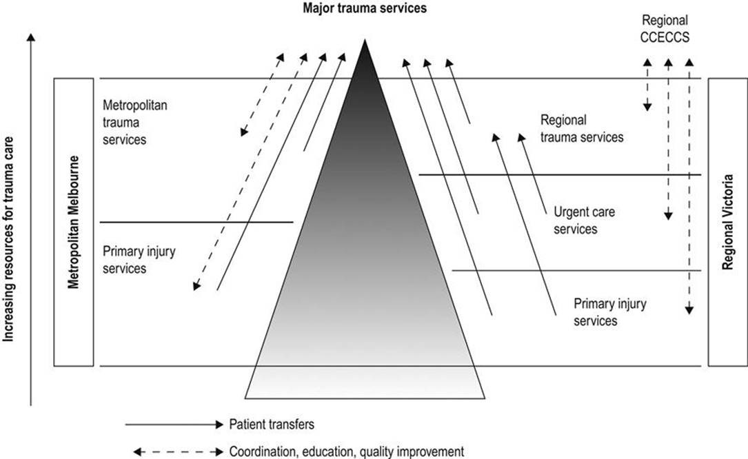
FIG. 3.1.1 Structure of the Integrated Victorian State Trauma System [13]. (From Atkin C, Freedman I, Rosenfeld JV, et al. The evolution of an integrated state trauma system in Victoria, Australia. Injury 2005; 36:1277–87.) CCECCS: Consultative Council on Emergency and Critical Care Services.
Criteria for identifying those patients who may require resuscitation at a tertiary level Trauma Centre or ‘Major Trauma Service (MTS)’ will depend on resources. In the most developed trauma systems, ‘mechanism of injury’ criteria are usually included in the pre-hospital triage tool. This ensures high sensitivity of the tool, but leads to considerable overtriage. In less-resourced settings, it may be appropriate to identify high-risk patients on the basis of abnormal vital signs and obvious major injury. The elements of a trauma system’s triage tool may include most or all of the predictors of life-threatening injury listed in Table 3.1.1.
Table 3.1.1
Predictors of life-threatening injury appropriate for use in pre-hospital trauma triage

The appropriate application of pre-hospital triage guidelines relies upon adequate resourcing. In regions with developed trauma systems, pre-hospital staff are expected to provide a range of advanced life support interventions including patient intubation and chest decompression, thereby ensuring that further organ injury is limited during the pre-hospital phase. The pre-hospital care providers armed with these skills may be doctors (as in many European countries) or highly trained paramedics (as in the USA, Australia and the UK).
The trauma system – intrahospital
Preparation
Effective pre-hospital communication, usually by phone and/or radio, allows timely preparation for the arrival of a trauma patient. Proper communication includes trauma team notification, staff and trauma bay identification and the adoption of universal precautions (gloves, gowns, etc.).
Trauma team notification might occur by phone or paging system and ensures the gathering of the trauma team prior to the patient’s arrival. Members of the trauma team may vary. An example of trauma team composition and roles in a Level 1 Trauma Centre is provided in Box 3.1.1. Variations to this list may occur in different settings depending upon the availability of skilled staff and the nature of specific injuries or physiological status prior to the patient’s arrival
Box 3.1.1
Team roles
Team leader (emergency physician or trauma surgeon)
Overview
Resuscitation
Assessment
Communication
Ambulance
Referrals
Investigations
Task allocation
Primary survey
Secondary survey
Airway doctor (anaesthetist)
Control of airway
Inline immobilization of cervical spine
Ventilation
Gastric tube
Procedure doctor (emergency registrar/trauma registrar)
Intravenous access/bloods
Intercostal catheter
Urinary catheter
ABG/art line
Nurses
Trauma nurse leader/scribe
Airway nurse
Circulation nurse
Radiographer
Orderly
Trauma team call-out criteria reflect the pre-hospital trauma triage criteria (see Table 3.1.1) and should be applied rigorously. Trauma team skill, functioning and leadership are essential to achieve the best patient outcome. The appropriate skill mix is reflected by the team membership listed in Box 3.1.1. Trauma team performance, including leadership and communication, will have an impact on patient outcome.
Initial management
The application of a consistent systematic approach to trauma resuscitation has been widely promulgated by training programmes, such as the Advanced Trauma Life Support (ATLS) [14]. The patient is brought directly to a prepared bay, the layout of which is illustrated in Figure 3.1.2. The principles of the initial identification and management of immediately life-threatening injuries, with prioritization accorded to airway (including protection of cervical spine), breathing, circulation, (neurological) disability and exposure are applied to all injured patients. This is followed by a secondary survey involving a head-to-toe examination. In most departments, parallel processing of the patient will occur simultaneously with management of ABCDE problems.

FIG. 3.1.2 The layout of a typical trauma resuscitation bay. (Reproduced with permission from Myers CT, Brown AF, Dunjey SJ, et al. Trauma teams: order from chaos. Emerg Med 1993; 5:34.)
Airway
It should be assumed that hypoxia is present in all patients who have sustained multiple injuries. Early expert airway intervention is essential. Every patient should receive initial supplemental oxygen via a well-fitting face-mask. If the airway is clear and protected, the neck should be immobilized with a semi-rigid collar but, if airway manoeuvres are necessary, it is often better to use manual inline immobilization without a collar, ensuring minimal neck movement with constant vigilance. The management of an obstructed airway in a trauma patient should be undertaken by an experienced senior clinician with significant anaesthetic experience. The first priority is to clear the upper airway by direct visualization, suction and removal of any foreign bodies. Insertion of an oropharyngeal or nasopharyngeal airway and the jaw-thrust manoeuvre are usually successful in clearing an upper airway obstruction. Insertion of a nasopharyngeal airway can be hazardous in patients with a fracture of the cribriform plate. The direction of insertion (backwards not upwards) is important. Chin lift is not recommended because it may cause additional movement of the cervical spine.
Early endotracheal intubation should be undertaken if the patient is apnoeic, has an unrelieved upper airway obstruction, has persistent internal bleeding from facial injuries, has respiratory insufficiency due to chest or head injuries or the potential for airway compromise (airway burns, facial instability, coma or seizures). Intubation may also be necessary for procedures such as computed tomography (CT) scanning or for the management of confused or disturbed patients. Any operator undertaking emergency intubation of major trauma victims should be skilled in rapid sequence induction and prepared for a difficult intubation. He/she must also be equipped with the skills for dealing with a ‘failed’ intubation (see Chapter 2.1) with a variety of interventions, including surgical cricothyroidotomy.
Breathing
Once the airway is secure, the patient’s breathing needs to be assessed. Particular attention is paid to optimizing oxygenation and maintaining normocapnia. During the primary survey, immediately life-threatening risks to breathing must be identified and dealt with. These injuries include tension pneumothorax, open pneumothorax, massive haemothorax, flail and pulmonary contusion. The specific features and management of these injuries is covered in Chapter 3.6.
Circulation
Shock is a clinical syndrome in which the perfusion of vital organs is inadequate to maintain function. Blood loss is the major cause of shock in the major trauma patient. Other less common causes of shock also need to be considered:
 Tension pneumothorax. This will cause rapid and severe disruption to the circulation.
Cardiogenic shock. This may be pre-existing (e.g. acute myocardial infarction (AMI) causing accident, drugs causing reduced cardiac compensation and hypovolaemia) or secondary to injury, i.e. myocardial contusion, valvular/septal injury or pericardial tamponade.
Neurogenic shock. This results from the loss of sympathetic tone. It may be caused by central brainstem injury and vasomotor instability or spinal cord injury and interruption of descending sympathetic tracts. It is characterized by bradycardia, but this may also occur in profound hypovolaemic states.
Anaphylactic shock and septic shock may coexist with hypovolaemic shock.
The initial stages of hypovolaemia can be difficult to detect. Reliance on systolic blood pressure to identify shock is dangerous. All patients who have sustained an injury that could be associated with significant blood loss, however remote the possibility, must be carefully monitored. In the initial phase, measurement of the clinical parameters will give some information about vital organ perfusion. However, more invasive monitoring will be required if hypovolaemia is severe or sustained.
It is essential to gain good venous access at the earliest phase in resuscitation. This is usually via two large-bore peripheral cannulae. In the absence of accessible arm veins, central venous access may be indicated. The recommended site (subclavian, jugular or femoral) depends on a number of factors. The subclavian vein is reliable in terms of patency, while the ease of access to the femoral vein is offset by its potential futility in major truncal haemorrhage. The internal jugular veins can be difficult to access in the immobilized trauma patient. Cut-downs of the saphenous veins and cubital fossa may also be used.
At the initial stages, where the blood pressure is unchanged, patients with potential blood loss can usually be managed without blood transfusion, namely crystalloid. There has been no benefit demonstrated in using non-blood colloids over crystalloids in traumatic haemorrhage [15–17]. Similarly, for hypertonic crystalloid the available data are inconclusive [18].
Where there is hypotension and tachycardia, blood transfusion should commence immediately, initially using O-negative blood and changing to group-specific or cross-matched blood as it becomes available. Chapter 3.12 covers the role and details of massive transfusion therapy in the trauma patient. Conversely, until the source of haemorrhage has been identified and haemostasis achieved, vigorous over resuscitation with fluid may actually result in a worse outcome [19,20].
The essential point is that after securing the airway and optimizing oxygenation and ventilation, the most important determinant of outcome in the major trauma patient with haemorrhagic shock is the time to definitive haemostasis. There is certainly no point in delaying surgery ‘to normalize the intravascular volume’. The major source of haemorrhage in the trauma patient must be identified early. The usual suspects are chest, abdomen, pelvis, long bone and/or external (e.g. scalp, major limb artery).
Disability
As the purpose of the primary survey is to identify immediate threats to life, the assessment for a head injury and its severity entails an examination of conscious state (Glasgow Coma Score) and neurological signs (pupils, limb weakness). If there is any risk of intracranial injury, a CT brain scan will be indicated immediately upon completion of the primary survey. Chapter 3.2 deals with the assessment and management of traumatic brain injury.
Exposure
Hypothermia is associated with worse outcomes in the major trauma patient [21]. Temperature control is now considered to be of critical importance in reducing the sequelae of major trauma (metabolic derangement, coagulopathy). The role of therapeutic hypothermia in isolated head injury remains controversial and is a subject of ongoing research. While maintaining normothermia, it is important to have fully exposed the patient, including a log roll with spinal immobilization, to enable a complete examination.
Next steps
By this stage, the trauma patient will have been received into a well-organized resuscitation area and the first life-saving procedures will have been initiated by an integrated and skilled team of doctors and nurses. Any immediately life-threatening conditions can be expected to have been identified and dealt with. Constant vigilance and reassessment are essential. Other occult injuries may be present in those patients identified with serious injuries.
While the trauma team leader continues to review the situation in the light of a constantly changing clinical scenario and, hopefully, the provision of more biomechanical data from the site of the incident, he or she should also be beginning to consider the next steps. The first of these is the calling in of other experts. Whereas it will have been clear that an airway doctor will be an essential part of the initial resuscitation team, it may be some minutes before it is known which other skills are required. Usually orthopaedic surgeons and neurosurgeons are near the top of the list. General surgery is not required as often as is commonly supposed [22], although general surgeons are often useful in coordinating ongoing care. Whichever specialty is required, the patient’s emergency problems demand experience, therefore, ‘if in doubt, refer’.
Radiographs are required at this stage. The initial films should be limited to those that will have a direct bearing on immediate management, including a chest AP view and a pelvis AP view.
Lateral X-ray of the cervical spine is no longer mandatory at this stage of assessment. Cervical immobilization is routine and it is not possible to exclude cervical injury with a lateral cervical spine X-ray. Therefore, a cervical X-ray does not alter initial management. Its utility at this stage would be to confirm an irretrievable injury, i.e. craniocervical dislocation.
Ideally, the resuscitation room should have an integrated X-ray facility but, if this is not available, portable films should be obtained. It is not appropriate to transfer a multiply injured unstable patient to a separate X-ray facility.
Other forms of imaging have become popular in localizing the source of haemorrhagic shock. The increasing availability of the focused assessment with sonography in trauma (FAST) has superseded diagnostic peritoneal lavage (DPL) as the bedside adjunct for detecting intraperitoneal haemorrhage.
Following the primary survey and investigations deemed necessary, the patient requires a secondary survey, the full head-to-toe examination for injuries not classified as immediately life-threatening.
Subsequent chapters deal with individual trauma problems, but it is essential that throughout the patient’s stay in hospital a single clinician has overriding responsibility for his or her care. In the resuscitation area, this is the ‘team leader’, who may be from any trauma-related discipline. Handover to the clinician responsible for ongoing care must be comprehensive, timed and well documented.
Trauma audit
Trauma kills people in a variety of ways; hence no one department in a hospital will see a large number of deaths. Many trauma victims die before they reach hospital, some in the ED and others scattered through the inpatient specialties and in intensive care. Hence from any one clinician’s perspective, trauma is not an outstanding problem. However, when looked at from a public health perspective, it is clearly a major issue, not least because some of the deaths are avoidable. Identifying these and the much more difficult-to-define group of patients who survive but whose outcome is not as good as expected is a major problem.
The most important variables to measure are the extent of the anatomical injury, the degree of physiological derangement that results, age and the previous well-being of the patient. All these have a direct effect on outcome and must therefore be measured before any comment can be made about the process of care. Outcome itself must also of course be measured. This is relatively easy in terms of mortality. However, disability is a much more difficult issue and, currently, there are no universally accepted measurement tools. The Glasgow Outcome Scale (GOSE) [23, 24], and the Short Form–36 (SF36) questions [25]are available tools that have been used. As 90% of major trauma patients survive their injury in a mature trauma system, it is important to measure disability and quality of life following major trauma when comparing outcomes [26].
Trauma audit was first formalized by Champion at the Washington Hospital Centre in the 1970s, using the Trauma Injury Severity Score (TRISS) methodology [27,28]. Most current trauma systems are now audited using some approach to examining risk-adjusted mortality. Injury severity adjustment, allowing intra- and intersystem comparison, usually requires measures of anatomical injury (e.g. Injury Severity Score), physiological effects (e.g. Revised Trauma Score) and age.
Trauma in developing countries
Globally, national governments are beginning to recognize the burgeoning human and economic cost of trauma, particularly road trauma. The public health achievements of the developed countries (seatbelts, helmets, alcohol and speed restrictions) are being implemented and, similarly, governments of developing countries are looking to implement trauma systems [29,30].
Research in developing countries reinforces the benefits of trauma systems previously described in countries with established emergency medical systems. For example, evidence indicates that people with life-threatening but potentially treatable injuries are up to six times more likely to die in a country with no organized trauma system than in one with an organized, resourced trauma system [31]. Trauma system development requires trauma outcome measurement [32,33]. As such, developing countries are likely to adopt trauma registries over the next several decades in an attempt to track the burden of trauma and the impact of system-wide interventions.
As developing countries embark upon trauma system development, it is becoming increasingly important to access standardized trauma care education through intensive short-courses. Advanced Trauma Life Support (ATLS) has been widely used. Other courses (such as Primary Trauma Care (PTC)) have also become popular in the developing world. Such courses are often less expensive and more flexible than ATLS.
Controversies
The number of major trauma patients necessary for a hospital to maintain high-quality trauma care.
The degree to which potential major trauma patients should be over-triaged to ensure that patients with major trauma are received at major trauma centres. There may be a greater risk in bypassing hospitals to take patients to a trauma centre, depending on distance and injury type. There is also the issue of deskilling of personnel from non-trauma centres and what effect this has on overall system outcomes.
The degree to which major trauma patients should be managed by protocol rather than clinical judgement. Clinicians are increasingly being asked to follow protocols in these critical situations. This prevents some adverse outcomes but may cause over-investigation and treatment.
The role of hypotensive resuscitation in blunt trauma has not been defined. Where victims have prolonged delays to theatre or the bleeding is not surgically correctable, then hypotensive resuscitation may cause more complications.
The role of controlled hypothermia in head-injured patients.
References
1. Centre for Disease Control and Prevention, National Center for Injury Prevention and Control: WISQARS Atlanta.<http://www.cdc.gov/injury/wisqars/facts.html>[Accessed Dec. 2012].
2. Australian Institute of Health and Welfare.<http://www.aihw.gov.au/deaths-faq/>[Accessed Dec. 2012].
3. Murray CJL, Vos T, Lozano R, et al. Disability-adjusted life years (DALYs) for 291 diseases and injuries in 21 regions, 1990–2010: a systematic analysis for the Global Burden of Disease Study 2010. Lancet. 2012;380:2197–2223.
4. Australian Bureau of Statistics. Causes of Death.<http://abs.gov.au/ausstats/>; 2010 [Accessed Dec. 2012].
5. Injury and poisoning mortality in England and Wales. Office for National Statistics.<http://www.ons.gov.uk/ons/publications/>[Accessed Dec. 2012].
6. Cameron PA, Gabbe B, Cooper DJ. A statewide system of trauma care in Victoria: effect on patient survival. Med J Aust. 2008;189:546–550.
7. Gabbe BJ, Simpson PM, Sutherland AM, et al. Improved functional outcomes for major trauma patients in a regionalized, inclusive trauma system. Ann Surg. 2012;255:1009–1015.
8. Trunkey DD. Trauma. Sci Am. 1983;249:28–35.
9. Cales RH, Trunkey DD. Preventable trauma deaths A review of trauma care systems development. J Am Med Assoc. 1985;254:1059–1063.
10. Roy PD. The value of trauma centres: a methodologic review. Can J Surg. 1987;30:17–22.
11. Pang JM, Civil I, Ng A, et al. Is the trimodal pattern of death after trauma a dated concept in the 21st century? Trauma deaths in Auckland 2004. Injury 2008; 39:102–106.
12. Eastman AB, Lewis FR, Champion HR, et al. Regional trauma system design: critical concepts. Am J Surg. 1987;154:79–87.
13. Atkin C, Freedman I, Rosenfeld JV, et al. The evolution of an integrated state trauma system in Victoria, Australia. Injury. 2005;36:1277–1287.
14. American College of Surgeons. ATLS (Advance Trauma Life Support) for Doctors Student Manual, 8th ed. Chicago: American College of Surgeons; 2008.
15. Roberts I, Alderson P, Bunn F. Colloids versus crystalloids for fluid resuscitation in critically ill patients (Cochrane Review). In: The Cochrane Library (4); 2004.
16. The SAFE Study Investigators. A comparison of albumin and saline for fluid resuscitation in the intensive care unit. N Engl J Med. 2004;350:2247–2256.
17. The SAFE Study Investigators. Saline or albumin for fluid resuscitation in patients with traumatic brain injury. N Engl J Med. 2007;357:874–884.
18. Bunn F, Roberts I, Tasker R. Hypertonic versus near isotonic crystalloid for fluid resuscitation in critically ill patients (Cochrane Review). In: The Cochrane Library (3); 2004.
19. Bickell WH, Wall MJ, Pepe PE, et al. Immediate versus delayed fluid resuscitation for hypertensive patients with penetrating torso injuries. N Engl J Med. 1994;331:1105–1109.
20. Civil IDJ. Resuscitation following injury: an end or a means? Aust NZ J Surg. 1993;63:921–926.
21. Ireland S, Endacott R, Cameron P, et al. The incidence and significance of accidental hypothermia in major trauma: a prospective observational study. Resuscitation. 2011;82:300–306.
22. Cameron PA, Dziukas L, Hadj A. Patterns of injury from major trauma in Victoria. Aust NZ J Surg 1995;65:830–4.
23. Jennett B, Bond M. Assessment of outcome after severe brain damage. Lancet. 1975;1:480–484.
24. Teasdale GM, Pettigrew LE, Wilson JT. Analysing outcome of severe head injury: a review and update on advancing the use of the Glasgow Outcome Scale. J Neurotrauma. 1998;15:587–597.
25. Garratt AM, Ruta DA, Abdulher MI. The SF36 Health Survey Questionnaire: an outcome measure suitable for routine use within the NHS? Br Med J. 1993;306:1440–1444.
26. Willis CD, Gabbe BJ, Cameron PA. Measuring quality in trauma care. Injury. 2007;38:527–537.
27. Boyd CR, Tolson MA, Copes WS. Evaluating trauma care: the TRISS method. J Trauma. 1987;27:370–378.
28. Champion HW, Copes WS, Sacco WJ, et al. The major trauma outcome study: establishing national norms for trauma care. J Trauma. 1990;30:1356–1365.
29. Mock C, Lormand JD, Goosen J, et al. Guidelines for essential trauma care Geneva: World Health Organization; 2004; http://whqlibdoc.who.int/publications/2004/9241546409.pdf.
30. Fitzgerald M, Dewan Y, O’Reilly G. India and the management of road crashes–towards a national trauma system. Ind J Surg. 2006;68:237–243.
31. Mock CN, Adzotor KE, Conklin E. Trauma outcomes in the rural developing world: comparison with an urban level 1 trauma center. J Trauma. 1993;35:518–523.
32. Mock C, Juillard C, Brundage S, et al. Guidelines for trauma quality improvement programmes Geneva: World Health Organization; 2009; <http://whqlibdoc.who.int/publications/2009/9789241597746_eng.pdf>.
33. O’Reilly GM, Cameron PA, Joshipura M. Global trauma registry mapping: A scoping review. Injury. 2012;43:1148–1153.
3.2 Neurotrauma
Gerard O‘Reilly, Lee Wallis and Peter Cameron
Essentials
1 Neurotrauma is a major cause of death in trauma.
2 A detailed history of the mechanics of the trauma experienced is invaluable.
3 Secondary brain injury is a major and potentially preventable cause of mortality and long-term morbidity.
4 Cerebral cellular dysfunction secondary to trauma is a result of both primary and secondary mechanisms and involves sodium, calcium and potassium shifts across the cell membrane, the development of oxygen free radicals and lipid peroxidation.
5 There are two features of prime importance to resuscitation in patients suffering neurotrauma: maintenance of airway/ventilation and maintenance of cerebral perfusion pressure.
6 Inline stabilization of the cervical spine during rapid sequence induction and orotracheal intubation is the preferred method for gaining definitive airway control in the head-injured patient.
7 Current emergency department and neurosurgical practice involves the use of CT scanning to investigate mild, moderate and severe head injury.
Introduction
Neurotrauma is a common feature in the presentation of multisystem trauma, particularly when associated with motor vehicle accidents and falls. Over 50% of trauma deaths are associated with head injury. The implications for the health system are enormous, with an annual rate of admission to hospital wards associated with head trauma approaching 300 per 100 000 population [1] and twice this in the elderly [2]. The long-term sequelae of moderate and severe neurotrauma are a major health resource drain and the morbidities associated with mild brain injury are becoming clearer.
Advances in preventative strategies, trauma systems, resuscitative therapies and rehabilitation management have improved outcomes. However, neurotrauma remains a serious health issue, predominantly affecting the productive youth of society.
Pathogenesis
Primary brain injury occurs as a result of the forces and disruptive mechanics of the original incident: this can only be avoided through preventative measures, such as the use of bicycle helmets.
Secondary brain injury is due to a complex interaction of factors and typically occurs within 2–24 hours of injury [3]. A principal mechanism of secondary injury is cerebral hypoxia due to impaired oxygenation or impaired cerebral blood flow. Cerebral blood flow is dependent on cerebral perfusion pressure (CPP), mean arterial systemic blood pressure (MAP) and intracranial pressure (ICP).

Intracranial pressure may be raised as a result of the mass effect of the haemorrhage or by generalized cerebral oedema. Cerebral vasospasm further reduces cerebral blood flow in patients in whom significant subarachnoid haemorrhage has occurred.
Cellular dysfunction is a result of both primary and secondary mechanisms and involves sodium, calcium, magnesium and potassium shifts across the cell membrane, the development of oxygen free radicals, lipid peroxidation and glutamate hyperactivity. Excessive release of excitatory neurotransmitters and magnesium depletion also occur [4].
Classification of primary injury in neurotrauma
Primary injuries are classified as:
skull fracture
concussion
contusion
intracranial haematoma
diffuse axonal injury
penetrating injury.
Skull fracture
The significance of skull fracture is not related to the specific bony injury but rather the associated neurotrauma. Fractures in the region of the middle meningeal artery in particular may be associated with acute extradural haemorrhage. Fractures involving the skull base and cribriform plate may be associated with CSF leak and the risk of secondary infection. Depressed skull fractures may compress underlying structures, cause secondary brain injury and require surgical elevation. Injury to underlying structures may result in secondary epilepsy.
Concussion
Concussion is a transient alteration in cerebral function, usually associated with loss of consciousness and often followed by a rapid recovery. The proposed mechanism is a disturbance in the function of the reticular activating system. Post-concussive syndromes, including headache and mild cognitive disturbance, are common [5,6]. Symptoms, particularly headache, are usually short-lived but may persist. ‘Second-impact syndrome’ describes a greater risk of significant re-injury following an initial injury causing a simple concussion. It is likely to be due to diffuse cerebral swelling [7]. In animal models, concussion may be associated with modest short-term increases in intracranial pressure and disturbances in cerebral cellular function [8].
Contusion
Cerebral contusion is bruising of the brain substance associated with head trauma. The most common mechanism is blunt trauma. Forces involved are less than those required to cause major shearing injuries and often occur in the absence of skull fracture. Morbidity is related to the size and site of the contusion and coexistent injury. Larger contusions may be associated with haematoma formation, secondary oedema or seizure activity. The most common sites for contusions are the frontal and temporal lobes [9].
Intracranial haematoma
Extradural
Extradural haematoma (EDH) is uncommon but classically associated with fracture of the temporal bone and injury to the underlying middle meningeal artery. Haemorrhage subsequently occurs, stripping the dura from the skull and expanding to cause a rise in intracranial pressure and eventually uncal herniation and death. Haemorrhage may be from vessels other than the middle meningeal artery (e.g. brisk arteriolar or venous bleeding). Signs will depend on the site of the haematoma.
Subdural
Subdural haematomata (SDH) may have an acute, subacute or chronic course. It generally follows moderate head trauma with loss of consciousness. In the elderly, SDH may be associated with trivial injury and, in children, with shaking (abuse) injury. Haemorrhage occurs into the subdural space, slowly enlarging to cause a space-occupying collection whose functional implications will vary according to location. Acute subdural haemorrhage carries a high mortality (>50%), similar to acute EDH. Subacute and chronic SDH is associated with a degree of cerebral dysfunction, headache or other symptomatology and is associated with a significantly lower mortality (up to 20%) [10].
Intracerebral
As with cerebral contusion, the most common sites of intracerebral haemorrhage associated with trauma are the temporal and posterior frontal lobes. Effects on function are variable, depending on the site. Intracerebral haemorrhage may progress from an initial contusion or be secondary to altered vascular characteristics. Symptom development and complications may be delayed as the size of the haemorrhage increases over time.
Subarachnoid and intraventricular haemorrhage
Subarachnoid blood is relatively common after major head injury. Intraventricular haemorrhage may also be evident. As in non-traumatic settings, the presence of subarachnoid blood may lead to cerebral vasospasm and secondary ischaemic brain injury.
Diffuse axonal injury
Diffuse axonal injury (DAI) is the predominant mechanism of injury in neurotrauma, occurring in up to 50% of patients [11]. Shearing and rotational forces on the axonal network may result in major structural and functional disturbance at a microscopic level. Disturbance to important communicative pathways sometimes results in significant long-term morbidity, despite non-specific or minimal changes on computed tomography (CT) scanning. The exact pathogenesis of diffuse axonal injury is incompletely understood. Specific injury in the regions of the corpus callosum and midbrain has been proposed; however, DAI is believed to be the mechanism for persistent neurological deficits seen in head-traumatized individuals with normal CT scans [12].
Penetrating injury
Penetrating neurotrauma is characterized by high levels of morbidity and mortality. This is especially true of gunshot wounds. Exposure of cerebral tissue through large compound wounds or through basilar skull structures is associated with a dismal outlook. Penetrating injury in the periorbital and perinasal regions is associated with high risk of infection.
Epidemiology
Neurotrauma is surprisingly common. In some settings, more than 30% of the population have suffered from a trauma brain injury. In addition to being a major cause of death in trauma, neurotrauma leads to significant morbidity. More than 40% of those who have sustained a traumatic brain injury will have residual disability one year later [13].
Common causes include motor vehicle accidents (including vehicle versus pedestrian and bicycle collisions), falls, assault and firearms. In young males, alcohol is often involved.
Prevention
Primary prevention of neurotrauma depends on the cause. Most preventative strategies are directed at vehicular traffic and include speed-calming measures, in-car safety devices and bicycle helmets. Improving roadside lighting and enhancing pedestrian visibility contribute to reduction of injury in this group.
Prevention of secondary injury involves maintenance of cerebral perfusion and oxygenation and is addressed under clinical management.
Clinical features
Definition
Neurotrauma may be classified according to severity as minimal, mild, moderate or severe (Box 3.2.1) [14]. Such a classification allows for directed investigation and management, but there is clearly a continuum of injury within the spectrum of neurotrauma.
Box 3.2.1
Neurotrauma severity
Minimal
No loss of consciousness, and
Glasgow Coma Score (GCS) 15, and
Normal alertness and memory, and
No neurological deficit, and
No palpable depressed fracture or other sign of skull fracture
Mild
Brief (<5 minutes) loss of consciousness, or amnesia for event, or
GCS 14, or
Impaired alertness or memory
No palpable depressed fracture or other sign of skull fracture
Moderate or potentially severe
Prolonged (>5 minutes) loss of consciousness, or
Persistent GCS<14, or
Focal neurological deficit, or
Post-traumatic seizure, or
Intracranial lesion on CT scan, or
Palpable depressed skull fracture
History
A detailed history of the mechanics of the trauma is essential. This should be followed by consideration of time courses, pre-hospital care, pre-sedative and pre-relaxant neuromuscular function and episodes and duration of hypotension or other decompensation. A history of previous health problems, allergies, medications and social setting is desirable.
Primary survey
As with all trauma patients, the initial assessment and therapy must be directed at maintenance of airway, ventilation and circulatory adequacy along standard Advanced Trauma Life Support (ATLS) principles. Early assessment of neurological disturbance using the Glasgow coma score (GCS) or AVPU scale (Alert: GCS 14–15; response to Verbal stimuli: GCS 9–13; response to Painful stimuli: GCS 6–8; or Unresponsive: GCS 3–5) is important. Simultaneous protection of the cervical spine by immobilization is fundamental. This management should commence in the pre-hospital setting and the level of care be maintained.
The greatest risks to the patient with a moderate to severe head injury are hypoxic injury and deficient cerebral perfusion due to systemic hypotension.
Secondary survey
A full secondary survey, including log-roll, should follow.
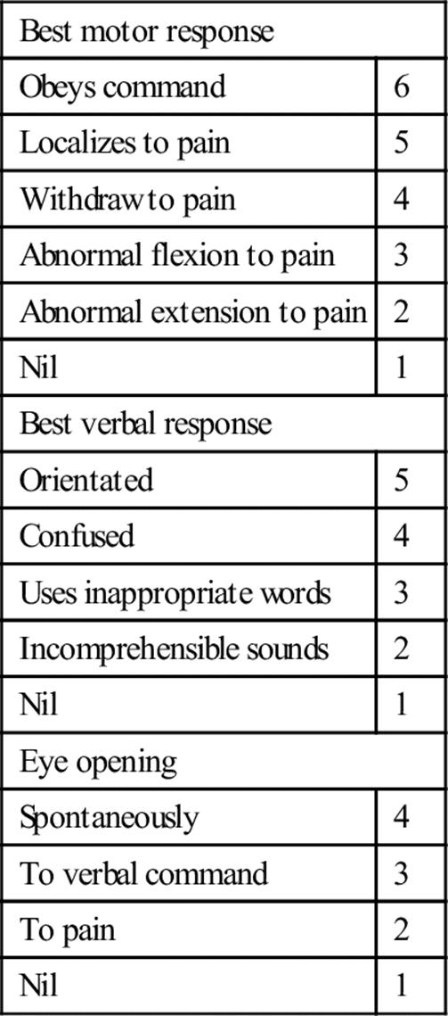
Clinical assessment of the neurological status of head-injured patients commences with formal documentation of the GCS (Table 3.2.1). The maximum score is 15 and the minimum 3. Coma may be defined in terms of the GCS, in which patients have a total score of 8 or less:
Fail to show eye opening in response to pain (eye-opening response=1)
Fail to obey commands (best motor response=5)
Make at best only incomprehensible sounds (best verbal response=2).
Table 3.2.1
Glasgow coma score
Examination of pupillary responses, particularly in the unconscious patient, is important as an indicator of increasing intracranial pressure; a non-responsive dilated pupil indicating ipsilateral herniation. However, a more common cause of abnormal pupil reactions in head injury is the presence of direct ocular trauma.
A general neurological examination, including reflexes, should be performed; the degree to which cooperation is possible and lateralization of signs being particularly important to document. Consideration of the pre-injury mental state is important, particularly where drug or alcohol intoxication is possible.
Clinical investigations
Minimal–mild head injury
In head injury associated with loss of consciousness or amnesia and a GCS of 14–15, CT scanning will demonstrate a relevant positive scan (i.e. cerebral contusion, haematoma, oedema, pneumocephalus) in 7–12% and a subsequent craniotomy rate of 1–3% [15–18].
On the weight of research evidence, current ED investigation of mild head injury should include CT scanning in all patients in this group [14,19–22]. Certain high-risk groups (such as the intoxicated, the elderly (>65 years), anticoagulated or demented patients) warrant CT scanning even after minimal presumed or possible head injury.
Despite considerable research within the minimal–mild head injury group, reliable risk stratification has not been achieved. The Canadian Head Rules detail five high-risk criteria for neurosurgical intervention in patients with GCS 13–15 and mild head injury [23]. The NICE head injury rules were based in part on the Canadian rules [24]; the NEXUS II [25] investigators showed that development of a simple head injury CT rule that is both sensitive and specific is extremely difficult. There have been conflicting results as to which has the best predictive power in adults and children, however, each has its critics for over-scanning [26,27].
Cervical spine imaging is indicated if the patient has neck pain, neurological abnormality, altered conscious state, intoxication or significant distracting injury.
Moderate–severe head injury
Urgent CT scanning is the investigation of choice in moderate-to-severe neurotrauma (GCS of 3–13); however, other investigations and therapy may take priority in the patient with multisystem trauma, particularly in the presence of unresponsive haemorrhagic shock.
In the absence of a CT scan, consultation with a neurosurgeon or early transfer to an appropriate facility is essential.
Imaging of the cervical spine is indicated in all patients with moderate to severe neurotrauma. A significant proportion of patients with severe head injury will have cervical spine fractures.
Treatment
Minimal–mild head injury
All patients with mild head injury must be counselled appropriately and discharged with written advice in the care of a responsible adult. Specific advice must be provided regarding expected duration of symptoms, possible risks or delayed complications and reasons for re-presentation to the ED (Box 3.2.2). Information should also be given about the second-impact syndrome and exclusions from sporting activity.
Box 3.2.2
Patient advice
General advice following head injury
The patient should read and understand these instructions:
Rest comfortably at home in the company of a responsible adult for the next 12–24 hours
Resume normal activity after feeling recovered
Drink clear fluids and consume a light diet only for the first 6–12 hours (a normal diet may be commenced as desired after that)
Mild pain killers (such as paracetamol) may be taken for headache as directed by the doctor
Following head injury, a small number of patients develop ongoing symptoms, such as recurrent mild headache, concentration difficulties, difficulty with complex tasks, mood disturbance, etc. If you notice such problems, consult your local doctor for appropriate referral
Avoid exposure to activities that may create risk of further head injury within the next 2 weeks
If you do not understand these instructions and advice, check with emergency department staff before your discharge or consult your local doctor
If you require a certificate for work please make this clear to emergency department staff
Report immediately the following problems
Persistent vomiting (more than twice)
Persistent drowsiness–unable to be woken up completely
Confusion or disorientation or slurred speech
Increased headache (not relieved by standard doses of paracetamol)
Localized weakness or altered sensation or incoordination
Blurred or double vision
Seizures, fits or convulsions
Neck stiffness
Follow up by a local medical officer should be arranged and neuropsychological assessment may be warranted for high-risk groups. Patients should be cautioned about making major life, occupational and financial decisions until they are free of post-concussive symptoms.
In minimal and mild head injury, a normal CT scan and the absence of neurological abnormality are reasonable criteria for patient discharge [3]. It is essential to assess for ongoing post-traumatic amnesia (PTA), as this is frequently overlooked in the ED. A simple screen to use is the modified Westmead PTA scale [6]. In the presence of these criteria, the persistence of mild symptoms (e.g. mild headache, nausea, occasional vomiting) is common and patients should be advised accordingly. In adults, such symptoms may be treated with mild analgesics (paracetamol, aspirin) and antiemetics (metoclopramide, prochlorperazine) and the patient discharged when comfortable. Advising patients that there will be problems with post-concussive symptoms (including short-term memory and information processing) and providing them with written material has been shown to improve outcomes at 3 months [6,28].
Currently there is no drug to treat the primary pathology in mild and minor head injury [29].
Moderate–severe head injury
Priority in the management of moderate to severe neurotrauma is given to maintenance of the airway and an adequate cerebral perfusion pressure. Hypotension (SBP<90) and hypoxia (PaO2<60 [8 kPa]) should be corrected immediately [30]. Control or modification of intracranial pressure has a place in the emergency management of neurotrauma. Avoidance of secondary brain injury and associated cerebral swelling is the mainstay of such therapy.
Intracranial pressure monitoring is generally indicated in patients with severe head injury (GCS<8) who remain comatose. Institutional variability exists in methods for measurement, as do specific indications for monitoring. Elevation of the head of the bed to 30° will reduce ICP modestly without altering CPP.
Mannitol (0.5–1.0 g/kg IV) may produce a short-term reduction in ICP. Mannitol causes an osmotic dehydration which is non-selective. Complications of mannitol therapy include fluid overload, hyperosmolality, hypovolaemia and rebound cerebral oedema. Mannitol may be used as a temporizing measure to enable a patient with a surgically remediable lesion to get to theatre.
Routine use of hyperventilation in head injury is contraindicated. Hypocarbia reduces cerebral blood flow (and ICP) through vasoconstriction which, if extreme, may reduce CPP to the point of exacerbation of secondary brain injury [31].
Anticonvulsant prophylaxis (phenytoin 15–18 mg/kg IV over 30–60 minutes) is indicated for the prevention of seizures within the first week after injury [32]. Seizures are managed acutely using standard therapies and guidelines (including benzodiazepines and phenytoin). The use of barbiturates, endotracheal intubation and mechanical ventilation may be indicated for status epilepticus or seizures that are refractory to therapy.
Antibiotic prophylaxis is indicated for compound fractures. Tetanus immunoprophylaxis is given as part of routine wound care. Steroid therapy has had varied support but is not recommended [33]; in 2005, the CRASH collaborators reported conclusively that intravenous corticosteroids should not be used in the treatment of head injury [34].
There has been considerable interest and experimental endeavour with regard to cerebral protection and salvage therapies. To date, no benefit has been demonstrated in the administration of aminosteroids, amino acids or monoamine antagonists in patients with head injury [35–37] and the role of calcium channel blockers remains unclear [38]. More recent research demonstrated worse outcomes for patients with diffuse traumatic brain injury who had a decompressive craniectomy [39]. The role of hypothermia is controversial; while animal studies have shown a benefit, prospective studies have shown either harm or no harm [40,41]. In summary, general supportive therapy, including the maintenance of thermoregulation, hydration, pressure care and nutrition are the mainstays of therapy.
Resuscitation in neurotrauma
There are two features of prime importance to resuscitation in patients suffering neurotrauma:
maintenance of airway and ventilation
maintenance of cerebral perfusion pressure.
With elevation of intracranial pressure and loss of autoregulation of cerebral circulation, relatively higher systemic blood pressures are required. The practice of minimal-volume resuscitation has no place in the patient with serious neurotrauma. Standard approaches to the management of hypovolaemia in head- injured patients should be adopted. The use of hypertonic solutions in resuscitation (including hypertonic saline) has been studied with variable conclusions [42]. But the only randomized controlled trial performed with hypertonic saline showed no improvement in outcome [43]. Albumin has also been shown to have detrimental effects in severe traumatic brain injury [44].
Indications for intubation and ventilation of the neurotrauma patient are inadequate ventilation or gas exchange (hypercarbia, hypoxia, apnoea); inability to maintain airway integrity (protective reflexes); a combative or agitated patient; and the need for transport where the status of the airway is potentially unstable (between hospitals, to CT, to angiography, etc.).
Disposition
In patients with minimal–mild head injury, recommendations with regard to a ‘safe’ period of observation, need for hospital admission or predictive value of injury mechanism are not consistent. Rural and isolated settings present logistic difficulties in the management of this group. Careful observation for a prolonged period is a reasonable alternative and early neurosurgical consultation, together with a low threshold to transfer to a neurosurgical centre, is prudent.
Patients with moderate to severe neurotrauma require hospital admission, preferably under the care of a neurosurgeon in a specialized neurosurgical unit or ICU. Rehabilitation and social readjustment is a focus of therapy from early in the clinical course.
Inter-hospital transfer of patients with significant neurotrauma requires the attendance of skilled transfer staff and the maintenance of level of care during transfer. Airway management must anticipate the potential for the patient to deteriorate en route. The presence of pneumocephalus precludes unpressurized (high) altitude flight. The use of teleradiology and neurosurgical consultation will be of value in the management of the remote head-injured patient.
Prognosis
The level of residual neurological impairment is a function of the severity of the degree of trauma and quality of care. A poor outcome is associated with prolonged pre-hospital time, delay of transfer to the appropriate facility, admission to an inappropriate facility and delay in definitive surgical treatment.
Overall mortality in severe head injury is of the order of 35%. A lower GCS at presentation is associated with a worse outcome. Approximately half the patients who remain comatose with GCS<9 for longer than 6 hours will die [12]. Acute subdural haematoma and diffuse axonal injury producing persistent coma are associated with the vast majority of neurotrauma deaths. Early neurological abnormalities are, however, not reliable prognostic factors and an initial period of maximally aggressive therapy is indicated in patients with closed neurotrauma.
Controversies
Intracranial pressure monitoring has not been shown to improve outcome from major head injury.
The role of CT scanning in minor head injury has become more widespread. Although it is increasingly accepted that CT is indicated, the timing or urgency of the investigation is controversial. Further studies are required to define discriminators and high-risk markers as guides to the most rational application of this investigation.
Consideration should be given to referral of patients with minor or worse head injury with persistent post-concussive symptoms for neuropyschological assessment in order to facilitate recovery and resumption of normal activities.
Acknowledgements
The chapter’s authors for this edition would like to acknowledge the important contribution of authors for previous editions, including Dr Marcus Kennedy.
References
1. Tennant A. Admission to hospital following head injury in England: incidence and socioeconomic associations. BMC Publ Hlth. 2005;5:21–29.
2. Jamieson LM, Roberts-Thomson KF. Hospitalised head injuries among older people in Australia 1998/1999 to 2004/2005. Injury Prevent. 2007;13:243–247.
3. Kay A, Teasdale MB. Head injury in the United Kingdom. World J Surg. 2001;25:1210–1220.
4. Morris JA, Limbird TJ, MacKenzie E. Rehabilitation of the trauma patient. In: Moore EE, Mattox KL, Feliciano DV, eds. Trauma. 2nd ed. Norwalk: Appleton & Lange 1991;815.
5. Lahaye PA, Gade GF, Becker DP. Injury to the cranium.. In: Moore EE, Mattox KL, Feliciano DV, eds. Trauma. 2nd ed. Norwalk: Appleton & Lange 1991;247.
6. Ponsford J, Willmott C, Rothwell A, et al. Factors influencing outcome following mild traumatic brain injury in adults. J Internatl Neurol Soc. 2000;6:568–579.
7. McCory P. Does second impact syndrome exist? Clin J Sport Med. 2001;11:144–149.
8. Goldman H, Hodgson V, Morehead M, et al. A rat model of closed head injury. J Neurotrauma. 1990;8:129.
9. Javid M. Head injuries. N Engl J Med. 1974;291:890.
10. Povlishock JT. Pathobiology of traumatically induced axonal injury in animals and man. Ann Emerg Med. 1993;22:980.
11. Meythaler JM, Peduzzi JD, Eleftheriou E, et al. Current concepts: Diffuse axonal injury-associated traumatic brain injury. Arch Phys Med Rehabil. 2001;82:1461–1471.
12. Statham PF, Andrews PJ. Central nervous system trauma. Baillière’s Clin Neurol. 1996;5:501.
13. Corrigan JD, Selassie AW, Orman JA. The epidemiology of traumatic brain injury. J Head Trauma Rehabil. 2010;25:72–80.
14. Stein S, Ross S. Minor head injury: a proposed strategy for emergency management. Ann Emerg Med. 1993;22:1193–1196.
15. Shackford S, Waid S, Ross SE, et al. The clinical utility of computed tomographic scanning and neurological examination in the management of patients with minor head injuries. J Trauma. 1992;33:385–394.
16. Stein S, Ross SJ. Mild head injury: a plea for routine early CT scanning. Trauma. 1992;33:11–13.
17. Richards KA, Lukin WG, Jones P. Minor head injuries. (Royal Brisbane Hospital, personal communication. Unpublished data, 1997).
18. Lenninger BE, Kreutzer JS, Hill MR. Comparison of minor and severe head injury emotional sequelae using the MMPI. Brain Injury. 1991;5:199–205.
19. Newcombe R, Merry G. The management of acute neurotrauma in rural and remote locations: a set of guidelines for the care of head and spinal injuries. J Clin Neurosci. 1999;6:85–93.
20. Victorian Road Trauma Committee. Report of the Consultative Committee on Road Traffic Fatalities. Victorian Institute of Forensic Pathology, Royal Australasian College of Surgeons, 1997.
21. McAllister TW. Neuropsychiatric sequelae of head injuries. Psychiatr Clin N Am. 1992;15:S395–S413.
22. Bullock R, Chesnut RM, Clifton G, et al. Guidelines for the management of severe head injury. Eur J Emerg Med. 1996;2:109–127.
23. Stiell I, Wells G, Vandenheem K, et al. The Canadian CT rule for patients with minor head injury. Lancet. 2001;357:1391–1396.
24. National Institute for Clinical Excellence. Clinical Guideline 56. Head Injury: triage, assessment, investigation and early management of head injury in infants, children and adults. London: NICE; 2007.<http://publications.nice.org.uk/head-injury-cg56>[Accessed Jan. 2013].
25. Mower W, Hoffman J, Herbert M, et al. Developing a clinical decision instrument to rule out intracranial injuries in patients with minor head trauma: methodology of the NEXUS II investigation. Ann Emerg Med. 2002;40:504–514.
26. Smits M, Dippel DW, De Hann GG, et al. External validation of the Canadian CT Head Rule and the New Orleans Criteria for CT scanning in patients with minor head injury. J Am Med Assoc. 2005;294:1519–1525.
27. Dunning J, Daly JP, Malhotra R, et al. The implications of NICE guidelines on the management of children presenting with head injury. Arch Dis Child. 2004;89:763–767.
28. NSW Ministry of Health. Adult Trauma Clinical Practice Guidelines. Initial Management of Closed Head Injury in Adults, 2nd ed. North Sydney; 2011.<http://www.itim.nsw.gov.au/images/3/3d/Closed_Head_Injury_CPG_2nd_Ed_Full_document.pdf>[Accessed Jan. 2013].
29. McCrory P. New treatments for concussion: The next millennium beckons. Clin J Sport Med. 2001;11:190–193.
30. Chesnut RM, Marshall LF, Klauber MR, et al. The role of secondary brain injury in determining outcome from severe head injury. J Trauma. 1993;34:216–222.
31. Fortune JB, Fenstel PJ, Graca L, et al. Effect of hyperventilation, mannitol and ventriculostomy drainage on cerebral blood flow after head injury. J Trauma Injury Infect Crit Care. 1995;39:1091–1099.
32. Temkin NR, Dikmen SS, Wilensky AJ, et al. A randomised, double blind study of phenytoin for the prevention of post-traumatic seizures. N Engl J Med. 1990;323:497–502.
33. Clausen T, Bullock R. Medical treatment and neuroprotection in traumatic brain injury. Curr Pharm Design. 2001;7:1517–1532.
34. Edwards P, Arango M, Balica L, et al. Final results of a randomised placebo controlled trial of intravenous corticosteroid in adults with head injury–outcomes at 6 months. Lancet. 2005;365:1957–1959.
35. Marshall LF, Maas AIR, Bowers MS, et al. A multicenter trial on the efficacy of using tirilazad mesylate in cases of head injury. J Neurosurg. 1998;89:519–525.
36. Willis C, Lybrand S. Bellamy N Excitatory amino acid inhibitors for traumatic brain injury. Cochrane Database Syst Rev. 2003;1:CD003986.
37. Forsyth RJ, Jayamoni B, Paine TC, Mascarenhas S. Monoaminergic agonists for acute traumatic brain injury. Cochrane Database Syst Rev. 2006;4:CD003984.
38. Langham J, Goldfrad C, Teasdale G, et al. Calcium channel blockers for acute traumatic brain injury. Cochrane Database Syst Rev. 2003;4:CD000565.
39. Cooper DJ, Rosenfeld J, Murray L, et al. Decompressive craniectomy in diffuse traumatic brain injury. N Engl J Med. 2011;364:1493–1502.
40. Sydenham E, Roberts I, Alderson P. Hypothermia for traumatic head injury. Cochrane Database Syst Rev. 2009;2:CD001048.
41. Ireland S, Endacott R, Cameron P, et al. The incidence and significance of accidental hypothermia in major trauma – a prospective observational study. Resuscitation. 2011;82:300–306.
42. Vassar MJ, Fischer RP, O’Brien PE, et al. A multicenter trial for resuscitation of injured patients with 7.5% sodium chloride The effect of added dextran 70 The Multicenter Group for the Study of Hypertonic Saline in Trauma Patients. Arch Surg. 1993;128:1003–1011.
43. Cooper JD, Myles PS, McDermott FT, et al. Prehospital hypertonic saline resuscitation of patients with hypotension and severe traumatic brain injury. J Am Med Assoc. 2004;291:1350–1357.
44. Myburgh J, Cooper J, Finfer S, et al. Saline or albumin for fluid resuscitation in patients with traumatic brain injury. N Engl J Med. 2007;357:874–884.
3.3 Spinal trauma
Pieter van Driel
Essentials
1 Cervical spine injury can be confidently eliminated in conscious, clear-headed patients younger than 65 years, using clinical examination criteria (as described in NEXUS and Canadian C-spine rules) alone.
2 Physical examination alone does not assist in the diagnosis of unstable vertebral injury unless the deformity is gross.
3 A lack of neurological symptoms and signs does not eliminate spinal column injury or spinal cord at risk.
4 A patient can be ambulant and still have a major vertebral injury, even a potentially unstable one.
5 The natural history of spinal cord injury may lead to progressively increasing symptoms commencing some hours after the incident.
6 Magnetic resonance imaging is evolving as the imaging modality of choice in patients with neurological signs.
7 The likelihood of significant vertebral injury in unconscious trauma victims is 10%; 2% of all trauma victims with significant altered conscious state have a spinal cord injury.
8 Although spinal immobilization is a standard of care for protecting the spine, the use of these devices can have adverse clinical effects.
9 Methylprednisolone is not recommended in most Australian centres but, if given, it should be within 8 hours after spinal cord injury in order to improve both motor function and functional outcome.
Introduction
Spinal cord injury is one of the most disabling traumas, causing major and irreversible physical and psychological disability to the patient and permanently affecting their lifestyle. The emotional, social and economic consequences affect the individual, family, friends and society in general.
Approximately 2% of adult victims of blunt trauma suffer a spinal injury and this risk is tripled in patients with craniofacial injury [1].
Motor vehicle collisions, falls and sporting injuries – notably diving and water sports – are the major causes of acute spinal cord injury in Australia [2]. Road traffic accidents account for about half of all spinal injuries. Despite the work to minimize spinal injuries in contact sports, such as rugby, serious spinal cord injuries still occur [2]. Spinal injuries occur mostly in young people, but minor falls in the elderly or low-impact injuries in people with pre-existing bony pathology can also cause spinal cord damage. Spinal cord injury due to pathological vertebral fractures may be the first presentation of malignancy.
Observations from two studies [3,4] suggest that possibly preventable neurological deterioration may be due to one or more of the following:
the injury not being recognized initially, e.g. not being specifically examined for, occult or masked by other injuries
the onset of the secondary effects of the spinal cord injury involving oedema and/or ischaemia
aggravation of the initial spinal cord lesion by inadequate oxygenation and/or hypotension
aggravation of the initial spinal cord lesion by inadequate vertebral immobilization.
Pathophysiology
Level of vertebral injury
The level of neurological injury in patients who sustain spinal injuries is variously reported. In studies from Victoria and New South Wales [4,5], the distribution of the level of injuries was cervical 60%, thoracic 30%, lumbar 4% and sacral 2%.
Spinal cord injuries occur most commonly at the level of the 5th, 6th and 7th cervical vertebrae, largely because of the greater mobility of these regions. The C5–6 and C6–7 levels account for almost 50% of all subluxation injury patterns in blunt cervical spinal trauma [6].
Associated injuries
There are three noteworthy observations [3,5] from associated injuries in patients with spinal injury:
Approximately 8–10% of patients with a vertebral fracture have a secondary fracture of another vertebra, often at a distant site. These secondary fractures are usually associated with the more violent mechanisms of injury, such as ejection or rollover. Secondary injuries are usually relatively minor and stable, e.g. fractures of the vertebral processes but, occasionally, they may be major and may also be associated with neurological damage. Therefore, when ‘thinking spine’, it is important to ‘think whole spine’ and, in particular, to attempt to avoid rotation of the vertebral column.
Owing to the mechanism of injury, many patients with spinal injuries often have other associated injuries, including head, intrathoracic or intra-abdominal injuries, which may modify management priorities [5].
Patients may complain of pain from other injuries and hence a back or neck injury may go unnoticed. Pain may often not be a significant feature despite severe vertebral column damage. Furthermore, spinal pain may take some time to become apparent because of other pathological processes modifying pain, such as swelling and inflammation.
Spinal trauma might result in several injuries directly related to the spinal cord. Specific injuries, such as vertebral injuries, spinal shock, spinal cord injuries and their neurological symptoms, are described later in this chapter.
Autonomic nervous system effects of spinal cord damage
Autonomic nervous system effects are mentioned here as important pathophysiological mechanisms must be understood to deliver optimum care and treatment to patients with spinal cord injuries.
The whole of the sympathetic nervous system and the pelvic parasympathetic outflow is transmitted in the spinal cord. In an injury higher than the upper thoracic vertebrae, there is significant impairment of total body sympathetic and pelvic parasympathetic functions. The extent and severity of autonomic dysfunction is dependent on the segmental level(s) and the extent or completeness of the neurological insult.
Direct effects
Direct effects include manifestations related to the cardiovascular, gastrointestinal, urogenital and thermoregulatory systems.
Cardiovascular effects
In complete quadriplegia, sympathetic denervation causes relaxation of resting vasomotor tone, resulting in generalized systemic vasodilatation. It is recognized by dry extremities with variable warmth and colour during initial assessment. In males, there may be penile engorgement or priapism. Owing to the peripheral vasodilatation, there is a drop in total peripheral resistance, with consequent hypotension (neurogenic shock). Under normal circumstances, this would result in a baroreceptor response in order to achieve compensation. However, as the effector arm of the sympathetic nervous system is paralysed, the normal compensatory effects of tachycardia and vasoconstriction do not occur. The vagus nerve carrying parasympathetic supply to the heart is unopposed, with resultant bradycardia. The higher and more complete the spinal cord injury, the more extensive the autonomic dysfunction.
The usual symptoms and signs of the shock process in response to hypovolaemia cannot occur, as tachycardia and vasoconstriction are mediated by the sympathetic nervous system, which has been interrupted by the high spinal cord lesion.
Gastrointestinal effects
Following spinal cord injury, a paralytic ileus develops. This is usually self-limiting and recovers over 3–10 days. Paralysis of sphincters occurs at the lower end of the oesophagus and at the pylorus; as a consequence, passive aspiration of the stomach contents, especially of fluid, is a potential problem. Furthermore, owing to thoracic and abdominal wall muscle paralysis, the capacity to cough and hence clear the airway is diminished. In quadriplegia and high paraplegia, occult fluid aspiration due to passive regurgitation of retained gastric content may not be recognized. The airway therefore requires close observation and active protection. A nasogastric tube must be inserted and gastric contents drained.
Urinary effects
Urinary retention is partly the consequence of acute bladder denervation and, in the early post-injury phase, due to spinal shock. Catheter insertion is required to prevent overdistension of the bladder in order to optimize recovery. It also permits measurement of urinary output.
Thermoregulatory effects
Following cervical or upper thoracic spinal cord injury, the spinal patient effectively becomes poikilothermic. In a cold environment, they are unable to vasoconstrict to conserve heat or shiver to generate heat. The patient is already peripherally vasodilated which promotes loss of heat and lowering of body temperature. In the warm environment, although the patient is already peripherally vasodilated, the capacity to sweat is sympathetically controlled and therefore lost.
Pre-hospital issues
Extrication and immobilization
Emergency medical services (EMS) personnel are sent to see trauma patients in difficult circumstances. Patients, for instance, could be stuck in vehicles, (partially) submersed in water or found in small and inconvenient places. These circumstances often make it hard initially to immobilize fully the (cervical) spine. Several devices have been developed to extricate a trauma patient from a crashed vehicle with maximum in-line protection of the spinal column.
Restlessness in patients, due to hypotension, hypoxia, drug abuse, anxiety or other causes, makes it even harder to immobilize fully the spine. Depending on local protocols, training and skills, EMS personnel should either be able to treat the cause of the restlessness or sedate these patients in order to immobilize the (cervical) spine.
Next to resuscitation interventions following the ABCDE approach, focus should be given to in-line immobilization of the total spine. Trauma patients should remain in immobilization devices until spinal trauma has been excluded and splinting of specific injuries can be effected. However, they do not need to be left in the devices applied by pre-hospital care providers: these are structured to provide rigid immobilization for initial stabilization and transport. Nor should they be left tied to spine boards or wrapped in extrication devices, as these are uncomfortable and can cause unwanted cutaneous pressure injuries. Tight webbing and wraps can interfere with respiratory excursion. In general, the pre-hospital devices are removed and replaced with more appropriate ones for the emergency department environment.
Immobilization of the spine
Immobilization of the spine continues to be a standard of care. However, the effectiveness of common techniques is largely unproven and there are side effects from unnecessary immobilization. The Cochrane Collaboration failed to infer a potential for good, in spite of the fact that splinting any suspected bony injury is universally considered standard management [7].
Although failure to detect and immobilize cervical spine injury in hospitalized patients is associated with a 7–10-fold risk of secondary neurological injury, it is unclear whether the secondary injuries occur in the out-of-hospital setting and can be prevented by spinal immobilization devices. Despite this, there is evidence that not immobilizing the cervical spine is not associated with an increase in neurological injury [8]. A benefit of applying a cervical collar can be to alert the medical team to the potential presence of spinal injury. The weight of opinion is in favour of splinting devices until spinal injury can be eliminated. Therefore, immobilization and the use of splinting devices remains commonplace in clinical practice.
Several types of devices exist and are used either alone or in combination. The common combination in out-of-hospital spine care comprises a cervical collar, spine board and associated padding to ensure a normal curvature of the spine. Other devices, such as extrication devices, not primarily designed as spinal immobilizers, have been used to splint the spine in special circumstances.
The various devices and techniques are variably effective and do not completely immobilize. However, they have generally been tested on uninjured subjects with normal muscular tone and posture.
As mentioned before, spinal immobilization can be harmful. Standard spinal immobilization applied to otherwise healthy subjects resulted in significant spinal pain in 100% of subjects [9]. Spinal immobilization can mask life-threatening injuries. Cervical collars have been shown to increase intracranial pressure. Spinal immobilization restricts pulmonary function in healthy adults and children. Prolonged immobilization of the cervical spine with rigid pre-hospital rescue collars and other immobilization devices may unnecessarily add to patient discomfort and the need for ongoing spinal nursing. Tissue perfusion in the sacral area is adversely affected within 30 minutes on a rigid spinal board [10]. This predisposes to pressure area problems and problematic decubitus ulceration. Therefore, upon arrival of the patient in the Emergency Department (ED), the pre-hospital devices should be removed as soon as possible (usually immediately after the primary survey) and replaced with more appropriate ones for the emergency department environment.
First treatment options
Primary survey
Patients presenting with a potential spinal cord injury are managed in keeping with the approach for any major trauma patient. Therefore, a standard approach of primary survey, resuscitation, secondary survey and definitive management is adopted.
Specific attention should be paid to the following issues important in the assessment and treatment of patients with (potential) spinal injury.
Airway
Assessment of the airway is vital in the management of suspected spinal cord injury, especially when the cervical spine is involved. Passive regurgitation and aspiration of fluid stomach contents may occur as a result of blunting or absence of cough, gag and vomiting responses. This is especially the case with higher cervical injuries. Therefore, the insertion of a nasogastric tube is of vital importance in minimizing the likelihood of aspiration. In quadriplegia and high paraplegia, unopposed vagal action owing to functional total or near-complete sympathectomy predisposes the patient to bradycardia on vagal stimulation of the pharynx. It is important that such patients have ECG monitoring and that atropine be immediately available to block these effects. Pretreatment with atropine prior to manipulation of the upper airway is a consideration.
Advanced airway management
Early endotracheal intubation and assisted ventilation should be considered in patients with quadriplegia and high paraplegia. Regular assessment of respiratory status is undertaken and includes continuous pulse oximetry and frequent vital capacity measurement, in order to detect fatigue.
Blind nasal or endoscopic-assisted intubation under local anaesthetic is the preferred mode of non-emergency intubation. Additionally, every manipulation to the head and neck of the patient should be done with extreme caution to minimize further damage to the vulnerable spine.
The literature suggests that video- laryngoscopy results in less overall movement during intubation and it does not seem to have an impact on cord injury.
Since the rocuronium antagonist sugammadex has become widely available, rocuronium has become the muscle relaxant of first choice in many settings because of the beneficial side-effect profile. Suxamethonium is therefore used less often, but still acceptable for a rapid-sequence intubation in the emergency setting. The hyperkalaemia associated with denervation is a concern in injuries more than 10–12 hours old (see Chapter 2.1).
Breathing
Ventilation in patients with spinal cord injury may be affected by the level of cord injury, aspiration and primary lung injury. In the absence of major airway obstruction and flail chest, the presence of paradoxical breathing is considered highly suggestive of cervical spine injury. Paradoxical breathing occurs because of loss of motor tone and paralysis of thoracic muscles innervated by thoracic spinal segments. Diaphragmatic action results in a negative intrapleural pressure. As a consequence of chest wall paralysis, the tendency is for the soft tissues of the thorax to ‘cave in’, producing paradoxical chest wall movement. The diaphragm needs to undertake the full work of breathing, including overcoming added resistance to ventilation caused by paradoxical chest wall movement. In addition to standard respiratory status assessment, continuous pulse oximetry and assessment of vital capacity is necessary. Early intubation should be considered if vital capacity is inadequate or falling.
Ventilation may be reduced for several reasons:
the diaphragm may simply fatigue and require assisted ventilation
a progressively ascending spinal cord injury owing to either further primary damage or secondary ascending spinal cord oedema may encroach upon the third to fifth cervical segments
the same segments may be involved with the initial injury and thus the diaphragm may itself be partially paralysed.
the consequences of coexisting chest trauma must also be taken into consideration, as respiration may be embarrassed by the natural progression of thoracic cage, pulmonary or intrapleural injuries.
Circulation
Volume resuscitation in the resuscitative phase of the primary survey is undertaken in keeping with usual practices. With the exception of perhaps diving injuries, hypotensive trauma victims should be considered as intravascular volume depleted and bleeding until proved otherwise. Standard initial volumes of resuscitation fluid will not adversely affect the haemodynamic welfare. Owing to peripheral vasodilatation, spinal cord trauma patients are relatively intravascular volume depleted and, therefore, volume preloading is appropriate. However, unnecessary volume overloading in an attempt to raise systolic blood pressure substantially will lead to acute pulmonary oedema.
After resuscitation fluids have been administered, haemorrhage controlled, ongoing losses replaced and fluid required for oedema responses to injury considered, routine maintenance fluids are all that is needed.
Paralysis of the sympathetic nervous system and, hence, the compensatory mechanisms for intravascular volume depletion, necessitates a heightened suspicion of ongoing bleeding, the signs of which may be dramatic or subtle. Progressive hypotension is a key sign. Paradoxically, the heart rate may rise progressively from a bradycardia of 50–60 beats per minute to more normally acceptable rates. It is uncertain by which mechanism this pseudo or relative tachycardia of quadriplegia occurs. One thought is that with progressive hypotension and brainstem hypoperfusion, the vagal effects are switched off by the brainstem, thus allowing the heart rate to rise towards a more normal or denervated range. The skin may develop patchy or blotchy cyanosis. This is due to a sluggish peripheral circulation and hence locally elevated levels of deoxygenated or desaturated haemoglobin.
In cases of spinal cord injury, the impact of functional sympathectomy will depend upon the level and completeness of the neurological injury. Complete injuries above T1, and perhaps T4, can be expected to have clinically significant manifestations of neurogenic shock. The clinical signs are bradycardia due to unopposed vagal action, peripheral vasodilatation and cessation of sweating. Peripheral vasodilatation is responsible for variable cutaneous manifestations. Initially, flushing can be expected, however, the skin may be pale or cyanosed and its temperature elevated, reduced or within normal limits. The state of the above signs is dependent on perfusion pressure, adequacy of oxygenation and the ambient temperature.
Priapism in a trauma patient is due to penile vasodilatation and is regarded as a highly suggestive sign of spinal cord injury.
Circulatory status is best assessed by conscious state, urine output and venous pressure monitoring. In the early phases of management, close urine output monitoring is of major importance. Early insertion of the urinary catheter allows measurement of urine output, may assist in identifying occult renal tract injury and also prevents undesirable bladder overdistension.
Inotropic support is often unnecessary [5]. However, satisfactory cerebral perfusion is essential. In order to maintain cerebral perfusion, a mean arterial pressure (MAP) of at least 60 mmHg is recommended. In the patient with a previously normal Mini Mental State examination, deterioration may suggest intracranial hypoperfusion due to either intracranial trauma or the neurogenic shock process. Chronotropic and vasoconstrictor agents are occasionally required. These are more likely to be necessary in older patients or those suffering from hypertension who are now relatively hypotensive despite volume loading. Chronotropic agents are occasionally required for patients prescribed β-blocker, peripheral and central vasodilator drugs. Likewise, patients with established cerebrovascular disease may require higher perfusion pressures than the resting pressure of the quadriplegic.
The degree of the physiological effects on the circulation will depend on the site and completeness of the injury. Spinal cord injury below the sympathetic outflow will have little effect on the circulation; complete spinal cord injury above the thoracic outflow will produce a total body sympathectomy. A complete spinal-cord injury in the mid-thoracic segments should result in preserved vasomotor function in the head, neck and upper limbs. Cardiac reflexes should also be relatively well preserved. Vasomotor tone to the abdominal cavity, pelvis and lower limbs will be paralysed. Likewise, incomplete lesions will have a varying affect depending on the site and completeness of the injury. Careful establishment of the segmental level and degree of spinal cord injury on secondary survey will assist in anticipating the likely extent of autonomic dysfunction.
The denervated lung is intolerant of volume overloading. Therefore, careful monitoring of fluid balance, including urine output and, in circumstances of low urine flow, central venous pressure, is required.
Disability
Spinal cord injury has an association with significant head trauma. In patients with altered conscious state due to head trauma, the early brief assessment of mental state and pupillary reflexes is important. All trauma victims with altered conscious state require spinal immobilization until spinal cord or unstable vertebral injury is excluded on physical examination and investigation.
In patients with injuries at or above T4, bilateral Horner’s syndrome may be present, with relative pupillary constriction.
Exposure
As a spinal cord injury may be one of several injuries, the patient should be fully exposed and then kept in a warming blanket in keeping with a routine approach to patients with multisystem trauma.
General management issues
The general management is in keeping with the approach to any victim of major trauma.
Analgesia and medications
Owing to the variable physiology of the peripheral circulation due to vascular tone denervation and sympathetic efferent interruption, the absorption of subcutaneous and intramuscular medications is unreliable. It is recommended that analgesia be provided by continuous intravenous infusion, with careful monitoring of vital signs. For similar reasons and where possible, all other medications are administered by the intravenous route.
Temperature
In complete quadriplegia, the patient has been rendered poikilothermic by the interruption of efferent sympathetic activity. Attention is directed to ensuring that the core temperature remains within the normal range. Such patients will demonstrate a core body temperature in keeping with changes in ambient temperature.
Clearing the spine
Clearing the cervical spine
Prolonged immobilization of the spine with rigid pre-hospital rescue collars and other rigid immobilization devices may unnecessarily add to patient discomfort, complications of the immobilization devices and the need for ongoing spinal nursing.
Although various algorithms exist for clearing the spine of significant injures and compliance with such clearing algorithms is high, none have been validated for clinical effectiveness. Most incorporate the elements of either the United States National Emergency X-Radiography Utilization Study (NEXUS) or the Canadian Cervical Spine Rules (Canadian C-Spine Rules [CCR]) thus restricting evidence-based decision rules to the cervical spine [11] (see Tables 3.8.7 and 3.8.8)).
It still is the emergency physician’s responsibility to minimize exposure to radiation. The need for imaging of the cervical spine can be safely determined by applying both the criteria of NEXUS and the CCR. The application of both of these two clinical tests essentially clears the c-spine in a number of patients.
The fundamental differences between the two tests are that the CCR incorporates the mechanism of injury, circumstances and examination findings of active movement of the cervical spine [12–14].
Following the (radiological) algorithm in Figure 3.3.1 is a safe way to approach the patient for cervical spine trauma.
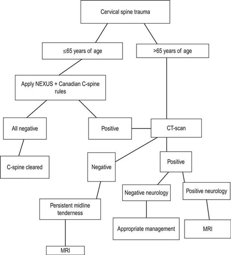
FIG. 3.3.1 Suggested radiological algorithm for cervical spine trauma.
Thoracic, lumbar and sacral spine clearance
There is little information available to provide evidence-based guidelines for clearing the thoracic, lumbar and sacral spine. The investigation and injury exclusion strategy is based on appropriate clinical reasoning (read the mechanism!) and an understanding of the effectiveness and limitations of medical imaging options, in both logistics and effectiveness. All patients with significant mechanisms of injury and pain or tenderness along the thoracic, lumbar and sacral spine should be imaged. Additionally, patients with multiple injuries and high-risk mechanisms should be routinely imaged.
Secondary survey, referral-disposition and definitive treatment
The secondary survey of spinal cord damage
The definitive diagnosis of a spinal cord injury is made from the findings on secondary survey. Two specific injury entities need to be considered: skeletal and neurological.
A head-to-toe clinical examination is conducted in keeping with the standard conventions used in examining any victim of major trauma. The following outlines the specific points of clinical examination pertinent to spinal injury.
Head and neck
An examination of the cervical spine is conducted maintaining immobilization. Palpation of the spine posteriorly may demonstrate generalized tenderness owing to diffuse muscular spasm. However, the point of maximal tenderness should be determined. In hyperextension injuries, the prevertebral and paravertebral muscles are often contused. This is a helpful sign when evaluating hyperextension–hyperflexion injuries in patients who were in stationary vehicles hit from behind. Longitudinal pressure to the head increases cervical pain. Such patients should be considered to have a higher likelihood of a significant vertebral injury.
The neck should be examined for swelling and bruising. Deformity will be noted if there is a dislocation with significant displacement. It should be remembered that significant bony and soft tissue injury frequently occurs without any major findings on external examination.
As prolonged immobilization of the cervical spine with rigid pre-hospital rescue collars and other rigid immobilization devices may unnecessarily add to patient discomfort, complications from the application of splinting devices and the need for ongoing spinal nursing, it is important to determine whether immobilization devices can be removed early during the assessment and treatment phases of management. Reasons for lengthy periods of immobilization include times to definitive radiological assessment and waiting for windows of opportunity to ensure vertebral stability (see also Immobilization of the spine and Clearing the spine).
A re-examination of the upper airway is required. A prevertebral haematoma can cause obstruction; the gag reflex may be blunted; airway protection may be embarrassed owing to paralysis of muscles below the neck, resulting in inefficient gag and cough. The patient will have gastric stasis and is at considerable risk of fluid aspiration.
The torso
The patient should either be lifted or rolled on to the side using a formal spinal-lifting technique, so that the back can be examined. The spine is examined for alignment, swelling, bruising and abrasion. Deformity is generally not a feature, except in the presence of major dislocation or disruption.
The rise and fall of the chest is noted. Paradoxical movement is a sign of thoracic cage muscular paralysis and will be more pronounced the higher the segmental level of injury. Careful examination of the thorax, abdomen and pelvis is required. In both quadriplegia and high paraplegia, serious injury may be masked by the use of analgesia and anaesthesia. Significant vertebral injury to the thoracic and lumbar spines is associated with major injuries to the thoracic, abdominal and pelvic organs.
The abdomen is specifically assessed for an evolving paralytic ileus.
Neurological assessment
A thorough examination of the peripheral nervous system is required. It is strongly recommended that both motor and sensory examinations be undertaken in accordance with the following convention. Examine motor, sensory and reflex components independently. Examination begins at the head and then progresses across the shoulders. The upper limbs are then examined. The torso evaluation begins from just below the clavicles, extending inferiorly to the groin; each lower limb is then assessed. Finally, the saddle area and pelvic floor are assessed.
This approach reduces the likelihood of an incorrect diagnosis of paraplegia by finding a ‘pseudo’ neurological level of injury just below the clavicles when upper limbs have not been examined. It is therefore important that the upper limbs be assessed before examining the torso.
Motor function
Muscle power is assessed in terms of neurological segments and not muscle groups. Muscle power in each segment is graded from 0 to 5 as shown in Table 3.3.1.
Table 3.3.1
Muscle power grading
|
Power grade |
Clinical finding |
|
Grade 0/5 |
No movement |
|
Grade 1/5 |
Flicker |
|
Grade 2/5 |
Movement present, but not a full range against gravity |
|
Grade 3/5 |
Full range of movement against gravity with no added resistance |
|
Grade 4/5 |
Full range of movement against gravity with added resistance but with reduced power |
|
Grade 5/5 |
Normal power |
It is often impossible to assess power grades in certain segments owing to the patient’s injuries. The upper limbs are the most easily examined. The strength of a cough provides some information as to the state of thoracic and abdominal musculature.
In the emergency setting, the state of the pelvic muscles is determined through a rectal examination by assessing rectal tone and requesting the patient to tighten the sphincter on the examiner’s gloved finger.
Sensory function
Dorsal column sensation is assessed using a piece of cotton wool and testing for light touch. Spinothalamic sensation is assessed using a pin or sharp object. Although proprioception, vibration and temperature can be assessed, these are not essential and add little to the emergency examination. When testing with a sharp object, a hypodermic injection needle or a trocar stylet must not be used: these are engineered to stab the skin as painlessly as possible, therefore they cause trauma and are unreliable.
The general convention described below should be followed. Sensory examination begins on the face which, as it is supplied by the trigeminal nerve and bypasses the spinal cord, acts as a reference point. It is an important axiom based on anatomy that ‘in the absence of head injury or local facial injury, sensation to the face is always normal in pure spinal cord injury’ (the trigeminal nerve comes from above the spinal cord). It is recommended that examination of the head, neck and upper torso is performed as follows. Start by examining the C2 dermatome laterally on the neck behind the mandible and beneath the ear. Extend examination onto the top of the shoulder, thus assessing the C3, C4 and C5 dermatomes. In the upper limbs, examine the dermatomes in segmental order. This should include T2 on the upper medial aspect of the arm. Then carry on examining the torso in the mid-clavicular plane or at the outer border of the surface marking of the rectus sheath.
Reflexes
Reflexes are examined in keeping with usual examination practices. Superficial abdominal reflexes should be noted. The anal and bulbocavernosus reflexes are important in assessing sacral segments.
Corticosteroids–methylprednisolone
A Cochrane review from 2012 found that administration of methylprednisolone within 8 hours after injury gave a significant recovery of motor function [15].
Administration for an additional 24 hours (totalling 48 hours) may give an additional improvement of motor neurological function and functional outcome. On the other hand, the use of methylprednisolone is not without complications. It is contraindicated in patients with heavily contaminated open injuries, other heavily contaminated situations, such as perforated bowel and established sepsis. It has the risk of developing acute adrenal insufficiency in these patients, which needs recognition and prompt treatment to prevent further complications. It is relatively contraindicated in diabetes mellitus. Prophylactic measures, such as for acute peptic ulceration and monitoring of blood glucose, should be instituted.
The benefit of steroids in spinal cord injury is therefore considered questionable. Despite this, their use remains a treatment option and several centres prefer to use high-dose methylprednisolone in the early management of patients with neurological injury. In Australia, spinal cord injury is not listed as an indication for high-dose methylprednisolone. Therefore, the decision to use high-dose corticosteroids should be made in conjunction with the specialist services, either the major trauma service or spinal injuries service that will be managing continuing care. If used, treatment must be commenced within 8 hours from the time of injury. The total treatment period should be for 24 hours if treatment is commenced within 3 hours of injury and 48 hours if commenced between 3 and 8 hours.
A guideline for the use of methylprednisolone in acute spinal cord injury is presented in Figure 3.3.2.

FIG. 3.3.2 Methylprednisolone for acute spinal cord injury.
Referral-disposition
Patients with a spinal cord injury should be referred to a centre with facilities for optimal management as soon as practicable. Specific treatments such as immobilization, specific therapy and transport considerations should be discussed with the continuing care provider or spinal injuries unit prior to transfer. If transport is delayed, it is appropriate that the spinal injuries unit be involved and contribute to the patient’s initial management, especially in areas of specific management, as soon as possible, even if transfer is to be delayed by several days.
Specific conditions
Vertebral injury [2]
Cervical spine fractures
Cervical spine injuries may result from one or more combinations of the following mechanisms:
hyperflexion
hyperextension
flexion–rotation
vertebral compression
lateral flexion
distraction.
Hyperflexion
Hyperflexion produces the following injuries:
a simple, stable wedge fracture
a fracture with an anterior teardrop
bilateral anterior subluxation
clay shoveller’s fracture
bilateral facet dislocation.
Flexion injuries can cause a vertebral body fracture with an anteroinferior extrusion teardrop fracture. This is often associated with retropulsion of a vertebral body fracture fragment or fragments into the spinal canal.
The clay shoveller’s fracture is a particular spinous process fracture produced by a sudden load on a flexed spine, with resulting avulsion of the C6, C7 or T1 spinous processes.
Hyperextension
Anterior widening of disc spaces, prevertebral swelling, avulsion of a vertebral body by the anterior longitudinal ligament, subluxation and crowding of the spinous processes are features of the hyperextension injury. Encroachment on the canal by an extruded disc or a posterior osteophyte may occur in patients with osteoarthritis of the cervical spine.
Flexion–rotation
This is responsible for unilateral facet dislocation or forward subluxation of the cervical spine.
Vertebral compression
This is the mechanism responsible for burst fractures. The intervertebral disc is disrupted and driven into the vertebral body below. In addition, disc material may be extruded anteriorly into prevertebral tissues and posteriorly into the spinal canal. The vertebral body may be comminuted to varying degrees, with fragments being extruded anteriorly and posteriorly into the spinal canal.
Lateral flexion
This may produce uncinate fractures, isolated pillar fractures, transverse process injuries and lateral vertebral compression.
Distraction
These injuries may result in gross ligamentous and intervertebral disc disruption. The hangman’s fracture may also occur by combined distraction and hyperextension mechanisms.
C1 – the atlas
Fractures of the atlas comprise 4% of cervical spine injuries. Mechanisms of injury generally involve hyperextension or compression. Around 15–20% of fractures may be associated with a C2 injury and 25% may be associated with a lower cervical injury. The Jefferson fracture is a blowout fracture of the ring. Other fractures include isolated injuries of the posterior arch, the anterior arch and the lateral mass.
C2 – the axis
Axis fractures comprise 6% of cervical spine injuries, with an association with concurrent C1 injury in the majority of cases.
Computed tomography (CT) scan images are examined for odontoid subluxation. Three types of odontoid fracture are described:
Type 1 is an avulsion of the odontoid tip. It is generally a stable injury and accounts for 5–8% of odontoid fractures
Type 2 injury is a fracture through the base of the dens and is generally unstable. It comprises 55–70% of odontoid injuries. In younger children, the epiphysis may be present and confused with a type 2 fracture
Type 3 is a subdental fracture of the odontoid extending into the vertebral body. It comprises 30–35% of odontoid fractures.
Other fractures of the odontoid include avulsion fractures of the lower anterior margin of the body due to a hyperextension injury. A hangman’s fracture is a bilateral neural arch fracture of C2. It is a hyperextension injury and is associated with prevertebral soft tissue swelling, anterior subluxation of C2 on C3 and avulsion of the anteroinferior corner of C2.
C3–C7
Fractures in this segment of the cervical spine are clearly picked up with CT scanning. Fractures are defined as unstable when:
the anterior and all of the posterior elements are disrupted
there is more than 3 mm overriding of the vertebral body above over the vertebral body below
the angle between two adjoining vertebrae is greater than 11°
the height of the anterior border of a vertebral body is less than two-thirds of the posterior border.
Fractures of the thoracic spine
Hyperflexion is the principal mechanism of injury to the thoracic spine, with resultant wedging of vertebral bodies. Owing to the rigidity of the thoracic cage and the associated costovertebral articulations, most thoracic spine injuries are stable. However, internal stabilization may be necessary where kyphosis is pronounced.
Thoracolumbar spine
Fractures of the thoracolumbar spine comprise 40% of all vertebral fractures responsible for neurological deficit. Most are flexion or hyperflexion-rotation injuries. Plain films may demonstrate facet joint disruption, evidence of interspinal ligament disruption, posterior bony fragments protruding into the spinal canal and burst fragments at the superior surface of the vertebral body. These fractures are generally unstable.
Lumbar spine
Injuries similar to those previously described do occur in the lumbar spine. Three specific injuries of the lumbar spine merit further discussion and are broadly considered posterior distraction injuries of the vertebral arch. They constitute a group known as seatbelt injuries, produced when a hyperflexion force is applied to a person wearing a lap-only type seatbelt. In unrestrained persons, a flexion injury generally flexes the spine around a point through the anterior spinal column, typically causing a wedge compression fracture of the body. In the restrained person, the point of flexion is moved forward to the anterior abdominal wall. This change in momentum forces converts the hyperflexion mechanism to one of distraction. These injuries are caused by deceleration from high speed, as seen in head-on road traffic accidents or aircraft crashes.
Plain film radiology remains the first-line imaging study. Suggestive findings include:
a vacant or empty appearance of the vertebral body on the AP film
discontinuity in the cortex of the pedicles or spinous processes on the AP view
fracture, with or without dislocation in the lateral view, which may be subtle.
CT or magnetic resonance imaging (MRI) are of value in further delineating architectural disruption. However, the exact nature of the fracture complex may be difficult to delineate on axial images, as the fractures are often orientated parallel to the scanning plane. Three-dimensional reconstruction of multislice CT images has greatly improved spinal injury imaging.
These injuries are often associated with concurrent intra-abdominal visceral injuries.
Chance fractures
These are characterized by an oblique or horizontal splitting of the spinous process and neural arch, extending the superior posterior aspect of the vertebral body into and damaging the intervertebral disc.
Horizontal fissure fracture
This fracture is very similar to the chance fracture, with the exception of the fracture line, which extends horizontally through the vertebral body to its anterior aspect.
Smith fracture
This spares the posterior spinous process. The fracture line involves the superior articular processes, the arch and a small posterior fragment of the superior posterior aspect of the vertebral body. Although the spinous process is intact, the posterior ligaments are disrupted.
Spinal shock
Spinal shock is often confused with the neurogenic shock of sympathetic interruption. They are different entities. Complete separation of the spinal cord from the brain abolishes voluntary movement and sensory perception and causes changes in cord physiology and reflex activity. Acute cord confusion is a simple explanation of the resulting pathophysiology. Spinal shock is manifested by the transient cessation of cord activity in the normal cord below the injury. The cord distal to the injury is unable to function as one would expect from a newly created upper motor neuron lesion. Spinal shock may last for a few hours to several weeks, depending on the segmental level and extent of the cord injury. During this period, both somatic and autonomic reflexes below the injured segments disappear. Spinal shock has been attributed to the sudden loss of descending facilitatory impulses from higher centres. Recovery from spinal shock is heralded by the return of the Babinski response, followed by the perineal reflexes. In quadriplegia and high paraplegia, as the cord recovers from spinal shock, either recovery of function (depending on the degree of injury resolution at the injury site) occurs or, more commonly, spasticity develops. If the cord injury is at the conus medullaris or the cauda equina, unless recovery occurs, a lower motor neuron pattern with areflexia remains.
Spinal cord injuries
Spinal cord injuries should be divided into primary and secondary injuries, as the causes affect the choice of treatment. Primary spinal cord injuries refer to the injuries directly caused by the trauma mechanism and its damaging energy onto the spinal cord. Secondary spinal cord injuries are caused by other mechanisms often related to the initial trauma, i.e. hypotension, hypoxia, etc.
Primary spinal cord damage (Fig. 3.3.3)
Transverse spinal cord syndrome
The spinal cord is completely damaged transversely across one or more adjacent spinal segments. No motor or autonomic information can be transmitted below the damaged area and ascending sensory stimuli from below the damaged spinal segments are blocked. The manifestations are: total flaccid paralysis, total anaesthesia, total analgesia and, usually, areflexia below the injured segment.

FIG. 3.3.3 Spinal core and syndromes. (From Clinical Symposia 1998; 34(2): 17. Comprehensive Management of Spinal Cord Injury, Plate 11. Redrawn with permission of Novartis Pty Ltd, Basel, Switzerland.)
The transverse cord syndrome can be incomplete, with partial paralysis, reduced sensation and pain sensibility below the injured part.
The term ‘sacral sparing’ implies that some sensibility with or without motor activity in the areas supplied by the sacral segments is preserved in an otherwise complete transverse cord syndrome. The presence of sacral sparing implies an incomplete injury, as some neurological transmission through the injured segments is preserved. It will be recalled that spinothalamic and corticospinal transmission to and from sacral segments are located in the outermost parts of the spinal cord and are, therefore, immediately adjacent to the origin of the spinal cord’s blood supply.
Acute central cervical cord syndrome
The central part or grey matter of the spinal cord is injured. Transmission in the outer rim of the spinal cord is essentially intact but impaired. The signs of this injury are:
motor function: there will be weakness in both upper and lower limbs, with weakness marked in the upper limbs
sensation: there is sensory loss in both upper and lower limbs, which is more severe in the upper limbs
reflexes are variable.
This is frequently caused by a hyperextension injury and is typically seen in older patients with cervical spondylosis. In this situation, the cord is compressed between posterior osteophytes and the intervertebral disc in front and the ligamentum flavum behind.
Acute anterior cervical cord syndrome
The anterior half of the spinal cord – the region supplied by the anterior spinal artery – is damaged (see Figs 3.3.3 and 3.3.5). There is motor loss or paralysis below the level of the injured segment(s). Spinothalamic transmission is impaired and thus there is analgesia with loss of temperature sensation and coarse touch. As the dorsal columns are relatively intact, there is some preservation of joint position, vibration sense and fine touch. In the context of an acute cord injury, the patient may not interpret dorsal column preservation in terms of joint position sense or light touch. Dorsal column function may be manifested as preservation of vague and poorly localized sensation in the extremities.
These injuries are frequently the result of flexion–rotation or vertical compression injuries.
Brown-Séquard’s syndrome
This syndrome is a functional cord hemisection with dissociated sensory loss. One half of the cord is damaged. In a pure Brown-Séquard lesion, ipsilateral motor function is impaired, as are light touch, joint position sense and vibration. Contralateral spinothalamic sensation – that is, pain and temperature – is impaired, whereas ipsilateral sensation is relatively preserved. Reflexes are variable.
Posterior cord syndrome
This is an uncommon injury that causes contusion or disruption to the dorsal columns, leading to impaired or disrupted proprioception, vibration and fine touch sensation.
This syndrome is usually the result of penetrating trauma to the back or a hyperextension injury in association with fractures of the vertebral arch.
Spinal cord concussion
This diagnosis implies a temporary cessation of spinal cord neurological function. In this instance, there is a near full recovery of cord function within 48 hours. The patient will be first assessed as suffering from either a complete or an incomplete spinal cord injury and will then recover within the above period. The patient has suffered an injury to the spinal cord that has been enough temporarily to cease electrical activity in the injured spinal segment, with no, or very little, mechanical or anatomical injury to the cord, such as haemorrhage or contusion. It is the pattern of recovery over a day or two that allows the diagnosis to be made. Unfortunately, this constitutes less than 1% of all spinal cord injuries.
In all the incomplete spinal cord syndromes, the location of cord pressure or damage varies in terms of incompleteness and segmental level(s) of cord injury and so will the range of symptoms and signs.
Secondary spinal cord damage
It is often believed that most spinal cord damage occurs at the time of injury, but it may occur subsequent to the initial injury [6]. This secondary damage may be caused by:
Inappropriate manual handling [4,5]. Subsequent mishandling causes significant movement at the site of the primary vertebral injury, leading to spinal cord damage. This can be prevented by careful handling of the patient. It is important to be aware of the possibility of a spinal cord injury and protect the spine until the diagnosis has been excluded. This involves standard cervical in-line immobilization, whole-spine immobilization using a spine board or a Jordan frame and ‘log roll’ for moving the patient.
Hypoxia and hypotension. These aggravate the primary injury, causing progressive neurological deterioration by mechanisms similar to those that cause secondary brain damage in head injury.
Acute response to injury. Intrinsic metabolic changes in the previously undamaged spinal cord at the region of the initial vertebral injury may also cause secondary deterioration due to oedema, haemorrhage and the release of metabolically active substances from damaged neurons. The culmination of the pathophysiological processes leads to cord ischaemia and oedema, thereby promoting further neurological damage. The oedema and haemorrhage tend to resolve within 10–14 days, with some improvement in neurological function. Resolving oedema results in local segmental recovery. However, residual ischaemic change in secondarily affected spinal cord adjacent to the primarily injured segments does occur, producing permanent neurological deficit.
Unconscious patients
As previously mentioned, the definitive diagnosis of spinal cord injury is a secondary survey consideration and hence identified primarily from symptoms and physical findings. There is no pathognomonic sign of a spinal cord injury in an unconscious patient. The following should alert the examiner to the possibility of a coexisting spinal cord injury in an unconscious trauma victim:
paradoxical breathing or chest wall movement (diaphragmatic breathing) in the absence of a major airway obstruction, stove-in or large flail chest suggests a cervical cord injury
priapism in the unconscious trauma victim suggests quadriplegia or high to mid-thoracic paraplegia
preserved facial grimace in the absence of a response to painful stimuli in the limbs
lower limb flaccidity in the presence of normal upper limb tone suggests paraplegia
observed upper limb movement in the absence of lower limb movement suggests paraplegia
the combination of the persistent bradycardia and hypotension despite volume challenge
where this is accompanied by a flaccid rectal sphincter, there is an increased likelihood of spinal cord injury.
Special attention, again, should go to protection of the C-spine in this category of patients. As the golden standard of imaging, the cervical spine CT is highly sensitive and may reliably exclude unstable injuries in patients with obtunded or intubated blunt trauma [17].
Documentation conventions
Two of the pitfalls in the management of any neurological injury are terminology and documentation. The following convention is recommended.
Motor function is recorded either using segmental terminology in written format or on a muscle chart (Fig. 3.3.4). It will be impossible to chart every segment accurately, but motor power in the upper and lower limbs should be able to be confidently recorded. Power should be graded using the 0–5/5 system.


FIG. 3.3.4 (A–C) Documentation of neurological injury.

FIG. 3.3.5 MRI scan of acute central cord syndrome.
Sensation is recorded more descriptively. Normaesthesia, hyperaesthesia, hypoaesthesia and anaesthesia are the descriptors for dorsal column function and testing for light touch. Normalgesia, hyperalgesia, hypoalgesia and analgesia are used in describing pain perception. These are recorded on sensory charts or described according to the following two examples.
In a patient with a transverse spinal cord syndrome, incomplete below C6 and complete below T1, the sensation is described as:
normaesthesia and normalgesia to C5
hypoalgesia and hypoaesthesia below C5
anaesthesia and analgesia below T1.
In a patient with an acute central cervical cord syndrome below C6, with total segmental paralysis in the C6–C8 segments and with some involvement of C5, the sensation might be described as:
normaesthesia and normalgesia to C4
hypoalgesia and hypoaesthesia below C4
anaesthesia and analgesia below C5
hypoalgesia and hypoaesthesia below T1.
Controversies/emerging issues
Recent research showed that patients with persistent midline cervical tenderness result in considerable healthcare costs [18]. With more and more understanding of the financial, social and psychological impact of spinal trauma, it is expected that more and more emphasis is going to be put on prevention, early detection and therapy in cases of (potential) spinal injury.
Clinical decision rules for cervical spinal clearance will need further testing with CT scanning as the new standard diagnostic modality of first choice. It is therefore expected that these rules will change according to results found in future research.
With the technical developments expected in the coming years, it is expected for MRI scanners to become more widely available, to be faster, to have higher diagnostic accuracy and to become cheaper. Also, since CT scanning has the disadvantage of the patient being exposed to radiation, it is expected that indications for MRI scanning of the spine will be broadened.
Acknowledgement
The author of this chapter in the previous edition of this textbook was Jeff Wassertheil. The text in this new edition is based on the original chapter for which Jeff Wassertheil must be acknowledged.
References
1. Lowery DW, Wald MM, Browne BJ, et al. Epidemiology of cervical spine injury victims. Ann Emerg Med. 2001;38:12–16.
2. Rotem TR, Lawson JS, Wilson FW, et al. Severe spinal cord injuries related to rugby union and league football in New South Wales, 1984–1996. Med J Aust. 1998;168:379–381.
3. Selecki BR, Berry G, Kwok B, et al. Experience with spine injuries in NSW. Aust NZ J Surg. 1986;56:567–576.
4. Toscano J. Prevention of neurological deterioration before admission to a spinal cord injury unit. Paraplegia. 1988;26:143–150.
5. Superspeciality Service Subcommittee of the Australian Health Ministers Advisory Council. Guidelines for acute spinal cord injury services Australian Institute of Health, AGPS 1990.
6. Goldberg W, Mueller C, Panacek E, et al. Distribution and patterns of blunt cervical spine injury. Ann Emerg Med. 2001;38:12–16.
7. Kwan I, Bunn F, Roberts I, on behalf of the WHO Pre-hospital Trauma Care Steering Committee. Spinal immobilization for trauma patients. Cochrane Database System Rev 2001;2.
8. Hauswald M, Ong G, Tandberg D, Omar Z. Out-of-hospital spinal immobilization: its effect on neurologic injury. Acad Emerg Med. 1998;5:214–219.
9. Chan D, Goldberg R, Tascone A, et al. The effect of spinal immobilization on healthy volunteers. Ann Emerg Med. 1994;23:48–51.
10. Keller BP, Lubbert PH, Keller E, Leenen LP. Tissue-interface pressures on three different support-surfaces for trauma patients. Injury. 2005;36:946–948.
11. Hoffman JR, Mower W, Wolfson AB, et al. Validity of a set of clinical criteria to rule out injury to the cervical spine in patients with blunt trauma. N Engl J Med. 2000;343:94–99.
12. Stiell IG, Wells GA, Vandemheen KL, et al. The Canadian C-Spine Rule for radiography in alert and stable trauma patients. J Am Med Assoc. 2001;286:1841–1848.
13. Stiell IG, Clement CM, McKnight RD, et al. The Canadian C-spine rule versus the NEXUS low-risk criteria in patients with trauma. N Engl J Med. 2003;349:2510–2518.
14. Zoe A, Naher CG, Verhagen AP, et al. Accuracy of the Canadian C-spine rule and NEXUS to screen for the clinically important cervical spine injury in patients following blunt trauma: a systematic review. Can Med Assoc J. 2012;184:E867–E876.
15. Bracken MB. Steroids for acute spinal cord injury. Cochrane Database Syst Rev 2012.
16. Brown D. Guidelines for the use of methylprednisolone in acute spinal cord injury Melbourne: Victorian Spinal Cord Service Austin & Repatriation Medical Centre; 2000.
17. Kirschner J, Sepaul RA. Does computed tomography rule out clinically significant cervical spine injuries in patients with obtunded or intubated blunt trauma. Ann Emerg Med. 2012;60:737–738.
18. Ackland HM, Wolfe R, Cameron PA, et al. Health resource utilisation costs in acute patients with persistent midline cervical tenderness following road trauma. Injury. 2012;43:1908–1916.
3.4 Facial trauma
Lewis Macken
Essentials
1 Facial trauma is a common problem in the emergency department (ED), usually as an isolated injury, often after an assault. It may also occur after multisystem injury.
2 Facial injuries that do not threaten the airway or risk life-threatening haemorrhage can usually be assessed and managed as part of the secondary survey.
3 Early diagnosis of facial injuries is important, as late diagnosis may lead to compromised function and an unsatisfactory cosmetic outcome.
4 CT scanning has replaced plain radiography for the delineation of facial fractures.
5 Visual assessment after facial trauma can be difficult, especially in the unconscious patient, but is an essential element of the clinical examination.
6 Penetrating trauma to the face is rarely fatal, but may result in significant morbidity. Attention to early airway intervention is vital after penetrating injuries, especially in patients with gunshot wounds to the face.
Introduction
Facial trauma is defined as injury to the facial soft tissues (including the ear) and bony skeleton. This covers a wide spectrum of injuries [1]. Patients with facial trauma are usually male, between the ages of 20 and 25 years and have usually sustained their injuries as a result of blunt trauma. Isolated facial injury after an assault accounts for most injuries, with motor vehicle injuries, contact sports and falls accounting for most of the remainder [2]. Up to 20% of patients with facial injuries as part of multisystem injury will have associated life-threatening injuries [3,4].
Management of blunt and penetrating trauma to the face can be challenging. Life-threatening facial injuries are those that result in airway compromise or ongoing haemorrhage. At the same time, attention must be given to preserving long-term cosmesis and the normal functions of sight, speech and mastication. After immediate resuscitation and stabilization, management of facial injuries requires a knowledge of anatomy, the injuries commonly associated with facial injury and an awareness of treatment methods for the differing injuries to the face [5,6]. Consultation should be sought when the injury threatens the restoration of normal function or appearance. Non-accidental injury should always be considered, as some studies report that over 20% of head, neck and facial injuries in women are the result of domestic violence [7].
History
The evaluation should commence with the history, with emphasis given to the mechanism of injury. The velocity of force directed to the face determines the degree of facial fracture [8]. Respiratory and haemodynamic observations and quantification of pre-hospital blood loss are important components of the history.
Examination
The physical examination involves evaluation of all facial areas by inspection, palpation and assessment of function. Inspection of the face may reveal deformity, loss of normal symmetry, changes in contour and localized areas of swelling. Skull-base fractures may be suggested by periorbital or postauricular bruising and the ears must be inspected to exclude haemotympanum or cerebrospinal fluid (CSF) otorrhoea. Gentle systematic palpation of the facial skeleton should follow, assessing for tenderness and feeling for asymmetry, bony margin irregularities, abnormal motion or crepitus. Specific attention should be given to the supraorbital and infraorbital margins, the zygomas, nasal bones, maxilla and mandible. Mobility associated with midfacial maxillary fractures is assessed by grasping the anterior maxilla with the thumb and index finger of one hand, while stabilizing the forehead at the nasal bridge with the other. Ophthalmological evaluation includes inspection for enophthalmos or exophthalmos and globe injury and assessment of pupillary responses, visual acuity, visual fields, extraocular movements and inquiry for diplopia. Although eye injury is an infrequent complication of blunt trauma, subjective impairment in visual acuity remains the most sensitive single predictor of eye injury [9] and examination that reveals the presence of an afferent pupillary defect or a non-reactive pupil is the most important factor in predicting the severity of eye injury [10]. The nose should be inspected for the presence of any septal deviation, haematoma (more common in children) or CSF rhinorrhoea. Oral examination is necessary to look for loose, broken or missing teeth, malocclusion of dentition, soft tissue lacerations and contusions. Examination of the mandible includes assessment of the temporomandibular joint in the open and closed positions.
Assessment of the function of facial structures is the final component of the examination of the face. Malocclusion, often identified by the presence of a new gap between the occlusal surfaces of the teeth when the patient closes his/her jaws, is a good indicator of maxillary or mandibular fracture. Similarly, pain on biting on a tongue depressor, loss of bite strength and limitation of jaw movement are strongly suggestive of a fracture; the patient should be asked if the bite has subjectively changed [11,12]. Motor function of the facial nerve should be assessed and all three branches of the trigeminal nerve should be evaluated. Commonly encountered symptoms and signs are hypoaesthesia or paraesthesia of the upper lip or upper alveolar margin, suggesting fracture of the maxilla causing injury to the alveolar branches of the infraorbital nerve. Sensory changes in the lower lip and lower alveolar margin suggest fracture through the mandibular canal, causing inferior alveolar nerve injury (a branch of the mandibular nerve) [11].
Radiographic examination
Computed tomography (CT) is the initial imaging modality of choice in evaluating patients with facial trauma. Although an orthopantomogram (OPG) view is very helpful in investigating a suspected fracture of the mandible, CT will be required for patients with clinically obvious complex facial injuries, especially of the upper and middle thirds of the face and for patients with multisystem injuries. CT and derived image reconstructions display soft tissue relations to the bony skeleton and the degree of skeletal distortion, displacement and comminution. CT assists with surgical planning and evidently allows other injured areas to be imaged at the same time for multitrauma patients.
Immediate management in the emergency department
It is important to prioritize injuries in the management of the patient with facial trauma because facial injuries that do not threaten the airway or risk life-threatening haemorrhage can usually be assessed and managed as part of the secondary survey. Proper attention should be given to potentially more significant head, chest and abdominal injuries before a non-life-threatening facial injury is thoroughly evaluated, no matter how impressive and disfiguring the facial injury appears. The association of facial injury with cervical spine or cord injury is questionable, especially if vehicular trauma was not the cause [13]. After blunt assault, the incidence of associated cervical spine injury is low, but is more likely if there is a fall after the assault [14]. Patients with clinically significant head injuries (especially Glasgow Coma Score [GCS]<9) are at greater risk of having cervical spine injuries than those with evidence of facial trauma [15–18]; intracranial injuries are more frequently associated with facial fractures than are cervical spine injuries [19].
Airway management
Asphyxia due to upper airway obstruction is the major cause of death from facial trauma. The airway must be rapidly assessed, respiratory obstruction relieved and an adequate airway established. Managing these patients in the supine position is often risky and early semi-elective intubation may be required. Signs of partial airway obstruction include tachycardia, tachypnoea, restlessness, fighting to sit up, noisy respirations, stridor and supraclavicular and intercostal retractions. This may lead to complete obstruction. An unobstructed airway in the presence of facial trauma should be closely monitored because increasing oedema and persistent bleeding may later compromise airway patency.
Fractures of the mandible and maxilla with posterior or inferior displacement, together with displaced soft tissues, blood, secretions or other foreign material, may lead to airway embarrassment. Simple airway measures, such as chin lift and jaw thrust should be performed and the mouth immediately examined. Any loose foreign material, such as food, broken dentures or teeth or bone fragments, should be removed and blood clots suctioned. If anterior traction on the fractured mandible or on the mobile segment of the maxilla (performed by inserting two fingers behind the soft palate and lifting the middle third of the face forwards and upwards) fails to relieve posterior pharyngeal obstruction and does not establish unobstructed ventilation, a definitive airway must be placed. Distorted facial anatomy may make bag and mask ventilation difficult. Rapid-sequence orotracheal intubation can usually be performed. Awake fibreoptic intubation is an option, but the presence of persistent bleeding makes the procedure technically difficult. Standard drills for anticipated intubation difficulties should be at hand and equipment for performing a cricothyroidotomy and transtracheal jet ventilation should be available. A surgical airway may be life saving when anatomical disruption makes intubation difficult. A tracheostomy performed under local anaesthesia in the operating theatre is often the safest option in a stable patient with significant midfacial injuries in whom airway difficulties are expected [20].
Control of haemorrhage
Traumatic facial haemorrhage can be massive, difficult to manage and potentially life threatening in approximately 5% of patients with midfacial fractures [21]. Bleeding from midface trauma mainly originates from branches of the external carotid artery (with branches of the internal carotid artery also supplying the nose). Most haemorrhage can be controlled with direct pressure, anterior nasal packs and double balloon catheters providing anterior and posterior nasopharyngeal tamponade (the placement of 12–14 G Foley catheters with 10 mL balloons inflated and taped under tension to the side of the face is an alternative) [22]. Care must be taken with nasal instrumentation: the incidence of associated skull-base fractures is more common with orbital wall or rim fractures than with more inferiorly located facial fractures. Also, a skull-base fracture is more likely in the presence of multiple facial fractures (the presence of three or more facial fractures is associated with an incidence of skull-base fracture of up to 33%) [23]. Attempts to clamp bleeding vessels in the emergency department (ED) should be avoided because of the associated risks of damaging important structures, such as the facial nerve, parotid duct or lacrimal apparatus. If simple measures fail to arrest the bleeding, operative reduction of the fractures, especially of the maxilla, and possible ligation of bleeding vessels should be undertaken, with angiography and selective embolization considered if other measures fail.
Disposition
After initial stabilization, immediate treatment of most facial injuries is mainly supportive. Fractures requiring operative management are often dealt with on a delayed basis, when soft tissue swelling is resolving. The need for antibiotics for patients with facial trauma depends on the mechanism of injury, the extent of the injury and the immune status of the patient. Systemic antibiotics are not required for most facial wounds. However, antibiotics should be given for bites and grossly contaminated wounds; wounds that extend into the oral cavity, nose and paranasal sinuses; wounds with exposed nasal or ear cartilage; crush wounds and wounds with considerable oedema; and for immunologically compromised patients. Specific antibiotic choices include amoxicillin–clavulanate or cephalexin. However, the evidence to support or refute the use of prophylactic antibiotics in facial trauma is weak. Tetanus immunity should be assessed.
Thorough documentation of injuries is important for medicolegal reasons, as facial injuries are frequently due to personal assault and so later litigation is often likely.
Specific injuries
Soft-tissue injuries
Soft-tissue injuries include abrasions, contusions, lacerations, avulsions and burns. The aim of treatment is to preserve appearance and function. The management of soft-tissue facial injuries involves consideration of how the wound should be repaired and whether it should be repaired in the ED. Possible reasons for delayed wound closure are more urgent coexisting injuries, severe crush injuries, the presence of a foreign body, severely contaminated wounds and an underlying fracture. In these cases, the wound should be irrigated, haemostasis achieved, covered with normal saline-soaked gauze and the patient referred to the appropriate specialty team.
Soft-tissue injuries that usually require repair in the operating theatre include ocular or significant eyelid injury; parotid gland or duct injury; facial nerve injury (injuries to the cheek between the tragus of the ear and a line drawn vertically through the pupil may be associated with damage to the facial nerve, parotid gland or duct); nasolacrimal apparatus injury; alveolar process wounds and significant tooth injuries; lacerations with significant tissue loss or contamination or requiring exact anatomical closure; or where difficulties with patient cooperation are expected. Careful consideration is required before areas of special concern are repaired in the ED, e.g. the lips and perioral area; tongue and oral cavity; nose; ears; periorbital structures; and eyebrows. The eyebrow should not be shaved. Subperichondrial haematomas of the ear require drainage within 7–10 days to avoid permanent cartilage injury (cauliflower ear) and to achieve best cosmetic results. For all wounds closed in the ED, careful attention must be given to thorough cleansing. This can be effectively achieved with pulsatile wound irrigation with normal saline and abrasions containing dirt or other foreign bodies must be scrubbed to prevent traumatic tattooing of the dermis. If the wound is gaping or if structures deeper than the skin and the subcutaneous layer are involved, then multiple-layer repair is usually advisable. This helps to prevent deep tissue space collections and may produce a better cosmetic result, with less scar depression or widening.
Visual assessment after facial trauma can be difficult, especially in the unconscious patient. Loss of vision after blunt facial trauma may be due to: direct injury to the globe; direct injury to the optic nerve (usually bony impingement); indirect injury to the optic nerve (deceleration injury); or due to raised local intraocular pressure (orbital compartment syndrome [OCS]) [24]. OCS is an uncommon ophthalmic emergency, but the diagnosis is clinical. Relief of the intraocular hypertension is time critical and usually requires a lateral canthotomy and inferior cantholysis [25].
Facial fractures
The facial skeleton is constructed to allow applied force to be dispersed via a series of small bone fractures, thereby protecting the skull and intracranial contents. The maxilla and mandible require three times the amount of applied force to cause a fracture as do the nasal bones [26]. Diagnosis of a facial fracture involves a combination of inspection, palpation and radiographic examination. Fractures other than undisplaced fractures of the nose, zygomatic arch or maxilla will usually require acute maxillofacial surgical review. All fractures should be managed initially with elevation of the patient’s head, if associated injuries allow this, and the application of ice.
Mandible
The horseshoe shape of the mandible disperses applied force, which leads to fractures occurring at vulnerable sites regardless of the point of impact and a high incidence of multiple fractures. Common sites of fracture are the condylar neck and angle and the body at the level of the first or second molar [27]. Fractures of the mandibular body usually demonstrate point tenderness, malocclusion and abnormal range of motion and interference with normal mastication. The integrity of the dental arch must be assessed. The application of a soft cervical collar may offer symptomatic relief by providing mandibular support.
Antibiotics should be given when there is the suggestion of a compound fracture with extension into the oral cavity. Most fractures will require some form of internal fixation. Complications of mandibular fractures include chin paraesthesia or hypoaesthesia, delayed union, non-union, infection and malocclusion [28].
Zygomatic arch
Isolated fractures of the zygomatic arch are uncommon and are more commonly part of a more extensive zygomatic complex fracture. An isolated fracture may be evidenced by a depression over the arch, point tenderness and limited or painful mouth opening owing to impingement on the coronoid process of the mandible by the fractured arch. Surgical reduction is required for cosmetic reasons or to correct restricted mandibular range of motion.
Zygomatic complex
Blunt trauma to the zygoma more commonly results in fractures at the articulations of the zygomatic bones with the frontal bone, maxilla and zygomatic process of the temporal bone. Separation at the zygomaticofrontal suture, the zygomaticotemporal suture and at the zygomaticomaxillary suture or infraorbital rim produces the tripod or tripartite fracture. Frequently, the lateral wall of the maxillary sinus and the lateral and central portions of the orbital floor (not to be confused with the orbital blowout fracture) will also fracture as part of the zygomaticomaxillary complex fracture.
Clinical signs of a tripod fracture include flattening of the cheek initially (owing to depression of the fracture segment); this is best seen by standing behind and above the patient, but it is soon replaced by significant swelling. Other signs are hypo-/hyperaesthesia or paraesthesia in the distribution of the infraorbital nerve; asymmetry of the ocular levels; a palpable step defect of the inferior orbital margin; and circumorbital and subconjunctival ecchymoses [11]. Diplopia is often also present and 10–20% of these fractures are accompanied by an ocular injury [29].
Orbital fractures
Fracture of the orbital floor may occur as part of a zygomaticomaxillary fracture or as an isolated injury – the less common orbital blowout fracture. This is a fracture of the orbital floor without fracture of the orbital margin. An increase in intraorbital pressure, as delivered by a fist or a small ball, is transmitted to within the orbit and the relatively weak orbital floor is disrupted, with possible herniation of the contents into the maxillary sinus. Very rarely, a supraorbital rim fracture may be part of a frontal sinus fracture, or lateral orbital wall fracture may be associated with a fracture of the zygoma or a medial orbital wall fracture may occur with a nasoethmoidal fracture. Clinical examination in cases of fracture of the orbital floor may reveal enophthalmos, a difference in pupillary levels, diplopia and impairment of upward gaze and infraorbital hypo-/hyperaesthesia or paraesthesia [6]. An irregular edge to the orbital rim may be evident on palpation. The integrity and function of the eye should be documented to exclude associated injury. CT scanning is necessary to define these fractures fully. Orbital apex fractures are uncommon, but clinical or radiological signs of optic nerve compression (e.g. retro-bulbar haematoma or bone fragment impingement) necessitate urgent surgical referral [30]. All patients with orbital margin or floor fractures require referral. Complications of surgical treatment of orbital floor fractures include persistent diplopia, hypo-/hyperaesthesia or paraesthesia, ectropion and epiphora [31].
Maxillary fractures
Fractures of the maxilla include fractures of the alveolar ridge of the maxilla, fracture of the anterolateral wall of the maxillary sinus and the Le Fort fractures. Isolated maxillary fractures are rare.
In Paris in 1901, Le Fort described a classification of patterns of midface fractures, following cadaveric experiments [32]: Le Fort I (horizontal maxillary fracture) involves only the maxilla at the level of the nasal fossae; Le Fort II (pyramidal fracture) is the most common midface fracture and involves the maxilla, nasal bones and medial aspect of the orbit; Le Fort III (craniofacial dysjunction) separates the midfacial skeleton from the base of the cranium, with the fracture extending through the base of the nose and ethmoid region and across the orbits and zygomatic arches bilaterally.
Most midface fractures are combination injuries, with different Le Fort patterns on each side of the face. Le Fort II and III fractures may require urgent reduction in the ED to improve airway compromise and to arrest ongoing haemorrhage. Patients with such injuries may demonstrate mid-face mobility, and these fractures are associated with skull-base fractures, leading to CSF rhinorrhoea. All patients require a complete eye examination and these injuries necessitate referral.
Nasal fractures
These are common facial fractures. Diagnosis is largely clinical, plain X-rays are unreliable and usually unnecessary and the major concerns for the emergency physician are control of epistaxis and exclusion of a septal haematoma. Displaced fractures should be reduced within 7–10 days.
Nasoethmoidal fractures are more complicated and are caused by trauma to the bridge of the nose. Disruption of the medial canthal ligaments may produce rounding of the palpebral fissures or widening of the intercanthal distance (telecanthus) [11]. Persistent epistaxis and CSF rhinorrhoea may also be evident. Referral is necessary for these patients.
Temporomandibular joint dislocation
Dislocation of the temporomandibular joint may follow trauma to the face or may occur as a result of simply opening the mouth widely. Patients complain of inability to close the mouth and moderate discomfort. X-rays should be performed to confirm that no fracture is present and dislocation will be evidenced by the appearance of the condyle anterior to the articular eminence of the fossa. A directed history will exclude extrapyramidal dystonia mimicking a dislocation. Reassurance, sedation and firm downward pressure of the physician’s thumbs on the patient’s posterior teeth, with upward tilting of the symphysis, is usually successful in relocating the mandibular condyles. Post-reduction X-rays are not always necessary. Analgesia and a soft diet should be prescribed and the patient warned to avoid wide opening of the mouth in the short term [33].
Penetrating injuries to the face
Penetrating trauma to the face from gunshot, stab wounds and impaling foreign bodies is often dramatic at the time of presentation. Such isolated injury is rarely fatal, but may result in significant morbidity due to the combination of soft-tissue and bone defects. The wounding capability of penetrating projectiles (bullets and pellets) is proportional to the energy imparted to the tissue; therefore, the mass of the slug, its velocity and design and the density of the body tissue penetrated determine the amount of tissue destruction [34].
Early aggressive airway management is necessary in patients with gunshot wounds to the face, as respiratory decompensation may be rapid: approximately one-third of patients will require emergency airway intervention [35]. Shotgun and stab wounds are less likely to require an emergency airway, although the presence of a significant vascular injury or oedema remains a universal indication for airway intervention and patients with mandibular entry sites are more likely to require an emergency airway than those with midfacial entry sites. Orotracheal intubation can usually be achieved, with cricothyroidotomy the preferred alternative if necessary. Central nervous system injuries are common after gunshot injuries and CT scans of the head and cervical spine will be required [36].
Arterial injury is suggested by evidence of active bleeding and an expanding haematoma; angiography (carotid and vertebral arteries), which is required in approximately 35–40% of cases [35,36], should be performed when the bullet trajectory suggests proximity to major vessels or the skull base or where the knife or foreign body is in close proximity to a major vascular structure [36–38].
Peripheral nerve injuries, especially of the facial nerve and the mandibular branch of the trigeminal nerve, are also frequently present [36]. Careful eye examination is necessary because ocular trauma is the most common overall complication of penetrating facial trauma [39].
Antibiotics and tetanus prophylaxis are indicated and wounds are managed with conservative debridement, closed reduction of facial fractures and early repair of palatal injuries. Open facial fracture reduction is usually delayed [35].
Conclusion
Facial trauma is common in the ED, encompasses many types of injury and, after rapid exclusion of life-threatening complications, requires thorough patient evaluation to exclude other more urgent injuries. The aim of management of isolated facial injuries is the maintenance of normal function and appearance.
Controversies
Consideration of an immediate surgical airway, rather than attempted oral intubation, in a patient with significant facial trauma and a compromised or deteriorating airway.
The role of angiography and selective embolization in the management of patients with significant haemorrhage from blunt and penetrating trauma.
Consideration of early intubation in the presence of midface fractures with ongoing haemorrhage in the supine patient with other system injuries.
References
1. McKay M, Mayersak R. Facial trauma. In: Marx JA, Hockberger RS, Walls RM, eds. Rosen’s emergency medicine – concepts and clinical practice. 7th ed Philadelphia: Mosby Elsevier; 2010;323–336.
2. Hogg NJ, Stewart TC, Armstrong JE. Epidemiology of maxillofacial injuries at trauma hospitals in Ontario, Canada, between 1992 and 1997. J Trauma. 2000;49:425–432.
3. Tung T, Tseng WS, Chen CT. Acute life-threatening injuries in facial trauma patients: a review of 1025 patients. J Trauma. 2000;49:420–424.
4. Back CPN, McLean NR, Anderson PJ. The conservative management of facial fractures: indications and outcomes. J Plastic Reconstruct Aesthet Surg. 2007;60:146–151.
5. Bailitz J. Trauma to the face. In: Tintinalli JE, ed. Emergency medicine: a comprehensive study guide. 7th ed New York: McGraw-Hill; 2010;1730–1738.
6. Carithers JS, Koch BB. Evaluation and management of facial fractures. Am Fam Phys. 1997;55:2675–2682.
7. Ochs HA, Neuenschwander MC, Dodson TB. Are head, neck and facial injuries markers of domestic violence? J Am Dent Assoc. 1996;127:757–761.
8. Rhee JS, Posey L, Yoganandan N. Experimental trauma to the malar eminence: fracture biomechanics and injury patterns. Otolaryngol Head Neck Surg. 2001;125:351–355.
9. Dutton GN, al-Qurainy I, Stassen LFA, et al. Ophthalmic consequences of mid-facial trauma. Eye. 1992;6:86–89.
10. Joseph E, Zak R, Smith S, et al. Predictors of blinding or serious eye injury in blunt injury. J Trauma. 1992;33:19–24.
11. Lynham AJ, Hirst JP, Cosson JA, et al. Emergency department management of maxillofacial trauma. Emerg Med Australas. 2004;16:7–12.
12. Alonso LL, Purcell TB. Accuracy of the tongue blade test in patients with suspected mandibular fracture. J Emerg Med. 1995;13:297–304.
13. Davidson JS, Birdsell DC. Cervical spine injury in patients with facial skeletal trauma. J Trauma. 1989;29:1276–1278.
14. Kulvatunyou N, et al. Incidence and pattern of cervical spine injury in blunt assault: it is not how they are hit, but how they fall. J Trauma. 2012;72:271–275.
15. Hills MW, Deanne SA. Head injury and facial injury: is there an increased risk of cervical spine injury? J Trauma. 1993;34:549–553.
16. Williams J, Jehle D, Cottington E, Head Shufflebarger C. Facial, and clavicular trauma as a predictor of cervical spine injury. Ann Emerg Med. 1992;21:719–722.
17. Lalani Z, Bonanthaya KM. Cervical spine injury in maxillofacial trauma. Br J Oral Maxillofac Surg. 1997;3:18–21.
18. Adams C, Januszkiewicz J, Judson J. Changing patterns of severe craniomaxillofacial trauma in Auckland over 8 years. Aust NZ J Surg. 2000;70:401–404.
19. Sinclair D, Schwartz M, Gruss J. A retrospective review of the relationship between facial fractures, head injuries, and cervical spine injuries. J Emerg Med. 1988;6:109–112.
20. Taicher S, Givol N, Peleg M. Changing indications for tracheostomy in maxillofacial trauma. J Oral Maxillofac Surg. 1996;54:292–295.
21. Ardekian L, Samet N, Shoshani Y. Life-threatening bleeding following maxillofacial trauma. J Craniomaxillofac Surg. 1993;21:336–338.
22. Murakami WT, Davidson TM, Marshall LF. Fatal epistaxis in craniofacial trauma. J Trauma. 1983;23:57–61.
23. Slupchynskyj OS, Berkower AS, Byrne DW. Association of skull base and facial fractures. Laryngoscope. 1992;102:1247–1250.
24. Perry M, Moutray T. Advanced Trauma Life Support (ATLS) and facial trauma: can one size fit all? Part 4: ‘Can the patient see?’ Timely diagnosis, dilemmas and pitfalls in the multiply injured, poorly responsive/unresponsive patient. Internatl J Oral Maxillofac Surg. 2008;37:505–514.
25. Lima V, et al. Orbital compartment syndrome: the ophthalmic surgical emergency. Surv Ophthalmol. 2009;54:441–449.
26. Luce EA, Tubb TD, Moore AM. Review of 1000 major facial fractures and associated injuries. Plastic Reconstruct Surg. 1979;63:26–30.
27. Dongas P, Hall GM. Mandibular fracture patterns in Tasmania, Australia. Aust Dent J. 2002;47:131–137.
28. Winstanley RP. The management of fractures of the mandible. Br J Oral Maxillofac Surg. 1984;22:170–177.
29. Larian B, Wong B, Crumley RL, et al. Facial trauma and ocular/orbital injury. J Craniomaxillofac Trauma. 1999;5:15–24.
30. Linnau KF, Hallam DK, Lomoschitz FM. Orbital apex injury: trauma at the junction between the face and the cranium. Eur J Radiol. 2003;48:5–16.
31. Whitaker LA, Yaremchuk MJ. Secondary reconstruction of posttraumatic orbital deformities. Ann Plastic Surg. 1990;25:440–449.
32. Le Fort R. Experimental study of fractures of the upper jaw. Rev Chirurg Paris. 1901;23:208–227 360–379. (Reprinted in Plastic Reconstruct Surg 1972; 50:497–506).
33. Luyk NH, Larsen PE. The diagnosis and treatment of the dislocated mandible. Am J Emerg Med. 1989;7:329–335.
34. Kaufman Y, Cole P, Hollier L. Contemporary issues in facial gunshot wound management. J Craniofac Surg. 2008;19:421–427.
35. Kihitir T, Ivatury RR, Simon RJ. Early management of civilian gunshot wounds to the face. J Trauma. 1993;35:569–575.
36. Dolin J, Scalea T, Mannor L. The management of gunshot wounds to the face. J Trauma. 1992;33:508–514.
37. Hollier L, Grantcharova EP, Kattash M. Facial gunshot wounds: a 4-year experience. J Oral Maxillofac Surg. 2001;59:277–282.
38. Doctor VS, Farwell DG. Gunshot wounds to the head and neck. Curr Opin Otolaryngol Head Neck Surg. 2007;15:213–218.
39. Chen AY, Stewart MG, Raup G. Penetrating injuries of the face. Otolaryngol Head Neck Surg. 1996;115:464–470.
3.5 Abdominal trauma
Garry J Wilkes
Essentials
1 One in 10 deaths from trauma is due to abdominal injuries.
2 Abdominal injuries are often occult, overshadowed by more apparent external and orthopaedic injuries and may be missed initially.
3 Detection of intra-abdominal injuries requires a high index of suspicion to avoid preventable morbidity and mortality.
4 CT scanning provides organ-specific diagnosis but requires sufficient stability for transfer from the resuscitation area.
5 Bedside investigations, such as focused assessment sonography in trauma (FAST) assist in the evaluation of suspected intra-abdominal trauma but have limitations.
6 Intra-abdominal trauma frequently coexists with other system trauma. Evaluation and disposition are greatly enhanced by early involvement of senior trauma surgeons.
Introduction
One in 10 deaths from trauma is due to abdominal injuries. These may be difficult to detect initially, as the abdominal cavity cannot be viewed with the naked eye, plain radiography is insensitive to intra-abdominal bleeding and solid organ injury and signs and symptoms of blood loss may be attributed to more obvious injuries. It is therefore not surprising that missed abdominal injuries are a major cause of preventable death in trauma patients. A high index of suspicion should be maintained for this important cause of morbidity and mortality.
A stepwise approach to the management of the multiply injured patient will address the possibility of significant intra-abdominal injury. The principles of initial management are to identify the presence or otherwise of such injury, the need for surgery and most appropriate timing of interventions. This process requires the presence of an experienced clinician at the earliest possible stage to direct and coordinate the trauma team. Ideally, the trauma surgeon should be present at the initial resuscitation.
The initial resuscitation, history, examination and specific investigations will be reviewed in turn.
Primary and secondary surveys
The abdomen does not normally form part of the primary survey. The unstable patient requiring continued fluid resuscitation without other sources of haemorrhage must be considered to have ongoing intra-abdominal bleeding. Specific points to remember are to expose the patient fully, including an examination of the back as well as the rectum and vagina.
History
The history and knowledge of the mechanism of injury will provide vital clues to the increased likelihood of significant intra-abdominal trauma (Box 3.5.1). Ambulance personnel will be able to provide valuable details of the incident. It is important to remember that trauma does not skip body regions and that significant intra-abdominal injuries frequently occur in the absence of external signs of abdominal trauma. Other important aspects of focused history are summarized by the acronym AMPLE: Allergies; Medications; Past medical history; Last ate and drank; Events associated with the trauma incident.
Box 3.5.1
Risk factors for intra-abdominal injury in trauma patients
High-speed vehicular collisions
Pedestrian struck by vehicle
Fall from greater than standing height
Hypotension or history of hypotension (systolic BP<100 mmHg) at any time
Presence of significant chest or pelvic injuries
Significant injuries on physically opposing sides of the abdomen
Abdominal examination
Penetrating injuries are overt and dramatic. Blunt trauma is more common and more difficult to assess on clinical grounds. Bruising and abrasions are associated with intra- abdominal pathology. The spleen and liver are the most commonly injured organs, with different patterns of injury seen in blunt and penetrating injuries (Table 3.5.1) [1]. Marks from lap-type seatbelts carry a high association with chance fractures (T12/L1), small bowel injury and pancreatic injury. Palpation of the abdomen may reveal local/generalized tenderness and evidence of peritonism, but is less reliable in detecting retroperitoneal injury and in the presence of altered sensorium. Auscultation is rarely useful; however, the absence of bowel sounds should increase the suspicion of intra-abdominal injury.
Table 3.5.1
Organ injuries associated with blunt and penetrating trauma [1]

Rectal examination may demonstrate frank blood from injured bowel, a high-riding or mobile prostate from urethral rupture and may allow direct palpation of fractures or breaches of bowel wall integrity. Vaginal examination is important for similar reasons and may detect an unrecognized gravid uterus. The examination of the abdomen is not complete until the back, buttocks and perineum have been fully exposed.
Although unexplained hypotension suggests intra-abdominal haemorrhage, not all patients with significant blood loss will display the typical pattern of hypotension and tachycardia [2]. This is especially true of the younger patient. Less than one-third of patients with significant blood loss will have both hypotension and tachycardia [3]. More important are trends in pulse and blood pressure with time and in response to fluid resuscitation. Continuing falls in blood pressure and rises in pulse rate indicate ongoing haemorrhage which, if no other source is identified, must be assumed to be intra-abdominal. Abdominal distension does not occur until several litres of blood have been sequestered and can initially be confused with obesity.
Once the patient has been examined, a urinary catheter should be inserted, unless there is suspicion of a urethral injury. Blood at the urethral meatus, scrotal haematoma and a high-riding or mobile prostate are suggestive of urethral injury and a urological opinion should be sought before attempting to insert a catheter. In these circumstances, a suprapubic catheter may be preferable. Patients with a suspicion of abdominal injury also require a gastric catheter. Nasogastric catheterization is more comfortable for the patient than the oral route, but is contraindicated by evidence of basilar skull fracture. Gastric decompression may be both diagnostic and therapeutic. Penetration of the stomach or proximal small bowel will produce a bloodied aspiration. Aspiration of air will relieve gastric tamponade, which is occasionally an unrecognized cause of hypotension from impaired venous return. Urinary and gastric catheterization is mandatory prior to diagnostic peritoneal lavage (DPL).
A penetrating object, such as a knife protruding from the abdomen, should be left in situ unless it is an immediate threat to life. While tempting to remove such objects from stable patients in order to examine the patient and the wound tract, following this impulse can lead to disastrous consequences if the object is adjacent to or penetrates vascular structures. The sudden release of a tamponade may be rapidly fatal. The only place to remove a penetrating object is in an operating theatre, with staff on hand capable of dealing with all possible complications.
Investigations
The initial resuscitation of all trauma patients includes blood drawn for full blood count, urea and electrolytes, blood sugar determination, cross-matching, blood gases (if available) and a trauma radiology series. There is little place for plain radiology of the abdomen.
Gunshot wounds are unpredictable in their path, can produce secondary missiles and cavitation effects and all require laparotomy. Stab wounds that have penetrated the peritoneum may also require laparotomy and need immediate assessment by an experienced trauma surgeon. In cases where immediate laparotomy is indicated, the patient should be escorted to theatre with no further investigations (Box 3.5.2). Further investigations, such as trauma ultrasound or DPL, may assist in determining the need for or timing of laparotomy if other urgent procedures are also required. The final order is determined by the trauma team leader.
Box 3.5.2
Indications for laparotomy
Immediate
Evisceration
Gunshot wound
Stab wound with peritoneum breached
Haemodynamic instability despite correction of estimated blood loss from extra-abdominal sites
Frank peritonism (initially or on repeat examination)
Free gas on plain radiography
Ruptured diaphragm
Emergent
Positive trauma ultrasound
Positive DPL
Unstable patients require surgical intervention as soon as possible. Stable patients can be investigated further, allowing better planning of further management. The difficulty arises in the common situation where there are multiple injuries and only a suspicion – not confirmation – of significant intra-abdominal pathology. Some patients are at risk of abdominal injury but cannot be assessed on clinical grounds. These include those with head, chest and spinal injuries, intoxicated or sedated patients and those who will be inaccessible while undergoing lengthy operations on other body regions. For these patients, it is important to make a further assessment of the presence or otherwise of intra-abdominal injury. Additional investigations of benefit are ultrasound, DPL and computed tomography (CT). Each has advantages and disadvantages (Table 3.5.2). They may also be consecutive and complementary, thereby minimizing the disadvantages of each individually. Areas poorly imaged by all modalities are hollow organ injuries, such as small bowel rupture and vascular compromise. Serial clinical examination is essential to detect these injuries, even if investigation results are normal.
Table 3.5.2
Comparison of abdominal CT, DPL and ultrasound for investigation of abdominal trauma
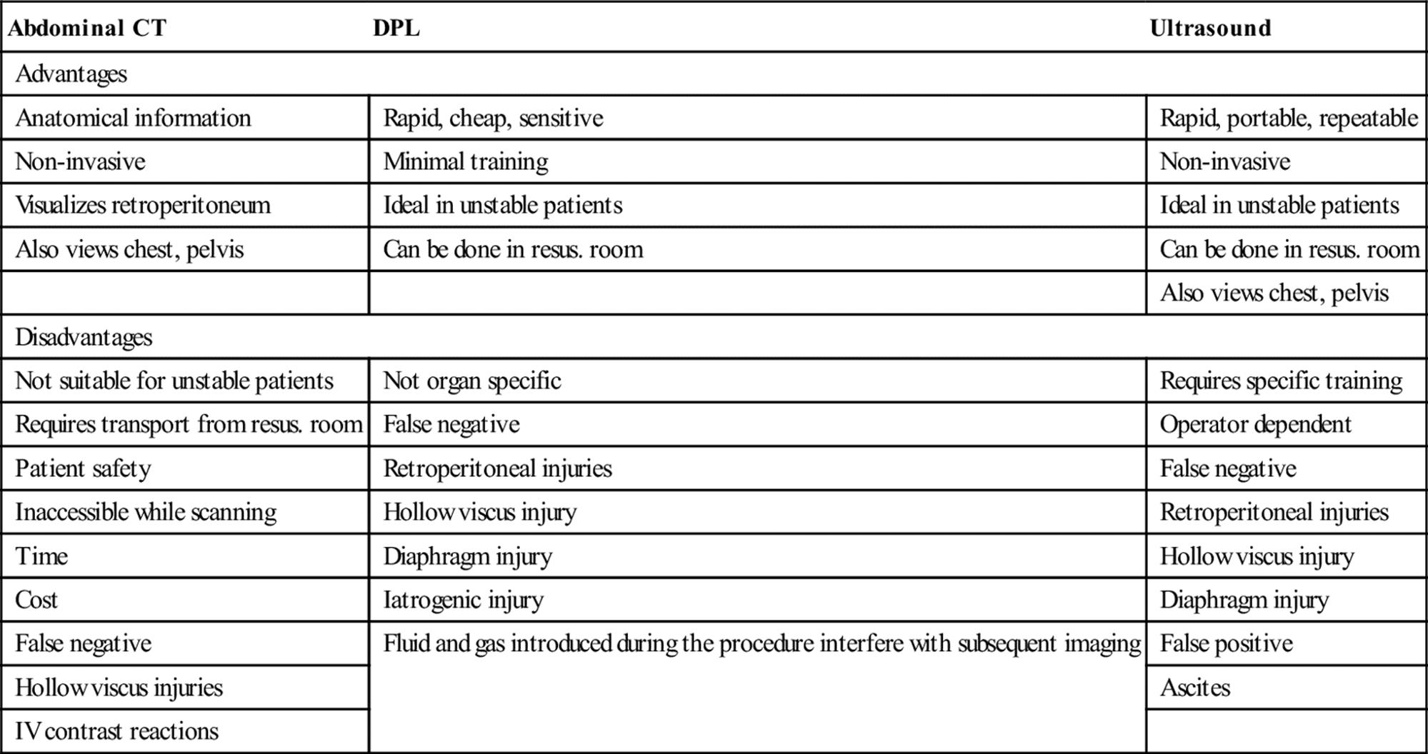
DPL was first described in 1965 and can be completed in less than 4 minutes by experienced operators [4,5]. However, DPL is of limited value when CT or FAST are available and the increasing availability of these modalities has largely resulted in this procedure no longer being used in trauma resususcitation.
Abdominal CT
Abdominal CT is non-invasive and provides precise anatomical details of intra-abdominal pathology. The major disadvantages are associated with the use of IV contrast and the need for the patient to be transported from the resuscitation area to the CT scanner, where they are not accessible during the procedure. Intravenous contrast may rarely produce allergic reactions and can precipitate or exacerbate renal impairment, particularly in higher doses and in the presence of renal hypoperfusion and hypofunction. Although modern machines complete scans in a single breath-hold, time is still required to load and unload the patient. Transport and transfer are times of maximum patient risk and minimum monitoring. Therefore, only stable patients are suitable for transfer for CT scanning. Unstable patients require further resuscitation or operative intervention if this is unsuccessful. The definition of stable is not agreed. The final decision can only be made by the most experienced physician available, although a systolic pressure of at least 90 mmHg and no requirement for additional fluids after correction of estimated losses would be minimum requirements.
Focused assessment sonography in trauma
Focused ultrasound is a skill practised by many clinicians involved in acute trauma management. Abdominal ultrasound is non-invasive, may be done at the bedside and can be repeated as needed. The technique can be easily learnt by clinicians and completed in less than 5 min without interfering with the function of a trauma resuscitation team [6]. Ultrasound combines the advantages of being rapid, accurate and non-invasive and can be performed at the bedside. It is at least as accurate as DPL and, in experienced hands, has similar results to CT in determining the presence of intraperitoneal injury. As a decision-making tool for identifying the need for laparotomy in hypotensive patients (systolic BP<90), FAST has a sensitivity of 100%, specificity of 96% and negative predictive value (NPV) of 100% [7,8]. It is the bedside investigation of choice if an experienced operator is available.
Laparoscopy
Laparoscopy has been investigated in some centres, but the skills required and the time needed for a thorough examination limit the widespread usefulness of this modality in acute blunt trauma. However, it is useful in excluding peritoneal penetration in abdominal stab wounds.
Penetrating injuries
Penetrating injuries produce a different pattern of injury (see Table 3.5.1) and are managed in a different manner from blunt injuries (Figs 3.5.1 and 3.5.2). Cavitation effects and the potential for secondary missiles causing widespread injury mandate formal laparotomy in all cases. Stab wounds with haemodynamic compromise or other indications should also proceed to laparotomy without delay for other investigations. Local exploration of stab wounds by experienced surgeons in patients without evidence of internal injury may be useful, as up to one-third of wounds do not breach the peritoneum. Selected patients may then be managed conservatively.

FIG. 3.5.1 Initial management of blunt abdominal trauma.
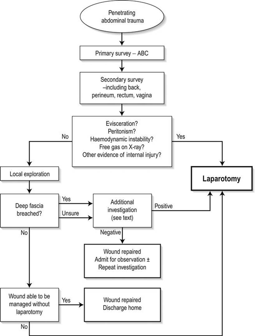
FIG. 3.5.2 Initial management of penetrating abdominal trauma.
Disposition
Disposition decisions may be difficult in the less seriously injured patient or those with suspected injury. Every patient suspected or at significant risk of intra-abdominal injury should be admitted for observation and serial examinations, ideally by the same individual. Injuries to the bowel wall or intestinal blood supply may not be evident on initial clinical examination or investigation and may take 24 h to declare themselves. A higher index of suspicion is required for patients who cannot be assessed clinically. Unconscious, intubated, head-injured and spinally-injured patients are all at increased risk of abdominal injuries and less able to declare them. Serial investigations and clinical vigilance are vital.
Future directions
The difficulty in managing abdominal trauma is determining the presence, location and details of intra-abdominal injury. Current imaging techniques have their disadvantages and limitations. The refinement of modalities, such as ultrasound and portable CT, will rapidly provide more detailed information at the bedside in the resuscitation area, without the need for invasive techniques. Non-operative management of injuries using radiological or minimally invasive surgical techniques continues to develop, thereby reducing the need for laparotomy.
Pharmacological agents such as Factor VIIa and transexamic acid have been assessed as adjuncts for haemorrhage reduction. Results are promising but more research is required.
Controversies
Haemodynamic stability is not dictated by a single reading of pulse or blood pressure. The need for ongoing fluids in order to maintain adequate perfusion indicates instability even in the presence of normal vital signs.
In the multiply injured patient, there is often difficulty in deciding whether to operate first on the head, chest, abdomen or limbs. All surgeons concerned must discuss the decision.
Perhaps the most difficult decision is which is the most appropriate investigation in the otherwise stable patient? Each modality – CT, DPL and FAST – has inherent strengths and weaknesses.
The role of newer pharmacological treatment, such as clotting factors, is promising and requires further evaluation. At present, the most important therapy is early surgery for injury with ongoing bleeding.
Stopping bleeding early is achieved by ‘damage control laparotomy’. This involves immediate attempts to stop the bleeding, either by ligation or tamponade with definitive surgery delayed until the patient is stabilized.
References
1. American College of Surgeons Committee on Trauma. Advanced Trauma Life Support Student Course Manual 9th ed. Chicago: ACS; 2012.
2. Wilkes GJ, McSweeney PA. The diagnosis of traumatic intra-abdominal haemorrhage in patients with normal vital signs. Emerg Med. 1996;8:19–23.
3. Grant RT, Reeve EB. Clinical observations on air-raid casualties. Br Med J. 1941;2:293–297.
4. Root HD, Hauser CW, La Fave JW, et al. Diagnostic peritoneal lavage. Surgery. 1965;57:633–637.
5. Sugrue M, Seger M, Gunning K, et al. A modified combination technique for performing diagnostic peritoneal lavage. Aust NZ J Surg. 1995;65:604–606.
6. Shackford SR, Rogers FB, Osler TM, et al. Focused abdominal sonogram for trauma: the learning curve of nonradiologist clinicians in detecting hemoperitoneum. J Trauma. 1999;46:553–562.
7. Brohi K. Focused assessment with sonography for trauma (FAST).<http://www.trauma.org/index.php/main/article/214>[Accessed Nov. 2012].
8. ACEM Policy on Credentialling for ED Ultrasonography: Trauma Examination and Suspected AAA.<http://acem.org.au/media/P22.pdf>[Accessed Nov. 2012].
3.6 Chest trauma
Mark Fitzgerald, Robert Gocentas and Jeremy Stevens
Essentials
1 Initial management priorities are oxygenation, ventilatory support if required, pleural and pericardial decompression when indicated, circulatory support, adequate analgesia and early imaging to identify evolving and potentially life-threatening injuries.
2 Less than 10% of blunt chest trauma patients require thoracic surgery.
3 Supine chest radiographs do not reliably exclude haemopneumothoraces, aortic transection, diaphragmatic disruption, cardiac tamponade or rib, sternal, thoracic spine and scapula fractures.
4 Multislice CT with IV contrast is the ‘gold standard’ screening and diagnostic tool for thoracic injuries. Sonography may demonstrate haemopneumothorax and cardiac tamponade.
5 Aseptic, percutaneous digital identification of the pleural space is the essential first step for pleural decompression. Drainage and insertion of a chest tube is a secondary priority. Needle thoracocentesis is an unreliable means of decompressing the chest of an unstable patient.
6 Pleural decompression and chest tube insertion during resuscitation is a procedure with a low complication rate.
7 There is a clear role for resuscitative thoracotomy in shocked patients with sonographic evidence of cardiac tamponade.
8 Non-invasive ventilation may avoid complications of mechanical ventilation in select patients with flail chest and pulmonary contusion.
Introduction
Incidence
Thoracic trauma is responsible for 25% of all trauma deaths and contributes to a further 25%. In Australasia and the UK, 90–95% of chest trauma is secondary to blunt injury.
Principles of initial management
The initial management priorities are oxygenation, ventilatory support if required, pleural and pericardial decompression when indicated, circulatory support, adequate analgesia and early imaging to diagnose evolving and potentially life-threatening injuries. The majority of chest trauma patients may be managed non-operatively. Only 10% of blunt thoracic trauma patients will require thoracotomy, the remainder requiring supportive care, including pleural decompression and drainage.
Supportive care, in particular resuscitation, is often suboptimal. Delayed or inadequate ventilatory resuscitation, inadequate shock management, insufficient monitoring of arterial blood gases, delay or failure to perform pleural decompression and drainage and delays in definitive diagnostic imaging remain identifiable problems that contribute to preventable morbidity and mortality [1,2].
Thoracic injuries evolve. Life-threatening injuries including flail chest, pulmonary contusion, thoracic aortic transection, pneumothorax, haemothorax, pericardial tamponade, respiratory insufficiency secondary to rib fractures and ruptured hemidiaphragm may not be apparent on initial presentation. These diagnoses need to be pursued and actively excluded. Supine chest radiographs do not reliably exclude these injuries. Multislice computed tomography (CT) with contrast is the ‘gold standard’ screening and diagnostic test for patients at high risk of potential life-threatening injuries [3]. However, life-saving procedures should be performed first (Table 3.6.1).
Table 3.6.1
Actions prior to CT
|
Situation |
Response |
|
Pneumothorax or haemothorax on initial supine chest X-ray |
Insert chest drain on affected side |
|
Spontaneously breathing patient with unilateral decreased air entry and normal chest X-ray, oxygenation and haemodynamic status |
Await CT |
|
Intubated and ventilated patient with unilateral decreased air entry and normal chest X-ray but hypoxic or hypotensive |
Insert chest drain on affected side, then reassess |
|
Intubated and ventilated patient with hypotension or hypoxia (no other apparent cause) |
Insert bilateral chest drains |
Oxygen
Hypoxia may be absent at the initial reception and resuscitation of chest trauma, but may develop as injuries evolve. Supplemental oxygen may be required for mild desaturation with the FiO2 titrated to the clinical response. Higher FiO2 can be achieved using positive airway pressure and invasive ventilation (CPAP, EPAP).
Increasing the normal I/E ratio may be beneficial in mechanically ventilated patients with severe hypoxia. The benefits of independent lung ventilation, inhaled nitric oxide, prone position, partial liquid ventilation and extracorporeal membrane oxygenation in the initial resuscitation setting are unproven strategies.
Support of pulmonary function
Pain, fatigue from increased work of breathing, disruption of lung mechanics and side effects of opiate analgesia may cause hypoventilation. The elderly as well as other patients with pre-existing poor chest wall compliance are particularly at risk of hypoventilation in the setting of chest wall injury.
Non-invasive ventilation
Patients with pulmonary contusions and high oxygen requirements do not necessarily require intubation but may be safely managed with non-invasive ventilation [4]. By avoiding mechanical ventilation, mortality from nosocomial infection is significantly reduced [5].
Mechanical ventilation
Contraindications to non-invasive ventilation in the trauma patient include the need for full spinal precautions, depressed conscious state and facial injury. Patients with pulmonary contusion and poor lung compliance require ventilation with low tidal volumes and low inspiratory pressures. This reduces barotrauma, secondary lung injury and mortality [6].
Fluid resuscitation
There is evidence that ‘permissive hypotension’ prior to surgical control of blood loss may improve survival in hypotensive penetrating torso injury [7]. It may also reduce blood product requirements, coagulopathy and early postoperative mortality [8]. It is unclear whether these findings translate equally to hypotensive blunt trauma. Once haemorrhage has been controlled, fluid therapy to maximize cardiac output and oxygen delivery may reduce trauma mortality [9].
Conservative fluid resuscitation has been recommended to minimize extravascular lung water in patients with pulmonary contusion [10]. However, under-resuscitation and tissue hypoperfusion may compound organ dysfunction and secondary lung injury. The early use of invasive monitoring to guide fluid replacement may be required.
Analgesia
Adequate analgesia reduces hypoventilation secondary to pain and facilitates coughing and chest physiotherapy. It reduces complications of atelectasis, consolidation and respiratory failure and improves pulmonary function [11]. Oral analgesia is often sufficient for single rib fractures. Parenteral narcotic analgesia, intercostal nerve block or thoracic epidural analgesia is usually required for multiple rib fractures. Intercostal nerve block involves a number of injections to treat multiple rib fractures. This limits its usefulness compared to the other techniques.
Indications for emergency thoracotomy
Although more than 90% of chest trauma patients may be managed non-operatively, the following categories warrant surgical intervention [12]:
cardiac tamponade
acute deterioration – cardiac arrest in patients with penetrating truncal trauma
vascular injury at the thoracic outlet
traumatic loss of chest wall
massive air leak from chest tube
massive or continuing haemothorax
mediastinal traversing penetrating injury
endoscopic or radiographic demonstration of oesophageal injury
endoscopic or radiographic demonstration of tracheal or bronchial injury
radiographic evidence of great vessel injury
thoracic penetration with industrial liquids (especially coal tar products).
Resuscitative thoracotomy
Left anterolateral thoracotomy as a resuscitative manoeuvre allows direct access to the heart and pericardial decompression for patients who have lost output following cardiac lacerations. Myocardial wounds can then be directly controlled. Right atrial catheterization facilitating IV fluid administration, pulmonary hilar clamping, cross-clamping of the descending aorta and open cardiac massage are adjunctive procedures performed if indicated.
Survival rates of better than 40% have been reported in some subgroups of penetrating trauma arrest, specifically precordial stab wounds. Survival was dependent on resuscitative thoracotomy performed within 10 minutes of arrest secondary to penetrating chest trauma and an organized cardiac electrical rhythm being present [13–16]. Left anterolateral thoracotomy allows pericardial decompression [17] in patients who have lost output following penetrating injury to the heart. Myocardial wounds can then be directly controlled. The role of resuscitative thoracotomy in blunt trauma arrest is more controversial, with a relatively low survival rate (<3%) [18].
Focused assessment with sonography in trauma (FAST) is an important triage tool in determining the presence of cardiac tamponade. Immediate use of ultrasonography can establish the diagnosis of haemopericardium and prompt repair of the injury may improve overall survival. Unresponsive hypotension with a systolic blood pressure of less than 70 mmHg and a FAST positive for pericardial tamponade is a consensus-based indication for immediate resuscitative thoracotomy. For patients with severe hypotension or in extremis, the treatment of choice is resuscitative thoracotomy, decompression of the pericardium and control of the cardiac injury [19].
Given the widespread availability of ultrasound, arguments about resuscitative thoracotomy for blunt trauma should include the important decision support provided by sonography. There is clearly a role for resuscitative thoracotomy in shocked patients with cardiac tamponade following blunt trauma. Procedural training and credentialing is recommended [19].
Thoracic injuries
Fractured ribs
Fractured ribs are a common sequela of focal trauma. Fractured ribs cause pain, which may then interfere with ventilation and coughing, causing ventilatory impairment and atelectasis. This impairment may not be manifest for hours and occasionally days after the injury.
Underlying structures are often injured concomitantly, particularly the lungs, pleura and intercostal vessels. Fractures of the lower left ribs are associated with splenic injury, the lower right with hepatic injury and the lower posterior ribs with renal injury. The first and second ribs are stronger and less easily injured and, when fractured, are usually indicative of significant force to the upper mediastinum. Although first and second rib fractures have been traditionally associated with thoracic aortic injury, the positive predictive value of this association has been questioned [20].
Rib fracture is essentially a diagnosis based on the clinical findings of local tenderness with or without deformity and crepitus. Up to 50% of fractured ribs are not apparent on the initial chest X-ray [21]. Reliance on the X-ray to diagnose fractured ribs inevitably results in under-diagnosis. This may lead to delays in diagnosis and therapy and an adverse outcome, particularly with elderly patients and those with coexisting airways disease.
The management of rib fractures centres on actively excluding associated injury as well as adequate pain relief including patient controlled administration of narcotics and local and regional anaesthetic blocks to allow breathing exercises, coughing and incentive spirometry. This in turn minimizes subsequent atelectasis and pulmonary sequelae.
Fractured sternum
This is a clinical diagnosis confirmed on CT scan or lateral chest X-ray. Associated intrathoracic injuries, specifically myocardial and other mediastinal injuries, need to be identified.
The possibility of underlying injury has been related to the mechanism of injury. For example, in North America, it is reported that up to 66% of patients with sternal fractures have intrathoracic injuries. It is believed that low seatbelt usage results in sternal fractures secondary to impact against the steering wheel. In Australasia, where seatbelt usage is high, sternal fracture is more often caused by the restraining belt. Therefore, comparatively lower deceleration forces are evident, resulting in a reduced association with underlying injury [22,23].
For isolated sternal fractures, admission for analgesia is usually required, although this may be only necessary for 1–2 days. Monitoring is not required unless the mechanism or subsequent investigations suggest underlying cardiac injury.
Vertebral column and spinal cord injury
Spinal stability must be determined prior to sitting the patient upright to improve ventilation and reduce VQ mismatch. Exclusion of thoracic spine fractures and spinal-cord injury forms part of the routine work-up of the chest trauma patient. Occult injuries are common and unstable injuries in ventilated patients require skilled nursing. Such injuries are easily overlooked.
Flail chest
Flail chest may occur where the continuity of the bony skeleton of the chest wall is disrupted in two places. It is characterized by paradoxical movement of the associated unanchored chest wall segment. Because of muscle spasm and splinting, this segment may not be apparent initially and may flail some time after the accident. Clinical features of a flail segment may also be masked by positive-pressure ventilation, which splints the chest wall internally. Elderly patients have a less compliant chest wall and are at greater risk of developing a flail segment.
Flail chest is often associated with ventilatory insufficiency. Ventilatory disturbance is caused by hypoventilation of the affected hemithorax due to the mechanical disruption and associated pain, compounded by the underlying pulmonary contusion. Therapy centres on maintaining oxygenation, ventilation and euvolaemia. Adequate analgesia should be supplemented with intercostal nerve blocks or epidural analgesia. In general, patients with a significant flail, which impairs ventilation, will require respiratory support. Hypoxia may be managed with non-invasive ventilation. Mechanical ventilation is required if non-invasive ventilation is contraindicated or unsuccessful.
A small number of patients with severe wall instability may require operative fixation to facilitate the weaning of mechanical ventilation [24]. Operative reduction and internal fixation using malleable, absorbable splints for the flail segment is a new approach associated with a reduction in ventilator days [25].
Ruptured hemidiaphragm
Diaphragmatic rupture may be difficult to diagnose. High-velocity lateral torso trauma or thoracoabdominal crush injuries as well as lateral rib fractures, penetrating left upper quadrant wounds and fractured pelvis, are linked to an increased incidence of diaphragmatic disruption. There may be respiratory compromise, with diminished air entry in the involved hemithorax. Placement of a radiopaque nasogastric tube will facilitate the diagnosis of left hemidiaphragmatic disruption on chest X-ray. Although gross rupture may be apparent initially, the classic radiological findings of viscera in the thoracic cavity, the nasogastric tube coiled in the thoracic cavity or marked hemidiaphragm elevation are present only 50% of the time, with no intrathoracic pathology seen on 15% of occasions [26]. CT scan will display gross disruption but may miss small defects. Diagnostic yield may be better with magnetic resonance imaging (MRI) [27]. Smaller diaphragmatic injuries may evolve, with visceral herniation developing over time. Positive pressure ventilation may mask this. Thus, many diaphragmatic injuries present late. Occult diaphragmatic lacerations are associated with penetrating injuries of the thoracoabdominal region and should be actively excluded by laparoscopy, thoracoscopy or open surgery. The treatment of diaphragmatic disruption is surgical repair.
Open pneumothorax
Open pneumothorax presents an immediate threat to life. An open chest wall defect disrupts the generation of a negative inspiratory pressure. If the opening is approximately two-thirds the diameter of the trachea, air will pass preferentially through the defect (a ‘sucking’ chest wound) and respiratory failure will occur [28].
Initial management includes covering the defect with a sterile dressing and taping it on three sides to achieve a flutter-valve effect, prior to placement of an intercostal catheter and sealing of the defect. Definitive surgical closure is required.
Pneumothorax
Simple pneumothorax is characterized by a visceral pleural rent and pleural air preventing expansion of the associated lung. Although small (<20%) pneumothoraces may be managed expectantly, larger ones mandate pleural decompression and drainage. There is no evidence that needle thoracotomy is a reliable means of pleural decompression (Fig. 3.6.1). The technique should be avoided during hospital trauma reception and resuscitation and used only as a technique of last resort. Blunt dissection and digital identification and decompression of the pleura using an aseptic technique should be the technique of first choice. Once successfully performed, it reduces the urgency of the situation and allows time for the subsequent placement of a chest tube [29].
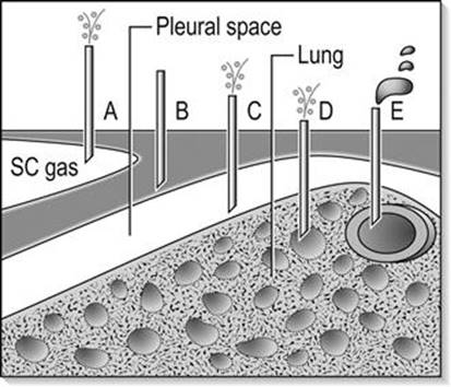
FIG. 3.6.1 Possible positions of needle thoracocentesis (NT). (A) False positive – as needle decompresses subcutaneous emphysema. (B) False negative – as needle does not reach pleural space. (C) Correct position of NT with decompression of tension pneumothorax. (D) False positive – with needle intrapulmonary in bulla or bronchial tree. If the tension pneumothorax is loculated due to pulmonary adhesions and missed by NT, a false-negative result may occur with intra-pulmonary placement. (E) True negative – with needle in a major vessel or the heart. This may be misinterpreted as a false positive for haemothorax. Only C will decompress a tension pneumothorax. A, B, D and E have all been associated with failure to decompress the pleural space and fatal outcomes. (From Fitzgerald M, Mackenzie CF, Marasco S, Hoyle R, Kossman T. Pleural decompression and drainage during trauma reception and resuscitation. Injury 2008;39:9–20, with permission.)
Intercostal catheters with underwater seal or flutter-valve drainage should also be inserted for pneumothoraces if positive-pressure ventilation is anticipated or has been commenced. If small traumatic pneumothoraces are not drained, the patient should be followed closely with repeat chest X-rays. Intercostal catheters should be placed if the patient is to be air-transported to another facility. Clinicians should be aware of common problems with chest drain placement (Fig. 3.6.2).

FIG. 3.6.2 Possible positions and complications of tube thoracostomy (TT). (A) Trauma to the intercostal neurovascular bundle. (B) Extrapleural placement. (C) Correct position in pleural space. (D) Intrafissural placement. (E) Intrapulmonary placement. (F) Mediastinal impingement or penetration. (G) Trans-diaphragmatic placement. (H) Infection. (From Fitzgerald M, Mackenzie CF, Marasco S, Hoyle R, Kossman T. Pleural decompression and drainage during trauma reception and resuscitation. Injury 2008;39:9–20, with permission.)
Thoracic CT scanning will demonstrate pneumothoraces that may not be apparent on plain radiographs [30]. This should prompt consideration of intercostal catheter placement in ventilated patients. Small (occult) pemuomothoraces in patients without major shunts may be managed expectantly [31].
While ultrasound is superior to initial supine chest X-ray in the diagnosis of pneumothorax, it does not change the management of the patient as the majority of occult pneumothoraces are treated conservatively. Conversely, a negative ultrasound for pneumothorax may not sufficiently exclude the need for pleural decompression in the unstable patient [32].
Tension pneumothorax
Tension pneumothorax occurs with the formation of a ‘one-way valve’ from the lung through disrupted visceral pleura. Air collects under tension in the hemithorax, collapsing the lung and displacing the mediastinum, impairing ventilation and obstructing venous return. Tension pneumothorax is more commonly associated with positive-pressure ventilation. It is essentially a clinical diagnosis, characterized by tachypnoea, tracheal deviation away from the affected hemithorax, diminished ipsilateral breath sounds, diminished compliance, oxygen desaturation and hypotension.
If clinically suspected, the affected hemithorax should be immediately decompressed and an intercostal catheter subsequently inserted. There should be no attempt to delay chest decompression in an unstable patient in favour of a chest X-ray.
Intubated and ventilated thoracic trauma patients may demonstrate subcutaneous emphysema on initial chest X-ray without a pneumothorax being visible. This should prompt immediate chest tube placement, as this is a precursor of ipsilateral tension pneumothorax. The subcutaneous tissues in communication with the air leak offer less initial resistance and display air under pressure, prior to tension developing within the pleural space.
Haemothorax
Blood may accumulate within the pleural space after lung laceration or laceration of a chest-wall vessel and, less commonly, after mediastinal injury. It is indicated by diffuse opacification of a hemithorax on supine chest X-ray or blunting of the costophrenic angle on an upright film. Once digital pleural decompression has occurred the haemothorax is best drained via placement of a 32 Fr or larger intercostal catheter, positioned in the fifth or sixth intercostal space in the mid-axillary line on the affected side. The use of suction (20 cm H2O) facilitates drainage.
Bleeding is usually self-limiting following drainage. Drainage of more than 1500 mL following initial intercostal catheter insertion (massive hemothorax) or a loss of more than 200 mL/h for more than 2 h, are indications for thoracotomy [33]. Large blood losses frequently come from intercostal arteries.
Clamping the intercostal catheter in an attempt to tamponade bleeding and ‘buy time’ for an intubated and ventilated, unstable patient with a massive and ongoing hemithorax could be considered if delays to thoracotomy arise. However, this technique is yet to be prospectively validated.
Pulmonary contusion
Pulmonary contusion is characterized by the leakage of blood into the alveoli and pulmonary interstitium, culminating in consolidation and atelectasis. Associated hypoxia may be profound. The initial chest X-ray may not demonstrate the severity of injury. Pulmonary contusion may take some time to be radiologically apparent, with 21% of experimentally incurred contusions still not visible on chest X-ray 6 hours after injury [34].
CT scans provide the most sensitive test for gauging the extent of pulmonary contusion, although arterial blood gases provide the best measure of physiological derangement requiring intervention. Therapy is based on ensuring adequate oxygenation, ventilatory support and fluid restriction. Ventilation should involve low-volume, low-pressure techniques to reduce barotrauma and secondary injury.
Tracheobronchial injury
Injuries to the trachea and bronchi are rare, accounting for less than 1% of injuries after blunt chest trauma. Eighty per cent of injuries occur near the carina, with mediastinal and cervical emphysema resulting. A persistent air leak post-intercostal drain insertion should alert the clinician to the possibility of a tracheobronchial injury. Fibreoptic bronchoscopy is the investigative modality of choice. Persistent air leaks often require operative repair.
Myocardial contusion
Although myocardial contusion is common, significant sequelae are rare. Cardiac failure and hypotension are uncommonly associated with myocardial contusion. Although the ECG is used as a predictor of myocardial contusion, it is non-specific and poorly portrays the right ventricle – the area most commonly injured. Cardiac enzyme elevation does occur but is non-predictive. Echocardiography may demonstrate dyskinesis of the ventricular wall. Patients with hyperacute ECG changes or conduction defects should be admitted and monitored for dysrhythmias.
Myocardial laceration and cardiac tamponade
Precordial penetrating injury is associated with myocardial laceration. Bedside sonography is useful in demonstrating myocardial injury and pericardial collections. Patients presenting with signs of pericardial tamponade (hypotension, diminished heart sounds, jugular venous distension) require urgent surgical intervention.
Patients who acutely deteriorate into cardiac arrest yet who had signs of life en route to hospital or on arrival, require a resuscitative thoracotomy in the emergency department [35]. Outcome for blunt trauma patients without initial signs is very poor (<2%), but penetrating injury has a higher survival rate. This procedure should only be undertaken when there is some chance of survival because of the infection risks to personnel. Prolonged (>9 minutes) external cardiac massage is futile for these patients.
Tension pneumopericardium
Tension pneumopericardium, albeit much less common than tension pneumothorax, is thought to arise via a similar ‘one-way valve’ mechanism, particularly after the institution of positive-pressure ventilation. It is characterized by raised jugular/central venous pressure and hypotension and requires urgent pericardiocentesis [36].
Thoracic aortic transection
Eighty-five per cent of patients with transection of the thoracic aorta die before reaching hospital. Lateral as well as frontal impact motor vehicle crashes are associated with aortic transection [37]. High deceleration forces cause the aorta to accelerate and twist against fixation points – usually the ligamentum arteriosum just distal to the left subclavian artery. The associated shearing forces transect and tear the artery. Tears of the aorta are commonly fatal at the time of injury or immediately after, as the aorta usually tears completely and the injured rapidly bleed to death. However, it has been estimated that up to 15% of patients with thoracic aortic injuries survive to reach hospital and this is due to the outer concentric layers of the aorta remaining intact. Fifty per cent die within the next 48 hours if not operated upon. Thus, early diagnosis and treatment of incomplete transection of the thoracic aortic injury is important.
Mediastinal widening on chest X-ray is 85% sensitive and 10% specific for aortic transection [38]. Associated fractures of the thoracic spine also cause mediastinal widening and make interpretation difficult. There is no thoracic skeletal injury that is a clinically useful predictor of acute thoracic aortic transection. Therefore, patients involved in high speed accidents require chest CT scans to exclude aortic injury.
Definitive treatment includes ensuring that the blood pressure is not elevated to reduce shearing and radiologically guided placement of a stent across the injured aorta [39–43].
Surgical repair of the injured thoracic aorta is uncommon and has a high mortality and morbidity. Published mortality rates of patients with thoracic aortic injury are: endovascular stent 9%, open surgical repair 19% and non-operative management without stent 46% [44].
Transoesophageal echocardiography has been used as a screening tool for aortic tears and is useful for patients in theatre or those unable to be moved to angiography [45,46].
Oesophageal perforation
Oesophageal rupture after blunt chest trauma is rare. The lower third of the oesophagus is the commonest site of rupture, presumably secondary to a forced Valsalva manoeuvre. Mediastinitis is a subsequent development. Retrosternal pain is common and mediastinal air may be seen on chest X-ray. Gastrograffin swallow and CT scanning is the study of choice. Mortality is directly related to time to operative repair [47].
Gunshot injuries across the truncal midline more commonly involve mediastinal and spinal structures and therefore have a much greater mortality than unilateral injuries [48]. It is important to exclude oesophageal injury early with penetrating and transmediastinal wounds, as there is significant morbidity and mortality in those patients who survive to hospital [49].
Likely developments
Better systems of care, improved imaging and newer technologies will result in earlier diagnosis and treatment of injuries preventing subsequent evolution and deterioration.
There is a trend towards the increased use of non-invasive ventilation and operative rib fixation for flail chest. The role of ultrasound is evolving and there is increasing use of ultrasound in the assessment of chest trauma. Conservative management of occult pneumothoraces is likely to increase – more prospective studies are required to prove the safety of conservative management.
Conclusion
The significance and severity of chest trauma may not be obvious on initial examination as injuries evolve. Most seriously injured patients with blunt thoracic trauma require supportive care including chest decompression and drainage. Indications for immediate chest decompression are ventilatory or respiratory compromise (Fig. 3.6.3). Patients with underlying airway disease and elderly patients with diminished compliance are at particular risk. Delayed or inadequate ventilatory resuscitation, inadequate shock management, insufficient monitoring of arterial blood gases, delay or failure to perform pleural decompression and drainage and inadequate diagnostic imaging remain identifiable problems in emergency departments.
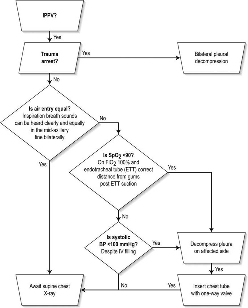
FIG. 3.6.3 Initial binary decision tree for pleural decompression. (From Fitzgerald M, Mackenzie CF, Marasco S, Hoyle R, Kossman T. Pleural decompression and drainage during trauma reception and resuscitation. Injury 2008;39:9–20, with permission.)
References
1. McDermott F. Severe chest injuries: problems in Victoria Issues and controversies in the early management of major trauma Melbourne: Alfred Hospital and Monash University, Department of Surgery; 1994.
2. Danne P, Brazenor G, Cade R, et al. The Major Trauma Management Study: an analysis of the efficacy of current trauma care. Aust NZ J Surg. 1998;68:50–57.
3. Mirvis SE. Imaging of acute thoracic injury: the advent of MDCT screening Semin Ultrasound. CT MRI. 2005;26:305–331.
4. Vidhani K, Kause J, Parr M. Should we follow ATLS guidelines for the management of traumatic pulmonary contusion: the role of non-invasive ventilatory support. 2002;52:265–8.
5. Gurnduz M, Unugenc H, Ozalevli M. A comparative study of continuous positive airway pressure (CPAP) and intermittent positive pressure ventilation (IPPV) in patients with flail chest. Emerg Med J. 2005;22:325–329.
6. ARDS Network. Ventilation with lower tidal volumes for acute lung injury and the acute respiratory distress syndrome. N Engl J Med. 2000;3:1301–1308.
7. Bickell WH, Wall Jnr MJ, Pepe PE, et al. Immediate versus delayed fluid resuscitation for hypotensive patients with penetrating torso injuries. N Engl J Med. 1995;38:780–787.
8. Morison CA, Carrick MH, Norman MA, et al. Hypotensive resuscitation strategy reduces transfusion requirements and severe post operative coagulopathy in trauma patient with haemorrhagic shock: preliminary results of a randomized controlled trial. J Trauma. 2011;70:652–663.
9. Bishol MH, Shoemaker WC, Appel PL, et al. Prospective, randomized trial of survivor values of cardiac index. J Trauma. 1995;38:780–787.
10. Richardson JD, Adam L, Flint LM. Selective management of flail chest and pulmonary contusion. Ann Surg. 1982;196:481.
11. Desai PM. Pain management and pulmonary dysfunction. Crit Care Clin. 1999;15:151–166.
12. Pickard LR, Mattox KL. Thoracic trauma and indications for thoracotomy. In: Mattox KL, Moore EE, Feliciano DV, eds. Trauma Norwalk: Appleton & Lange. 1988;315–320.
13. Tyburski JG, Astra L, Wilson RF. Factors affecting prognosis with penetrating wounds of the heart. J Trauma Injury Infect Crit Care. 2000;48:587–591.
14. Battistella FD, Nugent W, Owings JT. Field triage of the pulseless trauma patient. Arch Surg. 1999;134:742–746.
15. Renz BM, Stout MJ. Rapid right atrial cannulation for fluid infusion during resuscitative emergency department thoracotomy. Am Surg. 1994;60:946–949.
16. Mattox KL, Feliciano DV. Role of external cardiac compression in truncal trauma. J Trauma. 1982;22:934–936.
17. Mulder DS, Barkun JS. Injuries to the trachea, bronchus and oesophagus. In: Moore EE, Mattox KL, Feliciano DV, eds. Trauma Norwalk: Appleton & Lange. 1991;345–355.
18. Aihara R, Millham FH, Blansfield J, et al. Emergency room thoracotomy for penetrating chest injury: effect of an institutional protocol. J Trauma Injury Infect Crit Care. 2001;50:1027–1030.
19. Fitzgerald M, Tan G, Gruen R, et al. Emergency physician credentialing for resuscitative thoracotomy for trauma. Emerg Med Australas. 2010;22:332–336.
20. Lee J, Harris JH, Duke JD, Williams JS. Noncorrelation between thoracic skeleton injuries and traumatic aortic tear. J Trauma. 1997;43:400–404.
21. Hehir MD. Hollands MJ, Deane SA The accuracy of the first chest X-ray in the trauma patient. Aust NZ J Surg. 1990;60:529–532.
22. Brookes JG, Dunn RJ, Rogers IR. Sternal fractures: a retrospective analysis of 272 cases. J Trauma. 1993;35:46–54.
23. Hills MW, Delprado AM, Deane SA. Sternal fractures: associated injuries and management. J Trauma. 1993;35:55–60.
24. Lardinois D, Kreuger T, Dusmet M. Pulmonary function testing after operative stabilization of the chest wall for flail chest. Eur J Cardiovasc Surg. 2001;20:496–501.
25. Marasco SF, Davies AR, Cooper J, et al. Prospective randomized controlled trial of operative rib fixation in traumatic flail chest. J Am Coll Surg 2013;(12):01429 doi:pii: S1072-7515.
26. Brasel KJ, Borgstrom DC, Meyer P. Predictors of outcome in blunt diaphragm rupture. J Trauma. 1996;41:484–487.
27. Mirvis SE. Diagnostic imaging of acute thoracic injury. Semin Ultrasound CT MRI. 2004;25:156–179.
28. American College of Surgeons Committee on Trauma. Advanced trauma life support student manual Chicago: American College of Surgeons; 2004; 105.
29. Fitzgerald M, Mackenzie CF, Marasco S. Pleural decompression and drainage during trauma reception and resuscitation. Injury. 2008;39:9–20.
30. Blostein P, Hodgman CG. Computed tomography of the chest in blunt thoracic trauma: results of a prospective study. J Trauma. 1997;43:3–18.
31. Mowery NT, Gunter OL, Collier BR, et al. Practice management guidelines for management of hemothorax and occult pneumothorax. J Trauma Injury Infect Crit Care. 2011;70:510–518.
32. Alrahji K, Woo M, Vaillancourt C. Test characteristics of ultrasonography for the detection of pneumothorax – a systematic review and meta-analysis. Chest. 2012;141:703–708.
33. American College of Surgeons Committee on Trauma. Advanced trauma life support student manual Chicago: American College of Surgeons; 2004; 109.
34. Cohn S. Pulmonary contusion: review of the clinical entity. J Trauma. 1997;42:973–979.
35. Jorden RC. Penetrating chest trauma. Emerg Med Clin N Am. 1993;11:97–106.
36. Fitzgerald MC, Foord K. Tension pneumopericardium following blunt trauma. Emerg Med. 1993;5:74–77.
37. Katyal D, McLellan B, Brenneman F, et al. Lateral impact motor vehicle collisions: a significant cause of traumatic rupture of the thoracic aorta. J Trauma. 1997;45:769–772.
38. Fabian T, Richardson JD, Croce MA, et al. Prospective study of blunt aortic injury: multicentre trial of the American Association for the Surgery of Trauma. Journal of Trauma. 1997;42:374–383.
39. Creasy JD, Chiles C, Routh WD, et al. Overview of traumatic injury of the thoracic aorta. Radiographics. 1997;17:27–45.
40. Andrassy J, Weidenhagen R, Meimarakis G. Stent versus open surgery for acute and chronic traumatic injury of the thoracic aorta: a single center experience. J Trauma. 2006;60:765–772.
41. Hoornweg LL, Maarten KD, Goslings CJ. Endovascular management of traumatic rupture of the thoracic aorta: a retrospective multicenter analysis of 28 consecutive cases in The Netherlands 2006. J Vasc Surg. 2006;43:1096–1102.
42. Neschis DG, Moaine S, Gutta R. Twenty consecutive cases of endograft repair of traumatic aortic disruption: lessons learned. J Vasc Surg. 2007;43:487–492.
43. Reed AB, Thompson JK, Crafton CJ, et al. Timing of endovascular repair of blunt traumatic aortic transection. J Vasc Surg. 2006;43:684–688.
44. Lee WA, et al. Endovascular repair of traumatic thoracic aortic injury: clinical practice guidelines of the Society for Vascular Surgery. J Vasc Surg. 2011;53:187–192.
45. Cohn SM, Burns GA, Jaffe C, Milner KA. Exclusion of aortic tear in the unstable trauma patient: the utility of transoesophageal echocardiography. J Trauma. 1995;39:1087–1090.
46. Ben-Menachem Y. Assessment of blunt aortic-brachiocephalic trauma: should angiography be supplanted by transoesophageal echocardiography? J Trauma. 1997;42:969–972.
47. Jackimczyk K. Blunt chest trauma. Emerg Med Clin N Am. 1993;11:81–96.
48. Hirshberg A, Or J, Stein M, Walker R. Transaxial gunshot injuries. J Trauma. 1996;41:460–461.
49. Patel M, Malinoski D, Zhou L. Penetrating oesophageal injury: a contemporary analysis of the National Trauma Data Bank. Injury. 2013;44:48–55.
3.7 Limb trauma
Amit Maini and Alfredo Mori
Essentials
1 Optimal trauma resuscitation and fracture management will reduce limb and life-threatening complications.
2 Skin under pressure over a fracture is an orthopaedic emergency.
3 Fracture management consists of reduction, immobilization and rehabilitation.
4 Specific limb trauma assessment is part of the secondary survey.
5 Early renal replacement therapy is potentially life saving in crush syndromes.
Introduction
Injuries to the limbs account for the most trauma-related presentations to emergency departments (EDs) and are a common source of disability in this patient group. These injuries span a spectrum, from seemingly trivial and benign, to limb and life threatening. Injuries may involve the soft tissues, bones, as well as the neurovascular structures and can occur as discrete injuries in isolation or in combination as is the case with more severe trauma.
The major consideration in managing extremity injury in a major trauma patient is to consider the treatment of the patient as a whole, rather than be distracted by any particular injury or fracture. A full primary survey should be performed on all patients, with simultaneous assessment and management of life-threatening injuries as the priority. Orthopaedic injuries should be picked up as part of the secondary survey, although bleeding from long bone fractures (especially open femoral fractures) may be categorized under the circulation component of the primary survey.
Specific complications of open fractures and crush injury include bleeding, crush syndrome and hyperkalaemia as well as sepsis. Early consideration should be given to these with respect to early haemorrhage control and early administration of appropriate antibiotics. Other complications include compartment syndrome, as well as fat embolus syndrome which will be considered later.
The only immediate threat to life from fractures is haemorrhagic shock. Limb trauma may pose a therapeutic challenge in trauma resuscitation by limiting available vascular access. Deformed or injured limbs should, as a rule, be avoided when placing intravenous cannulae and patients with multiple-injured limbs may require early central venous access. Estimates of blood loss, in addition to external scene and ED blood loss, include:
1200–1500 mL for femoral fracture
500–1000 mL for tibial fracture
500 mL for humeral fracture.
The immediate goal of management in the multitrauma patient is control of haemorrhage, followed by limb salvage. The overall aim of limb trauma care is a return to full pain-free function and good cosmesis. The function of the upper limb is to communicate a person’s will to the external world and manipulate their surroundings. The function of the lower limbs is independent ambulation. Rehabilitation plays an essential part in recovery and must be considered early with regard to the role of splints from the outset of trauma care.
Fractures
A fracture is a soft-tissue injury with loss of bone continuity. The soft-tissue component is often underestimated. Tense or white skin over a closed fracture is an orthopaedic emergency requiring urgent reduction, even before imaging. Ischaemic skin over bone, such as in the area of the anterior tibia, has a high rate of necrosis and a poor response to skin grafting. This may result in the disastrous complication of limb amputation.
All transferred patients should have splints removed and the underlying tissues carefully assessed. No splint should remain over skin for more than 8 hours without removal and reassessment. Discharged patients should have clear instructions for returning should there be an increase in pain, tightness under a plaster or splint or numbness and pain in the limb distal to the fracture. Planned early follow up is essential.
Fractures where the overlying skin is intact are closed. Open or compound fractures are defined by their being exposed to the environment. Compound fractures may be classified as follows:
grade 1: wound<1 cm punctured from below (fracture fragment)
grade 2: wound up to 5 cm long with no contamination, crush, skin loss or necrosis
grade 3: large laceration with associated contamination or crush (closed after debridement). Periosteal stripping of bone (will require skin flap for closure)
grade 4: total or subtotal amputation.
Patients at high risk of fracture complications from single limb trauma include the elderly, the immunocompromised, alcoholics (from repeated falls and poor follow up) and patients with peripheral vascular disease. High-risk mechanisms in limb injury from multiple trauma include falls from over 3 m, pedestrians, motorcyclists and high-speed motorists. Haemodynamically unstable patients, those with open fractures, with delayed (>6 hours) presentation times and the severely head injured also form a group of associated injuries at high risk of complications. Severely head-injured patients are prone to coagulopathy, further increasing fracture bleeding. Clinical assessment of limb trauma may be affected by poor feedback from a head-injured conscious state or sedation and intubation [1–5].
Associated injuries
Vascular injury
Arterial injury is a limb- and (potentially) life-threatening emergency. Ischaemic times of 4–6 hours may result in permanent damage to tissues. Peripheral circulation and distal pulses must always be assessed and sides compared. All splints in transferred patients should be removed and underlying tissues and distal circulation assessed. High-risk patients include those that have been transferred by air with air splints in situ, unconscious patients and shocked patients, as the shock state may mask local limb ischaemia.
There may be a role for hypotensive resuscitation in complete arterial injury, whereby a reduced, controlled perfusion pressure may leave intact a clot formed as a result of complete arterial laceration. Aggressive fluid resuscitation in these patients, where not otherwise indicated, or inadequate analgesia, may cause a rise in mean arterial pressure, dislodgement of the clot at the site of injury and resumption of arterial bleeding.
The presence of a distal pulse does not exclude arterial injury, which may be incomplete. Other signs to consider in this diagnosis include the presence of a dislocation, limb deformity or open fracture in that limb, brisk bleeding from an open wound, reduced pulses compared to the other side (either clinically or on Doppler) and an expanding wound haematoma. Delayed signs include a false aneurysm or the presence of a bruit on examination.
Sites at specific risk of arterial injury include:
brachial artery in the upper limb
popliteal artery around the knee and adductor canal of the medial distal femur
deep femoral artery at the trochanter level of the femur
the anterior tibial artery in the tibia.
Computed tomography (CT) angiography has largely replaced formal angiography as the investigation of choice and, if a vascular injury is suspected, early discussion with a vascular surgeon is recommended.
Nerve injury
Nerve injury in limb trauma may be a direct result of laceration by foreign bodies or fracture fragments. Nerves may be crushed, bruised or stretched. Ischaemia must be excluded as a cause for neurological deficits. Nerve injury from penetrating injury ideally should be explored in the operating theatre.
Nerve injuries may be classified into three major groups:
Neuropraxia. This is a transient change in conduction. It usually follows crush or contusion or stretching of a nerve. There is usually some return of function within days and complete return of function within 8 weeks.
Axonotmesis. Complete denervation with an intact nerve sheath, usually as a result of blunt trauma causing severe bruising and stretching. Regeneration takes place over months along the intact nerve sheath.
Neurotmesis. Complete division of a nerve and its sheath. Spontaneous regeneration is not expected and surgical repair is required. This represents the most severe end of the spectrum and full recovery cannot be guaranteed.
The neurovascular status of the injured limb should be assessed and documented before and after any manipulation and relocation. Specific nerve injury presentations include:
wrist drop from radial nerve injury of the middle or distal third of the humerus
foot drop from peroneal nerve injury to the proximal fibula
shoulder skin numbness from axillary nerve injury in shoulder dislocation
lower limb numbness and weakness from sciatic nerve injury due to posterior dislocations of the hip
hand numbness and weakness from median nerve injury in distal fractures of the wrist and dislocations of the carpal bones
hand numbness and weakness from ulnar nerve injury in injuries to the medial forearm or humerus.
Presentation
History and examination
Injury history and pre-hospital care should be presented in the MIST format on arrival at hospital:
Mechanism.
Injuries identified or suspected. Specifically, attention to external blood loss, limb deformity (and correction) or amputation.
Symptoms and signs: in particular vital signs, whether the patient mobilized at the scene, areas of limb weakness or numbness and pale or pulseless limbs.
Treatments commenced and the responses to them: a note should be made of all splints placed and their type (hard, soft or anatomic).
The general history should also include patient’s normal state of health, medications and allergies, hand dominance, tetanus prophylaxis and fasting state. The history should be presented at the same time as the primary survey commences. Only when this is completed may a meticulous secondary survey start to exclude and treat limb trauma. All splints should be removed for limb trauma assessment, especially in patients transferred between hospitals, given the often long intervals before definitive assessment and treatment.
The assessment of limbs for trauma includes:
Looking for deformity, bruising, open fractures, bleeding, skin blistering (which denotes soft tissues under pressure) and white or pressured skin. Comparison should always be made with the other limb.
Feeling for local pain, crepitus or deformity. All peripheral pulses should be examined for and their absence investigated further.
Active (patient controlled) and passive (examiner controlled) movement. Joints with a full active range of movement are almost never dislocated. Full active movement of the elbow may exclude an elbow fracture and straight leg raising a major pelvic fracture. Passive movement should include an assessment of ligament stability, especially around the knee.
Peripheral vascular assessment includes pulses and capillary refill.
Peripheral neurological assessment includes motor power and sensation. The most accurate indicator of sensory function is two-point discrimination.
Vascular injury should be suspected in elbow and knee dislocations, regardless of whether the peripheral vascular examination is normal after reduction. Abnormal peripheral vascular signs include absent or decreased distal pulses, prolonged capillary refill, pale peripheries unilaterally, ongoing wound bleeding or an expanding haematoma.
Investigations
Plain radiography
Plain X-rays are the investigation of choice in the diagnosis of limb fracture. They may be performed in the trauma bay where available or in the radiology area once the patient is stable for transfer.
Two views in two planes are required for accurate diagnosis and planning of reduction. The joints above and below the injury site should also be imaged.
Other indicators of injury that may alter management include the presence of air or foreign bodies around injury sites and joints and soft tissue swelling, such as the sail sign in distal humerus fractures. Joint injury may be indicated by soft tissue swelling and lipohaemarthroses (radiopaque effusions), which may indicate an underlying fracture.
Joints and fractures should be X-rayed again after reduction. Timed repeated X-rays may be used in injuries where there is doubt about the presence of a fracture (e.g. the scaphoid in peripheral wrist injuries).
The multitrauma patient may have over 30 X-rays as part of the diagnostic and specific radiological screen. Such large numbers of X-rays, or where there is any doubt about the presence of a fracture, should be reviewed in conjunction with a radiologist or other senior clinician.
Ultrasound
Ultrasound is now commonplace in trauma centres and many EDs and is increasingly being used in the pre-hospital setting. Also, many emergency physicians and registrars are becoming proficient in its use. Bedside ultrasonography is cheap, reliable, safe, non- invasive and easily repeatable. Its role in major trauma is to exclude traumatic cardiac tamponade and haemoperitoneum. Doppler scanning may be used to identify peripheral pulses.
The role of ultrasound in limb trauma is less well defined, but includes the diagnosis of muscle or tendon ruptures (the rotator cuff and the Achilles tendon, respectively) and soft- tissue foreign bodies or free fluid. Therapeutically, ultrasound may aid in peripheral and central line placement, as well as the accurate placement of peripheral nerve blocks in the patient with limb trauma.
Computed tomography (CT)
CT scans have a limited role in the acute management of fractures. Indications may include further imaging and quantification of tibial plateau fractures, particularly the posterior component of the tibial plateau, and carpal and tarsal injuries that may by difficult to assess on plain X-ray. CT has been used in the diagnosis of suspected femoral neck fractures in the elderly.
CT angiography (CTA)
Although historically, digital subtraction angiography (DSA) was the preferred method for assessing vascular integrity, it has now (for the most part) been superseded by CTA due to widespread availability, being relatively non-invasive, shorter image acquisition times, and all the while maintaining diagnostic accuracy [6].
CT angiography is indicated in:
all dislocations or disruptions of the knee joint, as tears of the media of the popliteal artery may not be otherwise safely excluded
all limb injuries with vascular compromise distally, in particular high velocity injuries such as firearm wounds.
Angiography
In cases where CT angiography is diagnostically inconclusive, traditional angiography may be employed to delineate further vascular integrity in those patients where arterial injury is suspected. It would also be the primary choice in patients with penetrating trauma where shrapnel might cause considerable image artefact, for instance in blast or gunshot injuries [6].
Angiograms may be performed in the trauma centre, in the angiography suite or in theatre and specialist staff, such as interventional radiologists, must be alerted early, as angiography suites may require some time to staff and prepare. In major trauma centres, this may take up to an hour in out-of-hours scenarios. This, and the preparation of a transfer team, should be the role of the trauma team leader.
Magnetic resonance imaging (MRI)
The indications for emergency MRI do not include limb trauma. Compartment syndromes may be identified using MRI, but this is of limited value in the acute setting. The role of MRI is usually limited to acute spinal injury with neurological deficits.
Bone scan
There is no place for bone scans in the early management of limb trauma. Bone scans are most reliable 3 days after injury in the diagnosis of occult fractures. They may also be used in the diagnosis and assessment of osteomyelitis as a complication of fractures.
Manometry
Pressure manometry is used specifically in the measurement of compartment pressures. Tools, such as the Stryker manometer or a peripheral cannula connected to a blood pressure or arterial line manometer, may be used repeatedly in the ED.
Management
Resuscitation and the primary survey take precedence in limb trauma. Splints and limb injuries may distract the team or clinician from this process. Limb trauma may impede or limit the placement of peripheral cannulae. In the primary survey, limb trauma assessment is limited to control of visible haemorrhage by external pressure. Open wounds should be covered with sterile dressings and fractures splinted in the initial phase of care.
All rings, bracelets and other constricting foreign bodies, such as clothing, should be removed from the affected limbs.
Tetanus prophylaxis should be provided. Severely contaminated wounds should receive tetanus immunoglobulin and urgent debridement in theatre.
There is very good evidence that early systemic antibiotics reduce infection rates in open fractures, with a number needed to treat (NNT) of 13 in a recent Cochrane Review [7]. Antibiotics are not a substitute for good wound care, which includes decontamination, irrigation and early surgical debridement. Crushed, penetrating and macerated injuries should receive antibiotic prophylaxis against Staphylococcus aureus, Streptococcus pyogenes, Clostridium perfringens, and aerobic Gram-negative bacteria. Recommended antibiotic combinations include flucloxacillin, gentamicin and metronidazole or cephalothin and metronidazole [8]. Cephazolin is also commonly used.
Gentamicin and benzylpenicillin are indicated in severely soiled wounds, severe tissue damage or devitalized tissue to cover against Gram negatives and Clostridium perfringens, respectively.
Compound wounds should be protected from secondary injury and decontamination by gentle washing with normal saline and a sterile moist dressing placed over the wound. A Polaroid photograph may be taken of the wound and placed over the dressing until definitive care is provided.
Pain, even in the sedated or intubated patient, may cause life-threatening arrhythmias or emergent hypertension, especially in the multitrauma patient. Analgesia may be pharmacological or non-pharmacological. Non-pharmacological measures include splinting and fracture reduction.
Pharmacological analgesia may be general or local. General agents include narcotics, which should be titrated to comfort and physiological response. The use of ketamine is increasingly widespread in the pre-hospital transport of injured patients and in reduction of fractures and dislocation in the ED. It should be used by experienced clinicians in monitored, selected patients. Some procedures, such as reduction of disrupted joints, or in the uncooperative, intoxicated or polytrauma patient, may require general intravenous anaesthesia and intubation.
Local nerve blocks may prove useful. Specifically, in splinted femoral shaft fractures, the femoral nerve block is very useful in reducing quadriceps muscle spasm.
The role of splints
Splinting is almost universal in limb trauma management, being used in every stage of care, from scene to long-term rehabilitation.
The role of splints includes communication, analgesia, haemorrhage control, tissue protection, immobilization, facilitating transport and, perhaps, reduction of fat embolism. All splinted areas should be treated as fractured till proved otherwise. All splints should be noted and removed when possible and a full inspection made of the whole limb. Pain relief is assisted by less movement of injured tissue. Splinted, reduced injuries have less local bleeding and oedema. Definitive bone apposition will reduce fracture bleeding. Injured tissues may be protected during transport until definitive assessment and care. Immobilized limbs are less painful and bleed less. Patient immobilization may facilitate safe and efficient transfer to definitive care. Fat embolism may be reduced, though the role of early splinting is controversial and based on poor historical evidence.
Splints may be classified by area (general splints, such as spine boards, or local, such as cervical spine collars) or type (anatomical, such as the unaffected leg, soft, rigid, air or slings).
All splints are foreign bodies, with consequent complications: they may be distracting to other injuries or cause local skin pressure and necrosis, compartment syndromes, loss of limb function and distal hypoperfusion. A limb cannot be adequately assessed while a splint is in place.
The definitive management of fractures and dislocations is reduction, immobilization and rehabilitation. Ideally, all deformed, injured limbs should be splinted to an anatomically neutral position. Early reduction offers pain relief by distracting fracture edges and pressure on local innervated tissue. It also facilitates patient transport. Pressure on the overlying skin and nearby neurovascular structures is also reduced. Limb deformities with overlying skin under pressure are true orthopaedic emergencies which should be reduced before imaging. Any suspect penetrating joint injury should be reviewed under anaesthesia for assessment and lavage.
Injured limbs should be immobilized in the pre-hospital setting in the anatomical or neutral position where possible. The joints above and below an injured area should be immobilized. Some specific injuries, such as femoral shaft fractures, will require traction immobilization to overcome local muscle spasm. Common devices include the Donway splint and variations of the Thomas splint. Once applied, all distal areas of splinted limbs should be neurovascularly reassessed.
Rehabilitation of limb trauma commences in the ED. Early movement of uninjured limbs should be encouraged. Supervised practice with crutches and the removal and care of slings will improve outpatient independence and reduce complications from these devices. Timed follow up of all fractures, complicated wounds and patient groups otherwise at risk of complications is essential. Patients should be discharged home from the ED when limb- and life-threatening injuries have been excluded, when they are safely ambulant, are tolerating food and drink, have adequate oral analgesia and have planned follow up arranged.
Compound fractures and contaminated wounds are time-critical emergencies. There is a paucity of evidence regarding the ideal time to theatre, but ethically, the sooner these injuries are definitively attended, the better the expected outcome. Time to theatre is related to infection rates and necrosis of overlying soft tissue.
Wound management
The role of wound irrigation agents in the acute setting is controversial. There is no evidence to support the use of full-strength povidone– iodine and, if used, it should be diluted to less than 1%. Povidone–iodine has been shown to delay wound healing and increase infection rates in chronic wounds. Shaving of wounds should be avoided as it promotes local inflammation.
Gross contamination should be removed and the wound irrigated using normal saline. The efficacy of normal saline is related to the irrigation pressure. Pulsatile pressure at 7–10 psi (48–69 kPa) removes debris and bacteria without further dissemination of microorganisms in the tissue. This pressure may be produced with a 20 mL syringe and a 19 G needle with a splash guard. There is no evidence that high-pressure irrigation offers any benefit. Reviews of the techniques and materials used in wound irrigation recommend normal saline [9–11].
Tense haemarthroses (joint swelling from acute bleeding) should be assessed and drained. This may be diagnostic in revealing a lipohaemarthrosis (and thereby increase the suspicion of an underlying fracture), will facilitate joint assessment by increasing range of movement and is therapeutic in providing pain relief by reducing local joint pressure. A sterile field and an aseptic technique performed by experienced staff is essential to prevent iatrogenic septic arthritis.
Management of the mangled extremity
The mangled extremity, while often graphic in appearance, should not distract the trauma team from initiating rapid simultaneous assessment and management of life-threatening injuries in the multitrauma patient. A systematic approach in the ED will comprise restoration of anatomic alignment of the extremity, as well as evaluation for vascular and nerve injury.
The goals of management of the mangled extremity are:
Control of ongoing haemorrhage using direct pressure. If this fails, then application of a tourniquet may be life saving as a temporizing measure to prevent further major bleeding until definitive haemostasis is achieved in the operating theatre [12,13].
To achieve timely reperfusion of ischaemic tissues.
Early reduction of long bone fractures using traction or splints. This may also improve perfusion by relieving potential impingement of vasculature [14].
Providing adequate analgesia. Repeated, titrated doses of fentanyl may facilitate the humane manipulation of fractures. Ketamine has also emerged as a safe, useful adjunct to analgesia for experienced providers in the setting of extremity trauma.
Early communication with surgical specialists to expedite necessary early operative intervention.
Assessment and careful documentation of extremity neurological status.
Hyperbaric oxygen therapy
The role of hyperbaric therapy (HBOT) in acute limb injuries is controversial and remains unresolved. Theoretically, it enhances oxygen delivery to areas affected acutely by hypoxia and at risk of such by cellular and tissue oedema. This may reduce the number of cells at risk from delayed ischaemia and necrosis from local oedema. Animal and human case studies have demonstrated benefit in crush injury, compartment syndrome and malunited or non-united fractures [42,43]. The US Hyperbaric Society lists crush injury and compartment syndrome as indications for hyperbaric therapy. A systematic review has demonstrated a possible benefit of HBOT in the management of acute, difficult to heal wounds [15]. Clinicians should be aware of the recommendations and practice in their region.
Disposition
The ED is a critical care area, not a final disposition. Patients will be discharged home, admitted to a general or trauma ward, taken to theatre or admitted to the intensive care unit. In the interim, some patients may require transfer for angiography, CT scanning or MRI. Patients who have been completely managed in the ED may be discharged home with a written care plan and timed follow up at their GP, an injury or fracture clinic, or the ED. Elderly patients with splints should be assessed for mobilization safety and appropriate aids provided by an expert team. Adequate oral analgesia should be prescribed for at least a week, with specific care taken to cover weekend and holiday periods. Non-steroidal anti inflammatories (NSAIDs) should be avoided, particularly in the elderly, as they offer no benefit and may cause harm. Sleep with injured limbs may be interrupted and difficult. Slings should be removed during rest periods and adequate replacements, such as cushions, planned for. Minor sedatives may be prescribed in some cases.
Patients with a plaster should have documented evaluation of the plaster, the affected limb(s) and use of any splints or walking aids, such as crutches. Upper limb slings should have cushioned supports where they come in contact with the neck, especially at the site of any securing knot. All injured limbs should be elevated for the first 48 hours, preferably in a splint such as a sling, or with specific instructions, such as elevation of the leg above the height of the hip when sitting or lying. Patients should be instructed to return if their injury becomes too painful to cope with, even with discharge analgesia, if the distal area becomes numb, painful to move or pale or blue in colour. All initial plasters should be reviewed at 24 hours and removed at 1 week or earlier should they become tight, wet or damaged, and the injury and the patient reassessed.
Operating theatre
Urgent transfer to the operating theatre specifically for limb injury is indicated in:
uncontrollable haemorrhage
severely contaminated wounds or open fractures
limbs ischaemic for over 6–8 hours
crushed limbs requiring amputation as a life-saving procedure
infected limbs requiring amputation as a life-saving procedure.
In patients with complex polytrauma, patients in extremis with an otherwise high intra-operative mortality risk or in departments in which the surgical workload will overload theatre resources, damage control surgery may be indicated. In the 1970s, early fixation of fractures resulted in a dramatic fall in fat embolism syndrome and so became standard practice. Damage control orthopaedic surgery is the initial temporary fixation of fractures in patients in whom the overall burden of definitive surgery may be too great, with a definitive secondary procedure planned for a later date. The aims of damage control surgery are to control haemorrhage, contamination and wound swelling and reduce the potential risk of skin necrosis and fat embolism syndrome. The patient is then usually transferred to an intensive care unit for haemodynamic stabilization and correction of gross physiological derangements [16–18].
General or trauma ward
Patients transferred to a general or trauma unit ward should have the same documented attention as discharged patients. Specific issues include fasting status, fluid requirements, mobilization restrictions, analgesia with particular stress on systemic analgesia for breakthrough pain or pain after wound care on the ward. Considerable care should be given to adequate sighting, labelling and communication of any procedures planned. Other general care issues include bladder and bowel care, pressure care and elevation of injured limbs in splints or on pillows.
Complications
Compartment syndrome
Acute limb compartment syndrome (ALCS) is a limb- and (occasionally) life-threatening complication of limb trauma. It is caused by bleeding or oedema in a closed muscle compartment surrounded by fascia, interosseous membrane and bone. The syndrome leads to muscle and nerve ischaemia and the release of potentially lethal potassium and hydrogen ions and myoglobin. Untreated compartment syndrome leads to muscle necrosis, limb amputation and, if severe in large compartments, acute renal failure and death.
Clinical suspicion, elevation with local ice packs, occasional compartment pressure measuring and surgical decompression with fasciotomy are the mainstay of treatment.
Clinically, the outstanding sign is ischaemic muscle pain. That is, pain that is difficult to control and greater than expected for the injury seen. This may be brought on by passive flexion or extension of the distal digits. Peripheral pulses are usually present and their loss is a very late sign as mean arterial pressure is usually adequately maintained. Affected muscle compartments are firm, tense and tender on palpation.
Causes of compartment syndrome include crush injuries, closed fractures, injections or infusions into compartments, reperfusion of arterial ischaemia, snakebite, electric shock, burns, exercise and hyperthermia. Splinting of suspected limbs and removal of any circumferential casts, splints or dressings is essential so as not to increase compartment pressure further.
Areas in which ALCS occurs most commonly are the leg (anterior, lateral, superficial and deep posterior compartments), thigh (quadriceps) and forearm (volar and dorsal compartments). Less commonly, it may also occur in the buttocks (gluteals), the hand (interosseous muscles) and the arm (biceps and triceps).
The investigation of choice is compartment pressure monitoring. The use of this modality is controversial, although some centres advocate continuous monitoring. Normal compartment pressure is 4–8 mmHg. The pressure mandating fasciotomy remains controversial, but most departments would agree on an orthopaedic review with a view to fasciotomy for any pressure above 40 mmHg. Compartment pressure may be monitored with commercial devices, such as the Stryker pressure monitor, or by insertion of an intra-arterial pressure monitor and cannula.
Patients who should have compartment pressure measured include all those with tense compartments whose contralateral limbs cannot be clinically compared, patients with distracting injuries, such as compound fractures, and severely intoxicated or intubated patients.
The definitive management of compartment syndrome is surgical decompression with fasciotomy [19–21].
Fat embolism syndrome
Fat embolism – the passage of fat from one area of the body to another via the vascular system – is a normal consequence of long bone fractures and was first described in 1862. Fat embolism syndrome (FES), the self-limiting, life-threatening multiorgan syndrome affecting the lungs, brain cardiovascular system and skin, is very rare, occurring in perhaps less than 1% of all long bone fractures. The exact incidence is difficult to measure given that FES may be subclinical or masked by other syndromes, such as acute respiratory distress syndrome (ARDS), and its investigative diagnosis is non-specific and inconsistent. It usually follows 6–48 hours after long bone fracture. Other causes include closed cardiac massage, severe burns, liver injury, bone marrow transplantation and liposuction.
Clinically, patients deteriorate with hypoxaemia, chest X-ray changes, skin petechiae and an altered conscious state. The respiratory syndrome is similar to ARDS. There may be petechiae on the skin and conjunctivae.
Investigations are useful only in the exclusion of other causes, such as ARDS, pulmonary contusion or pulmonary embolism. Some tomographic changes may be more specific for FES and these are thought to represent the fat emboli themselves and the systemic inflammatory response to them. Treatment is both prophylactic and supportive. General ICU management includes adequate oxygenation and ventilation, haemodynamic stability and prophylaxis for deep vein thrombosis (DVT) and stress-related upper gastrointestinal bleeding.
Studies support early fixation of fractures to prevent recurrent FES. There is controversy regarding the role of reaming with intramedullary nails for the fractures of long bones, such as the femur and tibia, as by the nature of this technique, relatively large amounts of fat are released into the systemic circulation [22–29].
Crush syndrome
Crush syndrome is a life-threatening systemic manifestation of muscle damage resulting from pressure or crushing. Crush syndrome was first described in the early 20th century following the Messina earthquake of 1906 and work in Germany in World War 1 and by Beals and Bywater in London in 1941. Following the Armenian earthquake of 1988, the International Society of Nephrology established the Renal Disaster Relief Task Force in direct response to the overwhelming demand for dialysis of crush injury survivors in these earthquakes. Specific protocols for the prevention and management of renal failure due to crush syndrome have been established.
Crush syndrome is a result of both external pressure on muscles and time. Crushed or compressed muscle cells may immediately burst due to overwhelming external compressive force, releasing potassium, hydrogen ions (causing hyperkalaemia and acidosis, respectively) and myoglobin, oxygen free radicals and phosphate ions (causing acute renal injury and death from renal failure). The release of the above may occur in cells not initially crushed but at risk of cell wall breakdown from local ischaemia, as in compartment syndrome, or cell membrane damage without disruption from external compressive force. The toxic metabolites, listed above, are initially usually restricted to the local tissue environment as venous return is impeded by the crush injury itself. Creatinine kinase (CK) is also released and may be a measure of myoglobin load, predicting renal injury and dialysis. Hence the release of crushed tissue from a compressive environment and the re-establishment of local blood flow may release all of the above systemically. Therefore, pre-hospital fluids may be able to pre-empt renal injury and death before a limb is released from crush injury.
Diagnosis is from the history of a crush injury. Apart from earthquake survivors, other groups at risk include trapped motor vehicle accident victims, IV drug users who collapse unconscious on a limb or limbs and elderly collapsed patients who remain unattended for some time (e.g. after a hip fracture). Other causes of rhabdomyolysis are the destruction of skeletal muscle, heat stroke, severe exertion, cocaine and amphetamine use, serotoninergic syndrome and snakebites.
As with compartment syndrome, clinically, patients may exhibit tense, hard, tender muscles, with overlying skin that may be bruised or blistered due to high interstitial pressure. They may be hypothermic and shocked due to prolonged exposure and inadequate fluid intake. The urine is dark (like machinery oil or black tea) and reflects the presence of myoglobin and other toxic haem proteins. The bedside investigation of choice is an ECG to exclude the consequences of life-threatening hyperkalaemia. Blood tests may initially only demonstrate hyperkalaemia but, in time, will reflect metabolic acidosis and worsening acute renal failure. The CK is often raised above 5000 in significant crush injury. A CK over 75 000 is predictive of acute renal failure and death.
Early deaths from crush syndrome are due to arrhythmias from hyperkalaemia and hypovolaemic shock. At 3–5 days after injury, death is from renal failure, coagulopathy and haemorrhage (DIC) and sepsis. Treatment is aimed at stabilizing the cardiac milieu against hyperkalaemia, aggressive volume therapy to prevent shock and renal failure, enhancing haem protein elimination and limiting haem protein cytotoxicity.
Trapped patients should have aggressive fluid loading with normal saline before extraction. They may also receive calcium gluconate or bicarbonate intravenously to counter ensuing hyperkalaemia. In severe crush injury, fluid requirements in addition to baseline needs average 12 L in the first 48 hours to prevent renal failure.
Once in the ED, patients should be monitored and, given the large fluid load expected intravenously and the brisk diuresis desired, have an arterial line placed and an indwelling catheter and a central line considered.
Once urine flow is established an alkaline–mannitol diuresis is recommended, aiming for 2 mL/kg/h output. Mannitol increases renal tubular blood flow, is a renal vasodilator and free-radical scavenger. It is also an osmotoic diuretic. It may have an effect on compartment pressures, though compartment syndrome should be treated by fasciotomy, as mentioned. Urine pH should be maintained at over 5, at which myoglobin is over 50% soluble and thus prevented from precipitating into the renal tubules. Bicarbonate at 50 mmol/h after the first 3 L of normal saline will help achieve this. Clinicians should be aware that, if safe to do so, intravenous potassium may be given in addition to bicarbonate to further alkalinize urine.
Acute renal failure has a high prevalence following crush syndrome and, ideally, renal replacement therapy (RRT) should be utilized in the early stages of management where possible, especially in the anuric patient with refractory hyperkalaemia and fluid overload. This is logistically challenging in the disaster/earthquake scenario, as was found in the January 2010 earthquake in Haiti [30].
Local management of affected limbs and assessment as outlined above is mandatory [30–41].
Immobilization
It is worth reminding clinicians that, by definition, an injured limb will result in some loss of function of that limb until fully recovered. Some patients may therefore require prolonged periods of immobilization, usually in hospital, but also in rehabilitation facilities or at home. The consequences of prolonged immobilization include pressure sores and skin breakdown, muscle atrophy and weakness (with an increased risk of falls subsequently), postural hypotension, dependent-lung atelectasis and secondary pneumonia, constipation, insomnia, social isolation and depression. Management plans that are well communicated and documented should prevent and manage many of these complications.
Controversies
The type of fluid and technique for the irrigation of contaminated wounds.
The role of limb compartment pressure monitoring, in particular continuous pressure monitoring.
The role of early splinting in preventing fat embolism syndrome in long bone fractures.
The role of hyperbaric therapy in the management of acute limb trauma.
The timing of early fracture fixation and reduction to prevent fat embolism syndrome and skin necrosis.
References
1. Deitch EA, Dayal SD. Intensive care unit management of the trauma patient. Crit Care Med. 2006;34:2294–2301.
2. Sagraves SG, Toschlog EA, Rotondo MF. Damage control surgery – the intensivist’s role. J Intensive Care Med. 2006;21:5–16.
3. Perron AD, Brady WJ. Evaluation and management of the high-risk orthopedic emergency. Emerg Med Clin N Am. 2003;21:159–204.
4. Tintinalli J, Stapczynski J, Ma J. Emergency medicine: a comprehensive study guide 7th ed. McGraw Hill 2011.
5. Marx J, Hockberger R, Walls R. Rosen’s Emergency Medicine: concepts and clinical practice 7th ed. Elsevier 2009.
6. Uyeda JW, Anderson SW, Sakai O, Soto JA. CT angiography in trauma. Radiol Clin N Am. 2010;48:423–438.
7. Gosselin RA, Roberts I, Gillespie WJ. Antibiotics for preventing infection in open limb fractures. Cochrane Database Syst Rev 2004;CD003764.
8. Antibiotic Expert Group. Therapeutic guidelines: antibiotic Version 14 Melbourne: Therapeutic Guidelines Limited; 2010.
9. Crowley DJ, Kanakaris NK, Giannoudis PV. Irrigation of the wounds in open fractures. J Bone Joint Surg. 2007;89B:580–585.
10. Chatterjee JS. A critical review of irrigation techniques in acute wounds. Internatl Wound J. 2005;2:258–265.
11. Moreira ME, Markovchick VJ. Wound management. Emerg Med Clin N Am. 2007;25:873–899.
12. Kragh Jr JF, Walters TJ, Baer DG, et al. Survival with emergency tourniquet use to stop bleeding in major limb trauma. Ann Surg. 2009;249:1–7.
13. Beekley AC, Sebesta JA, Blackbourne LH, et al. 31st Combat Support Hospital Research Group Prehospital tourniquet use in operation iraqi freedom: effect on hemorrhage control and outcomes. J Trauma. 2008;64:S28–S37 Discussion S37.
14. Scalea TM, DuBose J, Moore EE, et al. Western Trauma Association critical decisions in trauma: management of the mangled extremity. J Trauma Acute Care Surg. 2012;72:86–93.
15. Eskes AM, Ubbink DT, Lubbers MJ. Hyperbaric oxygen therapy: solution for difficult to heal acute wounds? Systematic review. World J Surg. 2011;35:535–542.
16. Noonburg GE. Management of extremity trauma and related infections occurring in the aquatic environment. J Am Acad Orthopaed Surg. 2005;13:243–253.
17. Okike K, Bhattacharyya T. Trends in the management of open fractures A critical analysis. J Bone Joint Surg. 2006;88A:2739–2748.
18. Houston M, Hendrickson RG. Decontamination. Crit Care Clin. 2005;21:653–672.
19. Salcido R, Lepre SJ. Compartment syndrome: wound care considerations. Adv Skin Wound Care. 2007;20:559–565 Quiz 566–7.
20. Gourgiotis S, Villias C, Germanos S, et al. Acute limb compartment syndrome: a review. J Surg Educ. 2007;64:178–186.
21. Rush Jr RM, Arrington ED, Hsu JR. Management of complex extremity injuries: tourniquets, compartment syndrome detection, fasciotomy and amputation care. Surg Clin N Am. 2012;92:987–1007.
22. Taviloglu K, Yanar H. Fat embolism syndrome. Surg Today. 2007;37:5–8.
23. Husebye EE, Lyberg T, Roise O. Bone marrow fat in the circulation: clinical entities and pathophysiological mechanisms. Injury. 2006;37:S8–18.
24. Habashi NM, Andrews PL, Scalea TM. Therapeutic aspects of fat embolism syndrome. Injury. 2006;37:S68–S73.
25. White T, Petrisor BA, Bhandari M. Prevention of fat embolism syndrome. Injury. 2006;37:S59–S67.
26. Giannoudis PV, Tzioupis C, Pape HC. Fat embolism: the reaming controversy. Injury. 2006;37:S50–S58.
27. van den Brande FG, Hellemans S, De Schepper A, et al. Post-traumatic severe fat embolism syndrome with uncommon CT findings. Anaes Intens Care. 2006;34:102–106.
28. Pape HC, Krettek C. Management of fractures in the severely injured – influence of the principle of ‘damage control orthopaedic surgery’. Unfallchirurgie. 2003;106:87–96.
29. Dunham CM, Bosse MJ, Clancy TV, et al. Practice management guidelines for the optimal timing of long-bone fracture stabilization in polytrauma patients: the EAST Practice Management Guidelines Work Group. J Trauma. 2001;50:958–967.
30. Bartal C, Zeller L, Miskin I, et al. Crush syndrome: saving more lives in disasters Lessons learned from early-response phase in Haiti. Arch Int Med. 2011;171:694–696.
31. Buettner MF, Wolkenhauer D. Hyperbaric oxygen therapy in the treatment of open fractures and crush injuries. Emerg Med Clin N Am. 2007;25:177–188.
32. James T. Management of patients with acute crush injuries of the extremities. Internatl Anesthesiol Clin. 2007;45:19–29.
33. Vanholder R, van der Tol A, De Smet M, et al. Earthquakes and crush syndrome casualties: lessons learned from the Kashmir disaster. Kidney Internatl. 2007;71:17–23.
34. Sever MS, Vanholder R, Lameire N. Management of crush-related injuries after disasters. N Engl J Med. 2006;354:1052–1063.
35. Gonzalez D. Crush syndrome. Crit Care Med. 2005;33:S34–S41.
36. Garcia-Covarrubias L, McSwain NEJ, Van Meter K, Bell RM. Adjuvant hyperbaric oxygen therapy in the management of crush injury and traumatic ischemia: an evidence-based approach. Am Surg. 2005;71:144–151.
37. Greensmith JE. Hyperbaric oxygen therapy in extremity trauma. J Am Acad Orthopaed Surg. 2004;12:376–384.
38. Smith J, Greaves I. Crush injury and crush syndrome: a review. J Trauma. 2003;54:S226–S230.
39. Better OS, Rubinstein I, Reis DN. Muscle crush compartment syndrome: fulminate local edema with threatening systemic effects. Kidney Internatl. 2003;63:1155–1157.
40. Greaves I, Porter K, Smith JE. Consensus statement on the early management of crush injury and prevention of crush syndrome. J Roy Army Med Corps. 2003;149:255–259.
41. Eknoyan G. The Armenian earthquake of 1988: a milestone in the evolution of nephrology. Adv Renal Replacement Ther. 2003;10:87–92.
42. Butler J, Foex B. Best evidence topic report Hyperbaric oxygen therapy in acute fracture management. Emerg Med J. 2006;23:571–572.
43. Bennett MH, Stanford R, Turner R. Hyperbaric oxygen therapy for promoting fracture healing and treating fracture non-union. Cochrane Database Syst Rev 2005;CD004712.
3.8 Radiology in major trauma
Anthony P Joseph, Roger Harris and Simon Dimmick
Essentials
1 The initial trauma series X-rays should include a chest X-ray (CXR) and pelvic X-ray (PXR). If there is suspicion or risk of cervical spine injury, a comuted tomography (CT) scan is required.
2 The trauma team should be mindful of the risks of radiation for both the patient and the team members.
3 Evaluation of facial trauma requires an adequate clinical and radiological examination.
4 CT of the cervical spine will identify most bony cervical spine abnormalities.
5 Injuries to the thoracolumbar spine should be evaluated by sagittal reconstruction of axial CT scans of the chest/abdomen/pelvis or by plain X-rays.
6 Injury to the carotid or vertebral arteries should be suspected clinically and investigated by CT angiography in the first instance and may require formal digital subtraction angiography for definitive diagnosis.
7 Chest CT is a useful screening test for mediastinal or large vessel injury.
8 Pelvic CT is invaluable for the classification of pelvic fractures and angiography/embolization should be part of the treatment algorithm for haemodynamically unstable patients with pelvic fractures.
Emergency department reception
The initial trauma X-rays usually consist of a supine chest and pelvic X-ray. A lateral cervical (Cx) spine X-ray provides limited information and an axial computed tomography (CT) scan of Cx spine from occiput to T4/5 with sagittal and coronal reconstructions will accurately rule in or out bony injury. Many trauma centres no longer perform a lateral Cx spine X-ray in the resuscitation room if a CT scan of the cervical spine is required.
Hazards of radiation
Exposure of both trauma team members and patients to ionizing radiation should be minimized and staff should wear protective lead gowns and thyroid shields. These garments have been shown to protect against ionizing radiation within recommended occupational limits [1].
The number of X-rays taken in the resuscitation area should be kept to a minimum. As radiation exposure decreases inversely with the square of the distance from the source, staff should position themselves at a maximum distance from X-ray equipment in use whenever possible. The use of permanent lead barriers should be considered.
Ionizing radiation in X-ray and CT examinations may directly or indirectly damage DNA which may not be corrected by cellular repair mechanisms. This damage to DNA has been associated with an increased risk of developing cancer. This risk has, in part, been estimated from similar radiation exposures experienced in World War II by atomic bomb survivors in Japan [2].
The radiation dose from various diagnostic imaging examinations may be calculated as an ‘effective dose’ for the purpose of comparison and quantification of risk. Effective dose, evaluated in millisieverts (mSv), refers to the radiation dose from an examination averaged over the entire body and accounts for the relative sensitivities of the different tissues exposed [1–4].
A single CT scan gives tissue doses in the range of 10–30 mSv. Tissue doses in the range 50–200 mSv have been shown to cause an increase in cancer risk among atomic bomb survivors and the risk is higher for lower age at exposure [5–7]. The United States Food and Drug Administration estimates that CT examination with an effective dose of 10 mSv may carry a 1:2000 lifetime risk of inducing fatal cancer [8]. Table 3.8.1gives typical whole-body effective doses for selected radiological examinations.
Table 3.8.1
Whole-body effective doses (mSv)
|
Examination |
Radiation dose (mSv) |
|
Annual background radiation |
2.4 |
|
Chest X-ray [1] |
0.02 |
|
Pelvic X-ray [1] |
0.44 |
|
Skull X-ray [1] |
0.07 |
|
Cervical spine X-ray |
0.2 |
|
CT head [4] |
1–2 |
|
CT chest [4] |
5–7 |
|
CT abdomen/pelvis [4] |
8 |
Typical values cited for radiation dose should be considered as estimates, as they may vary with the size of the patient, the type of procedure and equipment and the operational technique used. This is particularly relevant for CT, where estimates of effective dose can vary widely.
The trauma series
The initial ‘trauma series’ of X-rays should consist of the lateral cervical spine, AP chest (CXR) and AP pelvic X-rays (PXR). The lateral cervical spine X-ray should be taken with a team member exerting gentle traction on the arms in order to pull down the shoulders and expose the lower cervical spine to the C7–T1 junction.
Systematic examination of this film includes assessment of Alignment, Bony structures, Cartilage and Soft tissue (ABCS) (Table 3.8.2, Fig. 3.8.1). Some trauma centres have abandoned the lateral cervical spine X-ray if the patient requires a CT brain scan and will perform a CT of the cervical spine with axial images plus coronal and sagittal reformations. This approach gives more information than the lateral cervical spine film, which poorly visualizes both the occipitoatlantal junction and the cervicothoracic junction and is essentially a screening test. If a patient continues to have pain or tenderness, particularly in the midline of the cervical spine, a CT scan with sagittal and coronal reconstructions will be required.
Table 3.8.2
Radiological examination of the lateral cervical spine
|
A |
Alignment |
|
B |
Bony structures |
|
C |
Cartilage spaces |
|
S |
Soft tissue |

FIG. 3.8.1 Lateral cervical spine X-ray. (1) Anterior vertebral line; (2) posterior vertebral line; (3) spino-laminar line; (4) spinous process line.
The CXR performed is usually a supine (AP) rather than an erect (PA) film owing to the inability to sit the patient up until the spine is cleared. The CXR should include both clavicles, ribs, lungs, mediastinum and diaphragm. If there is adequate penetration, the thoracic spine may be seen. The mediastinum may be falsely enlarged owing to the AP projection and this should be taken into account. The CXR will exclude life-threatening injuries, such as massive haemothorax or pneumothorax, and may show signs of major vessel injury indicated by a widening of the mediastinum (Figs 3.8.2 and 3.8.3).
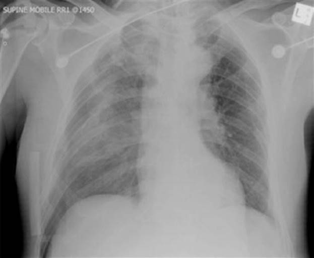
FIG. 3.8.2 Right-sided supine pneumothorax with tension.
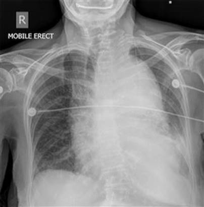
FIG. 3.8.3 Widened mediastinum secondary to aortic dissection and rupture.
The pelvic X-ray will include all the bony pelvic components and the hip joints (Fig. 3.8.4).
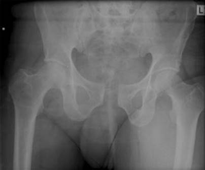
FIG. 3.8.4 Diastasis of the pubic symphysis.
Specific regional radiology
Head
Head trauma is responsible for 50–75% of the mortality associated with major trauma [9]. The spectrum of head injury ranges from mild concussion to diffuse axonal injury incompatible with life and includes all causes of intracranial haemorrhage.
A CT brain scan is the investigation of choice for all but minor head injuries (see Table 3.8.3 for CT indications in serious head injury). A non-contrast CT brain scan with bone windows is adequate for the detection of intracranial haematoma, cerebral oedema with or without midline shift and skull vault fractures.
Table 3.8.3
Indications for a CT brain scan in significant head injury
Glasgow Coma Score (GCS)<9 after resuscitation
Neurological deterioration of 2 or more GCS points
Drowsiness or confusion (GCS 9–13) that persists for longer than 2 hours
Persistent headache or vomiting
Focal neurological signs (e.g. pupillary abnormalities or focal neurological signs)
Skull fracture known or suspected
Penetrating injury known or suspected
Age over 50 years with a suspicious mechanism of injury
Any head injury in a patient on anticoagulation therapy
The Canadian CT Head Rule [10] for patients with minor head injury also provides guidance for CT brain scanning in patients with minor head injury (GCS 13–15). The authors found that the presence of any of the high-risk factors (Table 3.8.4) was 100% sensitive for predicting the need for neurological intervention. They also found that the presence of medium-risk factors was 97.2% sensitive for detecting clinically important brain injury.
Table 3.8.4
CT head rule is only required for patients with minor head injuries with any one of the following
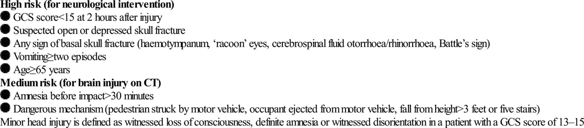
There are no indications for a skull X-ray in a trauma patient if a CT scanner is available, as it is extremely unreliable for the detection of either intra-or extracranial injuries.
If a compound or depressed fracture of the skull is suspected clinically, a CT brain scan should be performed. A compound depressed skull fracture is considered a neurosurgical emergency because of the increased risk of infection, such as meningitis or brain abscess (Fig. 3.8.5).

FIG. 3.8.5 CT brain with depressed skull fracture.
CT scan of the brain in children should be performed after careful consideration and explanation of the risks/benefits due to the exposure of the rapidly growing brain to a relatively high dose of radiation.
A magnetic resonance imaging (MRI) scan of the brain is not usually performed as first-line investigation in acute neurotrauma owing to the technical difficulties associated with the presence of patient monitoring and life support metallic equipment.
Classification of intracranial haemorrhage
Intracranial bleeding may be classified according to location. This includes: subdural, subarachnoid, extradural, intraventricular or parenchymal. These commonly coexist in the setting of trauma.
Epidural haematomas are commonly secondary to arterial bleeding due to a skull fracture with subsequent disruption of the middle meningeal artery. The haematoma is ovoid or lentiform in shape, does not cross cranial sutures but may cross the midline (Fig. 3.8.6).

FIG. 3.8.6 (A,B) Left frontal extradural haematoma with an associated fracture (arrows).
Subdural haematomas usually occur as a result of venous bleeding. These haematomas are cresentic in shape, may involve a larger area when compared to an epidural haematoma, may cross cranial sutures but do not cross the midline (Fig. 3.8.7).
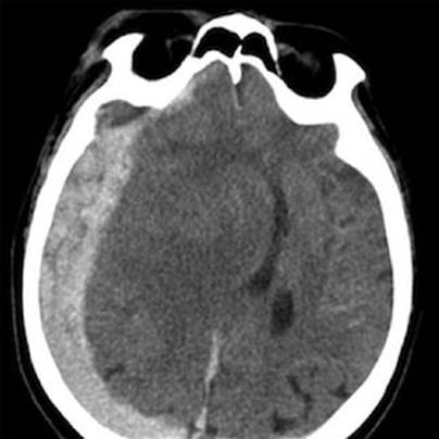
FIG. 3.8.7 Right cerebral convexity subdural haematoma.
Subarachnoid haemorrhage may be due to disruption of small subarachnoid vessels or by direct extension from a parenchymal contusion/haematoma. Haemorrhage may be visualized in the sulci of the cerebral convexities or within the subarachnoid cisterns at the base of the skull (Fig. 3.8.8).

FIG. 3.8.8 Subarachnoid haemorrhage (A) within the basal cisterns and (B) within cerebral sulci.
Intraventricular haemorrhage may be caused by tearing of subependymal veins on the surface of the ventricles or by direct extension from a parenchymal contusion/haematoma. These blood products tend to layer dependently on a CT scan with the patient imaged in a supine position, particularly within the occipital horns of the lateral ventricles.
Cerebral contusions represent foci of bleeding within the parenchyma of the brain. These may occur within superficial grey matter/subcortical white matter due to direct contact from bony protuberances of the calvarium or base of skull. Deeper parenchymal contusions are caused by disruption of intraparenchymal blood vessels. Cerebral contusions commonly increase in size and number within the first 24 hours post-trauma due to continued bleeding (Fig. 3.8.9). These haematomas also develop adjacent oedema which may increase the associated mass effect on the remainder of the intracranial structures.

FIG. 3.8.9 Multiple bilateral frontal cerebral contusions.
Also noteworthy is the increased presentation of patients taking anticoagulation or antiplatelet therapy who fall and suffer head injuries. The bleeding as a result of the injury may progress over 24 hours and may occur in all of the above locations despite both pharmacological and neurosurgical attempts at reversal.
Diffuse axonal injury occurs due to acceleration–deceleration forces and is a shearing-type injury to the brain. It may be initially difficult to visualize on a non-contrast CT but can be identified as tiny foci of petechial haemorrhage at the grey–white matter interface, within the corpus callosum or within the brainstem.
Non-contrast CT is also capable of identifying areas of acute established infarction. In the setting of trauma, this may be secondary to acute vascular injury or mass effect due to cerebral oedema (Fig. 3.8.10).

FIG. 3.8.10 Brain death. (A) Sagittal CT image demonstrating loss of supratentorial grey–white matter differentiation with preservation within the cerebellum (‘white cerebellar sign’). This represents global supratentorial parenchymal infarction. (B) Right and (C) left common carotid cerebral angiograms demonstrate contrast filling of the external carotid vessels bilaterally but no intracranial filling of the ICA bilaterally. This represents brain death.
Fractures of the base of skull, including the temporal bones may occur in trauma. Findings on CT include opacification of the mastoid air cells, fluid in the middle ear cavity and pneumocephalus. Longitudinal temporal bone fractures occur after a blow to the side of the head and constitute 70–90% of temporal bone fractures. In comparison, transverse fractures are secondary to a blow to the occiput or frontal region (E-Fig. 3.8.1).
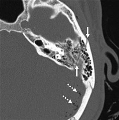
E-FIG. 3.8.1 Transverse fracture of the left temporal bone (solid arrows). The dashed arrows show pneumocephalus secondary to the fracture.
Blunt cerebrovascular injury (BCVI)
Blunt injury to the carotid or vertebral vessels (BCVI) occurs in about 0.1% of all trauma patients in the USA. Many of these are diagnosed after the development of symptoms and signs due to central nervous system ischaemia with neurological morbidity of up to 80% and mortality approaching 40%. However, when asymptomatic patients are screened for BCVI, the incidence rises to 1% for all admitted blunt trauma patients and up to 2.7% for those with an injury severity score (ISS)>15 [11].
The Denver Modification of Screening criteria (Table 3.8.5) provides both risk factors as well as symptoms and signs for BCVI [11]. CT angiography is a valuable screening tool for the detection of these injuries; however, digital 4 vessel cerebral angiography (DFVCA) remains the diagnostic investigation of choice [12]. Table 3.8.6 shows a grading scale for BCVI as proposed by Biffi et al.[13].
Table 3.8.5
Denver modification of screening criteria for BCVI
Symptoms/signs of BCVI
Arterial haemorrhage
Cervical bruit
Expanding cervical haematoma
Focal neurological deficit
Neurological findings incongruous with CT scan findings
Ischaemic stroke on secondary CT scan
Risk factors for BCVI
High energy transfer mechanism with:
Le Fort 2 or 3 fractures
Cervical spine fracture patterns: subluxation, fractures extending into the foramen transversarium, fractures of C1–3
Basilar skull fractures with carotid canal involvement
Diffuse axonal injury with GCS<7
Near hanging with anoxic brain injury
Table 3.8.6
Grading scale for BCVI
Grade 1: intimal irregularity with<25% narrowing
Grade 2: dissection or intramural haematoma with>25% narrowing
Grade 3: pseudoaneurysm
Grade 4: occlusion of lumen
Grade 5: transection with extravasation
[E-Figure 3.8.2 shows a dissection of the right vertebral artery due to blunt trauma in a patient with bilateral facet dislocation; E-Figure 3.8.3 demonstrates dissection of the right internal carotid artery with a large associated pseudoaneurysm; and E-Figure 3.8.4 shows a posterior inferior cerebellar artery infarct in a patient with a vertebral artery dissection.
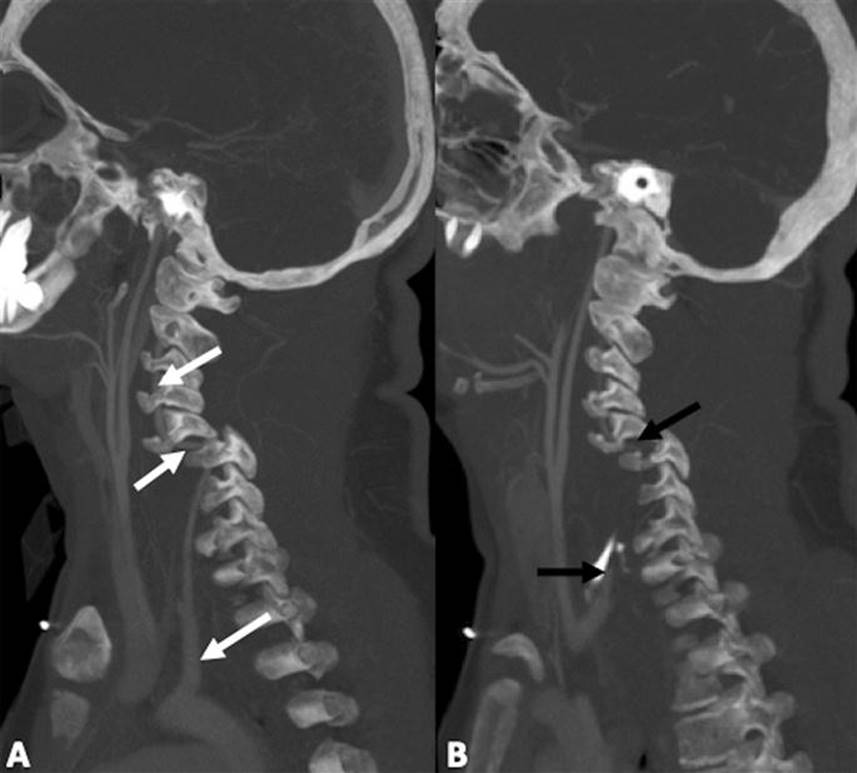
E-FIG. 3.8.2 Vertebral artery dissection. MIP images of a CT angiogram in a patient with bilateral C5/6 facet dislocation. (A) demonstrates the abnormal course of the left vertebral artery due to the dislocation (arrows). (B) shows an absence of contrast within the right vertebral artery due to dissection immediately distal to its origin to the level of the C5/6 neural exit foramen.

E-FIG. 3.8.3 Right internal carotid artery angiogram demonstrates a short segment dissection within the distal cervical portion (arrows) with a large associated pseudoaneurysm formation (dashed arrows).

E-FIG. 3.8.4 Vertebral artery dissection. (A) Axial CT image shows an undisplaced fracture involving the right transverse foramina of C5 (arrow). (B) Axial diffusion weighted MRI image demonstrates a right posterior inferior cerebellar artery infarct (thick arrow). (C) On MRA, there is absence of the right vertebral artery in its entirety due to a dissection (thin arrow indicates the origin of the vertebral artery).
CT is also capable of identifying injuries of the larynx (E-Fig. 3.8.5) which are usually due to a direct blow. Fractures of the thyroid or cricoid cartilage may be more difficult to visualize in younger patients who do not have calcification within their thyroid or cricoid cartilages.
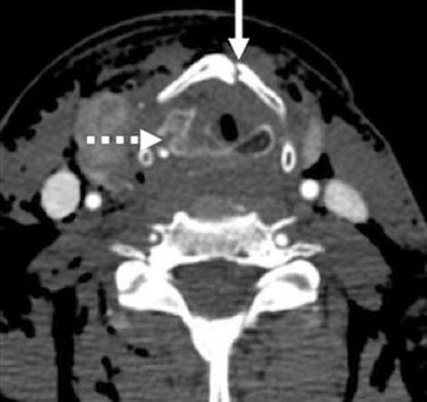
E-FIG. 3.8.5 Fractured larynx. There are fractures of the thyroid cartilage (solid arrow) and of the cricoid cartilage (dashed arrow).
Facial injury
Facial trauma may range from relatively trivial undisplaced nasal bone fractures to the life-threatening problems of airway protection and haemorrhage associated with midfacial (Le Fort) fractures. There may also be underlying cerebral injury associated with frontal bone fractures.
The commonest injury to the midface is the blowout fracture caused by a direct blow to the orbit, which results in a fracture of the orbital floor or the medial wall of the orbit in the region of the paper-thin lamina papyracea (E-Fig. 3.8.6). There may be tenderness over the fractured bone associated with diplopia due to entrapment of orbital contents or (less commonly), visual disturbance due to globe or optic nerve injury. These fractures are best seen on CT scans with multiplanar reconstructions. Blowout fractures with entrapment of orbital contents require surgical elevation.
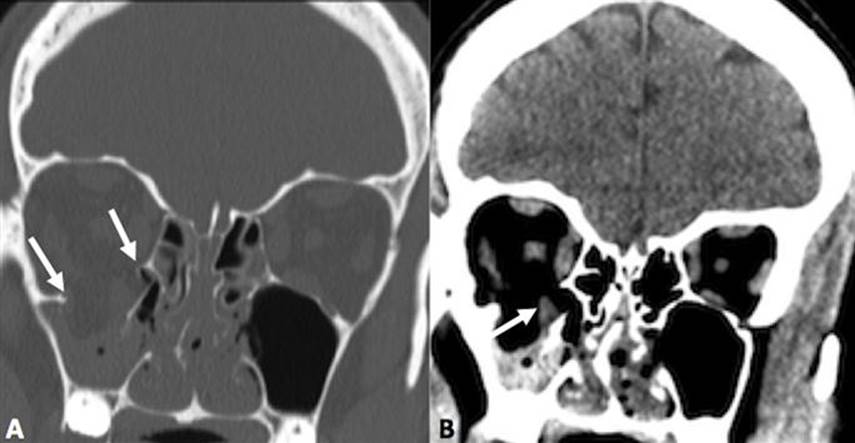
E-FIG. 3.8.6 (A) Fracture of the floor of the right orbit (arrows). (B) demonstrates herniation of the inferior rectus muscle into the bony defect (arrow).
Mandibular fractures are usually obvious clinically because of pain, malocclusion and drooling. Mandibular fractures may be difficult to demonstrate on standard PA and oblique X-ray views. A panoramic view or orthopantomogram (OPG) is more useful, but CT of the mandible provides optimal demonstration of mandibular fractures, including those involving the mandibular neck, condyle and temporomandibular joint (TMJ) (E-Fig. 3.8.7). Dislocation of the TMJ is also optimally diagnosed on CT (E-Fig. 3.8.8).

E-FIG. 3.8.7 Bilateral mandible fractures (arrows).
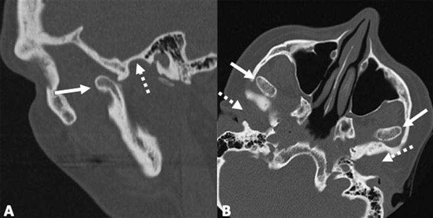
E-FIG. 3.8.8 Bilateral dislocated temporomandibular joints. The dashed arrows demonstrate the empty mandibular condyles and the solid arrows the dislocated mandibular heads. (A) sagittal image. (B) axial image.
Fractures of the zygoma are classified as (a) tripod fractures and (b) isolated fractures of the zygomatic arch. The tripod fracture or zygomaticomaxillary fracture separates the malar eminence of the zygoma from its frontal, temporal and maxillary attachments. Tripod fractures are usually caused by a significant force to the body of the zygoma or the malar eminence. The three fractures that constitute the tripod fracture are located in the inferior orbital margin, the lateral orbital margin or the zygomaticofrontal suture and the zygomatic arch. These fractures are best viewed on CT scans (Fig. 3.8.11).

FIG. 3.8.11 Tripod fracture (arrows).
The Le Fort fractures are caused by direct trauma to the midface. The Le Fort 1 fracture involves the maxilla at the level of the nasal floor and will allow mobility of the palate (‘floating palate’) (Fig. 3.8.12). Le Fort 2 passes through the nasal bones, as well as the medial, inferior and lateral walls of the maxillary antrum. The Le Fort 3 or ‘craniofacial dysjunction/floating face’ involves the nasal bones, the medial and lateral orbital walls and the zygomatic arch (E-Fig. 3.8.9).
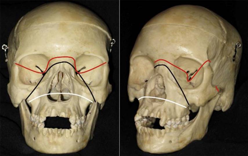
FIG. 3.8.12 Le Fort fracture classification. Le Fort 1, (white line), Le Fort 2 (black line) and Le Fort 3 (red line). (Courtesy of Associate Professor Alf Nastri, Department of Maxillofacial Surgery, Royal Melbourne Hospital.)

E-FIG. 3.8.9 Shattered skull and mandible.
Some facial fractures are unable to be classified owing to marked fragmentation of the bones and are termed as ‘central facial smash’ (Fig. 3.8.13). These fractures are best viewed by axial CT scans with multiplanar reformatting.

FIG. 3.8.13 (A) Lateral and (B) AP cervical spine X-rays.
Frontal sinus fractures commonly occur as a result of direct force and are often compound, with the risk of associated intracranial infection. There may be an associated intracranial haematoma or cerebral contusion. A CT scan will best evaluate these fractures and determine the involvement of the posterior sinus wall. These fractures often require surgical exploration for debridement and repair.
Spinal Injury
Cervical spine
Cervical spine injuries can be classified into those with:
fractures: stable or unstable
fractures and no neurological deficit
fractures associated with neurological deficit
a small group of patients with cord injuries associated with contusion, haemorrhage or oedema without bony injury.
The bony Cx spine is best evaluated by axial CT scan with coronal and sagittal reconstructions. The cervical cord, discs and ligaments are best imaged by MRI scan. X-rays of the Cx spine do not provide the same detail and accuracy as a CT scan in detecting bony injuries.
The incidence of adult cervical spine injury after blunt trauma is 2–6% and any blunt trauma patient with physical findings of posterior midline neck tenderness, altered mental status or neurological deficit is considered at high risk for cervical spine injury. In this group of patients, one study [14] found a Cx spine injury in 9.5% of patients.
Cervical spine injury occurs in 5–10% of patients with traumatic brain injury, and with potentially devastating consequences for missing an injury in these patients. Plain X-rays miss 12–16% of Cx spine injuries while swimmer’s views and/or oblique views identify more injuries but are frequently inadequate. A protocol at the Alfred Hospital in Melbourne [15] utilized multidetector CT (MDCT) with 1 mm cuts from C0–C3 and 3 mm cuts from C2–T4/5 with zero missed injuries. These authors also found that passive flexion/extension fluoroscopy in unconscious trauma patients was not sensitive for the detection of cervical instability and did not detect any injuries that had not been already seen in other imaging modalities.
MRI scan is invaluable for the detection of disco-ligamentous and cord injuries, however, its role in the routine clearance of the Cx spine in unconscious patients remains unclear. It is also problematic to transport critical care patients with metallic monitoring devices to the MRI scanner. There are also often delays to access the MRI scanner so that spinal precautions can be discontinued and the Cx collar removed. There are no prospective studies comparing modern MDCT and MRI for the evaluation of occult Cx spine injuries in unconscious patients and the use of either modality depends on clinician preference and level of suspicion with regard to the mechanism of injury. Of note, however, the Alfred Hospital investigators found three unstable Cx spine injuries detected by MRI scan which were not detected by MDCT [15].
There are also established criteria for identifying patients with a low risk for cervical spine injury who do not require imaging, as derived from the NEXUS Study (National Emergency X-Radiography Utilization Study) [16] (Table 3.8.7). The NEXUS study was a large validation study that identified 818 spinal injuries out of 34 000 patients and identified patients as low risk for cervical spine injury if all four high-risk clinical findings were absent. If the above criteria were met, there was no need for any imaging or further immobilization of the cervical spine. The results were 99.6% sensitive for clinically important cervical spine injuries. However, the specificity was only 12.9%.
Table 3.8.7
Low-risk criteria for radiological clearance of the cervical spine in a multitrauma patient (NEXUS criteria)
Disturbed conscious state, e.g. head injury, intoxication for any reason
Any neurological motor or sensory signs
Midline cervical tenderness
Other major distracting injuries in a multitrauma patient
The NEXUS study also found that cervical spine X-rays missed up to one-third of secondary spinal injuries where it was thought there was a single non-significant spinal injury and up to 25% of those missed injuries were non-contiguous with the original injury [17]. These findings have confirmed the use of CT in full evaluation of the cervical spine.
The Canadian C-Spine Rule for radiography in alert and stable trauma patients [18] may be more valuable clinically and is well validated in a prospective cohort study (Table 3.8.8). This rule demonstrated 100% sensitivity and 42.5% specificity for clinically important cervical spine injuries and there was good inter-observer agreement for each variable, with κ value>0.6 and a strong association with outcome (spinal injury) p<0.05.
Table 3.8.8
The Canadian C-spine rule
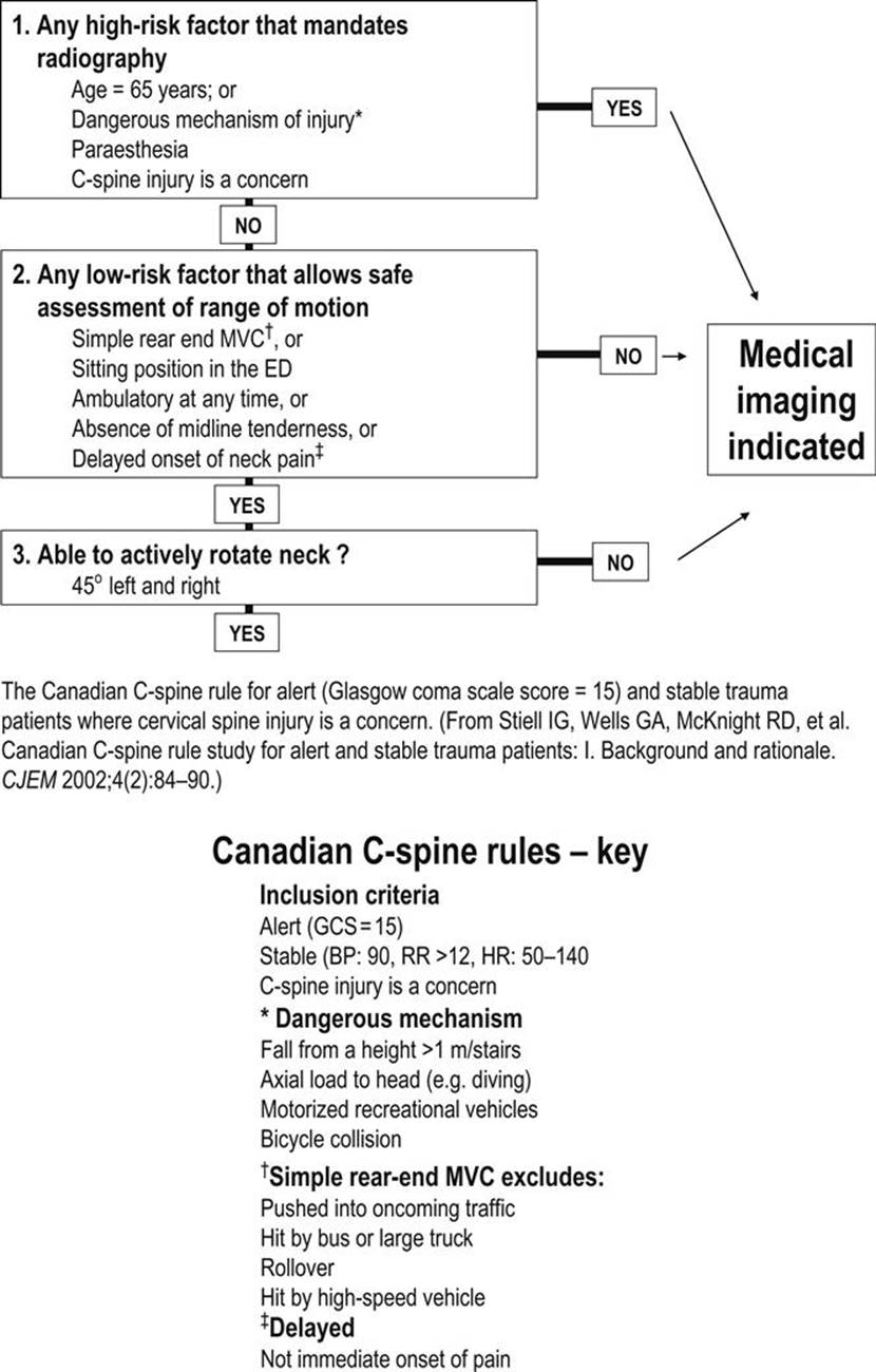
The above clinical decision rules should be applied with caution in the elderly, those with pre-existing spinal disease or the very young (<2 years). Owing to the relative immobility of the cervical spine or pre-existing spinal disease, such as ankylosing spondylitis, these patients may sustain cervical spine fractures even in the presence of a seemingly trivial injury.
If the cervical spine cannot be cleared clinically, then a radiological examination must be performed.
Cervical spine X-ray
The trauma series for cervical spine clearance consists of lateral, anteroposterior and open-mouth odontoid process (peg) view (Figs 3.8.13 and 3.8.14). Plain radiography of the Cx spine is now used infrequently in most trauma centres as it has been shown to have significantly less accuracy in detecting acute spinal fractures than is found in MDCT scans of the Cx spine from C0–T4/5.

FIG. 3.8.14 PEG view.
The lateral view must be adequate (to the C7–T1 junction) and show appropriate alignment of the three lines (see Fig. 3.8.1). As well as alignment and adequacy, one should inspect the bones (for fractures), cartilage and soft tissues.
Inspection of alignment and adequacy on the lateral cervical spine X-ray should include the four lordotic lines: anterior and posterior vertebral lines, the spinolaminar line and the line linking the tips of the spinous processes. In adults, up to 1.0 mm of anterior subluxation (and up to 3 mm in children) may be normal in a true lateral film taken at 1.8 m.
True pseudosubluxation is commonest in children up to the age of 8 years, but may be seen up to age 18 [19]. It commonly occurs between C2 and C3 and less commonly at the C3–4 and C4–5 levels. The key radiological feature is the preservation of the spinolaminar line in flexion/extension views of the lateral cervical spine [17].
The sagittal (AP) diameter of the spinal canal should be measured. At the C2 level, the lower limit radiographic measurement of the AP diameter of the spinal canal is 14 mm. At the C7 level, the lower limit of the AP canal diameter is 12 mm [20].
Bony canal
All the cervical vertebrae should be systematically examined, including vertebral body, pedicles, facet joints, laminae and spinous processes and the interspinous distance.
The ‘ring’ of increased radiodensity formed by the odontoid process and the facet joints of C1–C2 is known as the ‘Harris ring’. This ring should be intact anteriorly, superiorly and posteriorly, indicating an intact odontoid process and facet joints of C1–2. A type 2 fracture of the odontoid process through the body may be visible on the lateral cervical spine X-ray.
Cartilage
All the spaces between adjacent vertebrae should be inspected for equality.
Soft tissue
The prevertebral soft tissue should be inspected. A distance greater than 7 mm at C2 and 22 mm at C6 in the adult indicates the presence of a prevertebral haematoma [21]. If this is present, then a fracture or ligamentous disruption must be excluded. In children, the upper prevertebral space may be larger than in adults owing to the presence of increased nasopharyngeal lymphoid tissue and it may also increase in infants during crying.
Atlanto-occipital and atlantoaxial bony injuries
Atlanto-occipital dislocation is usually associated with a fatal injury, whereas atlanto-occipital subluxation is radiologically subtle and the patients usually survive.
The diagnosis is made by recognizing an abnormal basion–axial interval and/or an abnormal basion–dental interval. Normally, neither should exceed 12 mm [22] and this is best seen on the lateral cervical spine X-ray or the sagittal CT scan of the cervical spine (Fig. 3.8.15).

FIG. 3.8.15 Atlanto-occipital dislocation.
The space between the odontoid and the anterior arch of C1 measured at its most inferior margin should not exceed 3 mm in adults and may be up to 5 mm in children. If this distance is exceeded, there may be a rupture of the transverse atlantal ligament of the dens. This distance may also increase atraumatically in patients with rheumatoid arthritis (due to ligamentous destruction by pannus) or in Down’s syndrome.
Occipital condyle [23] and C1–C2 fractures are often missed on the lateral cervical spine X-ray, with the only indication of a fracture being an increase in soft-tissue swelling in this area. The initial lateral cervical spine X-ray should be accompanied by an open-mouth (odontoid process) and an AP view.
The open-mouth (odontoid) view should be inspected for alignment of the lateral masses of C1–C2, which is abnormal in the Jefferson fracture, fracture of the odontoid process (types 1–3) and rotatory subluxation of C1 on C2. Rotation of the head can simulate pathological malalignment in this region. The AP view of the cervical spine should be checked for alignment of the articular pillars and vertebral bodies. The spinous processes should be centred and deviation of these from the midline may indicate a unilateral facet dislocation. Widening of the interspinous distance may indicate subluxation or dislocation. Fractures and dislocations may cause malalignment or compression of the vertebral bodies.
CT scan of cervical (Cx) spine
Many trauma centres now routinely perform limited or nil plain Cx spine X-rays and a CT scan of the entire cervical spine from occipital condyles down to and including T4–5 (Table 3.8.9). There is now a limited role for swimmer’s and oblique views of the cervical spine, given the widespread use of CT scan (Fig. 3.8.16 [E-Fig. 3.8.10]).
Table 3.8.9
Imaging of patients with major trauma (based on Spinal Clearance Management Protocol, The Alfred Hospital, Melbourne, Australia)
Plain AP and lateral X-rays cervical spine
MSCT* 1 mm cuts C0–C3 (axial+sagittal and coronal reformats)
MSCT 3 mm cuts C2–T4/5 (axial+sagittal reformats)
*Multislice CT scans.

FIG. 3.8.16 Unilateral fracture dislocation. Sagittal CT image of the cervical spine. (A) Shows normal alignment of the left-sided cervical facet joints; (B) demonstrates a grade 1 anterolisthesis at C3/4 secondary to (C) a right-sided facet joint fracture dislocation.
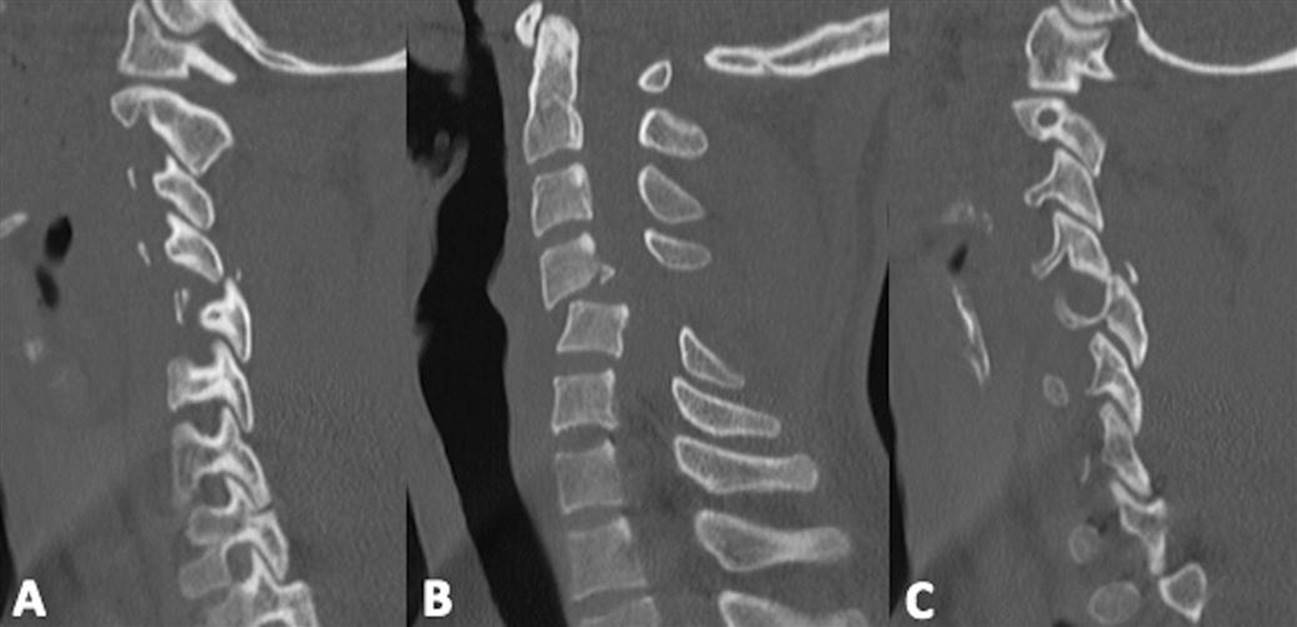
E-FIG. 3.8.10 Bilateral facet joint dislocation. There is a grade 2 anterolisthesis at C4/5 in (B) secondary to bilateral joint fracture/dislocations in (A) and (C).
Indications for MRI of the cervical spine include:
patients with complete or incomplete neurological deficit
deteriorating neurological status
suspected ligamentous or intervertebral disc injury.
MRI will provide clear and concise images of all structures, particularly the spinal cord, intervertebral discs and soft tissues (E-Fig. 3.8.11). Bone structures and bone oedema are also demonstrated, but fine bony detail is best seen on a CT scan. A range of different MRI sequences may be utilized in trauma patients to identify a number of spinal pathologies (Table 3.8.10). There is a small group of patients who will be unsuitable for an MRI scan (Table 3.8.11). An MRI scan also provides information regarding spinal cord injury patterns, such as central cord syndrome, which have been previously unavailable with other imaging modalities (Fig. 3.8.17).

E-FIG. 3.8.11 Ligamentous injury. Sagittal MRI image of the cervical spine demonstrates a significant ligamentous injury at C2/3. Solid arrows demonstrate rupture of the interspinous and supraspinous ligaments. The dashed arrow shows rupture of the ligamentum flavum. There is also widening of the C2/3 intervertebral disc and marked anterior and posterior soft tissue oedema.
Table 3.8.10
Spinal abnormalities seen on the MRI scan
Spinal cord injury (haemorrhage or oedema)
Disc herniation
Epidural haematoma
Epidural abscess
Occult bone fracture/dislocation
Ligamentous rupture
Facet joint – disruption or capsular injury
Nerve root avulsion and plexus injury (brachial or lumbosacral)
Table 3.8.11
Conditions unsuitable for MRI scan
Metallic components, e.g. bullets, aneurysm clips
Haemodynamically unstable patients
Patients requiring ventilation and extensive physiological monitoring

FIG. 3.8.17 Cord contusion. (A) Sagittal CT of the cervical spine shows fractures of the C5 and C6 verterbal bodies, with associated retropulsion. (B) Sagittal T2-weighted MRI demonstrates a cord contusion extending from C4 to C7 (solid arrows) and a focus of haemorrhage within the cord at C5 (dashed arrow).
There are limited indications currently for dynamic flexion/extension films when looking for evidence of ligamentous instability, where there is ongoing pain or tenderness of the cervical spine in a patient who is neurologically intact and fully alert. The patient must be able to flex and extend his/her neck voluntarily and these X-rays should be supervised by the medical officer who ordered the investigation. However, as discussed previously, these films often fail to demonstrate the entire Cx spine [15].
In the unconscious patient, there is controversy as to the best approach for clearing the cervical spine, as there is a clear need to remove the rigid immobilization collar in order to prevent the development of scalp pressure areas. Prolonged use of rigid Cx collars causes pressure ulceration in 31% of unconscious trauma patients with the median length of stay increasing in this group of patients from 10 days, if they do not develop ulcers, to 23 days if they do develop pressure areas [15].
Dynamic flexion/extension X-rays in unconscious patients have a poor sensitivity for detecting cervical instability, have high rates of inadequacy, are not cost-effective and are potentially dangerous.
Trauma centres should have agreed guidelines, for clearing the cervical spine in unconscious patients, which involve multidetector CT. If there is any abnormality seen on CT (such as malalignment of the vertebrae or a high cervical fracture) an MRI is performed to exclude ligament, disc or atlanto-occipital/atlantoaxial disruption. Limited MRI of the Cx spine is highly sensitive for detection of ligament injury, disc and spinal cord oedema or haemorrhage which is often not apparent on MDCT of the Cx spine [15]. Also MRI does not expose the patient to ionizing radiation. The Alfred Hospital group [15] believes there may be a clinical advantage to include MRI scan in routine Cx spine clearance protocols for unconscious patients in whom clinical examination is not possible.
Thoracolumbar spine
The second most frequently injured area of the spinal column, after the cervical spine, is the thoracolumbar junction (T11–L2). Cord injuries in this region comprise about 20% of all spinal cord injuries [24]. The main reasons for the susceptibility of this vertebral region are the abrupt transition from the rigidly fixed thoracic spine to the more mobile lumbar spine and the fact that the spinal canal in the thoracic region is smaller in diameter than the cervical or lumbar spinal canals, which results in increased risk to the spinal cord.
An important concept used for interpreting thoracolumbar injuries is the ‘three-column’ theory described by Denis [25]. This is a widely accepted concept which divides a vertebra into three columns: the anterior column, which consists of the anterior longitudinal ligament and the anterior half of the vertebral body; the middle column, which includes the posterior half of the vertebral body and the posterior longitudinal ligament; and the posterior column, which includes all the bony and ligamentous structures posterior to the posterior longitudinal ligament. Fractures involving the anterior column are considered stable, whereas fractures involving the anterior and middle columns or all three columns are considered unstable.
It is also of note that injuries in the T1–T10 region comprise 16% of cord injuries and lumbosacral injuries, such as cauda equina lesions and comprise approximately 4% of spinal neurological injuries [24].
As it is often difficult to obtain satisfactory images of the upper thoracic spine, particularly the T1–T4 region, multislice CT with multiplanar reconstructions is currently the most effective method of establishing the extent of bony injury. It is the practice of many trauma centres to perform routinely a CT of the cervical spine to T4–5 with multiplanar reconstructions, and it has become routine to perform sagittal and coronal reconstruction of the thoracolumbar spine when a CT scan of the thorax/abdomen/pelvis is performed.
Classification of thoracolumbar spine injuries
Stable fractures, which include transverse process fractures, spinous process fractures, pars interarticularis fractures and wedge compression fractures involving the anterior two-thirds only of the vertebral body.
Unstable fractures/dislocations, which include compression fractures with middle and/or posterior column disruption, the ‘Chance’ fracture (E-Fig. 3.8.12), the burst fracture (E-Fig. 3.8.13) and flexion/distraction injuries.

E-FIG. 3.8.12 T12 Chance fracture. (A) and (C) demonstrate fractures involving the articular pillars bilaterally at T12. (B) shows a fracture through the vertebral body of T12 and widening of the interspinous distance.
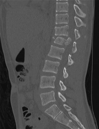
E-FIG. 3.8.13 L1 burst fracture.
Fractures in a fused thoracic spine (ankylosing spondylitis, diffuse interstitial spinal hypertrophy (DISH) and advanced degenerative disc disease with bridging osteophytes) constitute a unique subset. Due to the rigidity of the spine, they are likened to a long bone fracture and may be called a ‘carrot stick’ fracture (E-Fig. 3.8.14). These injuries are typically a result of hyperextension, usually involve three columns and are therefore unstable. Cord damage is common in this type of fracture.

E-FIG. 3.8.14 ‘Carrot stick injury/fracture’ in a patient with ankylosing spondylitis. Sagittal image (A) demonstrates widening of the intervertebral disc space at C7/T1 (arrow). Coronal image (B) demonstrates bilateral fractures through ankylosed facet joints.
If a patient is unable to be examined clinically because of pain or tenderness of the thoracolumbar spine or is unconscious, this area must be imaged. If a CT scan of the chest and abdomen/pelvis is performed, then sagittal and coronal reconstructions of the thoracolumbar (TL) spine can be done.
If CT of the torso is not performed, then plain X-rays of the thoracolumbar spine should be taken, with limited CT of any areas difficult to visualize or where there is clinical suspicion.
The Chance fracture is an example of a distraction or seatbelt injury, with the lap belt as the axis of rotation and failure of the spinal column in its posterior ligamentous and bony elements. This fracture is directed in a horizontal plane through the entire bony column, including vertebral body, pedicles, laminae and spinous processes and is, by definition, unstable. This fracture may also be associated with injuries to the abdominal contents, e.g. pancreas or duodenum.
Fractures of the lower lumbar spine and sacrum may involve the cauda equina and associated sacral nerve roots. There may be bladder, bowel and sexual dysfunction, as well as variable motor and sensory deficit in the lower limbs. There is often significant neuralgia that is disabling and difficult to treat. Plain X-rays will give some indication of the severity of the bony injury, but a CT scan is required for definitive information if surgical fixation is required.
Coccygeal fractures are due to direct blows and are both treated and diagnosed clinically. Radiology is not usually necessary for diagnosis, which is best made by CT scanning. There may be an associated rectal injury that requires operative repair but, otherwise, analgesia only is required.
Chest trauma
The chest X-ray still has a key role in the investigation of multiple trauma involving the thorax. The combination of CXR and ultrasound may provide the simplest and fastest assessments of unstable patients and should usually precede MDCT, particularly in cases of low energy trauma [26–28].The CT chest scan with IV contrast has become more accessible in recent years, and the use of both TOE (transoesophageal echo) and DSA (digital subtraction angiography) in assessing the mediastinal structures have become limited to situations where a CT cannot be done.
A study by Traub and colleagues [29] found that chest CT was more effective than X-rays in detecting lung contusions, pneumothoraces and mediastinal haematoma, as well as fractured ribs, scapula, sternum and vertebrae. The authors found that it was more likely than CXR to provide further diagnostic information in the presence of chest wall tenderness, reduced air entry and/or abnormal respiratory rate. However, the CXR can be done quickly and may provide urgent life-saving information regarding conditions, such as pneumothorax, haemothorax or malpositioned chest or endotracheal tubes [28].
The trauma room CXR should ideally be performed in the erect position with a nasogastric tube in situ. However, because it is often impossible to clear the cervical and/or thoracolumbar spine in the trauma room, the CXR is frequently taken in the supine position. This can create a number of difficulties in interpretation of the results. The AP projection of the X-ray beam will magnify the mediastinal structures and, when the patient is supine, the thoracic veins will passively distend and add to this appearance of mediastinal widening. Small pneumothoraces and haemothoraces are also difficult to detect on the supine CXR because the air distributes as a thin film anteriorly and blood as a thin homogeneous layer posteriorly. A haemothorax of 200–300 mL will normally be visible on a good-quality erect CXR, whereas it will usually require 800–1000 mL to produce the ‘fuzzy’ appearance of a haemothorax seen on the supine CXR [28].
Examination of the CXR will often begin with a review of the bones and soft tissues. The CXR is a poor diagnostic aid for rib fractures as it will miss up to 50% of anterior and lateral fractures [27,28], instead, the assessment should be directed more towards the complications of rib fractures, such as pneumothorax, haemothorax and lung contusion. It is also important to remember that the clavicles, glenohumeral joints and scapulae are visible on the CXR (E-Fig. 3.8.15). Fractures of these bones, along with fractures of the first and second ribs, are indicators of significant blunt thoracic trauma and should prompt a careful examination for underlying visceral and vascular injuries.

E-FIG. 3.8.15 Bilateral dislocated shoulders.
Sternal fractures may be seen on a lateral CXR but are best seen on CT. The significance of sternal fractures will largely direct the examination towards underlying mediastinal injuries. Brookes et al. [30], in a retrospective study, found a 2% incidence of sternal fractures associated with motor vehicle accidents. These patients had a very low incidence (1.5%) of cardiac arrhythmias due to cardiac muscle contusion requiring treatment and a mortality rate of less than 1%. The authors found that those at risk of cardiac arrhythmias requiring treatment were over 65 years of age and either had pre-existing ischaemic cardiac disease or were on digoxin treatment. They recommended that cardiac monitoring was not required unless the patient fulfilled the above criteria. They also found that the 12-lead ECG was not predictive for the development of arrhythmias requiring treatment.
In cases of penetrating chest trauma, a foreign body may be evident on the CXR. AP and lateral projections with appropriate skin markers will normally be required to help locate the position of the foreign object. In cases where the foreign object is embedded close to or in a pulsatile thoracic structure, the object may appear blurred on the film, indicating the proximity of the foreign body to the vessel.
Subcutaneous emphysema may be seen on the CXR and may result from injury to the lung, the tracheobronchial tree, the larynx, pharynx and oesophagus (Fig. 3.8.18). Subcutaneous emphysema should prompt a careful examination for evidence of a pneumothorax and pneumomediastinum. Subcutaneous emphysema and pneumothorax are common findings in traumatic injury to the lung and also occur in tracheobronchial injury.

FIG. 3.8.18 Subcutaneous emphysema. Extensive subcutaneous emphysema is visualized overlying the chest wall bilaterally and in the supraclavicular regions.
In cases of suspected tracheal laceration, where the patient has been intubated, the appearance of the endotracheal tube on the CXR should be carefully examined. The normal position of the balloon is 2.5 cm proximal to the tip of the endotracheal tube (Table 3.8.12).
Table 3.8.12
Chest X-ray signs of tracheal laceration
Subcutaneous emphysema/mediastinal emphysema/pneumothorax
Deviation of the endotracheal tube tip to the right relative to the tracheal lumen
Distension of the endotracheal tube balloon
Migration of the endotracheal tube balloon distally towards the tube tip
If a pneumothorax is suspected but not visible on the supine CXR, a CT scan is the definitive investigation.
In cases of penetrating chest trauma, the development of a detectable pneumothorax may be delayed and so it is recommended that check CXR be performed at 6 and 12 hours [29].
The lung parenchyma may become opacified by contusion, aspiration, pulmonary fat embolism and either cardiogenic or non-cardiogenic pulmonary oedema. Lung contusions will usually develop rapidly within 6 hours of an injury, whereas the changes of aspiration and pulmonary infarction are often delayed for 12–24 hours. Rib fractures are frequently associated with pulmonary contusions although, in paediatric patients and young adults, the ribs are more compliant and may bow in, causing a lung contusion without fracture.
Diaphragmatic injuries are more frequent in penetrating than in blunt trauma. In blunt trauma, however, 80% of diaphragmatic injuries occur on the left side because the liver and its ligamentous attachments protect the right side (Table 3.8.13).
Table 3.8.13
Signs of diaphragmatic injury on CXR
Elevated hemidiaphragm
Abnormal or indistinct contour of the diaphragm
Collapse of the lower lung fields
Inhomogeneous mass in the relevant hemithorax
Displacement of the mediastinum away from the injury
If a nasogastric tube is in situ, it may be seen to pass down into the abdomen and back up into the chest contained within the herniated stomach. Lower rib fractures are often seen in association with injuries to the diaphragm.
Thoracic aortic injury
Ninety per cent of injuries occur in the region of the aortic isthmus, i.e. that part of the proximal descending aorta between the origin of the left subclavian artery and the site of attachment of the ligamentum arteriosum (1.5 cm in length). The ascending aorta is involved in only 5% of cases [31].
As previously described, the supine AP CXR magnifies the mediastinal silhouette. Superior mediastinal widening is a common finding in cases of both penetrating and blunt trauma to the great thoracic vessels. The mediastinal width is measured at the top of the aortic knob. A width greater than 8.0–8.5 cm in a supine film or 6 cm in an erect film is suggestive of a mediastinal haematoma.
The sensitivity of a widened mediastinum on CXR for the detection of thoracic aortic injuries has been estimated at 90% and the specificity 10%, but approximately 7% of patients with aortic rupture have a normal chest radiograph [32] (Table 3.8.14).
Table 3.8.14
Signs of aortic disruption on chest X-ray [30]
Widened mediastinum
>6 cm in erect PA film
>8 cm in supine AP film
Deviation of the oesophagus/NG tube to the right of T4 spinous process
Obliteration of aortic knob
Opacification of the aortopulmonary window
Deviation of the trachea to the right of the T4 spinous process
Depression of the left main bronchus to below 40° from the horizontal
Increased right paratracheal stripe (>4 mm)
Increased left paravertebral stripe (>5 mm)
Left apical cap
Injuries to the oesophagus may occur in association with both blunt and penetrating chest trauma. The predominant X-ray finding in oesophageal injury is pneumomediastinum and this may be associated with subcutaneous emphysema, pneumothorax, a left pleural effusion or a widened mediastinum.
Thoracic CT scan
Thoracic MDCT has become a common diagnostic aid in investigating the multitrauma patient with chest injuries. The increasing speed and greater clarity of the MDCT scan gives a reliable and rapid means of screening for most intrathoracic injuries. The strength of CT lies in its ability to distinguish mediastinal haematoma from other causes of mediastinal widening detected on initial chest radiographs, e.g. magnification, mediastinal fat and tortuous vessels [26,27].
Chest CT is also a sensitive test for detecting pneumothorax, pneumomediastinum, pulmonary contusion and haemothorax and, with intravenous contrast, may demonstrate an intimal tear or pseudoaneurysm of the traumatized aorta (Fig. 3.8.19 [E-Figs 3.8.16–3.8.17]).

FIG. 3.8.19 Aortic transection with pseudoaneurysm formation (black arrow).

E-FIG. 3.8.16 Tension pneumothorax on CT. Coronal (A) and axial (B) images.

E-FIG. 3.8.17 Pulmonary contusion with pneumatocele. (A) and (B) demonstrate ground glass change within the right lower lobe (pulmonary contusion) with associated pneumatoceles (black arrows).
Mediastinal haematoma is an indirect sign of aortic injury and appears as a soft tissue density around mediastinal structures [26,27,32] or, in acute aortic dissection, a false lumen may be seen.
If mediastinal major vascular injury is initially suspected, a CT angiogram is the preferred screening investigation; however, DSA remains the gold standard. CT angiography with 2–3 mm slices using injections of 100–150 mL contrast may be reconstructed in multiple planes to produce detailed images of the aorta. Studies have shown that CT angiography is sensitive for traumatic aortic injury (83–100%) with a high negative predictive value (NPV) of 99–100% [32].
Angiography
Transfemoral angiography has been widely accepted as the gold standard for the diagnosis of major thoracic vascular injuries, particularly those involving the aorta and great vessels [32]. This investigation is not free from complications, although the morbidity and mortality are low. Significant complications, such as rupture at the site of injury during contrast injection, are rare, but have been reported [33].
Endoluminal repair of acute thoracic vascular injuries is now commonly undertaken. Formal angiography is therefore undertaken at the time of repair. Diagnostic information obtained on CT prior to repair is sufficient to confirm the diagnosis and provides images for surgical/endovascular planning.
Transoesophageal echocardiography (TOE)
TOE has many supporters of its value as both a screening and a diagnostic test in the investigation of suspected mediastinal haematoma. Some authors [34,35] suggest that TOE is more accurate than angiography in detecting aortic injuries, although it is acknowledged that interpretation is operator dependent.
The advantages of TOE are that it can be performed quickly in the resuscitation area, it is minimally invasive and it has a low rate of complications, such as aspiration and oesophageal perforation. It can demonstrate myocardial, pericardial and valvular injuries, which may not be demonstrated on MDCT. Disadvantages are that it may require sedation and intubation and may provide limited information about the distal ascending aorta, the aortic arch and the arch vessels [35]. Although the incidence of injury to the arch and major branch vessels is low, angiography is required when injury to these vessels is suspected.
Lung ultrasound
The use of ultrasound to detect evidence of pneumothorax has been reported for more than 20 years. It is easy to perform and safe. While the sensitivity of lung ultrasound in detecting pneumthoraces is significantly less than that of thoracic CT, it can be applied in conjunction with a plain CXR to provide timely information in the resuscitation room [36].
MRI is generally not practical for the diagnosis of traumatic aortic rupture.
Oral contrast studies
Oral contrast provides useful information in the investigation and diagnosis of oesophageal and diaphragmatic injuries. In cases of oesophageal perforation, Gastrografin is the preferred contrast medium, as it is less irritating than barium should there be a leak into the surrounding mediastinal tissues. A Gastrografin swallow is mandatory in the evaluation of suspected penetrating injuries of the oesophagus. If there is a risk of aspiration, Gastrografin should not be used as it produces a severe pneumonitis. In these circumstances, contrast designed for intravenous use can be administered orally in order to demonstrate oesophageal perforation.
A CT swallow study can also be performed. This would involve a non-contrast (control) study of the chest and upper abdomen, followed by a second study after the ingestion of oral contrast. In unconscious patients, a nasogastric tube is placed with its tip in the upper oesophagus and oral contrast is administered via this tube.
Flexible or rigid oesophagoscopy may also be used to exclude oesophageal perforation.
Abdomen/pelvis
Abdominal X-ray
The role of the plain abdominal X-ray (AXR) in the investigation of abdominal trauma is extremely limited. In cases of penetrating injuries, it may be useful in the detection and localization of foreign bodies and in the detection of free air under the diaphragm in hollow viscus rupture. An erect CXR may show free gas under the left hemidiaphragm more commonly than on the right. In cases of duodenal perforation, free retroperitoneal air may be seen as pockets of gas along the right psoas line (shadow) on a supine AXR. Importantly, all of these features will be better identified on an abdominal CT scan if it is available.
Abdominal CT scan
Abdominal CT is usually performed with both oral and intravenous contrast. However, as most multitrauma patients have delayed gastric emptying, the bulk of oral contrast tends to remain in the stomach and upper gastrointestinal tract. This phenomenon has led some authors to suggest that oral contrast is of little use in this setting [37]. The increased speed of the helical CT scanner has resulted in excellent resolution for the detection of vascular injuries involving the liver, spleen and kidneys after intravenous contrast. The American Association for the Surgery of Trauma (AAST) has developed a scoring system that grades the severity of injury to the solid intra-abdominal viscera including the spleen, liver, kidney, adrenal gland and the pancreas (Figs 3.8.20–22 [E-Figs 3.8.18-21], Tables 3.8.15–19) [38]. Scoring systems have also been developed for the hollow intra-abdominal viscera (see Table 3.8.18) [38].

FIG. 3.8.20 Shattered spleen with active bleeding (arrows) and a large perisplenic haematoma – grade V.

FIG. 3.8.21 Splenic lacerations (arrows) – grade III.
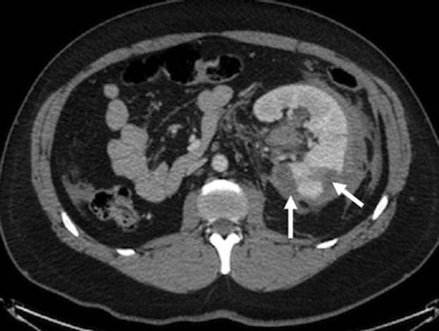
FIG. 3.8.22 Renal laceration (arrows) with surrounding perinephric haematoma – grade IV.

E-FIG. 3.8.18 Liver laceration (arrows) – grade III.
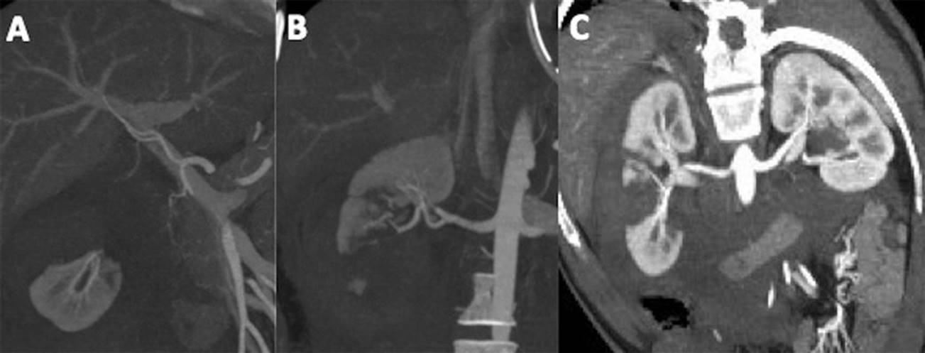
E-FIG. 3.8.19 (A, B) Coronal MIP images demonstrating transection of the right kidney. (A) Shows the transected upper pole and (B) the lower pole. (C) Oblique coronal image of the transection.

E-FIG. 3.8.20 Adrenal laceration with associated devascularisation and haematoma (arrow) – grade V. No discernable right adrenal gland is identified. The dashed arrow demonstrates the normal appearing left adrenal gland.
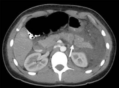
E-FIG. 3.8.21 Pancreatic transection (arrow) – grade III.
Table 3.8.15
Spleen injury scale (1994 revision)
|
Grade |
Type of injury |
Description of injury |
|
I |
Haematoma |
Subcapsular:<10% surface area |
|
Laceration |
Capsular tear:<1 cm parenchymal depth |
|
|
II |
Haematoma |
Subcapsular: 10–50% surface area; intraparenchymal,<5 cm in diameter |
|
Laceration |
Capsular tear: 1–3 cm parenchymal depth that does not involve a trabecular vessel |
|
|
III |
Haematoma |
Subcapsular:>50% surface area or expanding; ruptured subcapsular or parecymal hematoma; intraparenchymal hematoma≥5 cm or expanding |
|
Laceration |
>3 cm parenchymal depth or involving trabecular vessels |
|
|
IV |
Laceration |
Laceration involving segmental or hilar vessels producing major devascularization (>25% of spleen) |
|
V |
Laceration |
Completely shattered spleen |
|
Vascular |
Hilar vascular injury which devascularizes spleen |
Table 3.8.16
Liver injury scale (1994 revision)
|
Grade |
Type of injury |
Description of injury |
|
I |
Haematoma |
Subcapsular:<10% surface area |
|
Laceration |
Capsular tear:<1 cm parenchymal depth |
|
|
II |
Haematoma |
Subcapsular: 10 to 50% surface area; intraparenchymal<10 cm in diameter |
|
Laceration |
Capsular tear 1–3 cm parenchymal depth,<10 cm in length |
|
|
III |
Haematoma |
Subcapsular:>50% surface area of ruptured subcapsular or parenchymal hematoma; intraparenchymal hematoma>10 cm or expanding |
|
Laceration |
>3 cm parenchymal depth |
|
|
IV |
Laceration |
Parenchymal disruption involving 25 to 75% hepatic lobe or 1–3 Couinaud’s segments |
|
V |
Laceration |
Parenchymal disruption involving>75% of hepatic lobe or>3 Couinaud’s segments within a single lobe |
|
Vascular |
Juxtahepatic venous injuries; i.e. retrohepatic vena cava/central major hepatic veins |
|
|
VI |
Vascular |
Hepatic avulsion |
Table 3.8.17
Kidney injury scale
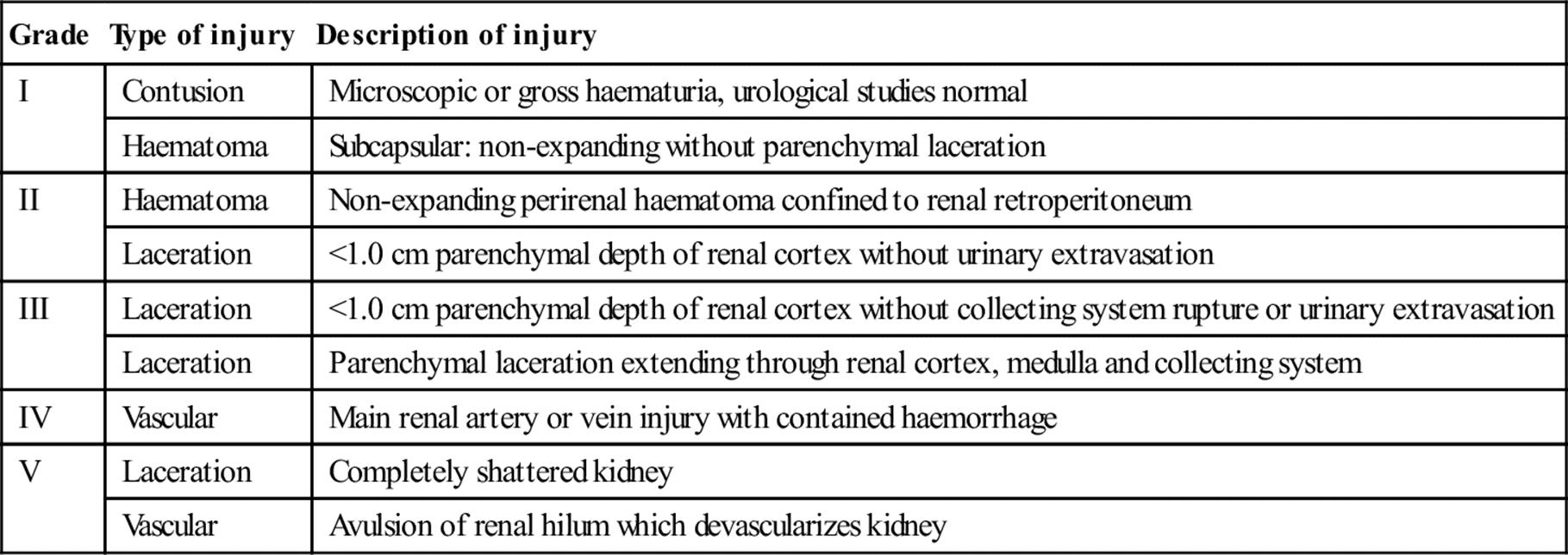
Table 3.8.18
Adrenal injury scale
|
Grade |
Description of injury |
|
I |
Contusion |
|
II |
Laceration involving only cortex (<2 cm) |
|
Ill |
Laceration extending into medulla (≥2 cm) |
|
IV |
>50% parenchymal destruction |
|
V |
Total parenchymal destruction (including massive intraparenchymal haemorrhage) |
|
Avulsion from blood supply |
Table 3.8.19
Pancreas injury scale

In stable patients with possible intra- abdominal injuries, the abdominal CT has become the investigation of choice because, as well as being non-invasive, it reliably identifies intraperitoneal fluid, solid organ and hollow visceral injury, retroperitoneal injuries and spinal and pelvic fractures (E-Figs 3.8.22 and 3.8.23). The use of intravenous contrast will also give some indication of both renal perfusion and function, as contrast is excreted into the ureters and bladder. One of the main limitations of abdominal CT is that the investigation must be carried out in the radiology department and so is inappropriate for any unstable patient. Injuries that may be missed on abdominal CT include upper intestinal perforation as well as injury to the diaphragm, pancreas and bladder.

E-FIG. 3.8.22 Bowel contusion with perforation. Solid arrows demonstrate mural thickening of the small and large bowel. The dashed arrows show free gas secondary to perforation.

E-FIG. 3.8.23 Traumatic abdominal wall hernia (arrows).
Focused assessment by sonography for trauma (FAST) examination
Since the introduction of the focused ultrasound examination for trauma in the early 1990s in North America and in the late 1990s in Australasia, there has been some debate regarding the sensitivity, specificity and accuracy of the examination compared to diagnostic peritoneal lavage (DPL). In those centres that use FAST on a regular basis, there has been a markedly reduced requirement for DPL. One of the criticisms of DPL has been its low specificity, resulting in an excessive non-therapeutic laparotomy rate of up to 30% in some centers [39,40]. The main utility of the FAST examination has been shown in the unstable trauma patient with intra-abdominal haemorrhage who requires urgent surgery and it has replaced DPL as the diagnostic modality of choice in these patients.
FAST requires the examination of four areas (Table 3.8.20). Its limitations include:
it requires training
it cannot differentiate between fluids (blood vs ascites vs urine)
poor-quality images in obesity, subcutaneous emphysema and dilated bowel loops.
Table 3.8.20
The FAST examination
1. The right upper quadrant (Morison’s pouch)
2. The left upper quadrant (splenorenal recess)
3. The subxiphoid area (pericardium)
4. The suprapubic area (pouch of Douglas/rectovesical pouch)
The FAST examination can be completed in 2–5 minutes, is non-invasive and is repeatable. It is very poor at detecting specific solid organ or hollow viscus intra-abdominal injuries but, if abdominal haemorrhage is ruled out and the patient is haemodynamically stable, then abdominal CT is indicated.
Many studies have reported a sensitivity of 80–100% and a specificity of 88–100% for the detection of intraperitoneal blood. It has also been consistently reported that FAST will not detect hollow viscus injuries, lacerations in the intra-abdominal solid organs, retroperitoneal or diaphragmatic injuries.
There is some evidence that FAST is of value in penetrating trauma. Boulanger et al. [41] found that the routine use of FAST in penetrating trauma was useful for the detection of pericardial and peritoneal fluid. However, they cautioned that a negative FAST did not exclude hollow viscus or diaphragmatic injuries.
Many centres have introduced FAST into the algorithm for the routine assessment of victims of trauma. Over 10 years ago, Boulanger et al. [42] demonstrated in a prospective study that a FAST-based algorithm for blunt abdominal injury was more rapid, less expensive and as accurate as an algorithm that used CT or DPL only. Hence, there is a growing body of evidence showing that the only indication for DPL (when no FAST is available) is for suspected bowel perforation, which is usually diagnosed either by clinical examination or by CT.
Radiology in pelvic trauma
In addition to plain radiology, pelvic CT scanning and angiography are becoming increasingly important in the diagnostic and therapeutic work-up of pelvic trauma. The trauma room AP X-ray of the pelvis should include all the bony pelvic components as well as both hip joints and the proximal femora, including greater and lesser trochanters (Fig. 3.8.23).
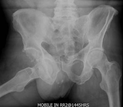
FIG. 3.8.23 Dislocated hip.
Most anterior pelvic fractures are seen on the AP film, but up to 30% of posterior fractures involving the sacrum and sacroiliac joints will not be seen on the plain radiology. These fractures will be best seen on a two-dimensional or reformatted 3D CT scan of the pelvis.
Acetabular fractures are often difficult to visualize on AP views and a CT scan of the pelvis may be required.
There are a number of radiological classifications of pelvic fractures that must be interpreted in association with the clinical impression of the fracture and the associated complications. The greater the AP disruption of the pelvic ring and hence the larger the pelvic cavity volume, the more there is potential for severe haemorrhagic shock and visceral damage.
A useful classification is that by Young and Resnik (Table 3.8.21) [43], which is a modification of the Pennel and Tile [44] classification of pelvic fractures. This classifies fractures by mechanism of injury into AP compression, lateral compression, vertical shear and a combination, and takes into consideration rotational and/or vertical instability of the pelvic ring (Fig. 3.8.24 [E-Figs 3.8.24 and 3.8.25]). If the pelvic ring is fractured anteriorly and posteriorly, stability is usually lost, with disruption of the posterior ligaments (sacroiliac, sacrotuberous and sacrospinous) and there will be widening of the sacroiliac joint(s) on the AP view. The classification provides a graded probability of bleeding related to the fracture, the development of haemorrhagic shock and associated organ damage.
Table 3.8.21
Young and Resnick classification of pelvic fractures
AP compression
Type 1: Disruption of the symphysis pubis with less than 2.5 cm diastasis; no significant posterior pelvic injury
Type 2: Disruption of the symphysis pubis of more than 2.5 cm with tearing of the anterior sacroiliac, sacrospinous and sacrotuberous ligaments
Type 3: Complete disruption of the pubic symphysis and posterior ligament complexes, with hemipelvic displacement
Lateral compression
Type 1: Posterior compression of the sacroiliac joint without ligament disruption; oblique pubic ramus fracture
Type 2: Rupture of the posterior sacroiliac ligament; pivotal internal rotation of the hemipelvis on the anterior SI joint with a crush injury of the sacrum and an oblique pubic ramus fracture
Type 3: Findings as in type 2 injury with evidence of an AP compression injury to the contralateral hemipelvis
Vertical shear
Complete ligament or bony disruption of a hemipelvis associated with hemipelvis displacement
This classification does not take into consideration isolated fractures outside the bony pelvic ring or acetabular fractures

FIG. 3.8.24 Vertical shear injury of the pelvis. Unilateral fractures involving the left iliac wing and superior and inferior pubic rami.
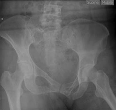
E-FIG. 3.8.24 Vertical shear injury. There is asymmetry of the heights of the iliac crest due to a vertical shear fracture of the right side of the sacrum. There are also bilateral fractures of the superior and inferior pubic rami.

E-FIG. 3.8.25 Vertical shear sacral fracture corresponding to the same patient as Figure 3.8.24. The CT scan better delineates the sacral fracture which can be difficult to visualize on plain film.
CT scan of the pelvis
CT and plain X-rays are complementary modalities in the evaluation of pelvic fractures. Patients with pelvic fractures associated with haemodynamic instability are not suitable for placing in the CT scanner.
If the patient with pelvic fractures is haemodynamically unstable, it is important to ascertain whether or not there is intra-abdominal bleeding. If the FAST examination excludes intra-abdominal bleeding, then the patient should proceed to angiography and embolization if pelvic arterial bleeding is demonstrated [45] or, alternatively, to surgical fixation if the pelvic ring is widely disrupted. There are limitations for both treatment options as set out below.
In stable patients, CT is useful for demonstrating posterior fractures involving the sacrum and sacroiliac joints as well as sacroiliac joint diastasis. Reformatted 3D images are particularly useful for the assessment of acetabular and pubic bone fractures.
The speed and definition of MDCT scanners have meant that contrast-enhanced MDCT is a highly accurate, non-invasive way of identifying ongoing arterial bleeding, which will then require urgent interventional radiological (IVR) treatment.
Visceral angiography and embolization
In haemodynamically stable patients, solid visceral injuries may be treated conservatively. In haemodynamically unstable patients with solid intra-abdominal visceral injuries and active bleeding, either surgical or endovascular management could be considered in centres where both interventions are available. Embolization in trauma is the intentional and controlled occlusion of vessels to stop haemorrhage [46]. Embolization agents may be classified as temporary or permanent. Permanent agents include metallic coils, glue and embolization particles, while gelatin sponge (Gelfoam) is a temporary agent. Gelfoam is able to stop active bleeding, however, allows for recanalization of the embolized vessel. The spleen and kidneys are the most commonly embolized viscera post-trauma [E-Figs 3.8.27 and 3.8.28].

E-FIG. 3.8.26 Active bleeding with embolisation. Axial CT image (A) shows active bleeding anteriorly within the pelvis (arrows) secondary to bilateral pubic rami fractures (not shown). (B) Superselected angiographic image of a right internal iliac artery branch with active bleeding (dashed arrow). (C) Shows a post embolisation angiographic image with a coil mass (black arrow) occluding the internal iliac artery branch and cessation of the active bleeding.
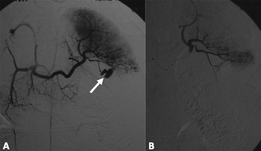
E-FIG. 3.8.27 Angiogram of the splenic artery (A) demonstrates a traumatic pseudoaneurysm of an inferior segmental branch (arrow), (B) demonstrates successful embolisation using particles and glue.
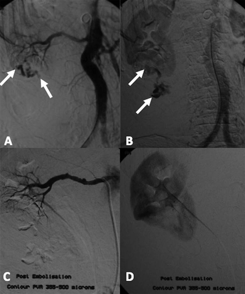
E-FIG. 3.8.28 Right renal angiogram demonstrating active bleeding from the inferior pole (arrows in A and B). (C, D) Show a successful embolisation using particles.
Pelvic angiography
Pelvic fractures that disrupt the posterior aspect of the pelvic ring have the potential to cause considerable arterial and/or venous injury. ‘Open-book’ or AP compression pelvic ring fractures are more likely to have venous rather than arterial bleeding and compression of the pelvic ring by external fixation should help to minimize this blood loss, although this practice has not been validated by prospective, randomized controlled trials.
There remains considerable controversy regarding the role of angiography and arterial embolization in pelvic trauma. Factors such as age>65, absence of long bone fractures and haemodynamic instability necessitating urgent angiography have been identified as predicting the likelihood of arterial bleeding in pelvic fractures [43]. Studies of the efficacy of angiographic embolization in select groups of haemodynamically unstable patients with pelvic fractures suggesting success rates for controlling bleeding in excess of 90% have led to a shift towards early radiographic intervention [47,48].
The femoral artery is catheterized and angiography of both internal iliac arteries performed. If arterial bleeding is identified, then the vessels can be selectively embolized (E-Fig. 3.8.26). There is a rich vascular supply to the pelvic viscera and major ischaemic complications are rare following pelvic embolization, but other problems, such as impotence, may occur.
Contrast studies
The main contrast studies used in pelvic fractures are the urethrogram and cystogram. Rupture of the membranous urethra may occur in association with pelvic fractures, particularly those involving distraction of the pubic symphysis or fractures involving both superior and inferior pubic rami. If there is clinical and radiological suspicion of potential urethral damage, an urethrogram should be performed. This is done by inserting a soft catheter into the urethral meatus and injecting contrast while screening with an image intensifier (Figs 3.8.25 and 3.8.26). The urethral passage, if patent, will be visualized and it may be possible to catheterize the urethra. If there is obstruction to the passage of dye or a false track is identified, a suprapubic catheter will be required. Further contrast is then injected into the bladder, then PA and oblique X-rays or CT scan can be performed to assess for extravasation of contrast suggesting bladder rupture.
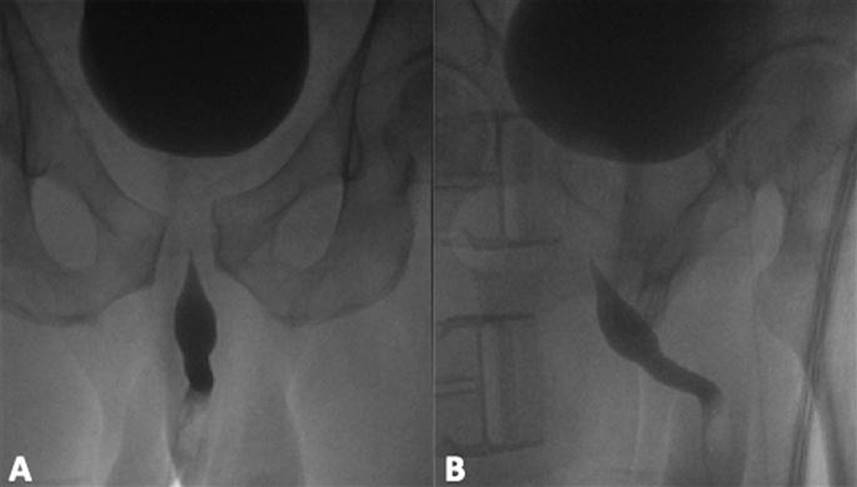
FIG. 3.8.25 Normal urethrogram in a patient with diastasis of the pubic symphysis.
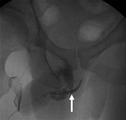
FIG. 3.8.26 Urethrogram in a patient with a fractured penis demonstrating active extravasation of contrast from the mid-penile urethra (arrow).
Extremities
Missed injuries occur in about 2–6% of blunt trauma patients. One retrospective study [50] found that musculoskeletal injuries and spinal fractures featured highly (6%) among the injuries not found after the initial primary and secondary surveys. The musculoskeletal injuries comprised mainly fractures and a small number of soft tissue injuries. Among the factors contributing to the missed injuries were the presence of closed head injury and intoxication. In comparison, one study found the rate of abdominal missed injuries to be 2% [51].
A careful clinical examination of all joints and limbs looking for swelling, deformity and crepitus must be made in order to direct radiological investigation. Fractures, dislocations and ligamentous instability are more likely to be missed in the smaller, peripheral bones. As these injuries may be a source of ongoing disability due to late diagnosis, they may also be a potential source of litigation. Dislocations of joints, such as the anterior shoulder and elbow, should be readily obvious, but less so are posterior shoulder and lunate/perilunate dislocation in the wrist. AP and lateral X-rays should be taken of any joint considered abnormal on examination during the secondary survey. In the lower limb, posterior dislocation of the hip and knee joints may cause serious sciatic nerve and popliteal artery damage, respectively, and require urgent reduction. If the viability of the limb or skin is threatened, a dislocation (e.g. knee or ankle) should be immediately reduced on clinical grounds and X-ray performed after reduction to check for position and bony fractures.
Bony fractures in the upper limb commonly missed include medial or lateral epicondylar fractures and supracondylar fractures of the elbow in children. In the adult, fractures of the carpal bones, in particular the scaphoid and triquetrum, may be missed unless carefully looked for and these injuries may result in significant disability. Fractures and dislocations involving the metacarpals and phalanges are also easily missed in the multitrauma patient. The skier’s (or gamekeeper’s) thumb [52] is an acute sprain or rupture of the ulnar collateral ligament at the metacarpophalangeal joint caused by forceful abduction of the thumb. This injury may be missed unless the joint is specifically examined for stability and stress views taken if indicated.
In the lower limb, fractures of the tibial plateau and calcaneus, which may occur as a result of a fall, may be missed unless sought both clinically and radiologically. Appropriate AP and lateral X-rays should be taken of these areas. In the foot, loss of Boehler’s angle (normal 25–40°) may indicate a depressed fracture of the subtalar part of the calcaneus (Fig. 3.8.27). Falls in general, and calcaneal fractures in particular, may be associated with fractures of the upper lumbar spine.

FIG. 3.8.27 Calcaneal fracture with loss of Bohler’s angle.
CT scans of complex fractures and dislocations may assist the orthopaedic surgeon in planning appropriate fixation (Fig. 3.8.28). Joints where this may be helpful include large joints, such as shoulder, hip and knee, e.g. tibial plateau fractures. Calcaneal fractures are often not clearly seen on plain X-rays and require a CT scan for a more accurate view. MRI is the investigation of choice for ligamentous or meniscal injuries in the knee.

FIG. 3.8.28 Lisfranc injury and tarsometatarsal joint complex dislocation.
Angiography is required when there is suspected or clinically obvious vascular compromise to either upper or lower limb. The axillary or brachial arteries may be damaged or transected in blunt or penetrating injuries to the upper limb. The brachial plexus can also be damaged in trauma around the shoulder joint and should be actively looked for in these injuries. The commonest serious vascular injury to the lower limb may be associated with posterior dislocation of the knee and intimal disruption of the popliteal artery. Angiography will give accurate information regarding the degree of arterial damage and the state of the collateral flow.
Conclusion
Radiology in the multitrauma patient requires judicious decision making and interpretation of X-rays and other specialized modalities, such as CT, MRI and ultrasonography. There is less reliance on plain X-rays and more emphasis on CT scans to rule in or out serious injury in the head, spine, chest, abdomen and pelvis. Radiation exposure should be considered for all CT scans, especially in younger trauma patients. Missed injuries that are not diagnosed in the first 24–48 hours often contribute significantly to patient morbidity and mortality. These injuries may involve the musculoskeletal system in the form of limb or spinal fractures and must be actively sought and excluded by appropriate clinical and radiological examination.
Controversies
Clearance of the cervical spine in obtunded trauma patients can be made on the basis of fine cuts in a multislice CT scan. MRI may be indicated if there is strong clinical suspicion and the need to exclude a ligamentous injury.
Dynamic flexion/extension X-rays of the Cx spine should only be performed on awake and cooperative patients, but they have low sensitivity and specificity for the detection of unstable spinal injuries.
CT angiography and digital subtraction angiography are invaluable in the investigation of blunt cerebrovascular injury to the neck and should also be performed if there is cervical spine bony injury involving the foramen transversarium.
Chest CT is the investigation of choice to exclude significant intrathoracic injury.
The FAST examination will detect haemoperitoneum only but not hollow viscus perforation or solid organ haematoma.
Pelvic angiography and embolization should be part of the resuscitation protocol in haemodynamically unstable patients with major pelvic fractures.
References
1. Tan GA, Van Every B. Staff exposure to ionizing radiation in a major trauma centre. Aust NZ J Surg. 2005;75:136–137.
2. Shah DJ, Sachs RK, Wilson DJ. Radiation-induced cancer: a modern view. Br J Radiol. 2012;85:e1166–e1173.
3. Furlow B. Radiation dose in computed tomography. Radiol Technology. 2010;81:437–450.
4. Aldrich JE, Bilawich AM, Mayo JR. Radiation doses to patients receiving computed tomography examinations in British Columbia. Can Assoc Radiol J. 2006;57:79–85.
5. http://www.radiologyinfo.org/en/safety/index.cfm?pg=sfty_xray&bhcp=1.
6. Pierce DA, Shimizu Y, Preston DL, et al. Studies on the mortality of atomic bomb survivors Report 12, Part 1, Cancer: 1950–1990. Radiat Rese. 1996;146:1–27.
7. Thompson DE, Mabuchi K, Ron E, et al. Cancer incidence in atomic bomb survivors Part 11, Solid tumours: 1958–1987. Radiat Res. 1994;137:S17–S67.
8. <http//www.pueblo.gsa.gov/cic_text/health/fullbody-ctscan/risks.htm>.
9. Valadka AB, Narayan RK. Injury to the cranium. In: Feliciano DV, Moore EE, Mattox KL, eds. Trauma. 3rd ed. Stamford CT: Appleton and Lange; 1996;267–278.
10. Stiel IG, Wells GA, Vandemheen K, et al. The Canadian CT Head Rule for patients with minor head injury. Lancet. 2001;357:1391–1396.
11. Bromberg WJ, Collier BC, Diebel LN, et al. Blunt cerebrovascular injury practice management guidelines: The Eastern Association for the Surgery of Trauma. J Trauma. 2010;68:471–477.
12. Emmett KP, Fabian TC, DiCicco JM, et al. Improving the screening criteria for blunt cerebrovascular injury: the appropriate role for computed tomography angiography. J Trauma. 2011;70:1058–1065.
13. Biffi WL, Moore EE, Offner PJ, et al. Blunt carotid arterial injuries: implications of a new grading scale. J Trauma. 1999;47:845–853.
14. Griffen MM, Frykberg ER, Kerwin AJ, et al. Radiographic clearance of blunt cervical spine injury: plain radiograph or computed tomography scan? J Trauma Injury Infect Crit Care. 2003;55:222–227.
15. Cooper DJ, Ackland HM. Clearing the cervical spine in unconscious head injured patients – the evidence. Crit Care Resus. 2005;7:181–184.
16. Hoffman JR, Mower WR, Wolfson AB, et al. Validity of a set of clinical criteria to rule out injury to the cervical spine in patients with blunt trauma National Emergency X-Radiography Utilisation Study Group. N Engl J Med. 2000;343:94–99.
17. Barrett TW, Mower WR, Zucker MI, et al. Injuries missed by limited computed tomographic imaging of patients with cervical spine injuries. Ann Emerg Med. 2006;47:129–133.
18. Stiell IG, Wells GA, Vandemheen K, et al. The Canadian C-spine rule for radiography in alert and stable trauma patients. J Am MedAssoc. 2001;286:1841–1848.
19. Berquist TH. Cervical spine trauma. In: Kricum ME, ed. Imaging of sports injuries Aspen Publications. 1992;31–64.
20. Lusted LB, Keats TE. The spine atlas of roentgenographic measurement 2nd ed. Chicago: Yearbook Medical Publications; 1967; 101–103.
21. Keene JG, Daffner RH. Spinal trauma. In: Rosen P, Doris PE, Barkin RM, eds. Diagnostic radiology in emergency medicine. St Louis: Mosby-Year Book; 1992;210–270.
22. Harris JH. The cervicocranium: its radiographic assessment. Radiology. 2001;218:335–337.
23. Noble EF. Smoker WRK The forgotten condyle: The appearance, morphology, and classification of occipital condyle fractures. Am J Neuroradiol. 1996;17:507–513.
24. Satisky E, Votey S. Emergency department approach to acute thoracolumbar spine injury. J Emerg Med. 1997;15:49–59.
25. Denis F. The three column spine and its significance in the classification of acute thoracolumbar spinal injuries. Spine. 1983;8:817–831.
26. Mirka H, Ferda J, Baxa J. Multidetector computed tomography of chest trauma: Indications, technique and interpretation. Insights Imag. 2012;3:433–449.
27. Oikonomou A, Prassopoulos PCT. Imaging of blunt chest trauma. Insights Imag. 2011;2:281–295.
28. Ho M, Gutierrez F. Chest radiography in thoracic polytrauma. Am J Roentgenol. 1992;200:599–612.
29. Traub M, Stevenson M, McEvoy S, et al. The use of chest computed tomography versus chest X ray in patients with major blunt trauma. Injury. 2007;38:43–47.
30. Brookes JG, Dunn RJ, Roger IR. Sternal fractures: a retrospective analysis of 272 cases. J Trauma. 1993;35:46.
31. Sammett EJ. Aorta, trauma.<www.emedicine.com/radio/topic44.htm>; 2003.
32. Creasy JD, Chiles C, Routh WD, et al. Overview of traumatic injury of the thoracic aorta. Radiographics. 1997;17:27–45.
33. Holtzman SR, Bettmann MA, Casciani T, et al. Expert Panel on Cardiovascular Imaging Blunt chest trauma-suspected aortic injury [online publication] Reston (VA): American College of Radiology (ACR); 2005.
34. Keaney PA, Wesley Smith D, Johnson SB, et al. Use of transoesophageal echocardiography in the evaluation of traumatic aortic injury. J Trauma. 1993;34:696–703.
35. Smith MD, Cassidy JM, Souther S, et al. Transoesophageal echocardiography in the diagnosis of traumatic rupture of the aorta. N Engl J Med. 1995;332:356–362.
36. Bouhemad B, Zhang M, Lu Q, Rouby J. Clinical review: Bedside lung ultrasound in critical care practice. Crit Care. 2007;11:205.
37. Tsang BD, Panacek EA, Brant WE, et al. Effect of oral contrast administration for abdominal computed tomography in the evaluation of acute blunt trauma. Ann Emerg Med. 1997;30:7–13.
38. Moore E, et al. Scaling system for organ specific injuries. <http://www.aast.org/Library/TraumaTools>.
39. Henneman PL, Marx JA, Moore EE, et al. Diagnostic peritoneal lavage: accuracy in predicting necessary laparotomy following blunt and penetrating trauma. J Trauma. 1990;30:1345.
40. Hsu JM, Joseph AP, Tarlinton LJ, et al. The accuracy of focused assessment with sonography in trauma (FAST) in blunt trauma patients: experience of an Australian major trauma service. Injury. 2007;38:71–75.
41. Boulanger BR, Kearney PA, Tsuei B, et al. The routine use of sonography in penetrating torso injury is beneficial. J Trauma. 2001;51:320–325.
42. Boulanger BR, McLellan BA, Brenneman FD, et al. Prospective evidence of the superiority of a sonography-based algorithm in the assessment of blunt abdominal injury. J Trauma. 1999;47:632–637.
43. Gill Cryer H, Johnson E. Pelvic fractures. In: Feliciano DV, Moore EE, Mattox KJ, eds. Trauma. 3rd ed. Stamford: Appleton and Lange; 1996;635–659.
44. Pennel GF, Time M, Waddell JP, et al. Pelvic disruption: assessment and classification. Clin Orthoped. 1980;151:12.
45. Martin J, Heetveld, Harris I, et al. Management of unstable patients with pelvic fractures. Practice Guidelines, Liverpool Hospital NSW. <http://www.swsahs.nsw.gov.au/livTrauma>; 2003.
46. Lopera JE. Embolisation in trauma: principles and techniques. Semin Intervent Radiol. 2010;27:14–28.
47. Velmahos GC, Toutouzas KG, Vassiliu P, et al. A prospective study of the safety and efficacy of angiographic embolization for pelvic and visceral injuries. J Trauma. 2002;52:303–308.
48. Miller PR, Moore PS, Mansell E, et al. External fixation or arteriogram in bleeding pelvic fracture: initial therapy guided by markers of arterial haemorrhage. J Trauma. 2003;54:437–443.
49. Fangio P, Asehnoune K, Edouard A, et al. The epidemiology of fractures and fracture-dislocations of the cervical spine. J Trauma. 2005;58:978–984 discussion 984.
50. Kremli MK. Missed musculoskeletal injuries in a University Hospital in Riyadh: types of missed injuries and responsible factors. Injury. 1996;27:503–506.
51. Sung C, Kim KH. Missed injuries in abdominal trauma. J Trauma. 1996;41:276–278.
52. Musharafieh RS, Bassim YR, Atiyeh BS. Ulnar collateral ligament injury in the emergency department. J Emerg Med. 1997;15:193–196.
3.9 Trauma in pregnancy
Steven Troupakis
Essentials
1 Trauma in pregnancy is the most common cause of non-obstetric maternal death, with most deaths due to head injury and haemorrhagic shock.
2 Fetal death occurs far more often than maternal death and is dependent on the severity of the maternal injuries. Placental abruption and direct fetal trauma cause most deaths.
3 Common causes of trauma are motor vehicle collisions, falls and assaults.
4 Important sequelae are bruising, fractures, premature labour, placental abruption, disseminated intravascular coagulopathy, fetomaternal haemorrhage, intra-abdominal injuries, uterine rupture and haemorrhagic shock.
5 The physiological changes that occur with pregnancy, such as the relative hypervolaemia and the gravid uterus, can make clinical assessment of the patient difficult.
6 Continuous cardiotocographic monitoring for at least 4 hours is the best predictor of placental abruption and fetal distress.
7 Bedside ultrasound allows early evaluation for the presence of intraperitoneal fluid and the fetal heart.
8 Maternal resuscitation remains the best method of fetal resuscitation.
Introduction
Trauma during pregnancy presents a unique set of challenges for the emergency department (ED), as the anatomical and physiological changes that occur during pregnancy will influence the evaluation and management of the patient. An appreciation of these changes is important. Aggressive resuscitation of the mother remains the best treatment for the fetus. A multidisciplinary approach with early obstetric consultation will help improve the outcome of these patients.
Anatomical and physiological changes in pregnancy
Cardiovascular
Blood volume increases by about 50% by the end of the third trimester [1]. With relative hypervolaemia, the patient may lose up to 35% of her blood volume before signs of haemorrhagic shock appear. Maternal cardiac output increases by 30–50% above normal by the end of the second trimester. The resting heart rate increases by 15–20 beats/min by the end of the third trimester. Systolic and diastolic blood pressure fall by 10–15 mmHg during the second trimester, but rise again towards the end of the pregnancy. ECG changes may occur with the cephalic displacement of the heart, such as left axis deviation by 15°, T-wave inversion or flattening in leads III, V1 and V2 and Q waves in III and AVF [1]. After 20 weeks’ gestation, supine positioning may cause inferior vena cava (IVC) obstruction by the gravid uterus, leading to a fall in cardiac output.
Haematological
A dilutional anaemia occurs with a fall in haematocrit (31–35% by the end of pregnancy). Pregnancy induces a leucocytosis, with levels up to 18 000/mm3 in the third trimester. Coagulation factors increase (fibrinogen, factors VII, VIII, IX, X), increasing the risk of venous thrombosis. The buffering capacity of the blood is reduced [2].
Respiratory
The diaphragm is elevated by about 4 cm. Tidal volume and minute volume increase by 40%. A respiratory alkalosis results, with a fall in PCO2 to 30 mmHg. The anteroposterior diameter of the chest is increased and the mediastinum is widened on chest X-ray.
Gastrointestinal
Cephalic displacement of intra-abdominal structures reduces gastro-oesophageal sphincter tone, combined with delayed gastric motility, there is an increased the risk of aspiration. The intestines are displaced to the upper part of the abdomen and may be shielded by the uterus. The peritoneum is stretched by the gravid uterus, which may make signs of peritonism less reliable [2]. Alkaline phosphatase levels may triple because of placental production.
Urinary
Dilatation of the renal pelvis and ureters occurs from the 10th week of gestation. The bladder becomes hyperaemic and is displaced into the abdomen from the 12th week, making it more susceptible to trauma.
Uterine
There is a massive increase in uterine size. Blood flow to the uterus increases from 60 to 600 mL/min by the end of the pregnancy.
Epidemiology
The incidence of trauma during pregnancy is approximately 6–7%, the causes being similar to those in the general population [1,3]. Blunt trauma is the commonest injury, with motor vehicle accidents, falls and assaults being the other common causes and in that order. Penetrating injuries are less common and usually the result of domestic violence. Stab wounds have a better prognosis for the fetus than do projectile wounds. Most trauma is of a minor nature, resulting in bruising, minor fractures and threatened premature labour. Maternal death from trauma is rare, but is the leading non-obstetric cause of death, with most fatalities due to head injuries and internal haemorrhage. Younger (age<20) and older (age>35) multiparous women at gestational ages of less than 28 weeks have a higher risk of adverse outcomes [4]. Women who are discharged undelivered continue to have delayed morbidity, with increased rates of placental abruption and low-birthweight infants. Fetal death occurs in about 1–2% of cases and is dependent on the gestational age and the pattern and severity of maternal injury. Most fetal deaths are due to placental abruption or direct trauma. High-speed (>80 km/h) and broadside motor vehicle accidents have a higher incidence of placental abruption and fetal and maternal death than do frontal collisions [5]. Similarly, ejection from a vehicle and motorcycle and pedestrian collisions are associated with poor fetal outcome [6]. Maternal hypotension and vaginal bleeding are associated with increased fetal loss. In one trauma series, pregnant patients with an injury severity score (ISS)>12 had a fetal death rate of 65%; those with an ISS<12 had no fetal deaths [7].
Specific injuries
Pelvic fracture
Pelvic fracture is often the result of a high-speed motor vehicle accident. Massive haemorrhage can occur from the uterus, as well as bladder, urethral and ureteric lacerations. Retroperitoneal haemorrhage occurs and may be difficult to diagnose. Direct fetal skull fractures can lead to fetal death. The majority of patients with a pelvic fracture can be delivered vaginally.
Placental abruption
Placental abruption complicates 1–5% of patients with minor trauma and between 20 and 50% of cases with major trauma [3,8]. The placenta separates from the underlying decidua because of shearing forces between the relatively inelastic placenta and the more elastic uterus. This leads to fetal hypoxia and death. Thromboplastin release may lead to the development of disseminated intravascular coagulopathy (DIC).
Uterine rupture
Uterine rupture is rare but leads to considerable haemorrhage and with 10% maternal mortality and almost 100% fetal mortality [3]. It usually occurs as a result of direct trauma to women with a uterine scar. It should be suspected when there is maternal shock, fetal death, difficulty defining a uterus, easily palpable fetal parts and intraperitoneal fluid on ultrasound.
Fetomaternal haemorrhage
Fetomaternal haemorrhage is the transplacental spread of fetal blood into the maternal circulation. It occurs in approximately 8–30% of trauma cases and may lead to Rhesus (Rh) sensitization of the mother, neonatal anaemia, fetal cardiac arrhythmias and fetal death [2]. The Kleihauer–Betke test is used to identify and quantify fetomaternal haemorrhage. This test relies on the principle that fetal cells are stable in acid (pH 3.2), whereas adult haemoglobin is eluted from maternal red cells. Microscopy will identify fetal red blood cells on blood smear. All Rh-negative pregnant trauma victims with a positive Kleihauer–Betke test should receive Rhesus immunoglobulin.
Presentation
History
Questions should be directed to determining the severity and type of trauma, as well as an obstetric history. In a motor vehicle accident, high speed, side collisions, ejection from the vehicle and improper use of seatbelts and lap belts alone are associated with a greater likelihood of serious injuries [3]. Direct trauma to the abdomen is more likely to cause fractures, splenic and hepatic injuries, whereas indirect trauma via shearing forces is more likely to cause placental abruption. Pelvic pain, uterine contractions and vaginal bleeding may indicate placental abruption. The gestational age (>22 weeks) is the main determinant for fetal viability. Lack of fetal movements may indicate fetal death.
Primary survey
The airway should be assessed and cleared. Intubation may be difficult because of aspiration risk, breast enlargement and cervical trauma. Breathing should be assessed and the patient given supplemental oxygen to improve both maternal and fetal oxygenation. If the patient is more than 20 weeks pregnant, she should be placed on her side (preferably the left) to relieve any caval compression. If spinal immobilization is necessary, wedges can be placed underneath a spinal board or, alternatively, the uterus pushed to the left manually. The blood pressure and circulation can then be assessed, remembering that signs of shock may present late because of relative hypervolaemia.
A quick assessment of conscious level and any major neurological deficits should be made. The patient should be adequately exposed for a thorough examination, but protected from a drop in temperature.
Secondary survey
The sequence of the secondary survey is the same as in the non-pregnant patient, but with an obstetric examination included in the abdominal examination. The uterus should be assessed for fundal height, tenderness, contractions, fetal heart tone, fetal movements and position. Focused abdominal sonography for trauma (FAST) should be performed to assess for intraperitoneal haemorrhage. Bedside ultrasound can also be used to assess the fetal heart rate. The availability of FAST scans and CTs have caused diagnostic peritoneal lavage to fall out of routine use [9]. An obstetrician should perform the pelvic examination, looking for trauma to the genital tract, cervical dilation, fetal presentation and station relative to the ischial spines. Nitrazine paper can be used to test for the presence of amniotic fluid: it turns blue in the presence of the alkaline fluid. Rectal examination and urinalysis are essential.
Investigations
Blood tests
Routine blood tests, such as full blood count, electrolytes, coagulation studies, group and hold, should be performed looking for evidence of anaemia and DIC. A Kleihauer–Betke test will indicate the necessary dose of Rhesus immunoglobulin in Rh-negative patients.
X-rays
In severe trauma, it is necessary to take cervical spine, chest and pelvic films. The abdomen should be shielded and repetition of films avoided. There is negligible risk to the fetus when radiation exposure has been limited to less than 0.1 Gy and after 20 weeks’ gestation radiation is unlikely to cause abnormalities [10]. A standard pelvic film delivers less than 0.01 Gy.
Ultrasonography
Ultrasonography is useful in determining gestational age, placental position and fetal well-being and estimating amniotic fluid volume [11]. Bedside FAST can be used to assess for free fluid in the peritoneum and in the pericardial and pleural cavities, especially in patients too unstable for CT. Ultrasonography will detect only 40–50% of placental abruptions [3].
Cardiotocography (CTG)
CTG monitoring beyond the 20th week of pregnancy has proved a sensitive way of diagnosing placental abruption early. It should be instituted early and continuously for at least 4 hours [2]. Fetal distress on CTG may be the earliest indicator of impending shock. Frequent uterine contractions and fetal distress are suggestive of placental abruption. In one study, no placental abruptions were missed if CTG monitoring remained normal for the first 4 hours [8].
Computed tomography
Computed tomography (CT) is an accurate and non-invasive way of assessing uterine and retroperitoneal structures, but it is time-consuming and involves a higher radiation dose than normal X-rays, with exposure for abdominal CT being between 0.05 and 0.1 Gy. Chest and head CT expose the fetus to far less radiation, especially if uterine shielding is used with radiation exposure of about 0.001 Gy [3,12].
Management
Maternal resuscitation is the best method of fetal resuscitation. If the injuries are severe, the patient should be in a resuscitation area with a multidisciplinary team approach to management and early surgical, anaesthetic and obstetrical consultation. Attention to adequate oxygenation, proper positioning and aggressive fluid replacement is important. Oximetry, ECG, blood pressure monitoring and cardiotocography should be started early. A nasogastric tube should be inserted to reduce the risk of aspiration, as should an indwelling catheter for urinalysis and to allow better assessment of the uterus. X-rays as indicated should be performed as well as a FAST scan and CT as necessary, to evaluate abdominal injuries. If the patient remains unstable with hypotension or continued bleeding, laparotomy is indicated. Ultrasound is particularly useful in the resuscitation phase to assess fetal heart rate and uterine bleeding.
If a thoracostomy is required, the entry point should be 1 or 2 intercostal spaces higher than normal to avoid the diaphragm and abdominal structures.
The presence of vaginal bleeding, abdominal tenderness or pain, hypotension, absent fetal heart sounds, fetal distress on CTG and amniotic fluid leakage requires an urgent obstetric opinion and possibly a caesarean section.
The use of leg veins for intravenous access should be avoided as the gravid uterus may affect venous return and compromise drug delivery. The uterine vasculature is very sensitive to catecholamines. If ionotropes are required, adrenaline and noradrenaline should be avoided. Ephedrine and dopamine at doses less than 5 μg/kg can be used to improve maternal BP without compromising uterine blood flow [1,3].
Premature labour can be treated with tocolytic agents, such as intravenous salbutamol. However, salbutamol causes maternal and fetal tachycardia, which may mask symptoms of hypovolaemia. Magnesium sulphate is recommended as an alternative tocolytic in abdominal trauma [3].
DIC may develop as a result of placental abruption, amniotic fluid embolism and fetal death. Clotting factors may need to be replaced.
Anti-D immunoglobulin should be administered to all Rhesus-negative mothers.
In general, penetrating injuries should be explored by laparotomy, especially if they involve the upper abdomen, where there is a high possibility of bowel perforation. Some authors argue that stab wounds over the uterus can be treated conservatively if there is no evidence of visceral injury, the entrance wound is below the fundus and the patient is stable [1].
Post-mortem caesarean section should be considered within the first 4 minutes of a maternal cardiac arrest. There have been many cases of fetal survival up to 20 minutes after maternal death. The fetuses that have the best chance of surviving neurologically intact are those delivered within 5 minutes of the arrest, who weigh more than 1000 g and are of more than 28 weeks’ gestation [1,12].
Disposition
Patients who are haemodynamically unstable and who have extensive head or chest injuries will require surgical intervention and intensive-care support. Patients who are stable but show signs of fetal distress should undergo caesarean section. All patients with minor injuries who are more than 20 weeks pregnant should have CTG monitoring for at least 4 hours, preferably in a labour ward.
Prognosis
Most women who sustain trauma during pregnancy suffer few complications. There is greater maternal and fetal mortality in pregnant women with higher injury severity scores. Placental abruption can still occur as a result of minor trauma 24–48 hours after the accident, but 4 hours of CTG monitoring should detect this group of patients [2].
Prevention
Properly worn seatbelts reduce both maternal and fetal mortality. In one study of serious motor vehicle accidents, maternal mortality following ejection from the vehicle was 33%, compared to only 5% in those who were not ejected: fetal mortality was 47% and 11%, respectively [3]. A three-point seat bar system should be used, with the lap portion as low as possible, preferably over the thighs and with the shoulder portion passing between the breasts and above the gravid uterus.
Controversies
The duration of CTG monitoring: most authors agree 4 hours should be enough to predict placental abruption, although some argue that 24–48 hours may be needed.
Exploration of penetrating wounds to the abdomen: some authors argue for a conservative approach to a wound below the uterine fundus, whereas others argue that all such wounds should be explored.
References
1. Muench MV, Canterino JC. Trauma in pregnancy. Obstet Gynecol Clin N Am. 2007;34:555–583.
2. Pearlman MD, Tintinalli JE, Lorenz RP. Blunt trauma during pregnancy. N Engl J Med. 1990;323:1609–1613.
3. Hill CC. Trauma in the obstetric patient. Women Hlth. 2009;5:269–285.
4. El Kady D, Gilbert WM, Anderson J, et al. Trauma during pregnancy: an analysis of maternal and fetal outcomes in a large population. Am J Obstet Gynecol. 2004;190:1661–1668.
5. Aitokallio-Tallberg A, Halmesmaki E. Motor vehicle accident during the second or third trimester of pregnancy. Acta Obstet Gynecol Scand. 1997;76:313–317.
6. Curet MJ, Schermer CR, Demarest GB, et al. Predictors of outcome in trauma during pregnancy: identification of patients who can be monitored for less than 6 hours. J Trauma. 2000;49:18–25.
7. Ali J, Yeo A, Gana TJ, McLellan BA. Predictors of fetal mortality in pregnant trauma patients. J Trauma. 1997;42:782–785.
8. Pearlman MD, Tintinalli JE, Lorenz RP. A prospective controlled study of outcome after trauma during pregnancy. Am J Obstet Gynecol. 1990;162:1502–1510.
9. Oxford CM, Ludmir J. Trauma in pregnancy. Clin Obstet Gynaecol. 2009;52:611–629.
10. Goldman SM, Wagner LK. Radiological management of abdominal trauma in pregnancy. Am J Roentgenol. 1996;166:763–767.
11. Bode PJ, Niezen RA, Van Vugt AB, Schipper J. Abdominal ultrasound as a reliable indicator for conclusive laparotomy in blunt abdominal trauma. J Trauma. 1993;34:27–31.
12. Meroz Y, Elchalal U, Ginosar Y. Initial trauma management in advanced pregnancy. Anesthesiol Clin 2007;25:117–129.
3.10 Wound care and repair
Gim Tan and Richard Waller
Essentials
1 Good cosmesis can be achieved in the emergency department with conservative treatment, thorough debridement and accurate apposition of everted skin edges.
2 Choose a suture that is monofilament, causes little tissue reactivity and retains tensile strength until the strength of the healing wound is equal to that of the suture.
3 Dirty, contaminated, open wounds should generally be cleansed, debrided and closed within 6 hours to minimize the chance of infection.
4 Suspected tendon injuries require examination of the full range of movement of joints distal to the wound while observing the tendon in the base of the wound for breaches. This is often done under anaesthesia.
5 The success of a tendon repair (as measured by function) relates in large part to the postoperative care and therapy, not simply to the suture and wound closure.
6 Appropriate splinting and elevation of limb wounds at risk of infection takes precedence over antibiotics in the postoperative prevention of infection.
7 If prophylactic antibiotics are used, they should be given intravenously prior to wound closure to achieve adequate concentrations in the tissues and haematomas that may collect. There is no need for antibiotics with simple lacerations not involving tendon, joint or nerves.
8 Wounds that breach body cavities, such as the peritoneum and joints, or involving flexor tendons, nerves and named arteries, should be referred to a specialist for consideration of repair and inpatient care.
9 Foreign bodies, such as clay, chemically impair wound healing.
10 Puncture wounds such as bites may be managed by either second-intention healing after thorough lavage or, better still, by excisional debridement, lavage, antibiotics and atraumatic closure, if less than 24 hours old (preferably less than 6 hours).
Introduction
Open wound injury comprises a significant component of emergency department (ED) workload. Data from the Victorian Injury Surveillance System [1] showed that 72% of all ED presentations for unintentional cutting and/or piercing injury that did not require admission were open wounds. In addition, open wounds may accompany other injuries, such as fractures. Of open wounds that occur in the home, 19% are in the paediatric age group (0–14 years), 62% occur in people under 35 and less than 10% in the over-65 s. Overall, 65% of patients are male.
Location data show that more than 53% of these wounds occur in the home [1], mostly during activity described as leisure. The three major causes are falls up to 1 m; contact with cutting or piercing objects; or having been struck or collided with. Most are unintentional and only 3% are due to an assault. Injuries to the face, head and neck comprise 12% and the upper extremity is involved in 62%. Eighty-eight per cent of all presentations are repaired in the ED and the patient is discharged home. Almost half are referred to GPs and specialists for review. It is those wounds suitable for ED repair that will be further discussed.
Clinical presentation
An initial general assessment of the patient is important as it defines the likely mode of repair and the injured structures and identifies factors for complications. The assessment includes the traditional history, examination and investigation of the patient.
It is important in the history to identify the time and mechanism of injury, the likely presence of foreign bodies and the patient’s tetanus immunization status. Past medical history, allergies to agents, such as local anaesthetics, antibiotics, preparation solutions and tapes, and current medications, such as warfarin or cytotoxics, all have a bearing on management. For example, there is a greater risk of infection and poor wound healing in diabetic patients with extremity wounds of the lower limbs sustained in a crush injury. Other relevant general conditions, particularly in the setting of dirty wounds, such as bites, include prior mastectomy and other causes of chronic oedema of the affected region, prior splenectomy, liver dysfunction, immunosuppression or autoimmune disease, such as systemic lupus erythematosus (SLE). Smokers have impaired collagen production in healing wounds [2].
The general examination comprises a search for all injuries sustained and concurrent medical illness that may have a bearing on the results of repair, such as poor circulation in patients with peripheral vascular disease. The patient needs to be recumbent (beware of syncope) and any clothing that may obstruct a thorough examination removed. Constricting rings or other jewellery that encircle the injured body part should also be removed. A general examination is performed, followed by a local examination of the wound coupled with initial cleansing. Function and nerve or vessel injury are then examined for. A detailed examination of the depth of the wound, which usually requires good anaesthesia, is then performed. A surface wound caused by the entrance of a foreign body does not necessarily mean that the foreign body has remained in the vicinity. A decision is made regarding the requirement for further investigations which include radiographs for fractures and some foreign bodies or ultrasound for radiolucent foreign bodies.
An injury to a tendon in the base of the wound may only be apparent when the joints over which it acts are in a particular position, reflecting the position of the limb at the time of injury. At other positions, the tendon injury may slide out of view. Marked pain with use may be a clue to a partial tendon injury.
Any tendon injury or other factors, such as nerve damage, indicate the need for referral to a plastic surgeon.
Wound cleansing
To provide optimum conditions for healing without infection, it is essential to remove all contaminants, foreign bodies and devitalized tissue prior to wound closure.
Universal precautions, including eye protection (goggles or similar), clothing protection (gown) and gloves, must be taken for all wound care and repair. Gloves should be powder free to avoid adding starch as a foreign body to the wound, which will delay healing and produce granulomas [3]. One must be aware of the risk of latex allergy to both the glove wearer and the patient [3].
If necessary, hair can be removed by clipping 1–2 cm above the skin with scissors. Shaving the area with a razor damages the hair follicle and is associated with an increased infection rate. Scalp wounds closed without prior hair removal heal with no increase in infection [4].
The skin surface should be cleaned using sterile normal saline. This has the lowest toxicity and there is no benefit in using antiseptic [5].
Recent studies have shown that the use of tap water in the cleaning of simple lacerations is as effective as normal saline [6].
A wide variety of cleansing solutions is available (Table 3.10.1), with differing attributes.
Table 3.10.1
Preparation solutions and their properties

Anaesthesia is necessary for wounds to be cleansed adequately. Extensive wounds, or particularly heavily contaminated wounds that need vigorous scrubbing, such as road debris tattooing, may require general anaesthesia. Local anaesthetic may be given by local infiltration or as a regional nerve blockade. Needles introduced through the wound cause less pain, but may theoretically track bacteria into the tissues, although this has not been demonstrated to be a problem clinically. After anaesthesia, irrigation with a pressure of at least 8 psi (55 kPa) [7,8] is required to dislodge bacteria and reduce the incidence of infection. This can be achieved with a 19 G needle, a 25–50 mL syringe, a three-way tap and a flask of fluid, such as sterile saline (Fig. 3.10.1) [9]. High-pressure irrigation (>20 psi, 138 kPa) may cause tissue damage [10].

FIG. 3.10.1 Wound irrigation set-up comprising flask of fluid, IV tubing, three-way tap, syringe and 19 G needle designed to deliver fluid at a pressure of at least 8 psi (55 kPa). (From an original drawing by Elaine Wheildon.)
Radiopaque foreign bodies, such as gravel, metal, pencil lead and glass>2 mm in size [11], may be identified using X-rays. A radiopaque marker, such as a paperclip, can be placed at the wound to help identify the position of the foreign body [12]. This is not sensitive for plastic or wood, however [13], which may be detectable with ultrasound if larger than 2.5 mm. However, if there is gas due to an open wound, this will make ultrasound less sensitive.
Adequate debridement of devitalized tissue has always been a tenet of surgical practice. More recently, there has been a change in emphasis from radical to meticulous debridement. If the skin is devitalized, it should be removed using a scalpel blade. Viable tissue will bleed when cut and viable muscle will contract when stimulated. If viability is in doubt, it may be better to wait for demarcation over the following days, with regular close observation. Fat and fascia are relatively avascular and, if semiviable, in contaminated wounds, should be removed. Semiviable muscle can usually be preserved when well drained [13]. Nerves, major vessels and tendons should not be debrided in the ED. Lavage and debridement should be continued until the wound is clean. Organic material and anionic soils, such as clay, pose the greatest risk of infection if not removed. The highly charged clay particles directly affect leucocytes, preventing phagocytosis of bacteria. They also react chemically with antibiotics, limiting their action.
Once the wound is clean the decision to close immediately or later is made.
Guidelines for delayed closure may include:
puncture wounds, such as with a tooth or a knife
wounds unable to be adequately debrided
contaminated wounds more than 6 hours old
too much tension in the wound, particularly with crush injury.
In some cases, such as thoroughly lavaged puncture wounds, it may be prudent to allow healing by secondary intention. If in doubt, consult with a plastic surgeon. When repair in the ED may be delayed, it is prudent to have nursing staff perform a preliminary preparation of the wound along the lines shown in Table 3.10.2.
Table 3.10.2
Preliminary wound preparation procedure instructions for nurses
Explain the procedure to the patient
Identify any allergies, especially to iodine-like products and adhesive tapes
Medicate the patient prior to the irrigation, as needed, for pain control
Protect patient clothing from soiling by the irrigation solution or wound drainage
Position the patient so that irrigating solution can be collected in a basin, depending on the wound’s location
Maintain a sterile field during the irrigation procedure as appropriate
Irrigate wound with appropriate solution, using a large irrigating syringe and set-up (see Fig. 3.10.1)
Instill the irrigation solution at 8 psi (55 kPa), reaching all areas
Avoid aspirating the solution back into the syringe
Cleanse from cleanest to dirtiest areas of the wound
Continue irrigating the wound until the prescribed volume is used or the solution returns clear
Position the patient after the irrigation to facilitate drainage
Cleanse and dry the area around the wound after the procedure
Dispose of soiled dressing and supplies appropriately
Lightly pack the wound with well wrung-out, saline soaked, lint-free, sterile gauze or an alginate dressing
Apply a sterile dressing as appropriate until repair is performed
Antibiotics are only necessary in wounds involving joints, tendons, nerves, vessels, significant crush injury or if due to human or animal bites [14].
Tetanus prophylaxis
The risk of tetanus is greatest in the very young and the very old, with an overall death rate of 1:10 in Australia [15], so prevention is all important. An average of 10 cases per year occur in Australia [13], usually in older adults who have not been immunized or who have allowed immunization to lapse. The anaerobic bacterium Clostridium tetani is present in soil and animal faeces. After incubation of 3–21 days after inoculation into a wound, the toxin produced by the bacteria causes severe muscle spasm and convulsions. Death occurs commonly as a result of respiratory failure. The types of wound at risk are listed in Table 3.10.3, but tetanus may occur after apparently trivial wounds.
Table 3.10.3
Wounds that are prone to tetanus
Compound fractures
Deep penetrating wounds
Wounds containing foreign bodies, e.g. wood splinters, thorns
Crush injuries or wounds with extensive tissue damage, e.g. burns
Wounds contaminated with soil or horse manure
Wound cleansing delayed more than 3–6 hours
Tetanus immunoglobulin is given into the opposite limb to the tetanus toxoid in patients with inadequate protection against tetanus (Table 3.10.4), providing passive protection.
Table 3.10.4
Tetanus vaccination schedule for acute wound management

*DTP: diphtheria, tetanus, pertussis for children before 8th birthday; DT: child diphtheria tetanus (CDT) if pertussis is contraindicated; Td: adult diphtheria tetanus (ADT) for children after their 8th birthday.
Adapted from Lammers R. Foreign bodies in wounds. In: Singer AJ, Hollander JE, (eds). Lacerations and acute wounds: an evidence-based guide. Philadelphia: FA Davis; 2003: 147.
Wound-healing mechanisms
Wounds never gain more than 80% of the strength of intact skin [16].
There are three phases of healing. Days 1–5 are the initial lag phase (inflammatory), where there is no gain in the strength of the wound. Days 5–14 are a period of rapid increase in wound strength, associated with fibroplasia and epithelialization. The wound has only 7% of its final strength at day 5. Wound maturation progresses from day 14 onwards, with production, cross-linking and remodelling of collagen.
The surgical maxim that wounds heal from side to side is only partly true: if left to heal by itself the entire wound will contract around its margin prior to epithelialization. This has been termed secondary closure or healing by second intention. Allowing the wound to close without intervention relies on healing up from the base and from the edges and often results in unsightly scars. Primary closure involves the apposition of wound edges, preferably within 6 hours of injury, with sutures, staples, tissue adhesive glue, etc. After a delay of 6 hours or more the chance of a wound infection increases. Delayed primary closure is performed 4–5 days after injury, when it is clear there is no infection. This may be used for contaminated wounds that present more than 6 hours post-injury.
Factors that affect the rate of wound healing include:
technical factors of the repair
anatomic factors (intrinsic blood supply, etc.)
drugs (steroids, cytotoxics, etc.)
associated conditions and diseases (diabetes, vitamin C, zinc deficiency, etc.)
the general nutritional state of the patient.
Suture types
Wounds may be closed with tape, staples, sutures or tissue adhesive.
Purpose-made commercial tapes reinforced with rayon provide an excellent means of closure. The adherence of tapes (Fig. 3.10.2) may be improved by the application of adhesive adjuncts, such as tincture of benzoin or gum mastic paint [17]. These adhesives must not be allowed to enter the wound [18] as they potentiate infection and cause intense pain. The rates of infection with tapes and staples are lower than with conventional sutures [19].
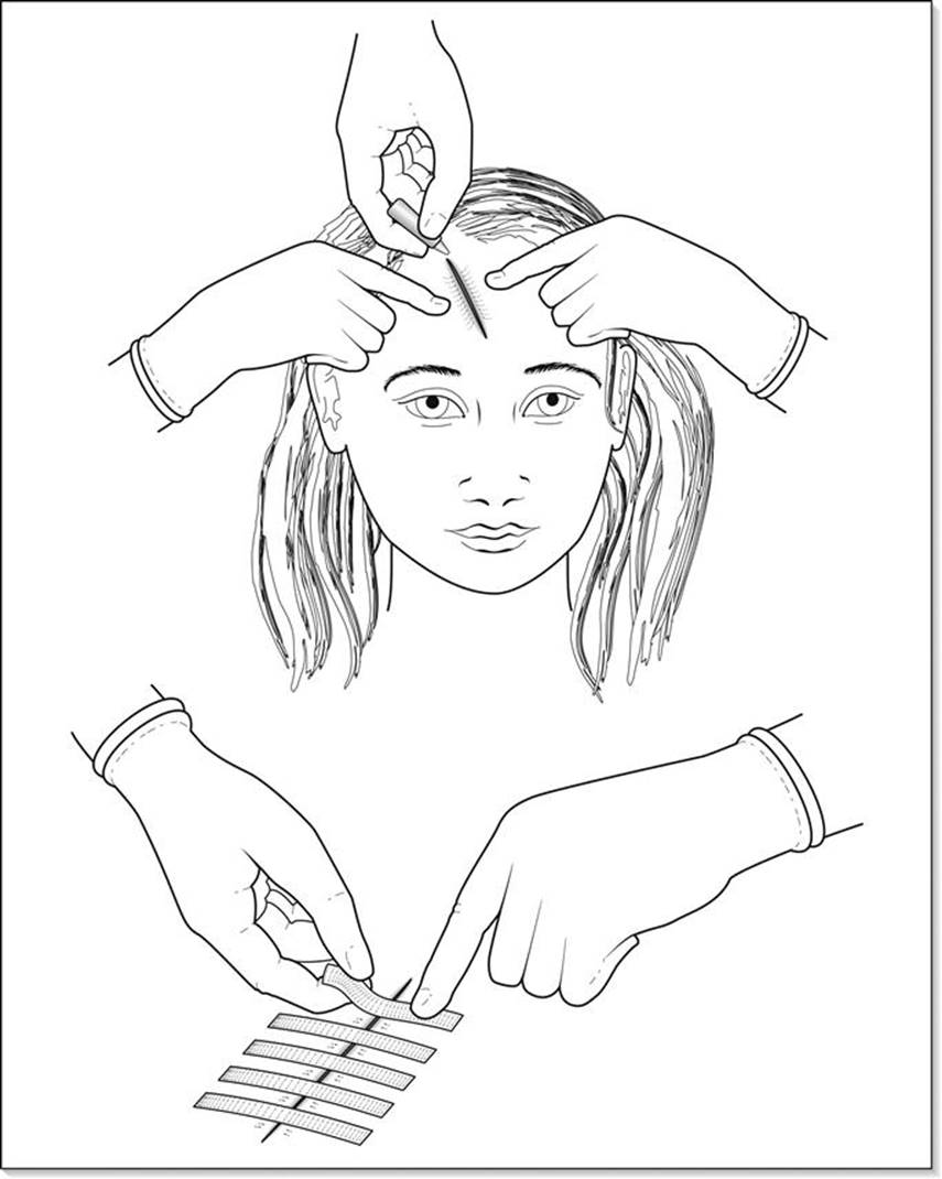
FIG. 3.10.2 Steristrips and glue are typically used for children in most simple split lacerations, thereby avoiding the use of needles. (From an original drawing by Elaine Wheildon.)
Staples have the advantage of rapid insertion and wound closure, particularly for extensive wounds. They are applied using a staple gun and must be removed using the appropriate device, which may be a problem with follow-up arrangements.
From horsehair in World War II [20] to today’s soluble monofilament plastics with prolonged tensile strength, necessity has seen the development of many different suture materials (Fig. 3.10.3) of different grades and using different types of needles. The ideal suture is monofilament, causes no tissue reaction, does not promote infection, is completely absorbed and yet has a tensile strength and secure knots that last until tissue strength has equalled that of the suture. It should stretch to accommodate wound oedema, recoil to its original length and be inexpensive. However, as yet no such suture exists.

FIG. 3.10.3 A simple classification of suture types in current usage. (Adapted from van Winkle W Jr, Hastings JC. Considerations in the choice of suture material for various tissues. Surg Gynecol Obstet 1972; 135:113–26.)
A key factor in choosing absorbable suture is the length of time over which it retains adequate strength. The inflammatory phase of healing lasts for 7 days. Catgut prolongs this phase and is removed by enzymatic action, whereas absorbable plastics simply hydrolyse. Braided sutures produce greater tissue reaction than monofilaments. Braided and catgut sutures should be avoided in contaminated wounds [21]as the interstices provide a haven for bacteria from phagocytes. Traditional absorbable sutures have included Vicryl and Dexon, both braided multifilament. Extensive studies have shown new monofilament absorbable sutures to have superior strength both initially and at 4 weeks: less interference with bacterial clearance; more secure knots requiring fewer throws; and lower drag forces through tissue, compared to the braided absorbable types [22].
Tissue adhesive agents such as Histoacryl (enbucrilate; B. Braun Surgical GmbH) – ‘superglue’ – have been developed particularly with the minor superficial paediatric wound in mind. The results can be excellent, provided good wound edge apposition is achieved prior to application of the glue on the surface (see Fig. 3.10.2).
In the future, biological tissue adhesive agents, such as fibrin sealant [20], for use in the wound may replace sutures as the means of wound closure. As yet these are experimental in sterile, surgically created wounds.
Needles
Early surgical needles had eyes like traditional sewing needles and caused tissue trauma as the bulk of folded-back thread and needle passed through the tissues. The first swaged needles were invented over 100 years ago and modern disposable swaged needles have largely replaced the reusable eyed needles. There are three parts to a needle: the swage, the body and the point (Fig. 3.10.4).

FIG. 3.10.4 Surgical needle characteristics and types. (From an original drawing by Elaine Wheildon.)
Advances in metallurgy have allowed the production of nickel stainless steel wire from which needles are cut. They may be straight or curved in arcs of varying degrees to produce portions of a circle, such as 90°, 135°, 180° and 225° parts. A compound curved needle comprises two different arcs, limiting the amount of supination necessary to pass it through tissue. Skin repair usually requires half-circle needles. The points of surgical needles may be tapered, cutting or a combination. Taper-point needles are generally round or oval bodied and are not suitable for skin as they are difficult to pass through the tightly bundled collagen fibres of the dermis. Their role is in repair of soft tissues, such as fascia, blood vessels and bowel, etc. Cutting needles are for skin and have a triangular point with sharp cutting edges to facilitate tissue penetration. Conventional cutting needles have the apex of the triangle towards the concavity of the curved needle (see Fig. 3.10.4). Reverse cutting needles have the apex on the convexity of the needle. This style of needle and suture will not cut out when the needle is passed through tissue or once the knotted suture is resting against a block of tissue rather than a cut. Such needles are structurally stronger [23]. Combination cutting at the point and taper for the remainder of the body are for slightly denser tissues, such as tendon or aponeurosis. Needle holders are generally used with curved needles and straight needles are handheld. The risk of needle-stick injuries with handheld needles makes their use hazardous.
Basic suture technique
Prior to closure, prophylactic antibiotics (see Chapter 9.9) should be given intravenously if required. This ensures that any haematoma that collects in the wound after or during closure will contain antibiotic.
Having prepared a sterile field with the contents of a suture tray (Table 3.10.5) laid out, the wound anaesthetized and cleaned and the sterile drapes placed around the wound, repair can begin. A very contaminated wound should be anaesthetized, lavaged and cleansed before re-preparing with antiseptic and draping for formal debridement, further lavage and repair.
Table 3.10.5
Surgical instruments required for wound repair
|
Contents of a typical simple suture tray |
Contents of a typical ‘plastics’ suture tray |
|
1×Nelson Hegar needle holder 6.5 in |
2×Mosquito forceps curved |
|
1×Curved artery forceps |
2×Mosquito forceps straight |
|
1×Gillies dissectors |
1×Hegar needle holder 5.5 in |
|
1×McIndoe dissectors |
1×Gillies needle holder |
|
2×Small bowls |
1×Straight Mayo scissors |
|
1×Kidney dish autoplas 255 mm |
1×Curved Mayo scissors |
|
1×Fenestrated drape |
1×Vein straight scissors |
|
1×Huck towel |
1×Vein curved scissors |
|
1×McIndoe dissectors 1×Adson dissectors |
|
|
1×Gillies toothed dissectors |
|
|
2×Skin hooks |
|
|
2×Catspaw refractors |
|
|
1×Bard–Parker handle no. 3 |
|
|
1×Bard–Parker handle no. 4 |
|
|
1×Vein hook Alcot |
|
|
1×Rampley sponge holder |
|
|
3×Gallipots |
|
|
1×Kidney dish |
|
|
3×Towel clips |
|
|
4×Huck towel |
One should choose the thinnest possible suture that will tolerate the tissue tensions and provide adequate strength. The needle holder must grasp the needle in the body, usually two-thirds of the length from the tip of the needle, rather than over the swage where the metal is relatively weak. Stretching the suture in the hands, supporting it at the needle swage, will remove its ‘memory’, making handling easier. The needle holder should be held in the palm of the hand and controlled with the index finger, using a supination/pronation action in the arc of the needle (Fig. 3.10.5). The placement of the first suture varies with the wound: in a small linear wound, it may be convenient simply to suture from one end to the other. In longer wounds without good corresponding landmarks on either side, it is helpful to subdivide the wound serially, to ensure that one does not finish up with a ‘dog-ear’. If an assistant is available, stretching the wound is helpful (Fig. 3.10.5). In more irregular complex wounds, it is helpful to approximate corresponding landmarks first: for example, the apex of a flap is best stitched first (Fig. 3.10.6).
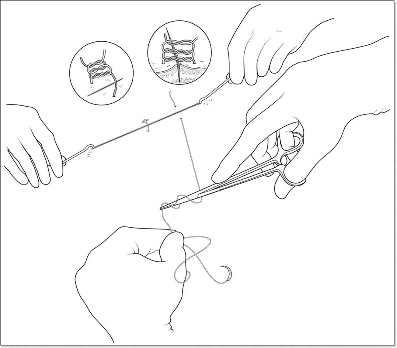
FIG. 3.10.5 The basic technique of how to hold a needle driver, put the wound on the stretch and suture a long wound in halves using surgical knots. For synthetic sutures, the reef knot with the third throw requires several twists, as illustrated, to prevent loosening. (From an original drawing by Elaine Wheildon.)
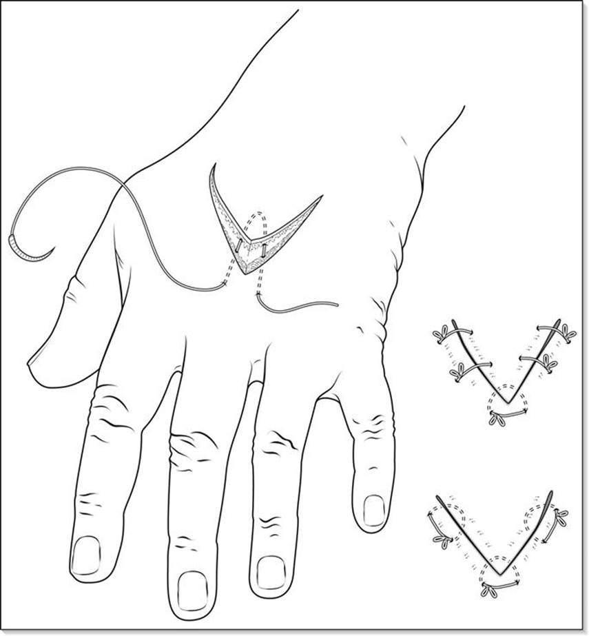
FIG. 3.10.6 Closure of a flap requires an initial suture of the apex, after which either simple or horizontal mattress sutures may be used. (From an original drawing by Elaine Wheildon.)
After wound contraction has occurred, the wound edge has a natural tendency to inversion, resulting in a shallow crater. To prevent this, the edges must be everted at closure. To do this, the skin near the wound edge is depressed (Fig. 3.10.7) or lifted with a skin hook or forceps, so that the needle enters and exits perpendicularly to the skin in both running and interrupted sutures. The sutures so placed may be interrupted with separate tied closed loops or continuous loops passing through tissue, tied at either end. Vertical mattress sutures (Fig. 3.10.8) and horizontal mattress sutures (Fig. 3.10.9) are designed to evert wound edges that are difficult to maintain in eversion with simple sutures.
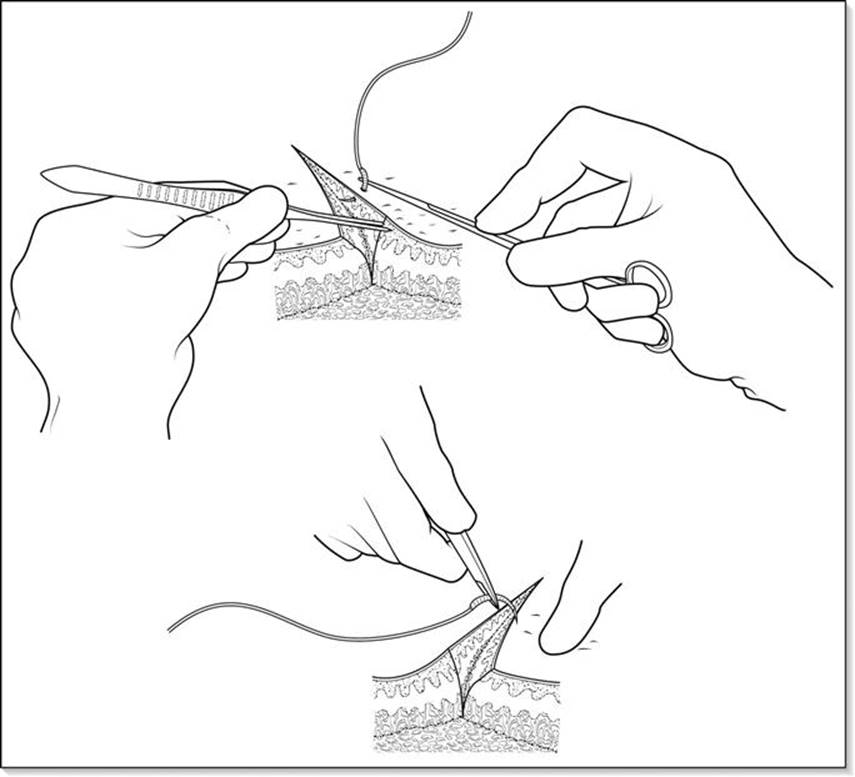
FIG. 3.10.7 Everting the wound edge using Gillies tissue forceps or digital pressure when placing a suture improves the cosmetic result. (From an original drawing by Elaine Wheildon.)
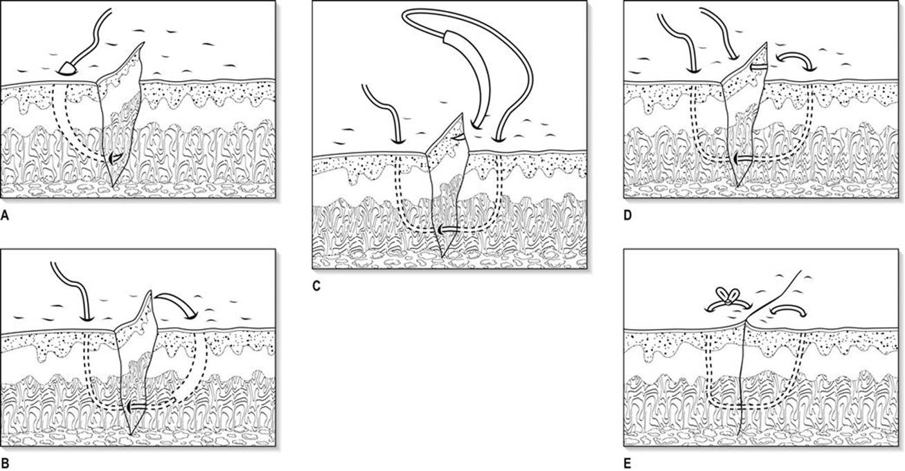
FIG. 3.10.8 (A–E) The vertical mattress suture technique is useful to evert wound edges with a natural tendency to roll inward despite correctly placed simple sutures. (From an original drawing by Elaine Wheildon.)
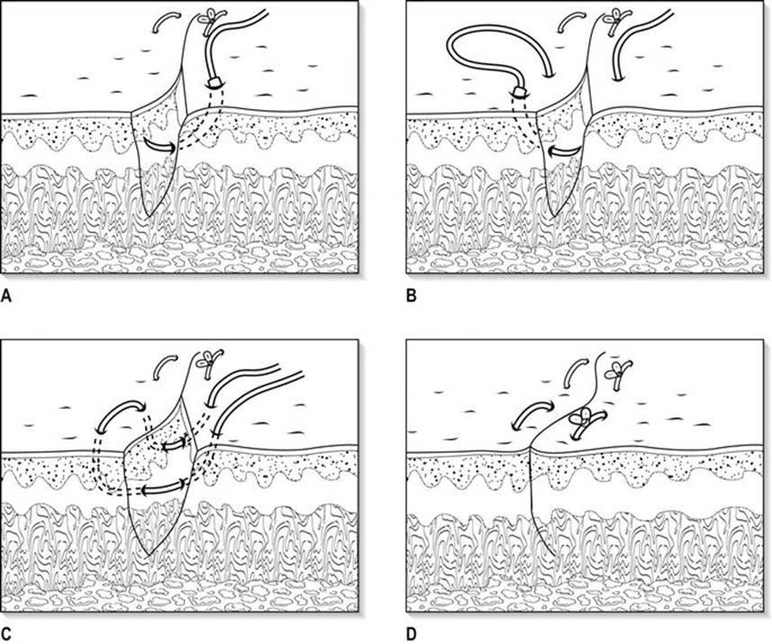
FIG. 3.10.9 (A–D) The horizontal mattress suture redistributes tension and everts wound edges. (From an original drawing by Elaine Wheildon.)
Knots are the weakest link in the suture, particularly for continuous sutures, where the failure of a knot will release the whole suture along the length of the wound. The knots may be tied with instruments or by hand. One must be careful, when using instrument ties, not to damage the suture by either crushing with the serrated jaws of a needle holder or tearing on the edges of the jaws. A reef knot with a snug third throw produces the best results for nylon or polypropylene. Synthetic monofilament sutures require several twists in the first and second throws to prevent unknotting (see Fig. 3.10.5). It is important that the wound be closed without excessive tension on the sutures.
Interrupted sutures have the advantage of individual removal to allow drainage of an infected wound or, for cosmetic reasons, to limit the time a suture stays in while retaining some sutures for wound strength; however, there is a trade-off in the time it takes to close a wound using multiple knots. Sutures tied too tightly, exacerbated by oedema in the wound and from the trauma created by the needle’s passage, will cause suture marks due to local ischaemia on the skin surface. An individual suture that is strangling tissue will continue to do so until it is cut. One way to avoid tissue strangulation is to use a loop throw in an interrupted suture (Fig. 3.10.10) [16].

FIG. 3.10.10 The loop suture method of avoiding excessive tension on a stitch [16]. (From an original drawing by Elaine Wheildon.)
Studies have shown no increase in wound infection or reduction in wound strength with the use of continuous sutures [24], which may be placed rapidly in long linear wounds, distributing tension evenly. However, if one knot fails or the stitch is cut, they will loosen along the length of the wound. Continuous sutures may be percutaneous or intradermal (subcuticular). If intradermal, they should surface every 3 cm to facilitate removal [25].
Intradermal sutures are most appropriate for surgical wounds. Monofilament polypropylene has a very low surface coefficient of friction and is thus easiest to remove in the setting of continuous percutaneous or subcuticular closure [26]. One should ensure that the suture glides easily through each segment and is not looped, otherwise removal may become very difficult. Recently, absorbable monofilament, such as glycolide caprolactone – Monocryl (Ethicon Inc.) – has supplanted polypropylene for continuous subcuticular suture as it does not have to be removed.
Historically, Halstead [27] considered it important to ‘obliterate with the greatest care all of the dead spaces of a wound’. In 1974, it was demonstrated that suture closure of dead space increases the incidence of infection secondary to the foreign body (the suture) in the wound, thereby eliminating the benefits of dead space closure [28]. Some authors [16] stress the importance of using buried sutures to obtain wound edge eversion and dead space closure. Modern hydrolysable monofilament sutures allow this. The long-term maintenance of dermal edge apposition, either with or without deep sutures, is the key to obtaining the narrowest possible scar. Techniques have been developed to encourage this and to avoid leaving buried sutures, with their attendant risk of wound infection. To allow the removal of a deep space-obliterating suture without disrupting the wound some creative methods have been devised (Fig. 3.10.11) [29].

FIG. 3.10.11 A deep closure method utilizing a variable number of loops, adapted from a Mayo Clinic stitch [29]. (From an original drawing by Elaine Wheildon.)
Wounds that slice obliquely through thick skin, such as on the back, can be trimmed with a scalpel blade perpendicular to the skin or sutured with a vertical mattress to prevent one bevelled edge sliding over the other. If necessary to prevent a wound edge step, adjustments in the height of the wound edges can be achieved by exiting the needle superficially on the high side and deeper on the low side, using either continuous or interrupted sutures [16].
Special sites and situations
Scalp lacerations may be closed using the ‘hair braiding’ technique [30,31], either on its own or combined with tissue adhesive. In this technique, four to five strands of hair from opposite sides are brought together, twisted once and tissue adhesive applied.
The face, particularly with dirty wounds, such as bites, requires early repair to achieve good cosmesis. Delay for up to 24 hours is acceptable, prior to definitive debridement and repair in the operating theatre, provided interim wound care is of a good standard. To enable adequate cleansing, local nerve blocks should be used.
A field block is generally required for ears. Ear cartilage must be aligned and skin coverage achieved to prevent perichondritis.
Injuries involving the eyelid need a good examination of the underlying globe to exclude scleral and conjunctival lacerations; also, canaliculi may be torn. A lacerated canaliculus should be microsurgically repaired and stented within 24 hours. Accurate apposition of eyebrows and vermilion border is essential. Never shave an eyebrow.
Damaged facial muscle must be repaired in the interests of facial symmetry. In cheek injuries, the facial nerve and parotid duct must be checked for intactness. The nerves are generally deep in the cheek. Terminal repair of nerves medial to the midpupillary line is unnecessary.
Tattooing should be removed within 12 hours to avoid tissue fixation. Use a sterile brush and magnification and be meticulous. It is useful to have sterile toothbrushes available in the ED for this. After 12 hours, a formal dermabrasion and/or debridement may be needed.
Complications of facial wounds are numerous and provide some special problems (Table 3.10.6).
Table 3.10.6
Complications of facial wounds
|
Complications |
Notes |
|
Infection with the brain |
Potentially fatal owing to the valveless venous communication |
|
AV fistulae |
Due to profuse vascularity – uncommon |
|
Scarring |
Producing facial asymmetry and cosmetic implications |
|
Deformity |
Due to unrecognized fractures, such as of the nose or malar bone |
|
Facial palsy |
Due to damaged facial nerves |
|
Epiphora damage |
With tissue loss or scarring everting the lower lid, or canaliculus |
|
Salivary fistula |
After disruption of the parotid duct |
|
Drooling |
With tissue loss, scar contracture or local nerve damage |
|
Corneal exposure |
With tissue loss, scar contracture or local nerve damage |
Special suture techniques
Techniques for relieving the tension in a wound include limited undermining and the use of horizontal mattress sutures (see Fig. 3.10.9). Very rarely should skin flaps be raised in acute trauma. These may be advancement (e.g. V–Y advancement), rotation or transposition in design. It is usually better to apply a split skin graft to heal the wound primarily and perform later scar revision or reconstruction. In some settings, V–Y flaps can be advanced or retreated, depending on the direction of tension (Fig. 3.10.12).
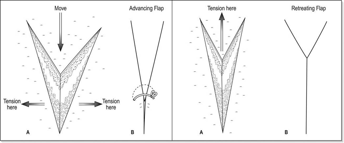
FIG. 3.10.12 The V–Y flap advancement or retreat is useful to redistribute and reduce tension across a wound. (From an original drawing by Elaine Wheildon.)
The ‘dog-ear’
The term ‘dog-ear’ refers to a conical pucker of redundant skin that may collect at the end of a wound towards the end of closure (Fig. 3.10.13), particularly in wounds with an elliptical area of skin defect. In order to avoid a ‘dog-ear’, the wound should be sutured in halves, placing each new stitch between the previous ones (see Fig. 3.10.5). There are several ways to remove a dog-ear [32]:
The direct overlap excision technique involves drawing the redundant skin from one side across the wound and excising along the line of the wound. Any remaining redundant skin is drawn across the wound from the other side and excised along the line of the wound (Fig. 3.10.13).
Unilateral dog-ears are best removed by elevating the redundant skin with a skin hook in the centre, followed by incising along the edge of the fold and then allowing the created flap to fall back along the line of the sutures, where it is trimmed off. This results in a J-shaped repair (Fig. 3.10.13).
An elliptical excision of the dog-ear in line with the closure can excise the defect (Fig. 3.10.13), but this also lengthens the wound.
In very large dog-ears, a V–Y excision and closure will provide good closure.
Thick dog-ears that are aligned perpendicularly to the original closure can be excised and closed in a T repair (Fig. 3.10.13).

FIG. 3.10.13 Various methods of dealing with a dog-ear. (A) Hockey-stick or back-cut technique; (B) double elliptical incision technique; (C) perpendicular elliptical T-repair technique; (D) direct overlap excision technique. (From an original drawing by Elaine Wheildon.)
Wound drainage
Fluid trapped within the closed wound predisposes to infection by:
progressive loss of opsonins
interfering with access of phagocytes to bacteria
providing a nutrient medium for bacterial growth
putting pressure on adjacent vasculature, compromising blood supply.
Fluid also prevents the apposition of healing tissues. The build-up of fluid can be prevented by immobilization, preventing shearing forces between tissue planes, firm but not tight dressings and drainage. The indications for drainage are:
dead-space elimination to prevent fluid accumulation (with an active suction drain or a compressive dressing with a passive drain)
removal of established fluid collections.
Suction drains are superior to passive drains which rely on gravity; however, blockage of drain holes and of the drain tube lumen can be a problem. There are many commercial closed suction systems on the market. A simple suction drain can be constructed from a ‘butterfly’ cannula and a vacuum blood specimen tube (Fig. 3.10.14) [7] by cutting off the syringe adapter and fenestrating the tubing prior to placement through a stab incision into the wound. The vacuum tube can be changed as necessary. Clamp the tube before changing it to prevent the ingress of contaminants into the wound via the drain. Patients with drains will need regular review, either in the ED or by the local doctor. Drains are generally removed at 48 hours unless they are draining copiously.

FIG. 3.10.14 A simple suction drain. (From an original drawing by Elaine Wheildon.)
Dressings
It has long been recognized that the dressing and subsequent wound care are as important as the operative technique [33]. The depths of the wound must be moist for healing, but the skin surface must not become macerated.
The appropriate style of dressing for abrasions is still debated. The ‘moist’ versus ‘dry’ debate revolves around saline packs, sterile paraffin, solugel, seaweed preparations, occlusive plastic film dressings and various foam preparations.
In covering the sutured wound, the dressing aims to keep the primarily apposed skin edges dry, wicking away any ooze, haemorrhage or exudate. It should only be changed if its capacity to absorb fluid is exceeded and, ideally, it should stay on until the time for suture removal. Where this is not possible, the wound may be bathed or showered 24 hours after closure, provided it is thoroughly dabbed dry and not immersed and soaked in water. In the case of scalp wounds, this allows showering and hair washing and avoids the problem of fixing a dressing to hairy skin. Wounds that are contaminated and at high risk of infection need review and re-dressing at 48 hours.
Immobilization
Wounds that traverse joints or which occur on highly mobile skin, such as in the hand, require immobilization. Splinting with plaster slabs is a cheap, traditional and reliable method. Apart from protecting and stabilizing the wound to allow healing, the splint also reduces the likelihood of infection. If practicable, potentially infected wounds of the upper limbs should be in a sling, elevated to reduce oedema. Lower limbs may be rested using crutches and elevated whenever possible.
Disposal/removal
Despite an apparently good cosmetic result at the time of suture removal (5–14 days) (Table 3.10.7), in head and neck wounds, there is evidence of a poor correlation with wound appearance 6–9 months later [34]. The degree to which different factors, such as wounding mechanism, wound repair technique and patient host factors, have a role remains to be determined. Keloid or hypertrophic scarring is more common in negroid and Asian races and in wounds located over the deltoid muscle or sternum.
Table 3.10.7
Guide to time for removal of sutures
|
Location |
Days to removal |
|
Scalp |
6–8 |
|
Face (incl. ear) |
4–5 |
|
Chest/abdomen |
8–10 |
|
Back |
12–14 |
|
Arm/leg* |
8–10 |
|
Hand* |
8–10 |
|
Fingertip |
10–12 |
|
Foot |
12–14 |
*Add 2–3 days for lacerations crossing extensor surfaces of joints and if early motion is required for rehabilitation, e.g. post-flexor tendon repair.
After Gusman D. Wound closure and special suture techniques. J Am Podiatr Med Assoc 1995; 85:2–10.
All percutaneous stitches will cause needle marks if left in situ longer than 8 days, as epithelium migrates down the needle track. Removal too early predisposes to wound dehiscence (Fig. 3.10.15), however, the wound may be supported by skin tapes. If tapes were the primary method of closure they may be left on for at least 10 days or until they fall off, provided the skin is not sensitive to the adhesive, as evidenced by erythema or bulla formation.

FIG. 3.10.15 The relationship between tensile strength and collagen deposition. (From an original drawing by Elaine Wheildon.)
Suture removal technique is also important. To avoid tissue trauma and additional scarring, stitches should be cut at the knots with iris scissors after gentle washing with saline to remove the eschar and the suture gently pulled through. So-called suture scissors are actually too big for the task.
Inelastic paper tape can be used to support a wound and help stop the scar from stretching until such time as the collagen is near maturation, beyond 3 months. Paper tape is also useful in the setting of keloid scarring in an attempt to provide pressure and encourage remodelling. In some cases, silicone gel pads and even pressure garments are required to control keloid scarring.
If the wound suppurates then the sutures will need to be removed, either partly or completely, to allow the egress of pus.
Likely developments over the next 5 to 10 years
There is a lot of work being done on knotless wound closure devices which will speed up suturing by eliminating the need for knot tying.
With surgical wounds, there are new developments with absorbable staples and fast setting cyanoacrylate.
Controversies
Drainage will remove fluid and haematoma that potentiate infection, but the drain itself may predispose to infection. This is less the case with suction drains.
Interrupted dermal sutures will close dead space, thereby reducing haematoma and wound infection, but may lead to infection in contaminated wounds. Their major role is to reduce skin tension and they should be used in large clean wounds.
The degree of debridement required for a dirty wound has moved from radical to conservative but meticulous, with an emphasis on preservation of viable skin to improve cosmesis.
Povidone–iodine packs, which are tissue toxic, are used by some surgeons in the setting of open wounds over compound fractures while the patient awaits transfer to the operating theatre for definitive repair.
Opinions as to the appropriate dressings for abrasions range from moist, such as plastic film, to dry, such as mercurochrome paint and dry gauze.
References
1. Clark B, Cassell E, Ashby K, et al. Hazard, edn no. 52, Spring. Victorian Injury Surveillance and Applied Research System; 2002.
2. Jorgensen LN, Kallenhave F, Christensen E, Siana JE. Less collagen production in smokers. Surgery. 1998;123:450–455.
3. Ellis H. Hazards from surgical gloves. Ann Roy Coll Surg Engl. 1997;79:161–163.
4. Howell JM, Morgan JA. Scalp laceration repair without prior hair removal. Am J Emerg Med. 1988;6:7.
5. Dire DJ, Welch AP. A comparison of wound irrigation solution used in the Emergency Department. Ann Emerg Med. 1996;19:704.
6. Bansal BC, Weike RA, Perkins SD, Abramo TJ. Tap water irrigation of lacerations. Am J Emerg Med. 2002;20:469.
7. Rodeheaver GT, Pettry D, Thacker JG, et al. Wound cleansing by high pressure irrigation. Surg Gynecol Obstetr. 1975;141:357–362.
8. Brown LL. Evaluation of wound irrigation by pulsatile jet and conventional methods. Ann Surg. 1978;187:170.
9. Gfeller RW, Crow DT. The emergency care of traumatic wounds: current recommendations. Vet Clin N Am. 1994;24:1249–1274.
10. Wheeler CB, Rodheaver GT, Tracker JG, et al. Side-effects of high pressure irrigation. Surg Gynecol Obstet. 1976;143:775–778.
11. Lammers R. Foreign bodies in wounds. In: Singer AJ, Hollander JE, eds. Lacerations and acute wounds: an evidence-based guide. Philadelphia: FA Davis; 2003;147.
12. Wyn T, Jones J, McNinch D, et al. Bedside fluoroscopy for the detection of foreign bodies. Acad Emerg Med. 1995;2:979–982.
13. Fackler MI, Breteau JP, Courbil CJ, et al. Open wound drainage versus wound excision in treating the modern assault rifle wound. Surgery. 1989;105:576–584.
14. Spicer WJ, Garland S, Christiansen K, et al. Skin and soft tissue infection: therapeutic guidelines antibiotic version 13. Melbourne: Therapeutic Guidelines Limited; 2006; 230–232.
15. National Health and Medical Research Council. The Australian Immunisation Handbook 9th ed. Canberra: NHMRC; 2008.
16. Moy RL, Lee A, Zalka A. Commonly used suturing techniques in skin surgery. Am Fam Phys. 1991;44:1625–1634.
17. Moy RL, Quan MB. An evaluation of wound closure tapes. J Dermatol Surg Oncol. 1990;16:721–723.
18. Panek P, Prusak MP, Bolt D. Potentiation of wound infection by adhesive adjuncts. Am Surg. 1972;38:343–345.
19. Edlich RF, Becker DG, Thacker JG, Rodeheaver GT. Scientific basis for selecting staple and tape skin closures. Clin Plast Surg. 1990;17:571–578.
20. Spotnitz WD, Falstrom MA, Rodeheaver GT. The role of sutures and fibrin sealant in wound healing. Surg Clin N Am. 1997;77:651–669.
21. Van Winkle W Jr Hastings. JC Considerations in the choice of suture material for various tissues. Surg Gynecol Obstet. 1972;135:113–126.
22. Rodeheaver GT, Beltran KA, Green CW, et al. Biomechanical and clinical performance of a new synthetic monofilament absorbable suture. J Long-Term Effects Med Implants. 1996;6:181–198.
23. Bendel LP, Trozzo LP. Tensile and bend relationships of several surgical needle materials. J Appl Biomat. 1993;4:161–167.
24. Mclean NR, Fyfe AH, Flint EF, et al. Comparison of skin closure using continuous and interrupted nylon sutures. Br J Surg. 1980;67:633–635.
25. Drake DB, Gear AL, Mazzarese PM, et al. Search for a scientific basis for continuous suture closure: a 30 year odyssey. J Emerg Med. 1997;15:495–504.
26. Pham S, Rodeheaver GT, Dang MC, et al. Ease of continuous dermal suture removal. J Emerg Med. 1990;8:539–543.
27. Halstead WS. The treatment of wounds with especial reference to the value of blood clot in the management of dead spaces. Bull Johns Hopkins Hosp. 1990–91;2:255.
28. De Holl D. Potentiation of infection by suture closure of dead space. Am J Surg. 1974;127:716–720.
29. Arnold PG. Space obliterating skin suture. Plast Reconstruct Surg. 1997;100:1506–1508.
30. Aoki N, Oikawa A, Sakai T. Hair braiding closure for superficial wounds. Surg Neurol. 1996;46:150.
31. Hock M, Ooi SBS, Saw SM, Lim SHA. randomised controlled trial comparing the hair apposition technique with tissue glue to standard suturing in scalp lacerations (HAT) study. Ann Emerg Med. 2002;40:19.
32. Gusman D. Wound closure and special suture techniques. J Am Podiatr Med Assoc. 1995;85:2–10.
33. Ivy RH, et al. Manual of standard practice of plastic and maxillofacial surgery Philadelphia: WB Saunders; 1943.
34. Hollander JE, Blasko B. Poor correlation of short and long-term cosmetic appearance of lacerations. Acad Emerg Med. 1995;2:983–987.
3.11 Burns
Tim Gray, Gerard O’Reilly and Biswadev Mitra
Essentials
1 Effective triage and advances in the treatment of severely burned patients, including fluid resuscitation, control of sepsis, early excision and use of skin substitutes have made previously lethal burns survivable.
2 Signs of impending or developed laryngeal oedema should prompt early intubation.
3 Burn resuscitation formulae should be considered as a guide only.
4 Meta-analysis of previous studies, suggests that resuscitation with colloid, as opposed to crystalloid, does not improve survival.
5 Extensive or complicated burns should be managed in a specialized burns unit.
6 Chemical burns, after decontamination and specific antidotes, are treated in a similar fashion to thermal burns.
Introduction
Advances in burn management over the last three decades have significantly reduced mortality and improved quality of life for victims. Appropriate fluid resuscitation, early debridement and the appropriate use of antibiotics have resulted in hypovolaemia and sepsis no longer being the major contributors to mortality in burns. Multiorgan failure is the most likely event leading to death, whereas age, burn surface area and inhalational injury are the major contributors to a poor outcome [1,3].
Pathophysiology
The skin is the largest organ of the body. Its most important functions are:
to act as a vapour barrier to prevent water loss from the body
to present the body’s major barrier against infection
temperature regulation.
The skin consists of two main layers. The epidermis is stratified squamous epithelium that acts as the major barrier to passive water loss from the body. The dermis contains the adnexal structures, namely sweat glands, hair follicles and sebaceous glands, as well as pain and pressure receptors and the cutaneous blood vessels, which play a major role in temperature regulation by controlling radiant heat loss (Fig. 3.11.1).
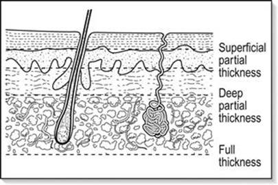
FIG. 3.11.1 Diagram of skin layers.
The adnexae are embryologic down-growths of the epidermis. Following burn injury, the epithelial cells of these structures undergo metaplastic change to stratified squamous epithelium, proliferate and gradually cover the wound. Thus, burns that partially or completely spare these structures will usually heal without scarring. Deeper burns involve greater loss of adnexal cells, resulting in poorer epithelial coverage and hence greater scarring.
Burned skin undergoes coagulative necrosis with three distinct zones of injury. A central zone of coagulation, in which irreversible cell death occurs, is surrounded by a zone of stasis, in which vasoconstriction and intravascular coagulation contribute to local ischaemia. A zone of hyperaemia surrounds the wound. In the early stages of the burn, evolution of these zones results in a progressive deepening of the wound, which may be minimized by appropriate early treatment [4,5].
Classification
Burns may be classified according to their depth as superficial, partial thickness or full thickness.
Superficial burns involve only the epidermis. Pain and swelling usually subside within 48 hours and the superficial epidermis peels off within a few days. Healing occurs by proliferation of undamaged cells of the germinal layer of the epidermis and is usually complete within 7 days.
Partial thickness burns involve destruction of the epidermis and superficial dermis. They are characterized by blister formation and may be further classified into superficial and deep partial thickness. Healing is dependent on the amount of intact epithelium in the adnexae.
Superficial partial thickness burns are typically bright red with a moist surface, are exquisitely sensitive to stimulus and heal in 2–3 weeks, generally with minimal scarring. Deep partial-thickness burns are typically dark red or yellow-white and take longer than 3 weeks to heal, as few epithelial elements survive. Hypertrophic scarring usually occurs.
Full-thickness burns involve the epidermis and dermis, including the epidermal appendages. Clinically, they appear charred or pearly white in appearance and are usually insensate. Because loss of epidermal adnexae is complete, full-thickness burns only heal by scarring or skin grafting.
Thermal burns
Presentation
History
History may be obtained from the patient, from witnesses and from fire or ambulance personnel. Details of the nature of the injury are important, especially the nature of the burning materials, duration of exposure, whether the patient was trapped in an enclosed space or lost consciousness or whether there was an associated fall, vehicular accident or blast injury.
A history of altered consciousness or confinement in a burning environment suggests the likelihood of carbon-monoxide poisoning. Past medical history, current medications, allergies and tetanus status should also be obtained.
Examination
The initial examination should be directed to identifying signs suggestive of airway burns as well as the presence of other injuries. Early haemodynamic compromise is rarely due to burn injury alone and should prompt a search for other causes.
Facial and oral burns, singed nasal hairs, carbonaceous sputum, tachypnoea and wheeze are clinical signs suggesting an increased risk of inhalation injury; however, in the absence of laryngeal oedema, inhalation injury may not become clinically evident for 12–24 hours [6,7].
Signs of laryngeal oedema, namely hoarseness, brassy cough or stridor, indicate the need for early endotracheal intubation as oedema formation may rapidly distort the anatomy, necessitating a surgical airway.
The adequacy of peripheral circulation should be assessed, particularly in the setting of circumferential limb burns.
Evaluation of burn area
The extent and depth of the burn must be assessed as accurately as possible. Representation of the burn area diagrammatically on a body chart aids assessment. The simplest method is the ‘Rule of nines’ where the adult body is divided into anatomical regions that represent 9% of the total body surface area.
In infants and young children, the Lund and Browder chart is used to correct for proportional variation at different ages: for instance, an infant’s head is approximately 18% of the total body surface area, compared to 9% in an adult (Fig. 3.11.2).

FIG. 3.11.2 (A) ‘Rule of nines’ diagram. (B) Lund and Browder chart.
Management
Pre-hospital
Pre-hospital care of the burned patient should be directed at stopping the burning process, assessing and stabilizing the airway, breathing and circulation and rapidly transferring the patient to hospital. Where possible, major burns should be triaged to a burn centre (Table 3.11.1) [8].
Table 3.11.1
Patients who fulfil the following criteria should be considered for transfer to a specialist burns unit
Partial-thickness burns>20% in all age groups, or>10% in the under 10 and over 50 age groups
Full-thickness burns>5% in any age group
Burns involving face, eyes, ears, hands, feet, genitalia, perineum or a major joint
Inhalation burns
Electrical burns, including lightning injury
Burns associated with other significant injuries
Smaller burns in patients with pre-existing disease that could complicate management
En route to hospital, recent burns should be covered with a clean dressing (e.g. Melolin), soaked in cool water or tea-tree/hydrogel dressing so as to limit the depth of burn by dissipation of heat. After this cooling process, or if it is not required, the burns can be covered with a clean dry dressing. Cling wrap is used for interhospital transfers. Prolonged exposure to cool water should be avoided and ice should never be applied directly to the wound as it may increase the depth of burn.
The patient should be kept warm, supplemental oxygen administered and, where prolonged transport times are anticipated, intravenous fluid therapy should be instituted.
Emergency department
Initial management
Supplemental oxygen should be administered and cardiac and oxygen saturation monitoring instituted.
Stabilization of the airway and treatment of life-threatening injuries take priority over management of the burn wound itself. Burns to the face, neck and airways result in massive fluid shifts from the circulating plasma into the interstitial space causing oedema and potentially resulting in rapid upper airway obstruction. Immediate intubation is indicated in apnoea or obstruction. Other indications for immediate intubation are the same as for any critically injured patients, including actual or impending cardiac arrest, impending respiratory failure and a decreased level of consciousness with inability to protect the airway.
Early intubation is indicated in burns to the face and neck where it is anticipated that oedema will make intubation difficult in the future or to facilitate initial pain and operative management. The nasal route is preferable in the presence of friable burnt oral tissue but requires a spontaneously breathing patient. The choice of an inducing agent is varied with thiopentone or propofol appropriate in haemodynamically stable patients. Succinylcholine may be used in the first 24 hours but, in later periods, has been associated with catastrophic potassium release leading to possible cardiac arrest and is best avoided [9]. Non-depolarizing muscle relaxants are preferred; however, the dose required may increase to 3–5 times normal. Uncut endotracheal tubes should be used and meticulously secured [10].
Airway patency alone does not guarantee adequate ventilation. Fumes and minute particles produced by the fire may cause bronchospasm, which can be treated by inhaled bronchodilators. Smoke particles also cause inflammation, hypersecretion and mucosal sloughing resulting in airway obstruction and atelectasis. Carbon monoxide (CO) poisoning should be suspected in anyone with a history of smoke exposure, an extended length of time in the fire or burns in an enclosed space. The features of CO poisoning include tachypnoea, tachycardia, vomiting, confusion and irritability to reduced conscious states and syncope. Oxygen saturation measured by pulse oximetry is not helpful in diagnosing CO toxicity, although recently developed oximetry devices display carboxyhaemoglobin levels. Blood gases and carboxyhaemoglobin levels aid in diagnosis. An admission carboxyhaemoglobin level>15% suggests significant smoke inhalation [4]. The affinity of CO for haemoglobin is substantially more than oxygen with a slow dissociation half-life of 3–4 hours. Administration of high concentrations of O2 can decrease the dissociation half-life to 50 minutes.
Cyanide poisoning can result from thermal decomposition of natural fibres, such as wool and silk, and manifests as persistent lactic acidosis with a high anion gap. Commercially available cyanide antidote kits contain amyl nitrite vials, sodium nitrite and sodium thiosulphate. The amyl nitrite is administered via inhalation and is intended for use when intravenous access may not be available. Sodium nitrite is given intravenously. The second stage of antidote therapy is administration of sodium thiosulphate, which acts synergistically to counteract cyanide formation [11,12].
Ventilation difficulties through parenchymal damage from burns share a similar pathophysiology to the development of acute respiratory distress syndrome (ARDS). The onset is generally slow and may be an issue in delayed presentations. An exception is blast injury, which can cause lung contusions and alveolar trauma leading to ARDS. The most effective initial treatment is the application of positive end expiration pressure (PEEP). The mechanism by which PEEP improves oxygenation is linked to the increase in residual functional capacity (RFC) obtained both by increasing the volume of partially collapsed alveoli and by reopening totally collapsed alveoli.
Fluid Resuscitation [7]
Intravenous fluid resuscitation with crystalloid solution as outlined below should be started in any patient with burns of more than 20% total body surface area (TBSA). There are many formulae for the fluid resuscitation of burns victims. Although considerable debate continues, the general principles of fluid resuscitation are:
In the first 24 hours, isotonic salt solution should be used to replace the large volumes lost to tissue oedema, with about half the fluid given in the first 8 hours after injury, coincident with the period of most rapid oedema formation.
The administration of colloid is unnecessary for patients with burns of less than 40% TBSA and during the first 8 hours. Meta-analysis of previous studies suggests that resuscitation with colloid does not result in improved survival [13].
Fluid resuscitation formulae are a guide only and the patient’s haemodynamic status must be monitored by cardiovascular parameters and hourly urine volume measurement. Increased resuscitation fluid volume (per kg) is required in children weighing less than 30 kg, in high-voltage electrical injuries, if resuscitation is delayed and in the presence of inhalation injury.
The initial goal of fluid resuscitation is the restoration of cardiac output and tissue perfusion. Multiple formulae estimating the initial fluid requirement in patients with major burns have been proposed including the Brooke, modified Brooke, Parkland, Evans, MGH and Monafo regimens [14,15]. These formulae use the weight of the patient and the percentage of body surface area burnt to estimate fluid requirements. The percentage of body surface area burnt can be easily estimated using a Lund and Browder chart. Weight should be measured if possible or estimated from collateral history. The Parkland formula allows for 4 mL/kg/% TBSA burned over 24 hours, with half the total fluid requirement to be given in the first 8 hours. In children under 30 kg, the above fluid should be given in addition to calculated maintenance fluid [1].
Patients generally receive more fluid than the Parkland’s formula dictates and this appears to be associated with better outcomes than historical data [16,17]. The proposed formula of (weight in kg×%TBSA) mL of fluid in the first 2 hours post-burns injury is approximately double that estimated by the Parkland formula and leads to maintenance of resuscitation endpoints without obvious adverse events [18]. This strategy has been currently adopted in the pre-hospital phase and needs to be followed by goal-directed management to maintain adequate resuscitation. Haemodynamic status may be difficult to evaluate in the severely burned patient. In patients with severe burns, the insertion of a central venous catheter allows the measurements of preload, tissue oxygenation via central venous oxygen levels, contractility and coronary perfusion pressure and may facilitate the measurement of afterload. Additional measures of tissue oxygenation are obtained by serial serum lactate measurements and monitoring urine output via an indwelling catheter. Inserting a urinary catheter with an electronic temperature probe will assist in monitoring core temperature.
Some strategies have been proposed to reduce the total volume of resuscitation fluid. These have included the use of colloid and hypertonic salt solutions. The generalized increase in capillary permeability that occurs with major burns results in the loss of plasma protein, particularly albumin, from the circulation. There is a coincident reduction in hepatic albumin production post-burn. Colloid administration helps maintain oncotic pressure, but does not reduce tissue oedema in the first 8 hours and has not been shown to improve clinical outcome compared to crystalloid [19,20]. There is currently inadequate evidence to support the routine use of colloids for burns resuscitation.
Hypertonic saline solution would appear to be useful in patients with limited cardiopulmonary reserve; however, there is considerable debate over the safety of this technique and, again, there is no evidence of outcome benefit compared to isotonic solution [6,7].
Subsequent management
Having stabilized the patient and initiated fluid resuscitation, a careful secondary survey should be performed, looking for associated injuries.
Adequate analgesia is an important facet of management. Small burns may be managed with a combination of cool compresses and oral analgesia; larger burns will require parenteral analgesia [8]. Opiates will generally be the first option; an infusion of ketamine can be useful for continuous analgesia where there are extensive burns.
Burns patients lose heat quickly. Wrapping the patient in blankets, foil or external warming devices may prevent hypothermia.
Simple initial management of the wound with cling-wrap is adequate in providing analgesia, protection from the environment and allowing visualization of pathological appearance (E-Fig. 3.11.1). Early definitive surgical therapy with excision and autologous skin grafting is currently widely practised for deep burns and constricting eschars [7,21,22]. Tangential escharectomies remove much of the necrotic tissue while leaving behind healthy tissue. Peripheries with circumferential burns are at a high risk of ischaemic necrosis and warrant urgent escharotomy. Such procedures can be undertaken in the emergency department where pulses are absent. In circumferential chest burns, escharotomy may be necessary to relieve chest wall restriction and improve ventilation. Routine fluorescein staining and examination of the eye in cases of facial burns result in early diagnosis and treatment of corneal burns.

E-FIG. 3.11.1 Simple pre-hospital dressing of major burns.
Tetanus following burn injury has been previously reported [23,24]. A tetanus vaccine booster is recommended for all patients with no history of tetanus vaccination in the last 10 years. Tetanus immunoglobulin should be considered in wounds grossly contaminated with soil. Systemic prophylactic antibiotics are not indicated. While there have been some suggestions of antibiotics to prevent toxic shock syndrome, its incidence is low and does not warrant routine prophylaxis for all burnt patients [25]. Antibiotics should be considered in the immunocompromised on an individual basis.
There is strong evidence for the use of histamine2-receptor antagonists for stress ulcer prophylaxis and they are recommended in all patients with major burns [26].
A nasogastric tube should be inserted in patients with major burns to avoid gastric dilatation.
Burn shock
The pathophysiology of burn shock is complex and involves a combination of haemodynamic and local tissue factors.
The early post-burn period, i.e. within the first 8 hours, is marked by the rapid formation of tissue oedema, predominantly in the wound itself, but also in non-burned tissue. Factors contributing to this fluid accumulation are not fully understood, but include local release of inflammatory mediators, particularly prostaglandins and leukotrienes. These increase capillary permeability both locally and systemically, in addition to increasing regional blood flow. Increased interstitial osmotic pressure in burned tissue due to the release of osmotically active cellular components and partial degradation of collagen also contributes to tissue oedema [4,7,22]. The combination of tissue injury, shock and dilution of coagulation factors through exogenous fluid administration leads to coagulopathy in about a third of major burns patients [27].
Major evaporative loss from burned skin due to loss of epithelial integrity significantly adds to fluid losses. In addition to the fluid shifts, cardiac output may fall by 30–50% in major burns, possibly due to a circulating myocardial depressant factor [4].
Inhalation injury
The presence of inhalation injury has a considerable negative impact on prognosis in the burns patient [3]. Direct thermal trauma below the larynx is rare, except in the case of steam inhalation.
Pulmonary complications are largely due to inhalation of toxic products of combustion, particularly in house or vehicular fires. Smoke consists of a particulate fraction – predominantly carbon – and a gaseous fraction, which may include carbon dioxide, carbon monoxide, oxides of nitrogen and sulphur, hydrogen cyanide and PVC, depending on the materials being burnt. These agents adhere to the moist respiratory mucosa, forming corrosive compounds that cause inflammation, hypersecretion and mucosal sloughing, resulting in airway obstruction and atelectasis. Smoke inhalation also triggers the release of thromboxane, resulting in increased pulmonary artery pressures [3,6].
Disposition
Patients with major burns should be managed in a specialist burns unit as outlined in Table 3.11.1. The patient should be discussed with the receiving unit prior to transfer, so that appropriate measures may be undertaken to stabilize them. Plastic cling wrap applied directly over the burn provides a good non-adherent dressing that will reduce heat and fluid loss. As noted previously, silver sulphadiazine (SSD) cream should not be applied to these burns as it interferes with subsequent evaluation. At the burns centre, burns will be dressed with a silver-impregnated, non-adherent, occlusive dressing (e.g. Acticoat).
Current management of full- and deep partial-thickness burns involves early excision and autologous skin grafting. In extensive burns, excision and autologous grafting may need to be staged, allowing sufficient skin to regenerate.
Less extensive burns (i.e. not meeting the criteria for burns centre transfer) may be admitted to a general or plastic surgery service. Loose skin and broken blisters should always be debrided. Blisters may otherwise be initially left intact, although some would advocate that all blisters be deroofed. A moist silver-impregnated dressing (e.g. Acticoat) may then be applied to areas of epithelial loss.
Superficial or partial-thickness burns involving less than 10% TBSA may be suitable for outpatient management subject to the criteria in Table 3.11.1 and depending on the social and psychological status of the patient. The choice of dressings for outpatient management depends on the depth of the burn, the extent and size of blisters and the amount of exudate from the burn surface [4,7].
Superficial burns can also be covered with a moist ointment (e.g. paraffin-based Dermeze) or moist dressing (e.g. Burnaid, which contains melaleuca-derived local anaesthetic properties). Superficial partial-thickness burns involve loss of epithelium with considerable exudate and hence are prone to infection. After gentle cleansing and debridement of loose tissue, a moist, non-adherent dressing, such as Bactigras, should be applied. These patients will need to be reviewed the next day, at which time the dressing will be changed. A silver-impregnated occlusive dressing is also an option to minimize crusting and dressing adherence.
Epithelialization commences at 7–10 days, by which time the burn surface should be drying out. At this stage, the more convenient hydrocolloid or film dressings may be used until epithelialization is complete.
If healing is not well established by 10–14 days, the patient should be referred for specialist opinion, as excision and grafting may be required.
Chemical burns
A wide range of products available in both the industrial and domestic environments can lead to burns. Although the mechanism is different, chemical burns demonstrate a similar spectrum of injury to thermal burns. Superficial burns are associated with itching, burning or pain; partial-thickness burns are associated with tissue oedema and the formation of bullae; and full-thickness burns are associated with damage extending through the dermis. The extent of tissue damage in chemical burns is determined by the nature and concentration of the chemical, as well as the extent and duration of contact [28].
In addition to the burn itself, toxicity may occur as a result of systemic absorption.
The majority of chemical burns are caused by acids and alkalis. Acids cause coagulation, with the formation of a tough eschar that may limit further tissue damage. Alkalis cause liquefactive necrosis, allowing deeper penetration. Many other types of chemical cause burns, but distinguishing them by mechanism of action is not relevant to the clinician as their management, apart from a few exceptions, is similar.
General principles
Chemical agents continue to damage tissue until they are removed or inactivated. Therapy then is directed to decontamination and, where appropriate, the use of specific antidotes as well as recognition and treatment of systemic toxicity.
Adequate protection of medical personnel to prevent secondary contamination is essential. Copious irrigation is the cornerstone of therapy, but contaminated garments should be removed and dry chemical particles brushed away before irrigation commences. Adherent or oily compounds may need to be removed with mild soap and a scrubbing brush and nails, hair and intertriginous areas should be carefully checked.
The duration of irrigation depends on the agent. Alkali in particular may require prolonged lavage owing to its tissue penetration. The use of litmus paper to determine wound pH may guide the duration of irrigation in acid and alkali burns.
Other than decontamination and treatment of systemic toxicity, management is similar to that for thermal burns.
Disposition
Most patients with chemical burns can be treated on an outpatient basis. Indications for admission include:
partial-thickness burns>15% TBSA
all full-thickness burns
burns involving hands, feet, eyes, ears or perineum
evidence of or potential for systemic toxicity
significant associated injuries or complicating medical conditions.
Specific chemicals
Hydrofluoric acid
Hydrofluoric acid is a relatively weak acid used in glass etching, electronics and oil-refining industries. It is also a component of many industrial and domestic rust removers. In strong solution it causes corrosion of tissue owing to the release of hydrogen ions; however, its major toxicity is caused by the dissociated fluoride ion that complexes calcium and magnesium to form insoluble salts. Cell destruction associated with severe pain results. In severe burns, hypocalcaemia and hypomagnesaemia may occur [28].
Contact with strong solution (>50%) causes immediate pain and tissue destruction; however, exposure to weaker solutions, particularly<20%, may cause little or no pain initially. Thus it may take up to 24 hours for the burn to become apparent. Once apparent, the burn causes excruciating pain that is difficult to control even with parenteral narcotics.
After irrigation, specific therapy is aimed at precipitation and hence neutralization of free fluoride ions. Methods depend on the severity and location of the burn. Calcium gluconate gel, made by mixing calcium gluconate with a water-soluble lubricant to make a 2.5–10% solution, should be applied directly to the affected area.
Relief of pain is the marker of adequate treatment. If pain is not relieved, or recurs, parenteral therapy is required. Generally, this consists of subcutaneous injection of calcium gluconate, aiming for 0.5 mL of 10% solution per square centimetre. Hand and digital burns pose a problem, as vascular compromise may occur if too much fluid is injected. Alternatives include intra-arterial injection of calcium gluconate or regional perfusion using Bier’s technique.
References
1. Wolf S, Rose J, Desai M, et al. Mortality determinants in massive paediatric burns. Ann Surg. 1997;225:554–569.
2. Miller S, Bessey P, Schurr M, et al. National Burn Repository 2005: a ten-year review. J Burn Care Res. 2006;27:411–436.
3. Fraser J, Mullany D, Traber D. Inhalational lung injury in patients with severe thermal burns. Contemp Crit Care. 2007;4:1–12.
4. Shaw A, Anderson J, Hayward A, Parkhouse N. Pathophysiological basis of burn management. Br J Hosp Med. 1994;52:583–587.
5. Singh V, Dengan L, Bhat S, Milner S. The pathogenesis of burn wound conversion. Ann Plast Surg. 2007;59:109–115.
6. Nguyen T, Gilpin D, Meyer N, Herndon D. Current treatment of severely burned patients. Ann Surg. 1996;233:14–25.
7. Monafo W. Initial management of burns. N Engl J Med. 1996;335:1581–1586.
8. Reed J, Pomerantz W. Emergency management of paediatric burns. Paediatr Emerg Care. 2005;21:118–129.
9. Jeevendra M. Succinylcholine hyperkalaemia after burns. Anaesthesiology. 1999;91:321–322.
10. Gillies M, Krone S, Sim K. Use of cut endotracheal tubes should be avoided in the initial resuscitation of the burned patient. Emerg Med J. 2003;20:109.
11. Peddy S, Rigby M, Shaffner D. Acute cyanide poisoning. Pediatr Crit Care Med. 2006;7:79–82.
12. Megarbane B, Delahaye A, Goldgran-Toledano D, Baud F. Antidotal treatment of cyanide poisoning. J Chin Med Assoc. 2003;66:193–203.
13. Roberts I, Alderson P, Bunn F, et al. Colloids versus crystalloids for fluid resuscitation in critically ill patients. Cochrane Database Syst Rev. 2004;18:CD000567.
14. Yowler C, Fratianne R. Current status of burn resuscitation. Clin Plast Surg. 2000;27:1–10.
15. Warden G. Burns shock resuscitation. World J Surg. 1992;16:16–23.
16. Mitra B, Fitzgerald M, Cameron P, Cleland H. Fluid resuscitation in major burns. Aust NZ J Surg. 2006;76:35–38.
17. Freiburg C, Igneri P, Sartorelli K, Rogers F. Effects of differences in percent total body surface area estimation on fluid resuscitation of transferred burn patients. J Burn Care Res. 2007;28:42–48.
18. Mitra B, Fitzgerald M, Wasiak J, et al. The Alfred pre-hospital fluid formula for major burns. Burns. 2011;37:1134–1139.
19. Ipaktchi K, Arbabi S. Advances in burn critical care. Crit Care Med. 2006;34:239–244.
20. Alderson P, Bunn F, Lefebvre C, et al. Human albumin solution for resuscitation and volume expansion in critically ill patients. Cochrane Database Syst Rev. 2004;18:CD001208.
21. McManus W, Mason A, Pruitt B. Excision of the burn wound in patients with large burns. Arch Surg. 1989;124:718–720.
22. Demling R. Improved survival after massive burns. J Trauma. 1983;23:179–184.
23. Karyoute S, Badran I. Tetanus following a burn injury. Burns. 1988;14:241–243.
24. Amy B, McManus W, Pruitt B. Tetanus following a major thermal injury. J Trauma. 1985;25:654–655.
25. Rashid A, Brown A, Khan K. On the use of prophylactic antibiotics in prevention of toxic shock syndrome. Burns. 2005;31:981–985.
26. Cook D, Reeve B, Guyatt G, et al. Stress ulcer prophylaxis in critically ill patients Resolving discordant meta-analyses. J Am Med Assoc. 1996;275:308–314.
27. Mitra B, Wasiak J, Cameron P, et al. Early coagulopathy of major burns. Injury. 2013;44:40–43.
28. Bretolini J. Hydrofluoric acid: a review of toxicity. J Emerg Med. 1992;10:163–168.
3.12 Massive transfusion
Biswadev Mitra
Essentials
1 Early prediction of massive transfusion and activation of massive transfusion guidelines can ease some of challenges of resuscitation.
2 Systems should be instituted for effective prenotification of patients at risk of massive transfusion and a team-based approach to resuscitation planned, with the emergency physician as the team leader.
3 Laboratory tests may require a considerable amount of time to provide results and are not always reliable in the setting of acidosis, hypothermia and ongoing bleeding.
4 It is recommended that massive transfusion guidelines are developed and followed in all centres expected to receive haemorrhaging patients.
5 Patients with coagulopathy in the setting of massive transfusion have been shown to be four times more likely to die than those without.
What is a massive transfusion?
A 70 kg male has an average circulating volume of 5 L of whole blood. Assuming a haematocrit of 0.40–0.50, this approximates to a red cell volume of just over 2–2.5 L. A leucocyte depleted unit of red blood cells, as distributed by the Australian Red Cross Blood Service, has a volume of 250–300 mL, with a haematocrit of 0.50–0.70. The traditional definition of massive transfusion of at least 10 units of packed red blood cells (PRBC) transfused in the first 24 hours was approximated from the total red cell volume in a 70 kg man. More recently, this definition has been challenged as being under-representative of patients during the acute resuscitative phase [1,2], as patients who die prior to receiving 10 units of red cells are excluded (mortality bias), as are patients whose transfusion requirements may not reach 10 units, while including patients who may not require transfusion during the acute resuscitative phase, but are transfused later secondary to surgical procedures or complications of management. Definitions using lower volumes of PRBCs in shorter times, such as at least 5 units in 4 hours or greater than 10 units in 6 hours, have also been used [3,4].
The definition of massive transfusion can be used to alert the clinician and blood bank to a massively haemorrhaging patient. A secondary use of the definition of ‘massive’ transfusion lies in transfusion research for selecting patients for prospective or retrospective studies to establish the guidelines for massive transfusion and have little use in clinical practice. Some prospective studies on massive transfusion have appropriately selected the patients based on perceived need, rather than a predetermined definition [5,6]. However, most retrospective studies studying components of a massive transfusion guideline have used the traditional definition of massive transfusion as the inclusion criterion. For these guidelines to be useful, a clinician must anticipate the patients who are likely to suffer with a certain level of blood loss over a specified time frame.
The volume of red blood cells transfused is associated with increased mortality [7]. There are other clinical and ethical reasons to reduce the amount of blood transfusions during resuscitation. Blood is a scarce resource and there are significant costs associated with the administration of blood banks. Transfusion of blood has also been associated with multiple adverse effects. Independent of shock severity, blood transfusion is a risk factor for mortality. Blood transfusions are independently associated with increased incidence of acute respiratory distress syndrome (ARDS) and the volume of transfusion has a dose response with later development of multiorgan failure. Rarer risks associated with transfusions include minor allergic reaction, blood-borne viral infections, bacterial infection, anaphylactic shock, clinically significant immunosuppression and graft-versus-host disease.
The adverse clinical risks of transfusion and the limited supply of blood have resulted in a trend in modern resuscitation protocols to limit the volume of blood transfused. Appropriate use of blood in the reception and resuscitation of the massively haemorrhaging patient can be achieved by the early definitive control of haemorrhage, restrictive transfusion practice in select patients, external warming and correction of coagulopathy.
Predicting massive transfusion
Massive transfusion (MT) post-injury is relatively infrequent, but presents major challenges to emergency departments (EDs) and blood banks. It is important to note that the most common indication for massive transfusion in the ED in non-trauma centres is for patients with gastrointestinal haemorrhage. In the hectic phase of reception and resuscitation of patients with critical bleeding, in addition to diagnosis and management of the underlying pathology, the complex processes of rapid checking and delivery of blood products, monitoring of accurate ratios and, later, goal-directed management of coagulopathy, must occur. Early prediction of MT and activation of MT protocols can ease some of these challenges, leading to the formulation of several predictive scoring tools (Table 3.12.1).
Table 3.12.1
Examples of scores to predict massive transfusion

GCS: Glasgow coma scale; SBP: Systolic blood pressure; HR: Heart rate; BD; Base deficit.
*Maximum score.
The primary utility of current predictors of MT is in mass casualties or combat, although in those scenarios it might result in directing limited resources away from patients with higher scores, just the opposite of its purpose in civilian trauma care. When used clinically, the primary benefit in scoring is the ability to select accurately patients who will undergo an MT due to the high specificity of the scores. This enables blood and products to be ready with minimal wastage. However, due to the current low sensitivity of the scores, a high clinical suspicion needs to be maintained for all patients and MT protocols promptly activated where clinically indicated.
Preparation
Systems should be instituted for effective prenotification of patients at risk of massive transfusion. Pre-hospital staff should be encouraged to contact receiving hospitals as early as possible. Upon notification, relevant staff should be informed. The most senior emergency physician should assume the role of team leader in all cases. Medical and nursing roles should be allocated for management of the airway, breathing and circulation, ensuring flexibility at the discretion of the team leader to reallocate according to patient needs. The role of transfusion specialists to monitor blood and blood-product administration has been reported, but has current limited availability. The blood bank, surgical, radiological and theatre staff should be notified. Allied health staff should be on standby to aid in the transport of blood products and equipment, transport of patient and cater to the needs of relatives.
Reception
On reception, the patient should be managed in a trauma or resuscitation cubicle with full physiological monitoring. The principles of reception of all critically ill patients apply and have been previously discussed. The team leader must prepare the team for specific procedures to assist in the diagnosis and management of the severely haemorrhaging patient. Where relevant, focused assessment with sonography for trauma (FAST) should be performed by staff trained and credentialled in its use. FAST screening in the haemodynamically unstable population has a higher sensitivity and specificity when compared with the stable population [8]. The likelihood ratio for presence of haemorrhage given a positive FAST is about 12.0. Where delays to the operating theatre are expected, preparation should be made for thoracotomy or laparotomy in the ED for appropriate indications.
History
History of haemorrhage should be obtained from the patient or collateral history from paramedics or family members if present. Essential items on history include:
age, gender, mechanism of injury or bleeding
history of external bleeding
previous sources of bleeding, e.g. oesophageal varices, angiodysplasias
bleeding disorders and coagulopathies:
pre-existent, e.g. associated with liver disease, use of antiplatelet and anticoagulants
acquired, e.g. dilutional through massive fluid administration or pre-hospital blood cell transfusion
pre-hospital management, including transfusions, fluid administration, use of procoagulant (or antiplatelet) medications
if available, history of previous transfusions, blood typing and previous transfusion reactions
family history, as well as the possible intake of medicinal herbs, including homeopathy, should be explored.
Consent for transfusions should be obtained as early as possible. If unable to obtain consent from patients, alternate sources, such as the next of kin, should be approached. However, transfusion in life-threatening situations should not be delayed where consent cannot be obtained.
Examination
Clinically assessed blood loss when the patient is awake using advanced trauma life support (ATLS) guidelines of shock classes is outdated and rarely useful in the clinical setting. Significant haemorrhage should be suspected when signs, such as tachycardia, hypotension, oliguria, deficient peripheral perfusion, venous collapse and pulmonary capillary bed collapse (increase of dead space with low end tidal (ET) CO2 and hypercapnoea), are observed along with base deficit and increased lactate levels.
Observation of wound for clot formation can sometimes be enough to diagnose the coagulopathic sequelae of the massive haemorrhage. A thorough secondary survey will usually locate the source of the massive haemorrhage.
Investigations
Investigations should be initially directed at accurately determining the source of bleeding and then to facilitate transfusion management. In the setting of haemodynamic instability, investigations for accurate diagnosis may need to be delayed in preference of explorative surgery. Where available, radiological investigations, such as red cell scans or angiograms, may assist in diagnosis while facilitating management through embolization.
Laboratory tests may require a considerable amount of time to provide results and they are not always reliable when there is acidosis, hypothermia and ongoing bleeding. Blood samples should be taken to identify blood group and Rhesus status, perform cross-match compatibility tests, full blood examination (including platelet count), acid–base balance and lactate. Standard coagulation tests do not identify the pathophysiological mechanisms of haemorrhage. A prolonged activated partial thromboplastin time (aPTT) can be due to deficiency of intrinsic factors of coagulation, fibrinogen deficit, hypothermia, blood heparinization or increased fibrinolysis. Each requires a different approach and tests usually do not help to choose the appropriate therapy. Finally, common laboratory tests (international normalized ratio [INR] and aPTT) are carried out at 37°C without platelets and red blood cells; therefore, they are unable to determine the presence of coagulopathy associated with hypothermia and platelet dysfunction of fibrinolysis.
However, testing of coagulation status may have a prognostic value. A prolonged aPTT>1.8 normal value is related with significant haemorrhage and has been associated with an increase of>300% mortality rate in injured patients [9]. INR is a predictive factor independent of mortality in traumatized patients when it reaches>1.5–1.8.
Normal quantitative values do not ensure platelet function in patients with anaemia, hypothermia, hypocalcaemia or hypomagnesaemia. A decreased number of platelets is a phenomenon with high personal variability and is not predictive of mortality in injured patients.
There has been renewed interest in the use of near-patient functional tests of coagulation, such as thromboelastometry for the diagnosis of coagulopathy. These devices (Rotational Thromboelastometry [ROTEM], Thromboelastography [TEG]) may be suitable for EDs. These tools are in routine use in some elective surgery settings, such as cardiac and liver transplant surgery. TEG and ROTEM are representative of a total coagulation process as well as thrombus formation and lysis. Blood samples are processed at the patient’s temperature, including hypothermia in the dysfunction analysis. These procedures are easy to use and interpret and results are available within 15 minutes. Blood tests should be repeated every 30 minutes depending on the clinical condition.
Circulatory management
The goal of blood replacement is to maintain tissue perfusion and cellular oxygenation to avoid multiorganic failure from shock. In the setting of concomitant brain injury (traumatic and atraumatic) and the elderly, it is recommended that, during active bleeding, arterial pressure is kept at minimum safe values (mean arterial pressure 60–70 mmHg) to maintain perfusion of vital organs (kidney, heart and central nervous system). Patients with traumatic brain injury should maintain a cerebral perfusion pressure, despite increased intracranial pressure, by optimizing mean arterial pressure to ensure cerebral perfusion pressure at 60 mmHg. This may require the use of vasoconstrictors and inotropic drugs and may worsen bleeding if not first surgically controlled.
Permissive hypotension in the setting of blunt trauma resuscitation has been advocated recently, but level I evidence for the practice exists only in patients with penetrating truncal trauma. In other situations, the risks of reduced tissue perfusion must be weighed against potential benefits of preserving clot strength.
The specific goals of circulatory management are:
surgical control of bleeding
replacement of intravenous fluids (maintaining circulatory volume and oxygen transport)
normothermia, acidosis and hypocalcaemia correction
avoidance of hyperventilation and excessive positive end expiratory pressure (PEEP).
These goals are interactive and their correction should be simultaneously performed. The endpoint of resuscitation may be reached when vital signs are normal or even hyperdynamic as measured by cardiac output, arterial pressure, central venous pressure, haematocrit stable between 20 and 30% (according to patient’s physical condition) and coagulation test results are normal.
Group O Rh-negative red blood cells may be used while awaiting results of the group. If the group O-Rh negative supply becomes compromised, the patient should be maintained with group O positive until such time as the patients’ group can be determined. It is recommended to switch to the patient’s specific group unless the patient’s group cannot be determined. Group AB plasma may be used if necessary while awaiting results of the group. It is recommended that massive transfusion guidelines are developed at all centres expected to receive haemorrhaging patients and followed.
Massive transfusion guidelines
A guideline is defined as ‘a systematically developed statement that assists in decision making about appropriate healthcare for specific clinical situations’. Due to the varied level of evidence on the management of the massively haemorrhaging patient, these guidelines remain largely variable across different regions. The following section discusses the key components that are likely to be uniform across most guidelines.
Fresh frozen plasma
Fresh frozen plasma (FFP) is a key component in massive transfusion protocols and most of the evidence has been gleaned for resuscitation post-trauma. The landmark study to suggest high dose fresh frozen plasma was in 2006, by Borgman et al. Combat casualties (n=246) admitted to a combat hospital in Baghdad, Iraq who were given≥10 units PRBCs (PRBCs or fresh whole blood) in the first 24 hours were divided into three groups and analysed according to low (1:12–1:5), intermediate (1:3.0–1:2.3) and high ratios (1:1.7–1:1.2) of FPP:PRBCs units. Overall mortality was 28%, but mortality in patients receiving a high ratio was significantly lower at 19%. This study was limited by being retrospective in design and included fresh whole blood which was viewed as 1:1:1 PRBCs:FFP:platelets (PLTs). Furthermore, the military setting was unlikely to be generalizable to a community setting with a high degree of penetrating trauma and pre-hospital care with short transit times involving standardized regimental care. There was a significantly higher incidence of thoracic trauma in the low ratio group with more severe injuries and lower initial haemoglobin levels in those who died early [10].
There have been multiple retrospective reviews to date on the topic, with similar weaknesses and with survival bias rarely controlled. Studies to date have provided inadequate evidence to support or refute the use of a high FFP:PRBC ratio in patients with severe trauma. It could be that the benefits accorded to the 1:1 strategy are solely due to survival. Specifically, those patients who survive injury are simply able to receive more plasma transfusions, as opposed to those who die from overwhelming injury from acute haemorrhagic shock immediately.
In the setting of massive haemorrhage, early treatment with thawed FFP is recommended with an initial dose of 10–15 mL/kg. Further doses should be guided by coagulation monitoring and the amount of other blood products administered. In patients with ongoing red blood cell requirement, best current evidence supports a ratio of 1:2 FFP:PRBC.
Platelets
The role of early platelet transfusion in the setting of haemorrhagic shock also continues to be debated. Platelets are obtained by two methods: (1) an apheresis machine separates anticoagulated blood into components with retention of the platelets and a portion of plasma to create a standard adult dose of platelets. The remaining elements may be returned to the donor. The platelet apheresis unit is then divided into four packs of equal volume to produce a paediatric platelet component. This is to reduce donor exposure for small paediatric transfusions and to minimize product wastage. (2) An adult dose of platelets derived from whole blood from ABO identical donors and resuspended in a nutrient additive solution to produce a platelet pooled leucocyte depleted component. Leucocyte depletion is performed during or soon after collection to remove most leucocytes.
In Australia, both apheresis and pooled platelets are irradiated before release from the Australian Red Cross Blood Service, unless other specific arrangements have been made with the receiving laboratory/institution. As with FFP, recent military reports have promoted routine administration of apheresis platelets to the injured patient. However, a similar survival bias has been suggested to explain the apparent benefit of early platelet administration.
Studies from more than two decades ago evaluating clotting factor and platelet counts in massively transfused patients concluded that a platelet count of 100 000/mm3 is the threshold for diffuse bleeding and that thrombocytopaenia was not a clinically significant problem until transfusions exceeded 15–20 units of blood. Specifically, patients with a platelet count>50 000/mm3 had only a 4% chance of developing diffuse bleeding. Although the classic threshold for platelet transfusion has been 50 000/mm3, a higher target level of 100 000/mm3 has been suggested for multiply injured patients and patients with massive haemorrhage. However, the relationship of platelet count to haemostasis and the contribution of platelets to formation of a stable clot in the injured patient remain largely unknown. Furthermore, platelet function, irrespective of number, is also of crucial importance. The complex relationship of thrombin generation to platelet activation requires dynamic evaluation of clot function. Accordingly, at this time, there is inadequate evidence to support an absolute trigger for platelet transfusions during resuscitation. Best evidence suggests a low threshold for transfusion, without specific defined levels.
Cryoprecipitate
The evidence for cryoprecipitate use during resuscitation remains similarly scant. It has been suggested that cryoprecipitate can rapidly increase the concentrations of fibrinogen and von Willebrand’s factor, but the advantages of higher than normal concentrations remain speculative. Cryoprecipitate administration is recommended at a fibrinogen count of<1.0 g/L.
Calcium
Calcium in the extracellular plasma exists either in a free ionized state (45%) or bound to proteins and other molecules in a biologically inactive state (55%). The normal concentration of the ionized form ranges from 1.1 to 1.3 mmol/L and is influenced by the pH. A 0.1 unit increase in pH decreases the ionized calcium concentration by approximately 0.05 mmol/L. The availability of ionized calcium is essential for the timely formation and stabilization of fibrin polymerization sites and a decrease in cytosolic calcium concentration precipitates a decrease in all platelet-related activities. In addition, contractility of the heart and systemic vascular resistance are compromised at low ionized calcium levels. Combining beneficial cardiovascular and coagulation effects, the level for ionized calcium concentration should therefore be maintained above 0.9 mmol/L [11]. Early hypocalcaemia following haemorrhage shows a significant correlation with the amount of infused colloids, but not with crystalloids, and may be attributable to colloid-induced haemodilution [12]. Also, hypocalcaemia develops during massive transfusion as a result of the citrate employed as an anticoagulant in blood products and is rare prior to the start of transfusion. Citrate exerts its anticoagulant activity by binding ionized calcium and hypocalcaemia and is most common in association with FFP and platelet transfusion because these products contain high citrate concentrations. Citrate undergoes rapid hepatic metabolism and hypocalcaemia is generally transient during standard transfusion procedures. Citrate metabolism may be dramatically impaired by hypoperfusion states, hypothermia and in patients with hepatic insufficiency.
There are currently no evidence-based guidelines on calcium management during massive transfusion. It is recommended that ionized calcium levels be monitored during massive transfusion and that calcium chloride be administered during massive transfusion if ionized calcium levels are low or electrocardiographic changes suggest hypocalcaemia.
Synthetic agents
Pharmacological interventions that inhibit fibrinolysis (aprotonin, e-aminocaproic acid, tranexamic acid) or increase von Willebrand’s factor release (desmopressin) have been used to decrease bleeding and reduce blood-product usage in selected settings. Agents that have shown promise in randomized controlled trials for the bleeding patient are recombinant activated factor VII (rFVIIa) and tranexamic acid.
Recombinant activated factor VII is approved for treatment of bleeding in haemophilia patients with inhibitors to factors VIII and IX. It has also been used during surgery to control haemorrhage and shown to be safe in these settings. To date, no effect of rFVIIa on mortality or thromboembolism has been demonstrated in the trauma population, but a significant reduction in blood usage and ARDS has been found. The use of rFVIIa should only be considered in the setting of continuing bleeding refractory to routine management [13].
Tranexamic acid (TXA) is an antifibrinolytic that inhibits both plasminogen activation and plasmin activity, thus preventing clot breakdown rather than promoting new clot formation. TXA (trans-4-(aminomethyl) cyclohexanecarboxylic acid) is a small molecule (molecular weight 157.2). It occupies the lysine-binding sites on plasminogen, thus preventing its binding to lysine residues on fibrin. This reduces plasminogen activation to plasmin. Similarly, blockade of lysine-binding sites on circulating plasmin prevents binding to fibrin and thus prevents clot breakdown. Tranexamic acid is 10 times more potent in vitrothan an older drug of the same class, aminocaproic acid. At therapeutically relevant concentrations, TXA does not affect platelet count, aggregation or coagulation parameters. It is excreted largely unchanged in urine and has a half-life of about 2 hours in circulation.
CRASH-2 was a landmark study in the use of TXA for trauma [14]. The authors also reported a reduction in relative risk (RR) of death as a result of bleeding as 15% (4.9% vs 5.7%; RR, 0.85; CI, 0.76–0.96; p=0.0077). Similarly, they reported an RR reduction in death as a result of bleeding on the day of randomization of 20% (2.8% vs 3.5%; RR, 0.80; CI, 0.68–0.93; p=0.0036). Generalizability of the results of CRASH-2 to severely injured patients in mature trauma systems has been questioned. It is likely that patients most severely injured and those with acute traumatic coagulopathy were excluded from the study. Trauma mortality in mature trauma systems is also significantly lower than reported in CRASH-2. Together with advanced trauma reception, resuscitation, intensive care and rehabilitation in mature trauma systems, any additional benefits of TXA as a routine agent in this setting is debatable [15]. There may be some benefit in the pre-hospital phase where the management of acute traumatic coagulopathy is minimal and trials are underway to evaluate this question.
Prothrombin complex concentrates are indicated for bleeding in the setting of vitamin-K dependent oral anticoagulants (warfarin). There is no evidence for the routine use of desmopressin, but it may be considered in patients with refractory bleeding using antiplatelet agents. Antithrombin III should be avoided until further studies on its safety profile are conducted.
Acute traumatic coagulopathy
Acute traumatic coagulopathy (ATC) is a unique entity defined by coagulation disorders precipitated by tissue injury and shock. It has been shown that nearly 25% of major trauma patients arrive in the ED with a clinically significant coagulopathy. The existence of this early coagulopathy has been verified, with remarkably similar results, despite subtle differences in the definition of coagulopathy.
The key measures used in defining ATC are:
Prothrombin time (PT): a measure of the extrinsic pathway of coagulation. PT measures function of factors I, II, V, VII and X. The reference range for PT is usually around 10–13 seconds. The prothrombin time is most commonly measured using blood plasma. Blood is drawn into a test tube containing liquid citrate, which acts as an anticoagulant by binding the calcium in a sample. The plasma is analysed at 37°C and excess of calcium is added (thereby reversing the effects of citrate), which enables the blood to clot again. Tissue factor (also known as factor III) is added and the time the sample takes to clot is measured optically. The prothrombin time was described by Quick in 1935 and the test is sometimes referred to as the ‘Quick’s test’.
Prothrombin ratio (PTr): the prothrombin ratio is the prothrombin time for a patient, divided by the result for control plasma.
International normalized ratio (INR): the result (in seconds) for a prothrombin time performed on a normal individual will vary according to the type of analytical system employed. This is due to the variations between different batches of manufacturer’s tissue factor used in the reagent to perform the test. The INR was devised to standardize the results. Each manufacturer assigns an ISI value (International Sensitivity Index) for any tissue factor they manufacture. The ISI value indicates how a particular batch of tissue factor compares to an international reference tissue factor. The ISI is usually between 1.0 and 2.0. The INR is the ratio of a patient’s prothrombin time to a normal (control) sample, raised to the power of the ISI value for the analytical system used.
Activated partial thromboplastin time: the partial thromboplastin time (PTT) or activated partial thromboplastin time (aPTT or APTT) are all performance indicators of both the intrinsic and the common coagulation pathways. Blood samples are collected in tubes with oxalate or citrate to arrest coagulation by binding calcium. In order to activate the intrinsic pathway, phospholipid, an activator (such as silica, celite, kaolin, ellagic acid) and calcium (to reverse the anticoagulant effect of the oxalate) are mixed into the plasma sample. The time is measured until a thrombus forms. The test is termed ‘partial’ due to the absence of tissue factor from the reaction mixture.
Acute traumatic coagulopathy has been associated with the presence of both tissue injury and shock together. Its incidence appears to be very low when one of these two factors is present without the other. Patients with this acute coagulopathy from trauma have been shown to be four times more likely to die than those without. It should also be remembered that up to 30% of coagulopathic patients do not receive massive transfusions and therefore may not be amenable to massive transfusion guidelines. With no other guidelines to manage the acute coagulopathy that results secondary to tissue injury and shock, these patients potentially have delayed management of their coagulopathy.
Future directions
The varied nature of massive transfusion guidelines and the low level of evidence for its components have been barriers to adequately powered outcome studies. Future clinical trials are required to focus on improvements in the quality of supporting evidence for these agents.
The benefits of high volumes of fresh frozen plasma and the limitations of a relatively low level of evidence to guide this practice were highlighted above. Despite randomized controlled trials, the trials of rFVIIa and TXA suffered from deficiencies in design and the known difficulties of performing research in critical care environments. Uncertainties about these trials have led to a lack of consensus. Associated with the agents mentioned above, fibrinogen may be the key element of blood coagulation and is the first element to reach critically low levels. In addition to tissue injury, fibrin polymerization can be compromised by colloids, but it has been shown that this form of blood coagulation compromise can be reversed by the administration of fibrinogen. Fibrinogen quantity and function in vitro can be improved by multiple agents, including direct administration of fibrinogen concentrate and should be a key measure in future studies examining improvements in the management of the acutely haemorrhaging patient.
References
1. Mitra B, Mori A, Cameron PA. Massive blood transfusion and trauma resuscitation. Injury. 2007;38:1023–1029.
2. Kashuk JL, Moore EE, Johnson JL, et al. Postinjury life threatening coagulopathy: is 1:1 fresh frozen plasma:packed red blood cells the answer? J Trauma. 2008;65:261–270.
3. Mitra B, Mori A, Cameron P. Fresh frozen plasma (FFP) use during massive blood transfusion in trauma resuscitation. Injury. 2010;41:35–39.
4. Mitra B, Cameron PA, Gruen RL, et al. The definition of massive transfusion in trauma: a critical variable in examining evidence for resuscitation. Eur J Emerg Med. 2011;18:137–142.
5. Cotton BA, Au BK, Nunez TC. Predefined massive transfusion protocols are associated with a reduction in organ failure and postinjury complications. J Trauma. 2009;66:41–48 Discussion 48–9.
6. Cotton B, Dossett L, Au B. Room for (performance) improvement: provider-related factors associated with poor outcomes in massive transfusion. J Trauma. 2009;67:1004–1012.
7. Marik PE, Corwin HL. Efficacy of red blood cell transfusion in the critically ill: a systematic review of the literature. Crit Care Med. 2008;36:2667–2674.
8. Fitzgerald MC, Chan JY, Ross AW, et al. A synthetic haemoglobin-based oxygen carrier and the reversal of cardiac hypoxia secondary to severe anaemia following trauma. Med J Aust. 2011;194:471–473.
9. MacLeod JB, Lynn M, McKenney MG. Early coagulopathy predicts mortality in trauma. J Trauma. 2003;55:39–44.
10. Borgman MA, Spinella PC, Perkins JG, et al. The ratio of blood products transfused affects mortality in patients receiving massive transfusions at a combat support hospital. J Trauma. 2007;63:805–813.
11. Dutton RP, Mackenzie CF, Scalea TM. Hypotensive resuscitation during active hemorrhage: impact on in-hospital mortality. J Trauma. 2002;52:1141–1146.
12. Vivien B, Langeron O, Morell E, et al. Early hypocalcemia in severe trauma. Crit Care Med. 2005;33:1946–1952.
13. Mitra B, Cameron PA, Parr M, Phillips P. Recombinant factor VIIa in trauma patients with the ‘triad of death’. Injury. 2012;43(9):1409–1414.
14. CRASH-2 trial collaborators. Effects of tranexamic acid on death, vascular occlusive events, and blood transfusion in trauma patients with significant haemorrhage (CRASH-2): a randomised, placebo-controlled trial. Lancet. 2010;376:23–32.
15. Gruen RL, Mitra B. Tranexamic acid for trauma. Lancet. 2011;377:1052–1054.