
Edited by Anne-Maree Kelly and Anthony Brown
OUTLINE
4.1 Injuries of the shoulder 163
4.2 Fractures of the humerus 167
4.3 Dislocations of the elbow 171
4.4 Fractures of the forearm and carpal bones 173
4.5 Hand injuries 178
4.6 Pelvic injuries 183
4.7 Hip injuries 187
4.8 Femur injuries 191
4.9 Knee injuries 192
4.10 Tibia and fibula injuries 199
4.11 Ankle joint injuries 202
4.12 Foot injuries 205
4.13 Osteomyelitis 210
4.1 Injuries of the shoulder
Crispijn van den Brand and Anne-Maree Kelly
Essentials
1 Most clavicular fractures heal despite displacement, therefore reduction is not necessary.
2 Injuries to the shoulder region may also involve injury to local neurovascular structures.
3 Acromioclavicular joint injuries and fractures of the scapula are usually treated conservatively.
4 Posterior sternoclavicular dislocations require reduction.
5 In dislocation of the shoulder, careful examination of the axillary (circumflex) nerve, brachial plexus and axillary artery is mandatory both before and after reduction.
6 In anterior dislocation of the shoulder, surgical repair of the capsule is recommended for recurrent dislocators and first-time dislocators who are young and engaged in high-risk sports.
Fractures of the clavicle
Fractures of the clavicle account for 2.6–5% of all fractures and usually result from a direct blow on the point of the shoulder, but may also be due to a fall on the outstretched hand. The most common site of fracture is the middle third of the clavicle, which accounts for 69–82% of clavicular fractures. Most other clavicular fractures are in the outer third. There are varying degrees of displacement of the fracture ends, with overlapping fragments and shortening being common. Owing to the strategic location of the clavicle, injury to the pleura, axillary vessels and/or brachial plexus is possible but, fortunately, these complications are rare. They should be excluded by directed examination.
The clinical signs of clavicular fracture are a patient supporting the weight of their arm at the elbow coupled with local pain and tenderness, often accompanied by deformity.
In non-displaced or minimally displaced fractures, treatment consists of an elbow- supporting sling (e.g. broad arm sling) for 2–3 weeks. For comfort, this may be worn under clothes for the first few days. The sling may be discarded when local tenderness has subsided. Note that clinical union usually precedes radiological union by weeks. Early shoulder movement should be encouraged within the limits of pain and immobilization should be discontinued if clinical union has occurred, even if there is not yet radiological union. Non-union is rare.
Midshaft fractures with complete displacement, comminution or fractures in the elderly or women with osteoporosis have a higher rate of non-union and poorer functional outcome. Recent evidence suggests that this group may benefit from surgical stabilization with either plate-and-screw fixation or intramedullary devices.
Fractures of the outer third of the clavicle may involve the coracoclavicular ligaments. These fractures are generally displaced. If so, surgical management should be considered because these fractures have a high incidence of non-union (30%). Displaced fractures of the medial third of the clavicle are often associated with other serious injuries and warrant further examination. Early orthopaedic consultation is recommended for all (displaced) fractures of the medial and outer third of the clavicle.
Late complications of clavicular fractures include shoulder stiffness and a local lump at the site of fracture healing, which is rarely of cosmetic significance.
Acromioclavicular joint injuries
Acromioclavicular (AC) joint injuries usually result from a fall where the patient rolls onto his/her shoulder. The degree of the injury relates to the number of ligaments damaged; about two-thirds of AC injuries are incomplete and involve only part of the AC and coracoclavicular ligaments (CC) (types I and II).
AC dislocations are classified according to the Tossy/Rockwood classification system (Fig. 4.1.1):
 type I: partial tear of the AC ligament, CC ligament intact. Tenderness over the AC joint, no deformity
type II: complete tear of AC ligament, partial tear of CC ligament. Radiographs show partial elevation of the distal clavicle
type III: complete tear of AC and CC ligaments. Radiographs show substantial elevation of distal clavicle and increased CC distance
type IV: complete tear of AC and CC ligaments with dislocation of the distal clavicle posterior into or through the trapezius muscle
type V: complete tear of AC and CC ligaments along with disruption of the muscular attachments of the distal clavicle
type VI: complete disruption of AC and CC ligaments and muscular support. The distal clavicle is forced behind the tendons of the biceps and coracobrachialis.

FIG. 4.1.1 The Tossy/Rockwood classification system of AC dislocations.
On clinical examination of the standing patient, the outer end of the affected clavicle may be prominent and there will be local tenderness over the AC joint. The degree of damage can be ascertained by taking standing X-rays of both shoulders with the patient holding weights in both hands (stress X-rays) and by ultrasound. Stress X-rays may be normal in mild strains, but dynamic ultrasonographic techniques may better define the injury.
Treatment is with a broad arm sling. For minor injuries (Rockwood type I/II) 1–2 weeks is usually sufficient. For type II injuries, heavy lifting and contact sports should be avoided for 4–6 weeks to avoid conversion to a type III injury. The treatment of type III injuries is controversial with some authors recommending conservative treatment and others surgery. Types IV to VI injuries are usually treated surgically.
Sternoclavicular subluxation and dislocation
Sternoclavicular dislocations are uncommon and usually due to a direct, high velocity blow to the medial clavicle or medial compression of the shoulder girdle. Subluxation is more common than dislocation, with the affected medial end of the clavicle displaced forwards and downwards. Dislocations may be anterior or, rarely, posterior. In the latter case, the great vessels or trachea may be damaged.
Clinical features include local tenderness and asymmetry of the medial ends of the clavicles. The diagnosis is essentially clinical. X-rays are difficult to interpret and are not necessary for subluxations. For dislocations, contrast enhanced CT scanning should be obtained.
Subluxations should be treated in a broad arm sling for 2–3 weeks. Anterior sternoclavicular joint instability should also be treated conservatively; however, there is a significant risk of ongoing instability that is usually well tolerated and of little, if any, functional impact. For patients with posterior dislocation, expeditious diagnosis and treatment are important. Closed reduction, performed under general anaesthesia, is usually stable and the joint can then be managed in a brace or sling for 4–6 weeks. Operative stabilization is required if closed reduction is unsuccessful or there is persistent instability.
Fractures of the scapula
Fractures of the scapula are uncommon, accounting for less than 1% of all fractures. They typically occur after high-energy trauma. Up to 90% of patients have other associated injuries.
Fractures of the blade of the scapula are most common and are usually due to direct violence. Clinical features are local tenderness, sometimes with marked swelling. Healing is usually rapid, even in the presence of comminution and displacement, with an excellent functional outcome. Treatment is usually non-operative, with a broad arm sling and early mobilization. There is growing acceptance of surgical treatment for highly displaced fractures. However, there is no evidence comparing outcome for surgical versus non-surgical treatment.
Fractures of the scapula neck are often comminuted and may involve the glenoid. Swelling and bruising of the shoulder may be marked. Clinical examination and X-rays should ensure that the humeral head is enlocated. Computed tomography (CT) scans may be useful in defining the anatomy and the degree of involvement of the glenoid, including any steps in the articular surface. Surgery is often indicated for fractures involving the scapular neck or glenoid.
The ‘floating shoulder’ is an uncommon injury pattern. Although it is usually defined as an ipsilateral fracture of the clavicle and scapular neck, recent studies suggest that ligamentous disruption associated with a scapular neck fracture can give the functional equivalent of this injury pattern, with or without an associated clavicle fracture. Because the degree of ligament disruption is difficult to assess, indications for non-surgical and surgical management are not well defined. Minimally displaced fractures typically do well with conservative management. The degree of fracture displacement and ligament disruption that results in poor outcome with conservative management is not well defined and the indications for surgery are controversial, as is choice of surgical technique. Options include fixation of the clavicular fracture, which often indirectly reduces the scapular fracture, or fixation of both fractures.
Supraspinatus tendon injuries
Rotator cuff tears most commonly affect the supraspinatus tendon and become more common with advancing age as degeneration weakens the cuff. Indeed, the presence of asymptomatic partial or complete tears identified on ultrasound or magnetic resonance imaging (MRI) may be as high as 40% in patients aged over 50.
Symptomatic injuries may follow minor trauma or the sudden application of traction to the arm. Many are acute or chronic in nature, rather than truly acute. This can be defined with ultrasound or MRI if required.
The clinical features of a strain include a painful arc of abduction centred at 90° of abduction, weakness in external rotation and tenderness under the acromion. If the tear is complete, no abduction at the glenohumeral joint will occur, although some abduction to 45–60% is possible by scapular rotation. In both partial and complete injuries, there is a full passive range of abduction. Another useful test to isolate the supraspinatus and test its integrity is the ‘empty can’ test. The patient abducts the arm to 30° with 30° of forward flexion and full internal rotation (i.e. thumb pointed down) and is then asked to forward flex the shoulder first without and then against resistance. Pain or weakness against resistance suggests supraspinatus injury.
The goals of emergency care for rotator cuff injuries are to provide pain relief and prevent further disability. For the acute symptoms, an arm sling can provide support but prolonged immobilization should be avoided. Treatment of supraspinatus tears is controversial, with no clear evidence guiding the choice of operative versus non-operative therapy or the components or duration of non-operative treatments. Most experts would still recommend a trial of non-operative therapy before considering surgery. An exception to this may be the patient with a previously asymptomatic shoulder who sustains trauma with resultant weakness (after the pain from the injury subsides) in whom imaging studies indicate an acute full-thickness tear.
Dislocation of the shoulder
Dislocation of the shoulder results in the humeral head lying anterior, posterior or inferior to the glenoid. Of these, anterior dislocation is the most common.
Anterior dislocation
Anterior glenohumeral dislocation is most often due to a fall resulting in external rotation of the shoulder, for example, the body rotating internally over a fixed arm. It is most common in young adults, often being related to sports. There is inevitable damage to the joint capsule (stretching or tearing) and there may be associated damage to the subscapularis.
Anterior dislocations are associated with several fractures including Hill–Sachs deformities, (bony) Bankart lesions and greater tuberosity fractures. A Hill–Sachs deformity is an impression fracture of the humeral head caused by the glenoid and is present in 35–100% of all anterior dislocations. It is unclear if this is prognostically important. Bony Bankart lesions are caused by a disruption of the glenoid labrum with an avulsion of the glenoid. These occur in about 5% of patients. Another common fracture is of the greater tuberosity of the humerus. Other complications may include damage to the axillary (circumflex) nerve (resulting in inability to contract deltoid and numbness over the insertion of deltoid) and, rarely, the axillary vessels and the brachial plexus.
Clinical features include severe pain, reluctance to move the shoulder and the affected arm being supported at the elbow, often in slight abduction. The contour of the shoulder is ‘flattened off’ and there is a palpable gap just under the acromion where the humeral head usually lies. The displaced humeral head may be palpable anteriorly in the hollow behind the pectoral muscles.
Dislocation is confirmed by X-ray. The dislocation may be evident on the AP film but cannot be ruled out on a single view. Additional views (e.g. an axial lateral, translateral, tangential lateral) are required. These may reveal an associated fracture of the greater trochanter, but this does not influence initial management.
The principles of management are the provision of adequate analgesia as soon as possible, reduction of the dislocation and immobilization followed by physiotherapy. There are more than 20 described methods for the reduction of anterior dislocations, with reported success rates ranging from 60 to 100%. These include the FARES technique, the Spaso technique, the modified Kocher’s manoeuvre, the Milch technique and scapular rotation techniques (www.youtube.com/watch?v=NXFPWxSTK5c). There is no high-quality evidence to assist in selecting the most effective method. That said, the Hippocratic method is not recommended as the traction involved may damage neurovascular structures. Gravitational traction (the Stimson technique), having the patient lie face down with a weight strapped to the limb, is occasionally successful and may be worthwhile if there will be a delay until reduction by another method. All reduction methods require adequate analgesia. Intra-articular local anaesthetic may also be useful. Sedation, in an appropriately controlled environment, may be of assistance in augmenting analgesia and providing a degree of muscle relaxation and amnesia, but is not required in most cases. Failure of reduction under analgesia/sedation is rare and mandates reduction under general anaesthesia.
If there is an associated fracture of the greater trochanter, it usually reduces when the shoulder is reduced. If it remains displaced, open reduction and internal fixation may be required.
Post-reduction X-rays confirm reduction and neurovascular status must be rechecked. Post-reduction care includes immobilization in a broad arm sling followed by physiotherapy. Available evidence suggests that there is no benefit from immobilization for more than 1 week. It was suggested that bracing in external rotation might reduce the incidence of recurrent dislocation but this has not been borne out in validation studies.
Primary surgery, usually by arthroscopic techniques, is recommended for patients having suffered recurrent dislocations and should be considered for first-time dislocators, especially those who are young, as surgery has been shown to significantly reduce the risk of recurrent dislocation.
Recurrence is rare in the elderly, but is common (64–68%) in young patients.
Reduction techniques (www.youtube.com/watch?v=NXFPWxSTK5c)
Most anterior glenohumeral dislocations can be reduced without anaesthesia or procedural sedation, although appropriate analgesia and a patient, gentle technique is required. Intra-articular lignocaine (lidocaine) has been shown to be a safe, effective alternative to procedural sedation for reduction of dislocated shoulders.
FARES technique
The patient may be in the supine or prone position. Hold the patient’s wrist and apply traction to the affected limb in a neutral position. Move the limb anteriorly and posteriorly in small oscillating movements (about 5–10 cm) while continuing to apply traction slowly abducting the limb. Once the limb is abducted to 90°, externally rotate the limb at the shoulder, with ongoing traction and oscillating anterior/posterior movements. Continue slowly to abduct the limb past this position. Reduction is usually achieved once the limb is abducted to about 120°. Success rate of the order of 89% has been reported.
Spaso technique
The patient is placed in the supine position. The affected arm is held by the forearm or wrist and gently lifted vertically, applying traction. While maintaining vertical traction, the shoulder is then externally rotated, resulting in reduction. If necessary, countertraction by downward pressure over the shoulder joint may be applied. Success rate of the order of 75% has been reported.
Modified Kocher’s manoeuvre
While applying traction to the arm by holding it at the elbow, the shoulder is slowly externally rotated, pausing if there is muscle spasm or resistance. External rotation to about 90° should be possible and reduction often occurs during this process. The elbow is then adducted until it starts to cross the chest and then internally rotated until the hand lies near the opposite shoulder.
Scapular rotation
This technique is traditionally performed with the patient prone, but can be performed on a seated patient. For both variations, the scapula is manipulated by adducting (medially displacing) the inferior tip using thumb pressure while stabilizing the superior aspect with the other hand.
Posterior dislocation
Posterior dislocation is frequently mentioned in medicolegal reports as it is easy to miss, especially in the unconscious patient. It may result from a fall on the outstretched or internally rotated hand or from a blow from the front. It is also associated with seizures and electrocution injuries, where it is not uncommonly bilateral. The dislocation is usually not apparent on an AP film, so additional views are required. Reduction is performed by traction on the limb in the position of 90° abduction, followed by external rotation. Aftercare is the same as for anterior dislocation.
Posterior dislocation is prone to recurrence. Good functional outcomes are associated with early detection and treatment, a small osseous defect and stability following closed reduction. Poor prognostic factors include late diagnosis, a large anterior defect in the humeral head, deformity or arthrosis of the humeral head, an associated fracture of the proximal part of the humerus and the need for an arthroplasty. The indications for surgery are controversial.
Inferior dislocation (luxatio erecta)
This type of dislocation is rare and usually obvious, as the arm is held in abduction. Neurovascular compromise is a significant risk requiring careful examination and prompt reduction. Reduction is by traction in abduction followed by swinging the arm into adduction. Aftercare is the same as for anterior dislocation.
Controversies
The role of surgery for midshaft clavicular fractures.
Optimal treatment for Rockwell type III AC joint disruptions.
Surgical treatment of scapular fractures.
Immobilization method after reduction of dislocation of the shoulder.
The best technique for reduction of anterior dislocation of the shoulder.
Surgery for first dislocations of the shoulder.
Further reading
1. Beitzel K, Cote MP, Apostolakos J, et al. Current concepts in the treatment of acromioclavicular joint dislocations. Arthroscopy. 2013;29:387–397.
2. Bicos J, Nicholson GP. Treatment and results of sternoclavicular injuries. Clin J Sport Med. 2003;22:359–370.
3. Canadian Orthopaedic Trauma Society. Non-operative treatment compared with plate fixation of displaced midshaft clavicular fractures A multi-center randomized clinical trail. J Bone Joint Surg. 2007;89A:1–10.
4. Cole PA, Gauger EM, Schroder LK. Management of scapular fractures. J Am Acad Orthop Surg. 2012;20:130–141.
5. Dannenbuam J, Krueger CA, Johnson A. A review of techniques for anterior glenohumeral joint dislocations. J Spec Oper Med. 2012;12:83–92.
6. Fitch RW, Kuhn JE. Intraarticular lidocaine versus intravenous procedural sedation with narcotics and benzodiazepines for reduction of the dislocated shoulder: a systematic review. Acad Emerg Med. 2008;15:703–708.
7. Kuhn JE. Treating the initial anterior shoulder dislocation – an evidence-based medicine approach. Sports Med Arthrosc Rev. 2006;14:192–198.
8. Oh LS, Wolf BR, Hall MP, et al. Indications for rotator cuff repair: a systematic review. Clin Orthopaed Relat Res. 2007;455:52–63.
9. Paterson WH, Throckmorton TW, Koester M, et al. Position and duration of immobilization after primary anterior shoulder dislocation: a systematic review and meta-analysis of the literature. J Bone Joint Surg Am. 2010;92:2924–2933.
10. Sayegh FE, Kenanisis EI, Papavasiliou KA, et al. Reduction of acute anterior dislocations: a prospective randomized study comparing a new technique with the Hippocratic and Kocher methods. J Bone J Surg Am. 2009;91:2775–2782.
11. Shoulder reduction techniques.<www.youtube.com/watch?v=NXFPWxSTK5c>.
4.2 Fractures of the humerus
Raymond Chi Hung Cheng and Timothy H Rainer
Essentials
1 Fractures of the proximal humerus occur primarily in the elderly, whereas distal humerus fractures occur more often in children.
2 Falls producing fractures in elderly patients are often precipitated by an underlying medical problem that should be sought and managed.
3 Most proximal humeral fractures do not require surgical intervention.
4 The aim of treatment is to minimize pain, to maximize the return of normal function as soon as possible and to achieve acceptable cosmesis.
5 Humeral shaft fractures, displaced distal humeral fractures and fractures associated with neurovascular compromise require early orthopaedic review.
6 Low-force fractures, especially in the elderly, suggest the presence of osteoporosis. ‘At-risk’ patients not already identified as having osteoporosis should be referred for bone density scans, vitamin D testing and treatment.
Introduction
The function of the upper limb depends on an intact shoulder girdle that is, in turn, affected by the integrity of muscles, tendons and ligaments, bones, joints, blood vessels and nerves. Fractures of the humerus severely limit efficient function of the upper limb and may be divided into proximal (proximal to the surgical neck), middle (shaft) and distal (supracondylar) segments.
Fractures of the proximal humerus
Patterns of injury
Fractures of the proximal humerus represent 5% of all fractures presenting to emergency departments (ED) and 25% of all humeral fractures. The fracture typically occurs as a result of an indirect mechanism in elderly, osteoporotic patients who fall on their outstretched hand with an extended elbow. The majority do not require surgical intervention and may initially be treated in the ED. A subset with a non-viable humeral head requires early surgical intervention and it is therefore important to identify this group. Fractures of the humerus may also occur in patients with multiple injuries or in the elderly with associated fractures of the neck of femur.
Clinical assessment
Patients typically present soon after injury holding their arm close to the chest wall. They complain of pain and exhibit swelling and tenderness of the shoulder and upper arm. Although crepitus and bruising may occur, the former should not be elicited because it causes excessive and unnecessary pain. Bruising is usually delayed, occurring several days after injury. It appears around the lower arm rather than at the fracture site as a result of gravity and blood tracking distally.
A neurovascular examination is essential as the axillary nerve, brachial plexus and/or axillary artery may be damaged. The axillary nerve is the most commonly injured and presents with altered sensation over the badge area (insertion of the deltoid) and reduced deltoid muscle contraction (which may be hard to assess because of pain). The axillary artery is the commonest vessel to be injured and may present with any combination of limb pain, pallor, paraesthesia, pulselessness and paralysis.
As these injuries frequently occur in elderly patients, careful attention must be paid to the reason for the fall, as an underlying acute medical condition may have precipitated the event and require management in its own right.
Clinical Investigations
Three radiographic views – anteroposterior, lateral and axillary – will allow most proximal humeral fractures to be correctly diagnosed.
Fracture classification
Although the majority of these fractures are easily managed in the ED, the challenge is to differentiate these from the minority that require orthopaedic intervention.
Neer classification system
In this system, fractures are classified first according to the number of the four anatomical sites (humeral head, humeral shaft, greater and lesser tuberosities) that were involved in the injury; second, according to the degree of fracture displacement, defined as 1 cm separation or>45° angulation (Figs 4.2.1 and 4.2.2).
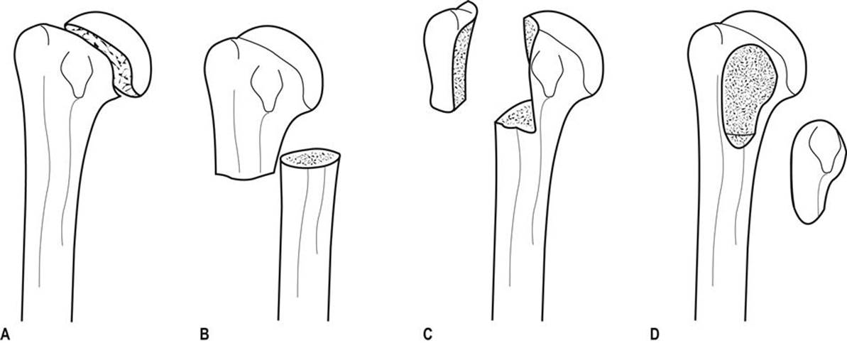
FIG. 4.2.1 Neer classification with two-part fractures of (A) the anatomical neck, (B) the surgical neck, (C) the greater tuberosity and (D) the lesser tuberosity.

FIG. 4.2.2 Neer classification with three-part fractures of (A) the greater tuberosity and anatomical neck, (B) the lesser tuberosity and anatomical neck and (C) four-part fracture involving the anatomical neck, greater tuberosity and lesser tuberosity.
One-part fracture One-part fractures account for 80% of proximal humeral fractures. Any number of fracture lines may exist, but none are significantly displaced.
Two-part fracture Two-part fractures account for 10% of proximal humeral fractures and one fragment is significantly displaced or angulated. Two-part fractures of the humerus may involve the anatomical neck (see Fig. 4.2.1A), the surgical neck (see Fig. 4.2.1B), the greater tuberosity (see Fig. 4.2.1C) or the lesser tuberosity (see Fig. 4.2.1D).
Three- and four-part fractures Three-and four-part fractures account for the remaining 10% of proximal humeral fractures, with two or three significantly displaced or angulated fragments (see Fig. 4.2.2A–C).
Treatment
One-part fractures and two-part fractures can be treated with a collar and cuff sling, adequate analgesia and follow up. Early mobilization is important and the prognosis is good.
Definitive management of the displaced fragment in two-part fractures may include open or closed reduction depending upon neurovascular injury, rotator cuff integrity, associated dislocations, likelihood of union and function. Early orthopaedic assessment is recommended.
For three- and four-part fractures, the consensus is for open reduction and internal fixation. However, a review has suggested that there is little evidence that surgery is superior to the non-operative approach.
For displaced proximal humeral fractures, surgical management remains varied and controversial. A recent systematic review suggested that non-operative treatment of proximal humerus fractures has a high rate of radiological healing, good functional outcomes but a lower complication rate when compared with the operative approach. Small, randomized controlled trials suggest that external fixation may confer some benefit over closed manipulation and that conservative treatment is better than tension band osteosynthesis. Another study suggests that the decision should be made according to the viability of the humeral head. Locking plate technology may also provide better outcomes in patients with unstable displaced humeral fractures having a viable humeral head. Other small-scale studies suggest that some bandaging styles may be better than others and that early physiotherapy may improve functional outcome.
Special cases
Fracture of the anatomical neck and articular surface
Fractures at these sites are uncommon, but are important to recognize as they have a high incidence of compromised blood supply to the articular segment, may result in avascular necrosis and may require a humeral hemiarthroplasty.
Fracture dislocations
Fractures of the greater tuberosity accompany 15% of anterior glenohumeral dislocations and may be associated with rotator cuff tears. Although the fracture may be grossly displaced, reduction of the dislocated shoulder usually also reduces the fracture. In patients who require the full range of movement of their shoulders, surgical repair of the cuff may be required.
Fractures of the lesser tuberosity are associated with posterior glenohumeral dislocations.
Disposition
Most patients with undisplaced one- and two-part fractures may be discharged from the ED with a collar and cuff sling, analgesia, early mobilization and appropriate follow up. High-risk cases, including displaced three- and four-part fractures, all open fractures and the special proximal humeral fractures described above, require orthopaedic consultation and admission, as do those with medical problems requiring investigation or treatment.
Low-energy fractures, especially in the elderly, suggest the presence of osteoporosis. ‘At-risk’ patients not already identified as having osteoporosis should be referred for bone density scans, vitamin D testing and treatment.
Fractures of the shaft of humerus
Patterns of injury
Fractures of the humeral shaft commonly occur in the third decade (active young men) and in the seventh decade of life (osteoporotic elderly women). The commonest site is the middle third, which accounts for 60% of humeral fractures. The close proximity of the fracture to the radial nerve and brachial artery commonly leads to neurovascular deficits.
Direct blows tend to produce transverse fractures, whereas falls on the outstretched hand produce torsion forces and hence spiral fractures. Combinations of the two mechanisms may produce a butterfly segment. Pathological fractures are also common, most resulting from metastatic breast cancer.
The angle and degree of displacement of the fracture depends on the site of injury and its relationship to the action and attachment of muscles on either side of the injury (Fig. 4.2.3).

FIG. 4.2.3 Relationships between humeral fracture site and the actions of inserting muscles determine bony angulation and displacement.
Clinical assessment
Patients typically present complaining of pain and supporting the forearm of the injured limb, flexed at the elbow, and held close to the chest wall. Examination of the limb reveals tenderness, swelling, shortening and possibly deformity. The skin should be assessed for tension or disruption and particular attention should be paid to the shoulder and elbow regions for associated fractures or dislocations. Initial and post-reduction assessments of the brachial artery and vein and ulnar, median and radial nerves are essential.
The commonest complication is radial nerve injury resulting either from the injury itself or reduction of the fracture and is evidenced by wrist drop and altered sensation in the first dorsal web space. A recent systemic review reported that radial nerve injury occurs in 11% of midshaft humerus fractures.
Clinical investigations
Two radiographic views – anteroposterior and lateral – will allow the correct diagnosis in most cases.
Treatment and disposition
Uncomplicated, closed fractures account for the majority of injuries and may be treated conservatively by immobilization and analgesia. Immobilization can be by a hanging cast, U-shaped cast or with functional bracing and a broad arm or collar and cuff sling. The acceptable deformity is 20° anterior/posterior angulation and 30° valus/valgus deformity. The rate of fracture union is usually higher than 90%. Early specialist follow up is recommended.
Some authors prefer a functional humeral brace rather than U-shaped plaster for immobilization, as the former may permit greater functional use without affecting healing or fracture alignment. For oblique/spiral fractures, some orthopaedic surgeons prefer an operative approach for a better functional outcome.
Open fractures and complications affecting the vessels require surgical repair. Although the majority of radial nerve injuries are neuropraxia and recover without surgical intervention, each case should be considered individually by an orthopaedic surgeon with a view to possible operative exploration.
Fractures of the distal humerus
Classification and patterns of injury
Unlike in children, fractures of the distal humerus in adults are very uncommon and patterns of injury tend to reflect the anatomical two-column construction (condyles) of the humerus. Several classification methods have been used, such as the Riseborough and Radin, Mehne and Matta classifications, but the simplest and most commonly used are the AO/ASIF classifications. These classify injuries into three categories: type A are extra-articular fractures, type B are partial articular and type C are complete articular fractures. Practically, distal humeral fractures may be classified into supracondylar, intercondylar and other types. Supracondylar fractures lie transversely, whereas intercondylar T or Y fractures include an additional vertical extension between the condyles.
Mechanisms of injury usually involve a direct blow to the flexed or extended elbow. In the former, the olecranon is driven upwards, thereby either splitting the condyles apart producing a ‘T’ or a ‘Y’ pattern, or shearing off one condyle.
Clinical assessment
Patients typically present with a swollen, tender, deformed elbow. As very little subcutaneous or other tissue separates the bone from skin, any disruption of the skin should be carefully examined for the possibility of a compound fracture. Distal neurological and vascular injury must be assessed carefully, as the possibility of nerve injury has been reported to be as high as 12–20%.
Clinical investigations
Two radiographic views – anteroposterior and lateral – should be obtained. Some authors suggest that an internal oblique view may improve the diagnostic accuracy. Pain and inability to extend the elbow often result in poor-quality radiographs. Although high-quality radiographs are essential for operative planning, repeat films should not be attempted in the ED as they rarely provide the desired result. When there is any suspicion of severe injury, either from the history or from gross soft- tissue swelling, early computed tomography (CT) scanning should be considered to give better detail, especially of intra-articular fractures.
Undisplaced fractures may not be visible on radiography but may be suggested by posterior or anterior fat pad signs, which result from fat displaced by an underlying haemarthrosis. Ultrasonography, CT and magnetic resonance imaging may all improve diagnostic precision. They alter management and improve outcome in patients with occult fractures, mostly of the intra-articular type.
Treatment and disposition
Uncomplicated, undisplaced, closed fractures with minimal swelling should be immobilized for 3 weeks in 90° flexion with an above-elbow cast and a broad arm sling, followed by active mobilization.
Patients with severe swelling, compound fractures, displaced fractures or neurovascular compromise require orthopaedic intervention.
Controversies
For humeral shaft fractures, it is unclear whether hanging plasters or functional braces are better than U-shaped plasters for fracture healing and position.
Although the union rate of humeral shaft fracture treated with bracing method is high, the functional outcomes after brace treatment are still under investigation.
Low-intensity pulsed ultrasound may be useful in the treatment of non-union. Whether it may enhance normal fracture healing is not known.
The role of magnetic resonance imaging in the diagnosis of bone bruising and humeral fracture has not been studied.
Further reading
1. Camden P, Nade S. Fracture bracing the humerus. Injury. 1992;23:245–248.
2. Diana JN, Ramsey ML. Decision making in complex fractures of the distal humerus: current concepts and potential pitfalls. Orthopaed J. 1998;11:12–18.
3. Handol HHG, Madhok R. Interventions for treating proximal humeral fractures in adults. Cochrane Database Syst Rev. 2003;4:CD000434.
4. Iyengar JJ, Devcic Z, Sproul RC, et al. Nonoperative treatment of proximal humerus fractures: a systematic review. J Orthopaed Trauma. 2011;25:612–617.
5. Mulhall KJ, Ahmed A, Khan Y, Masterson E. Simultaneous hip and upper limb fracture in the elderly: incidence, features and management considerations. Injury. 2002;33:29–31.
6. Ramachandran M, Birch R, Eastwood DM. Clinical outcome of nerve injuries associated with supracondylar fractures of the humerus in children, the experience of a specialist referral centre. J Bone Joint Surg. 2006;88B:90–94.
7. Ring D, Chin K, Taghinia AH, Jupiter JB. Nonunion after functional brace treatment of diaphyseal humerus fractures. J Trauma. 2007;62:1157–1158.
8. Rommens PM, Heyvaert G. Conservative treatment of subcapital humerus fractures Comparative study of the classical Desault bandage and the new Gilchrist bandage. Unfallchirurgie. 1993;19:114–118.
9. Shao YC, Harwood P, Grotz MRW, et al. Radial nerve palsy associated with fractures of the shaft of the humerus: a systematic review. J Bone Joint Surg. 2005;87B:1647–1652.
10. Vallier HA. Treatment of proximal humerus fractures. J Orthopaed Trauma. 2008;21:469–476.
11. Weber E, Matter P. Surgical treatment of proximal humerus fractures – an international multicenter study [In German]. Swiss Surg. 1998;4:95–100.
12. Zyto K, Ahrengart L, Sperber A, Tornkvist H. Treatment of displaced proximal humeral fractures in elderly patients. J Bone Joint Surg. 1999;79:412–417.
4.3 Dislocations of the elbow
Raymond Chi Hung Cheng and Timothy H Rainer
Essentials
1 Elbow dislocations are the third most common large joint dislocation.
2 Surgical intervention is rarely required for simple elbow dislocations.
3 Surgical intervention may be required when fractures of the radius, ulnar and humerus are associated with elbow dislocation or when neurovascular injury occurs.
4 The commonest neurovascular complication involves the ulnar nerve.
5 After reducing elbow dislocations, it is important to reassess joint stability and potential neurovascular complications.
Introduction
Elbow dislocation, along with glenohumeral and patellofemoral joint dislocations, is one of the three most common large joint dislocations. The elbow joint is a hinge-like articulation involving the distal humerus and proximal radius and ulna. Owing to its strong muscular and ligamentous supports, the joint is normally quite stable and rarely requires operative intervention, even for acute instability after dislocation.
Elbow dislocations can be classified as either anterior or posterior. Posterior dislocation is the most common type and can be further divided into posteromedial or posterolateral. It usually results from a fall on the outstretched hand with some degree of flexion or hyperextension at the elbow. The radius and ulna commonly dislocate together. Similarly, anterior dislocation can also be divided into anteromedial or anterolateral. This type is less common and is usually due to a direct blow to the dorsal side of the elbow.
Uncommonly, the radius or ulna alone may dislocate at the elbow. In such cases, there is always a fracture of the other bone. One common example is in Monteggia fractures, where anterior or posterior radio-humeral dislocation occurs alongside a fracture of the proximal one third of the ulna shaft (Fig. 4.3.1). A rarer example is a posterior ulna-humeral dislocation with fracture of the radial shaft. So, although elbow dislocations may appear to be isolated, it is essential to look for associated intra-articular or shaft fractures.

FIG. 4.3.1 Monteggia fracture dislocation. Fracture of the ulnar shaft may be associated with (A) anterior radio-humeral dislocation or (B) posterior radio-humeral dislocation.
Clinical assessment
History and examination
Patients typically present holding the lower arm at 45° to the upper arm and there is swelling, tenderness and deformity of the elbow joint. The three-point anatomical triangle of olecranon, medial and lateral epicondyles should be assessed for abnormal alignment, as this strongly suggests dislocation.
The commonest neurovascular injury involves the ulnar nerve, reported in 10–15% of elbow dislocations, but the median and radial nerves and the brachial artery may also be affected.
The differential diagnosis is a complex distal humerus fracture which, in a swollen elbow, may be hard to differentiate clinically from an elbow dislocation.
Clinical investigations
Anteroposterior and lateral radiographic views should be obtained and scrutinized for associated fractures of the coronoid process, radial head, capitellum and olecranon.
Magnetic resonance imaging (MRI) characterizes bony injury more accurately than radiography in children with elbow injuries, but its potential role for diagnosis and guiding management in adults has not been well evaluated. Duplex Doppler ultrasound can be use to identify early brachial artery injury.
Treatment
Simple dislocation can be reduced using a closed method. With adequate sedation, gentle traction and counter-traction, the joint relocates quite easily. Medial and posterolateral dislocations may also require sideways correction. Dislocation of the stable elbow joint produces severe soft-tissue injury and resultant instability, therefore, after reduction, signs and symptoms of compartment syndrome should be sought.
Joint instability should be tested by valgus and varus testing and by lateral pivot-shift test. The reduced elbow joint should move smoothly. Any crepitation or resistance, particularly during the mid-range, suggests incongruent reduction or soft tissue interposition, which is commonly associated with coronoid process or epicondylar fractures. Inability to fully flex or extend the elbow suggests a loose bone or cartilaginous fragment or a capsular tear.
Post-reduction films should be assessed, not only for correct joint relocation, but also for associated fractures. After successful reduction, the elbow should be placed in a posterior plaster slab in 90° of flexion. Cylinder casts are contraindicated because of the likelihood of severe soft-tissue swelling.
There is little evidence that surgical intervention improves outcome in patients with medial or lateral elbow instability after dislocation. A recent systematic review found that there is no difference in outcome between surgical repair of the ligament and plaster immobilization for simple elbow dislocation. Patients with functional treatment have a better range of movement, less pain, better functional scores, shorter disability and shorter treatment time when compared with plaster immobilization. The management of Monteggia fracture- dislocation is discussed in Chapter 4.4. Compound fracture dislocation should be reduced by the open method. Patients with irreducible dislocations, neurovascular complications, associated fractures or open dislocations require orthopaedic intervention.
Ulnar nerve injuries can occur both before and after closed reduction. The reported rate varies between 10 and 15%. Most of them are neuropraxia and will recover with conservative measures. The most sensitive sign and symptoms are numbness over the little fingers.
Disposition
Current practice is that most patients may be discharged from the emergency department with analgesia, pressure bandage for stable joints and plaster immobilization for unstable joints. A broad arm sling with appropriate follow up should be arranged after reduction.
A recent prospective, randomized study suggested that early mobilization is superior to plaster immobilization in terms of functional recovery, without any increased instability or a recurrence of dislocation for patients with uncomplicated posterior dislocations. The duration of immobilization should not be longer then 14 days to prevent joint stiffness. Patients with irreducible dislocations, neurovascular complications, associated fractures or open dislocations require admission.
Controversies
There are no large-scale randomized studies comparing operative and non-operative management of elbow dislocation. It is therefore unclear whether one method may produce better outcomes than another.
Early mobilization may be superior to prolonged plaster immobilization after reduction of uncomplicated posterior dislocations.
The epidemiology of elbow injury, including dislocation in patients presenting to emergency departments, has not been well described and requires further studies.
Roles for computed tomography and magnetic resonance imaging in evaluating acute elbow injury and influencing management require further study.
Further reading
1. Ergunes K, Yilik L, Ozsoyler I, et al. Traumatic brachial artery injuries. Texas Heart Inst J. 2006;33:31–34.
2. Griffiths JF, Roebuck DJ, Cheng JCY, et al. Comparison of radiography and magnetic resonance imaging in the detection of injuries after paediatric elbow trauma. Am J Roentgenol. 2001;176:53–60.
3. Haan JD, Schep NWL, Tuinebreijer WE, et al. Simple elbow dislocations: a systematic review of the literature. Arch Orthopaed Trauma Surg. 2010;130:241–249.
4. Lam TP, Ng BKW, Ma RF, Cheng JCY. Monteggia fractures in children – a review of 30 cases. J Jap Pediatr Orthoped Assoc. 2004;13:193–195.
5. McRae R. Practical fracture treatment Edinburgh: Churchill Livingstone; 1994.
6. Partio EK, Hirvensalo E, Bostman O, Rokkanen P. A prospective controlled trial of the fracture of the humeral medial epicondyle – how to treat? Ann Chirurg Gynaecol. 1996;85:67–71.
7. Rafai M, Largab A, Cohen D, Trafeh M. Pure posterior luxation of the elbow in adults: immobilization or early mobilization A randomized prospective study of 50 cases. Chirurg Main. 1999;18:272–278.
8. Reynders P, De Groote W, Rondia J, et al. Monteggia lesions in adults A multi-centre Bota study. Acta Orthopaed Belg. 1996;62:78–83.
9. Robert S, David R. Current concepts review: the ulnar nerve in elbow trauma. J Bone Joint Surg. 2007;89A:1108–1116.
10. Uehara DT, Chin HW. Injuries to the elbow and forearm. In: Tintinalli JE, Kelen GD, Stapczynski JS, eds. Emergency medicine A comprehensive study guide. New York: McGraw-Hill; 2000;1763–1772.
11. Willet K. Upper limb injuries. In: Skinner D, Swain A, Peyton R, Robertson C, eds. Cambridge textbook of accident and emergency medicine. Cambridge: Cambridge University Press; 1997;601–617.
4.4 Fractures of the forearm and carpal bones
Crispijn van den Brand
Essentials
1 Forearm fractures are among the most common fractures seen in the emergency department (ED).
2 When assessing the need for or success of reduction, the external appearance of the limb is a key feature.
3 Median nerve function must be assessed before and after reduction of all distal radial fractures.
4 Splinting or functional bracing may be sufficient for stable fractures. Early movement and load bearing aids functional recovery.
5 General indications for orthopaedic referral include fractures which are compound, unstable, associated with intra-articular or neurovascular injury and those that have failed reduction in the ED.
6 Displaced, isolated fractures of the ulna or radius may be associated with a dislocation of the radius or ulna respectively (Monteggia and Galeazzi fracture dislocations). These should be carefully sought, as there is high risk of long-term disability.
7 Significant or persistent symptoms with the absence of a visible fracture on plain X-ray may be due to an undetected fracture or significant soft-tissue injury. A high index of suspicion and early review are recommended. Further investigation with bone scintigraphy, CT or MRI may be indicated.
Radial head fractures
Clinical features
History
Radial head fractures occur frequently, usually as a result of a fall onto an outstretched hand or, less frequently, following a direct blow to the lateral side of the elbow. Radial head fractures present with pain and restricted movement at the elbow.
Examination
Usually, there is swelling and tenderness over the radial head. Sometimes, with more subtle injuries, rotating the forearm while palpating the radial head may be necessary to elicit tenderness. Elbow extension and forearm rotation are limited. Severely comminuted fractures may have proximal displacement of the radius, which can be associated with disruption of the interosseous membrane and subluxation of the distal radioulnar joint (Essex–Lopresti fracture dislocation).
Clinical investigations
Imaging
Standard anteroposterior (AP) and lateral X-rays of the elbow are required. A radiocapitellar view may be necessary if the fracture is subtle. The presence of an anterior fat pad sign alone on X-ray is associated with an underlying radial head or neck fracture in up to 50% of patients. In this case, a fracture should be assumed to be present if there is an appropriate mechanism and local signs. A follow-up X-ray or computed tomography (CT) scan is indicated only in the presence or persistent pain, stiffness or locking.
Classification
Radial head fractures are usually classified according to the (modified) Mason classification (Fig. 4.4.1). About two-thirds of fractures are Mason type I.

FIG. 4.4.1 (A–D) Mason–Hotchkiss classification of radial head fractures.
The Mason classification is as follows:
Mason type I, displaced less than 2 mm
Mason type II, displacement more than 2 mm
Mason type III, comminuted fractures of the entire radial head
Mason type IV, radial head fracture with associated elbow dislocation.
Treatment
All non-displaced (type I) radial head fractures and those type II fractures without mechanical block may be managed with a bandage and sling. Mobilization should be started as early as possible. If there is severe pain, a posterior splint may be useful but should not be applied for more than 2 days. Prognosis is good, but full extension may not be possible for many months.
Displaced or complex radial head fractures (type II or III) may be treated in the acute setting with a sling or posterior splint. These patients should have early orthopaedic review (within days). The treatment of displaced or complex radial head fractures remains controversial and should be determined by an orthopaedic surgeon.
Mechanical block can be difficult to assess acutely due to pain. Intra-articular injection of bupivacaine may assist early assessment or assessment may be deferred until pain has settled. Surgical options include open reduction and internal fixation and excision of the radial head with or without implantation of a prosthesis.
Radial neck fractures with up to 20° tilts can be managed conservatively. More severe tilt can be reduced using intra-articular local aneasthesia. The forearm is pronated until the most prominent part of the radial head is felt. Then traction is applied to the forearm and pressure applied to the radial head. Open reduction is indicated if closed methods fail or displacement is severe.
Complications
Neurovascular complications and compartment syndrome are uncommon. Most complications relate to disturbance of the relationships of the proximal radio-ulnar and radiocapitellar articular surfaces causing limitation of movement. This is uncommon with minor fractures.
Shaft fractures
Clinical features
History
This type of injury requires great force, typically from a motor-vehicle accident, a fall from a height or a direct blow. These fractures are commonly open and nearly always displaced.
Examination
The forearm is swollen and tender and may be angulated and rotated. Examination looking for an open wound, local neurovascular compromise, compartment syndrome or musculotendinous injury is required. Given the mechanism of injury, other injuries should also be sought.
Clinical investigations
Imaging
AP and lateral X-rays of the forearm, including the wrist and elbow joints, are needed. Displacement and angulation are easily determined, but torsional deformity may be subtle. Because the ulna and radius are rectangular in cross-section rather than circular, a change in bone width at the fracture site indicates rotation. The radial and ulnar styloid processes normally point in opposite directions to the bicipital tuberosity and coronoid process, respectively. A change in this alignment also suggests torsion.
Treatment
Adult forearm fractures are less stable than those in children and lack of remodelling limits tolerance to incomplete reduction. Undisplaced fractures may be managed with an above- elbow cast, but must be reviewed at 1 week for displacement and angulation. Most fractures, however, are displaced and require open reduction and internal fixation.
Complications
Early complications include wound infection, osteomyelitis, neurovascular injury and compartment syndrome. Later, non-union, malunion, reduced forearm rotation and reflex sympathetic dystrophy are possible complications.
Specific fracture types
Isolated fracture of the ulnar shaft
These fractures are due to a direct blow to the ulna, often when raised in defence; hence they are also known as ‘nightstick’ fractures. Patients present with localized pain and swelling. AP and lateral X-rays delineate the location of the fracture and degree of angulation. Look for associated dislocation of the radial head if displacement is present (Monteggia fracture dislocation).
Fractures displaced less than 50% of the ulna width heal well with a non-union rate of 0–4%. Traditional treatment involves fixing the forearm in mid-pronation with a plaster cast, extended above elbow if the middle or proximal thirds of the ulna are fractured. The cast is removed once union occurs, usually in about 8 weeks. Other proven options include a below-elbow plaster (BEPOP) for proximal fractures, early mobilization with bandage after 1–2 weeks in BEPOP or functional bracing after 3–5 days, which allows movement at wrist and elbow.
Fractures with more than 10° of angulation or displaced more than 50% of the diameter of the ulna require surgical intervention.
Monteggia fracture dislocation
This is a rare fracture of the proximal ulna with dislocation of the radial head. It occurs either through a fall onto the outstretched hand with hyperpronation or through a force applied to the posterior aspect of the proximal ulna. Patients present with pain, swelling and reduced elbow movement. The forearm may appear shortened and the radial head may be palpable in the antecubital fossa. Associated posterior interosseous nerve injury is common.
On X-ray the fracture is obvious, but the dislocation is commonly missed. Check that a line through the radial shaft bisects the capitellum on both views. There are four types of Monteggia fracture depending upon displacement of the radial head (Bado classification). Dislocation is anterior in 60% (Bado type I), but may be lateral or posterior.
All Monteggia fractures require open reduction and internal fixation. Common complications include malunion and non-union of the ulnar fracture and an unstable radial head.
Isolated radial shaft fracture
Isolated fractures of the proximal two-thirds of the radial shaft are uncommon and are usually displaced. Rare undisplaced fractures can be treated similarly to isolated ulnar shaft fractures. Displaced fractures require open reduction and internal fixation.
Galeazzi fracture dislocation
Fractures of the distal third of the radial shaft occur as a result of a fall onto the outstretched hand or a direct blow. There may be an associated subluxation or dislocation of the distal radioulnar joint (DRUJ), known as the Galeazzi fracture dislocation. Patients have pain and swelling at the radial fracture site. Those with a Galeazzi injury will also have pain and swelling at the DRUJ and a prominent ulnar head.
X-rays show the radial fracture, which is tilted ventrolaterally. Widening of the DRUJ space on the AP X-ray and dorsal displacement of the ulnar head on the lateral X-ray are seen (Fig. 4.4.2). An ulnar styloid fracture is seen in 60% of cases.
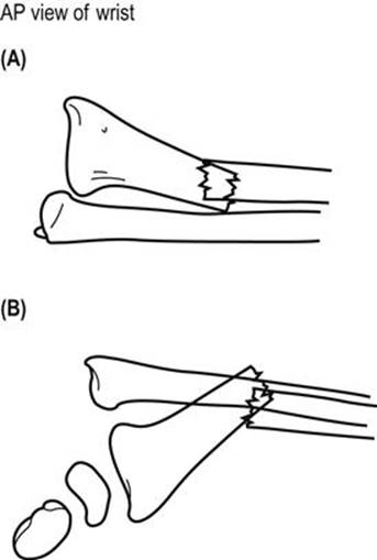
FIG. 4.4.2 The Galeazzi fracture dislocation.
All Galeazzi fracture dislocations require surgical management. Complications include malunion or non-union of the radial fracture and subsequent instability of the DRUJ.
Fractures of the distal radius and ulna
Fractures of the distal radius and ulna are common, particularly in children and elderly women. Fractures in the latter group are indications for evaluation of bone-mineral density.
Clinical features
History and examination
Fractures usually occur after a fall onto the outstretched hand resulting in bending, shearing or impaction forces being applied to the distal metaphysis, or from a direct blow. Patients present with pain, tenderness and variable degrees of swelling and deformity. It is important to examine for associated injuries to carpal bones, radial and ulnar shafts, elbow and shoulder joints, for median nerve injury, vascular compromise and for extensor tendon injury.
Clinical investigations
Imaging
Anteroposterior and lateral X-rays of the wrist demonstrate most injuries. For patients with significant symptoms or signs and a normal X-ray, consider an occult undisplaced fracture or ligamentous injury.
Although this chapter uses eponymous names, it is important to be aware that orthopaedic circles have moved to more formal classification systems for distal radial fractures. Several have been proposed and are beyond the scope of this text. The author recommends being familiar with anatomical descriptions and fracture features associated with need for reduction, instability of reduction and indications for operative intervention.
Treatment
Management
Prompt attention to analgesia, splinting and elevation is essential while awaiting X-rays.
Reduction is indicated in the following circumstances to improve long-term function:
visible deformity of the wrist
loss of volar tilt of the distal radial articular surface beyond neutral
loss of>5° of the radial inclination of the distal radius (normally approximately 20°)
intra-articular step of>2 mm
radial shortening>2–3 mm.
Greater deformity can be accepted in low-demand, elderly patients.
Anaesthetic options for reduction include haematoma block, Bier’s block and procedural sedation. Reduction is traditionally maintained with an encircling plaster cast moulded to oppose displacement forces and extending from volar metacarpal crease to proximal forearm for 6 weeks. Displaced or comminuted fractures at high risk of swelling, especially in the elderly or coagulopathic patients, are immobilized with non-encircling splints.
Factors associated with instability of the distal fragment and failure to maintain reduction include:
intra-articular component (especially involving the distal radio-ulnar joint)
shearing fractures (Barton-, Hutchinson’s type)
palmarly displaced fractures (Smith type)
the magnitude of the initial displacement or comminution.
Weekly X-rays for 2–3 weeks with orthopaedic follow up are recommended for all displaced fractures, those with intra-articular extension and potentially unstable fractures.
Stable, undisplaced, extra-articular fractures can be managed more conservatively with splinting and referral to a family doctor for early mobilization after 4 weeks.
Indications for operative management are debated, but should be considered for:
comminuted, displaced, intra-articular fractures
open fractures
associated carpal fractures
associated neurovascular or tendon injury
failed conservative treatment (failed reduction or unstable after reduction)
bilateral fractures/impaired contralateral extremity.
Complications
Median nerve injury may occur acutely due to the injury, as a result of reduction or later due to pressure effects from the plaster. Median nerve function must be documented before and after reduction.
Loss of reduction may require delayed surgical intervention. Malunion with chronic wrist pain, arthritis and secondary radioulnar and radiocarpal instability are associated with intra-articular extension of the fracture.
Long-term complications include osteoarthritis, residual disability and complex regional pain syndrome (CRPS). The incidence of CRPS following distal radius fractures ranges in the literature from less than 1% to 22%. Prophylactic vitamin C may reduce the incidence of CRPS, the advised dose is 500 mg/day for 50 days.
Specific fractures
Colles’ fracture
Colles’ fracture is a metaphyseal bending fracture. The wrist has a classic ‘dinner-fork’ appearance, often with significant swelling of the soft tissues. This appearance is reflected in the radiographs (Fig. 4.4.3). There is often associated damage to the radio-ulnar fibrocartilage. There may be comminution, commonly dorsally, which can extend into the radiocarpal or radio-ulnar joints.

FIG. 4.4.3 Colles’ fracture. A fracture of the distal radial metaphysis with six classic deformities. The lateral view shows anterior angulation, dorsal displacement and impaction. The AP view reveals radial displacement, ulnar angulation and an ulnar styloid fracture.
The aim of reduction is to restore radial length, volar tilt and radial angulation. A minimum of 0° tilt is acceptable if full reduction is not possible. Reduction is achieved by first disimpacting the fracture with traction in the line of the forearm. If this fails, traction in extension or hyperextension should be tried. Volar tilt is then restored with volar pressure over the dorsum of the distal fragment while traction is maintained. Lastly, correct radial tilt and radial displacement with ulnar pressure over the radial side of the distal fragment. Reduction is successful in 87%, but almost two-thirds lose reduction over 5 weeks, most of this occurring during cast immobilization.
The commonly accepted cast immobilization position is with the wrist joint in 15° palmar flexion, 10–15° ulnar deviation and slight pronation. However, some evidence suggests better outcomes are achieved with the wrist in dorsiflexion and mid-supination. The cast must be carefully moulded over the dorsum of the distal fragment and the anteromedial forearm. Functional bracing allowing wrist movement has also shown good outcomes.
Smith’s fracture
This metaphyseal bending fracture of the distal radius occurs through a direct blow or fall onto the back of the hand or a fall backward onto the outstretched hand in supination.
AP and lateral X-rays of the wrist show a ‘reverse Colles’ fracture’ with a similar AP appearance, but with volar displacement and tilt on the lateral X-ray view.
Closed reduction to achieve anatomical radial length and volar tilt should be attempted. Traction is first applied to restore length, followed by dorsal pressure over the volar surface of the distal radius to reverse displacement and angulation. A full above-elbow cast is applied with the wrist in supination and dorsiflexion to prevent loss of reduction. However, most Smith’s fractures are unstable and require operative management. Early orthopaedic follow up is mandatory.
Barton’s fracture
Barton’s fractures are dorsal or volar intra- articular fractures of the distal radial rim (Fig. 4.4.4). The mechanisms of injury are similar to those seen with Colles’ and Smith’s fractures, respectively. There is often significant soft-tissue injury and the carpus is usually dislocated or subluxed along with the distal fragment. These fractures are complicated by arthritis of the radiocarpal joints and carpal instability.

FIG. 4.4.4 Barton’s fractures demonstrated on lateral views of the wrist.
Minimally displaced fractures involving less than 50% of the joint surface and without carpal displacement may be reduced along the lines of a Colles’ or Smith’s fracture. Immobilization should occur with wrist flexed for dorsal Barton’s and extended for volar Barton’s. However, most fractures are unstable and potentially disabling, requiring early operative management, especially in younger patients. Early orthopaedic follow up is mandatory.
Radial styloid (Hutchison’s or chauffeur’s) fracture
This oblique intra-articular fracture of the radial styloid is caused by a direct blow or fall onto the hand. Displacement is associated with carpal instability and long-term arthritis. The fracture is seen best on AP X-rays of the wrist (Fig. 4.4.5). Undisplaced fractures can be treated with a cast for 4–6 weeks. Displaced fractures should be referred to an orthopaedic surgeon for anatomical reduction and fixation.

FIG. 4.4.5 Radial styloid (Hutchison’s or chauffeur’s) fracture.
Ulnar styloid fracture
An isolated fracture can occur through forced radial deviation, dorsiflexion, rotation or a direct blow. Avulsion fractures involving the lesser portion of the ulnar styloid are not associated with significant instability of the distal radio- ulnar joint (DRUJ). In contrast, fractures involving the base of the ulnar styloid disrupt the major stabilizing ligaments of the distal ulna and the triangular fibrocartilage complex (TFCC) and may lead to subsequent DRUJ instability. Fractures should be treated with a splint or cast with the wrist in mid-supination and ulnar deviation, patients should be referred to an orthopaedic surgeon to assess DRUJ stability.
Carpal fractures and dislocations
Carpal fractures predominantly occur in young men. The bones in the proximal carpal row are more commonly involved, especially scaphoid fractures which account for 82–89% of all carpal fractures. Most other isolated carpal fractures are triquetral fractures. Management depends on the degree of displacement and damage and stability. Generally, undisplaced fractures with minimal comminution can be managed by cast immobilization. Given the importance of wrist function, early orthopaedic review should be sought for patients with displaced or comminuted fractures or where instability or an associated carpal dislocation is suspected.
Specific fractures
Scaphoid fracture
The most common mechanism of injury is a fall on the outstretched hand with the wrist in radial deviation. This mechanism also puts the distal radius and the scaphoid-lunatum (SL) ligament at risk. Clinical features include wrist pain and local swelling and tenderness over the scaphoid, palpated dorsally or via the anatomical snuffbox. Imaging with AP, lateral and scaphoid views will detect at most 70% of all scaphoid fractures.
Fractures of the scaphoid are classified by their location (proximal third, waist, distal third or tubercle) and by their stability. Stable fractures are undisplaced with little comminution and unstable fractures are displaced with considerable comminution. Stable fractures are generally treated with a below-elbow cast for 10–12 weeks. There is no evidence that cast immobilization with inclusion of the thumb leads to better outcome. Unstable fractures require surgical intervention. Complications include non-union and avascular necrosis of the proximal segment.
Some patients have clinical features suggestive of scaphoid fracture without confirmatory X-ray evidence. In the past, cast immobilization for 1–2 weeks followed by repeat X-ray was advocated. Although this is still advocated by some, it is not recommended. The additional sensitivity is low and scaphoid fractures are often missed. A number of alternative diagnostic approaches have been suggested, including bandaging with clinical review at 7–10 days followed by CT if clinical features persist, or early primary CT, magnetic resonance imaging (MRI) or bone scintigraphy. All of these imaging modalities have their advantages and shortcomings. Bone scintigraphy is recommended as a useful diagnostic modality to rule out occult scaphoid fractures. Bone scintigraphy can rule out scaphoid fracture with a sensitivity close to 100% but with the disadvantage of up to 25% false positives.
Dislocations of the wrist
Dislocations involving the wrist usually result from high-energy falls on the outstretched hand (such as from a height) that result in forced hyperextension. The distal row of carpal bones is commonly displaced dorsal to the proximal row as a result of a scaphoid fracture, a scapholunate dislocation or a perilunate dislocation. Trans-scaphoid perilunate fracture dislocation is slightly more common than perilunate dislocation.
Clinical features
Clinical features include mechanism of injury, wrist pain, swelling and tenderness and possibly reduced grip strength.
Clinical investigations
Imaging requires PA and lateral X-rays. The normal PA view should show two rows of carpal bones in a normal anatomic position with uniform joint spaces of no more than 1–2 mm. No overlap should be seen between the carpal bones or between the distal ulna and the radius. On the lateral film, a longitudinal axis should align the radius, the lunate, the capitate and the third metacarpal bone.
Radiographic features include:
Lunate dislocation: on the usual PA image, the lunate has a triangular shape rather than its usual trapezoidal shape. On the lateral film, the lunate has a ‘C-’ or ‘half-moon’ shape. The rest of the carpal bones are in a normal anatomic position in relation to the radius.
Perilunate dislocation: on the lateral film, the lunate is in a normal anatomical position with respect to the radius and with the rest of the carpal bones displaced dorsally. On the PA film, crowding is evident between the proximal and distal carpal bones.
Scapholunate dislocation: on a PA radiograph, the scapholunate space is greater than 4 mm (also known as the Terry-Thomas sign). The scaphoid rotates, producing the classic signet-ring sign. Associated carpal fractures, especially of the scaphoid, may be evident.
Treatment
All wrist dislocations require orthopaedic consultation and prompt reduction.
Controversies
Optimal management for Mason type II radial head fractures.
Optimal immobilization for distal radial fractures.
Operative versus non-operative management of distal radial fractures, particularly in the elderly.
Vitamin C for prevention of CRPS following distal radius fractures.
Optimal management strategy for suspected scaphoid fracture with normal initial X-rays.
Further reading
1. Connolly JF. Nonoperative fracture treatment. In: Bucholz RW, Heckman JD, Court-Brown C, eds. Rockwood and Green’s fractures in adults. 6th ed. Baltimore: Lippincott Williams & Wilkins; 2005.
2. Cruikshank J, Meakin A, Braedmore R, et al. Early computerized tomography accurately determines the presence or absence of scaphoid and other fractures. Emerg Med Australas. 2007;19:223–228.
3. Hanel DP, Jones MD, Trumble TE. Wrist fractures. Orthopaed Clin N Am. 2002;33:35–57.
4. Mackay D, Wood L, Rangan A. The treatment of isolated ulnar fractures in adults: a systematic review. Injury. 2000;31:565–570.
5. Rhemrev SJ, Ootes D, Beeres FJP, et al. Current methods of diagnosis and treatment of scaphoid fractures. Internatl J Emerg Med. 2011;4:4.
6. Ruch DS. Fractures of the distal radius and ulna. In: Bucholz RW, Heckman JD, Court-Brown C, eds. Rockwood and Green’s fractures in adults. 6th ed. Baltimore: Lippincott Williams & Wilkins; 2005.
7. Sarmiento A, Latta L. The evolution of functional bracing for fractures. J Bone Joint Surg. 2006;88B:141–148.
8. Uehara DT, Chin HW. Injuries to the elbow and forearm. In: Tintinalli JE, Kelen GD, Stapcznski JS, eds. Emergency medicine. 5th ed. New York: McGraw Hill; 2000.
9. Uehara DT, Chin HW. Wrist injuries. In: Tintinalli JE, Kelen GD, Stapczynski JS, eds. Emergency medicine. 5th ed. New York: McGraw Hill; 2000.
10. Van Glabbeek F, Van Riet R, Verstreken J. Current concepts in the treatment of radial head fractures in adults A clinical and biomechanical approach. Acta Orthopaed Belg. 2001;67:430–441.
11. Villarin Jr LA, Belk KE, Freid R. Emergency department evaluation and treatment of elbow and forearm injuries. Emerg Med Clin N Am. 1999;17:843–858.
12. Zollinger PE, Tuinebreijer WE, Breederveld RS, et al. Can vitamin C prevent complex regional pain syndrome in patients with wrist fractures? A randomized, controlled, multicenter dose-response study. J Bone Joint Surg. 2007;89A:1424–1431.
4.5 Hand injuries
Peter Freeman
Essentials
1 Hand injuries are common and most carry a good prognosis if treated early and competently.
2 A comprehensive knowledge of hand anatomy and function is essential for appropriate initial management of the injured hand.
3 Aftercare and rehabilitation are essential for return to normal function.
Introduction
Hand injuries are common and up to 10% of emergency department (ED) attendances involve injury to the hand. Presentations may be due to wounds (≈35%), contusions (≈20%), fractures (≈20%), sprains (≈10%) or infections (≈5%). Males injure their hands more than females. The complex anatomy and tactile function of the hand mean that hand injuries can profoundly affect an individual. The importance of correct assessment and care of hand injuries cannot be overstated. Apart from the initial pain and trauma, occupational and psychological concerns play a major role in the aftermath of these injuries. Even a relatively minor fingertip injury can result in an individual being away from work for several days, with consequent loss of earnings and concerns for long-term function and appearance. It is therefore essential that initial assessment and management are appropriate. The role of ED management is as much about identifying cases that require specialist referral as it is about treating straightforward injuries.
Clinical features
History
Time taken eliciting a focused history of the mechanism of injury is essential in cases of hand injury. Key questions include:
When did the injury occur?
What was the position of the hand at the time?
Was the hand injured with a sharp implement, such as glass, or crushed in a machine? Incised wounds caused by sharp implements tend to damage structures, such as nerves and tendons, whereas crush injuries may cause fractures and lacerations.
Was there brisk bleeding and does any part of the hand feel numb? These symptoms are important as, in the fingers, the digital nerves lie adjacent to the arteries.
What was the environment of the injury?
Is it likely that the wound is contaminated or contains foreign material, such as glass?
Injury to the dominant hand should be noted as well as occupation and key leisure activities. It is also important to record medications and allergies to guide analgesia and antibiotic choice. Tetanus prophylaxis status should be determined.
Examination
The injured hand must be examined in a well-lit area. Temporary dressings may need to be soaked off if they have been allowed to dry out and become adherent. At triage, an initial moist dressing is preferred, with firm pressure and elevation if there is significant bleeding.
Hand and finger injuries are painful and suitable analgesia should be given prior to full examination. Local infiltration of local anaesthetic without adrenaline around a wound or as a digital nerve block will allow examination of all aspects except sensation, which must be tested and recorded prior to anaesthesia. A wrist block is useful when some or all of the hand needs to be anaesthetized (Fig. 4.5.1). In this instance, longer-acting local anaesthetic is generally used to prolong the effect.

FIG. 4.5.1 Palmar wrist block. (Reproduced with permission from American Society for the Surgery of the Hand. The Hand, 2nd edn. Boston: Churchill Livingstone; 1990.)
Testing sensation is achieved by point touch in the distribution of the three main nerves that supply the hand (Fig. 4.5.2). The median nerve supplies the palmar aspect of the thumb, index, middle and half of the ring finger, extending to supply the fingertip and nailbed. The ulnar nerve supplies both palmar and dorsal aspects of the other half of the ring finger and the little finger. The radial nerve supplies the radial dorsum of the hand, thumb, index, middle and radial aspects of the ring finger. If the patient is unable to describe sensation because they are too young or unconscious, it is useful to remember that the digital nerves also carry the sympathetic supply to the fingers and that division will cause a dry finger in the distribution of the digital nerve.

FIG. 4.5.2 The nerve supply to the hand.
The hand examination should be holistic and not just concentrate on the obvious injury. Inspection of the hand will provide information about the perfusion of the tissues, local swelling and position of wounds. The resting position of the hand may be a clue to tendon injury, as the normal uninjured position is held with the fingers in increasing flexion from the index to the little finger (Fig. 4.5.3A). A pointing finger may indicate a flexor tendon injury (Fig. 4.5.3B). Testing for pinch grip is important if there is concern about the stability of the first metacarpal. Obvious bone or joint deformity should be recorded. The metacarpals and phalanges are all easily palpable subcutaneously and local tenderness may indicate underlying fracture.

FIG. 4.5.3 (A) The normal resting hand. (B) The pointing finger. (Reproduced with permission from American Society for the Surgery of the Hand. The Hand, 2nd edn. Boston: Churchill Livingstone; 1990.)
Functional testing should be performed for all injured hands. Tendon integrity is tested by asking the patient to perform specific movements. Some tendon injuries may be obvious, however, two flexor tendons supply each finger and simply asking the patient to flex the finger will not exclude a divided flexor digitorum superficialis tendon. The profundus tendon flexes the distal interphalangeal joint and is tested by asking the patient to flex the tip of each finger in turn while the examiner holds the proximal interphalangeal joint in extension. The superficialis flexor tendon is tested by asking the patient to flex each finger individually, while the examiner holds the other fingers straight. The extensor tendons to the fingers are tested by asking the patient to extend the fingers against resistance. It is important to remember that the broad interconnections between the extensor tendons make it possible to extend to near neutral in the presence of a divided tendon. Partial tendon injuries may still exist despite normal functioning of the fingers. A functioning hand should allow full extension of all fingers and comfortable flexion of the fingers into the palm.
Displaced fractures or dislocations may be apparent as deformity. More subtle rotational deformity will be detected by a finger crossing its neighbour when flexed.
Clinical investigations
Most information will be obtained from a focused history and examination. Radiology of the hand and fingers will be necessary if bone/joint deformity or tenderness is elicited. Even obvious dislocations should be X-rayed prior to correction as post-reduction X-rays may be overly reassuring despite significant soft-tissue damage. Glass is radiopaque to a varying degree and, if a wound is caused by glass, an X-ray should be done prior to closure. Organic foreign bodies and infections may be detected by ultrasound using a small-parts soft-tissue probe and this modality is becoming increasingly available in the ED. Ultrasound is also useful to establish tendon integrity but this is a more specialized examination.
Laboratory investigations are rarely of use in the injured hand unless there are signs of infection.
Magnetic resonance imaging (MRI) can be useful in selected injuries as it shows the soft tissues of the hand clearly, but it is relatively unavailable acutely and should be reserved for conditions where emergent treatment is dependent on the integrity of the soft structures in the hand which are not apparent on examination alone.
Treatment
Appropriate analgesia should be provided as previously described. Rings should be removed from injured fingers to prevent subsequent compromise of circulation as the finger swells. Irrigating wounds with tap water does not increase the risk of infection and is economic. Simple hand and finger wounds can be treated along conventional lines with judicious use of local anaesthetic and skin approximation with fine (5.0) sutures or skin closures. Hand wounds generally heal well and a recent randomized controlled trial showed similar cosmetic and functional outcomes from either conservative treatment or suturing of small, uncomplicated hand wounds. Digital nerve block is useful for managing finger injuries. This technique involves infiltrating local anaesthetic around the digital nerve at the base of the finger or in the palm. Approaching the digital nerve from the dorsum of the finger is less painful but the palmar approach is more accurate as the digital nerves lie just deep to the palmar aponeurosis. A short, fine gauge (e.g. 30 gauge) needle is used with small amounts (≈1 mL) of local anaesthetic for each nerve. Choice of anaesthetic will depend on the desired length of effect and consideration should be given to using long-acting agents for crush or bone injury when a prolonged analgesic effect is desirable. Studies have shown that the use of adrenaline with lignocaine is safe and also prolongs the anaesthetic effect.
Hand dressings can be held in place with a conforming crepe bandage to provide a degree of compression. Stable injuries to the fingers can be managed with ‘buddy’ strapping which allows for some joint movement. Elevation is essential after hand injury to reduce swelling. Minor injuries can be successfully managed in the ED, but more significant injuries usually require referral for surgical opinion.
Fingertip injuries
The fingertips have an excellent blood supply and will usually heal with good cosmetic and tactile function if basic wound care principles are followed. Fingertip injuries may involve skin, subcutaneous tissue, nail or terminal phalanx.
The most complex to manage is when the terminal phalanx is exposed and, in these cases, referral for surgical treatment is advised. If there is injury involving less than 50% of the nail and no bone is exposed conservative treatment is often the best option. Small tuft fractures of the underlying terminal phalanx are stable and will be supported by the dressing or nailbed repair.
Care of the fingertip will require haemostasis followed by a non-adherent dressing. There is good evidence that this kind of dressing promotes healing and re-epithelialization of the fingertip. Occlusive fingertip dressings are quick to apply, easily removed and comfortable for the patient. Most other dressings adhere to the wound and pull epithelial cells off when removed. Alternatives to conservative management include full-thickness skin grafts to the fingertips, advancement flaps and cross-finger flaps. These should be performed by surgeons trained in the specialist techniques and reserved for injuries involving large areas of tissue loss.
Major amputations of the fingertip or crush injuries may require terminalization of the finger. This should be fully discussed with the patient, who may be prepared to forgo finger length in exchange for early return of function. Patients requiring terminalization of a finger should be referred to a specialist hand service. Occasionally, patients will bring amputated pieces of the injured fingertip with them into the ED. Recently amputated fingers can be wrapped in moist gauze and then placed in a bag and packed with ice if re-implantation is being considered by specialist hand surgeons. If there is any doubt about the viability of fingertip tissue, the patient should be referred to a specialized hand service. No attempt should ever be made to resuture avascular tissue.
Digital nerve injuries
Nerve repairs distal to the distal interphalangeal joint are rarely required as the terminal branches are very fine. Any sensory loss with these distal injuries is minimal and improves with time. More proximal injuries can be repaired by hand or plastic surgeons using microsurgery. Good results are achieved with early repair of digital nerves when the ends can be approximated without tension using a fine (>8/0) suture. The return of protective sensation depends on the extent of damage, level of repair and axon regeneration.
Nailbed injuries
These injuries are frequently underestimated, often because of a reluctance to remove the nail. A displaced fracture or growth-plate slip of the terminal phalanx will usually be associated with nailbed disruption. Current practice is to leave a nail when the nail remains adherent to the underlying bed. Small painful subungual haematomas can be released using a hot paperclip or trephine burr. Often, damage to the nailbed results in spontaneous separation of the nail, followed by new nail growth which pushes any residual nail off. Assuming the nail root is intact, a new nail will grow back at a rate of 1 mm per week; thus full growth of a new nail takes approximately 80 days.
If required, removal of a displaced nail is achieved under digital nerve block using blunt dissection with a pair of fine forceps or scissors. The nail should be retained for use as a dressing later. Underlying fractures should be reduced with pressure and fracture haematoma irrigated away to achieve anatomical approximation of the bone ends. Fractures distal to the insertion of the profundus tendon are stable. Repair of the fragmented nailbed can be performed with fine (5/0 or 6/0) absorbable suture on an atraumatic needle. Care needs to be taken not to cut out with the needle as the nailbed is extremely friable. A prospective, randomized controlled trial has shown nailbed repair using tissue ‘glue’ provides similar cosmetic and function results to suture and is faster. Procedural haemostasis can be achieved with the prior application of a finger tourniquet or firm pressure over the digital arteries. Ideally, the nail is trimmed and reapplied as an organic splint and dressing.
Distal interphalangeal joint injuries
Acute flexion injuries of the terminal phalanx may either rupture the extensor tendon at the level of the distal interphalangeal joint (DIPJ) or avulse its insertion into the terminal phalanx. This produces an acute flexion deformity of the DIPJ, known as a mallet finger. An X-ray of the finger should be taken, as an intra-articular fracture involving more than one-third of the joint surface may require internal fixation. Small avulsion fractures and tendon ruptures are best treated by the application of a correctly fitting mallet finger splint, which should be retained for at least 8 weeks. Persisting mallet finger deformity after treatment or late presentations are best treated conservatively as the finger is still functional despite the mallet deformity and operative repair is usually less than satisfactory.
Hyperextension of the DIPJ can cause avulsion of the profundus tendon from the terminal phalanx and requires operative repair. In this injury, there is an inability to flex the DIPJ.
Simple dislocations of the DIPJ can be reduced in the ED and rarely cause long-term instability. However, prior radiography should be performed to differentiate dislocation from the more complicated intra-articular fractures. When associated with a palmar wound, copious irrigation is required prior to closure. Follow up is required and a course of antibiotics.
Middle phalangeal injuries
The middle phalanx takes the insertion of the flexor superficialis tendon slips through which passes the profundus tendon. Fracture of the middle phalanx can disrupt the fibrous tunnel of the profundus tendon and cause adhesions. These fractures need to be accurately reduced and may require internal fixation. They are usually unstable owing to the pull of the tendons. Palmar wounds at this level are likely to divide the profundus tendon or digital nerves and should be explored by a specialized hand service if these injuries are suspected on clinical grounds.
Proximal interphalangeal joint injuries
This is the joint that causes most long-term complications, owing to stiffness and joint contracture. It is also the most commonly dislocated joint in the hand. The proximal interphalangeal joint (PIPJ) is mechanically complex and is supported dorsally by the extensor apparatus and, on the palmar aspect, by the strong fibrous volar plate. Lateral stability is provided by the collateral ligaments. Rupture of either the extensor apparatus or the volar plate will result in joint instability and potential long-term disability. Tears in the extensor apparatus may result from relatively minor blunt trauma. Dislocations of the PIPJ invariably displace both structures. Hyperextension of the PIPJ, often from basketball or netball injuries, can result in an avulsion injury of the volar plate and a small fragment from the middle phalanx may be visible on lateral finger X-ray. Reduction of dislocations should be followed by extension splinting and early follow up. The boutonnière deformity (flexion of the PIPJ accompanied by hyperextension of the DIPJ) is a hand surgeon’s nightmare and, ideally, should be prevented by careful attention to the extensor apparatus at the level of the PIPJ. These injuries should not be underestimated. Ultrasound can be used to aid in early diagnosis.
Proximal phalangeal injuries
Both flexor tendons pass along the palmar aspect of the proximal phalanx and, therefore, fractures of this bone tend to be unstable. Rotational deformity is particularly disabling and may not be noticeable with the finger held straight. These fractures usually require internal fixation. The lateral X-ray will often be the most useful in determining the degree of angulation or displacement. Wounds may damage digital nerves or either or both of the flexor tendons. Examination of the finger should detect these injuries and referral to a specialized hand service will be required.
Metacarpophalangeal joint injuries
Subluxation of the metacarpophalangeal (MCPJ) may occur in the older patient after a fall on the outstretched hand. The clinical appearances are subtle and the injury is easy to miss on X-ray. The clue is the inability of the finger to extend fully. In recent injuries, reduction is achieved by traction on the finger, although once the displacement is established, reduction becomes difficult even with open procedures.
MCPJ injuries caused by a fist and tooth impact (fight bite) are common and should be assumed to be infected. The extensor tendon may be divided and X-ray may show fracture of the metacarpal head. These injuries should be treated aggressively by joint irrigation, splinting and antibiotics.
Rupture of the ulnar collateral ligament (gamekeeper’s or skier’s thumb) results from an abduction injury of the thumb and, when complete, results in MCPJ instability. The ligament when completely ruptured may become folded back outside the adductor aponeurosis which prevents healing. X-rays may be taken to identify avulsion fractures of the base of the proximal phalanx. Stress X-ray views can demonstrate joint instability, but MRI will confirm injury. Treat suspected ulnar collateral ligament injuries in a thumb spica splint and refer for specialist assessment as early surgical repair gives the best outcome.
Metacarpal injuries
These injuries can be caused by punching, crush injury or falls onto the closed fist. The commonest injury is fracture of the neck of the fifth metacarpal, which is often treated conservatively. Correction of significant angulation (>45°) should be attempted but it is rare to achieve complete correction. Spiral fractures of the shaft of a metacarpal will result in shortening of the bone and loss of the contour of the knuckle. Angulation of index and middle finger metacarpal fractures should be corrected, but up to 20° of angulation in the ring and little fingers is acceptable. Conservative management of these fractures should involve splinting the hand in intrinsic plus position (Fig. 4.5.4) with the metacarpophalangeal joint flexed to 70%. The fingers must be splinted almost straight, with support extending to the fingertip. Abduction injuries of the thumb may cause a Bennett’s fracture, which is an intra-articular fracture of the base of the thumb metacarpal. Bennett fractures, when displaced, should be referred for specialist opinion.

FIG. 4.5.4 Intrinsic plus – recovery position. (Reproduced with permission from American Society for the Surgery of the Hand. The Hand, 2nd edn. Boston: Churchill Livingstone; 1990.)
Dorsal hand injuries
Wounds on the dorsum of the hand may divide the extensor tendons, which are relatively superficial. Complete division may be apparent by loss of full extension of a digit (extensor lag). Extensor tendons have extensive cross-insertions, so over 50% of the tendon can be divided without extensor lag. Visualization of the intact tendon gliding throughout its range of movement in a wound is the only safe way to exclude damage. Repair of these tendons is relatively straightforward as both ends of the tendon are usually visible within the wound. It should, however, only be performed by clinicians with appropriate training and experience. The extensor pollicis longus tendon can retract and, therefore, should be treated in a similar manner to divided flexor tendons and be referred for specialist repair.
Palmar hand injuries
Penetrating wounds on the palm of the hand are likely to divide flexor tendons or main digital nerves. These injuries should be detected by examination of the function of the individual fingers as mentioned previously. Briskly bleeding wounds proximal to an area of anaesthesia are a clue to digital nerve injury because of coexisting damage to both neurovascular structures. Neurovascular and flexor tendon damage will require referral for specialist repair.
Foreign bodies in the hand can be notoriously difficult to find and damage to other structures can result from injudicious exploration. The best results are achieved in a bloodless field with full anaesthesia. Nail-gun injuries require an X-ray prior to removal of the nail to establish its location with respect to bone and to see whether the nail has barbs that will make removal difficult. High-pressure grease or paint-gun injuries result in extensive tissue penetration and should not be underestimated. The extent of penetration may be seen on X-ray. Wide exposure and decompression of the tract will require the care of a specialist hand service.
Disposition
Many minor hand injuries can be well managed in an appropriately equipped ED ambulatory care area. No attempt should be made to operate surgically on a hand without experience, good instruments, adequate lighting and fine sutures. After treatment, the hand should be elevated in a high arm sling and suitable analgesia provided. More complex injuries will require access to a specialized plastic or orthopaedic hand service. When in doubt, early consultation is advisable.
Prognosis
Hand injuries recover best with early definitive treatment, as badly managed injuries can be very difficult to salvage at a later date. Stiffness and loss of function can be avoided if good surgical principles of wound management are adhered to. Appropriate initial splinting and guarded mobilization are the cornerstones of rehabilitation. The injured hand recovers best when splinting has been in a functional position. Whenever possible, the hand should be immobilized with the fingers straight and the MCPJ flexed to 70°. This can be achieved in even the most swollen hand by careful application of a volar plaster slab. Early referral for definitive surgery and subsequent rehabilitation will be essential for severe or complex injuries. An explanation to the patient of the need to prevent joint stiffness is important when the finger requires dressings for more than 3 weeks.
Prevention
Hand and finger injuries can be prevented. Strategies for prevention involve providing data for public awareness, identifying strategies (e.g. safety equipment, machinery modification) to prevent occupational injuries and lobbying officials to legislate for sensible measures to prevent injury.
Controversies
Choice of wound dressings. There is good evidence that a sterile moist environment promotes re-epithelialization of fingertips. There is no doubt that dressings that adhere to wounds are uncomfortable to remove, damage new epithelial cells and delay healing.
Solutions for wound irrigation. EDs have long used sterile solutions to cleanse wounds. In countries with clean drinking water, there is no evidence that using tap water results in more infected wounds and is certainly cost effective.
Foreign body removal from the hand can range from being entirely straightforward to being excessively difficult and damaging. A judgement needs to be made on the likely ease of removal and the facilities available. The first attempt is usually the easiest. Wood and glass can be very difficult to find in the tissues without precise localization and a bloodless field. Ultrasound is increasingly being used to locate non-radiopaque foreign bodies which, if left, can predispose to infection.
To suture or not? Injudicious suture of an acutely injured finger can compromise circulation and confer a secondary injury. Skin closures may be used to bring the skin edges together or, where there is gross swelling, dressings may be used to maintain the anatomy of the finger. Conservative non-suture management of small, uncomplicated hand and finger wounds is quick and safe.
Use of antibiotics. Antibiotics have no role in the initial management of clean hand injuries. The exception to this is the grossly contaminated injury and those known to be caused by bites. Open fractures of the hand bones will need to be admitted for surgical debridement.
Further reading
1. Abraham M, Scott S. The emergent evaluation and treatment of hand and wrist injuries. Emerg Med Clin N Am. 2010;28:789–809.
2. American Society for the Surgery of the Hand. The hand – primary care of common conditions 2nd ed. Boston: Churchill Livingstone; 1990.
3. Angermann P, Lohmann M. Injuries to the hand and wrist. J Hand Surg. 1993;18B:642–644.
4. de Boer P, Collinson PO. The use of silver sulphadiazine occlusive dressings for fingertip injuries. J Bone Joint Surg. 1981;63B:545–547.
5. Harrison B, Holland P. Diagnosis and management of hand injuries in the ED. Emerg Med Pract. 2005;7:2.
6. Quinn J, Cummings S, Callaham M, Sellers K. Suturing versus conservative management of lacerations of the hand (RCT). Br Med J. 2002;325:299–300.
7. Strauss EJ, Weil WM, Jordan C, et al. A prospective, randomized, controlled trial of 2-octylcyanoacrylate versus suture repair for nail bed injuries. J Hand Surg. 2008;33A:250–253.
8. Valente JH, Forti RJ, Freundlich LF, et al. Wound irrigation in children: saline solution or tap water? Ann Emerg Med. 2003;41:609–616.
9. Wilhelmi BJ, Blackwell SJ, Miller JH, et al. Do not use epinephrine in digital blocks: myth or truth? Plast Reconstruct Surg. 2001;107:393–397.
4.6 Pelvic injuries
Peter Allely and Michael Cadogan
Essentials
1 Pelvic fractures account for 3% of skeletal fractures.
2 Fractures are either stable or unstable. Unstable fractures are associated with considerable mechanical forces and result in concomitant injuries, with a significant overall mortality.
3 Understanding the mechanism of injury and recognizing the pelvic fracture pattern on X-ray provide insight into the potential for complications, such as associated haemorrhage or urogenital injuries.
4 Isolated stable pelvic fractures are usually treated conservatively.
Anatomy
The pelvic ring is formed by two innominate bones and the sacrum. The innominate bones are made up of the ileum, ischium and pubis and are joined anteriorly at the symphysis pubis and posteriorly at the left and right sacroiliac joints. The lateral surface of the innominate bone forms a socket, the acetabulum, contributed to by the ileum, ischium and pubis.
Stability of the pelvic ring is dependent on the strong posterior sacroiliac, sacrotuberous and sacrospinous ligaments. Disruption of the ring can result in significant trauma to the neurovascular and soft tissue structures it protects.
Classification of pelvic fractures
Pelvic fractures can be open or closed, major or minor, stable or unstable, depending on the degree of ring disruption and may be associated with haemodynamic compromise and/or hollow viscus or neurological injury.
Young and Resnik classification
The Young and Resnik pelvic fracture classification (also outlined in Chapter 3.8) classifies pelvic fractures by the mechanism of injury and the direction of the causative force. It does not include isolated fractures outside the bony pelvic ring or acetabular fractures, which are discussed later.
Most pelvic fractures result from lateral compression, anteroposterior compression or vertical shear forces. These injuries may be suggested by the history mechanism and are confirmed radiographically.
Lateral compression injuries
Lateral compression accounts for 50% of pelvic fractures and commonly occurs when a pedestrian or motor vehicle occupant is struck from the side. Most of these injuries are stable but, as a result of the considerable forces involved, there is a high potential for associated injury. This mechanism of injury can produce several fracture patterns involving anterior and posterior pathology.
Anteriorly, there is a transverse fracture of at least one set of pubic rami. These fractures may be unilateral or bilateral and can include disruption of the pubic symphysis. The posterior element of lateral compression fractures is important, but may be overlooked when concentrating on the anterior findings. However, it is critical in determining the functional stability of the pelvic ring and defining associated injuries.
Type 1 fractures
Type 1 fractures are the most common and involve compression injury to the sacrum posteriorly and oblique pubic rami fractures anteriorly.
These injuries occur on the side of impact and are usually stable, involving impaction of the cancellous bone of the sacrum without ligamentous disruption. X-rays confirm discontinuity of the sacral foramina posteriorly.
Type 2 fractures
Type 2 fractures result from greater lateral compressive forces. The iliac wing is fractured posteriorly, with the fracture line often extending to involve part of the sacroiliac joint. This leaves part of the ileum firmly attached to the sacrum.
Anteriorly, there are associated fractures of the pubic rami. Stability is determined by the degree of sacroiliac joint disruption and mobility of the anterior hemi-pelvis involved. These fractures are usually stable to external rotation and vertical movement, but are more mobile to internal rotation.
Type 3 fractures
Type 3 fractures usually occur when one hemi-pelvis is trapped against the ground and a lateral force rolls over the mobile hemi-pelvis. This produces a lateral compression injury to the side of primary impact and an unstable anteroposterior compressive injury to the contralateral sacroiliac joint.
Anteroposterior compression injuries
Anteroposterior compression injuries of the pelvis account for 25% of pelvic fractures. They result from anterior forces applied directly to the pelvis or indirectly via the lower extremities to produce an open-book type injury.
Type 1 injuries
Type 1 injuries result from low-energy forces that stretch the ligamentous constraints of the pelvic ring. The pubic symphysis is disrupted anteriorly, but with less than 2.5 cm diastasis seen radiographically. These fractures are stable and there is usually no significant posterior pelvic injury.
Type 2 injuries
Type 2 injuries classically cause an open-book fracture. They involve rupture of the anterior sacroiliac, sacrospinous and sacrotuberous ligaments posteriorly and disruption of the pubic symphysis anteriorly. There is widening of the anterior sacroiliac joint with diastasis of the pubic symphysis by more than 2.5 cm on radiology; occasionally, there is avulsion of the lateral border of the lower sacral segments.
Considerable force is involved to disrupt these ligaments and neurovascular injuries and complications are common. The pelvis is unstable to external rotation and external compression will ‘spring’ the pelvis, although this manoeuvre is no longer recommended diagnostically.
Type 3 injuries
Type 3 injuries occur when an even greater force is applied and involves disruption of all the pelvic ligaments on the affected side. Rupture of the posterior sacroiliac ligaments leads to lateral displacement and disconnection of the affected hemi-pelvis from the sacrum. They are grossly unstable and associated with the highest rate of haemorrhage and neurological injury and haemorrhage (Fig. 4.6.1).

FIG. 4.6.1 ‘Open-book’ pelvic fracture, with pubic symphysis diastasis and sacroiliac disruption, following an anteroposterior compression injury.
Vertical shear injury (Malgaigne fracture)
These injuries account for only 5% of pelvic fractures. They usually occur following a fall from a height or during a motor vehicle accident, when the victim reflexly extends their leg against the brake pedal before impact. These mechanisms force the hemi-pelvis in a vertical direction and result in complete ligamentous or bony disruption, with cephaloposterior hemi-pelvis displacement.
Anterior disruption occurs through the pubic symphysis or pubic rami. Posteriorly, dissociation usually occurs through the sacroiliac joint, but may occur vertically through the sacrum. These fractures are usually unilateral, but may be bilateral and are associated with significant bleeding and/or intra-abdominal injury.
Clinical assessment
A standard trauma management protocol is adhered to in managing the multitrauma patient, with usual attention being paid initially to the airway, breathing and circulation (ABCs) in the primary survey and resuscitation phases of care (see Chapter 3.1).
General examination
The back is examined to assess for external evidence of injury to the lumbar spine, sacroiliac regions and coccyx, with inspection and palpation. Abdominal, perineal, rectal and a vaginal examination are performed according to suspected injury. The rectal examination in the absence of overt urethral trauma includes observation for fresh blood and an assessment of anal sphincter tone and position of the prostate. A thorough perianal and lower limb neurovascular examination is performed.
Pelvic examination
The pelvis is briefly examined as part of the cardiovascular assessment in the ABC approach to trauma. The suprapubic, pelvic and urogenital regions are inspected for signs of bruising, abrasions, open wound and obvious deformity. In males, the urethral meatus is assessed for the presence of frank blood and the scrotum for bruising. Flank bruising may indicate retroperitoneal haemorrhage.
Pelvic compression or ‘pelvic springing’ has been in widespread use ostensibly as a means to assess for pelvic injury and to assess the stability of a fracture. It adds little to the assessment of a patient beyond gentle palpation. As it may dislodge clots in an injured pelvic venous plexus resulting in catastrophic bleeding, it is no longer recommended in anyone with haemodynamic compromise and/or an obvious pelvic fracture.
Radiology
The AP pelvic X-ray is an initial film that will usually rapidly alert clinicians to anterior fractures, such as pubic rami, diastasis and suggest complex hemi-pelvic injuries. Posterior fractures are difficult to visualize and further plain X-rays or usually a computed tomography (CT) scan are needed.
Injuries associated with pelvic fractures
Haemorrhage
Haemorrhage is the most serious complication of a pelvic fracture. It may result from bleeding at fracture sites, local venous or arterial tears and/or disruption of a major vessel. Catastrophic bleeding can result from disruption of the internal iliac arteries, their tributaries and accompanying veins as they pass over the anterior aspect of the sacroiliac joint.
Severe hypovolaemia due to persistent haemorrhage without major vessel disruption is a significant cause of mortality. Up to 4 L of blood may be lost into the retroperitoneal space before tamponade occurs. Anteroposterior type 3 injuries and vertical shear injuries disrupt the sacroiliac joint and are associated with significant haemorrhage.
Treatment to minimize or stop haemorrhage associated with a pelvic fracture includes the early application of a pelvic binder across the level of the greater trochanters. Treatment beyond that is complex and best led by a senior specialist team leader using a pre-agreed algorithm. This will vary between institutions and involve any combination of external fixation, interventional radiology with angiography and embolization, ‘damage control’ laparotomy with pelvic packing and/or open reduction with internal fixation.
Genitourinary and bladder injuries
Pelvic fractures are associated with injury to the lower urinary tract in up to 16% of cases. These are more prevalent in males who sustain a higher rate of urethral injury. Pelvic trauma may also result in bladder rupture. The bladder is normally protected by the pelvis and rupture usually indicates significant disruption of the pelvic ring.
Almost 90% of blunt trauma patients with bladder rupture have an associated pelvic fracture. Patients are usually hypotensive with frank haematuria, although gross haematuria is a non-specific sign of genitourinary trauma and does not necessarily indicate bladder rupture. Therefore, a retrograde urethrogram is performed to delineate any urethral trauma prior to performing retrograde cystography. However, this should be not performed before other emergent advanced imaging, such as CT scan of the abdomen and pelvis, as the contrast or spillage may obscure other important injuries.
Urethral and genital injuries
Urethral rupture is rare in females. Rupture of the urethra secondary to blunt trauma commonly occurs to the anterior bulbous urethra just distal to the urogenital diaphragm. It is associated with bilateral fractures of the pubic rami, pubic symphysis disruption and vertical shear injuries.
Suspect a urethral rupture in the adult male with a pelvic fracture, blood at the urethral meatus, perineal haematoma and urinary retention. A ‘high-riding prostate’ may be found on rectal examination, however, not all these signs may be present. A retrograde urethrogram is diagnostic and must be performed prior to urethral (Foley) catheterization when indicated clinically (see above).
Injury to the female genitalia is uncommon and often overlooked. Vaginal laceration is associated with a pelvic fracture in 4% of cases. These normally present with bleeding, but may be occult. A bimanual pelvic examination is necessary in women with a pelvic fracture, which may necessitate anaesthesia due to patient discomfort. Complications, such as abscess formation and sepsis, are severe, particularly if the injury is missed.
Management of the unstable pelvic fracture
The mainstay of pelvic fracture management in the emergency department (ED) is to identify and assess the degree of pelvic injury, to provide pain relief and to provide fluid resuscitation to minimize life-threatening haemorrhagic shock. Early identification of major pelvic trauma with mobilization of general surgical, orthopaedic, vascular, interventional radiology and intensive care specialties is essential, ideally using a pre-agreed algorithm approach.
Fluid resuscitation
Commence initial fluid resuscitation with intravenous crystalloid in the hypotensive patient with pelvic trauma using two large-bore peripheral intravenous cannulae, but rapidly change to blood and blood products if the hypotension is not immediately reversed. An average blood transfusion requirement for anteroposterior compression fractures is 15 units, for a vertical shear injury is 9 units and for lateral compression injuries is 3.5 units.
Therefore, activate a Major Transfusion Protocol (MTP) with blood and blood products, such as fresh frozen plasma (FFP) and platelets in a ratio that may approach 1:1:1 according to local protocol. Also give 1 g tranexamic acid IV in 100 mL normal saline over 10 min, followed by 1 g infusion over 8 h, providing these are commenced within a maximum of 3 h of injury.
Pelvic immobilization
Pelvic binder or sling
Immobilization of the pelvis with attempted reapproximation of bony fragments creates a tamponade effect that reduces the risk of haemorrhage prior to definitive treatment. This is best achieved using a proprietary radiolucent pelvic binder device with ratchet mechanism or pelvic sling to apply compression. Alternatively, simply brace the pelvis in a sheet, support it laterally with sandbags and internally rotate the hips with the lower legs splayed apart.
External fixation
External fixation is a rapid and simple procedure designed to immobilize and stabilize the anterior pelvis in the ED to reduce pelvic haemorrhage prior to definitive treatment. Three pins are placed through each iliac crest and are then clamped to an external frame to reduce the displaced pelvic ring injury.
The advantages of external fixation are that it is quick, effective and can proceed in the ED without delaying the continued management of the multiply injured patient. Disadvantages include a lack of support for the posterior component of the pelvic ring fracture, difficulty of placement in the obese patient and reduced pelvic surgical access in the event of laparotomy being required.
Embolization
Life-threatening arterial haemorrhage is estimated to occur in 5–20% of patients with blunt pelvic fracture. Emergency angiography is both diagnostic and therapeutic to control primary haemorrhage and (where available) has become the treatment of choice in patients with haemodynamic instability due to a pelvic fracture, particularly where a CT scan has shown an ‘arterial blush’ indicating ongoing bleeding.
Early recognition of these patients with organization of transfer to a hospital with angiography capabilities and mobilizing an interventional radiologist reduce mortality, but is a logistical challenge. The success of the procedure is operator dependent, time-consuming and does not address venous blood loss, which still requires appropriate replacement of blood and blood products and consideration of laparotomy with pelvic packing.
Laparotomy with pelvic packing
Continuing pelvic bleeding with haemodynamic instability when due to venous haemorrhage and/or when interventional radiology is unavailable or delayed may require laparotomy with pelvic packing, with continued blood and component therapy to prevent or treat coagulopathy paying attention to base deficit, coagulation profile and temperature; followed by admission for intensive-care monitoring. Surgical expertise is necessary to perform this temporizing procedure, prior to subsequent pack removal and definitive management that may include later open reduction with internal fixation.
Open pelvic fracture
Open pelvic fractures are rare and associated with increased morbidity and mortality of up to 40–50%. Open fractures with pelvic ring disruption lose any tamponade effect and can result in massive and fatal haemorrhage, as well as high risk of intra-abdominal injury and/or late sepsis.
Management
Control of haemorrhage is the priority in an open pelvic injury, with early surgery to avoid the increased risk of infection. Sterile gauze packed into the wound applies direct pressure tamponade. Urgent repair of associated open bowel and/or bladder injuries and to debride bleeding wounds is paramount, with stabilization of the pelvic fracture as the last step in treatment. The mortality remains high despite advances in imaging and aggressive treatment.
Acetabular fractures
Acetabular fractures account for 20% of pelvic fractures and are usually associated with lateral compression forces. They also occur with posterior forces applied distally through the femur. Their classification is complex.
Clinical features
Acetabular fractures are caused by direct impaction of the femoral head that may be associated with a central hip dislocation. These fractures are associated with sciatic and femoral nerve injury, depending on the position of the hip dislocation. A thorough neurovascular examination is mandatory.
In addition, these fractures are often associated with other pelvic injuries, knee injury, hip fractures and dislocations, which should all be looked for.
Management
Standard radiographs of the hip and pelvis may define the fracture, but a CT scan is necessary, particularly to show the anterior and posterior fragments and involvement of the ilioischial and iliopubic columns. All fractures are referred for inpatient orthopaedic management.
Stable fracture of the pelvis
Isolated pubic ramus fracture
These injuries are commonly seen in the elderly with direct trauma following a fall. The patient has difficulty in weight bearing and there is local pain and tenderness in the groin. These should be carefully looked for in any patient unable to bear weight with a suspected hip fracture, particularly when X-ray of the hip is normal.
Pain is usually reproduced with the FABER test. The ipsilateral foot is placed on the contralateral knee, forcing the ipsilateral hip to be Flexed, ABducted and Externally Rotated, which exacerbates the pain. Pelvic radiographs confirm the diagnosis.
Iliac wing fracture (Duverney fracture)
Direct lateral trauma may result in an isolated iliac wing fracture, known as the Duverney fracture. Patients complain of severe pain on weight bearing and walk with a waddling gait. Localized tenderness and bruising occur over the site of injury, associated with abdominal guarding, ileus and lower quadrant tenderness.
These fractures are usually minimally displaced, rarely comminuted and are readily visualized on AP pelvic X-ray.
Isolated avulsion fractures
These are often sustained by young adults following acute stress to the muscular and ligamentous insertions onto the bony pelvis. They include anterior superior iliac spine fracture, anterior inferior iliac spine fracture and the ischial tuberosity fracture.
Anterior superior iliac spine fracture
The anterior superior iliac spine may be fractured in jumping activities due to powerful contraction of the sartorius muscle and tensor fascia lata. Such injuries cause pain on weight bearing, with local tenderness and swelling at the fracture site. Active flexion and abduction of the thigh reproduces the pain. There is usually minimal displacement of the avulsed fracture on the AP film of the pelvis. Treatment is usually conservative, although operative management is possible for significant displacement.
Anterior inferior iliac spine fracture
Forceful contraction of the rectus femoris muscle in sports that involve sprinting or kicking (forceful hip extension) may avulse the anterior inferior iliac spine. These patients complain of a sharp pain in the groin and are unable actively to flex the hip. The fracture is usually evident on plain AP pelvic views, with the fragment being displaced distally. Conservative treatment is common.
Ischial tuberosity fracture
Fracture of the ischial tuberosity is rare and occurs with forceful contraction of the hamstrings, usually in young adults whose apophyses are not fully united. They are associated with hurdling and other jumping activities. Pain may be reproduced by local palpation and by active flexion of the hip with the knee extended. Plain X-rays of the pelvis reveal minimal displacement of the apophysis from the ischium. Conservative treatment is common.
Coccyx fracture
These fractures are more frequent in women and are caused by a fall onto the buttocks with both hips flexed. Patients have difficulty in mobilizing and have local pain, swelling, bruising and tenderness over the lower sacral region. X-ray confirmation is unnecessary if physical examination confirms an isolated injury.
Management of isolated stable fractures
Pubic ramus fractures, iliac wing fractures and avulsion fractures are treated conservatively with non-steroidal anti-inflammatory drugs (NSAIDs) and non-weight-bearing crutches for 10 days. Mobilization and physiotherapy allow resumption of normal activities in 3–4 weeks, followed by graduated return to high impact sporting activities. Coccygeal fractures require rest, analgesia and stool softeners. As sitting is painful, a doughnut-ring foam cushion is helpful.
Controversies
Optimal multidisciplinary management in the ED of the hypotensive pelvic trauma patient; what protocol, who to call and when.
The role of external pelvic fixation devices in the ED.
The optimum timing of angiography and arterial embolization.
Further reading
1. Blackmore CC, Cummings P, Jurkovich G, et al. Predicting major hemorrhage in patients with pelvic fracture. J Trauma. 2006;61:346–352.
2. Burgess AR, Eastridge BJ, Young JW, et al. Pelvic ring disruptions: effective classification system and treatment protocols. J Trauma. 1990;30:848–856.
3. Dalal SA, Burgess AR, Siegel JH, et al. Pelvic fracture in multiple trauma: classification by mechanism is key to pattern of organ injury, resuscitative requirements, and outcome. J Trauma. 1989;29:981–1002.
4. Fallon B, Wendt JC, Hawtrey CE. Urological injury and assessment in patients with fractured pelvis. J Urol. 1984;131:712–714.
5. Gokcen EC, Burgess AR, Siegel JH, et al. Pelvic fracture mechanism of injury in vehicular trauma patients. J Trauma. 1994;36:789–796.
6. Kellam JF. The role of external fixation in pelvic disruptions. Clin Orthopaed Relat Res. 1989;241:66–82.
7. Mattox KL, Bickell W, Pepe PE, Mangelsdorff AD. Prospective randomized evaluation of antishock MAST in post-traumatic hypotension. J Trauma. 1986;26:779–786.
8. Pennal GF, Tile M, Waddell JP, et al. Pelvic disruption: assessment and classification. Clin Orthopaed Relat Res. 1980;151:12–21.
9. Rothenberger DA, Velasco R, Strate R, et al. Open pelvic fracture: a lethal injury. J Trauma. 1978;18:184–187.
10. Sarin EL, Moore J, Moore E, et al. Pelvic fracture pattern does not always predict the need for urgent embolization. J Trauma. 2005;58:973–977.
4.7 Hip injuries
Peter Allely and Michael Cadogan
Essentials
1 Trauma to the hip is a major cause of morbidity and mortality in the elderly, which has a large impact on healthcare and resources.
2 Hip injuries are frequently a pathological disease of the elderly. However, there is an increased incidence of hip fractures and dislocations in young people sustaining high-energy trauma.
3 Extracapsular neck of femur fractures are associated with significant haemorrhage.
4 Avascular necrosis (AVN) of the femoral head is a complication of intracapsular femoral neck fractures, as well as hip dislocation.
5 The hip joint is least stable when flexed and adducted and prone to dislocation. Posterior hip dislocations are an orthopaedic emergency, as they are associated with sciatic nerve injury and avascular necrosis.
6 Anterior hip dislocations are associated with femoral neurovascular injury and occult hip joint fractures.
Anatomy
The hip joint is a large ball and socket articulation encompassing the acetabulum and proximal femur. The hip joint provides a high degree of stability and mobility.
Blood supply
The head and intracapsular portion of the femoral neck receive the majority of their blood supply from the extracapsular trochanteric anastomosis arterial ring, with a minor supply arising from the foveal branch of the obturator artery, via the ligamentum teres to the femoral head.
Retinacular arteries from the extracapsular ring pass under the reflection of the hip capsule to supply the femoral neck and head in a retrograde manner. Intracapsular fractures disrupt this ‘distal to proximal flow’ and so may result in avascular necrosis of the femoral head.
Avascular necrosis
Avascular necrosis (AVN) following hip injury refers to ischaemic bone death within the femoral head due to compromise of its blood supply. Increased bone density of the femoral head is the radiographic feature of AVN, but this may take up to 6 months to become manifest.
AVN results primarily from the disruption of the trochanteric anastomosis in femoral neck fractures and is the commonest early complication of these fractures. Traumatic haemarthrosis, with or without a fracture, may also result in intracapsular tamponade. AVN occurs when the intracapsular pressure exceeds the diastolic blood pressure.
AVN is also seen following posterior dislocation and is related to the degree of trauma and the length of time the femoral head is out of the joint. Early management is thus an orthopaedic emergency, as reduction within 6 h results in an AVN rate of less than 10%.
In addition, chronic pancreatitis, alcohol abuse, sickle cell anaemia, vasculitis, irradiation, decompression illness (DCI) and the prolonged use of corticosteroids may all result in AVN.
Classification of hip fractures
Hip fractures are either intracapsular or extracapsular. Intracapsular fractures involve the femoral neck or head. Extracapsular fractures include intertrochanteric, trochanteric and subtrochanteric types and are four times more common than intracapsular fractures.
The incidence of hip fractures increases exponentially with age, with the fracture rate doubling for every decade over 50 years. Hip fractures occur most frequently in white postmenopausal women as 50% of 65-year-old women and 100% of women over the age of 85 have a bone mineral density below fracture threshold level (osteoporosis).
Intracapsular fractures
Femoral head
Femoral head fractures are uncommon and are usually associated with dislocations of the hip. They often occur in young patients, 75% of cases being associated with motor vehicle incidents.
Classification
Fractures of the superior aspect of the femoral head are usually associated with anterior dislocation, whereas inferior femoral head fractures occur with posterior dislocation. Fractures may involve a single fragment (type 1) or comminution (type 2).
Clinical evaluation
Symptoms and signs of femoral head injuries are usually those of the associated dislocation rather than the fracture itself. Femoral head fractures are not always picked up on initial X-rays. In the absence of abnormality on plain radiography, further imaging with a computed tomography (CT) scan should be performed in the presence of persistent pain following reduction of a hip dislocation.
Management
Immediate orthopaedic referral is essential as prompt reduction of the dislocation and appropriate stabilization of the fracture reduce the risk of AVN, increasing the chances of a return to full mobility. The prognosis is related to the severity of the initial trauma, time to definitive reduction and the number of failed closed relocation attempts.
Complications
AVN occurs in 15–20% of cases, post-traumatic arthritis in 40% and myositis ossificans in 2%.
Femoral neck fractures
Intracapsular fractures are four times more common in females than males. There are four main causes of this type of injury:
elderly, with minimal trauma following a fall onto the greater trochanter (pathological fracture)
elderly, with torsion or twisting injury prior to fall (pathological fracture)
young person involved in high-energy trauma (excessive loading)
repetitive stress or cyclical loading injuries (stress fracture).
Classification
The Garden classification system is commonly used to describe intracapsular neck of femur fractures.
Garden I: incomplete, impacted or stress fractures that are stable. Trabeculae of the inferior neck are still intact and, although they may be angulated, they are still congruous.
Garden II: undisplaced fracture across the entire femoral neck. The weight-bearing trabeculae are interrupted, without displacement. These fractures are inherently unstable and must be fixed.
Garden III: complete femoral neck fracture with partial displacement. There is associated rotation of the femoral head, with non-congruity of the head and acetabular trabeculae.
Garden IV: complete subcapital fracture with total displacement of fracture fragments. There is no congruity between proximal and distal fragments, but the femoral head maintains a normal relationship with the acetabulum.
These fractures may be further simplified into non-displaced (Garden I and II) and displaced (Garden III and IV).
Clinical assessment and management
Non-displaced fractures
Non-displaced fractures include stress fractures, Garden I and Garden II fractures. Stress fractures are usually the result of repetitive abnormal forces on normal bone in fit, active young people, such as military recruits or marathon runners, but may occur with repetitive normal stresses on abnormal bones, such as in rheumatoid arthritis or patients taking long-term steroids.
These present with pain that is gradual in onset and worse after activity, radiating from the groin to the medial aspect of the knee. Patients walk with a limp and often present late. Physical examination reveals no obvious deformity, although there is mild discomfort on passive movement at the extremes of motion and percussion tenderness over the greater trochanter.
Additional radiological examination with a bone scan and/or MRI is indicated when initial X-rays are normal but there is persistent pain. MRI is the investigation of choice, being more sensitive than bone scans in the first 24 hours. It is of similar accuracy to bone scans in fracture assessment at 72 hours.
Stress fractures and Garden I impacted fractures are considered stable and may be treated conservatively under close orthopaedic supervision. Garden II fractures, although non- displaced, are inherently unstable and must be fixed internally.
Displaced fractures
Elderly patients with displaced fractures usually present with pain in the hip area and markedly reduced hip movement. The lower limb is shortened, abducted and externally rotated distal to the fracture, albeit less than with intertrochanteric fractures.
X-ray reveals the fracture and the degree of posterior comminution of the proximal fragment. Parenteral analgesia and a femoral nerve block reduce discomfort. Skin traction will also reduce pain and helps preserve femoral head vascularity.
Traumatic femoral neck fractures in the young adult are uncommon and usually involve normal bone. These fractures are outside of the Garden classification. They follow a large degree of force and have up to a 35% risk of AVN and up to a 57% risk of non-union.
Complications
Mortality
Femoral neck fractures are associated with a mortality of 14–36% in the first year after injury, with the rate returning to the pre-fracture level after this. Mortality is increased threefold in those who were institutionalized prior to the fracture, with increased risk factors for mortality being male gender, older age, malnutrition, multiple medical problems and end-stage renal failure.
Morbidity
AVN is the most common complication despite optimal treatment. Non-union, postoperative infection and osteomyelitis are also seen.
Extracapsular femur fractures
Intertrochanteric femur fractures
Fractures of the proximal femur that occur along a line between the greater and lesser trochanters are referred to as intertrochanteric. They are usually pathological, occur in the elderly and have a female preponderance.
Mechanism
A simple fall with a direct force applied to the greater trochanter in the elderly is enough to cause an intertrochanteric femoral fracture. In young adults, they are associated with high-speed motor vehicle incidents or falls from a height.
Clinical assessment
Patients sustaining an intertrochanteric fracture are unable to bear weight and have significant pain on hip movement. There is often a large haematoma overlying the greater trochanter, owing to the highly vascular bone that is fractured without any intracapsular containment. Examination reveals a markedly shortened, abducted, significantly externally rotated lower limb.
X-rays confirm the fracture in most cases. However, internal rotation of the hip on the AP view may obscure the fracture. The lateral view depicts the size, location and degree of comminution of the fracture fragments and determines stability.
Classification
Numerous classification systems are available for intertrochanteric fractures, the simplest of which is by Evans. This divides intertrochanteric fractures into stable and unstable. However, for the emergency physician, an anatomical description of the fracture detailing the degree of comminution, subtrochanteric extension and the presence of displaced posterior fragments is adequate (Fig. 4.7.1).
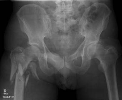
FIG. 4.7.1 Unstable comminuted intertrochanteric fracture with subtrochanteric extension.
Management
A complete evaluation is essential to formulate an early treatment plan as intertrochanteric fractures occur most frequently in the elderly. Patients may lose up to 1.5 L of blood from a comminuted fracture and are often dehydrated, malnourished and in significant pain on arrival in the emergency department (ED). Parenteral analgesia and fluid resuscitation are important in preparation for theatre.
Skin traction or immobilization with sandbags prevents further soft-tissue damage and bony comminution and reduces blood loss. Full preoperative evaluation requires a search for associated injuries, such as rib fractures, distal radial fractures and vertebral compression fractures at the level of T12 and L1.
An ECG, bloods and chest X-ray help elucidate the cause of the fall and may indicate the need for associated medical treatment.
Treatment is with open reduction with internal fixation (ORIF) which produces better anatomical alignment, a shorter hospital stay and improved function with reduced mortality by comparison with conservative management.
Complications
Survival is directly related to the patient’s age and pre-existing medical factors.
Greater trochanteric fracture
Mechanism
Isolated fractures of the greater trochanter are uncommon. They usually occur between 7 and 17 years of age and involve true epiphyseal separation secondary to indirect trauma. Forceful muscular contraction by the gluteus medius causes avulsion of the apophysis. The displaced, non-comminuted fragment may be separated by up to 6 cm.
Greater trochanteric fractures in adults are rare and usually result from direct trauma, causing a comminuted fracture whose fragments are rarely displaced and usually involve only part of the trochanter.
Clinical assessment
Patients with a greater trochanter injury are tender to palpation over the area of avulsion or comminution, but bruising is uncommon. There is often an associated flexion deformity of the hip as a result of pain and muscle spasm and weight bearing causes a limp.
Management
The prognosis is good after these fractures. Most are treated with bed rest for 3 days, followed by non-weight bearing and crutches for 4 weeks. Open reduction and internal fixation are indicated for marked separation of the bony fragment.
Lesser trochanteric fracture
Isolated fractures of the lesser trochanter usually occur in children and young athletes, with 85% occurring before the age of 20.
Mechanism
Lesser trochanter fractures are usually an apophyseal avulsion injury secondary to forceful contraction of iliopsoas.
Clinical assessment
Patients complain of pain on flexion and internal rotation of the hip. Examination reveals tenderness in the femoral triangle. The patient is unable to flex the hip and raise the foot off the ground in a seated position (Ludloff sign specific for the iliopsoas muscle).
Radiology is often inconclusive, as there may not be complete separation of the bony fragment; comparison views may be required.
Management
Ten days of bed rest and slow mobilization result in full recovery. Open reduction and internal fixation are not indicated, even with wide apophyseal separation.
Subtrochanteric femoral fractures
The subtrochanteric region of the femur lies between the lesser trochanter and a point 5 cm distally. Fractures in this region are termed subtrochanteric. They account for 11% of hip fractures and occur in the elderly with osteoporosis, bone metastases or end-stage renal failure. High-energy injuries in young adults with normal bone are less common.
Mechanism
Ninety per cent of these fractures result from blunt trauma, either due to a simple fall in the elderly or following a high-speed motor vehicle accident (MVA) or fall from a height in young adults. In some countries, up to 10% are due to high-energy gunshot wounds.
Classification
A variety of classification systems is available, but none is widely used. As with intertrochanteric fractures, it is best to describe the location, presence of comminution and the position of the lesser trochanter proximal or distal to the fracture line.
Clinical assessment
A subtrochanteric fracture is usually isolated in the elderly. However, as substantial force is required in young adults, the presence of other injuries must be sought. The limb distal to the fracture is usually held in abduction, flexion and external rotation. Haemorrhage from a comminuted subtrochanteric fracture may be up to 2 L. Assess the patient’s circulatory status and commence fluid and blood.
Management
The affected limb is immobilized in a splint following parenteral analgesia and a femoral nerve block. Suitable splints include proprietary splints, such as the Donway or Hare. Fluid resuscitation is started as required. The older, more laborious Thomas splint is now rarely used.
Orthopaedic referral is essential for open reduction and internal fixation of these fractures.
Complications
There is up to a 20% mortality associated with these fractures within the first year in the elderly. They are associated with a higher rate of non-union and implant failure as subtrochanteric bone is cortical thus, unlike the cancellous bone involved in intertrochanteric fractures, these fractures often lack the vascularity for adequate new bone growth and repair. The further down the shaft of femur the fracture line is located, the greater the degree of non-union and implant failure.
Hip dislocation
The hip joint is inherently stable and considerable force is required to produce a dislocation. Associated injuries must always be sought. Hip dislocations are classified anatomically into anterior and posterior, depending on the final position of the femoral head relative to the acetabular rim.
Non-prosthetic hip dislocations are an orthopaedic emergency as the femoral head’s blood supply is precarious and also due to the proximity of the sciatic nerve. Failure to reduce a hip dislocation within 6 hours dramatically increases the risk of AVN and sciatic nerve ischaemic damage.
Posterior hip dislocation
Mechanism
Posterior dislocations represent 85–90% of traumatic hip dislocations. Classically, a direct distal force applied to the flexed knee, with the hip in varying degrees of flexion as when seated in the front of a car, causes a posterior dislocation of the hip. The hip and knee are usually flexed to 90° and the hip adducted, which is the least stable position for the hip to be in.
The force applied by the dashboard in a head-on collision to a seated individual may produce an isolated posterior dislocation. The abducted and partially flexed hip in the same scenario is more stable and, if the force of impact is great enough, will result in a posterior dislocation with displaced acetabular fracture.
Clinical assessment
Examination of the affected limb reveals shortening, adduction, internal rotation and some degree of flexion. A single AP pelvis radiograph is usually adequate to confirm a posterior dislocation. However, as up to half of these dislocations are associated with an acetabular, femoral head or femur fracture, further radiological imaging is essential. Judet views, AP hip with internal rotation and AP and lateral femoral views have been used extensively in the past, but are now largely superseded by CT.
Neurological examination
Neurological examination is essential in a posterior dislocation, particularly with marked internal rotation which may compress the sciatic nerve and its branches. This results in neurological deficit particularly in the peroneal nerve distribution. Associated injuries, such as ipsilateral knee ligament disruption with a posterior cruciate rupture, must be looked for as well.
Management
The orthopaedic team is consulted early. A thorough search for associated periarticular and distal limb injuries, neurological evaluation and adequate imaging are essential in the ED.
Closed reduction
Closed reduction of a posterior hip dislocation may be performed in the ED under procedural sedation, unless there is immediate access to an operating theatre (see Chapter 22.3).
Allis manoeuvre
There are numerous methods of relocation, many requiring significant physical strength. The most common is the Allis manoeuvre, whereby the patient lies supine with assistants on either side stabilizing the pelvis by downward pressure on the anterior superior iliac spines. The operator applies longitudinal traction to the lower leg with the hip slightly flexed in the line of the femur and knee in 90° of flexion. The leg is internally and externally rotated until the femoral head is rearticulated with the acetabulum. Lateral traction to the inside of the thigh may assist.
Other techniques include the lateral traction–countertraction method and the Whistler technique.
Complications
The risk of developing AVN is directly proportional to the length of time the hip remains dislocated and increases dramatically if the dislocation is not reduced within 6 hours of injury. Sciatic nerve neuropraxia may occur in 15% of cases but is usually relieved by reduction.
Permanent ischaemic changes with neurological deficit secondary to pressure necrosis have been reported in up to 3% of cases, usually in the peroneal nerve distribution. Missed knee injuries occur in up to 15% of cases as well as patellar, tibial plateau and posterior cruciate injuries.
Anterior hip dislocation
Anterior dislocations account for 10–15% of traumatic hip dislocations and are associated with femoral neurovascular injury and occult hip joint fracture. They usually result from a direct blow to the abducted and externally rotated hip. When the hip is in abduction, the femoral neck or greater trochanter impinges on the rim of the acetabulum. A direct force applied distally can lever the head out of the acetabulum and tear the anterior capsule of the hip.
Classification
Anterior dislocations may be superior or inferior. Type I or superior dislocations occur when the hip is extended at the time of injury. These are also known as iliac dislocations. Type II or inferior dislocations occur when the hip is flexed at injury and are also known as obturator dislocations.
They may be further subclassified as simple dislocation, associated femoral neck fracture or associated acetabular fracture.
Clinical assessment
The superior type of injury causes an extended, externally rotated and slightly abducted distal limb. The distal limb in the inferior type of dislocation is externally rotated, abducted and in flexion. The femoral head may be palpated around the anterior superior iliac spine in superior types and in the obturator foramen in inferior types.
A neurovascular examination is essential in anterior dislocation, particularly the superior type, where trauma to the femoral artery, vein and nerve is common. Hip and pelvis radiographs must be studied carefully for associated fractures of the acetabulum and femoral head. Further imaging with CT is indicated, particularly for persistent post-reduction pain.
Management
General examination looking for associated life-threatening injuries is essential as this type of hip dislocation is usually associated with high-energy trauma. Orthopaedic consultation is mandatory because of the high probability of vascular injury and the need for closed reduction under general anaesthesia.
Complications
Early complications in superior dislocations result from direct pressure on the femoral vessels with the potential for distal neurovascular compromise. Late complications include post-traumatic arthritis and AVN. Recurrent dislocation is common when anterior capsular healing is incomplete following inadequate immobilization after reduction.
Controversies
Efficacy of applying skin traction and immobilization to reduce extracapsular femoral fractures in the ED.
Early use of CT and MRI to evaluate the reduced non-prosthetic hip to limit (missed) associated morbidity.
Whether the hip reduction should take place in the ED or in the operating theatre; it is essential to treat hip dislocations early.
Further reading
1. Dahners LE, Hundley JD. Reduction of posterior hip dislocations in the lateral position using traction–countertraction: safer for the surgeon? J Orthopaed Trauma. 1999;13:373–374.
2. Garden RS. The structure and function of the proximal end of the femur. J Bone Joint Surg. 1961;43B:576–589.
3. Hirasawa Y, Oda R, Nakatani K. Sciatic nerve paralysis in posterior dislocation of the hip. Clin Orthoped. 1977;126:172–175.
4. Holmberg S, Conradi P, Kalen R, Thorgren KG. Mortality after cervical hip fracture: three thousand two patients followed for six years. Acta Orthopaed Scand. 1986;57:8–11.
5. Jazayeri M. Posterior fracture dislocations of the hip joint with emphasis on the importance of hip tomography in their management. Orthoped Rev. 1978;7:59–64.
6. Keller CS, Laros GS. Indications for open reduction of femoral neck fractures. Clin Orthoped. 1980;152:131–137.
7. Walden PD, Hamer JR. Whistler technique used to reduce traumatic dislocation of the hip in the emergency department setting. J Emerg Med. 1999;17:441–444.
4.8 Femur injuries
Peter Allely and Michael Cadogan
Essentials
1 Early femoral fracture reduction and immobilization in traction reduces mortality.
2 Haemorrhagic shock is a major complication, with a closed femoral fracture average blood loss of 1200 mL.
3 Femoral shaft fractures are associated with other significant injuries including those to the pelvis, hip, knee and/or multitrauma.
Femoral shaft fracture
Mechanism
Considerable force is required to break the adult femur in the absence of osteoporosis or metastatic disease with a bony secondary. The majority of femoral shaft injuries occur in young adults following road traffic incidents, falls from a height or a gunshot wound.
Classification
No universally accepted classification system exists for fermoral shaft fractures. A precise description of the fracture provides the orthopaedic specialist with an indication of the potential for blood loss and the urgency of definitive management.
Femoral fractures are either open or closed and may be transverse, oblique, spiral or segmental. They may occur within the proximal third, midshaft or distal third of the femur. The degree of fracture comminution, soft-tissue involvement and neurovascular status should also be described.
The majority of fractures occur in young adults with healthy bones and are transverse. Greater mechanical force usually results in comminution (Fig. 4.8.1). Minimal force with pathological bone tends to produce metaphyseal fractures with propagation into the shaft.

FIG. 4.8.1 Comminuted femoral fracture.
Stress fractures
Stress fractures of the femoral shaft are becoming increasingly common. They occur when repetitive mechanical forces are applied to the femur, such as in marathon running or military recruits. They are associated with pain in the midthigh and apparently normal X-rays, although a bone scan will detect the fracture. Low-impact training, such as cycling, is used in rehabilitation. They are rarely displaced.
Clinical evaluation
The clinical diagnosis of femoral shaft fracture is usually straightforward. The thigh is shortened and externally rotated, with the hip held in slight abduction. Palpation reveals tenderness over the fracture site and extreme pain on attempted movement. Neurovascular injuries are rare, but the distal pulses, capillary refill and distal sensation must be carefully examined.
Vascular damage
Vascular damage is usually limited to rupture of the profunda femoris perforating branches in closed fractures. The resulting tense, swollen haematoma is limited to the thigh and is not associated with distal circulatory compromise. However, penetrating trauma from gunshot wound and open fractures may cause femoral artery disruption with distal circulatory compromise, so repeated vascular evaluations are important.
Any evidence of an expanding haematoma or diminished distal pulses requires further investigation with Doppler imaging or arteriography.
Associated injuries
Commonly associated injuries include fractures of the pelvis, the femoral head and neck, dislocation of the hip and soft-tissue disruption of the knee. Up to 50% of closed femur injuries are associated with meniscal and collateral ligament injuries in the knee, although it is usually impossible to evaluate reliably these injuries in the acute setting. Up to 1.5 L of blood may extravasate into the surrounding soft tissues.
Management
The treatment of any associated multitrauma to the head, neck, thoracic or pelvic injury should take priority. However, early reduction of a femoral fracture is an important part of haemorrhage control. Administration of analgesia, fluid resuscitation and fracture reduction and splinting are ideally performed prior to X-ray of the lower limb, unless this is immediately available.
Analgesia
Adequate pain relief is essential in the emergency department (ED). Intravenous opioid analgesia is necessary, and titrated to effect. A femoral nerve block is an important adjunct that should be performed prior to fracture reduction and splinting (see Chapter 22.2).
Reduction and splinting
Early fracture reduction and splinting in traction decreases overall mortality and pain, limits blood loss and reduces the risk of fat embolism. Fractures are returned to near anatomical alignment using longitudinal traction following appropriate analgesia with the knee in extension.
Proprietary splints, such as the pneumatic Donway or Hare traction splint, have replaced the old skin traction (Thomas) splint in the ED.
Traction is an interim procedure prior to definitive management as it cannot hold a constant force of sufficient magnitude to maintain the length and alignment of an adult femur fracture.
Fluid resuscitation
Haemorrhagic shock is a major complication with an average blood loss from a closed femoral fracture of 1200 mL. All patients must be resuscitated with intravenous fluid and blood, kept fasted and an indwelling catheter inserted to monitor fluid balance.
Orthopaedic management
Early operative fixation, typically intramedullary nailing, is indicated in adults within 8 hours. Open fractures require immediate operative debridement with antibiotic cover such as flucloxacillin 2 g IV or cephazolin 2 g IV, followed by delayed intramedullary nailing.
Complications
Complications include fat embolus syndrome, haemorrhagic shock and adult respiratory distress syndrome, with a higher incidence in comminuted fractures. Long-term complications of shortening, malalignment and non-union may result in post-traumatic arthritis.
Early mobilization following intramedullary nailing greatly reduces complications associated with prolonged immobilization. Patients older than 60 years with closed femoral fractures have a complication rate of 54% and a mortality of 17%.
Controversies
Arteriography, particularly in distal third femoral fractures following proximity penetrating trauma, even in the absence of initial vascular compromise.
Diagnosis of stress fractures from repetitive exercise.
Further reading
1. Provost R, Morris J. Fatigue fracture of the femoral shaft. J Bone Joint Surg. 1969;51A:487–498.
2. Russell RH. Fracture of the femur A clinical study (abridged by Peltier LF). Clin Orthoped. 1987;224:4–11.
3. Taylor M, Banerjee B, Alpar E. Injuries associated with a fractured shaft of the femur. Injury. 1994;25:185–187.
4. Vanganess C, DeCampos J, Merritt P. Meniscal injury associated with femoral shaft fractures An arthroscopic evaluation of incidence. J Bone Joint Surg. 1993;75:207–209.
5. West H, Turkovich G, Donnell C. Immediate prediction of blood requirements in trauma victims. South Med J. 1989;82:186–189.
4.9 Knee injuries
Michael Baker and Michael Cadogan
Essentials
1 The knee is the most commonly injured joint in the body.
2 Knee injuries often occur in the young, usually associated with sport.
3 The mechanism of injury is an essential part of the history; examination should include the hip and ankle joint.
4 Anterior cruciate ligament disruption is associated with meniscal and collateral ligament injuries in 50% of cases.
5 Lateral tibial plateau fractures are associated with anterior cruciate and medial collateral ligament disruption, whereas medial tibial plateau fractures are associated with posterior cruciate and lateral collateral ligament disruption.
6 Knee dislocations require urgent reduction with assessment for a popliteal artery injury.
Anatomy
The knee is the largest, most complicated joint in the body. It is a synovial, complex hinge joint comprising the patellofemoral and tibiofemoral joints. Movement ranges from 10° of extension to 140° of hyperflexion, with up to 12° of rotation present through the full arc.
The ligaments of the knee are classified as extracapsular or intracapsular. The main extracapsular ligaments are the medial and lateral collaterals (MCL and LCL). The main intracapsular ligaments are the anterior and posterior cruciate ligaments (ACL and PCL), which are extrasynovial. The collateral ligaments provide lateral stability and stability in extension, whereas the cruciate ligaments provide knee stability in flexion.
Knee stability is further enhanced by muscular extensions, such as the vastus medialis giving patella stability, the fibrous extension of vastus lateralis and medialis (the patellar retinaculum) strengthening the knee anteriorly and the iliotibial tract strengthening the knee in slight flexion.
Clinical assessment
An exact history of the mechanism of injury, degree of force, presence of immediate swelling and the ability to bear weight straight after the injury are essential to guide the diagnosis of soft-tissue injuries. Injury may be due to direct or indirect trauma and may involve valgus or varus stress.
Knee physical examination
Always examine both legs with the patient undressed and lying supine on a trolley (not sitting). Visual inspection may reveal swelling, bruising, erythema, deformity and/or an associated wound.
Swelling appearing within the first few hours of trauma is usually associated with a haemarthrosis due to a vascular response to subchondral, bone or synovial injury. Swelling developing gradually over several hours to days is more likely due to a serous effusion from a synovial reaction.
Knee palpation
Start palpation away from the point of trauma to detect warmth, swelling, crepitus, muscle mass and neurovascular status and then to localize the areas of maximal tenderness to define the underlying pathology. Assess the insertion points of the quadriceps tendon, patellar tendon, collateral ligaments and the medial and lateral joint lines, as well as the bony structures of the knee joint.
Assess active and passive movements of the knee joint, noting the degree of flexion, extension and internal and external rotation. Always test for the ability to straight leg raise while supine, to assess for potential damage to the extensor mechanism of the knee.
Anterior and posterior drawer tests
Complete the examination with an assessment of the knee’s functional stability. The stability of the anterior and posterior cruciate ligaments may be crudely determined with the anterior and posterior drawer tests. Ligamentous laxity decreases with age, so comparison with the opposite knee is more important than absolute laxity.
The patient must be supine with the hip flexed at 45°, the knee flexed at 90° and the hamstrings relaxed. The examiner sits on the patient’s foot to stabilize the limb and attempts to demonstrate abnormal forward movement of the tibia on the femoral condyles (positive anterior drawer test) and/or abnormal backward movement of the tibia on the femoral condyles (positive posterior drawer test). However, the accuracy of the anterior drawer test, as defined by subsequent arthroscopy, is only 56% for rupture of the ACL, whereas posterior displacement of the tibia by more than 5 mm is indicative of PCL ruptures with a specificity of 85%.
Lachman’s test
Lachman’s test is a more sensitive manoeuvre in the acute setting for testing ACL integrity, with a sensitivity of 86% and specificity of 91% [1]. The operator supports the distal femur with one hand with the knee in 20–30° of flexion and uses the other hand to draw the tibia forwards on the femoral condyles. Increased anterior displacement of the proximal tibia compared to the unaffected limb indicates a positive test.
Collateral laxity
The collateral ligaments are assessed by applying a varus or valgus stress to the knee in 0° and 30° of flexion. The degree of ligamentous laxity is determined by the amount of movement produced between the tibia and fibula, compared to the normal side.
McMurray’s test
McMurray’s test is used to demonstrate a meniscal injury. The patient lies supine and the knee is passively flexed and extended. One hand is placed over the knee to feel for crepitus while the other hand rotates the tibia on the femur. Internal rotation tests the lateral meniscus and external rotation tests the medial meniscus. Pain and crepitus at the extremes of movement indicate a positive test.
Apley’s test
Apley’s test is also used to demonstrate a meniscal injury. This is performed with the patient lying prone with the knee flexed to 90°. The tibia is rotated on the femur with downward pressure on the heel. Meniscal tears are associated with pain on downward pressure at the extremes of movement, and relieved by the release of pressure.
Radiology
Clinical decision rules to determine the requirement for knee radiography aim to reduce emergency department (ED) radiographs, waiting times and costs. The most widely used is the Ottawa knee rule.
Ottawa knee rule
The Ottawa knee rule states an X-ray is indicated for acute knee injury in adults with any of the following:
age>55 years
tenderness at the head of the fibula
isolated tenderness of the patella
inability to flex knee to 90°
inability to bear weight (take four steps) immediately and in the ED.
This rule has been validated in a number of studies, with a pooled sensitivity of 98.5% and specificity of 48.6% [2].
Standard knee X-rays
Standard knee X-ray evaluation includes AP and lateral views. The AP view assesses for the integrity of the medial and lateral joint spaces and the femoral tibial angle. It also shows the size, position and integrity of the patella.
Lateral view may identify a lipohaemarthrosis effusion, seen as a horizontal line demarcating darker, more radiolucent fat floating on lighter, more radiodense blood. This is indicative of an intra-articular fracture and is most helpful when the actual injury is hard to see, such as with an undisplaced condylar fracture, patellar or tibial spine fracture.
Oblique X-rays are helpful in elucidating a tibial plateau fracture. The tunnel view enhances the intercondylar region.
A skyline X-ray is taken to evaluate further the patella and patellofemoral joint, particularly following reduction of a patellar dislocation. It can identify undisplaced vertical fractures of the patella and subtle subluxation not seen on the conventional views.
Computed tomography
Computed tomography (CT) is important to define fractures, such as those of the tibial plateau. Magnetic resonance imaging (MRI) is reserved for evaluation of complex soft-tissue knee injuries, unless arthroscopy is preferred.
Fractures around the knee joint
Distal femur
Distal femoral fractures account for 4% of femoral fractures. They are usually associated with high-energy injuries secondary to a fall or a direct blow to the femur in a motor vehicle incident.
Classification
Distal femoral fractures are divided anatomically into supracondylar, intercondylar and isolated condylar fractures. Supracondylar fractures are extra-articular and occur immediately above the femoral condyles. Intercondylar fractures involve separation of the femoral condyles. Although the fracture line may extend through the supracondylar region, in general, these are treated as intra-articular fractures.
Isolated condylar fractures are uncommon and occur when a varus or valgus force is applied to a weight-bearing, extended knee. The tibial eminence is driven into the femoral intercondylar notch, creating an intra-articular fracture associated with significant ligamentous disruption.
Clinical assessment
Patients with an injury to the distal femur are in significant pain and unable to bear weight. Examination may reveal swelling, deformity, rotation and shortening. The joint is tender to palpate along the medial or lateral joint lines and an acute haemarthrosis secondary to associated ligamentous injury or intra-articular involvement is common.
Examine the whole lower limb to exclude ipsilateral hip dislocation, associated tibial fracture and quadriceps damage. Assess for any neurovascular deficit, including loss of sensation in the web space between the first and second toes due to deep peroneal nerve injury.
Anteroposterior and lateral X-rays of the femur and knee reveal the fracture and its degree of displacement or comminution. A pelvic X-ray is necessary to exclude an associated proximal femur fracture or hip dislocation.
Management
Administer adequate analgesia and apply a splint in the ED to prevent movement at the fracture site. Distal femoral fractures are a complex orthopaedic problem and long-term complications of malunion, quadriceps adhesion and osteoarthritis are common.
Early orthopaedic input is required in all cases. Fractures with joint incongruity or displacement require open reduction and internal fixation. Cast immobilization alone may be sufficient for undisplaced or impacted fractures without joint involvement, particularly in the elderly patient [3].
Tibial plateau fracture
The tibial plateaus are the superior articulating surfaces of the medial and lateral tibial condyles and are covered by hyaline cartilage and a fibrocartilaginous meniscus. Their integrity is vital for knee alignment, articulation and stability.
Mechanism
Tibial plateau fractures account for 1% of all skeletal fractures. They are most common in the elderly, often as a result of a simple fall. They occur when a valgus or varus deforming force is applied to the weight-bearing knee. Lateral tibial plateau fractures are twice as common as medial injuries, but both tibial plateaus are involved in 10–30% of cases.
Anterior fractures occur when the knee is in extension and posterior fractures when the knee is flexed. High-energy complicated fractures can also occur, often in the younger age group, and are associated with extensive ligamentous and soft-tissue injury.
Classification
Fracture classification is complex owing to the varying degrees of comminution, displacement and compression of the plateaus. The most widely used system is that of Schatzker, which divides the factures into six different types [4]. Fracture types 1, 2 and 3 involve the lateral tibial plateau with increasing articular depression (Fig. 4.9.1). Type 4 involves the medial plateau. Fracture types 5 and 6 involve both tibial plateaus with increasing comminution and joint instability.

FIG. 4.9.1 Schatzker type 3 tibial plateau fracture.
Segond fracture
A tibial plateau avulsion fracture at the site of lateral capsular ligament insertion is called a Segond fracture. It appears as an elliptical, vertical fragment of bone parallel to the lateral condyle just distal to the plateau. They are associated with excessive internal rotation and varus stress to the flexed knee and are usually associated with sporting injuries. They are an important marker of ACL disruption and/or medial meniscus injury and indicate severe rotatory instability.
Clinical assessment
Patients present with a painful, swollen knee and are usually unable to bear weight. Pain and haemarthrosis limit active and passive movements of the knee. Focal tenderness is palpated at the fracture site and over any associated collateral ligament tears.
Distal circulatory compromise may be secondary to compression of the popliteal artery by comminuted subcondylar fragments [5]. Peroneal nerve neurapraxia and paralysis may complicate displaced lateral condylar fractures, resulting in foot drop. Soft tissue injuries occur in up to 35% of injuries. Generally, lateral tibial plateau fractures are associated with ACL and MCL disruptions, whereas medial plateau fractures are associated with PCL and LCL disruptions.
Radiology
Most tibial plateau fractures are evident on standard knee X-rays, although oblique views may be required to elucidate a subtle fracture and to help classify a fracture. CT is important to evaluate further non-displaced and comminuted fractures and for operative planning. An MRI is preferred to quantify the degree of any associated ligamentous damage [6].
Management
Orthopaedic consultation is essential. Lateral fractures with<2 mm displacement and less than 5° of angulation may be treated conservatively with a cast or splint, but comminuted fractures with articular surface disruption require open reduction and internal fixation [7]. Other surgical indications include open injuries, fractures with vascular injury and a fracture associated with unstable ligamentous injury.
Common complications include undiagnosed neurovascular injury, compartment syndrome and osteoarthritis.
Fractures of the tibial spine and intercondylar eminence
The tibial spine separates the medial and lateral tibial condyles and is divided into anterior and posterior areas by the intercondylar eminence. These areas provide flat surfaces for the attachment of the ACL and PCL, respectively. The intercondylar eminence is divided into a medial and a lateral tubercle visible on anteroposterior X-rays, although nothing actually attaches to them.
Mechanism
Most tibial spine and intercondylar eminence fractures occur in children as the cruciate ligaments are stronger than the skeletal physeal plates [8]. Considerable force is required for these fractures to occur in an adult. The tibial spine is usually fractured during violent knee twisting movements.
The anterior tibial spine fractures 10 times more frequently than the posterior. Intercondylar eminence fractures are associated with severe hyperextension or hyperflexion injuries.
Clinical assessment
The patient complains of severe pain, immediate swelling of the knee and inability to bear weight. The knee is usually held in slight flexion and cannot be fully extended. Examination confirms the presence of an acute haemarthrosis and limited knee movement. An associated ACL disruption may be confirmed with a positive Lachman’s or anterior drawer test, although pain may prevent these.
Radiology
AP and lateral X-rays plus tunnel or oblique views are used to confirm the diagnosis. MRI is preferred to quantify the degree of any associated ligamentous damage.
Management
Most injuries are treated conservatively, but refer a displaced fracture with marked ligamentous injury for open reduction and internal fixation.
Patellar fracture
The patella is the largest sesamoid bone in the body and lies within the quadriceps tendon. It improves the stability, strength and mechanical advantage of the extensor mechanism and offers some protection to the femur.
Mechanism
Patellar fractures account for 1% of skeletal injuries and occur predominantly in males between the ages of 20 and 50 years as a result of direct or indirect trauma. Direct trauma to the anterior aspect of the patella results in an incomplete, stellate, comminuted or vertical patellar fracture. These commonly occur in motor vehicle incidents when the knee strikes the dashboard. There is usually little or no separation of the bony fragments as the medial and lateral quadriceps expansions remain intact.
Indirect trauma occurs when stumbling or falling forwards [9]. The combination of powerful quadriceps contraction proximally and the strong patellar insertion distally overcomes the intrinsic strength of the patella and leads to a transverse fracture. These fractures account for up to 80% of patellar fractures and occur mainly in the central and lower third of the patella. The extent of the fragment separation is dependent on the degree of quadriceps expansion tear.
Clinical assessment
Examination reveals pain, swelling and bruising over the patella. The ability to walk and extend the knee actively are dependent on the type of fracture and are important when considering surgical repair. Test for the ability to perform a straight leg raise while in a supine position in order to confirm the integrity of the knee extensor mechanism.
A patient with a non-displaced fracture may be ambulatory and able to demonstrate active knee extension against gravity. Patients with displaced transverse patellar fractures are unable to extend the knee actively.
Radiology
Patellar fracture is diagnosed by X-ray with additional skyline views. A fracture needs to be differentiated from a bipartite patella, which represents failure of the patella ossification centres to fuse, seen in 1–6% of the population. The bipartite patella has smooth borders that are well corticated, with minimal separation between fragments.
Management
Fractures with fragment displacement of more than 2 mm are associated with disruption of the extensor mechanism and require referral to an orthopaedic specialist for open reduction and internal fixation with tension band wiring.
Treat a non-displaced patella fracture conservatively with an intact extensor mechanism with a long-leg cast in full extension for 6 weeks.
Dislocations around the knee joint
Dislocation of the knee
Knee dislocations are rare and usually occur in males in their third decade. They are an orthopaedic emergency that is associated with vascular damage and require urgent reduction.
Mechanism
Tibial femoral knee dislocation usually involves rupture of both cruciate ligaments and one collateral ligament. Such injuries are associated with high-velocity injuries, such as from a motorcycle incident. They are described with respect to the displacement of the tibia in relation to the femur. Anterior dislocations are the most common.
Clinical assessment
Spontaneous reduction prior to the emergency department is common, so a high index of suspicion and careful assessment are required. Examination usually reveals gross distortion of the knee, with the clinical deformity being easily palpable. Knee dislocations are associated with a high rate of peroneal artery (20–80%) and popliteal nerve injury, so a careful neurovascular assessment is essential.
Compression and distortion of the posteriorly placed popliteal artery and vein may cause distal vascular compromise, although 10% of vascular injuries are associated with normal pedal pulses. Peroneal nerve dysfunction is present in up to 50% of patients suffering knee dislocation, causing foot drop and sensory impairment of the dorsum of the foot and lateral border of the foot and leg.
Radiology
Immediate plain X-rays confirm the dislocation, but should not delay analgesia and reduction.
Management
Prompt consultation with the orthopaedic and vascular teams is essential, with early reduction under procedural sedation in the ED if necessary. Neurovascular status should then be reassessed and documented. The risk of developing a compartment syndrome and/or needing amputation is increased when reduction is not performed within 6 hours. Failed reduction secondary to buttonholing of the femoral condyle is uncommon and requires open reduction under general anaesthesia.
In the past, angiography was performed in all cases of knee joint dislocation. There is now a move towards serial examinations if the ankle brachial index (ABI) is>0.90 (the ratio of the systolic blood pressure [SBP] measured at the ankle to that measured at the brachial artery) and all distal pulses are equal and present [10].
Patella dislocation
Traumatic patellar dislocation is common and may become recurrent, with further patellar subluxation or dislocation. The majority of dislocations occur in the setting of patellofemoral dysplasia or malalignment syndromes secondary to hypoplastic vastus medialis, a shallow trochlear groove or genu valgum. Lateral dislocations are overwhelmingly the most common, usually caused by a direct blow to the anterior or medial surface of the patella. The medial retinaculum is disrupted by being stretched in subluxations and torn in a dislocation.
Clinical assessment
Patients complain of the knee suddenly ‘giving way’, accompanied by immediate pain and swelling. They are unable to bear weight or extend the knee. Palpation reveals an anterior defect, a laterally deviated patella, swelling and medial joint line tenderness. In a spontaneously reduced injury, the ‘apprehension test’, performed by applying gentle pressure on the medial border of the patella, may confirm the diagnosis.
Standard AP and lateral X-rays confirm the diagnosis and are important to exclude an associated osteochondral fracture. However, particularly in recurrent dislocations, X-ray can follow immediate reduction.
Management
Some dislocations reduce spontaneously or are reduced prior to arrival at the ED. Closed reduction is performed following suitable analgesia or procedural sedation. Apply anteromedial pressure to the lateral aspect of the patella with the thumbs while gently extending the knee. Immobilize the knee in an extension splint for 3–6 weeks after post-reduction X-rays, to allow the medial retinaculum time to heal.
Complications
Up to 50% of patients suffer symptoms of instability or anterior knee pain following traumatic dislocation. Recurrent dislocation occurs in over 15% of cases and may require surgical repair.
Proximal tibiofibular joint dislocation
Mechanism
The proximal tibiofibular joint is supported by a capsule anteriorly, the popliteus muscle posteriorly and the LCL superiorly. Tibiofibular joint dislocation is rare and only possible when the LCL support is relaxed with the knee in flexion. Thus, they occur mainly in violent athletic twisting injuries, such as during the shot put.
Clinical assessment
The patient holds the knee flexed at 20–30° and is able to bear weight with difficulty, with point tenderness over the fibula head. Common peroneal nerve neuropraxia is unusual.
Radiology
AP and lateral comparison views reveal the dislocation, which is usually anterolateral.
Management
Reduction is by firm pressure over the head of fibula towards the centre of the knee, under procedural sedation. Success is associated with a satisfying ‘click’. Surgical intervention is rarely needed.
Soft-tissue knee injuries
Collateral ligaments
Medial and lateral ligament damage is often associated with sporting events. They are graded 1 to 3, indicating the degree of disruption to the ligamentous fibres:
grade 1: stretching of the fibres only
grade 2: partial tear, mild instability but firm endpoint on stress testing
grade 3: complete disruption of fibres, clear instability with no endpoint on stress testing.
Medial collateral ligament
The medial collateral ligament (MCL) complex comprises a long superficial ligament with a distal point of insertion and a short deep ligament attached to, and stabilizing, the medial meniscus. The MCL provides medial stabilization to the knee joint in conjunction with the capsule and semimembranosus, resisting valgus laxity and medial rotational instability.
MCL injuries are the most common isolated knee ligament injury. They occur when an excessive valgus force is applied to the knee, usually by a direct blow to the lateral aspect. The greater the degree of the valgus deforming force, the greater the risk of an associated ACL disruption.
Lateral collateral ligament
The lateral collateral ligament (LCL) is the phylogenetically degenerate part of peroneus longus. It is a cord-like ligament running from the lateral epicondyle of the femur to the head of the fibula. It is separated from the lateral meniscus by the popliteal tendon. The LCL is the major lateral stabilizer of the knee, providing the main resistance to varus deforming forces, especially when the knee is extended.
LCL injuries are less common, but more debilitating, than MCL injuries. The lower incidence of LCL injuries is a result of the lateral ligament’s mobility and the protective effect of the opposite leg. They result from a direct blow to the medial aspect of the knee. Associated injuries to the insertion of biceps femoris and to the common peroneal nerve at the fibular head must be excluded.
Clinical assessment
Examine for point tenderness at the site of injury, demonstrable laxity and a haemarthrosis. Gently stress the affected ligament complex to check for reproducible pain. Complete rupture of the ligament complex is associated with instability and stress testing causes the joint line to open up on the affected side.
Test the MCL in 0° and 30° of flexion. Apply pressure to the lateral joint line with one hand while the other hand creates a valgus stress by gently pushing the medial malleolus laterally. At 0°, the medial complex is reinforced by the ACL but, at 30°, testing is specific for MCL rupture.
Lateral instability is assessed with pressure applied to the medial joint line with one hand and by a varus stress performed by moving the lower leg medially.
Radiology
Standard X-rays can only reveal collateral ligament injury when there has been a bony avulsion. Calcification at the origin of the MCL occurs in chronic injuries (Pellegrini–Stieda). In complex cases, an MRI helps delineate the degree of ligamentous disruption and highlights associated injuries.
Management
The majority of isolated collateral ligament injuries are treated conservatively, provided damage to the ACL and PCL complexes has been excluded [11]. Discomfort is reduced by immobilizing the knee in a proprietary splint, such as a three-panel Velcro knee immobilizer, or by an elastic knee support, with ice massage and anti-inflammatory drugs.
Quadriceps strengthening exercises are essential to aid recovery and early return to movement using a hinged splint. Early orthopaedic opinion is sought for a grade 3 injury to consider surgical repair, as this is best done within 48 hours of injury.
Cruciate ligaments
The cruciate ligaments are the primary stabilizers of the knee in flexion and extension.
Anterior cruciate ligament
The anterior cruciate ligament (ACL) extends from the medial aspect of the lateral femoral condyle to the anterior inter-condylar area of the tibia. It prevents backward displacement of the femur on the tibial plateau and limits extension of the lateral condyle of the femur. It helps control the rotation of the knee in twisting and turning activities and is much more commonly injured than the PCL.
Mechanism
The ACL is commonly injured during sporting activities, such as skiing and rugby, with patients readily able to define the causative mechanism. Injury results from direct trauma as the tibia is forcefully displaced anteriorly on the femur or the femur posteriorly on the tibia or by indirect injury when the flexed knee suffers a sudden twisting movement with the foot firmly planted on the ground.
Clinical assessment
ACL injuries are classically associated with sudden severe pain and an audible ‘pop’, with an acute haemarthrosis and inability to bear weight. Immediate swelling of the knee indicates serious intra- articular pathology.
The anterior drawer test or Lachman’s test is used to assess ACL integrity. ACL disruption is associated with meniscal and collateral ligament injuries in 50% of cases, with the most common combination involving the triad of ACL and MCL disruption with a lateral meniscal tear.
Radiology
X-rays may show avulsion of the anterior tibial spine, although an MRI scan is necessary to determine ACL rupture with certainty, having over 90% sensitivity and specificity.
Management
Arthroscopy is the gold standard in assessing the integrity of the ACL and has the advantage of allowing simultaneous debridement and repair. The decision to undertake reconstruction is dependent on a number of factors including the patient’s age, level of activity, the degree of instability, presence of other knee structures injured and the time post-injury (ideally within 2 weeks).
Both autograft and allograft materials have been utilized in the repair. However, meta- analysis shows insufficient evidence to determine superiority of surgical repair over conservative management as regards long-term function and joint stability [12].
Posterior cruciate ligament
The posterior cruciate ligament (PCL) extends from the lateral aspect of the medial femoral condyle to the posterior intercondylar area of the tibia. It prevents excessive forward displacement of the femur on the tibia and is essential in providing mechanical support when walking downhill or down stairs, as it is the only stabilizing structure in the flexed, weight-bearing knee.
Mechanism
PCL rupture is normally caused by a posteriorly directed force on the proximal tibia, such as with falls onto the tibial tubercle, knee dislocation and dashboard injuries. It is less commonly associated with sporting injuries than ACL ruptures.
Clinical assessment
Immediate pain and swelling are common with PCL rupture which, unlike the ACL, rarely causes any popping or tearing sensation. Stability is usually adequate to allow partial weight bearing. Isolated PCL ruptures result in posterior ‘sag’ of the tibia compared to the unaffected limb, with the posterior drawer test performed to assess PCL integrity. Associated MCL and ACL disruptions are common and must be actively sought.
Radiology
X-rays may reveal avulsion of the posterior tibial spine but, as with ACL injuries, MRI demonstrates over 90% sensitivity and specificity for PCL rupture and may be more reliable than arthroscopic examination.
Management
The treatment of isolated PCL rupture is largely non-operative and focuses initially on pain management and non-weight-bearing immobilization. When a PCL injury is combined with other ligamentous injuries, operative intervention within 2 weeks is common.
Patellar tendon rupture
The patellar tendon is the final connection of the extensor mechanism from the inferior pole of the patella to the tibial tuberosity. Rupture usually occurs under the age of 40 years, often associated with a previous history of patellar tendonitis or steroid injections. Injury is associated with stressful sporting activity and occurs with forceful quadriceps contraction, with significant pain.
Examination
Examination reveals a palpable defect, which may be masked by significant swelling. Test the extensor mechanism by asking the patient to straight leg raise against gravity. This is impossible in a complete rupture and is painful and difficult in an incomplete injury. Comparison lateral X-ray views of both knees may reveal a high-riding patella.
MRI is indicated in complex cases to differentiate partial and complete tears. Partial tears are treated non-operatively with cast immobilization in extension for 6 weeks. Complete tears of the patellar tendon are referred to the orthopaedic specialist for surgical intervention.
Quadriceps tendon injury
The quadriceps tendon is a trilaminar junction of the quadriceps muscle. Rupture is commonest in the older age groups as the tendinous blood supply declines. Young persons usually suffer a muscular disruption. Rupture occurs three times more commonly than patellar tendon rupture, usually due to a direct blow to the knee or a hyperextension injury. It is associated with intense pain and the patient is unable to walk without assistance.
Examination
Examination reveals a tender, palpable defect more apparent on attempted knee extension. Swelling secondary to a haemarthrosis and bruising are usually present. The straight leg raise is impossible in complete rupture, whereas extension of the knee from a flexed position cannot be performed in a partial tear.
Comparison lateral knee X-rays may demonstrate a low-lying patella in the affected knee. In doubtful cases, MRI is indicated to distinguish between a partial and a complete rupture.
Management
A partial tear is treated non-operatively, but a complete rupture requires early surgical intervention for the best results.
Patellar and quadriceps tendonitis (jumper’s knee)
Both these extensor tendons are susceptible to tendonitis secondary to repetitive overloading. Patellar tendonitis is more common than quadriceps tendonitis. Patients present with anterior knee pain with point tenderness over the inferior or superior pole of the patella, commonly in athletes who participate in running and jumping activities.
Inflammation and pain in patellar tendinitis at the insertion point of the patella tendon into the patella is six times more common than at the insertion to the tibial tuberosity. Patellar tendonitis may be associated with fragmentation of the inferior pole of the patella on X-ray.
Initial treatment includes rest, ice and anti-inflammatory medication. Longer-term recovery and prevention requires conditioning and training of the extensor musculature.
Meniscal injury
The menisci are semilunar fibrocartilaginous structures found on the medial and lateral sides of the superior aspect of the tibia. They enhance the fluidity of articulation between the femoral and tibial condyles and increase the stability of the tibiofemoral articulation.
The medial meniscus is immobile, being firmly attached to the deep portion of the medial collateral ligament and joint capsule. The lateral meniscus has a uniform thickness and a larger tibial area than the medial. It has no attachment to the LCL and is more mobile than the medial meniscus, making it twice as likely to be injured.
Mechanism
Meniscal injuries are usually associated with collateral or cruciate ligament injury, which should be sought when examining the acutely injured knee. Chronic degenerative processes account for only a small percentage of injuries. The menisci are uncommonly injured in isolation, but suspect an isolated meniscal injury in the young athlete sustaining a violent twisting or rotational injury to the weight-bearing knee.
Clinical assessment
The patient is able partially to bear weight following meniscal injury and usually complains of medial or lateral joint line pain. Delayed swelling, intermittent locking and a sensation of the knee ‘giving way’ with sudden loss of stability are clues to meniscal damage.
Examination usually confirms the presence of an effusion and joint line tenderness, especially in the extremes of flexion and extension. McMurray’s test may be positive, but is not pathognomonic and, acutely, the pain prevents adequate hyperflexion for the test to be accurate.
The ‘locked’ knee is held in 30° of flexion, with a springy block to extension on examination and associated pain. A bucket-handle meniscal tear is classically associated with a true ‘locked’ knee. They are longitudinal tears, usually of the medial meniscus, and frequently associated with ACL disruption.
Radiology
Routine X-rays do not show any direct evidence of meniscal damage but are useful to exclude commonly associated bony injuries. MRI may determine both meniscal and ligamentous injuries in complex cases.
Management
Arthroscopy is used to evaluate and treat meniscal injuries, revealing the extent of damage and determining whether resection of the torn cartilage or meniscectomy are required. An acutely locked knee should be referred for urgent arthroscopy.
Controversies
Evaluating and validating new clinical decision tools for radiography in acute knee injuries.
Replacing cylindrical plaster cast treatment of patellar fractures and some soft-tissue injuries with three-panel Velcro knee immobilizer splints and hinged supports.
Role of CT scan in condylar and tibial plateau knee injuries.
Indications for MRI over arthroscopy in complex soft tissue knee injuries.
References
1. Scholten R, Opstelten W, Van Der Plas C, et al. Accuracy of physical diagnositic tests for assessing ruptures of the anterior cruciate ligament: a meta-analysis. J Fam Pract. 2003;52:689–694.
2. Bachmann LM, Steurer J, Ter Riet G, et al. The accuracy of the Ottawa knee rule to rule out knee fractures: a systematic review. Ann Intern Med. 2004;140:121–124.
3. Crist B, Della Rocca G, Murtha Y. Treatment of acute distal femur fractures. Orthopedics. 2008;31:681–690.
4. Markhardt B, Gross J, Monu J. Schatzker classification of tibial plateau fractures: use of CT and MRI imaging improves classification. Radiographics. 2009;29:585–597.
5. Bandyk DF. Vascular injury associated with extremity trauma. Clin Orthoped. 1995;318:117–124.
6. Kode L, Lieberman JM, Motta AO, et al. Evaluation of tibial plateau fractures: efficacy of MR imaging compared with CT. Am J Roentgenol. 1994;163:141.
7. Fenton P, Porter K. Tibial plateau fractures: a review. J Trauma. 2011;13:181–187.
8. Kendall NS, Hsu SY, Chan KM. Fracture of the tibial spine in adults and children. J Bone Joint Surg. 1992;74B:848.
9. Scolaro J, Bernstein J, Jaimo A. Patellar fractures. Clin Orthop Relat Res. 2011;469:1213–1215.
10. Nicandri G, Dunbar R, Wahl C. Are evidence-based protocols which identify vascular injury associated with knee dislocation underutilized? Knee Surg Sports Traumatol Arthrosc. 2010;18:1005–1012.
11. Coen A, Chad J, Steinar J, et al. Injuries to the medial collateral ligament and associated medial structures of the knee. J Bone Joint Surg. 2010;92A:1266–1280.
12. Linko E, Harilainen A, Malmivaaran A, Seitsalo S. Surgical versus conservative interventions for anterior cruciate ligament ruptures in adults. Cochrane Database Syst Rev. 2005;18:CD001356.
Further reading
1. Roberts D, Stallard T. Emergency department evaluation and treatment of knee and leg injuries. Emerg Med Clin N Am. 2000;18:67–84.
4.10 Tibia and fibula injuries
Michael Baker and Michael Cadogan
Essentials
1 Tibial shaft fractures are the commonest long bone fracture and the subcutaneous nature of the tibia leaves it vulnerable to open injury.
2 Neurovascular injury and compartment syndrome are a risk in tibial shaft fractures.
3 Proximal fibula fractures are associated with common peroneal (lateral popliteal) nerve injury.
4 Tibial tubercle injuries range from apophysitis to fracture.
Anatomy
The tibia is the weight-bearing strut of the lower leg. Proximally, the tibia articulates with the femoral condyles and distally, the bony extension provides medial stability to the ankle joint. Its shaft is triangular in cross-section and is subcutaneous anteromedially.
The fibula head is proximal and connects to the fibular shaft by the neck. Distally, the fibula is palpated subcutaneously as the lateral malleolus.
The tibia and fibula are connected by superior and inferior tibiofibular joints and a dense interosseous membrane. Distally, this union is strengthened by a syndesmosis, which enhances the stability of the ankle mortise.
Lower leg fascial compartments
The lower leg is divided into four compartments by bone and fascia. Each compartment contains a sensory nerve and muscles with specific functions. Increased pressure within a compartment is evaluated clinically by impaired function according to the functional anatomy.
Anterior compartment
The anterior compartment contains the tibialis anterior and the long toe extensor muscles (extensor hallucis longus and extensor digitorum longus) that dorsiflex the ankle and foot. The deep peroneal nerve supplies these muscles and the first web space of the foot. The anterior tibial artery is contained within the compartment down to the ankle, where it becomes the dorsalis pedis artery.
Lateral compartment
The lateral compartment contains the peroneus longus and peroneus brevis which evert the foot and the superficial peroneal nerve that supplies sensation to the dorsum of the foot.
Superficial posterior compartment
The superficial posterior compartment contains the gastrocnemius, plantaris and soleus muscles which plantarflex the ankle. The sural nerve lies in this compartment before piercing the fascia to supply the lateral side of the foot and distal calf.
Deep posterior compartment
The deep posterior compartment contains the tibialis posterior and long toe flexor muscles (flexor hallucis longus and flexor digitorum longus) which plantarflex the toes. The tibial nerve is within the compartment and supplies sensory function to the sole of the foot. The posterior tibial and peroneal arteries also lie in this compartment.
Fractures of the tibia
Tibial shaft fracture
Tibial shaft fractures are the most common long bone fracture and are usually easily recognized. They are also the commonest open fracture owing to the subcutaneous nature of the tibial shaft.
A considerable amount of direct or indirect energy is needed for the tibial shaft to fracture. Direct injuries may occur secondary to bending forces or a direct blow. Direct violence, such as in a motor vehicle incident or when a pedestrian is struck, cause deformation at the site of contact, resulting in transverse or comminuted, usually open, fractures. High-energy injuries have an increased degree of displacement, comminution, soft tissue injury and fibular involvement. They are associated with marked vascular, interosseous and bony involvement and are unstable, with a high risk of compartment syndrome. Up to 15% are complicated by malunion or non-union.
Indirect torsional forces applied to the tibia produce a spiral fracture as the body rotates about a fixed foot. Such injuries are common in skiing incidents and have increasing degrees of comminution depending on the amount of energy applied.
Classification
The description of the fracture must be clear and concise in relation to the following (see also Table 4.10.1 for the AO classification of tibial shaft fractures) [1]:
skin integrity: open or closed
anatomical site: proximal, middle or distal third
fracture type: transverse, oblique, spiral or comminuted
angulation of the distal fragment in relation to the proximal fragment, expressed in degrees and direction (anterior, posterior, varus or valgus)
degree of displacement and rotation
involvement of the fibula
any joint involvement.
Table 4.10.1
AO classification of tibial shaft fractures
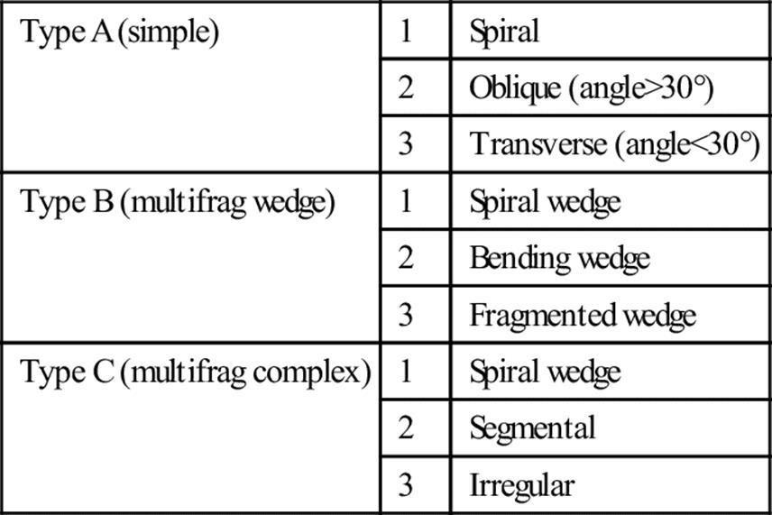
Clinical assessment
Pain at the site of fracture is usually severe. The patient is unable to bear weight and inspection reveals swelling and deformity of the leg. The skin is checked for integrity and to identify areas of pressure caused by any displaced fragments. The neurovascular status of the lower leg and foot are assessed as a matter of urgency, including skin colour, capillary refill and the distal dorsalis pedis and posterior tibial pulses. Associated injuries of the ipsilateral femur, hip, knee, foot and pelvis must be excluded.
Nerve injury
Contusion of the peroneal nerve may occur in high-energy injuries with proximal fibular fractures, although direct peroneal nerve injury can occur rarely in a closed tibial shaft fracture. The motor function of the deep peroneal nerve is tested by active ankle and toe dorsiflexion and the sensory function is tested in the first dorsal web space. The motor function of the superficial peroneal nerve is tested by active foot eversion and the sensory function is tested over the dorsal lateral aspect of the foot.
Radiology
AP and lateral views of the lower leg must include the entire tibia and fibula, from the knee to the ankle, to document tibial shaft fracture, identify associated fibula fracture, the fracture pattern and any degree of comminution and/or displacement (Fig. 4.10.1). X-ray the knee and ankle joint to look for associated joint involvement.
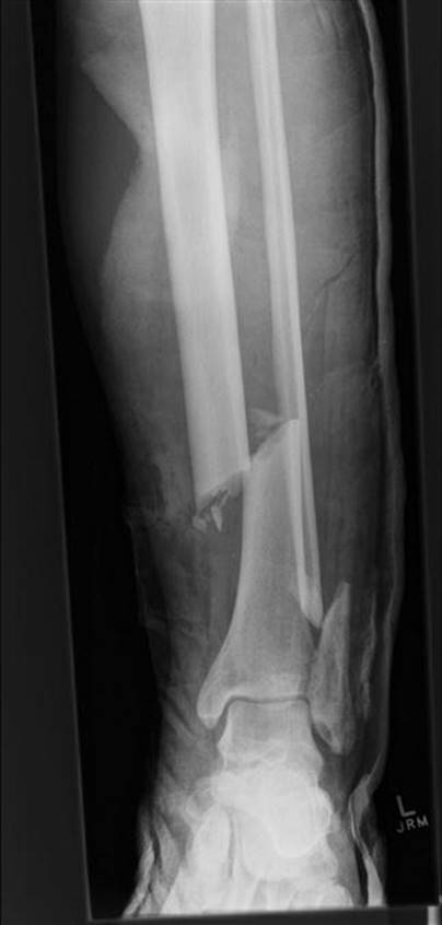
FIG. 4.10.1 Open, oblique, distal third tibial fracture with displaced varus deformity and fibula involvement.
Management
Parenteral analgesia is the first priority, usually an intravenous opiate, followed by reduction of any displaced and/or compound fractures and immobilization in a long leg cast as early as possible.
Compartment syndrome
Emergency department (ED) documentation of the neurovascular status is essential to exclude acute neurovascular injury, as well as to detect actual or potential compartment syndrome. Development of a compartment syndrome may occur in up to 20% of closed injuries and can take up to 24 hours to appear. It is less common in compound injuries.
Increasing pain despite reduction and casting are an early indicator of compartment syndrome. This should be suspected if there is weakness of muscle action, pain on passive movement and diminished sensation over the distal sensory nerve territory. Pulses can still be present, so a strong pedal pulse does not exclude the presence of a compartment syndrome. The deep posterior compartment is most commonly affected, followed by isolated elevation of pressure in the anterior compartment.
Compartment pressures
If there is concern, compartment pressures may be measured. A compartment syndrome can occur if the compartment pressure is>30 mmHg or within 30 mmHg of mean arterial pressure (MAP). Routine continuous pressure monitoring in tibial shaft fractures does not result in an improved outcome and is not recommended [2].
Open wounds
Open wounds are assessed for depth and associated soft tissue damage, then dressed to avoid further contamination. Reduce a displaced, rotated or angulated fracture in the ED under appropriate analgesia and procedural sedation (see Chapter 22.3). Reduction aims to stop local swelling, release the tension of any skin ‘tented’ over a displaced fracture and relieve associated soft tissue damage. Exposed bone is returned under the skin after appropriate decontamination. Check the tetanus status and give parenteral antibiotics such as flu/dicloxacillin 2 g 6-hourly IV or cephazolin 2 g 8-hourly IV.
Immobilize the leg following reduction in 20° of knee flexion and request post-reduction X-rays to confirm the position. Re-check and document the neurovascular status of the lower leg and foot.
Definitive orthopaedic management
There are various options for definitive management including conservative, closed reduction, open reduction and internal fixation (ORIF) and intramedullary rods. No approach is superior, with a recent meta-analysis finding insufficient evidence to support any particular management option [3].
Non-operative management
Non-operative management in an above-knee plaster of Paris (POP) cast is appropriate for a low- energy fracture without significant comminution, shortening or displacement. Conservative management may also be appropriate for fractures with>50% cortical contact,<5–10° of varus/valgus angulation,<10–15° of anterior or posterior bowing,<5–7° of rotation and no more than 10–15 mm of shortening [4].
Operative management
Operative management is considered for patients with a high-energy displaced fracture, compound fracture or who have failed closed treatment. Intramedullary nailing results in shorter hospital stay, fewer outpatient visits and earlier return to work [5]. ORIF is also considered for displaced intra-articular fractures of the tibia involving the knee or ankle.
Tibial tubercle fracture
The tibial tubercle lies proximally on the anterior border of the shaft of the tibia. It is readily palpable beneath the infrapatellar bursa and receives the insertion of the patellar tendon. Fractures of the tubercle are uncommon and occur in adolescents, typically as a result of indirect injury sustained during sports involving jumping, such as sudden acceleration or deceleration of the extensor mechanism of the knee producing an avulsion fracture. Risk factors for this condition include Osgood–Schlatter’s disease and osteogenesis imperfecta [6]. Similar to other tibial fractures, up to 20% are complicated by compartment syndrome.
Three grades of injury are described by Watson–Jones. In type I injuries, the tubercle is hinged upwards without displacement; type II injuries involve avulsion of a small portion of the tubercle proximally; and type III injuries are intra-articular. The fragment is displaced and may be comminuted.
Examination reveals pain and tenderness over the anterior aspect of the knee and proximal tibia. There may be a haemarthrosis and loss of active extension, depending on the severity of the injury.
Plain X-rays confirm the diagnosis. The lateral tibial view reveals the avulsion fragment, its degree of displacement and comminution.
Management is dependent on the degree of displacement and the presence of joint involvement. Watson–Jones type I and II injuries are treated with cylindrical long leg casts until healed. Type III injuries require open reduction and internal fixation with tension band wiring and fixation screws [7].
Osgood–Schlatter’s disease (traction apophysitis of the tibial tubercle)
The commonest differential diagnosis of tibial tuberosity fracture is Osgood–Schlatter’s disease, that refers to traction apophysitis of the tibial tubercle caused by repeated microtrauma to the growing tubercle during adolescence. It is chronic and, unlike tubercle fractures, is not accompanied by a haemarthrosis. Active knee extension is possible, albeit painful.
Treatment is conservative with rest, ice, compression and non-steroidal anti-inflammatory drugs (NSAIDs), followed by graded return to sporting activities. This is a self-limiting condition with full recovery expected in 1–2 years, when the apophysis closes.
Tibial stress fractures
Tibial stress fractures are common, affecting the proximal third of the tibia in adolescents and the junction of middle and distal thirds of the tibia in runners. Clinically, there is point tenderness over an area of induration. X-rays may appear negative early or show periosteal reaction after 3–5 weeks. A bone scan or MRI can detect these injuries earlier.
The differential diagnosis includes ‘shin splints’ (see below), fascial hernias and exertional compartment syndrome. Management is conservative, reducing activity and impact on the tibia. Symptoms may persist for over 12 months.
Shin splints
Shin splints is also known as ‘medial tibial stress syndrome’ and is characterized by exercise-induced pain in the midsection of the leg, with tenderness along the posteromedial border of the middle and distal thirds of the tibia. The tenderness is usually more diffuse than the localized tenderness of a stress fracture. There may be periostitis near the origin of the soleus and flexor digitorum longus muscles. It is rare in children under 15 years of age.
Fractures of the fibula
Proximal fibula fractures may occur in isolation (uncommon) or in association with tibial and ankle injuries.
Associated tibial shaft fracture
Most fibula fractures are associated with a fracture of the tibial shaft and are managed as for tibial fractures. The pattern of the associated fibular fracture indicates the degree of energy imparted. Severe comminution of the fibula or tibiofibular diastasis implies disruption of the interosseous membrane and indicates an unstable fracture.
The fibula usually heals well with whatever treatment is selected for the tibia, with a better rate of union. Complications of fibula fractures associated with tibial shaft fractures are rare.
Isolated proximal fibula fractures
Isolated proximal fibula or fibula shaft fractures are less common. They are usually associated with a direct blow to the lateral aspect of the leg, causing local tenderness, swelling, bruising and difficulty walking. A neurovascular assessment is important as the common peroneal nerve passes around the neck of fibula and may be contused or disrupted in these isolated injuries. Rarely, thrombosis of the anterior tibial artery may occur.
Full-length AP and lateral X-rays of the tibia and fibula, including the ankle and knee joints, will confirm the fracture pattern.
Management
Non-displaced fractures associated with little pain are treated with ice, compression bandage, analgesia and non-weight-bearing crutches for 3 weeks. Weight bearing is progressive as tolerated. Mildly displaced fractures or those with significant pain may require a long leg cast for up to 6 weeks. A severely displaced fracture or one associated with peroneal nerve deficit, such as foot drop, requires orthopaedic consultation and consideration for fixation.
Maisonneuve fracture
A medial malleolus or distal tibial fracture associated with a proximal fibula fracture is termed a Maisonneuve fracture. These are unstable and occur when an external rotatory force is applied to the ankle, resulting in partial or complete disruption of the syndesmosis between the tibia and fibula. Palpation of the proximal fibula following a complex ankle injury is therefore essential to exclude this fracture. Refer all these fractures to the orthopaedic team for operative fixation.
Controversies
Use and value of compartment pressure monitoring in the ED.
Which clinical features or actual compartment pressure require active management.
Indications for operative intervention in closed tibial shaft fractures.
References
1. Muller ME, Nazarian S, Koch P. The AO classification of fractures New York: Springer-Verlag; 1988.
2. Harris I, Kadir A, Donald G. Continuous compartment pressure monitoring for tibia fractures: does it influence outcome? J Trauma. 2006;60:1330–1335.
3. Littenberg B, Weinstein L, McCarren M, et al. Closed fractures of the tibial shaft A meta-analysis of three methods of treatment. J Bone Joint Surg. 1998;80A:174–183.
4. Wheeless’ Textbook of Orthopaedics.<http://www.wheelessonline.com/ortho/cast_treatment_of_tibial_fractures>[Accessed Feb. 2013].
5. Hooper G, Keddell R, Penny I. Conservative management or closed nailing for tibial shaft fractures: a randomized prospective trial. J Bone Joint Surg. 1991;73B:83–85.
6. Frey S, Hosalker H, Cameron D, et al. Tibial tuberosity fractures in adolescents. J Child Orthop. 2008;2:469–474.
7. Balmat P, Vichard P, Pem R. The treatment of avulsion fractures of the tibial tuberosity in adolescent athletes. Sports Med. 1990;9:311–316.
Further reading
1. Roberts D, Stallard T. Emergency department evaluation and treatment of knee and leg injuries. Emerg Med Clin N Am. 2000;18:67–84.
4.11 Ankle joint injuries
Michael Baker and Michael Cadogan
Essentials
1 Ankle injuries are common and occur as isolated injuries or related to high-energy multitrauma.
2 Lateral malleolar fractures are the most common ankle fracture.
3 The Ottawa Ankle Rules (OAR) are used to determine the need for imaging of the ankle (or foot) in adults with an isolated acute ankle injury.
4 The Weber and Henderson (Potts) classifications are the most commonly used for describing ankle fractures.
5 Ankle sprains should be mobilized early.
6 The calf-squeeze test (Thompson or Simmond’s test) is used to confirm the diagnosis of Achilles tendon rupture.
Anatomy
The ankle joint is a complex hinge joint that permits articulation between the tibia, fibula and talus, providing a stable but mobile support for the body. It helps absorb the forces of ambulation, maintain an upright posture and allows for uneven terrain.
The stability of the ankle joint relates to the bony architecture, joint capsule and the ligaments. The bones and ligaments are best visualized as a ring structure centring on the talus, which provides stability. This ring is made up of the tibial plafond, medial malleolus, the medial (deltoid) ligament, calcaneus, lateral collateral ligaments, lateral malleolus and the syndesmotic ligaments. The joint becomes unstable when more than one element of this ring structure is disrupted.
Bony mortise
The lateral malleolus of the distal fibula, the medial malleolus of the distal tibia and the distal tibial plafond form the bony mortise of the joint. This provides intrinsic stability constraining the wedge-shaped talus distally. The medial ligament of the ankle or deltoid ligament fans out from the tip of the medial malleolus to attach to the tuberosity of the navicular, the medial aspect of the talus and the sustentaculum tali of the calcaneus. The lateral ligament comprises three discrete parts, the anterior and posterior talofibular ligaments and the calcaneofibular ligament.
Most ankle joint injuries are a result of abnormal movement of the talus within the mortise. Movement causes stress to the encompassing ring of structures of the ankle joint, with instability arising when disruption of the malleoli or their associated ligaments results in distraction of the talus within the mortise.
Clinical assessment
Injuries around the ankle include fractures to the ankle and adjacent tarsal bones, ligamentous sprains, dislocations and tendon ruptures. All these are considered when assessing the patient with an ankle injury.
History
Inability to bear weight and the presence of swelling immediately following an injury imply significant pathology. Additional essential information includes the circumstances of the injury, position of the foot at the time and the magnitude and direction of loading forces applied, particularly rotational. A history of inversion injury should prompt the examiner to assess also the base of the fifth metatarsal for an avulsion fracture by the insertion of peroneus brevis.
Examination
Give the patient analgesia, ice and elevate the affected limb. Examination of the ankle includes the entire lower leg and begins with a comparison between the injured and non- injured sides. Note the skin integrity and presence of bruising, swelling or deformity.
Palpate for point tenderness to localize ligament, bone or tendon injury, which should commence at a site away from the area of obvious injury. The entire length of the tibia and fibula as well as the base of the fifth metatarsal, calcaneus and Achilles tendon are examined. Palpation of the posterior aspects of the malleoli should commence 6–10 cm proximally and include both ends of the collateral ligament attachments. The anterior plafond and the medial and lateral aspects of the talar dome are then palpated in plantarflexion.
Then assess the range of active and passive movement at the ankle joint, including inversion, eversion, dorsiflexion and plantarflexion. A soft tissue injury is likely when there is a significant difference between the active and passive ranges of movement.
Finally, always check the foot for motor or sensory impairment, capillary return, the presence of dorsalis pedis and posterior tibial pulses and injury to the base of the fifth metatarsal.
Stress testing for ligamentous instability
Stress testing for ligamentous instability of the acutely injured ankle and/or an evaluation of weight-bearing ability should only proceed when clinical suspicion of a fracture is low. All require appropriate analgesia for evaluation.
The talar tilt test assesses the calcaneofibular ligament by applying a gentle inversion stress to the calcaneum.
The anterior and posterior drawer tests assess the anterior and posterior talofibular ligaments by gentle forward traction on the heel.
Radiology: Ottawa ankle rules
Standard radiography of the acutely injured ankle includes anteroposterior, lateral and mortise views. All patients with an obviously deformed fracture or dislocation should have immediate X-ray following analgesia.
The need for imaging of the ankle or mid-foot in a patient with less obvious injury may be determined using the Ottawa ankle rules (OAR). When used on a competent patient, the OAR are more than 98% sensitive for detecting clinically relevant ankle fractures in adults [1,2] and 98% sensitive in children [3,4].
Ottawa ankle rules
These rules specify that an ankle X-ray series is only required if there is any pain in the malleolar region and any one of:
bone tenderness over the posterior aspect or inferior tip of the distal 6 cm of the lateral malleolus
bone tenderness over the posterior aspect or inferior tip of the distal 6 cm of the medial malleolus
inability to bear weight for at least four steps, both immediately after the injury and at the time of emergency department (ED) evaluation.
The OAR also include indications for taking an additional foot X-ray series (see Foot injuries, Chapter 4.12).
Other imaging
Computed tomography (CT) is used to evaluate further complex fractures and magnetic resonance imaging (MRI) for difficult or recalcitrant ligamentous injuries.
Ankle fracture classification
Several classification systems are used to describe ankle fractures and dislocations, some more complicated than others.
Weber classification
The Weber classification system (1972) divides ankle fractures into three types, based on the level at which the fibula fractures. The more proximal the fibula fracture, the greater the associated syndesmosis disruption and potential for ankle instability.
Type A fractures involve the distal fibula below the level of the tibial plafond; type B involve an oblique or spiral fracture at the level of the syndesmosis; and type C occur when the fibula is fractured above the level of the syndesmosis (ankle joint).
The original Weber classification system does not take into account medial or posterior malleolar fractures. The AO system applies three subdivisions to each Weber fracture type to account for these injuries and to define further ankle stability [5].
Henderson or Pott’s classification
The Henderson or Pott’s classification is a simple system based on radiographic findings:
Unimalleolar fractures affecting the lateral or medial malleolus. The stability of these fractures is dependent on the integrity of contralateral ligaments and the inferior tibiofibular joint.
Bimalleolar fractures affecting the medial and lateral malleoli, which are usually unstable.
Trimalleolar fractures involving the medial, lateral and posterior tibial plafond, which are always unstable.
Fracture management
A grossly displaced fracture is reduced and splinted promptly in the ED, under procedural sedation, prior to imaging if distal ischaemia is identified and/or the skin integrity compromised (Fig. 4.11.1). These will require elevation and orthopaedic admission for operative management.

FIG. 4.11.1 Unstable bimalleolar ankle fracture.
Non-displaced fractures
Non-displaced (<3 mm) unimalleolar Weber A fractures with an intact mortise joint (no talar shift) on X-ray are treated non-operatively in a below-knee plaster of Paris (POP) cast in a neutral position, that is, with the ankle at 90° with no inversion or eversion. Refer these injuries for orthopaedic follow up, with advice that fracture movement may occur and that operative intervention may still be required.
The management of an isolated non-displaced Weber B injury, with the fracture line at the level of the syndesmosis, is controversial. If the deltoid ligament is intact and there is no talar shift, a conservative approach results in good outcomes with low rates of subsequent surgical intervention [6].
Displaced fractures
Displaced and potentially unstable fractures require early orthopaedic consultation, including all bimalleolar and trimalleolar fractures, those unimalleolar fractures with contralateral ligamentous injuries and Weber C injuries. The majority of these injuries will require operative intervention (open reduction and internal fixation [ORIF]).
Tibial plafond (pilon) fractures
Tibial plafond (pilon=hammer) fractures involve the distal tibial metaphysis and result from high-energy injuries directed through the talus into the distal tibia, with tibial plafond disruption. The frequency has increased with greater numbers of motor vehicle incidents and falls from heights. They are usually associated with other multiple injuries and often open, comminuted or associated with extensive soft tissue deformity.
Reduce and splint the fracture under appropriate analgesia and procedural sedation to decrease the potential for massive soft tissue swelling, with conversion of a closed fracture to an open one as a result of overlying skin necrosis. Treatment usually requires operative fixation.
Maisonneuve fracture
The Maisonneuve fracture is a fracture of the proximal end of the fibula associated with a medial malleolus fracture or disruption of the medial (deltoid) ligament, resulting from external rotation. The proximal fibula fracture is associated with disruption of the interosseous membrane from the tibiofibular syndesmosis up to the proximal fibular head. It may be complicated by a common peroneal nerve injury. They are unstable and require operative fixation.
Ankle dislocations
Ankle dislocations are frequently associated with a fracture, but can occur in isolation and may be open or closed. They result from considerable energy, such as when force is directed against the plantarflexed foot, squeezing the talus out of the mortise. Posterior dislocations are the most common. All require orthopaedic advice for consideration of internal fixation and ligamentous repair.
Closed dislocations
Closed dislocations are associated with marked soft-tissue disruption and skin tethering, although neurovascular compromise is uncommon. These dislocations are reduced promptly in the ED to minimize associated soft-tissue injury, using gentle manipulation under appropriate analgesia and procedural sedation (just as for the grossly displaced fracture).
Despite the potential for ligamentous disruption, they usually have an excellent outcome following immobilization for 8 weeks.
Open dislocations
Open dislocations may be associated with disruption of the dorsalis pedis and posterior tibial vessels. They require surgical debridement in theatre, but again should initially be reduced and splinted with POP in the ED. Open injuries are associated with more long-term complications than closed, in particular traumatic arthritis and reduced mobility.
Soft-tissue injuries
Ligamentous injuries
Ankle sprains
Ankle sprains are one of the most common injuries presenting to the ED: 75% of injuries to the ankle are sprains and 90% of these affect the lateral ligament complex, predominantly the anterior talofibular ligament. Typically, injuries to the lateral ligament proceed from anterior to posterior as increasing force is applied.
Medial ligament disruption is more frequently associated with lateral malleolar fractures or Maisonneuve-type injuries involving the proximal fibula and syndesmosis disruption.
Lateral ligament injuries
Lateral ligament injuries are graded according to the degree of fibre disruption and reflect the progression of injury from anterior to posterior as well as subsequent stability of the ankle joint. However, an accurate assessment of joint stability is usually not possible in the ED setting due to pain limiting examination.
Grade I: partial tear, usually of the anterior talofibular ligament. Patients are usually able to weight bear with minimal swelling and normal stress testing.
Grade II: partial tear, usually extending to the calcaneofibular ligament. There is pain at rest, difficulty weight bearing, significant swelling and mild to moderate joint instability.
Grade III: complete tear of two or more elements of the lateral ligament. Patients are unable to weight bear due to severe pain, with immediate swelling and marked joint instability.
Grade I and most grade II injuries are treated conservatively with rest, ice, compression, elevation and non-steroidal anti-inflammatory drugs for 48 hours. Early functional treatment is likely better than immobilization in the treatment of lateral ligament injuries [7].
Operative intervention for grade III lateral ligament injuries is controversial, with conservative and surgical therapy associated with similar clinical outcomes. Surgery may improve perceived stability and reduce pain and is more commonly undertaken in the athlete [8].
Conservative treatment includes cast immobilization for 6–8 weeks with orthopaedic follow up. Delayed surgical repair or reconstruction has similar results to early intervention.
Achilles tendon rupture
Achilles tendon rupture is traditionally associated with sedentary middle-aged individuals during a burst of unaccustomed strenuous physical activity, although young, fit athletes also sustain this condition.
Predisposing medical conditions when present include rheumatoid arthritis, systemic lupus erythematosus (SLE), chronic renal failure, gout, hyperparathyroidism and long-term steroid or fluoroquinolone use. The segment of the Achilles tendon particularly prone to rupture lies 2–6 cm proximal to the tendon’s insertion into the calcaneus, as blood vessels that supply this area are prone to atrophy. The resultant reduction in collagen cross-linking leads to a reduced tensile strength in the tendon, with the majority of tears complete.
History
Rupture usually occurs while pushing off with a weight-bearing foot, but may occur with sudden dorsiflexion or direct trauma. The sensation of a direct blow to the back of the ankle and even an audible ‘pop’ are followed by difficulty in walking. The patient often states that they thought they had been hit or kicked from behind.
Examination
Examination reveals a visible and/or palpable deficit in the tendon, although swelling around the tendon sheath rapidly masks these signs. Some degree of plantarflexion of the ankle joint is preserved by the other long flexors of the ankle, foot and toes. This should therefore not be used to determine if the Achilles tendon is intact, although the patient cannot stand on tiptoe.
Calf-squeeze test (Thompson test or Simmond’s test)
The calf-squeeze test (Thompson or Simmond’s test) confirms the diagnosis of a rupture with a sensitivity of 96%. Perform this with the patient kneeling on a chair with the feet hanging free over the edge. Alternatively, it is often more comfortable for the patient to lie prone with the feet and ankles extended hanging freely beyond the end of the examination couch.
Demonstrate normal plantar flexion initially on the unaffected calf, by gently squeezing just distal to its maximal girth. Absence of plantarflexion in the affected limb is a positive test and confirms Achilles rupture. Ultrasound is useful if the diagnosis is in doubt and can demonstrate partial or full-thickness tears of the tendon, as well as measure the size of the defect.
Management
The choice of operative or non-operative treatment is controversial [9]. Surgery is more likely to be offered to younger patients, those who are diagnosed early and in those with a larger defect. Operative risks include fistula formation, skin necrosis and infection. However, the procedure has a lower rate of muscle atrophy, a lower re-rupture rate and allows earlier resumption of physical activity. Minimally invasive techniques have resulted in reduced infection rates and time to return to work.
Non-operative management includes applying a POP cast to the ankle in equinus (full plantar flexion) to bring the two ends of the ruptured tendon into apposition. One regimen involves a cast for 4 weeks in equinus, 4 weeks in partial plantarflexion and then 2 weeks in the neutral position. Complications of non- operative management include a higher re-rupture rate (requiring surgical intervention).
Controversies
Whether internal fixation results in an improved outcome compared to conservative management in Weber type B fractures.
Role of bracing, topical anti-inflammatories, laser and ultrasound therapy in the management of ankle sprains.
Operative versus conservative management of Achilles tendon rupture.
References
1. Stiell I, Greenberg G, McKnight R, et al. Decision rules for the use of radiography in acute ankle injury Refinement and prospective validation. J Am Med Assoc. 1993;269:1127–1132.
2. Bachmann L, Kolb E, Koller M, et al. Accuracy of Ottawa ankle rules to exclude fractures of the ankle and mid-foot: systematic review. Br Med J. 2003;326:417–423.
3. Libetta C, Burke D, Brennan P, et al. Validation of the Ottawa ankle rules in children. J Accident Emerg Med. 1999;16:342–344.
4. Plint A, Bulloch B, Osmond M, et al. Validation of the Ottawa ankle rules in children with ankle injuries. Acad Emerg Med. 1999;6:1005–1009.
5. Muller ME, Nazarian S, Koch P. The AO classification of fractures New York: Springer-Verlag; 1988.
6. Martin A. Weber B ankle fracture: an unnecessary fracture clinic burden. Injury. 2004;35:805–809.
7. Jones M, Amendola A. Acute treatment of inversion ankle sprains: immobilization versus functional treatment. Clin Orthopaed Relat Res. 2007;445:169–172.
8. Pijnenburq A, Van Dijk C, Bossuyt P, et al. Treatment of ruptures of the lateral ankle ligaments: a meta-analysis. J Bone Joint Surg. 2000;82A:761–773.
9. Weatherall J, Mroczek K, Tejwani N. Acute Achilles tendon ruptures. Orthopedics. 2010;33:758–764.
Further reading
1. Borrer R, Famo-Salek M, Totten V, et al. Managing ankle injuries in the emergency department. J Emerg Med. 1999;17:651–660.
2. Wedmore I, Charette J. Emergency department evaluation and treatment of ankle and foot injuries. Emerg Med Clin N Am. 2000;18:85–113.
4.12 Foot injuries
Michael Baker and Michael Cadogan
Essentials
1 Most calcaneal fractures are intra-articular, associated with a Bohler’s angle of less than 20° and are at risk of developing compartment syndrome.
2 Major talar fractures have a significant risk of subsequent avascular necrosis.
3 Navicular body fractures may require internal fixation.
4 Fractures of the base of the second metatarsal are pathognomic of Lisfranc injury (Fleck sign).
5 CT imaging is indicated in complicated talar, calcaneal, navicular as well as Lisfranc injuries.
Anatomy
The foot is composed of 28 bones with 57 articular surfaces. It may be divided into three anatomical regions: the hindfoot containing the talus and calcaneum; the midfoot containing the navicular, cuboid and cuneiforms; and the forefoot containing the metatarsals and phalanges.
The subtalar joint collectively describes the three articulations of the inferior aspect of the talus with the calcaneus. It allows inversion and eversion of the hindfoot. The midtarsal joints incorporate the talonavicular and calcaneocuboid joints that connect the hindfoot and midfoot and allow abduction and adduction of the forefoot. The five tarsometatarsal joints (Lisfranc joint complex) connect the midfoot and forefoot and form an arch, which gives stability to the foot.
Clinical assessment
History
Injury to the foot occurs as a result of direct or indirect trauma. Direct trauma is often associated with considerable soft-tissue swelling and fracture. Indirect trauma from a twisting injury usually results in minor avulsion-type injuries. Record any pain, swelling, loss of function, reduced sensation and deformity or associated ankle injury.
Examination
Inspect the area with the patient lying on a bed with both lower limbs exposed and compare to the unaffected limb to identify bruising, swelling, deformity, skin wounds, pallor or cyanosis.
Start gentle palpation over the entire foot away from the area of maximal pain. Point tenderness or crepitus may be elicited at the site of fracture. Specific areas to palpate include the Achilles tendon, calcaneus, base of the fifth metatarsal, the navicular and the area under the head of the second metatarsal.
Ask the patient to demonstrate active foot movements before performing gentle passive movements and compare with the other foot. Evaluate subtalar motion with the foot in a neutral position, with one hand on the lower leg and the other holding the heel. The heel is inverted and everted and should attain 25° of movement.
Midtarsal motion is assessed with one hand stabilizing the heel while the other hand grasps the forefoot at the bases of the metatarsals. The forefoot is pronated, supinated, adducted and abducted. Finally, forefoot motion is evaluated by individually flexing and extending the metatarsophalangeal (MTP) and interphalangeal (IP) joints.
Ask the patient to stand and walk if no obvious focus of the pain is found during the initial examination. Finally, assess the circulation by observing capillary refill, skin colour and the presence of the dorsalis pedis and posterior tibial pulses.
The posterior tibial pulse is palpable behind the medial malleolus, unless there is excessive swelling or damage to the artery. The dorsalis pedis is more variable, being too small to feel easily, or is absent in 12% of the population. Doppler may be needed to determine the presence of flow when there is doubt. Neurological assessment includes motor and sensory function.
Radiology
Standard imaging includes AP, lateral and 45° internal oblique projections. The lateral view visualizes the hindfoot and soft tissues, whereas the oblique and AP projections image the midfoot and forefoot. An axial calcaneal view should also be requested as clinically indicated to best visualize the hindfoot and may reveal a subtle calcaneal fracture.
Ottawa ankle and foot rules
The Ottawa ankle and foot rules provide indications for X-ray for suspected midfoot fractures [1]. All patients with obvious deformities should have X-rays. However, if clinical findings are more subtle, a foot X-ray is only required if there is pain in the midfoot region and any one of:
bone tenderness over the navicular
bone tenderness at the base of the fifth metatarsal
inability to bear weight for at least four steps, both immediately after the injury and at the time of emergency department (ED) evaluation.
This clinical decision rule has a sensitivity approaching 100% and routine use has been predicted to reduce unnecessary X-rays by 30–40% [1]. These rules do not apply to suspected hindfoot or forefoot fractures.
Other imaging
A bone scan is indicated when a stress fracture is suspected and may become positive 2–3 weeks before conventional radiographs demonstrate a fracture. Computed tomography (CT) is used for imaging the calcaneum, subtalar joint and Lisfranc joint in more complex injuries or when a fracture is strongly suspected but plain X-rays are inconclusive.
Hindfoot injuries
Calcaneal fractures
The calcaneus is the largest bone in the foot and is the most commonly fractured tarsal bone. It forms the heel of the foot, provides vertical support for the body’s weight and functions as a springboard for locomotion. The majority of fractures of the calcaneus occur as a result of direct axial compression during a fall from a height. Seven per cent are bilateral. Lower-extremity injuries are present in 25% of cases and vertebral compression fractures are found in 10%, so these regions must be examined as well.
Mechanism and classification
Patients usually present following a fall with direct trauma to the heel. Seventy-five per cent of calcaneal fractures are intra-articular. These fractures may be non-displaced, displaced or frequently comminuted owing to cancellous bone in the calcaneus and the magnitude of associated force (Fig. 4.12.1).

FIG. 4.12.1 Intra-articular comminuted calcaneal fracture.
An isolated fracture of the anterior process of the calcaneus is commonly misdiagnosed as an ankle sprain [2]. It results from inversion causing an avulsion fracture or forced dorsiflexion producing compression against the cuboid.
Clinical assessment
The patient may be able to walk, but weight bearing on the heel is impossible. Examination reveals pain, swelling and tenderness over the heel, with bruising that may extend over the sole of the foot. Associated fractures are common, so examination of the vertebral column, pelvis, affected lower extremity and opposing calcaneus is essential.
Radiology
Standard X-rays usually reveal most comminuted calcaneal fractures, whereas more subtle fractures are visualized with the aid of specific axial (Harris) calcaneal views or on CT scan. The AP view demonstrates the anterosuperior calcaneus and calcaneocuboid joint. The lateral view may reveal compression fractures of the body and posterior facet.
Bohler’s angle
Bohler’s angle is formed by the intersection of a line drawn from the most cephalic point on the tuberosity to the highest point of the posterior facet, with the line from the latter to the most cephalic part of the posterior process of the calcaneus. It normally ranges from 20° to 40° measured on the lateral X-ray and a compression fracture is likely if Bohler’s angle is less than 20° [3]. A CT scan is necessary to define complex fractures and is useful in preoperative planning.
Management
Calcaneal fractures are notoriously difficult to manage and frequently have a poor outcome, with up to 50% suffering chronic pain and functional disability. Intra-articular, displaced and comminuted fractures are prone to gross swelling of the foot with a risk of compartment syndrome. Admit patients with these fractures for elevation, further imaging, such as CT, and consideration of surgical intervention. Operative intervention may be indicated in younger patients and those with greater degrees of Bohler’s angle disruption [4].
Extra-articular fractures and fractures of the anterior process of the calcaneus are usually non-displaced and are treated conservatively in a posterior non-weight-bearing cast for 6 weeks.
Talar fractures
The talus provides support for the body when standing and bears more weight per surface area than any other foot bone. It has no muscular attachments and is held in place by the malleoli and ligaments and comprises a head, neck and body.
The head has articulations with the navicular and calcaneus and the body articulates with the tibia, fibula and calcaneus. The neck joins the head and body and is extra-articular. The blood supply to the talus arises from an anastomotic ring from the peroneal, posterior and anterior tibial arteries and is tenuous and easily disrupted, leading to avascular necrosis.
Mechanism and classification
Major or minor talar fractures are the second most common tarsal fracture. Minor fractures are caused by inversion injuries to the plantar- or dorsiflexed foot, often from minimal trauma, and may present as an apparent ankle sprain. They include avulsion fractures, lateral process fractures commonly seen in snowboarders and posterior talar process fractures. A high index of suspicion is needed to identify these injuries and avoid long-term complications from a delay in diagnosis and treatment [2].
Talar dome fracture
Talar dome fracture is difficult to diagnose on plain films, although a large ankle joint effusion may be apparent. Request specific plain X-ray talar views, although a CT scan is frequently required to confirm the diagnosis. These fractures are important to diagnose as they involve the weight-bearing articular surface of the talus within the ankle joint and missed injury may result in chronic pain and osteoarthritis.
Talar neck fractures
Major talar fractures of the neck, body or head follow significant force, such as a motor vehicle incident, or involve axial loading in a fall from a height (when they are associated with calcaneal fracture). They are commonly accompanied by a subtalar dislocation.
Talar neck fractures account for 50% of major talar injuries and are related to extreme dorsiflexion injuries. The Hawkins classification is used to describe these fractures. Type I fractures are non-displaced with the fracture line entering the subtalar joint between the middle and posterior facets. The risk of avascular necrosis (AVN) with this injury is<10%. Type II fractures are identified by any degree of displacement or subtalar subluxation and have a 30% incidence of AVN. Type III injuries involve displaced talar neck fracture with dislocation from both the subtalar and ankle joints. The incidence of AVN is up to 90%. Commonly associated injuries include vertebral compression, calcaneal and medial malleolar fractures.
Talar head fractures
Talar head fractures are uncommon and result from a compressive force applied to the plantarflexed foot and are associated with disruption of the talonavicular joint, navicular fractures and anterior malleolar fractures.
Clinical evaluation
Minor talar fractures are usually subtle. The patient presents following an inversion injury with mild swelling around the ankle joint and is able partially to bear weight. Active plantar- and dorsiflexion are possible, but inversion and eversion at the subtalar joint is painful. Major talar fractures are associated with large compressive forces and cause considerable swelling and tenderness dorsally.
Radiology
Standard X-rays of the foot reveal all but the most subtle avulsion fractures. A CT scan is required when there is clinical suspicion of talar fracture, but plain films are inconclusive and/or for preoperative planning.
Management
Major talar fractures have a significant risk of subsequent avascular necrosis. A displaced fracture, especially if associated with neurovascular or cutaneous compromise, should be reduced in the ED under appropriate analgesia and procedural sedation by grasping the hindfoot and midfoot and applying longitudinal traction in plantarflexion. Apply a plaster of Paris (POP) posterior splint with the ankle dorsiflexed at 90°. Refer major fractures to an orthopaedic specialist for open reduction and internal fixation.
Minor talar fractures are treated with a below-knee, non-weight-bearing posterior cast with orthopaedic follow up.
Subtalar dislocation
Subtalar dislocations are rare and follow considerable deforming forces. Such injuries involve the simultaneous dislocation of the talonavicular and talocalcaneal joints, with preservation of the tibiotalar joint.
Mechanism and classification
Subtalar dislocations are associated with motor vehicle incidents, but a number occur during sport, particularly basketball. They are described in terms of the final position of the foot in relation to the talus following dislocation. Medial dislocations account for 85% of these injuries and are caused by forceful foot inversion in plantarflexion. Ten per cent of subtalar dislocations are open and 50% are associated with proximally located injuries [5].
Clinical assessment
Subtalar dislocations are associated with obvious deformity, swelling and tension of the skin over the opposing joint margin. Neurovascular status is rarely compromised. Standard X-rays are difficult to interpret because of the distortion of the foot. The most helpful is the AP view, which confirms disruption of the talonavicular joint.
Management
Reduce a closed subtalar dislocation in the ED under appropriate analgesia and procedural sedation to minimize the chance of tented skin over the head of the talus becoming necrotic.
Closed reduction of a medial subtalar dislocation requires firm longitudinal traction applied to the foot, with countertraction on the leg with the knee flexed to relax the tension from the Achilles tendon on the calcaneum, thereby increasing the mobility of the hindfoot. The foot is initially inverted to accentuate the deformity and then everted with digital pressure over the head of the talus to reverse the deformity. Eighty per cent of dislocations can be reduced non-operatively. Following reduction, the ankle is placed in a posterior POP splint in 90° of dorsiflexion. Orthopaedic consultation is required.
Midfoot fractures
The midfoot comprises the navicular, cuboid and cuneiform bones. It is inherently stable and is rarely injured. However, midfoot fractures are associated with a delay in diagnosis owing to the difficulty in X-ray interpretation and poorly localized pain. The Ottawa ankle rules are accurate in determining which patients with midfoot pain require imaging [1].
Navicular fractures
The navicular is a curved bone with extensive articulations. It has a tenuous blood supply and, like the talus, is susceptible to avascular necrosis.
Mechanism and classification
The navicular is the most commonly injured midfoot bone, although the overall incidence is rare. Fractures may involve the dorsal surface, the tuberosity or the body. The dorsal avulsion fracture is the most common, due to an eversion injury and is associated with deltoid ligament or talonavicular capsular injury. Tuberosity fractures also result from eversion injuries with avulsion of the posterior tibial tendon insertion. Body fractures from axial loading are rare and are frequently comminuted.
Clinical evaluation
Point tenderness is elicited over the dorsum and medial aspect of the midfoot. Passive eversion and active inversion reproduce the pain. Standard X-rays usually reveal the fracture, but a CT scan may be required.
Management
Refer all intra-articular, displaced or comminuted fractures to the orthopaedic specialist, as they are frequently complicated by avascular necrosis. Treat dorsal avulsion and tuberosity fractures conservatively in a walking cast for 6 weeks. Navicular body fractures may require internal fixation.
Cuboid fractures
Isolated cuboid fractures are rare and are most commonly associated with Lisfranc-type injuries with lateral subluxation of the midtarsal joint, the so-called ‘nutcracker’ compression fracture of the cuboid between the calcaneus and lateral metatarsal heads, and fractures of the posterior malleolus. They are best visualized with an oblique foot X-ray.
Management
All cuboid fractures require orthopaedic consultation. Treatment ranges from weight-bearing POP casts for undisplaced fractures to operative fixation for displaced and comminuted fractures.
Cuneiform fractures
These fractures are extremely rare and usually occur from direct trauma. An associated Lisfranc injury should be excluded. Displaced fractures require orthopaedic intervention, but non-displaced fractures are treated conservatively in a cast.
Lisfranc fractures and dislocations
The Lisfranc joint complex includes the articulation of the first three metatarsal bases with their respective cuneiforms and articulation of the fourth and fifth metatarsal bases with the cuboid. The second metatarsal is the most important structure within this complex and holds the key to stability.
Mechanism and classification
Injury results from rotational forces applied to the fixed forefoot, axial loads and crush injury. Although commonly associated with vehicular crashes, Lisfranc injuries may also occur in sports that involve fixation of the forefoot, such as horse-riding and rowing.
There are three types classified by the direction of dislocation in the horizontal plane. Divergent dislocations usually involve medial and lateral splaying of the first and second metatarsals. In ipsilateral (homolateral) dislocations, all five metatarsals are displaced in the same direction, either medially or laterally. In isolated dislocations, one or more of the metatarsals is displaced away from the others.
Lisfranc dislocations
A Lisfranc dislocation is usually associated with fracture of the metatarsals, particularly the second metatarsal base and, in 40% of cases, with fracture of the midfoot [6]. Although vascular compromise is uncommon, significant haemorrhage can occur with disruption of the dorsalis pedis branch to the plantar arch as it passes between the first and second metatarsal bases.
Clinical assessment
Lisfranc injuries should be suspected when a midfoot fracture is present. They are associated with severe midfoot pain and inability to bear weight on the toes. Examination reveals deformity, swelling and bruising over the dorsum of the foot. Point tenderness over the joint, with pain on passive abduction and pronation, may also be present.
Radiology
Standard X-rays are sufficient to visualize most Lisfranc injuries. The AP view identifies Lisfranc fractures and oblique views determine their alignment. Lateral views delineate the soft tissues and identify the presence of dorsal or plantar displacement. Fracture of the base of the second metatarsal is pathognomic of a Lisfranc injury (fleck sign, indicating avulsion of the Lisfranc ligament), sometimes with diastasis between the first and second metatarsals. CT scan is frequently necessary to define the degree of disruption to the Lisfranc joint complex.
Management
Refer all Lisfranc injuries for orthopaedic consultation. Most are treated with closed reduction and screw and/or K-wire fixation, followed by non-weight-bearing for 12 weeks [7]. Despite aggressive management, chronic pain, reflex sympathetic dystrophy and degenerative arthritis are common.
Forefoot fractures and dislocations
Metatarsal shaft fractures
Metatarsal shaft fractures occur as a result of direct trauma or a rotational injury to the fixed forefoot. Metatarsal fractures are associated with difficulty in weight bearing and ill-defined tenderness and bruising over the plantar aspect of the foot. They are also commonly associated with Lisfranc injuries and phalangeal fractures. As the second and third metatarsals are relatively fixed, they are prone to stress fractures, typically occurring with repetitive trauma, such as long distance running.
Standard X-rays will detect most fractures and determine their alignment, angulation and displacement. Occult stress fractures may only be evident on CT or bone scan imaging.
Management
Undisplaced closed shaft fractures of the second to fifth metatarsals are treated in a below-knee walking cast for 3–4 weeks. Closed reduction and a non-weight-bearing cast for 6 weeks are necessary when these fractures have>3 mm of displacement or 10° of angulation [8].
Hallux (great toe) metatarsal
Injury to the great toe metatarsal requires more aggressive treatment because of its load-bearing function. Non-displaced fractures require 4–6 weeks in a non-weight-bearing cast, whereas displaced fractures require operative treatment. Orthopaedic consultation is required for multiple or displaced fractures and all fractures of the hallux metatarsal.
Metatarsal head and neck fractures
These fractures usually result from direct trauma and are often multiple. Treat a non- displaced fracture with a walking cast for 4–6 weeks. Displaced fractures require closed reduction to maintain the integrity of the transverse plantar arch.
Fractures of the base of the fifth metatarsal
These are the most common of the metatarsal fractures, with two distinct types. The commonest fracture is to the fifth metatarsal tuberosity, occuring when the plantarflexed foot suddenly inverts. It is caused by avulsion of the lateral band of the plantar aponeurosis and is transverse and usually extra- articular. Rarely, the fracture line extends into the cuboid–metatarsal articulation, but not into the joint between the fourth and fifth metatarsals.
Jones fracture
The second type of fracture is known as the Jones fracture and is defined as a transverse fracture through the diaphysis of the fifth metatarsal from 15 to 31 mm distal to the proximal end of the bone. This fracture is intra-articular as it involves the intermetatarsal articulation of the fourth and fifth metatarsals and is prone to non-union [7]. A Jones fracture occurs when a load is applied to the lateral aspect of the foot without inversion. Activities, such as jumping and dancing, are typically associated with such injury, which may also occur as a more distal ‘stress-type’ injury due to repetitive strain.
The patient has difficulty weight bearing with both types of fracture. There is point tenderness over the fifth metatarsal tuberosity and passive inversion is painful.
In children, the normal growth plate at the base of the fifth metatarsal should not be confused with an acute fracture. Fracture lines usually pass transversely through the base of the fifth metatarsal, whereas growth plates run in a longitudinal or oblique direction.
Management
Tuberosity fractures heal well regardless of size or degree of displacement and are treated symptomatically with either a compression bandage or, less commonly, a POP walking cast for 3 weeks [9]. A non-displaced Jones fracture that does not extend beyond the distal limit of the fourth/fifth intermetatarsal articulation is treated in a non-weight-bearing cast for 6 weeks. Jones fractures that are significantly displaced or extend distal to the intermetatarsal articulation should be considered for surgical fixation. There is a lower threshold to advise surgery in an athlete as it results in faster time to union and return to sport [10].
Metatarsophalangeal (MTP) dislocations
MTP dislocations are uncommon. The fifth MTP joint is most commonly dislocated laterally when the little toe is snagged on an object. First or hallux MTP joint dislocation is usually dorsal and follows violent hyperextension injury. They are usually obvious, with the metatarsal head palpable on the plantar surface. Other dislocations are more subtle.
Management
Most MTP joint dislocations are readily reduced with longitudinal traction under local anaesthesia. After reduction they are managed with a buddy strap.
First MTP joint dislocations are more difficult to reduce and may require open reduction if there is buttonholing of the joint capsule. If reduced, they are treated in a POP walking cast with a toe-plate extension for 3 weeks.
Phalangeal fractures and dislocations
Phalangeal fractures are common and usually occur with direct trauma, most often involving the proximal phalanx. They are associated with pain, deformity and difficulty walking.
Management
Non-displaced fractures heal well and are ‘buddy strapped’ to reduce pain and prevent displacement. Place gauze between the splinted toes to prevent skin maceration. Pain may be expected for up to 3 weeks until the fracture is stabilized by callus.
Reduce a displaced fracture with traction under digital nerve anaesthesia. Operative fixation may be indicated if the fracture is unstable, especially if it is intra-articular, is rotated or involves the hallux.
Interphalangeal dislocations are uncommon and usually involve the hallux. They are reduced with longitudinal traction under digital nerve anaesthesia. Those involving the great toe require a toe-plated walking cast for 3 weeks following reduction. All other interphalangeal dislocations are treated with a buddy strap once reduced.
Controversies
Optimal use of nurse-initiated X-ray in lower limb injuries.
Surgical versus conservative management of Jones fractures.
Role of the bone scan, CT and MRI in foot injuries.
References
1. Bachmann L, Kolb E, Koller M, et al. Accuracy of Ottawa ankle rules to exclude fractures of the ankle and midfoot: systematic review. Br Med J. 2003;326:417–423.
2. Judd DB, Kim DH. Foot fractures frequently misdiagnosed as ankle sprains. Am Fam Phys. 2002;66:785–794.
3. Chen M, Bohrer S, Kelly T, et al. Bohler’s angle: a reappraisal. Ann Emerg Med. 1991;20:122–124.
4. Bajammal S, Tornetta P, Sanders D, et al. Displaced intra-articular calcaneal fractures. J Orthop Trauma. 2005;19:360–364.
5. Merchan E. Subtalar dislocations: long-term follow-up of 39 cases. Injury. 1992;23:97–100.
6. Vuori J, Aro H. Lisfranc joint injuries: trauma mechanisms and associated injuries. J Trauma. 1993;35:40–45.
7. Watson T, Shurnas P, Denker J. Treatment of Lisfranc joint injury: current concepts. J Am Acad Orthop Surg. 2010;18:718–728.
8. Armagan O, Shereff M. Injuries to the toes and metatarsals. Orthop Clin N Am. 2001;32:1–10.
9. Polzer H, Polzer S, Mutschler W, et al. Acute fractures to the proximal fifth metatarsal bone: development of classification and treatment recommendations based on the current evidence. Injury. 2012;43:1626–1632.
10. Mologue T, Lundeen J, Clapper M, et al. Early screw fixation versus casting in the treatment of acute Jones fractures. Am J Sports Med. 2005;33:970–975.
4.13 Osteomyelitis
Varadarajulu Suresh and Trevor Jackson
Essentials
1 Staphylococcus aureus is the most frequent pathogen in all age groups.
2 Surgery, trauma and diabetes predispose to chronic infection in adults.
3 Diagnosis may be difficult, relying on a combination of clinical features, imaging studies and microbiological cultures. Laboratory testing is often unhelpful.
4 Successful treatment requires appropriate parenteral antibiotics with complete surgical clearance of any necrotic bone.
Introduction
Osteomyelitis is an inflammatory process of the bone secondary to infection usually with a pyogenic organism. It is an infrequent but important presentation to the emergency department (ED).
Aetiology, pathogenesis and pathology
Osteomyelitis predominantly occurs in children and the aged, via haematogenous spread in the former and associated with co-morbidity, such as trauma, surgery, vascular insufficiency and diabetes in the latter. Osteomyelitis may also be due to spread of infection from contiguous structures.
Common bacterial pathogens
Haematogenous spread in children typically affects the long bones whereas, in adults, it is most common in the spine [1]. Common bacterial pathogens are listed in Table 4.13.1 according to age group [2].
Table 4.13.1
Bacterial causes of osteomyelitis
|
Age group |
Typical bacteria |
|
Children<2 years |
Staphylococcus aureus including MRSA |
|
Streptococcus spp. |
|
|
Older children |
Staphylococcus aureus including MRSA |
|
Adults |
Staphylococcus aureus, Streptococcus spp. |
|
Unusual organisms |
Anaerobic bacteria, Brucella spp., Mycobacterium tuberculosis, Fungi |
MRSA: methicillin-resistant Staphylococcus aureus.
Pathology
Inoculation of bone by bacteria causes alterations in pH and capillary permeability which contribute to regional oedema, cytokine release, tissue breakdown, leucocyte recruitment and decreased oxygen tension. These processes increase local pressure leading to small vessel thrombosis and bone deterioration [3].
As the infection spreads into the medullary cavity, increased pressure causes extension into the cortex with subsequent spread into the subperiosteal space and, finally, to the periosteum and adjacent soft tissues, forming an abscess. Necrosis of cortical bone follows with the formation of bone fragments or sequestra harbouring bacteria. At this stage, the infection is considered as chronic osteomyelitis.
Rarely, infection also stimulates a layer of new bone deposition from stripping of the periosteum, known as an involucrum. Tracts may perforate the involucrum with an opening known as a cloaca. A tract reaching the skin surface is termed a sinus [4].
Epidemiology
Acute osteomyelitis affects 0.1–0.8% of the otherwise healthy adult population in the USA. Over the last decade, methicillin resistant Staphylococcus aureus (MRSA) strains have emerged, especially after surgery in hospital. A rising trend in infection rates related to increased surgical procedures has been noted.
In countries with limited medical resources, tuberculosis may be an important infection as well as brucellosis. Agricultural injuries, industrial incidents and traumatic wounds, where prompt and adequate debridement and repair are uncommon, as well as a lack of laboratory facilities plus effective antimicrobial agents, account for the increased incidence of osteomyelitis [2].
Clinical features
New onset of localized bone pain and fever is suspicious. Enquire about a history of injury including soft tissue, which may serve as a nidus of secondary bone infection. Enquire also about a history of diabetes, surgery or a compound injury. Intravenous drug abuse is associated with infection in unusual sites, such as the spine and clavicle. In the paediatric population, the onset may be insidious.
Risk factors
The risk factors for osteomyelitis are recent surgery including joint replacement, trauma including a puncture wound, other wound infections, peripheral vascular disease, diabetes especially in the presence of a diabetic foot ulcer, immune suppression such as chemotherapy, steroids, alcohol and intravenous drug abuse, sickle cell disease and iatrogenic such as a peripheral intravenous cannula or central line [1,2,5].
Examination
Patients may not appear toxic or unwell, but look for mild fever with warmth, tenderness and swelling at the site of pain. The elderly may present febrile, with non-traumatic back or neck pain and localized tenderness due to involvement of vertebral bodies. Joint movement may be restricted if osteomyelitis is periarticular or involves a joint space.
Diabetics may present with a painless foot ulcer due to associated neuropathy. The presence of a scar, ulcer or sinuses may signify chronic infection. Children can present with malaise, fatigue and irritability.
Investigations
Laboratory tests
Laboratory tests although useful are non-specific. The white cell count is unreliable in confirming or excluding osteomyelitis. An erythrocyte sedimentation rate (ESR) and C-reactive protein (CRP) will be elevated in acute infection, but are also elevated in conditions other than osteomyelitis.
Conversely, a normal ESR and CRP may occur particularly in chronic infection, however, when the clinical suspicion is low they are reassuring that no further urgent investigation is required [6].
Imaging studies
Plain X-rays
Plain radiographs help to suggest the correct diagnosis and exclude other differential diagnoses [7]. In pyogenic infection, the first changes in bone are periosteal elevation then focal lucency, bony resorption or radiodense, avascular areas known as sequestra.
Note that as X-ray changes are not seen until the infectious process has been present for 10 days to 2–3 weeks or more, they are of limited value in diagnosing early osteomyelitis.
Ultrasonography and bone scan
Musculoskeletal ultrasonography helps to localize the site and extent of infection and provides guidance for diagnostic aspiration or bone biopsy. As it is readily accessible, it may be performed without delay.
Nuclear medicine scans, though sensitive, lack specificity but are useful when magnetic resonance imaging (MRI) is contraindicated and or metalwork affects the computed tomography (CT) images.
CT and MRI
CT provides excellent images and identifies subtle changes, particularly in long bones; it is also used for spinal infection if MRI is not available.
MRI allows the earliest detection of osteomyelits usually within 3–5 days after the onset of infection and demonstrates the extent of involvement and activity of the disease. MRI is the investigation of choice in vertebral osteomyelitis and helps exclude extension to discitis or an epidural abscess.
Differential diagnosis
Arthrits, tumours such as a Ewing’s sarcoma or osteoid osteoma, traumatic injury and gout all should be considered. Septic arthritis may coexist with osteomyelitis in joints, such as hip and shoulder.
Microbiology
Microbial cultures are essential to the diagnosis and treatment of osteomyelitis [5]. Positive culture from bone biopsy and histopathology are the key to the definitive diagnosis of osteomyelitis.
Blood cultures are positive in over 50% of infections, especially if spread is by the haematogenous route. However, a superficial wound culture does not contribute significantly to a diagnosis of osteomyelitis.
Chronic infections are more likely to have polymicrobial involvement including anaerobic, mycobacterial and fungal organisms. Specific cultures or microbiologic testing are needed for suspected pathogens. See Table 4.13.2 for criteria for the diagnosis, in order of decreasing diagnostic value [8].
Table 4.13.2
Criteria for diagnosis of osteomyelitis (in decreasing order of diagnostic utility) [8]
Bone biopsy with positive bacterial culture
Imaging studies demonstrating contiguous soft-tissue infection or bone destruction
Clinical signs of exposed bone, persistent sinus tract*
Chronic wound over a surgical site or fracture*
Laboratory evaluation – positive blood cultures, elevated ESR, CRP
Reproduced with permission from American Society of Plastic Surgeons. Evidence-based Clinical Practice Guideline: Chronic Wounds of the Lower Extremity. http://www.plasticsurgery.org/Documents/medical-professionals/health-policy/evidence-practice/Evidence-based-Clinical-Practice-Guideline-Chronic-Wounds-of-the-Lower-Extremity.pdf [Accessed Feb 2013].
*Chronic osteomyelitis; ESR: erythrocyte sedimentation rate; CRP: C-reactive protein.
Management
Hospitalization may be needed with multispecialty evaluation, imaging and treatment. Early admission under the orthopaedic team can shorten the length of hospital stay. Antibiotic therapy should be aimed at stopping disease progression as well as avoiding the development of resistance. Early surgical intervention helps confirm the infection, identify the aetiological agent and remove dead or devitalized tissue.
Treatment needs to be guided by the results of Gram stain and culture. All initial antibiotic regimens should include an anti-staphylococcal agent as this organism accounts for over 80% of cases. This should be with vancomycin if MRSA is suspected. Empiric antibiotic therapy for osteomyelitis is suggested in Table 4.13.3, but it is essential to seek expert microbiology advice on local organisms and their sensitivities [1,2,5].
Table 4.13.3
Empiric antibiotic therapy for osteomyelitis [1,2,5]
|
Risk factor |
Likely infecting organism |
Antibiotic regimen |
|
Nil or MRSA unlikely |
Staph. aureus |
Di/flucloxacillin 2 g IV 6-hourly. Use lincomycin 600 mg IV 8-hourly if allergic to penicillin |
|
Postoperative, with or without orthopaedic implant |
Staph. aureus and coagulase-negative staphylococci |
Vancomycin 1.5 g IV 12-hourly |
|
Elderly, haematogenous spread |
Staph. aureus including MRSA, Gram-negative bacteria |
Vancomycin 1.5 g IV 12-hourly with piperacillin–tazobactam 4.5 g IV 6-hourly |
|
Diabetes mellitus or vascular insufficiency |
Polymicrobial: Staph. aureus and Streptococcus pyogenes plus coliforms and anaerobes |
Vancomycin 1.5 g IV 12-hourly with piperacillin–tazobactam 4.5 g IV 6-hourly |
|
IV drug use |
Staph. aureus including MRSA and Pseudomonas aeruginosa |
Vancomycin 1.5 g IV 12-hourly with piperacillin–tazobactam 4.5 g IV 6-hourly |
|
Sickle cell anaemia |
Salmonella, Gram-negative bacteria |
Ceftriaxone 2 g IV daily |
Note: essential to seek microbiology advice about local organisms and sensitivities.
Prognosis
The outcome for osteomyelitis depends on predisposing factors, underlying disease processes, the bone involved and treatment duration, although this may not be clear at the start of therapy. The duration of follow up is uncertain and the final outcome and morbidity may be influenced by the treatment and complicating factors.
Chronic osteomyelitis and sinus tracts will not be controlled by antibiotic therapy alone, and with surgery essential for eradication. A squamous cell carcinoma in a tract is a rare, long-term complication.
Prevention
The risk of osteomyelitis is reduced by eliminating sources of infection and by infection control measures prior to surgery. Prompt treatment of infections and effective surgical debridement of an injury may help avoid subsequent infection. Awareness of, and meticulous attention to infections and ulcers of the foot and sacrum in diabetics, particularly with neurological impairment, are imperative [2].
Controversies
The optimum duration for parenteral antibiotics.
Balance between parenteral and oral routes.
Inpatient versus outpatient therapy.
Role of surgery in complicated cases.
References
1. Tintinalli JE, Stapczynski JS, Cline DM, et al. Tintinalli’s emergency medicine: a comprehensive study guide 7th ed. New York: McGraw Hill Medical; 2011.
2. Longo DL, Fauci AS, Kasper DL, et al. Harrison’s principles of internal medicine 18th ed. New York: McGraw Hill Medical; 2011.
3. Tsukayama DT. Pathophysiology of posttraumatic osteomyelitis. Clin Orthop Relat Res. 1999;360:22–29.
4. Resnick D, Niwayama G. Osteomyelitis, septic arthritis and soft tissue infection; mechanisms and situations. In: Resnick D, ed. Diagnosis of bone and joint disorders. 3rd ed. Philadelphia: WB Saunders; 1995;2325–2418.
5. Hatzenbuehler J, Pulling TJ. Diagnosis and management of osteomyelitis. Am Fam Phys. 2011;84:1027–1033.
6. Harris J, Caesar D, Davision C, et al. Review article: How useful are laboratory investigations in the emergency department evaluation of possible osteomyelitis? Emerg Med Australas. 2011;23:317–330.
7. Pineda C, Espinosa R, Pena A. Radiographic imaging in osteomyelitis: the role of plain radiography, computed tomography, ultrasonography, magnetic resonance imaging, and scintigraphy. Semin Plast Surg. 2009;23:80–89.
8. American Society of Plastic Surgeons. Evidence-based clinical practice guideline: chronic wounds of the lower extremity.<http://www.plasticsurgery.org/Documents/medical-professionals/health-policy/evidence-practice/Evidence-based-Clinical-Practice-Guideline-Chronic-Wounds-of-the-Lower-Extremity.pdf>[Accessed Feb. 2013].