Muscles of the Back: Overview
![]() The muscles of the back are divided into two groups, the extrinsic and the intrinsic muscles, which are separated by the superficial layer of the thoracolumbar fascia. The superficial extrinsic muscles are considered muscles of the upper limb that have migrated to the back; these muscles are discussed in Unit 4.
The muscles of the back are divided into two groups, the extrinsic and the intrinsic muscles, which are separated by the superficial layer of the thoracolumbar fascia. The superficial extrinsic muscles are considered muscles of the upper limb that have migrated to the back; these muscles are discussed in Unit 4.
Fig. 2.1 Superficial (extrinsic) muscles of the back
Posterior view. Removed: Trapezius and latissimus dorsi (right). Revealed: Thoracolumbar fascia. Note: The superficial layer of the thoracolumbar fascia is reinforced by the aponeurotic origin of the latissimus dorsi.

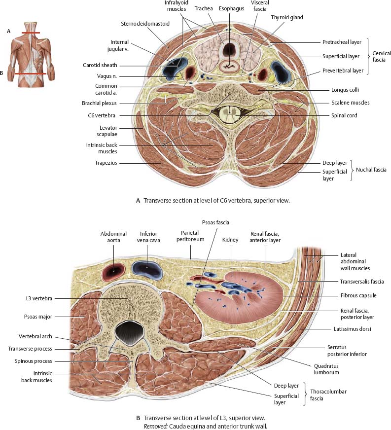
Fig. 2.2 Thoracolumbar fascia
Transverse section, superior view. The intrinsic back muscles are sequestered in an osseofibrous canal, formed by the thoracolumbar fascia, the vertebral arches, and the spinous and transverse processes of associated vertebrae. The thoracolumbar fascia consists of a superficial and a deep layer that unite at the lateral margin of the intrinsic back muscles. In the neck, the superficial layer blends with the nuchal fascia (deep layer), becoming continuous with the cervical fascia (prevertebral layer).
Intrinsic Muscles of the Cervical Spine
Fig. 2.3 Muscles in the nuchal region
Posterior view. Removed: Trapezius, sternocleidomastoid, splenius, and semispinalis muscles (right). Revealed: Nuchal muscles (right).
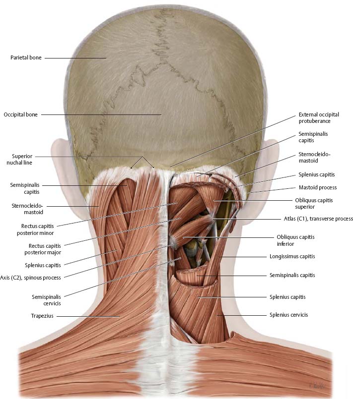
Fig. 2.4 Short nuchal muscles
Posterior view. See Fig. 2.6.

Intrinsic Muscles of the Back
![]() The extrinsic muscles of the back (trapezius, latissimus dorsi, levator scapulae, and rhomboids) are discussed in Unit 4. The serratus posterior, considered an intermediate extrinsic back muscle, has been included with the superficial intrinsic muscles in this unit.
The extrinsic muscles of the back (trapezius, latissimus dorsi, levator scapulae, and rhomboids) are discussed in Unit 4. The serratus posterior, considered an intermediate extrinsic back muscle, has been included with the superficial intrinsic muscles in this unit.
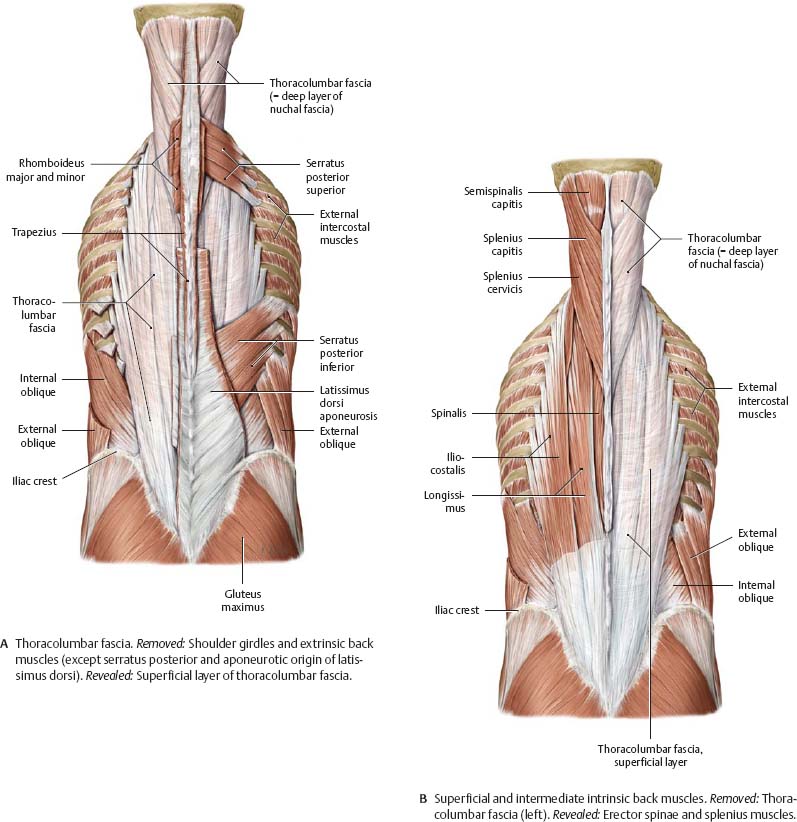
Fig. 2.5 Intrinsic muscles of the back
Posterior view. Sequential dissection of the thoracolumbar fascia, superficial intrinsic muscles, intermediate intrinsic muscles, and deep intrinsic muscles of the back.

Muscle Facts (I)
Fig. 2.6 Short nuchal and craniovertebral joint muscles
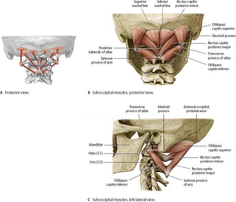
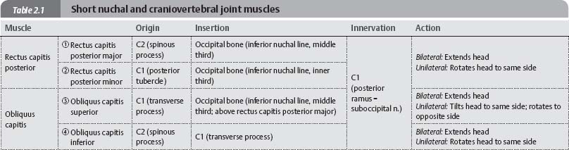
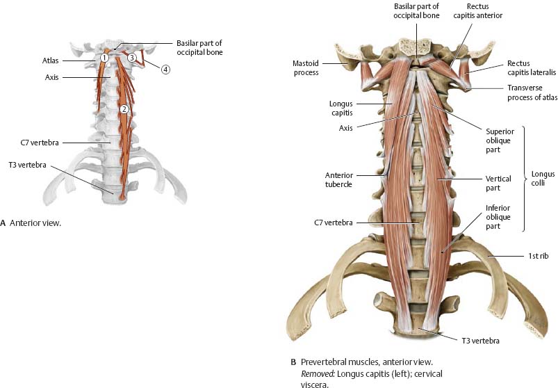
Fig. 2.7 Prevertebral muscles

Muscle Facts (II)
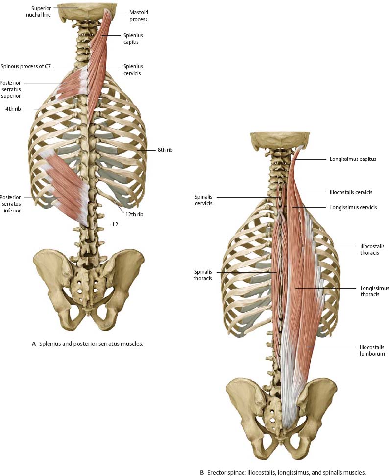
![]() The intrinsic back muscles are divided into superficial, intermediate, and deep layers. The posterior serratus muscles are extrinsic back muscles, innervated by the ventral rami of intercostal nerves, not the dorsal rami, which innervate the intrinsic back muscles. They are included here as they are encountered in dissection of the back musculature.
The intrinsic back muscles are divided into superficial, intermediate, and deep layers. The posterior serratus muscles are extrinsic back muscles, innervated by the ventral rami of intercostal nerves, not the dorsal rami, which innervate the intrinsic back muscles. They are included here as they are encountered in dissection of the back musculature.
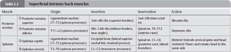
Fig. 2.8 Superficial intrinsic back muscles (schematic)
Right side, posterior view.

Fig. 2.9 Intermediate intrinsic back muscles (schematic)
Right side, posterior view. These muscles are collectively known as the erector spinae.
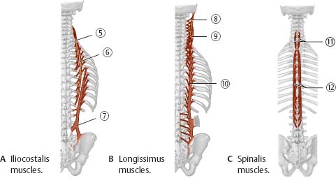

Fig. 2.10 Superficial and intermediate intrinsic back muscles
Posterior view.
Muscle Facts (III)
![]() The deep intrinsic back muscles are divided into two groups: transversospinal and deep segmental muscles. The transversospinalis muscles pass between the transverse and spinous processes of the vertebrae.
The deep intrinsic back muscles are divided into two groups: transversospinal and deep segmental muscles. The transversospinalis muscles pass between the transverse and spinous processes of the vertebrae.

Fig. 2.11 Transversospinalis muscles (schematic)
Posterior view.

Fig. 2.12 Deep segmental muscles (schematic)
Posterior view.

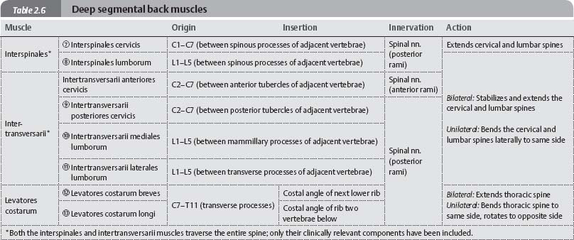
Fig. 2.13 Deep intrinsic back muscles
Posterior view.
