Arteries & Veins of the Back
Fig. 3.1 Arteries of the back
The structures of the back are supplied by branches of the posterior intercostal arteries, which arise from the thoracic aorta or directly from the subclavian artery.

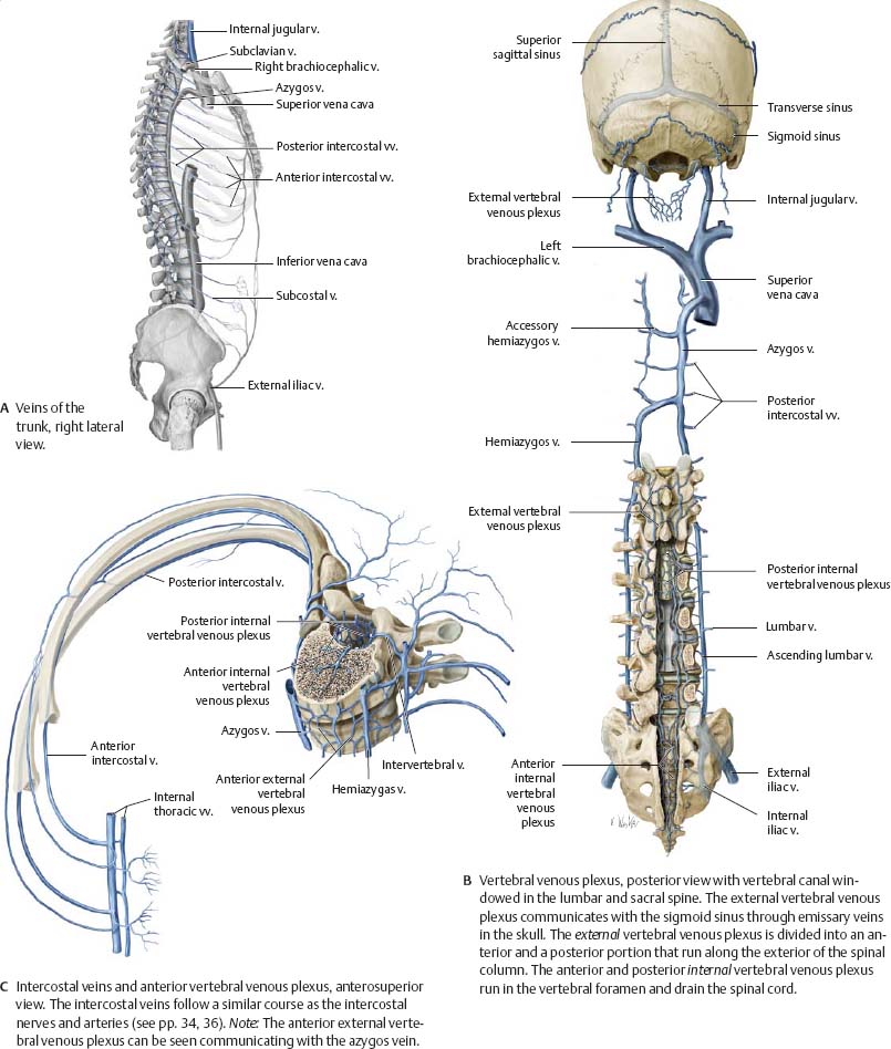
Fig. 3.2 Veins of the back
The veins of the back drain into the azygos vein via the superior intercostal veins, hemiazygos veins, and ascending lumbar veins. The interior of the spinal column is drained by the vertebral venous plexus that runs the length of the spine.
Nerves of the Back
![]() The back receives its innervation from branches of the spinal nerves. The posterior rami of the spinal nerves supply most of the intrinsic muscles of the back. The extrinsic muscles of the back are supplied by the anterior rami of the spinal nerves.
The back receives its innervation from branches of the spinal nerves. The posterior rami of the spinal nerves supply most of the intrinsic muscles of the back. The extrinsic muscles of the back are supplied by the anterior rami of the spinal nerves.
Fig. 3.3 Nerves of the back
The anterior rami of spinal nerves T1–T11 form the intercostal nerves, which course along the ribs and give rise to lateral and anterior cutaneous branches.

Fig. 3.4 Nerves of the nuchal region
Right side, posterior view. Like the back, the nuchal region receives most of its motor and sensory innervation from the posterior rami of the spinal nerves. The posterior rami of C1–C3 have specific names: suboccipital nerve (C1), greater occipital nerve (C2), and third occipital nerve (C3). The lesser occipital and great auricular nerves arise from the anterior rami of the C1–C4 spinal nerves and innervate the skin of the anterolateral head and neck. The anterior rami of C1–C4 also give rise to the ansa cervicalis, which innervates the infrahyoid muscles (see p. 562).

Fig. 3.5 Cutaneous innervation of the back

Neurovascular Topography of the Back
Fig. 3.6 Neurovasculature of the nuchal region
Posterior view. Removed: Trapezius, sternocleidomastoid, splenius capitis, and semispinalis capitis. Revealed: Suboccipital region. See p. 60 for the course of the intercostal vessels.

Fig. 3.7 Neurovasculature of the back
Posterior view. Removed: Muscle fascia (except superficial layer of thoracolumbar fascia); latissimus dorsi (right). Reflected: Trapezius (right). Revealed: Transverse cervical artery in the deep scapular region.
