The syndrome of autoimmune hepatitis (AIH) (Table 10.1) was recognized in the 1950s in young women, with hypergammaglobulinemia, acnelike skin rashes, myalgia, amenorrhea, liver disease (including hepatomegaly and splenomegaly), and a fluctuating course (13,26,33). The term “lupoid hepatitis” was adopted to distinguish AIH from systemic lupus erythematosus (LE) because some patients had circulating LE cells (25), antinuclear antibodies (ANAs) in the sera, and other serologic abnormalities, including the presence of antibodies directed against smooth muscle antibody (SMA). However, the term has appropriately been abandoned because an LE test is positive in fewer than 20% of patients and because liver involvement is distinctly unusual in classical systemic lupus erythematosus.
Autoimmune mechanisms contribute to injury in many liver diseases (47), including acute and chronic hepatitis caused by hepatitis A, B, and C viruses, primary biliary cirrhosis (PBC), primary sclerosing cholangitis (PSC), and drug-induced chronic hepatitis (6,41,44), but also in alcoholic liver disease, Wilson disease, and perhaps α1-antitrypsin deficiency. However, the autoimmune liver diseases are principally thought of as (a) AIH (e-Figs. 10.1-10.16), (b) primary biliary cirrhosis (PBC), (c) primary sclerosing cholangitis (PSC), (d) drug-induced/associated autoimmune hepatitis (e-Figs. 10.17-10.20), and, in recent years, (e) autoimmune cholangiopathy (e-Figs. 10.21-10.29). In addition, some patients have (f) “overlap syndrome,” with combined clinical and/or serologic features of AIH and one of the cholangiopathies (e-Figs. 10.30-10.32) (4,58). AIH can recur after transplantation (e-Figs. 10.33-10.36). In recent years the development of AIH has been recognized in individuals who had undergone liver transplantation for other reasons (“de novo autoimmune hepatitis”) (e-Fig. 10.37-10.41) (26).
AUTOIMMUNE HEPATITIS
The term “autoimmune chronic active hepatitis” is no longer used because “autoimmune” and “chronic” are redundant terms and, more important, because AIH may present as an acute, or occasionally fulminant, disorder difficult to distinguish from acute viral hepatitis (3,8,16,17,27). Also, biopsy does not always show typical features of chronic active hepatitis (3).
|
TABLE 10.1 Synonyms for Autoimmune Hepatitis |
||||||||
|
The usual patient is a young woman, 15 to 35, or perimenopausal. Men and children of both sexes may be affected (9,26). More than 50% of patients present with episodic jaundice, anorexia, and fatigue, and premenopausal women are often amenorrheic. Epistaxis, bleeding gums, and easy bruisability, as well as right upper quadrant abdominal pain and tenderness occur. As many as 20% are febrile. Many patients present with advanced disease, including cirrhosis and portal hypertension. Rarely, AIH may be asymptomatic at discovery. Various extrahepatic manifestations (e.g., thyroiditis, vasculitis, uveitis, ulcerative colitis (26,41), arthritis, Coombspositive hemolytic anemia, glomerulonephritis, neuropathy, vitiligo (43), alopecia (43), mixed connective tissue disorders, and pneumonitis are seen. An international panel established diagnostic criteria (Table 10.2) (1,25).
AIHis fatal if untreated. The typical patient responds to corticosteroid therapy, with improvement in symptoms and normalization of laboratory test results, but often, the histologic picturemay not change and the disease may progress. Fibrosis can decrease (44). With treatment, the 5-year survival is approximately 85%. Patients with liver-kidney microsomal (LKM) antibody-positive disease may have a somewhat poorer prognosis (23). Death is generally due to cirrhotic hepatocellular failure with bleeding esophageal varices or end stage liver disease. Rarely deathmay follow a fulminant hepatitis-like course. Other causes of death include sepsis and hepatocellular carcinoma.AIHmay recur after liver transplantation (14,38) and may also occur in patients who have been transplanted for other diseases (de novo AIH) (26,58).
Genetics
There is a relationship between AIH and certain human leukocyte antigens (HLAs) (12,13). HLA A1-B8-DR3 and DR4 have strong, and probably independent, associations (18,23,36,61). Familial occurrence is distinctly unusual (9,22). However, first-degree relatives may demonstrate circulating ANAs, SMAs, and antimitochondrial antibodies (AMAs). An increased familial incidence of C4 deficiency in relatives of children with AIH has been shown and a familial reduction in suppressor T-cell activity in AIH patients who express the HLA B8-DR3 haplotype.
|
TABLE 10.2 International Autoimmune Hepatitis Group Scoring (Modified) |
||||||||||||||||||||||||
|
||||||||||||||||||||||||
Mechanisms of Injury
Predominantly an antibody-dependent cytotoxic cellular reaction, the immune response is most likely activated by helper T lymphocytes and mediated by K lymphocytes elaborating an antibody directed against antigens at the hepatocyte surface, with the probable involvement of cytokines, including tumor necrosis factor (TNF) and interferon-g (24). The lymphocytes may directly stimulate the production of autoantibodies (30). Cell injury could also be independent of antigen-specific mechanisms, as a kind of defective immune response to the hepatocyte that is triggered by various infections. For example, P450IID6, the putative antigen against which LKM-1 antibody is thought to act, is not on the surface of the hepatocyte and consequently is not exposed to possible antibody-mediated cell lysis. Infectious agents implicated as potential initiators of the defective immune response have been enterobacteria, rubeola, rubella, and cytomegalovirus, in addition to hepatitis C virus (HCV). Medications have also been associated (44).
Laboratory Tests
The serum levels of transaminases, aspartate aminotransferase (AST), and alanine aminotransferase (ALT) are generally moderately elevated, with AST values generally between 200 and 1,000 U. Bilirubin and alkaline phosphatase values may also be moderately elevated. Most patients demonstrate polyclonal hypergammaglobulinemia, with levels as high as 50 to 70 g/L, as well as abnormal serum copper and ceruloplasmin values.
Autoantibodies
Various circulating autoantibodies have been found (1,16,26). The identification of specific antibodies has led to the recognition of clinically, as well as serologically, distinct groups. The most intensely studied autoantibodies are listed in Table 10.3. The demonstration of SMAs (70%) and/or ANAs (50%) is classic in AIH. Fewer than 20% have AMAs, many (? all) of these as overlap. These intracellular target antigens may not be exposed to the immune effector mechanisms thought responsible for AIH. Antibodies react with P450IID6 (19,36,46,60), and LKM-2 with P450IIC11. Thus far, there is no conclusive evidence that any AIH-associated autoantibodies directly contribute to the development of AIH. Antibodies directed against gastric parietal cells and against thyroid may also be found in AIH patients.
|
TABLE 10.3 Autoantibodies Described in Association with Autoimmune Hepatitis |
||||||||||||||||||||||||||||||||
|
Antineutrophil cytoplasmic antibodies (ANCAs), autoantibodies reacting with neutrophil cytoplasmic antigens, are demonstrable in AIH and PSC, but not PBC, chronic hepatitis as a result of HBV or HCV, or other liver disorders (20,28). Various “liver-specific” antibodies have been evaluated, the most important of which are those directed against liver and kidney microsomes, including variants against different cytochrome P450 components in smooth endoplasmic reticulum, with at least five related antibodies (54). Some AIH patients have anti-LKM-1 (16), the most widely studied of the LKM antibodies, which is directed against cytochrome P450s from the IID subfamily (35). It is also in sera of HCV-infected patients (43), but the antigenic sites recognized by the sera from patients with HCV differ from those recognized by sera from patients with AIH (61).
Classification
Four AIH subgroups can be distinguished (16,26,38) (Table 10.4).
Type 1 is the classical and most common form (e-Figs. 10.1-10.16) (25), characterized by the demonstration of SMA and ANA in the serum.
Type 2 AIH patients have LKM antibodies but not ANAs. LKM-associated AIH is uncommon in the United States (e-Fig. 10.42-10.44). Two subgroups have been recognized (Table 10.5). One resembles type 1 AIH, with typical histologic findings. A more common subgroup also has antibodies to HCV, as well as antibodies to GOR, suggesting previous infection with HCV (16). HCV may induce autoimmune antibodies to both GOR and LKM-1 usually in Mediterranean men. Liver biopsy findings resemble those of hepatitis C.
Two other AIH subtypes are rare. Patients with type 3 have antibodies against soluble liver antigens (SLAs) and AMAs, and sometimes SMAs (40). Type 4 occurs principally in children who demonstrate high SMA titers, but without SLA or AMA (38).
|
TABLE 10.4 Classification of Autoimmune Hepatitis |
||||||||||||||||||||||||||||||||||||||||||||||||||
|
||||||||||||||||||||||||||||||||||||||||||||||||||
|
TABLE 10.5 LKM-Positive Autoimmune Hepatitis |
||||||||||||||||||||||||||||||||||||||||||||||||
|
||||||||||||||||||||||||||||||||||||||||||||||||
Pathology
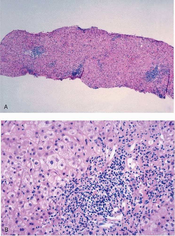
AIH is a progressive disease (Fig. 10.1, e-Figs. 10.4-10.9) that, if untreated, progresses inexorably from the early, inflammatory stages to cirrhosis.
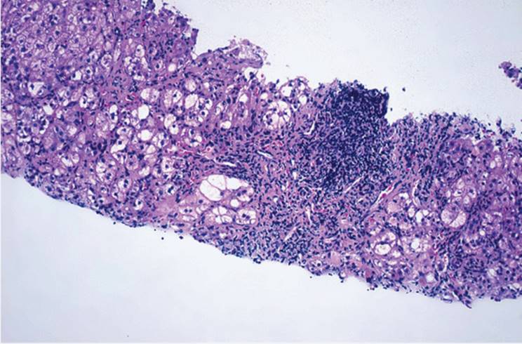
Liver biopsy findings are not specifically diagnostic. In spite of this, liver biopsy can be useful in cases in which the diagnosis has not been clinically established. In general, the biopsy shows the picture of chronic hepatitis and other causes for this pattern, such as chronic viral hepatitis, Wilson disease, α1-antitrypsin deficiency, and drug-associated chronic hepatitis, should be considered and clinically or morphologically excluded (3,27). Typically, the biopsy shows an irregularly distributed relatively heavy portal infiltrate, with periportal or paraseptal interface hepatitis (Figs. 10.1B, 10.2, e-Figs. 10.4, 10.5, 10.10 ), with increased numbers of plasma cells and eosinophils in addition to lymphocytes (Fig. 10.2). Interface hepatitis is usually quite prominent, and there may also be portal-to-portal or portal-to-central bridging necrosis (e-Figs. 10.10, 10.11, 10.15, 10.16), also with plasma cells. Early in the development of AIH, there may be zone 3 necrosis (63).
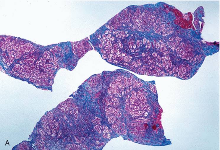
Lobular hepatitis is often seen, especially when there is an acute clinical relapse (25). With significant lobular involvement, which affects zone 1 (periportal) hepatocytes predominantly, there is pseudoacinar (rosette) formation (e-Figs. 10.4-10.6), generally without obvious lumen formation (Figs. 10.2, 10.3). This pattern is typical of AIH, as is the presence of increased numbers of plasma cells. However, these features are not pathognomonic and are not seen in every biopsy from an AIH patient.
Both rosettes and increased numbers of plasma cells can be seen in association with chronic hepatitis as a result of other causes. In some patients with AIH, giant cell transformation (“giant cell hepatitis”) may be seen.
|
|
|
FIGURE 10.1 The progressive course of autoimmune hepatitis. A 52-year-old woman presented with anorexia, easy fatigability, and mild jaundice, with elevated levels of antinuclear antibodies. Liver biopsy, obtained approximately 2 years after presentation (A, B), showed portal tracts expanded by a chronic inflammatory cell infiltrate composed of approximately equal numbers of plasma cells and lymphocytes, with scattered eosinophils and histiocytic cells, with interface hepatitis and scattered foci of lobular inflammation. Four years later (C, D) the biopsy showed definite but incomplete nodularity with interface hepatitis, bridging fibrosis and inflammation, and a portal infiltrate consisting mostly of lymphocytes, with scattered plasma cells and only rare eosinophils. Four years after the second biopsy, and almost 10 years after the onset of symptoms, the patient underwent orthotopic liver transplantation. The explant was a cirrhotic, predominantly macronodular liver, with areas of collapse and a few macroregenerative nodules (hematoxylin-eosin; A, original magnification ×200; B, ×400; C, ×40; D, ×400). |
|
|
|
FIGURE 10.1. (Continued) |
Features to distinguish AIH from chronic hepatitis C include a more severe degree of lobular inflammation and necrosis, greater numbers of plasma cells, more marked interface hepatitis, and broad areas of parenchymal collapse (Table 10.6). Biliary changes are uncommon in AIH, and their presence may be indicative of some other disorder, such as PBC, PSC, or, to a lesser degree, HCV. In a patient in whom ANAs are demonstrable, bile duct injury should suggest the diagnosis of autoimmune cholangiopathy, discussed below. In chronic HBV, plasma cells may also be prominent. Histologic diagnosis for these three entities has high specificity and predictability but relatively low sensitivity. Acute hepatitis A, rarely biopsied, can be indistinguishable from AIH, with many plasma cells.
|
|
|
FIGURE 10.2 Autoimmune hepatitis. Liver biopsy from a 14-year-old with easy fatigability and malaise. Both antinuclear antibodies and anti-smooth muscle antibodies were demonstrable. The biopsy demonstrates a severe inflammatory process with almost complete effacement of the portal tract structure and with bridging inflammation and rosette formation (hematoxylin-eosin, original magnification ×40). |
The biopsy in LKM-associated AIH is similar to that of other forms (Fig. 10.4, e-Figs. 10.42-10.44). There are no known light microscopic correlates for the various autoantibodies. Fulminant hepatitis in AIH is indistinguishable from other forms of massive and submassive necrosis.
|
|
|
FIGURE 10.3 Autoimmune hepatitis. Liver biopsy from a 19-year-old man with a history of approximately 4 years of clinical liver disease and high levels of antinuclear antibodies, showing portal and periportal fibrosis, with prominent rosette formation and moderate portal and lobular inflammation (Masson trichrome; A, original magnification ×40; B, ×200). (See Color Figure 10.3A following page 129.) |
|
|
|
FIGURE 10.3. (Continued) |
The cirrhotic stage usually shows a greater degree of inflammation than cirrhosis arising from other causes. Septa are easily recognized, with obvious areas of prior parenchymal collapse, separating nodules of varying size (Fig. 10.1C,D). There may be dysplastic (macroregenerative) nodules and small hepatocellular carcinomas.
Immunohistochemical Studies
The great majority of portal tract lymphocytes are T cells, and CD4 cells predominate (21,29,49). In contrast, the T lymphocytes in the area of interface hepatitis are mostly CD8 antigen-positive and CD11b antigen-negative and therefore are phenotypically cytotoxic T cells. Regulatory T cells (Tregs) are typically deficient in AIH (32).
|
TABLE 10.6 Histologic Features Most Useful in Differentiating Autoimmune Hepatitis from Chronic Hepatitis C |
||||||||||||||||||||||||||||||
|
||||||||||||||||||||||||||||||
|
|
||||||||||||||||||||||||||||||
|
FIGURE 10.4 Liver-kidney microsomal (LKM) autoimmune hepatitis. Photomicrographs of biopsy from a 48-year-old man with chronic hepatitis. Antinuclear antibodies were not demonstrable, but his serum showed elevated titers of anti-LKM antibodies. There is extensive interface hepatitis with bridging and lobular inflammation (hematoxylin-eosin, original magnification ×40). |
||||||||||||||||||||||||||||||
Class I and class II antigens of the major histocompatibility complex have a major role in the induction and modulation of the immune response (48). Most hepatocytes are class II reacting cells.
What is the Relationship of Hepatitis C to Autoimmune Hepatitis?
An etiopathogenic relationship has been suggested for HCV and AIH (14,31,40). This seems unlikely (31,50). The association of LKM-1 with HCV is mostly in southern European countries, with much less evidence for this in the United States, western Europe, or Australia (27).
Key features of AIH are listed in Table 10.7.
AUTOIMMUNE CHOLANGIOPATHY
Autoimmune cholangiopathy is a variant of AIH. It is serologically indistinguishable from type 1, often with high ANA titers, and can show clinical, biochemical, and histologic features (Fig. 10.5) of PBC, but without demonstrable AMAs (7,22,37,56). Liver biopsies show the bile duct damage characteristic of PBC (e-Figs. 10-21-10.29) and may also show granuloma formation. Patients with this condition have previously been designated as having atypical PBC or AMA-negative PBC; it may be that they represent a unique entity, although there is not complete agreement on this (18,57). Response to corticosteroid therapy has not been uniformly confirmed (66). Extrahepatic manifestations may be seen (2). This histologic picture has also been described in association with drug sensitivity (42). Bile duct injury can also be seen in AIH (3,11).
|
TABLE 10.7 Key Features of Autoimmune Hepatitis |
||||||||||||||||||||
|
||||||||||||||||||||
|
|
||||||||||||||||||||
|
FIGURE 10.5 Autoimmune cholangitis. Liver biopsy from a 66-year-old woman with clinical and laboratory evidence of chronic hepatitis. Transaminase levels were approximately 200 U, and the alkaline phosphatase determination was >1,600 U. Antimitochondrial antibodies were not demonstrable on at least three occasions. The biopsy shows markedly expanded portal tracts with minimal piecemeal necrosis. The inflammatory cell infiltrate consists mostly of lymphocytes, with significant numbers of plasma cells. A bile duct (upper left) shows epithelial cell variation and infiltration by lymphocytes. The bile duct adjacent to the small artery (center) is almost completely destroyed (hematoxylin-eosin, original magnification ×200). |
||||||||||||||||||||
|
TABLE 10.8 Autoimmune Disease Variants |
|||||||||||||||
|
|||||||||||||||
OVERLAP SYNDROMES
Table 10.8 lists overlap syndromes. Some patients have mixed clinical and/or histologic features, and definitive diagnosis may be difficult to establish, usually requiring correlation of signs and symptoms, laboratory test results, and histopathology.
REFERENCES
1. Alvarez F, Berg PA, Bianchi FB, et al. International Autoimmune Hepatitis Group Report: a review of criteria for diagnosis of autoimmune hepatitis. J Hepatol 1999;31: 929-938.
2. Archimandritis A, Tjivras M, Tsirantonaki M, et al. Sjogren's syndrome with antimitochondrial-negative primary biliary cirrhosis: a case of autoimmune cholangitis. J Clin Gastroenterol 1995;20:268-270.
3. Bach N, Thung SN, Schaffner F. The histological features of chronic hepatitis C and autoimmune chronic hepatitis: a comparative analysis. Hepatology 1992;15:572-577.
4. Ben-Ari Z, Czaga AJ. Autoimmune hepatitis and its variant syndromes. Gut 2001;49: 589-594.
5. Beuers U, Rust C. Overlap syndromes. Semin Liv Dis 2005;25:311-320.
6. Bourdi M, Larrey D, Nataf J, et al. Anti-liver endoplasmic reticulum autoantibodies are directed against human cytochrome P-450IA2: a specific marker for dihydralazineinduced hepatitis. J Clin Invest 1990;85:1967-1973.
7. Corte G, Kaplan M, Olivera M, et al. Prevalence of antinuclear antibodies (ANA) in patients with negative antimitochondrial antibodies (AMA) primary biliary cirrhosis (PBC). Hepatology 1992;16:189A.
8. Crapper RM, Bhathal PS, Mackay IR, et al. “Acute” autoimmune hepatitis. Digestion 1986;34:216-225.
9. Czaja AJ. Natural history, clinical features, and treatment of autoimmune hepatitis. Semin Liv Dis 1984;4:1-12.
10. Czaja AJ, Carpenter HA, Santrach PJ, et al. Genetic predisposition for the immunological features of chronic active hepatitis. Hepatology 1993;18:816-822.
11. Czaja AJ, Carpenter HA. Autoimmune hepatitis with incidental histologic features of bile duct injury. Hepatology 2001;34:659-665.
12. Donaldson PT, Doherty DG, Hayllar K, et al. Susceptibility of autoimmune chronic active hepatitis: human leukocyte antigens DR4 and A1-B8-DR3 are independent risk factors. Hepatology 1991;13:701-706.
13. Eddleston AL, Donaldson PT, Hegarty JE, et al. Immunological aspects of liver disease. Gut 1991(Suppl):S40-S46.
14. Fried MW, Draguesku JO, Shindo M, et al. Clinical and serological differentiation of autoimmune and hepatitis C virus-related chronic hepatitis. Dig Dis Sci 1993;38:631-636.
15. Frust TW. Recurrent primary biliary cirrhosis, primary sclerosing cholangitis, and autoimmune hepatitis after transplantation. Liver Transpl 2000;7(11 Suppl 1):S99-S108.
16. Geller SA. Autoimmune hepatitis. Pathol: State of the Art Rev 1994;3:57-76.
17. Gish RG, Mason A. Autoimmune liver disease. Current standards, future direction. Clin Liver Dis 2001;5:287-314.
18. Goodman Z, McHally PR, Davis D, et al. “Autoimmune cholangitis”: a variant of primary biliary cirrhosis. Dig Dis Sci 1995;40:1232-1242.
19. Gueguen M, Boniface O, Bernard O, et al. Identification of the main epitope on human cytochrome P450 IID6 recognized by anti-liver kidney microsome antibody. J Autoimmun 1991;4:607-615.
20. Hardarson S, Labrecque DR, Mitros FA, et al. Antineutrophilic cytoplasmic antibody in inflammatory bowel and hepatobiliary diseases. High prevalence in ulcerative colitis, primary sclerosing cholangitis, and autoimmune hepatitis. Am J Clin Pathol 1993;99:277-281.
21. Hashimoto E, Lindor KD, Homburger HA, et al. Immunohistochemical characterization of hepatic lymphocytes in primary biliary cirrhosis in comparison with primary sclerosing cholangitis and autoimmune chronic active hepatitis. Mayo Clin Proc 1993;68:1049-1055.
22. Hatzis GS, Vassiliou VA, Delladetsima JK. Overlay syndrome of primary sclerosing cholangitis and autoimmune hepatitis. Eur J Gastroenterol Hepatol 2001;13:203-206.
23. Homberg JC, Abuaf N, Bernard O, et al. Chronic active hepatitis associated with antiliver/kidney microsome antibody type 1: a second type of “autoimmune hepatitis.” Hepatology 1987;7:1333-1339.
24. Hussain MJ, Mustafa A, Mowat AP. Immunohistochemical detection of TNF-a and IFNg in the liver of children with autoimmune chronic active hepatitis and primary cholangitis. J Hepatol 1992;16:S59.
25. Johnson PJ, McFarlane IG. Meeting report: International Autoimmune Hepatitis Group. Hepatology 1993;18:998-1005.
26. Krawitt El. Autoimmune hepatitis. N Engl J Med 2006;354:54-66.
27. Lenzi M, Johnson PJ, McFarlane BM, et al. Antibodies to hepatitis C virus in autoimmune liver disease: evidence for geographical heterogeneity. Lancet 1991;338:277-280.
28. Lo SK, Chapman RWG, Cheeseman P, et al. Antineutrophil antibody: a test for immune primary sclerosing cholangitis in childhood? Gut 1993;34:199-202.
29. Lohr H, Manns M, Kyriatsoulis A, et al. Clonal analysis of liver-infiltrating T cells in patients with LKM-1 antibody-positive autoimmune chronic active hepatitis. Clin Exp Immunol 1991;84:297-302.
30. Lohr H, Treichel U, Poralla T, et al. Liver-infiltrating T helper cells in autoimmune chronic active hepatitis stimulate the production of autoantibodies against the human asialoglycoprotein receptor in vitro. Clin Exp Immunol 1992;88:45-49.
31. Lohse AW, Gerken G, Mohr H, et al. Relation between autoimmune liver diseases and viral hepatitis: clinical and serological characteristics in 859 patients. Z Gastroenterol 1995;33:527-533.
32. Longhi MS, Hussain MJ, Mitry RR, et al. Functional study of CD4+CD25+ regulatory T cells in health and autoimmune hepatitis. J Immuno 2006;176:4484-4491.
33. Lunel F, Abuaf N, Franguel L, et al. Liver/kidney microsome antibody type 1 and hepatitis C virus infection. Hepatology 1992;16:630-636.
34. Magrin S, Craxi A, Fiorentino G, et al. Is autoimmune chronic active hepatitis a HCVrelated disease? J Hepatol 1991;13:56-60.
35. Manns MP, Johnson EF, Griffen KJ, et al. Major antigen of liver kidney microsomal autoantibodies in idiopathic autoimmune hepatitis is cytochrome P450 db1. J Clin Invest 1989;83:1066-1072.
36. Manns MP, Griffin KJ, Sullivan KF, et al. LKM-1 autoantibodies recognize a short linear sequence in P450IID6, a cytochrome P450 monooxygenase. J Clin Invest 1991;88: 1370-1378.
37. Michieletti P, Wanless IR, Katz A, et al. Antimitochondrial antibody negative primary biliary cirrhosis: a distinct syndrome of autoimmune cholangitis. Gut 1994;35:260-265.
38. Odievre M, Maggiore G, Homberg JC, et al. Seroimmunologic classification of chronic hepatitis in 57 children. Hepatology 1983;3:407-409.
39. Oo JH, Neuberger J. Recurrence of nonviral diseases. Clin Liv Dis 2007;11:377-395.
40. Pawlotsky J-M, Deforges L, Bretagne S, et al. Hepatitis C virus infection can mimic type 1 (antinuclear antibody positive) autoimmune chronic active hepatitis. Gut 1993;(Suppl): S66-S68.
41. Rabinovitz M, Demetris AJ, Bou-Abboud CF, et al. Simultaneous occurrence of primary sclerosing cholangitis and autoimmune chronic active hepatitis in a patient with ulcerative colitis. Dig Dis Sci 1992;37:1606-1611.
42. Ryley NG, Fleming KA, Chapman RW. Focal destructive cholangiopathy associated with amoxycillin/clabulanic acid (Augmentin). J Hepatol 1995;23:278-282.
43. Sacher M, Blumel P, Thaler H, et al. Chronic active hepatitis associated with vitiligo, nail dystrophy, alopecia and a new variant of LKM antibodies. J Hepatol 1990;10:364-369.
44. Schvarcz R, Glaumann H, Weiland O. Survival and histological resolution of fibrosis in patients with autoimmune chronic active hepatitis. J Hepatol 1983;18:15-23.
45. Scully LJ, Clarke D, Barr RJ. Diclofenac induced hepatitis. Dig Dis 1993;38:744-761.
46. Seelig R, Renz M, Bunger G, et al. Anti-LKM-1 antibodies determined by use of recombinant P450 2D6 in ELISA and Western blot and their association with anti-HCV and HCV-RNA. Clin Exp Immunol 1993;92:373-380.
47. Seki T, Kiyosawa K, Inoko H, et al. Association of autoimmune hepatitis with HLABw54 and DR4 in Japanese patients. Hepatology 1990;12:1300-1304.
48. Senaldi G, Lobo-Yeo A, Mowat AP, et al. Class I and class II major histocompatability complex antigens on hepatocytes: importance of the method of detection and expression in histologically normal and diseased livers. J Clin Pathol 1991;44:107-114.
49. Senaldi G, Portmann B, Mowat AP, et al. Immunohistochemical features of the portal tract mononuclear cell infiltrate in chronic aggressive hepatitis. Arch Dis Child 1992;67: 1447-1453.
50. Strassburg CP, Obermayer-Straum P, Manns MP. Autoimmunity in hepatitis C and D virus infection. J Viral Hepatitis 1996;3:49-59.
51. Talwalker JA, Keach JC, Angulo P, et al. Overlap of autoimmune hepatitis and primary biliary cirrhosis: an evaluation of a modified scoring system. Am J Gastroenterol 2002;97:1191-1197.
52. Taylor SL, Dena PJ, Riely CA. Primary autoimmune cholangitis. Am J Surg Pathol 1994;18:91-99.
53. Thung SN, Bach N, Fasy TM, et al. Hepatocellular carcinoma associated with autoimmune chronic active hepatitis. Mt Sinai J Med 1990;57:165-168.
54. Todros L, Touscoz G, D'Urso N, et al. Hepatitis C virus-related chronic liver disease with autoantibodies to liver-kidney microsomes (LKM). Clinical characterization from idiopathic LKM-positive disorders. J Hepatol 1991;13:128-131.
55. Tomsic M, Ferlan-Marolt V, Kveder T, et al. Mixed connective tissue disease associated with autoimmune hepatitis and thyroiditis. Ann Rheum Dis 1992;51:544-546.
56. Vierling JM. Autoimmune cholangiopathy. Clin Liver Dis 1999;3:571-584.
57. Washington K. Autoimmune cholangitis: not just AMA-negative primary biliary cirrhosis. Adv Anat Pathol 2002;9:244-250.
58. Washington K. Autoimmune liver disease: overlap and outliers. Mod Pathol 2007;20(Suppl 1): S15-30.
59. Yabe H, Noma K, Tada N, et al. A case of CREST syndrome with rapidly progressive liver damage. Intern Med 1992;31:69-73.
60. Yamamoto AM, Mura C, Morales MG, et al. Study of CYP2D6 gene in children with autoimmune hepatitis and P450 IID6 autoantibodies. Clin Exp Immunol 1992;87: 251-255.
61. Yamamoto AM, Cresteil D, Homberg JC, et al. Characterization of anti-liver-kidney microsome antibody (anti-LKM1) from hepatitis C virus-positive and -negative sera. Gastroenterology 1993;104:1762-1767.
62. Yousuf M, Kiyosawa K, Sodeyama T, et al. Development of hepatocellular carcinoma in a man with auto-immune chronic active hepatitis. J Gastroenterol Hepatol 1992;7:66-69.
63. Zen Y, Notsumata K, Tanak N, et al. Hepatic centrilobular zone necrosis with positive antinuclear antibody: a unique subtype or early disease of autoimmune hepatitis. Hum Pathol 2007;38:1669-1675.