This chapter summarizes some major aspects of hepatotoxicity (80). Many examples of response to drug or toxic injury are shown in the electronic figures. They are listed alphabetically for easy reference. Most are also cited in the text.
Drug-induced liver injury (DILI) encompasses a wide spectrum of clinicopathologic alterations ranging from mild biochemical abnormalities to acute liver failure. There are two broad categories: intrinsic and idiosyncratic hepatotoxicity.
INTRINSIC TOXICITY
Intrinsic toxicity refers to drugs that have a high incidence of predictable hepatotoxicity and that are dose dependent. Their effects can be induced in experimental animals (64). There are two groups: direct and indirect hepatotoxic effect. Both cause cytotoxic and cholestatic liver injury.
In direct cytotoxicity, liver cell injury occurs at the subcellular level, affecting various organelles with subsequent steatosis, necrosis, or both. An example is carbon tetrachloride poisoning (71). Another example is phosphorus poisoning, now only of historical significance (27). Paraquat causes cholestatic direct hepatotoxic injury, with biliary epithelium as the primary target of injury (56).
Indirect toxicity refers to cell injury caused by alterations of various metabolic pathways or selective effects on membrane cell receptors, DNA and RNA molecules, either within the nuclei or in the cytosol. Many drugs cause indirect cytotoxic injury, leading to steatosis or hepatocellular necrosis, including phalloidin, dimethylnitrosamine, acetaminophen, aflatoxin B, pyrrolizidine alkaloids, tetracycline, galactosamine, and inorganic arsenic (19,62). Some of the effects of alcohol are also reflections of altered metabolic pathways. Drugs that cause cholestatic indirect toxic injury by selective interference with bile excretion and uptake from the blood include contraceptive steroids, C17 alkylated anabolic steroids, and lithocholic acid.
IDIOSYNCRATIC HEPATOTOXICITY
Idiosyncratic drug-induced liver injury (DILI) (also termed “immunemediated” and “immunogenetic”) can have various systemic reactions, including fever, rash, and peripheral eosinophilia. Symptoms usually develop weeks following exposure, and, more important, symptoms recur with re-exposure. Effects are not experimentally demonstrable (65).
Although drug-associated acute liver failure is uncommon, as many as 15% of all acute liver failure cases are attributed to idiosyncratic drug reactions (e-Figs. 11.1-11.9, 11.18, 11.19, 11.35, 11.36, 11.44). Chronic disease occurs in up to 6%, even after withdrawal of the offending drug. Antibiotics and NSAIDs are the most common causes of DILI acute liver failure.
The mechanisms of DILI are complex and multifactorial. They involve a complex interplay between intracellular stress and tumor necrosis factor (TNF)-alpha-activated apoptosis/necrosis, coupled with proinflammatory responses of the innate and adaptive immune systems. Risk factors include age, sex, and genetic polymorphisms of drug-metabolizing enzymes such as cytochrome P450.
Many drugs produce DILI by interfering with metabolism and by producing toxic metabolites. Examples are halothane (e-Figs. 11.40-11.44), phenytoin (e-Fig. 11.29), paraaminosalicylic acid (e-Fig. 11.61), sulfonamides, phenylbutazone (e-Fig. 11.62), valproic acid (e-Fig. 11.96), isoniazid (e-Figs. 11.45-11.48), and chlorpromazine (e-Fig. 11.21) (8,23,34,49,57,70). A wide range of morphologic responses that mimic many hepatic disorders can be induced (Table 11.1).
|
TABLE 11.1 Morphologic Lesions in the Liver Associated with Drug-Induced Injury |
|
|
STEATOSIS
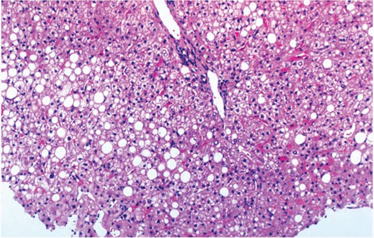
Drugs and toxins produce variable degrees of macrovesicular or microvesicular steatosis or both. Zonal distribution is generally not prominent except in phosphorus poisoning, in which steatosis is predominantly in zone 1 (periportal), and tetracycline-induced toxicity, in which steatosis is predominantly in zone 3 (perivenular) (21,62) (Fig. 11.1). Alcohol, at first, shows zonal distribution, initially in zone 3.
Macrovesicular steatosis is common in alcoholic liver disease after total parenteral nutrition (TPN) (e-Figs. 11.92-11.95) (4,5) and methotrexate therapy (e-Figs. 11.49-11.53) (2,49,68). Focal microvesicular steatosis may occur with macrovesicular steatosis, however, and so-called foam cells may be present.
Steatosis and necrosis occur concomitantly with carbon tetrachloride - (e-Figs. 11.18, 11.19), tannic acid-, and amanitin-induced injury, but in the latter, steatosis predominates (71).
Microvesicular steatosis is common in tetracycline-induced toxic injury (62), Reye syndrome, acute fatty liver of pregnancy, and after use of valproic acid (23), salicylates (73,79), and cocaine (40).
Steatosis mostly affects hepatocytes. Perisinusoidal stellate (Ito) cells can also contain significant numbers of fat droplets, particularly in hypervitaminosis A (e-Figs. 11.102-11.104) (Table 11.2) (28,30,38,53).
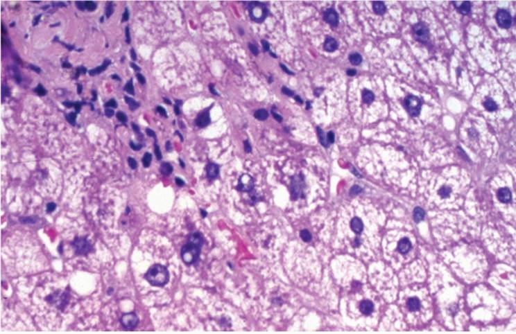
Phospholipidosis is a special form of drug-induced steatosis, first described in patients given coronary vasodilator medications, such as perhexiline maleate (61) and after amiodarone (e-Fig. 11.11) (Table 11.2) (31,39). Hepatocytes contain many small fat droplets (foam cells), and electron microscopy shows lamellar or crystalloid cytoplasmic inclusions, identical to those seen in metabolic types of phospholipidosis. Kupffer cells may also be affected (Fig. 11.2). Cirrhosis can result.
|
|
|
FIGURE 11.1 Predominantly macrovesicular steatosis in a patient treated with methotrexate for rheumatoid arthritis (hematoxylin-eosin, original magnification ×100). |
|
TABLE 11.2 Medications Associated with the Picture of Steatosis and Steatohepatitis |
||||||||
|
||||||||
|
|
||||||||
|
FIGURE 11.2 Microvesicular steatosis, focal nonalcoholic hepatitis, and Mallory material in a patient treated with amiodarone (hematoxylin-eosin, original magnification ×200). |
|
|
|
FIGURE 11.3 Perivenular and submassive necrosis in acetaminophen toxicity (hematoxylin-eosin, original magnification ×100). |
NECROSIS
Perivenular (zone 3) liver cell necrosis is typical of toxicity induced by the poison mushroom Amanita phalloides (e-Figs. 11.56), paracetamol, halothane (e-Figs. 11.40-11.44), and, well shown experimentally, carbon tetrachloride (e-Figs. 11.18, 11.19). Perivenular hemorrhagic necrosis is associated with pyrrolizidine alkaloids and aflatoxinB(27,50,66) (Fig. 11.3). Periportal (zone 1) liver cell necrosis is commonly associated with phosphorus poisoning and ferrous sulfate toxicity.Necrosis limited to zone 2 has not been describedwith drug-induced injury. Extensive zonal necrosismay lead to diffuse and/ormassive necrosis, as in some cases of phenytoin toxicity (69).
ACUTE AND CHRONIC HEPATITISLIKE PICTURE
Many drugs cause histopathologic changes of DILI indistinguishable from acute or chronic viral hepatitis (e-Figs. 11.22, 11.23, 11.38, 11.39, 11.45, 11.48, 11.54, 11.55, 11.59-11.61, 11.88-11.91, 11.97-11.101), including halothane, a-methyldopa, indomethacin, isoniazid, nitrofurantoin, and the recently reported anticholesterol drug ezetimibe (3,7,11,37,49,55,60,67). Nitrofurantoin-induced hepatitis usually affects females, often with autoimmune serologic markers, making it difficult to differentiate from autoimmune hepatitis. Furthermore, nitrofurantoin-induced chronic hepatitis may progress to fibrosis/cirrhosis.
CHOLESTASIS
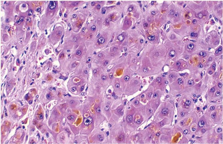
Cholestatic injury can follow use of chlorpromazine, anabolic and contraceptive steroids, antibiotics, and paraquat (1,18,35,36,51,56) (Fig. 11.4, e-Figs. 11.14-11.16, 11.21-11.23, 11.64, 11.69, 11.75-11.79 (Table 11.3).
|
|
|
FIGURE 11.4 Canalicular cholestasis in a patient on long-term oral contraceptives (hematoxylin-eosin, original magnification ×200). |
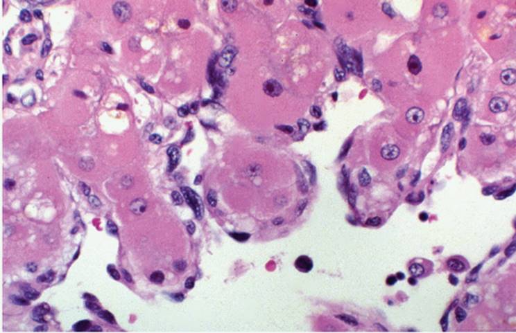
In chlorpromazine-induced injury (e-Fig. 11.21), in addition to canalicular cholestasis, there is an associated portal tract inflammatory reaction of variable degree, sometimes with many eosinophils. This type of injury, with both cholestatic and inflammatory components, has been described as hypersensitivity cholestasis or hepatocanalicular cholestasis (35) (Figs. 11.5, 11.6).
In contrast, cholestasis induced by steroids, both anabolic and contraceptive, usually shows little, if any, portal inflammation and has been described as bland or canalicular (36). When bile duct injury predominates, as in paraquat toxicity or with the no-longer used ajmaline (e-Fig. 11.10), the cholestasis is described as ductal or cholangiodestructive (56). Bile duct loss (vanishing bile duct syndrome) can rarely be seen (e-Figs. 11.79-11.82).
Cholestatic drug injuries generally have a short, acute, and self-limited course but can sometimes lead to chronic liver disease, similar to primary biliary cirrhosis or drug-induced secondary sclerosing cholangitis (12,13,22,48). Mixed forms, including cholestatic and parenchymal damage, can be seen with phenytoin (69) and amoxicillin/clavulanic acid preparations.
|
TABLE 11.3 Medications Associated with a Cholestasis Picture |
||||
|
|
|
|
FIGURE 11.5 Severe cholestasis in a patient treated with chlorpromazine (hematoxylin-eosin, original magnification ×100). |
GRANULOMAS
Granulomas may be one of the manifestations of DILI (52). This can be isolated or associated with other features, including portal tract inflammatory reaction and cholestasis. As many as 30% of granulomas in liver biopsies are attributed to approximately 60 drugs, including allopurinol (75), aspirin, carbamazepine (46), cephalexin, chlorpromazine, dapsone (37), diazepam, gold, halothane, isoniazid (11), nitrofurantoin, oxacillin, phenylbutazone (8), procainamide (69), quinidine (17), sulfadiazine, sulfasalazine, tolbutamide, and others. There is no zonal predilection (Table 11.4). Allopurinol granulomas can resemble fibrin ring-type granulomas typically seen in Q fever (Fig. 11.7). Granulomas may resolve completely without sequelae after the drug has been discontinued.
|
|
|
FIGURE 11.6 Ductopenic reaction in the same patient (hematoxylin-eosin, original magnification ×200). |
|
TABLE 11.4 Medications Causing Hepatic Granulomas |
||||
|
FIBROSIS AND CIRRHOSIS
Fibrosis develops in patients treated with high doses of methotrexate for various indications (e-Fig. 11.53) (2,10,29,59,68), with variable periportal/pericellular fibrosis. In early methotrexate-induced toxicity, macrovesicular steatosis alone may be present (e-Figs. 11.49-11.51). Portal and focal pericellular fibrosis is also seen in chronic hypervitaminosis A (30,43). Drug-induced fibrosis occurs after exposure to arsenic and vinyl chloride (64). Fibrosis develops in various drug/toxin settings, including nonalcoholic steatohepatitis, phospholipidosis, and after drug-induced chronic hepatitis (e.g., nitrofurantoin) (60).
|
|
|
FIGURE 11.7 So-called fibrin-ring granulomas in allopurinol toxicity (Masson trichrome, original magnification ×200). |
VASCULAR LESIONS
Venous outflow obstruction (hepatic vein thrombosis or Budd-Chiari syndrome) occurs in patients using oral contraceptives (47) (Table 11.5). Venoocclusive disease (VOD) (e-Figs. 11.102-11.104) can be caused by pyrrolizidine alkaloids, alcohol, azathioprine (20,58), thioguanine, and irradiation therapy.
Peliosis hepatis (e-Figs. 11.12, 11.13, 11.27, 11.28), pseudocystic (lakelike) transformation of sinusoids, occurs in patients taking anabolic steroids, azathioprine, contraceptive pills, and tamoxifen (12,20,35,58,72). Sinusoidal dilatation and peliosis may occur together (e-Figs. 11.24-11.28) (2,35).
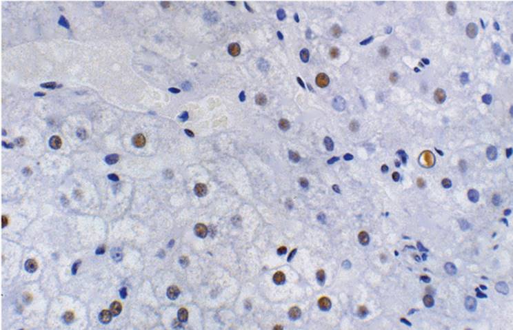
Striking regenerative activity in liver cells and liver nuclei can be seen in patients taking oral contraceptives for only a few months, highlighted by strong reactivity of liver cells with proliferating cell nuclear antibody (Fig. 11.8). Chapter 19 contains a more detailed discussion of hepatic vascular disorders.
|
TABLE 11.5 Medications Causing Vascular Lesions |
||||||||
|
|
|
|
FIGURE 11.8 Striking regenerative and proliferative nuclear activity in a patient on oral contraceptives (antibody for proliferative cell nuclear antigen, immunoperoxidase avidinbiotinperoxidase complex, original magnification ×100). |
NEOPLASMS
Both benign and malignant neoplasms, as well as nonneoplastic proliferating lesions, have been described in association with various drugs and toxic agents (Table 11.1).
Liver cell adenoma was exceedingly rare before the introduction of oral contraceptives. This tumor is no longer rare, particularly in women taking oral contraceptives (35). Adenoma can regress when contraceptives are discontinued. Adenoma also develops after treatment with danazol and testosterone enanthate (15,24). An association between oral contraceptives and focal nodular hyperplasia remains controversial.
Malignant tumors, including hepatocellular carcinoma and cholangiocarcinoma, have been associated with thorium dioxide (Thorotrast) exposure (36) (Fig. 11.9). Hepatocellular carcinoma has been described in patients on long-term anabolic steroid and contraceptive steroid therapy (26).
Occupational exposure to vinyl chloride, long-term exposure to inorganic arsenic, and injection of thorium dioxide are associated with primary angiosarcoma of the liver (e-Figs. 11.85-11.87), an otherwise rare neoplasm (64).
As one example of a proliferative lesion, nodular hyperplasia can be seen in association with 6-thioguanine therapy for inflammatory bowel diseases (e-Figs. 11.70-11.74)
CELLULAR INCLUSIONS
Hepatocytes can undergo various adaptive changes after exposure to various drugs.
|
|
|
FIGURE 11.9 Angiosarcoma of the liver in a patient previously exposed to vinyl chloride (hematoxylin-eosin, original magnification ×100). |
“Ground-glass” hepatocytes are enlarged hepatocytes with pale eosinophilic cytoplasm similar to ground-glass hepatocytes containing hepatitis B surface antigen. Drug-induced ground-glass hepatocytes have hyperplastic and dilated smooth endoplastic reticulum. These cells are diastase resistant and paraaminosalicylic acid positive; they also resemble the cells seen in glycogenosis IV. In contrast to both hepatitis B and glycogenosis, drug-induced ground-glass cells are in zone 1 rather than scattered. Drug-induced ground-glass cells occur with chlorpromazine, barbiturates, and after long-term therapy with steroids, azathioprine, and phenytoin (e-Fig. 11.29) (20,55,70). Ground-glass cells are particularly prominent in patients treated with cyanamide and disulfiram (6,14).
Mallory material, typically associated with alcoholic liver disease, can be seen in amiodarone toxicity (39).
PIGMENTS
Pigments associated with drugs and toxins may be endogenous (e.g., hemosiderin, copper, bilirubin) or exogenous (e.g., gold, thorium dioxide, titanium).
Hemosiderin is often seen with alcoholic liver disease, primarily in sinusoidal cells. Hepatocyte deposition can be prominent, and iron analysis may be necessary to exclude hereditary hemochromatosis. Hemosiderin is also found with alcohol-induced porphyria cutanea tarda. Patients on long-term hemodialysis have hemosiderosis to varying degrees. Copper and copper-binding protein can accumulate in hepatocytes in long-standing cholestasis, including primary biliary diseases such as primary biliary cirrhosis and primary sclerosing cholangitis. Copper is also found in Kupffer cells after occupational exposure to copper salts. Thorium dioxide (Thorotrast), with a half-life of decades to years, is found in the liver long after exposure, recognized as granular, somewhat refractile gray-brown material, either in single or in clustered histiocytic cells (e-Figs. 11.83-11.85) (36). Gold can be demonstrated in patients with rheumatoid arthritis treated with gold salts. The fine black granules of gold in Kupffer cells show golden birefringence with polarized light (24). Titanium accumulates in intravenous drug abusers.
IMMUNOSUPPRESSIVE DRUGS AND THEIR EFFECT ON THE LIVER
Immunosuppressive drugs administered after solid organ transplantation, particularly liver transplantation (LT), include cyclosporine, azathioprine, corticosteroids, antilymphocytic preparations (OKT3), tacrolimus (FK-506), RS-61443, and rapamycin. In addition, patients may receive various antiviral and antifungal medications, including trimethoprim/sulfamethoxazole, cephalosporins, amphotericin, ketoconazole, fluconazole, isoniazid, ganciclovir, hyperalimentation preparations, interferon-a, and, more recently, lamivudine for recurrent forms of hepatitis B and C (20,41,42,74). Some antirejection drugs, such as cyclosporine, OKT3, and tacrolimus cause relatively mild histopathologic changes and only rarely cause significant hepatotoxicity (16,20,41,43,74,75).
DILI should be considered in the differential diagnosis whenever the histopathologic features of the liver biopsy after transplantation appear unusual. Both TPN and azathioprine can induce cholesta tic hepatitis, and both are used in liver transplantation patients. Cyclos porine causes cholestasis, without other changes. Corticosteroids cause sinusoidal dilatation and a variable steatosis. There has been no evidence of hepatotoxicity with tacrolimus (74) or mycophenolate mofetil (43). Antiretroviral drugs used for treatment of HIV-positive patients can cause various changes ranging from mild nonspecific changes, nonalcoholic fatty liver disease (NAFLD), cholestasis, to acute liver failure (44,63).
TOTAL PARENTERAL NUTRITION
The spectrum of histopathologic findings associated with TPN includes cholestasis, cholestatic hepatitis, steatosis, steatohepatitis, phospholipidosis, fibrosis, and cirrhosis (4,5,13).
Cholestasis is common, especially in children. Canalicular cholestasis may be accompanied by cholestatic hepatitis. Giant cells may be seen. Portal tracts usually contain an inflammatory infiltrate composed of lymphocytes and histiocytes, often with eosinophils. Kupffer cells are prominent. After long-term TPN treatment, fibrosis and biliary cirrhosis may develop (e-Figs. 11.92-11.95) (4,5,13).
In adults, steatosis, initially zone 1, is the first and predominant feature, developing in the first weeks of treatment. Cholestasis is often present, and features mimicking bile duct obstruction may develop, including bile ductular reaction (proliferation), fibrosis, and biliary cirrhosis (4,5,13).
In TPN-induced phospholipidosis, the histopathologic changes are similar to those seen in amiodarone-induced phospholipidosis.
ACUTE HEPATITIS ASSOCIATED WITH HERBAL PRODUCTS
Herbal products, popular in North America as potential remedies for various conditions, can be hepatotoxic. Patients develop acute hepatitis as early as 2 weeks and as long as a year after ingestion (77,78), with fatigue, hepatomegaly, nausea, abdominal pain, jaundice, vomiting, peripheral eosinophilia, and pruritus. Children can have central nervous system and respiratory depression along with bradycardia. Symptoms resolve within 2 to 30 weeks, but reuse of the product causes recurrence (78). Hypersensitivity reaction, direct hepatotoxic effect, and an idiosyncratic reaction have all been individually or synergistically implicated.
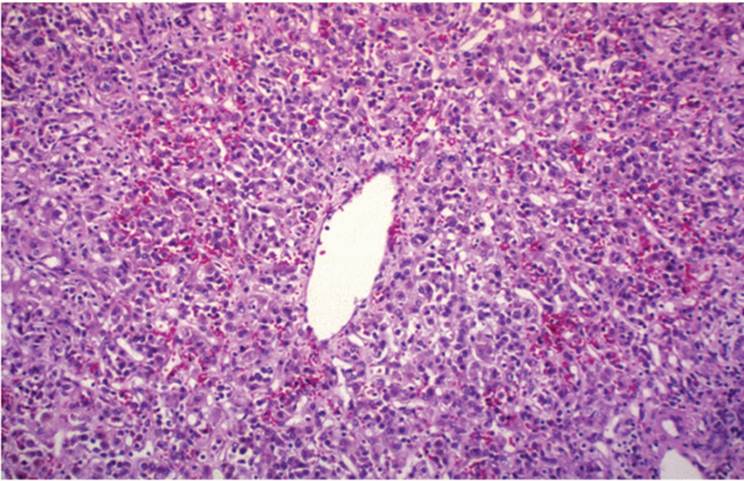
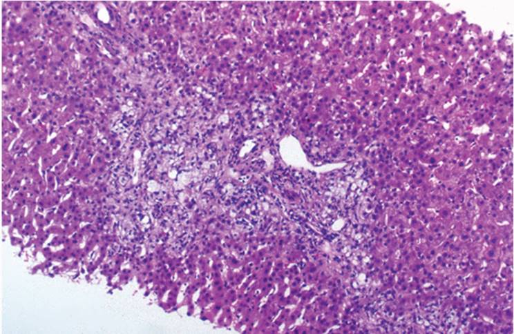
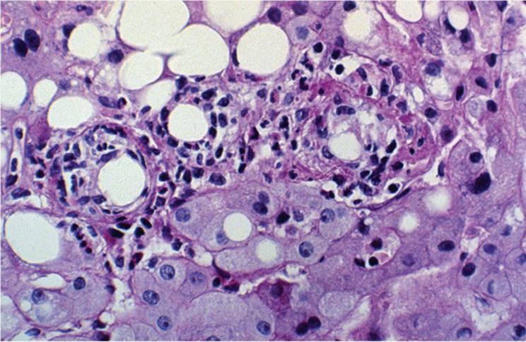
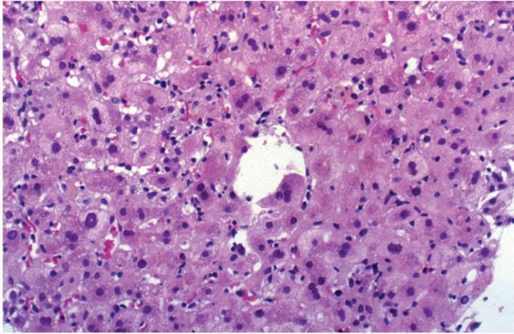
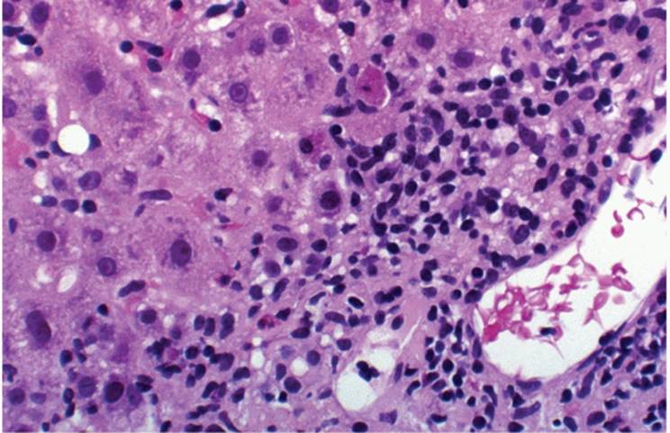
Many “natural” herbal products, including germander, chaparral, senna, mistletoe, skullcap, comfrey, crotalaria, and various herbal teas, are hepatotoxic (9,32,33,42,45,54,77). Their effect is focal lobular necrosis, with variable numbers of eosinophils (Figs. 11.10, 11.11). Focal steatosis may be present.
|
|
|
FIGURE 11.10 Acute hepatitis in a patient taking herbal product (jin bu huan) (hematoxylin-eosin, original magnification ×100). |
|
|
|
FIGURE 11.11 Portal inflammation in the same patient (hematoxylin-eosin, original magnification ×200). |
REFERENCES
1. Ammann R, Neftel K, Hardmeier T, et al. Cephalosporin-induced cholestatic jaundice. Lancet 1982;2:336-337.
2. Aponte J, Petrelli M. Histopathologic findings in the liver of rheumatoid arthritis patients treated with long-term bolus methotrexate. Arthritis Rheum 1988;31:1457-1461.
3. Arranto AJ, Sotaniemi EA. Morphologic alterations in patients with alpha-methyldopainduced liver damage after short- and long-term exposure. Scand J Gastroenterol 1981; 16:853-863.
4. Baker AL, Rosenberg IH. Hepatic complications of total parenteral nutrition. Am J Med 1990;82:489-497.
5. Balistreri WF, Bove KE. Hepatobiliary consequences of parenteral alimentation. Prog Liver Dis 1990;9:567-601.
6. Bartle WR, Fisher MM, Kerenyi N. Disulfiram-induced hepatitis: report of two cases and review of the literature. Dig Dis Sci 1985;30:834-837.
7. Benjamin SB, Goodman ZD, Ishak KG. The morphologic spectrum of halothaneinduced hepatic injury. Hepatology 1985;5:1163-1171.
8. Benjamin SB, Ishak KG, Zimmerman H, et al. Phenylbutazone liver injury: a clinicalpathologic survey of 23 cases and review of the literature. Hepatology 1981;1:255-263.
9. Beuers U, Spengler U, Pape GR. Hepatitis after chronic abuse of senna [letter]. Lancet 1991;337:372-373.
10. Bjorkman DJ, Hammond EH, Lee RG, et al. Hepatic ultrastructure after methotrexate therapy for rheumatoid arthritis. Arthritis Rheum 1988;31:1465-1472.
11. Black M, Mitchell JR, Zimmerman HJ, et al. Isoniazid-associated hepatitis in 114 patients. Gastroenterology 1975;69:289-374.
12. Blackburn AM, Amiel SA, Mills RR, et al. Tamoxifen and liver damage. Br Med J 1984;289:288.
13. Body JJ, Bleiberg H, Bron D, et al. Total parenteral nutrition-induced cholestasis mimicking large bile duct obstruction. Histopathology 1982;6:787-792.
14. Bruguera M, Pares A, Heredia D, et al. Cyanamide hepatotoxicity. Incidence and clinicalpathological features. Liver 1987;7:216-222.
15. Carrasco D, Prieto M, Pallardo L, et al. Multiple hepatic adenomas after long-term therapy with testosterone enanthate. Review of the literature. J Hepatol 1985;1:573-578.
16. Carrier M, Jenicek M, Pelletier L. Value of monoclonal antibodies (OKT3) in solid organ transplantation—a meta analysis. Transplant Proc 1992;24:2586-2591.
17. Chajek T. Quinidine and granulomatous hepatitis [letter]. Ann Intern Med 1975;82:282.
18. Davies MH, Harrison RF, Elias E, et al. Antibiotic-associated acute vanishing bile duct syndrome: a pattern associated with severe, prolonged, intrahepatic cholestasis. J Hepatol 1994;20:112-116.
19. Decker K, Keppler D. Galactosamine induced liver injury. Prog Liver Dis 1972;4:183-199.
20. De Pinho R, Goldberg CS, Lefkowitch JH. Azathioprine and the liver. Evidence favoring idiosyncratic mixed cholestatic-hepatocellular injury in humans. Gastroenterology 1984;86:162-165.
21. Diaz-Rivera RS, Collazo PJ, Pons ER, et al. Acute phosphorus poisoning in men: a study of 56 cases. Medicine 1950;29:269-298.
22. Doria MI, Shepard KV, Levin B, et al. Liver pathology following hepatic arterial infusion chemotherapy. Cancer 1986;58:855-861.
23. Eadie MJ, Hooper WD, Dickinson RG. Valproate-associated hepatotoxicity and its biochemical mechanisms. Med Toxicol 1988;3:85-106.
24. Fermand JP, Levy Y, Bouscary D, et al. Danazol-induced hepatocellular adenoma. Am J Med 1990;88:529-530.
25. Fleishner GM, Morecki I, Manaichi T, et al. Light and electron microscopical study of a case of gold salt induced hepatotoxicity. Hepatology 1991;14:422-425.
26. Forman D, Vincent TJ, Doll R. Cancer of the liver and use of oral contraceptives. Br Med J 1986;292:1357-1361.
27. Galler GW, Weisenberg E, Brasitus TA. Mushroom poisoning: the role of orthotopic liver transplantation. J Clin Gastroenterol 1992;15:229-232.
28. Geubel AP, DeGalocsy C, Alves N, et al. Liver damage caused by therapeutic vitamin A administration: estimate of dose-related toxicity in 41 cases. Gastroenterology 1991;100: 1701-1709.
29. Gilbert SC, Klinmalm G, Menter A, et al. Methotrexate-induced cirrhosis requiring liver transplantation in three patients with psoriasis. A word of caution in light of the expanding use of this “steroid-sparing” agent. Ann Intern Med 1990;150:889-891.
30. Gurascio P, Portmann B, Visco G, et al. Liver damage with reversible portal hypertension from vitamin A intoxication: demonstration of Ito cells. J Clin Pathol 1983;36: 759-771.
31. Harrison RF, Elias E. Amiodarone-associated cirrhosis with hepatic and lymph node granulomas. Histopathology 22:80-82.
32. Harvey J, Colin-Jones DG. Mistletoe hepatitis. Br Med J (Clin Res Ed) 1981;282:186-187.
33. Huxtable RJ, Luthy J, Zweifel U. Toxicity of comfrey-pepsin preparations [letter]. N Engl J Med 1986;315:1095.
34. Ishak KG, Irey NS. Hepatic injury associated with phenothiazines. Clinicopathologic and follow-up study of 36 patients. Arch Pathol 1972;49:630-648.
35. Ishak KG, Zimmerman HJ. Hepatotoxic effects of the anabolic/androgenic steroids. Semin Liver Dis 1987;7:230-236.
36. Ito Y, Kojiro, Nakashima T, et al. Pathomorphologic characteristics of 102 cases of thorotrastrelated hepatocellular carcinoma, cholangiocarcinoma and hepatic angiosarcoma. Cancer 1982;62:1153-1162.
37. Jayalakshmi P, Ting HC. Dapsone-induced liver necrosis. Histopathology 1990;17:89-91.
38. Jorens PG, Michielsen PP, Pelckmans PA, et al. Vitamin A abuse: development of cirrhosis despite cessation of vitamin A. A six-year clinical and pathological follow-up. Liver 1992; 12:381-386.
39. Kalantzis N, Gabriel P, Mouzas J, et al. Acute amiodarone-induced hepatitis. Hepatogastroenterology 1991;38:71-74.
40. Kanel GC, Cassidy W, Shuster L, et al. Cocaine-induced liver cell injury: comparison of morphological features in man and experimental models. Hepatology 1990;11:646-651.
41. Kassianides C, Nussenblatt R, Palestine AG, et al. Liver injury from cyclosporine A. Dig Dis Sci 1990;35:693-697.
42. Katz M, Saibil F. Herbal hepatitis: subacute hepatic necrosis secondary to chaparral leaf. J Clin Gastroenterol 1990;12:203-206.
43. Klintmalm GB, Ascher NL, Busuttil R, et al. RS-61443 for treatment of resistant human liver rejection. Transplant Proc 1993;25:697.
44. Lai KK, Gang DL, Zawacki JK, et al. Fulminant hepatic failure associated with 2′,3′dideoxyuridine (ddI). Ann Intern Med 1991;115:283-284.
45. Larrey D, Vial T, Pauwels A, et al. Hepatitis after germander (Teucrium chamaedrys) administration: another instance of herbal medicine hepatotoxicity. Ann Intern Med 1992;117:129-132.
46. Levy M, Goodman MW, Van Dyne BJ, et al. Granulomatous hepatitis secondary to carbamazepine. Ann Intern Med 1981;95:64-65.
47. Lewis JH, Tice H, Zimmerman HJ. Budd-Chiari syndrome associated with oral contraceptive steroids. Review of treatment of 47 cases. Dig Dis Sci 1983;28:673-683.
48. Ludwig J, Kim CH, Wiesner RH, et al. Floxuridine-induced sclerosing cholangitis: an ischemic cholangiopathy? Hepatology 1989;9:215-218.
49. Maddrey WC. Isoniazid-induced liver disease. Semin Liver Dis 1981;1:129-133.
50. Maddrey WC. Hepatic effects of acetaminophen. Enhanced toxicity in alcoholics. J Clin Gastroenterol 1987;9:180-185.
51. Mallat A, Dhumeaux D. Cocaine and the liver. J Hepatol 1991;12:275-278.
52. Mc Master KR, Hennigar GR. Drug-induced granulomatous hepatitis. Lab Invest 1981; 44:61-73.
53. Minuk GY, Kelly JK, Hwang W-S. Vitamin A hepatotoxicity in multiple family members. Hepatology 1988;8:272-275.
54. Mostefa-Kara N, Pauwels A, Pines E, et al. Fatal hepatitis after herbal tea. Lancet 340:674.
55. Mullick FG, Ishak KG. Hepatic injury associated with diphenylhydantoin therapy. A clinicopathologic study of 20 cases. Am J Clin Pathol 1980;74:442-452.
56. Mullick FG, Ishak KG, Mahabir R, et al. Hepatic injury associated with paraquat toxicity in humans. Liver 1981;1:209-221.
57. Munoz SJ, Martinez-Hernandez A, Maddrey WC. Intrahepatic cholestasis and phospholipidosis associated with the use of trimethoprim-sulfamethoxazole. Hepatology 1990;12: 342-347.
58. Nadell J, Kosek J. Peliosis hepatis. Twelve cases associated with oral androgen therapy. Arch Pathol Lab Med 1977;101:405-410.
59. Newman M, Auerbach R, Feiner H, et al. The role of liver biopsies in psoriatic patients receiving long-term methotrexate treatment. Improvement in liver abnormalities after cessation of treatment. Arch Dermatol 1989;125:1218-1224.
60. Paiva LA, Wright PJ, Koff RS. Long-term hepatic memory for hypersensitivity to nitrofurantoin. Am J Gastroenterol 1992;87:891-893.
61. Paliard P, Vitrey D, Fournier G, et al. Perhexiline maleate-induced hepatitis. Digestion 1978;17:419-427.
62. Peters RL, Edmondson HA, Mikkelsen W, et al. Tetracycline induced fatty liver in non-pregnant patients. Am J Surg 1972;113:622-632.
63. Petrovic LM. HIV/HCV co-infection: histopathologic findings, natural history, fibrosis, and impact of antiretroviral treatment: a review article. Liver Int 2007;27(5):598-606.
64. Popper H, Thomas LB. Alterations of liver and spleen among workers exposed to vinyl chloride. Ann NY Acad Sci 1975;246:172-194.
65. Popper H, Geller SA. Pathogenetic considerations in the histologic diagnosis of drug-induced liver injury. Prog Surg Pathol 1981;2:233-246.
66. Portmann B, Talbot IC, Day DW, et al. Histopathological changes in the liver following paracetamol overdose: correlation with clinical and biochemical parameters. J Pathol 1975;117:169-181.
67. Qiang L, Tobias H, Petrovic LM. Drug-induced hepatitis caused by ezetimibe therapy. Dig Dis Sci 2007;52(2):602-605.
68. Rabinowitz M, Van Thiel DH. Hepatotoxicity of non-steroidal anti-inflammatory drugs. Am J Gastroenterol 1992,87:1696-1704.
69. Rollins BJ. Hepatic veno-occlusive disease. Am J Med 1986;81:297-306.
70. Rotmensch HH, Yust I, Siegman-Igra Y, et al. Granulomatous hepatitis: a hypersensitivity response to procainamide. Ann Intern Med 1978;89:646-647.
71. Roy AK, Mahoney HC, Levine RA. Phenytoin-induced chronic hepatitis. Dig Dis Sci 1993;38:740-743.
72. Ruprah M, Mant TGK, Flanagan RJ. Acute carbon tetrachloride poisoning in 19 patients: implications for diagnosis and treatment. Lancet 1985;1:1027-1029.
73. Shepherd P, Harrison DJ. Idiopathic portal hypertension associated with cytotoxic drugs. J Clin Pathol 1990;43:206-210.
74. Starko KM, Mullick FG. Hepatic and cerebral pathology findings in children with fatal salicylate intoxication: further evidence for a causal relationship between salicylate and Reye's syndrome. Lancet 1983;1:326-329.
75. Sterncek M, Wiesner R, Ascher N, et al. Azathioprine hepatotoxicity after liver transplantation. Hepatology 1991;14:465-471.
76. United States Multicenter FK 506 Liver Study Group. Use of FK 506 for the prevention of recurrent allograft rejection after successful conversion from cyclosporine for refractory rejection. Transplant Proc 1993;25:635-637.
77. Verhamme M, Ramboer C, Van de Bruaene P, et al. Cholestatic hepatitis due to amoxycillin/clavulanic acid preparation. J Hepatol 1989;9:260-264.
78. Woolf GM, Petrovic LM, Rojter SE, et al. Acute hepatitis associated with the Chinese herbal product jin bu huan. Ann Int Med 1994;121:729-735.
79. Zafrani ES, Pinaudeau Y, Dhumeaux D. Drug-induced vascular lesions of the liver. Arch Intern Med 1983;143:495-502.
80. Zimmerman HJ, Ishak KG. Valproate-induced hepatic injury: analysis of 23 fatal cases. Hepatology 1982;2:591-597.