The principal disorders affecting intrahepatic bile ducts are primary biliary cirrhosis (PBC) and primary sclerosing cholangitis (PSC). They show similar histologic changes and reflect, to some extent, immune-mediated bile duct injury and, eventually, bile duct destruction and loss. They are also alike as progressive, chronic liver diseases leading to liver failure in either the precirrhotic or the cirrhotic stage. Clinically, they can also behave similarly as chronic, relentless, cholestatic disorders, with similar patterns of biochemical abnormalities. Medical treatment is generally not effective, and liver transplantation is often the only life-saving measure. However, the conditions differ in terms of the patients affected.
PRIMARY BILIARY CIRRHOSIS
Primary biliary cirrhosis (PBC) is a progressive nonsuppurative destructive cholangitis affecting mostly middle-aged women, with peak incidence between 40 and 60 years (30). The onset is usually insidious, with patients asymptomatic for years. In contrast with PSC, with its strong association with inflammatory bowel disease, there are no known predecessor disorders. Patients frequently present with cholestatic itching, which can be disabling. Jaundice is usually not seen early; cholestasis in the biopsy is a poor prognostic sign (22,24,25). PBC can be associated with various autoimmune conditions, including sicca syndrome, CREST (calcinosis, Raynaud phenomenon, esophageal motility disorders, sclerodactyly, and telangiectasia) syndrome, celiac sprue, membranous glomerulonephritis, and interstitial lung disease (3,5,10,21,23,27,29). Hepatocellular carcinoma is uncommon (7,9,26). Elevated serum alkaline phosphatase value and high titers of antimitochondrial antibody (AMA) are virtually diagnostic.
Antimitochondrial antibodies are a family of immunoglobulins, including anti-M2, anti-M4, anti-M8, and anti-M9, all of which are directed against different epitopes and all of which may be associated with primary biliary cirrhosis. Anti-M2, the antibody against the multienzyme complexes at the inner mitochondrial membrane, is present in all PBC patients; its presence is considered highly specific and virtually diagnostic (24).
Despite the presence of AMA, the etiopathogenesis of PBC is unclear. Autoimmune mechanisms may have a role in its development. Impairment of cell-mediated immunity, particularly involving the T-lymphocyte population, circulating immune complexes, and complement activation, has been implicated (13,14). There is up-regulation of human leukocyte antigen (HLA) class I antigens as well as aberrant expression of HLA class II antigens on bile duct epithelium in PBC. This is not specific and is seen in other immunologically mediated bile duct conditions, including acute allograft rejection and graft versus host disease (GVHD).
The possibility that PBC is an atypical manifestation of different infectious diseases, such as tuberculosis or sarcoidosis, has been considered. A possible association with Escherichia coli, which is antigenically similar to biliary epithelium, has been suggested (molecular mimicry) (12). Chlamydia pneumoniae has also been implicated in the pathogenesis of PBC (1,19).
The diagnosis is usually established clinically. Liver biopsy confirms the diagnosis but is particularly useful for determining the stage of the disease and in monitoring progression.
Pathology
Four distinct stages are generally recognized although there is a significant overlap among them, and as the disease progresses those overlapping features can be found in any given biopsy. Several staging classifications have been proposed (Table 17.1) (18,24). The Scheuer staging method emphasizes an early, florid bile duct injury stage with subsequent bile ductular proliferation (reaction) as particularly important in the development of the histopathologic changes (23,25). The Ludwig approach, in contrast, emphasizes the inflammatory cell component (18).
Despite the commonly accepted name, cirrhosis is only the last stage in the development of the disease, and PBC encompasses a wide spectrum of relentlessly progressive changes. Stage 1 (early primary biliary cirrhosis) is characterized by florid duct lesion and portal inflammation (e-Figs. 17.1-17.4). Bile duct injury is segmental, affecting small and medium-sized interlobular bile ducts. Bile duct epithelium is irregularly involved. Hyperplasia of the epithelium occurs early in association with infiltration by mature lymphocytes. The basement membrane is disrupted, and bile duct rupture takes place. The injured bile duct is usually surrounded by inflammatory infiltrate predominantly composed of lymphocytes, plasma cells, some histiocytes, and a variable number of eosinophils (23,24). Lymphoid aggregates may form, sometimes including true lymphoid follicles with germinal centers, resembling those seen in chronic hepatitis C infection (see Chapter 9) (Fig. 17.1).
|
TABLE 17.1 Staging of Primary Biliary Cirrhosis |
||||||||||||||||||||
|
||||||||||||||||||||
|
|
||||||||||||||||||||
|
FIGURE 17.1 Early (stage 1) primary biliary cirrhosis. Florid bile duct injury and associated predominantly mononuclear inflammatory infiltrate in the portal tract (hematoxylin-eosin, original magnification ×400). |
||||||||||||||||||||
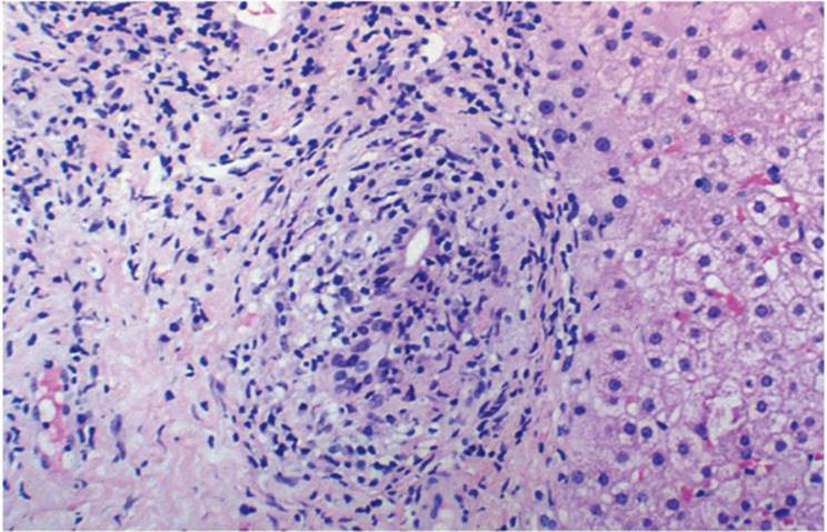
In as many as 40% of stage 2 early-stage biopsies (e-Figs. 17.5-17.11) characteristic, but not pathognomonic, epithelioid and histiocytic noncaseating granulomas can be seen immediately adjacent to a damaged bile duct (Figs. 17.2, 17.3, e-Figs. 17.5-17.9, 17.11) (17). Granulomas can also be seen in later stages but are uncommon with advanced fibrosis. The absence of granulomatous bile duct damage does not exclude the diagnosis (23,24). Because the bile duct injury in PBC is segmental, any biopsy may fail to show diagnostic changes, and step sections, and occasionally a subsequent biopsy, may be required (Fig 17.4).
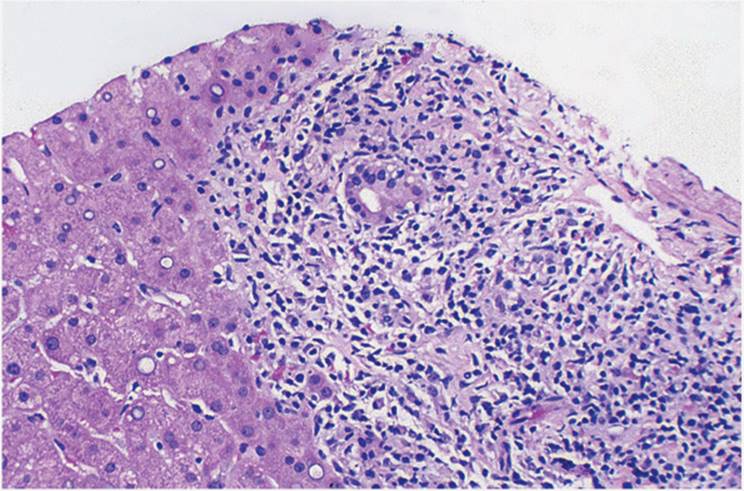
The stage of bile duct injury and portal inflammation is followed by extension of the inflammatory infiltrate beyond the limiting plate into the lobule, strongly resembling the lymphocytic interface hepatitis (lymphocytic piecemeal necrosis) of various forms of chronic hepatitis, particularly chronic hepatitis C. Kupffer cells are frequently prominent, and varying degrees of parenchymal inflammation, with sinusoidal inflammation, are invariably present. Lobular foci of necrosis can be seen, with accumulations of lymphocytes and even scattered small granulomas or clusters of histiocytes (Fig. 17.5, e-Figs. 17.5-17.11). Cholestasis is usually not prominent in early PBC. Once significant fibrosis or frank cirrhosis has developed, parenchymal inflammatory activity is usually minimal.
|
|
|
FIGURE 17.2 Early (stage 1) primary biliary cirrhosis. Granulomatous inflammation and bile duct injury (hematoxylin-eosin, original magnification ×200). |
|
|
|
FIGURE 17.3 Primary biliary cirrhosis. Extensive interface hepatitis is present (lymphocytic piecemeal necrosis) (hematoxylin-eosin, original magnification ×200). |
Early PBC can sometimes be associated with portal hypertension, perhaps because of narrowing of intrahepatic portal vein branches by the inflammatory process. In some patients, nodular hyperplasia may contribute to portal hypertension (Fig. 17.6) (4).
|
|
|
FIGURE 17.4 Primary biliary cirrhosis. Smaller bile ducts are affected, and some of them have already disappeared (hematoxylin-eosin, original magnification ×200). |
|
|
|
FIGURE 17.5 Primary biliary cirrhosis. Focal bile ductular proliferation is present (hematoxylin-eosin, original magnification ×200). |
Late primary biliary cirrhosis (periportal or progressive stage) is manifested not only by more extensive bile duct injury but also by expansion of the portal tracts with associated lymphocytic piecemeal necrosis, as well as marked bile ductular reaction (proliferation), giving rise to so-called biliary interface hepatitis. Superimposed on this is increasing fibrosis, sometimes referred to as “fibrotic interface hepatitis” (24,25). These three changes interrupt the limiting plate and are generally easily seen.
|
|
|
FIGURE 17.6 Primary biliary cirrhosis. Mild lobular inflammation and microgranulomas are present (hematoxylin-eosin, original magnification ×200). |
Prolonged cholate stasis gives a characteristic appearance to the periportal/paraseptal hepatocytes (e-Figs. 17.12-17.14). They are slightly enlarged and pale, with rarefied cytoplasm, sometimes containing abundant copper-associated protein (e-Fig. 17.15), demonstrable with Victoria blue or orcein stain. Mallory hyalin may also be present. In fully developed stage 3 and 4 PBC, intracanalicular cholestasis can be prominent and most of the original interlobular bile ducts are no longer seen. Granulomatous inflammatory injury may affect the remaining bile ducts. Clusters of xanthomatous and pseudoxanthomatous cells are also seen.
However, the most significant and characteristic feature of late-stage PBC is progressive bile duct loss (Fig. 17.7). The normal portal tract generally has at least one interlobular bile duct with one to two accompanying hepatic artery branches (see Chapter 4). The site of the original bile ducts is sometimes marked by collections of histiocytes or lymphoid aggregates, adjacent to undamaged arteries. The fibrous scarring typical of PSC is not seen in PBC. Eventually fibrous septa extend from portal tracts and intersect parenchymal architecture, with development of irregular (jigsaw puzzle-shaped) regenerative nodules (Fig. 17.8, e-Figs. 17.6-17.18).
PBC can recur after liver transplantation (e-Figs. 17.19-17.22).
Differential Diagnosis
Several conditions affecting the liver show variable degrees of bile duct injury. In addition to PBC and PSC, other conditions, such as suppurative cholangitis, sarcoidosis, graft versus host disease (GVHD), and acute cellular and chronic ductopenic rejection have bile duct injury as a significant feature. In each of these, the injury may be destructive and progressive, leading to bile duct loss.
|
|
|
FIGURE 17.7 Primary biliary cirrhosis. Slender fibrous septa (late stage 3, 4), and periseptal cholestasis and cholate stasis (hematoxylin-eosin, original magnification ×100). |
|
|
|
FIGURE 17.8 Primary sclerosing cholangitis (PSC). Bile duct injury in the early (stage I) of PSC. Bile duct epithelium is irregular and atrophic (hematoxylin-eosin, original magnification ×200). |
In contrast, some degree of bile duct damage, without loss, may be seen in viral hepatitis, particularly hepatitis C and autoimmune hepatitis, some drugs/toxins, HIV-associated cholangiopathy (with or without cryptosporidiosis), sepsis and septicemia, toxic shock syndrome, parasitic infestation, and Hodgkin disease.
The progressive periportal stage of PBC can be difficult to differentiate from chronic hepatitis with periportal activity. In most forms of chronic hepatitis, there is only mild bile duct epithelial injury and no recognizable bile duct loss. Lobular activity with focal necrosis, acidophilic body formation, and inflammation is usually more pronounced in chronic viral hepatitis than in PBC. Differentiating between PBC and autoimmune hepatitis can also be difficult. Clinical information is invaluable in these cases.
PRIMARY SCLEROSING CHOLANGITIS
Primary sclerosing cholangitis (PSC) is characterized by the presence of inflammation and fibrosis with segmental stricturing along the biliary tree. In many patients there is little or no intrahepatic bile duct change until late in the disease, and in part for this reason, the biopsy is not a standard for diagnosis. Small duct PSC is a variant that affects only intrahepatic bile ducts (20). In small duct PSC, liver biopsy is the principal means of establishing the diagnosis.
Approximately 70% of patients with PSC also have inflammatory bowel disease (IBD), mostly ulcerative colitis (UC). Conversely, fewer than 10% of patients with UC have associated PSC. PSC can sometimes precede the onset of PSC. PSC occurs at any age, with peak incidence in the fourth decade, with an incidence in males twice that of in females (25). The onset may be insidious, with malaise associated with increased serum alkaline phosphatase values. In contrast with PBC, there are no specific associated serum antibodies. In approximately 65% of patients, however, high titers of perinuclear antineutrophil cytoplasmic antibody (p-ANCA) are demonstrable and anti-smooth muscle antibody (SMA) titer may be increased (8). Approximately 10% of chronic PSC patients develop cholangiocarcinoma.
The etiopathogenesis of PSC is not well understood. Both nonimmune and autoimmune mechanisms are thought to contribute (15). Although PSC and IBD often occur concurrently, they can also occur independently of each other, suggesting that different factors influence their occurrence. Familial cases indicate some genetic predisposition with associated HLA-B8 and DR2 and DR3 haplotypes. In a few cases, PSC and autoimmune hepatitis occur simultaneously, so-called overlap syndrome (28).
Although the diagnosis of PSC can sometimes be established on liver biopsy, definitive diagnosis is most often based on characteristic cholangiographic findings. The biliary tree is distorted with alternating segments of extrahepatic bile duct stenosis and dilatation, imparting a typical and virtually diagnostic “beaded” appearance. Other possible causes of secondary sclerosing cholangitis must be excluded including previous biliary tract surgery or prior instances of cholecystitis and cholelithiasis. They may have similar histopathologic and cholangiographic findings.
Pathology
Early (stage 1) changes (e-Figs. 17.23-17.27) may be inconspicuous and, even when present, not seen in all portions of the liver, with resultant unremarkable biopsies (20). Interlobular bile ducts may show only mild epithelial irregularity with focal atrophy. The hyperplastic epithelial changes seen in PBC are not seen in PSC. Instead, partial lumen obliteration and focal branching of bile ducts occur (Fig. 17.9). Portal tracts have a mild chronic inflammatory cell infiltrate, mostly lymphocytes, confined to the portal tract. Interface hepatitis is unusual. Lymphoid aggregates are seen but without germinal centers. Granulomas are only rarely seen.
|
|
|
FIGURE 17.9 A and B. Primary sclerosing cholangitis. Bile duct injury is very subtle, with epithelial atrophy, pleomorphic epithelial nuclei, and early branching of the intrahepatic ducts (hematoxylin-osin, original magnification ×200). |
|
|
|
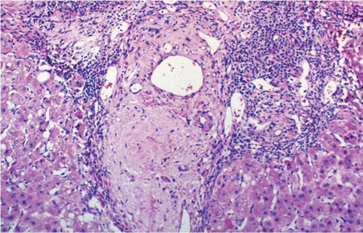
FIGURE 17.10 Primary sclerosing cholangitis. The inflammatory infiltrate in the portal tract is predominantly mononuclear, and only focal interface hepatitis is present (hematoxylin-eosin, original magnification ×200). |
|
|
|
FIGURE 17.11 Primary sclerosing cholangitis. Portal tract fibrosis, slender fibrous septa and “smudgy” fibrous scar, the site of the original interlobular bile duct (hematoxylin-eosin, original magnification ×200). |
Stage 2 PSC (e-Fig. 17.28) is characterized by more extensive inflammatory interface hepatitis, sometimes mimicking chronic hepatitis C or autoimmune hepatitis. Bile ductular proliferation is usual but may be minimal and only focal. Mild fibrosis of the portal tracts may be evident (Fig. 17.10, e-Figs. 17.29-17.33). Parenchymal changes are prognostically more important than bile duct alterations (19) (Fig. 17.11).
|
|
|
FIGURE 17.12 Primary sclerosing cholangitis (PSC). Focal bile ductular proliferation and periseptal cholestasis and cholate stasis are present, usually in the late stages (stage 3, 4) of PSC (hematoxylin-eosin, original magnification ×200). |
Stage 3 PSC has a characteristic, but not pathognomonic, pattern of periductal fibrosis (e-Figs. 17.28-17.31). The “onion skinning” pattern of fibrosis becomes more obvious in later stages but can be present earlier (e-Fig. 17.28). Ultimately, there is bile duct loss and complete replacement of the bile duct with a characteristic fibrous scar, described as “smudgy” (Fig. 17.12 e-Figs. 17.32-17.35). Unfortunately, this is not uniformly distributed and often is not seen in biopsy samples.
At this stage progressive, biliary-type fibrosis is more extensive and portal-to-portal bridging usual (e-Figs. 17.33-17.35). The inflammatory infiltrate is less prominent. With loss of many interlobular bile ducts, portal tracts contain only arteries and portal tributaries. Periportal/periseptal hepatocytes show cholate stasis, similar to that of PBC, with accumulation of copper and copper-binding protein (e-Fig. 17.36) and occasionally Mallory hyalin (e-Fig. 17.37). Despite extensive loss of interlobular bile ducts, there is little bile ductule proliferation. Centrolobular cholestasis is only seen when large bile ducts are affected.
Stage 4 PSC has extensive biliary-type fibrosis imparting the typical picture of biliary-type cirrhosis (e-Fig. 17.38) (11). The advanced stages of PBC and PSC may be indistinguishable, and the correct diagnosis requires clinical information.
PSC can recur after liver transplantation (e-Figs. 17.39, 17.40)
Differential Diagnosis
Secondary (acquired) sclerosing cholangitis can be indistinguishable from PSC. Secondary sclerosing cholangitis was seen in most patients who were treated with intra-arterial infusions of fluorodeoxyuridine for metastatic adenocarcinoma (6). Patients with AIDS can have various bile duct changes, including true cholangiopathy (AIDS cholangiopathy), as well as acalculous cholecystitis and sclerosing cholangitis, sometimes with papillary stenosis. These occur with or without either cryptosporidiosis or cytomegalovirus infection.
In immune deficiency syndromes in children, such as familial combined immunodeficiency and immunodeficiency with hyperimmunoglobulin M, the features of sclerosing cholangitis may be present. Superimposed bacterial/opportunistic infection in these conditions can also cause changes that resemble PSC. GVHD, autoimmune cholangitis, portal vein thrombosis, and Langerhans cell histiocytosis (histiocytosis X) can all cause changes that resemble PSC.
REFERENCES
1. Abdulkarim A, Petrovic L, Kim WR, et al. Primary biliary cirrhosis: an infectious disease caused by Chlamydia pneumoniae? J Hepatol 2004;40(3):380-384.
2. Baggenstoss AH, Foulk WT, Butt HR, et al. The pathology of primary biliary cirrhosis with emphasis on histogenesis. Am J Clin Pathol 1964;42:259-276.
3. Ballardini G, Mirakian R, Bianchi FB, et al. Aberrant expression of HLA-DR antigens on bile duct epithelium in primary biliary cirrhosis: relevance to pathogenesis. Lancet 1984; 2:1009-1013.
4. Colina F, Pinedo F, Solis JA, et al. Nodular regenerative hyperplasia of the liver in early histological stages of primary biliary cirrhosis. Gastroenterology 1992;102:1319-1324.
5. Culp KS, Fleming CR, Duffy J, et al. Autoimmune association in primary biliary cirrhosis. Mayo Clin Proc 1982;57:365-370.
6. Demetris AJ, Markus BH, Esquivel C, et al. Pathologic analysis of liver transplantation for primary biliary cirrhosis. Hepatology 1988;8:939-947.
7. Farinati F, Floreans A, DeMaria N, et al. Hepatocellular carcinoma in primary biliary cirrhosis. J Hepatol 1994;21:315-316.
8. Farrant JM, Hayllar KM, Wilkinson ML, et al. Natural history and prognostic variables in primary sclerosing cholangitis. Gastroenterology 1991;100:1710-1717.
9. Gores GJ. Yes, hepatocellular cancer does occur in primary biliary cirrhosis. Liver Transpl 2002;8:570-571.
10. Gores GJ, Moore SB, Fisher LD, et al. Primary biliary cirrhosis: associations with class II major histocompatibility complex antigens. Hepatology 1987;7:889-892.
11. Jeffrey GP, Reed DW, Carrello S, et al. Histological and immunohistochemical study of the gall bladder lesion in primary sclerosing cholangitis. Gut 1991;32:424-429.
12. Jones DE, Palmer JM, Burt AD, et al. Bacterial motif DNA as an adjuvant for the breakdown of immune self-tolerance to pyruvate dehydrogenase complex. Hepatology 2002; 36:679-686.
13. Kaplan MM. Primary biliary cirrhosis: past, present, and future. Gastroenterology 2002; 123:1392-1394.
14. Keefe NA, Gershwin ME. Immunopathogenesis of primary biliary cirrhosis. Semin Liver Dis 2002:22:291-302.
15. Lee TJ, Wanless IR, Tsui FW, et al. Apoptosis of biliary epithelial cells in primary biliary cirrhosis and primary sclerosing cholangitis. Liver 2002;22:228-234.
16. Ludwig J, Dickson ER, McDonald GS. Staging of chronic nonsuppurative destructive cholangitis (syndrome of primary biliary cirrhosis). Virchows Arch A Pathol Anat 1978; 379:103-112.
17. Ludwig J. New concepts in biliary cirrhosis. Semin Liver Dis 1987;7:293-301.
18. Mackay IR, Gershwin ME. Primary biliary cirrhosis: considerations on pathogenesis based on identification of the M2 autoantigens. Springer Semin Immunopathol 1990; 12:101-119.
19. Marangoni A, Donati M, Cavrini F, et al. Chlamydia pneumoniae replicates in Kupffer cells in mouse model of liver infection. World J Gastroenterol 2006;28:12(40):6453-6457.
20. Markus BH, Dickson ER, Grambsch PM, et al. Efficacy of liver transplantation in patients with primary biliary cirrhosis. N Engl J Med 1989;320:1709-1713.
21. McMahon RF, Babbs C, Warnes TW. Nodular regenerative hyperplasia of the liver, CREST syndrome and primary biliary cirrhosis: an overlap syndrome? Gut 1989;30:1430-1433.
22. Michieletti P, Wanless IR, Katz A, et al. Are patients with antimitochondrial antibody negative primary biliary cirrhosis a distinct syndrome of autoimmune cholangitis? Gut 1994;35:260-265.
23. Portmann B, Popper H, Neuberger J, et al. Sequential and diagnostic features in primary biliary cirrhosis based on serial histologic study in 209 patients. Gastroenterology 1985; 88:1777-1790.
24. Sherlock S, Scheuer PJ. The presentation and diagnosis of 100 patients with primary biliary cirrhosis. Lancet 1987;2:493-496.
25. Shimizu M, Yuh K, Aoyama S, et al. Immunohistochemical characterization of inflammatory infiltrates at the site of bile duct injury in primary biliary cirrhosis. Liver 1986;6:1-6.
26. Singson RC, Fraiman M, Geller SA. Hepatocellular carcinoma with fibrolamellar pattern in a patient with autoimmune cholangitis. Mt Sinai J Med 1999;66:109-112.
27. Wee A, Ludwig J, Coffey RJJ, et al. Hepatobiliary carcinoma associated with primary sclerosing cholangitis and chronic ulcerative colitis. Hum Pathol 1985;16:719-726.
28. Wiesner RH, Grambsch PM, Dickson ER, et al. Primary sclerosing cholangitis: natural history, prognostic factors and survival analysis. Hepatology 1989;10:430-436.
29. Wiesner RH, LaRusso NF, Ludwig J, et al. Comparison of the clinicopathologic features of primary sclerosing cholangitis and primary biliary cirrhosis. Gastroenterology 1985;88: 108-114.
30. Yamada S, Howe S, Scheuer PJ. Three-dimensional reconstruction of biliary pathways in primary biliary cirrhosis: a computer-assisted study. J Pathol 1987;152:317-323.