Disorders affecting the large extrahepatic bile ducts are common. Modern biochemical tests and sophisticated imaging techniques often make liver biopsy unnecessary in contrast with past years when large duct obstruction (LDO) was one of the most important indications for liver biopsy.
LARGE DUCT OBSTRUCTION
Acute Large Bile Duct Obstruction
Acute large bile duct obstruction (LDO) often has a dramatic clinical presentation with sudden onset of jaundice and right upper quadrant abdominal pain, generally due to obstruction of the common hepatic duct or the ampulla of Vater.
Histopathologic changes are characteristic but not pathognomonic. They can be seen with obstruction of the right or left hepatic ducts as well as the smaller intrahepatic ducts into which portal tract interlobular ducts drain. When either intrahepatic duct obstruction or obstruction above the point of juncture of the right and left hepatic ducts is suspected, biopsy of tissue from both liver lobes may be useful. Changes will be bilateral with obstructions below the juncture of the right and left hepatic ducts, but will be one-sided when only one is involved or when the obstructed duct is intrahepatic.
Acute LDO is most commonly associated with gallstones and can persist after passage of the gallstone. Any process causing obstruction or stenosis of the bile ducts can lead to similar changes, including neoplasms (13), strictures following surgical procedures (8), and congenital biliary atresia (see Chapter 6), for example. Rare causes include inflammatory bile duct polyps, choledochal cyst or diverticulum, parasites such as Clonorchis sinensis, annular pancreas compressing the common duct, and pancreatitis and its complications (7,10,11,16,21,23).
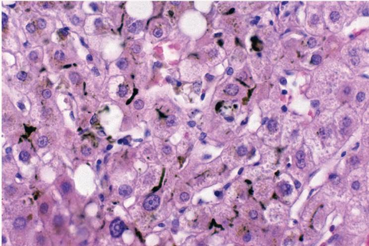
PATHOLOGY. The earliest change, seen as early as 2 or 3 days after onset, is canalicular cholestasis, predominantly in zone 3 of the acinus (Fig. 18.1). The bile is visible as green or brown-green pigment in the form of bile plugs or bile thrombi. In addition, the perivenular (zone 3) area has prominent, bile-laden Kupffer cells. In resolving acute LDO, the bile plugs disappear first, but pigment-rich Kupffer cells may persist for weeks. Periodic acid-Schiff-positive Kupffer cells may be the only remaining evidence of cholestasis after resolution of the obstruction (23).
|
|
|
FIGURE 18.1 Acute large bile duct obstruction (early, first couple of days). Centrolobular canalicular cholestasis and mild reactive changes in the hepatocytes (hematoxylin-eosin, original magnification ×200). |
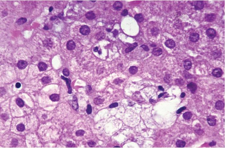
In acute LDO, hepatocytes are not primarily damaged. If obstruction and cholestasis persist, however, secondary reactive changes become prominent (4). Cholestasis up-regulates class I human leukocyte antigen expression in hepatocytes leading to focal liver cell injury and necrosis (6). Zone 3 hepatocytes have distended, swollen cytoplasm, and liver cell nuclei vary in size with nuclear hyperchromasia. The cytoplasm of some hepatocytes becomes rarified with fine, weblike reticulation, traditionally recognized, and described as “feathery degeneration” (Fig. 18.2). Individual liver cells may be necrotic (acidophilic bodies). Mitoses are common. Ultrastructurally there is hypertrophy of endoplasmic reticulum.
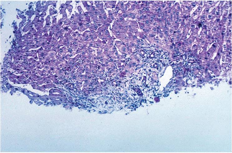
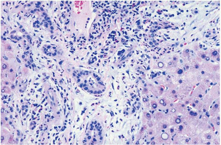
In the first week after onset of LDO, portal tracts are edematous, with pale, loose connective tissue and early fibroblast proliferation (Fig. 18.3, e-Figs. 18.1-18.4). Interlobular or septal bile ducts can show reactive epithelial changes including irregularity of epithelium, variation in size and nuclear hyperchromasia, and, occasionally, true necrosis. Bile duct epithelium may later become atrophic, particularly once periductal fibrosis is well developed. In spite of this, interlobular bile duct loss is exceedingly rare (Fig. 18.4).
The portal tract inflammatory infiltrate varies. Early, there is little or no inflammatory cell accumulation. When present, lymphoid and histiocytic cells predominate (Fig. 18.4). As LDO persists, the number of polymorphonuclear leukocytes (PMNs) increases in number, usually accompanying so-called marginal bile ductular proliferation. Proliferating bile ductules are themselves chemotactic and nonspecifically stimulate a PMN response in many conditions associated with ductular proliferation.
|
|
|
FIGURE 18.2 Large bile duct obstruction (first week). Centrolobular cholestasis and feathery degeneration of some hepatocytes (hematoxylin-eosin, original magnification ×200). |
Marginal bile ductular reaction (proliferation) is a reliable, but not pathognomonic, feature of LDO, seen in more than 80% of portal tracts (e-Fig. 18.4). Although proliferated bile ductules may have a dilated lumen, there is usually no visible bile. When dilated ductules are prominent, sepsis and septicemia should also be considered (17). Superimposed acute cholangitis with intraluminal PMNs and chronic inflammatory cells focally infiltrating the bile duct epithelium can be seen. Proliferated ductules, especially before a luminal space is apparent, can be highlighted with keratin immunohistochemistry, using CK19 or keratin AE 1,3.
|
|
|
FIGURE 18.3 Acute large bile duct obstruction (early, first 2 to 3 days). Portal tract is edematous and contains only mild predominantly mononuclear inflammatory infiltrate (hematoxylin-eosin, original magnification ×100). |
|
|
|
FIGURE 18.4 Acute large bile duct obstruction (first week). Edematous portal tract with inflammatory infiltrate composed of lymphocytes, histiocytes, and polymorphonuclear leukocytes and with associated marginal bile ductular proliferation (hematoxylin-eosin, original magnification ×200). |
The origin of ductules is controversial. Possibilities include (a) multiplication of pre-existing bile ductules, (b) proliferation from a putative progenitor (stem) cell, and (c) metaplasia of periportal hepatocytes. It may be that they are all active at different stages (9,24,27,29,30,31). Multiplication of the preexisting bile ductules seems more important in acute cholestasis, whereas ductular periportal metaplasia is more often associated with chronic cholangiopathies, including primary biliary cirrhosis and primary sclerosing cholangitis. Hepatocytes undergo phenotypic changes with expression of biliary-type cytokeratin, integrins, blood group antigens, and tissue peptide antigen (20,29,30,31). Cholestatic rosette formation is commonly seen (Fig. 18.5).
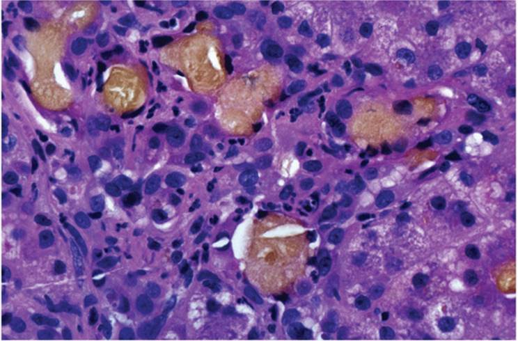
Periportal bile lakes or bile infarcts are the hallmark of LDO, although seen in only a small percentage of patients. Bile within the bile lake is often admixed with fibrin. Hepatocytes immediately adjacent show variable degrees of degeneration and lytic-type necrosis. Bile can escape from bile ducts into portal tracts as bile extravasation with local histiocytic phagocytic reaction, sometimes causing giant cell reaction (Fig. 18.6).
Long-Standing Large Duct Obstruction
The clinical diagnosis of long-standing LDO obstruction is rarely problematic.
PATHOLOGY. In long-standing LDO, lasting weeks to months, fibrosis gradually replaces the portal inflammatory infiltrate. A characteristic pattern of periductal, concentric fibrosis, similar to that of primary sclerosing cholangitis, develops (Figs. 18.7, 18.8). Portal-to-portal fibrous bridging ensues, and secondary biliary cirrhosis eventually develops. Liver cell injury is not prominent, and fibrous septa are highly irregular. The nodules that form are not circular as are typical regenerative nodules of cirrhosis following hepatitis and have a geographic or jigsaw-puzzle appearance (e-Figs. 18.5, 18.6) (5,7,8). In the absence of portal-to-central shunting and true regenerative nodules, secondary biliary cirrhosis is not a true cirrhosis and initially manifests as portal hypertension without hepatocellular dysfunction (see Chapter 20). Cholestasis has an inhibitory effect on liver cell regeneration by interfering with expression of growth-promoting genes (4,28). The term biliary fibrosis is more appropriate than biliary cirrhosis in these cases.
|
|
|
FIGURE 18.5 Large bile duct obstruction. In addition to reactive changes and feathery degeneration, occasional cholestatic rosettes are formed (hematoxylin-eosin, original magnification ×400). |
|
|
|
FIGURE 18.6 Large bile duct obstruction. Bile infarct formation is pathognomonic of bile duct obstruction (hematoxylin-eosin, original magnification ×200). |
|
|
|
FIGURE 18.7 Large bile duct obstruction (long-standing, several weeks). Portal inflammation and fibrosis and bile ductular proliferation (hematoxylin-eosin, original magnification ×400). |
|
|
|
FIGURE 18.8 Large bile duct obstruction (long-standing, at least several weeks). Periductal concentric fibrosis. Only reactive epithelial changes are present in the bile duct (hematoxylin-eosin, original magnification ×200). |
Other parenchymal changes can be seen with long-standing cholestasis secondary to LDO. Feathery degeneration (Fig. 18.2), and cholestatic rosettes (Fig. 18.5), representing a circular arrangement of hepatocytes around a central bile plug, may be seen. Antibody directed against cytokeratin CAM 5.2 will highlight these tubule-like structures. Clusters of foamy (xanthomatous) cells are seen. Perpiportal or paraseptal hepatocytes are pale and rarified (cholate stasis). These hepatocytes often have increased copper-associated protein, demonstrated with Victoria blue or orcein (Shikata) stain (14,25,26). Mallory hyalin can be present (Fig. 18.9), but, unlike in alcoholic liver disease, the intracytoplasmic hyalin is almost invariably periportal or paraseptal and not distributed throughout the lobule (see Chapter 13). Portal and periportal fibrosis develops (e-Figs. 18.7-18.9). Fibrous septa are usually regular, broad, and paucicellular, with loosely arranged collagen fibers.
The changes of acute LDO are entirely reversible. When LDO is eliminated, the inflammatory and reactive changes and the cholestasis resolve within a few weeks. However, the reversibility of established fibrosis is somewhat controversial (1,5). Extensive fibrosis of established secondary biliary cirrhosis is probably irreversible, but there have been reports of resolution of biliary cirrhosis in children (32).
Cholangitis Associated with Bile Duct Obstruction
PMNs accompany bile ductular proliferation following prolonged LDO in the connective tissue (acute pericholangitis). True acute ascending cholangitis, rare with modern antibiotic therapy, should be diagnosed only when numerous PMNs fill the bile duct lumen. In ascending cholangitis there may also be suppurative necrosis of the bile duct epithelium and eventual abscess formation.
|
|
|
FIGURE 18.9 Large bile duct obstruction (long-standing). Mallory material is present in some periportal hepatocytes with swollen cytoplasm (hematoxylin-eosin, original magnification ×400). |
Differential Diagnosis of Intrahepatic Cholestasis
Cholestasis is the presence of visible bile in liver tissue and can be canalicular (intracanalicular), intracellular (intrahepatocytic) or can involve ducts and/or ductules.
Canalicular (intracanalicular) cholestasis is the most frequently observed form of acute cholestasis. In LDO, canalicular cholestasis is seen within a few days of onset. Green or green-yellow bile plugs are in dilated canaliculi, particularly in zone 3. Histochemical stains can be used to demonstrate bile, but canalicular cholestasis is generally easily recognized with hematoxylin-eosin. In prolonged cholestasis, the cytoplasm of liver cells also contains bile pigment (intracytoplasmic cholestasis). With excessive accumulation of bile, liver cells form tubular, glandlike structures (cholestatic liver cell rosettes) (19).
Canalicular cholestasis is not pathognomonic for LDO and can be seen in other conditions, including acute hepatitis, whether viral or drug-induced, preservation (harvesting) injury in liver transplantation patients, and “pure” cholestatic syndromes associated with oral contraceptives, benign recurrent cholestasis, pregnancy, sepsis, and some malignancies including Hodgkin disease and other lymphoproliferative disorders. Several drugs can also cause a pure cholestatic jaundice in the absence of other morphologic changes (15).
Chronic cholestasis is often associated with long-standing chronic biliary diseases. Even in the absence of bile in the canaliculi and hepatocytes, cytoplasmic changes develop secondary to compromised bile flow, so-called cholate stasis (22). The term and the concept of cholate stasis are not universally accepted, despite the constellation of changes that are easily recognized, including swelling and rarefaction of the hepatocytes in periportal or paraseptal areas. These swollen cells contain a variable amount of finely granular copper-associated protein within autophagic vacuoles, demonstrated with Victoria blue or orcein stains (14,25,26). There may also be excessive metallic copper, stainable with rubeanic acid or rhodamine methods. Hepatocytes can contain Mallory hyaline-like material (12).
Cholate stasis is seen as a pale rim of hepatocytes at the periphery of the nodule. There is usually associated ductular proliferation.
Ductal and, when present, ductular cholestasis are commonly seen in sepsis, as well as in acute pancreatitis, and can follow ischemic cholangitis in the early posttransplantation period. In congenital hepatic fibrosis and bile duct hamartomas, larger bile duct-like structures can contain bile in their lumen in the absence of obstructive changes, hepatitis, or other causes of cholestasis.
Intrahepatic Cholestasis
The list of conditions contributing to intrahepatic cholestasis is extensive and includes congenital and inherited syndromes as well as various acquired disorders (see Chapter 26) (8,18).
REFERENCES
1. Abdel-Aziz G, Lebeau G, Rescan PY, et al. Reversibility of hepatic fibrosis in experimentally induced cholestasis in rat. Am J Pathol 1990;137:1333-1342.
2. Afroudakis A, Kaplowitz N. Liver histopathology in chronic common bile duct stenosis due to chronic alcoholic pancreatitis. Hepatology 1981;1:65-72.
3. Bouche H, Housset C, Dumont J-L, et al. AIDS-related cholangitis. Diagnostic features and course in 15 patients. J Hepatol 1993;17:34-39.
4. Bucher NLR, Robinson GS, Farmer SR. Effects of extracellular matrix on hepatocyte growth and gene expression: implication for hepatic regeneration and repair of liver injury. Semin Liver Dis 1990;10:11-19.
5. Bunton GL, Cameron R. Regeneration of liver after biliary cirrhosis. Ann N Y Acad Sci 1963;111:412-421.
6. Calmus Y, Arvieux C, Gane P, et al. Cholestasis induces major histocompatibility class I expression in hepatocytes. Gastroenterology 1992;102:1371-1377.
7. Desmet VJ. Cholestasis: extrahepatic obstruction and secondary biliary cirrhosis. In: MacSween RNM, Anthony PP, Scheuer PJ, et al., eds. Pathology of the Liver. 3rd Ed. Edinburgh: Churchill Livingstone, 1994:425-477.
8. Desmet VJ, Callea F. Cholestatic syndromes of infancy and childhood. In: Zakim D, Boyer TD, eds. Hepatology. A Textbook of Liver Disease, vol 2, 2nd Ed. Philadelphia: WB Saunders, 1990:1355-1395.
9. De Vos R, Desmet V. Ultrastructural characteristics of novel epithelial cell types identified in human pathologic liver specimens with chronic ductular reaction. Am J Pathol 1992;6:1441-1450.
10. Ecktein RP, Bambach CP, Stiel D, et al. Fibrolamellar carcinoma as a cause of bile duct obstruction. Pathology 1988;20:326-331.
11. Flinn WR, Olson DF, Oyasu R, et al. Biliary bacteria and hepatic histopathologic changes in gallstone disease. Ann Surg 1977;185:593-597.
12. Gerber MA, Orr W, Denk H, et al. Hepatocellular hyaline in cholestasis and cirrhosis: its diagnostic significance. Gastroenterology 1973;64:89-98.
13. Gerber MA, Thung SN, Bodenheimer HC Jr, et al. Characteristic histologic triad in liver adjacent to metastatic neoplasm. Liver 1986;6:85-88.
14. Gurasciao P, Yentis F, Cevikbas U, et al. Value of copper-associated protein in diagnostic assessment of liver biopsy. J Clin Pathol 1983;36:18-23.
15. Herrmann G, Lorenz M, Kirkowa-Reimann, et al. Morphological changes after intra-arterial chemotherapy of the liver. Hepatogastroenterology 1987;34:5-9.
16. Lee KC, Sakai K, Kinoshita H, et al. Resection of hepatocellular carcinoma with obstructive jaundice caused by compression of the common hepatic duct. J Surg Oncol 1988;39: 201-205.
17. Lefkowitch JH. Bile ductular cholestasis: an ominous histopathologic sign related to sepsis and “cholangitis lenta”. Hum Pathol 1982;13:19-24.
18. Ludwig J, Kim CH, Wiesner RH, et al. Floxuridine-induced sclerosing cholangitis: an ischemic cholangiopathy? Hepatology 1989;9:215-218.
19. Nagore N, Howe S, Scheuer PJ. Liver cell rosettes: structural differences in cholestasis and hepatitis. Liver 1989;9:43-51.
20. Nakanuma Y, Sasaki M. Expression of blood group-related antigens in the intrahepatic biliary tree and hepatocytes in normal livers and various hepatobiliary diseases. Hepatology 1989;10:174-178.
21. Pol S, Romana CA, Richard S, et al. Microsporidia infection in patients with the human immunodeficiency virus and unexplained cholangitis. N Engl J Med 1993;328:95-99.
22. Popper H. Morphological and immunological studies on chronic aggressive hepatitis and primary biliary cirrhosis. In: Smith M, Williams R, eds. Immunology of the Liver. London: Heinemann, 1971:17-27.
23. Scheuer PJ, Lefkowitch J. Biliary Disease. In: Liver Biopsy Interpretation. 5th Ed. London: WB Saunders, 1994.
24. Sell S. Is there a liver stem cell? Cancer Res 1990;50:3811-3815.
25. Sipponen P. Orcein positive hepatocellular material in long-standing biliary diseases. 1. Histochemical characteristics. Scand J Gastroenterol 1976;11:545-552.
26. Sipponen P. Orcein positive hepatocellular material in long-standing biliary diseases. II. Ultrastructural studies. Scand J Gastroenterol 1976;11:553-557.
27. Slott PA, Liu MH, Tavoloni N. Origin, pattern and mechanism of bile duct proliferation following biliary obstruction in the rat. Gastroenterology 1990;99:466-477.
28. Tracy TF Jr, Bailey PV, Goerke ME, et al. Cholestasis without cirrhosis alters regulatory liver gene expression and inhibits hepatic regeneration. Surgery 1991;34:280-289.
29. Van Eyken P, Desmet VJ. Development of intrahepatic bile ducts, ductular metaplasia of hepatocytes, and cytokeratin patterns in various types of human hepatic neoplasms. In: Sirica AE, ed. The Role of Cell Types in Hepatocarcinogenesis. Boca Raton: CRC Press, 1992:227-263.
30. Van Eyken P, Sciot R, Desmet VJ. A cytokeratin immunohistochemical study of cholestatic liver disease: evidence that hepatocytes can express ‘bile duct-type’ cytokeratins. Histopathology 1989;15:125-135.
31. Volpes R, Van den Oord JJ, Desmet VJ. Distribution of the VLA family of integrins in normal and pathological human liver tissue. Gastroenterology 1991;101:200-206.
32. Yeong ML, Nicholson GI, Lee SP. Regression of biliary cirrhosis following choledochal cyst drainage. Gastroenterology 1988;10:332-335.