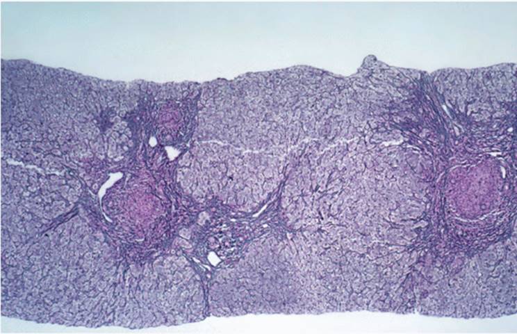
The full understanding of cirrhosis requires that it be recognized as a pathophysiologic entity, rather than just a morphologic change. Cirrhosis is the clinical and pathologic constellation of portal hypertension, intrahepatic vascular shunting of blood, developing or developed hepatic dysfunction, and morphologic alterations of the liver itself. Fibrosis is not synonymous with cirrhosis (Table 20.1), although in many instances fibrosis precedes cirrhosis. The coarsely nodular liver of advanced syphilis (hepar lobatum) or the portal fibrosis of schistosomiasis is not cirrhosis. Portal hypertension may be present, but there is no hepatic dysfunction until late in the course of disease. Similarly, primary bile duct disorders, such as biliary atresia, primary biliary cirrhosis (PBC), and primary sclerosing cholangitis (PSC), may have significant portal hypertension, requiring liver transplantation, before there is evidence of a defect of synthetic or degradative function of the liver because the fibrosis that causes the portal hypertension is, at first, not accompanied by changes in liver plate architecture. In addition, the fibrous septa are portal to portal, and anastomotic connections between the portal vein and hepatic vein occur later.
A MORPHOLOGIC DEFINITION FOR THE PHYSIOLOGIC STATE OF CIRRHOSIS
Two key morphologic alterations, required for the clinical entity of cirrhosis, are seen: vascular septa and regenerative nodules (1,2,13,21,27,32,40) (Table 20.2).
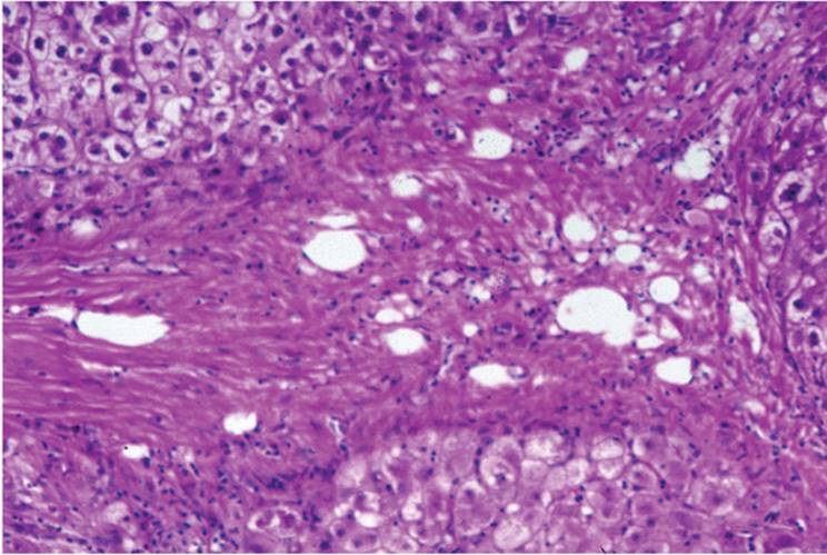
Vascular septa (Fig. 20.1) bridge portal to central, normally connected only via the sinusoids. These septa are not simple areas of fibrosis or scarring but are dynamic alterations in the pattern of blood flow in the liver. Normally, blood flows into the liver via the portal vein and hepatic arteries and then traverses the sponge-like liver to reach the hepatic venous system and return to the general circulation. In cirrhosis, a considerable portion of the blood bypasses hepatocytes, passing almost directly from the hilum to the hepatic veins via the vascular septa (vascular shunting) (16,29). Since much of the liver's blood does not directly contact hepatocytes and does not participate in the usual physiologic activities of the liver, xenobiotics are incompletely removed from the circulation and, conversely, biosynthetic products, such as albumin and coagulation factors, do not enter the circulation. Vascular septa contribute to portal hypertension by increasing the resistance to blood flow through the liver (40). In the normal liver, the flow of blood from portal veins through sinusoids to hepatic veins is relatively unimpeded. The septa of cirrhosis are significantly less compliant than the sponge-like hepatic parenchyma.
|
TABLE 20.1 Examples of Noncirrhotic Hepatic Fibrosis |
|||||
|
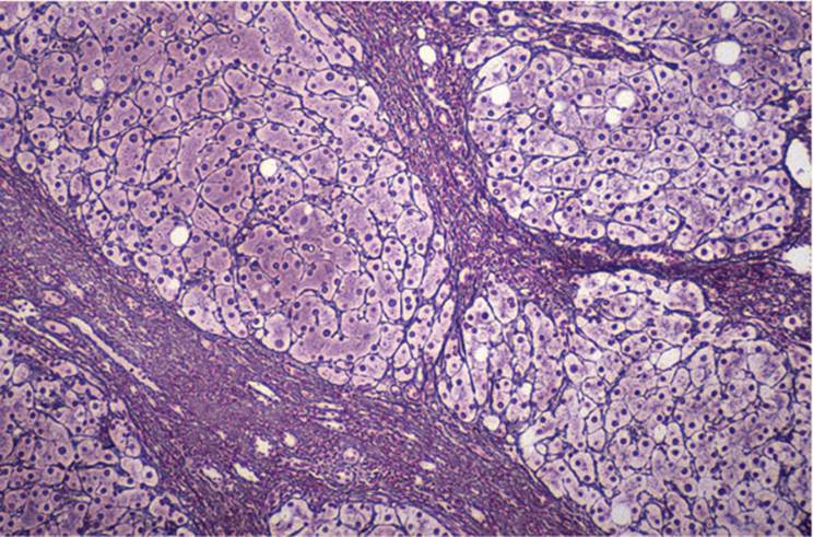
The regenerative nodule (Fig. 20.2) is the second component of cirrhosis. After liver cell injury and/or necrosis, the liver regenerates. As the injured portion of the liver plate repairs, normal modulators of growth do not act and the usual one-cell-thick liver plate is replaced by liver plates two or more cells thick (1,2,24,32,37). Growth factors and cytokines can also cause even previously uninjured liver cells to proliferate leading to expansion of liver plates, so that most of the liver consists of liver plates containing two or more liver cells, with relative reduction of sinusoidal space. There is significantly less surface contact between blood and hepatocyte, further limiting the passage of chemical substances to and from the cell. In addition, expanding nodules compress the fibrous septa and the vessels within the septa, further contributing to portal hypertension (32,40).
The development of the vascular septa and the formation of regenerative nodules generally take place concurrently. However, in some instances, particularly when the hepatic injury is primarily to portal tract structures, as in congenital biliary atresia and PBC, liver cell injury is a relatively late event and the liver plate and sinusoidal structure may be maintained in the face of extensive septum formation. Similarly, in chronic hepatitis regenerative nodules can be seen with relatively little fibrosis.
|
TABLE 20.2 Key Features of Cirrhosis |
||||||||||
|
||||||||||
|
|
|
FIGURE 20.1 Septum from a cirrhotic liver showing multiple vascular channels (hematoxylin-eosin, original magnification ×100). |
Hepatocyte pleomorphism and polyclonality are characteristic of the regenerative nodule. Different cell populations may be discerned. In a few cirrhotic livers, the degree of variation becomes sufficient to warrant the designation of dysplasia (3,5,6,10,19,20,26). The cells may be enlarged with hyperchromatic, irregular nuclei having prominent nucleoli (large cell dysplasia) (e-Figs. 20.1-20.3), with relatively little increase in the nuclear/cytoplasmic ratio. This pattern is common in chronic hepatitis B infection and has been shown in experimental models of hepatocarcinogenesis (7,12). However, there is controversy about the relevance of large cell dysplasia in the development of hepatocellular carcinoma (HCC) in humans. Another form of dysplasia is characterized by cells that are equal in size to, or more often smaller than, the usual hepatocyte (small cell dysplasia) (e-Fig. 20.4), but having greatly increased nuclear/cytoplasmic ratio. This pattern is generally accepted to be a precursor to hepatocellular carcinoma in man (6,10).
|
|
|
FIGURE 20.2 Regenerative nodules showing liver plates two or more cells thick (hematoxylin-eosin, original magnification ×100). |
The hepatocyte of the regenerative nodule may be markedly enlarged with marked eosinophilic granularity (oncocytic change) (e-Fig. 20.5). Various cell products can also be found in the hepatocytes of the cirrhotic liver, including Mallory hyalin, eosinophilic globules composed of α1-antitrypsin or fibrinogen, and, rarely, fat that may be present in only a few nodules. Bile is not usually seen in the hepatocyte of the regenerative nodule, and its presence in the absence of a cholestatic state may be indicative of HCC.
Macroregenerative nodules (MRNs) are 1 cm or more in diameter and can be exceedingly difficult to appreciate in the biopsy. They are more likely to be sites of liver cell dysplasia and HCC.
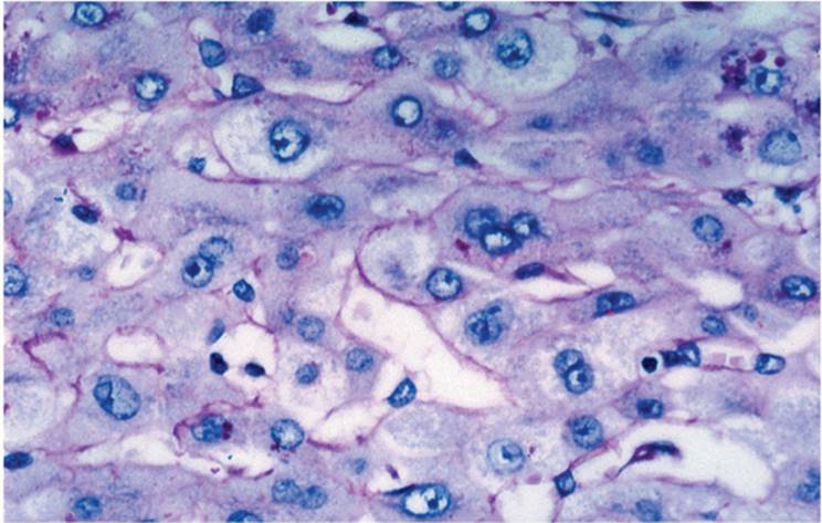
A third feature, difficult to appreciate in routine sections, is basement membrane development in the space of Disse (36) (Fig. 20.3) (capillarization), which contributes to impaired liver cell function. Sinusoids do not normally have a basement membrane. The sieve-like endothelial lining cells are highly permeable, allowing for relatively free passage of a large variety of substances. Capillarization further impairs hepatic function.
|
|
|
FIGURE 20.3 Basement membrane-like structure (capillarization) in the space of Disse in a cirrhotic liver (periodic acid-Schiff reaction, original magnification ×200). |
|
|
|
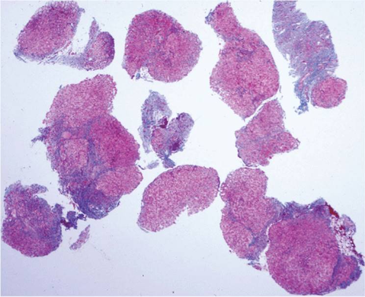
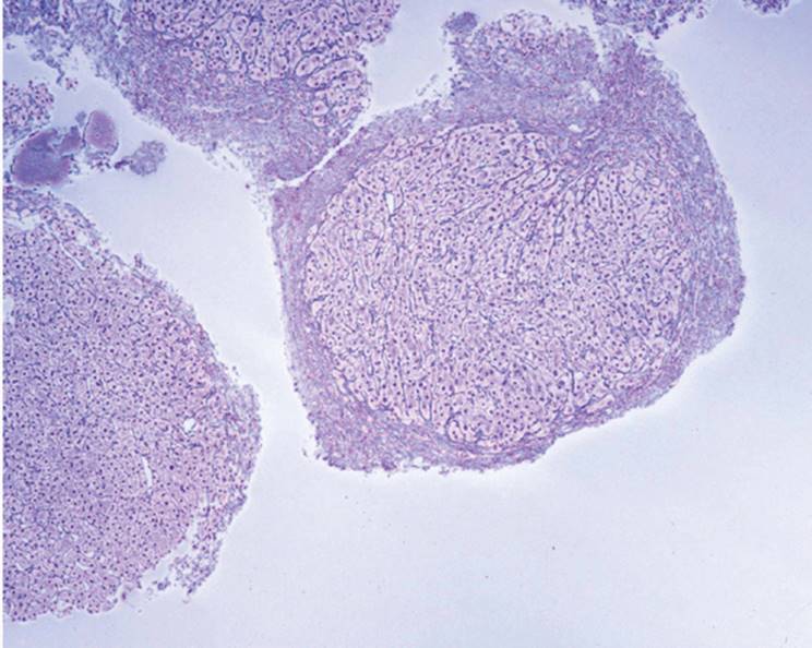
FIGURE 20.4 Fragmented cirrhosis biopsy, with regenerative nodules partially rimmed by vascular septa (hematoxylin-eosin, original magnification ×40). |
A fourth key feature, not easily seen in biopsy, is that cirrhosis, when fully developed, it is almost always diffuse, although the degree may vary in parts of the liver. Exceptions are rare and generally pertain to the caudate lobe that, for reasons not always clear but possibly a result of blood vessel supply variability, can be relatively normal.
The last important feature of the cirrhotic nodule is its tendency to progress, the likely basis of the ultimate development of HCC in at least some instances of cirrhosis, when a specific oncogene is not active. Various cytokines, such as transforming growth factors, may be key.
DIAGNOSIS OF CIRRHOSIS
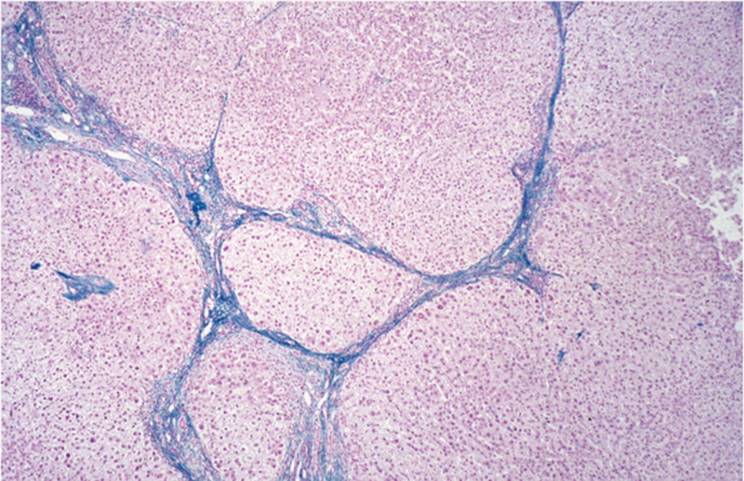
The histologic diagnosis of well-developed cirrhosis is not always straightforward (1,17,28,37). When an adequate sample is obtained, the diagnosis can be made on the basis of the hematoxylin-eosin-stained slide alone (Fig. 20.4, e-Fig. 20.6) or trichrome (e-Fig. 20.7). In this instance, well-defined nodules are surrounded by vascular septa. The diagnosis becomes more difficult when a suboptimal biopsy is obtained, either because of the inexperience of the individual performing the biopsy or, more often, because a needle providing a small sample is used (see Chapters 5 and 9). Fragmented biopsies suggest, but do not prove, cirrhosis unless there is demonstration of reticulin fibers encircling nodules (Fig. 20.5, e-Figs. 20.8, 20.9). The diagnosis can also be difficult when the cirrhosis is not fully developed and the features of cirrhosis are not well expressed in all areas of the liver (e-Fig. 20.10) (1,32,37,39).
|
|
|
FIGURE 20.5 Fragmented biopsy from a cirrhotic patient showing reticulin fibers outlining a nodule (reticulin, original magnification ×200). |
Generally, needle biopsy is more valuable than wedge biopsy in establishing the diagnosis of cirrhosis (17,37). The wedge obtains relatively peripheral, subcapsular liver tissue that may show misleading degrees of fibrosis often misinterpreted as cirrhosis (28).
The vascular septa contain portal tract structures. Degrees of inflammation and ductular reaction vary, depending on the cause (1,2,4,11,31,32). Typically, septa are rich in elastic fibers, demonstrable with elastic stain, orcein stain, or Victoria blue (38) (Fig. 20.6). Ductular reaction can occur, to some degree, in any cirrhotic liver (1,2,22), tending to be more prominent in biliary/cholangiopathic disorders and after large duct obstruction.
|
|
|
FIGURE 20.6 Cirrhotic septum showing elastic fibers (Victoria blue, original magnification ×100). |
Inactive cirrhosis refers to cases with relatively little inflammatory activity in the septa and little or no inflammation or necrosis within the lobule. In contrast, active cirrhosis has the features of chronic hepatitis, with interface hepatitis and varying degrees of necrosis in the lobule. Inflammation varies (1,2,4,11,31,32) and may be quite prominent, presumably contributing to further progression. There may be a discrepancy between the clinical and biochemical features and the morphologic features at any given time (31,34,36).
CAUSES OF CIRRHOSIS
Cirrhosis is the end stage of various disorders (Table 20.3). In a few cases, an etiologic factor cannot be established (cryptogenic).
|
TABLE 20.3 Causes of Cirrhosis |
||||||||||||||||||||||||||||||||||||||||||||||||||||||||||||||||||||||||||||||||
|
||||||||||||||||||||||||||||||||||||||||||||||||||||||||||||||||||||||||||||||||
|
|
|
FIGURE 20.7 Primary sclerosing cholangitis showing swollen, pale hepatocytes (cholate stasis) at the periphery of the jigsaw-puzzle-shaped nodules, adjacent to the septum. This change is typical of chronic cholestasis (hematoxylin-eosin, original magnification ×200). |
Morphologic Clues to the Pathogenesis of Cirrhosis
Cirrhosis can follow various injuries to the liver (Table 20.3). Etiologic factors can sometimes be determined with appropriate special studies, but often, clinical correlation is needed (e-Figs. 20.6-20.10). Except for the irregular (“jigsaw puzzle”) nodules developing bile duct disorders (e-Fig. 20.11), it can be difficult to establish cause with hematoxylin-eosin sections alone.
Structural alterations are helpful. In adults, loss of interlobular bile ducts is often a result of PBC or PSC. Ductular reaction can be seen in any cirrhotic liver but is most suggestive of a primary biliary injury. Ductular reaction can be accentuated and can mimic either a benign bile duct tumor or even cholangiocarcinoma. Sclerosis of terminal hepatic venules (central veins) is often seen in alcoholic liver disease (9,25,30,33,45).
Nodules that form after the viral hepatitides tend to be round. Nodules of the cholangiopathic disorders, such as PBC, PSC, and congenital biliary atresia, tend to be highly irregular (jigsaw puzzle) (e-Fig. 20.6) because the injury initially leads to portal-to-portal fibrosis with irregular dissection of lobules. This characteristic pattern is difficult to see in the biopsy. Generally, however, chronic cholestasis changes, including marked swelling of the paraseptal hepatocytes (cholate stasis, feathery degeneration) and ductular reaction, typical of these disorders, can suggest the diagnosis (15) (Fig. 20.7). Regenerative activity may not develop until relatively late. Vascular occlusion is seen in venous outflow obstruction but it is not a specific finding and can also be seen in cirrhosis resulting from other causes (25). Progression of cirrhosis can lead to the formation of dysplastic MRNs and/or HCC (19,26,42,43) (Fig. 20.8).
|
|
|
FIGURE 20.8 Small cell dysplasia occurring in a macroregenerative nodule, in a liver biopsy from a patient with chronic hepatitis C (hematoxylin-eosin, original magnification ×200). |
Previously, the macroscopic pattern of cirrhosis was thought to be diagnostic of the cause, with terms such as “Laennec,” “postnecrotic,” and “posthepatitic” cirrhosis used. These terms are no longer used because they are not reliable indicators. As one example, Laennec pattern (now called “micronodular”) was thought diagnostic of alcoholic liver disease. It is now clear that this pattern in alcoholic liver disease can progress from the Laennec pattern to the more irregular posthepatitic (macronodular) pattern, and other features (e.g., loss of bile ducts in PBC, ground-glass cells in HBV, stainable iron in hereditary hemochromatosis), as well as clinical information, are to be used in establishing cause.
INCOMPLETE SEPTAL FIBROSIS/CIRRHOSIS
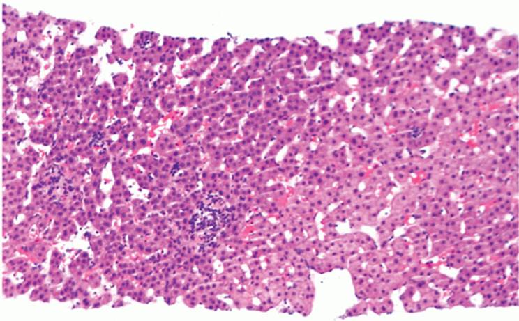
In developing cirrhosis, nodularity may be indistinct without special stains, and the septa may not yet connect anatomic structures. Early septa appear as slender fibrous extensions that end blindly (39) (Fig. 20.9, e-Figs. 20.12, 20.13). Reticulin staining highlights the early regenerative change and extensions of fibrosis from portal tracts. The histologic picture of incomplete cirrhosis could be evidence of regression of cirrhosis (44). Regression of fibrosis has been recognized for many years in both experimental models and humans. It is not yet clear, however, that clinically manifest cirrhosis can regress (14).
NONCIRRHOTIC PORTAL HYPERTENSION
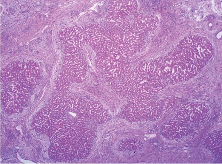
“Noncirrhotic portal hypertension” is the term for a distinct and rare condition with intrahepatic portal fibrosis associated with portal hypertension, without cirrhosis or extrahepatic portal obstruction, and without a recognizable cause (8,18,23,27,35,41). Noncirrhotic portal hypertension manifests as rapidly developing liver disease or even fulminant hepatic failure. The main histologic feature is the presence of paucicellular, dense portal tract fibrosis (Fig. 20.10, e-Fig. 20.14) and establishing the diagnosis requires exclusion of various clinically recognizable disorders that also contribute to portal hypertension in the absence of cirrhosis (Table 20.4) including true sclerosis of intrahepatic portal veins (see Chapter 1). Either injury or thrombosis of portal venous branches has been implicated in the pathogenesis of this condition. However, the obliterated veins may not be seen in biopsy material.
|
|
|
FIGURE 20.9 Incomplete septal cirrhosis, showing a slightly blunted, partial septum ending in the middle of the lobule and not connecting two architectural structures (hematoxylin-eosin, original magnification ×100). |
|
|
|
FIGURE 20.10 Portal fibrosis in a patient with idiopathic portal hypertension (Masson trichrome, original magnification ×10). |
|
TABLE 20.4 Noncirrhotic Portal Hypertension |
|||||||||||||||||||||||
|
REFERENCES
1. Anthony PP, Ishak KG, Nayak NC, et al. The morphology of cirrhosis: definition, nomenclature, and classification. Bull WHO 1977;55:521-540.
2. Anthony PP, Ishak KG, Nayak NC, et al. The morphology of cirrhosis. Recommendations on definition, nomenclature, and classification by a working group sponsored by the World Health Organization. J Clin Pathol 1978;31:395-414.
3. Anthony PP, Vogel CL, Barker LF. Liver cell dysplasia: a premalignant condition. J Clin Pathol 1973;26:217-223.
4. Baggenstoss AH, Staufer MH. Posthepatitic and alcoholic cirrhosis: clinicopathologic study of 43 cases of each. Gastroenterology 1952;22:157-180.
5. Borzio M, Bruno S, Roncalli M, et al. Liver cell dysplasia and risk of hepatocellular carcinoma in cirrhosis: a preliminary report. Br Med J 1991;302:1312.
6. Crawford JM. Pathologic assessment of liver cell dysplasia and benign liver tumors: differentiation from malignant tumors. Semin Diagn Pathol 1990;7:115-128.
7. Cullen JM, Sandgren EP, Brinster RL, et al. Histologic characterization of hepatic carcinogenesis in transgenic mice expressing SV40 T-antigens. Vet Pathol 1993;30:111-118.
8. Eckhauser FE, Appleman HD, Knol JA, et al. Non-cirrhotic portal hypertension: different patterns of disease in children and adults. Surgery 1983;94:721-728.
9. Edmondson HA, Peters RL, Reynolds TB, et al. Sclerosing hyalin necrosis of the liver in the chronic alcoholic: a recognizable clinical syndrome. Ann Intern Med 1963;59:646-673.
10. Ferrell LD, Crawford JM, Dhillon AP, et al. Proposal for standardized criteria for the diagnosis of benign, borderline, and malignant hepatocellular lesions arising in chronic advanced liver disease. Am J Surg Pathol 1993;17:1113-1123.
11. Gall EA. Posthepatitis, postnecrotic and nutritional cirrhosis: a pathologic analysis. Am J Pathol 36:241-271.
12. Geller SA, Nichols WS, Kim S, et al. Hepatocarcinogenesis is the sequel to hepatitis in Z#2 alpha-1-antitrypsin transgenic mice: histopathological and DNA ploidy studies. Hepatology 1994;19:389-397.
13. Geller SA. Coming or going? What is cirrhosis? Arch Pathol Lab Med 2000;124:1587-1588.
14. Gerber MA, Popper H. Relation between central canals and portal tracts in alcoholic hepatitis: a contribution to the pathogenesis of cirrhosis in alcoholics. Hum Pathol 1972;3:199-207.
15. Gerber MA, Orr W, Denk H, et al. Hepatocellular hyalin in cholestasis and cirrhosis: its diagnostic significance. Gastroenterology 1973;64:89-98.
16. Gross G, Perrier CV. Intrahepatic portasystemic shunting in cirrhotic patients. N Engl J Med 1975;293:1046-1047.
17. Imamura H, Kawasaki S, Bandai Y, et al. Comparison between wedge and needle biopsies for evaluating the degree of cirrhosis. J Hepatol 1993;17:215-219.
18. Kage M, Arakawa M, Fukuda K, et al. Pathomorphologic study on the extrahepatic portal vein in idiopathic portal hypertension. Liver 1990;10:209-216.
19. Kondo F, Ebara M, Sugiura N, et al. Histological features and clinical course of large regenerative nodules: evaluation of their precancerous potentiality. Hepatology 1990;12:592-598.
20. Lefkowitch JH, Apfelbaum TF. Liver cell dysplasia and hepatocellular carcinoma in non-A, non-B hepatitis. Arch Pathol Lab Med 1987;111:170-173.
21. MacSween RNM, Scott AR. Hepatic cirrhosis: a clinicopathologic review of 320 cases. J Clin Pathol 1973;26:936-942.
22. Masuko K, Rubin E, Popper H. Proliferation of bile ducts in cirrhosis. Arch Pathol 1964; 78:421-431.
23. Mikkelsen WP, Edmondson HA, Peters RL, et al. Extra- and intrahepatic portal hypertension without cirrhosis (hepatoportal sclerosis). Ann Surg 1965;162:602-618.
24. Morgan JD, Martcroft WS. Juvenile liver: age at which one-cell-thick liver plates predominate in the human liver. Arch Pathol 1961;77:86-88.
25. Nakanuma Y, Ohta G, Doishita K. Quantitation and serial section observations of focal venoocclusive lesions of hepatic veins in liver cirrhosis. Virchows Arch A Pathol Anat Histopathol 1985;405:429-439.
26. Nakanuma Y, Terada T, Ueda K, et al. Adenomatous hyperplasia of the liver as a precancerous lesion. Liver 1993;13:1-9.
27. Ohnishi K, Saito M, Sato S, et al. Portal hemodynamics in idiopathic portal hypertension (Banti's syndrome). Gastroenterology 1987;92:751-758.
28. Petrelli M, Scheuer PJ. Variation in subcapsular liver structure and its significance in the interpretation of wedge biopsies. J Clin Pathol 1967;20:743-748.
29. Popper H, Elias H, Petty DE. Vascular pattern of the cirrhotic liver. Am J Clin Pathol 1952;22:717-729.
30. Popper H, Szanto PB, Elias H. Transition of fatty liver into cirrhosis. Gastroenterology 1955;28:183-192.
31. Popper H, Rubin E, Krus S, et al. Postnecrotic cirrhosis in alcoholics. Gastroenterology 1960;39:669-685.
32. Popper H. Pathologic aspects of cirrhosis. Am J Pathol 1977;87:228-259.
33. Reynolds TB, Hidemura R, Michel H, et al. Portal hypertension without cirrhosis in alcoholic liver disease. Ann Intern Med 1969;70:497-506.
34. Rubin E, Krus S, Popper H. Pathogenesis of postnecrotic cirrhosis in alcoholics. Arch Pathol 1962;73:288-289.
35. Sarin SK. Non-cirrhotic portal fibrosis. Gut 1989;30:406-415.
36. Schaffner F, Popper H. Capillarization of hepatic sinusoids in man. Gastroenterology 1963;44:239-242.
37. Scheuer PJ. Liver biopsy in the diagnosis of cirrhosis. Gut 1970;11:275-278.
38. Scheuer PJ. Hepatic fibrosis and collapse: histological distinction by orcein staining. Histopathology 1980;4:487-490.
39. Sciot R, Staessen D, Van Damme B, et al. Incomplete septal cirrhosis: histopathological aspects. Histopathology 1988;13:593-603.
40. Shibayama Y. On the pathogenesis of portal hypertension in cirrhosis of the liver. Liver 1988;8:95-99.
41. Tandon BN, Lakshminarayanan R, Bhargava S, et al. Ultrastructure of the liver in noncirrhotic portal fibrosis with portal hypertension. Gut 1970;11:905-910.
42. Terada T, Terasaki S, Nakanuma Y. A clinicopathologic study of adenomatous hyperplasia of the liver in 209 consecutive cirrhotic livers examined by autopsy. Cancer 1993;72:1551-1556.
43. Theise ND, Schwartz M, Miller C, et al. Macroregenerative nodules and hepatocellular carcinoma in forty-four sequential adult liver explants with cirrhosis. Hepatology 1992;16:949-955.
44. Wanless IR, Nakashima E, Sherman M. Regression of human cirrhosis. Morphologic features and the genesis of incomplete septal cirrhosis. Arch Pathol Lab Med 2000;124:1599-1607.
45. Worner TM, Lieber CS. Perivenular fibrosis as precursor lesion of cirrhosis. JAMA 1985; 254:627-630.