Systemic disorders often affect the liver, and morphologic changes and the clinical presentation are usually mild. Some typical biopsy changes can be seen, however (22).
DIABETES MELLITUS
Morphologic changes are common in diabetes mellitus (DM), although clinical manifestations relating to the liver are quite rare (10).
Histopathologic Findings
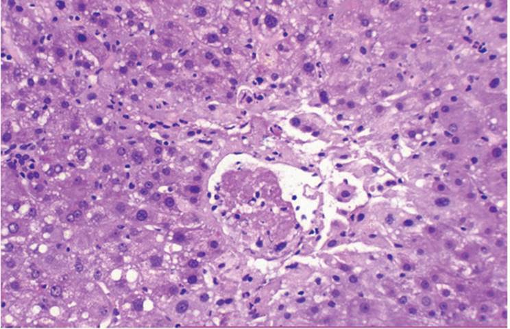
Glycogenated (glycogen) nuclei, occurring especially in zone 1, and variable degrees of steatosis are the most common changes. Glycogenated nuclei are optically clear nuclei with a denser-than-usual nuclear membrane (Fig. 21.1, e-Figs. 21.1, 21.2). This phenomenon, whose pathogenesis is not clear, is presumed to reflect cytoplasmic accumulation of glycogen as a large vacuole that protrudes into the nucleus. In contrast with the usual ultrastructural rosette-like appearance of intracytoplasmic glycogen, the glycogen of the glycogenated nucleus appears dispersed. Glycogenated nuclei may be seen many years before the clinical onset of DM (10).
Glycogenated nuclei can also be present in Wilson disease (see Chapter 16), but are also present in otherwise normal adolescents and elderly patients.
Nonalcoholic Fatty Liver Disease (NAFLD)
Nonalcoholic fatty liver disease (NAFLD), including the nonalcoholic steatohepatitis (NASH), occurs in patients with DMand must be differentiated fromtrue alcoholic steatohepatitis (ASH) (Chapter 13).Macrovesicular steatosis is common in DM and generally not zonal (Fig. 21.2, e-Figs. 21.3-21.5). In some cases the steatosis is mixed, microvesicular and macrovesicular.
Although there is still some disagreement in how to apply these terms, we use NAFLD as a comprehensive term that includes a spectrum of histopathologic changes ranging from steatosis, mild or marked, without obvious evidence of liver injury, NASH characterized by marked steatosis, balloonedhepatocytes, sometimeswithpolymorphonuclear leukocyte (PMN) infiltration and accumulations of Mallory hyalin, and fibrosis/cirrhosis (Fig. 21.3, e-Figs. 21.6, 21.7). Mallory material is generally not as abundant as in ASH, is generally not as strongly eosinophilic, and is predominantly periportal. Portal tracts contain mild to moderate inflammatory cell infiltrate, mostly lymphocytes with some plasma cells and PMNs. Glycogenated nuclei, often seen in NAFLD, are not particularly prominent in ASH.
|
|
|
FIGURE 21.1 Diabetes mellitus. Glycogenated nuclei are seen as optically clear with denser than usual nuclear membrane (hematoxylin-eosin, original magnification ×400). |
|
|
|
FIGURE 21.2 Diabetes mellitus. Severe steatosis (hematoxylin-eosin, original magnification ×40). |
|
|
|
FIGURE 21.3 Diabetes mellitus with nonalcoholic steatohepatitis, showing marked steatosis with acute inflammatory cells and Mallory hyalin (hematoxylin-eosin, original magnification ×200). |
NAFLD may progress to fibrosis and cirrhosis. Collagenization of the space of Disse, with deposition of type IV collagen, may precede the development of cirrhosis and has been described as a part of the spectrum of diabetic microangiopathy (Fig. 21.4, e-Fig. 21.8) (10,11,50).
The understanding of NAFLD/NASH has increased greatly with various morphologically based definitions and staging systems in the recent literature (13,16,47).
|
|
|
FIGURE 21.4 Diabetes mellitus. Collagenization of the space of Disse (hematoxylin-eosin, original magnification ×200). |
|
|
|
FIGURE 21.5 Sepsis. There is ductular proliferation, and the dilated ductules contain inspissated bile. Surrounding ducts and ductules are polymorphonuclear leukocytes (hematoxylin-eosin, original magnification ×100). |
Other Conditions
Hepatic adenoma (29), nodular hyperplasia (100), and primary sclerosing cholangitis (PSC) (2) have been reported in association with DM.
SEPSIS
The liver is often affected in sepsis, but biopsy is rarely obtained, except in the setting of liver transplantation (see Chapter 25) (8).
Histopathologic Findings
Three sepsis patterns have been recognized: (a) perivenular canalicular cholestasis with mild steatosis, associated prominence of Kupffer cells, and nonspecific portal inflammatory infiltrate; (b) marked bile ductular reaction/proliferation with the perivenular canalicular cholestasis (Fig. 21.5) and ductular proliferation at the margin of the portal tracts, with ductules dilated and filled with inspissated bile (52), along with periductal and periductular infiltration with PMNs (pericholangitis) with secondary, reactive epithelial injury; and (c) nonbacterial cholangitis, most commonly associated with toxic shock syndrome (40). This variant may be a result of circulating staphylococcal toxin. PMNs are seen in the bile duct lumen, with necrosis of the epithelium.
HUMAN IMMUNODEFICIENCY VIRUS AND ACQUIRED IMMUNE DEFICIENCY SYNDROME (HIV/AIDS)
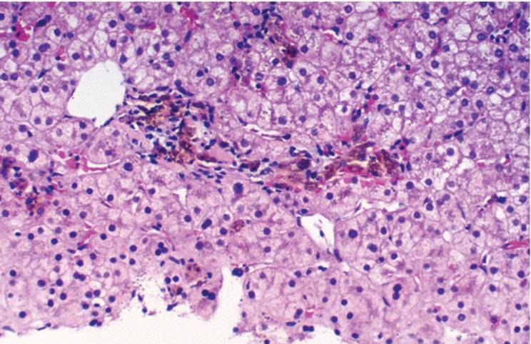
A wide spectrum of liver diseases occur in patients infected with human immunodeficiency virus (HIV) (Table 21.1) (7,25,32,41,49,61,70,83,87,105). The liver changes are secondary and do not reflect primary liver disease, although hepatitis primarily associated with HIV infection may occur in children and adults (66). The most frequently encountered features in both HIV-positive patients and patients with acquired immune deficiency syndrome (AIDS) are variable degrees of bile duct injury (AIDS cholangiopathy), prominence of Kupffer and sinusoidal endothelial cells, often with hemosiderosis, and steatosis (Fig. 21.6) (32,71).
|
TABLE 21.1 Hepatic Involvement in HIV/AIDS |
||||||||||||||||||||||||||||||
|
||||||||||||||||||||||||||||||
Bile Duct Injury and AIDS Cholangiopathy
Bile duct injury varies from mild epithelial damage to changes that mimic bile duct obstruction with significant cholestasis and bile ductular proliferation (Fig. 21.7, e-Figs. 21.9, 21.10) (61). Sometimes significant periductal fibrosis is seen, indistinguishable from PSC (36,57). These changes also seen with either cryptosporidiosis or cytomegalovirus (CMV) infection affecting the biliary tree (61).
HIV gag protein p24 can be detected in sinusoidal endothelial and Kupffer cells but not in hepatocytes (Fig. 21.8). There is little evidence that HIV has a direct cytopathic effect in the liver (38), although propagation of HIV virus is feasible in vitro in hepatoma cell lines (95).
Many HIV patients also have hepatitis B or C chronic hepatitis (17,71). Granulomas, without an identifiable cause, are seen in 20% of patients, and 15% show nonspecific reactive changes. Cirrhosis, of varying causes, can also be seen. Peliosis hepatis and bacillary angiomatosis occur. Opportunistic infections affecting the liver are relatively uncommon. Kaposi sarcoma occurs sporadically.
|
|
|
FIGURE 21.6 A and B. Acquired immune deficiency syndrome. Bile ducts show mild injury, and there is prominence of sinusoidal endothelial and Kupffer cells (hematoxylin-eosin, original magnification ×200). |
Opportunistic Infections
Poorly formed microgranulomas should raise suspicion for Mycobacterium avium-intracellulare (MAI) infection (e-Fig. 21.11). Spindle cell proliferation can be prominent (pseudosarcoma), with the spindle cells containing innumerable acid-fast bacilli (37,75). Mycobacterium tuberculosis var. hominis, including miliary dissemination, also occurs in AIDS, with caseating or noncaseating granulomas.
Fungal Infections
Fungal infections occur usually with widespread dissemination. Special stains (periodic acid-Schiff, Gomori methenamine silver) highlight organisms, including Candida albicans, Histoplasma capsulatum, Cryptococcus neoformans, and Coccidioides immitis. Blastomycosis, aspergillosis, and kala azar are rarely seen (26,76,87,110).
|
|
|
FIGURE 21.7 Acquired immune deficiency syndrome. Bile duct injury is moderately severe, and there is ductular proliferation and cholestasis suggesting large duct obstruction (hematoxylin-eosin, original magnification ×200). |
Protozoal Infections
Cryptosporidium can infect the biliary tree, with an associated cholangiopathy, but is rarely seen in biopsy samples (35). Hepatitis caused by microsporidia has been described (73,98).
|
|
|
FIGURE 21.8 Acquired immune deficiency syndrome. Human immunodeficiency virus gag protein p24 is present in sinusoidal endothelial and Kupffer cells. (immunoperoxidase, antip24, original magnification ×100). |
Cytomegalovirus
CMV infection was, prior to the development of effective antiviral therapy, frequently seen in AIDS patients. Characteristic intranuclear inclusions are generally easily seen, but in situ hybridization may be necessary (35,47).
VASCULAR DISORDERS
HIV-Associated Peliosis
HIV-associated peliosis of the liver is indistinguishable from peliosis hepatis seen in immunocompetent patients. Cystically dilated sinusoidal spaces are partially lined by nonneoplastic endothelial cells and filled with blood (Fig. 21.9, e-Figs. 21.13, 21.14) (21,70,90).
Bacillary Angiomatosis
Bacillary angiomatosis is a distinct lesion associated with HIV infection (30,51,70,90,96,104). It affects various organs, including the spleen and liver. In most cases there are cystically dilated spaces filled with blood, with variable surrounding fibrous and fibromyxoid stroma alternating with angioproliferative spindle cells. Clusters of lymphocytes and PMNs are admixed with blood and adjacent fibrous stroma and endothelial cells (Fig. 21.10, e-Figs. 21.15-21.18). Rod-shaped bacillary organisms (Rochalimaea henselae) can be demonstrated with Warthin-Starry silver stain (93,104). The organism is sensitive to erythromycin, and the lesion can resolve completely.
|
|
|
FIGURE 21.9 Peliosis hepatis in a patient with human immunodeficiency virus infection (hematoxylin-eosin, original magnification ×100). |
|
|
|
FIGURE 21.10 Bacillary angiomatosis in a patient with acquired immune deficiency syndrome showing cystically dilated vascular spaces with spindle cell proliferation (hematoxylin-eosin, original magnification ×100). |
Kaposi Sarcoma (Kaposi Angioproliferative Lesion)
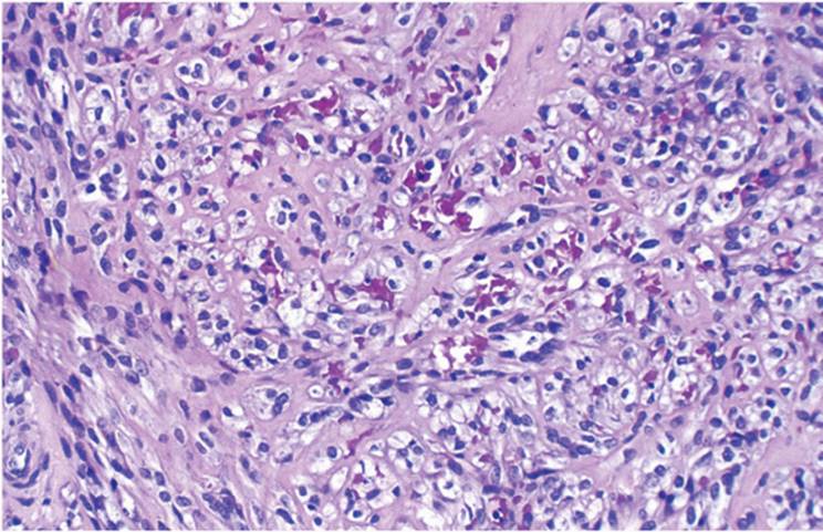
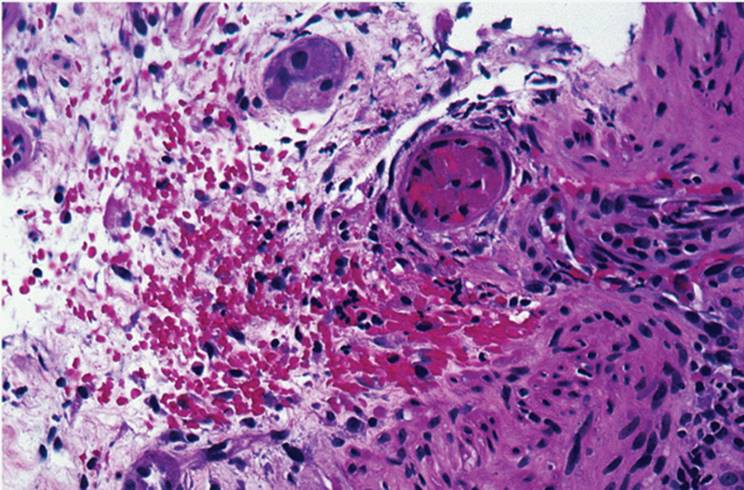
Kaposi sarcoma is generally seen as a disseminated malignancy and often affects the liver (90,96). The lesions are usually only a few millimeters in diameter and may not always appear in biopsy, despite being abundant in the liver. The angioproliferative lesion has irregular fascicles of spindle cells with extravasated red blood cells (Fig. 21.11, e-Figs. 21.19-21.24).
Extracellular hemosiderin is seen, as well as intracellular eosinophilic globules of uncertain origin. Lymphocytes and plasma cells may be present. The proliferated spindle cells are most likely of endothelial cell origin, either vascular or lymphatic, with reactivity to antibodies routinely used for other neoplasms of endothelial cell origin, CD31 and CD34 (Fig. 21.12) (91).
|
|
|
FIGURE 21.11 Kaposi sarcoma in a patient with acquired immune deficiency syndrome, showing tightly packed spindle cells and extravasated red blood cells (hematoxylin-eosin, original magnification ×100). |
|
|
|
FIGURE 21.12 Reactivity of proliferating spindle cells in acquired immune deficiency syndrome to antibody directed against CD34 (hematoxylin-eosin, original magnification ×200). |
Lymphoproliferative Disorders
The incidence of lymphoproliferative disorders is increased in AIDS. The liver is affected in approximately 25% of patients, as either a solitary mass or as multiple nodules. Lymphoma rarely involves bile ducts, primarily, clinically resembling PSC.
Most HIV-associated lymphomas are of the non-Hodgkin B-cell type. They are usually high grade, with either a predominantly large cell immunoblastic population or a Burkitt type, with small noncleaved cells (33,48).
In contrast with the posttransplantation immunocompromised patients, only half of AIDS-associated lymphoproliferative disorders are Epstein-Barr virus (EBV) associated (77,92). A c-myc gene rearrangement is more commonly seen in these patients (92).
Viral Hepatitides in AIDS
As many as 75% of AIDS patients have serologic evidence of HBV infection (32,71). The effect of HBV on the progression of HIV infection is controversial. Coinfection with HCV and HIV is also common (71). HIV adversely affects chronic HCV infection, leading to more severe and rapidly progressive disease (e-Figs. 21.22-21.26) (71,86). HCV infection can affect the progression of HIV in a similar fashion (56,71,72), to some extent dependent on the HCV genotype (94).
Drug Toxicity in Immunocompromised Patients
Drugs used for treatment of patients with AIDS may be hepatotoxic. These include antifungal agents, such as ketoconazole and fluconazole, as well as antimycobacterial drugs, such as rifampicin and isoniazid. The histologic distinction between viral and drug-induced hepatitides may be particularly difficult (71). Some antiretroviral drugs (e.g., dideoxyinosine) cause fulminant liver failure (71).
PREGNANCY
Benign Recurrent Cholestasis
The pathogenesis of benign recurrent cholestasis is not entirely clear but may be related to elevated estrogen levels. Histologically, perivenular intracanalicular cholestasis is seen without significant inflammation or liver cell necrosis (81,101). Recognition of the clinical setting is obviously helpful in differentiating this type of cholestasis from sepsis or drug-induced cholestasis.
Acute Fatty Liver of Pregnancy
Acute fatty liver of pregnancy is a serious condition associated with high mortality (68,81,82). The fat may be zonal, mostly involving zones 3 (perivenular) and 2, or diffuse, affecting the entire acinus (e-Figs. 21.27, 21.28). Steatosis is generally microvesicular, sometimes difficult to recognize on routine paraffin sections, but easily seen in cryostat-sectioned liver stained for fat. Zone 3 intracanalicular cholestasis and portal and lobular lymphocytic inflammatory infiltrate are usually seen (81,101). The portal infiltrate can be prominent, suggesting acute viral hepatitis. When only zone 3 fat is seen, tetracycline-induced toxicity (which can also be seen during pregnancy) must be considered.
Preeclampsia and Eclampsia
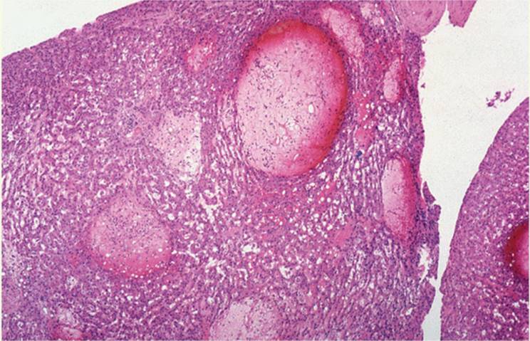
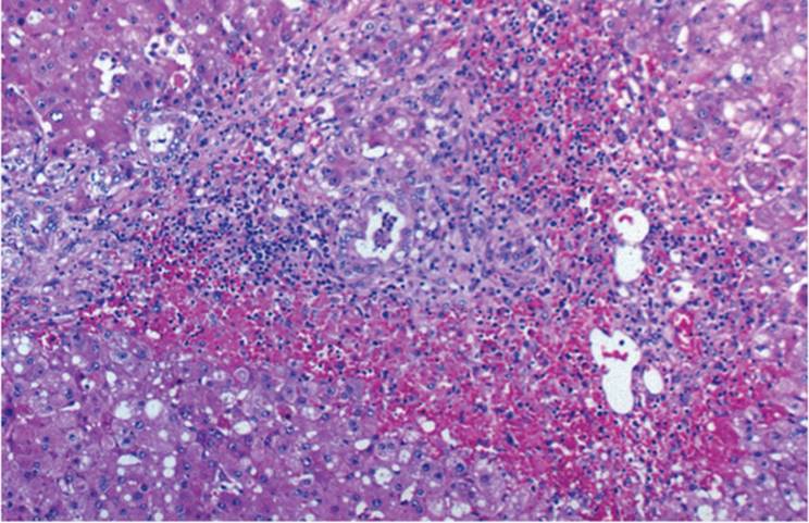
Preeclampsia is characterized by hypertension, proteinuria, and peripheral edema (82,85). If uncontrolled, it can lead to the convulsive, hyperreflexive condition of eclampsia. Liver biopsy is only rarely obtained. Parenchymal hemorrhage and necrosis are seen. Sinusoids may contain fibrin, evidence of intravascular coagulation, and fibrin thrombi can form, particularly in zone 1 (Figs. 21.13, 21.14, e-Figs. 21.29-21.32). With progression, fibrin thrombi are seen in many sinusoids and even arterioles (Fig. 21.15).
HELLP Syndrome
HELLP syndrome (hemolysis, elevated liver function tests, and low platelets) can be morphologically indistinguishable from, and may represent a mild form of, eclampsia (e-Figs. 21.33, 21.34) (6,9,88). Sometimes the liver shows nonspecific changes, including portal inflammatory infiltrate, focal lobular necrosis, and glycogenated nuclei.
|
|
|
FIGURE 21.13 Eclampsia, showing deposition of fibrin in periportal sinusoids, with associated hepatocellular necrosis (hematoxylin-eosin, original magnification ×100). |
HEMATOLOGIC DISORDERS
The liver is often affected in hematologic disorders, including lymphoproliferative and myeloproliferative conditions (67,106,109). Kupffer cell hyperplasia with associated hemosiderosis is commonly seen, usually attributable to multiple blood transfusions. Patients with hemophilia have a higher risk of infection with HBV and HCV, as well as HIV.
|
|
|
FIGURE 21.14 Eclampsia, with demonstration of fibrinogen in fibrin deposits (peroxidaseantiperoxidase, original magnification ×100). |
|
|
|
FIGURE 21.15 Eclampsia with demonstration of intravascular thrombus formation (Masson trichrome, original magnification ×100). |
Anemia
Various nonspecific changes are seen with long-standing anemia, including mild macrovesicular steatosis and secondary hemosiderosis (Fig. 21.16). Chronic hemolysis can also lead to gallstones and changes of large duct obstruction (see Chapter 18).
|
|
|
FIGURE 21.16 Secondary hemosiderosis, with demonstration of iron in Kupffer cells, in hemolytic anemia (Perls, original magnification ×200). |
|
|
|
FIGURE 21.17 Sickle cell disease, showing clumped sickle-shaped red blood cells distending sinusoids (hematoxylin-eosin, original magnification ×200). |
In sickle cell anemia, sinusoidal aggregation of sickle-shaped cells, with associated sinusoidal dilatation and congestion, and eventual liver cell necrosis is seen, in addition to secondary hemosiderosis. These changes are typical of patients in crisis (54) and only rarely encountered in biopsy samples (Fig. 21.17, e-Figs. 21.35, 21.36) (97).
Large vein thrombosis, affecting portal and/or hepatic veins, occurs in patients with paroxysmal nocturnal hemoglobinuria (55,97).
Lymphoproliferative Disorders
Liver involvement by lymphoid leukemias, non-Hodgkin lymphoma, and Hodgkin lymphoma is generally not clinically dominant. Liver biopsy is often performed to either identify the cause of associated fever or, particularly in Hodgkin lymphoma, for staging purpose. Rarely, massive infiltration by lymphoid cells contributes to fulminant failure (e-Figs. 21.37-21.40) (67,105,109).
LYMPHOID LEUKEMIA. Infiltration in the lymphoid leukemias tends to be primarily portal (109). This is particularly prominent in lymphoblastic leukemia, in which the lymphoid cells are relatively primitive (e-Figs. 21.41, 21.42) (109).
HAIRY CELL LEUKEMIA. The characteristic monocytoid hairy cells usually infiltrate sinusoids in a linear pattern. Other findings include dilated sinusoids and angiomatous (peliosis-like) lesions surrounded by rings of leukemic cells (15,108).
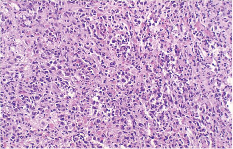
NON-HODGKIN LYMPHOMA. Liver involvement is common in non-Hodgkin lymphoma, and the biopsy shows diffuse lymphoid infiltration (Fig. 21.18) (108).
|
|
|
FIGURE 21.18 Non-Hodgkin lymphoma, with replacement of hepatic parenchyma by sheets of lymphoid cells (hematoxylin-eosin, original magnification ×100). |
HODGKIN LYMPHOMA. When Hodgkin lymphoma involves the spleen, the liver is almost always involved. However, the characteristic Hodgkin lymphoma infiltrate is seen in only 15% of biopsies. Classic Reed-Sternberg cells are rarely seen (e-Fig. 21.43). Usually, a mixed inflammatory cell infiltrate, not specifically diagnosable as Hodgkin disease, is seen (Fig. 21.19). As many as 10% of biopsies show noncaseating epithelioid granulomas, similar to those of sarcoidosis. Sinusoidal dilatation and peliosis hepatis are seen (15). In some patients the only finding is intrahepatic cholestasis (53). Uncommonly, a lesion resembling PSC may be seen (39), and the clinical and pathologic syndrome of vanishing bile duct syndrome can develop (39).
|
|
|
FIGURE 21.19 Hodgkin disease. There is an extensive lymphoproliferative infiltrate in this biopsy performed for acute liver failure (hematoxylin-eosin, original magnification ×100). |
|
|
|
FIGURE 21.20 Budd-Chiari syndrome in a patient with lymphoproliferative disorder, showing characteristic atrophy and necrosis of zone 3 hepatocytes (hematoxylin-eosin, original magnification ×100). |
Myeloproliferative Disorders
All of the myeloproliferative disorders, including the myelogenous leukemias, polycythemia vera, myeloid metaplasia and myelofibrosis, and essential thrombocythemia, can affect the liver. In myelogenous leukemia the infiltrating cells are seen in both sinusoids and portal tracts.
The most common finding in the liver in the nonleukemia myeloproliferative disorders is infiltration of sinusoids by hematopoietic cells originating from all three hematopoietic lineages (24,69), sometimes causing portal hypertension. Sinusoidal dilatation from increased hepatic blood flow is often seen. Budd-Chiari syndrome occurs in myeloproliferative disorders (Fig. 21.20) (18,24). Nodular regenerative hyperplasia develops either secondary to ischemia or as a result of drug-induced hepatotoxicity (24). Extramedullary hematopoiesis is seen, particularly in myeloid metaplasia and myelofibrosis (Fig. 21.21).
|
|
|
FIGURE 21.21 Extramedullary hematopoiesis in a patient with myelofibrosis (hematoxylin-eosin, original magnification ×200). |
|
|
|
FIGURE 21.22 Multiple myeloma. There is deposition of light-chain immunoglobulins in the space of Disse (immunoperoxidase, anti-kappa light chain, original magnification ×200). |
Isolated circulating megakaryocytes can also be seen in sinusoids as a manifestation of stress and not necessarily as a component of a myeloproliferative disorder (e-Figs. 21.44, 21.45).
MYELOMA. The liver is variably involved in myeloma. There may be sinusoidal and parenchymal infiltration by plasma cells or there may be tumor-like replacement. In myeloma, as well as in Waldenström's macroglobulinemia, there may be portal hypertension, nodular regenerative hyperplasia, and peliosis hepatis, as well as deposition of light chain immunoglobulins or macroglobulins in the space of Disse (Fig. 21.22) (99,102).
EPSTEIN-BARR VIRUS-RELATED DISORDERS
Infectious Mononucleosis
Infectious mononucleosis can be complicated by EBV-associated hepatitis. Sinusoidal infiltration with lymphoid cells almost always includes immunoblastic forms (Fig. 21.23). Kupffer cells are usually hyperplastic. There is little or no hepatocellular injury or necrosis. Small microgranulomas may also be present. Differential diagnosis includes leukemia and extramedullary hematopoiesis. Diagnosis is generally established after correlating serologic and histopathologic findings (44).
|
|
|
FIGURE 21.23 Infectious mononucleosis, showing sinusoidal infiltration by lymphoid cells, some of which are slightly immature and atypical (hematoxylin-eosin, original magnification ×200). |
Viral-Associated Hemophagocytic Syndrome
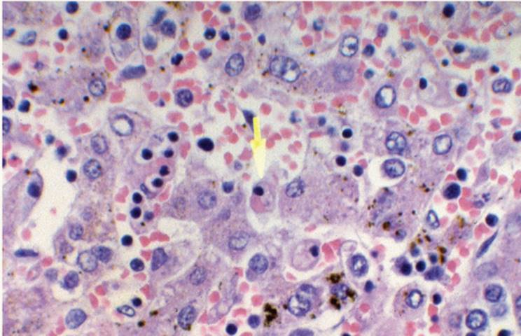
Viral-associated hemophagocytic syndrome is a systemic viral infection generally associated with infection with either EBV or CMV and characterized by progressive, often profound, anemia (28). Histiocytes show phagocytosed hematopoietic cells. Most Kupffer cells contain red blood cells (Fig. 21.24). The Kupffer cells can be markedly hyperplastic and can resemble the infiltrate of malignant histiocytosis; with appropriate immunohistochemical studies, however, differentiation is generally easy (60).
|
|
|
FIGURE 21.24 Viral-associated hemophagocytic syndrome. Kupffer cells contain phagocytosed red blood cells (hematoxylin-eosin, original magnification ×100). |
Chronic Granulomatous Disease
Chronic granulomatous disease (CGD) is a result of impaired hydrogen peroxide production leading to failure of the myeloperoxidase-H2O2-halide killing system. Chronic granulomatous disease is an inherited, generally Xlinked disease of male children and infants, characterized by recurrent infections and death at an early age, most often because of infection by catalasepositive organisms such as Staphylococcus aureus. Adults are rarely affected. Poorly formed necrotizing granulomas are seen in the parenchyma. Histiocytes surrounding the area of necrosis contain dusty brown lipofuscin pigment. Pigment-laden histiocytes are also in portal tracts with other inflammatory cells, particularly lymphocytes. Other infectious causes or drug-induced granulomas have to be excluded (63).
Reye Syndrome
Reye syndrome typically occurs in children treated with aspirin (103). Aspirin toxicity mimics Reye syndrome. Although histopathologic changes may be similar, ultrastructurally they appear to be unrelated. In Reye syndrome, hypoglycemia is often profound and is typically followed by the rapid development of encephalopathy and coma.
The characteristic histopathologic feature is diffuse microvesicular steatosis without significant inflammation, necrosis, or cholestasis (e-Figs. 21.47-21.49) (14,79). Fat droplets can be difficult to appreciate in routine histologic sections, and frozen sections stained with a fat stain (oil red O or Sudan IV) may be required. Fat droplets are generally smallest in zone 3 (Fig. 21.25). Alternatively, formalin-fixed wet tissue can be postfixed with osmium tetroxide to retain the fat (e-Fig. 21.50). The ultrastructural changes are virtually diagnostic. Mitochondria are enlarged with decreased matrix density and fragmentation of the cristae (58). The enlargement of mitochondria correlates with the stage of encephalopathy.
|
|
|
FIGURE 21.25 Reye syndrome. Zone 3 hepatocytes show microvesicular steatosis electron microscopy. |
Acute fatty liver of pregnancy, exposure to certain drugs (e.g., tetracycline, aflatoxin, and valproic acid), Jamaican vomiting sickness caused by ingestion of the unripe fruit of the ackee tree, defects of the urea cycle, and systemic carnitine deficiency are histologically similar, but clinical circumstances are different.
PARAPROTEIN DISORDERS
Amyloidosis
Amyloidosis is a systemic disease with at least four recognized forms: (a) primary or myeloma-related amyloidosis, associated with plasma cell dyscrasias, multiple myeloma, B-cell malignancies, and Waldenström disease, in which amyloid type A is found (19,46); (b) secondary or reactive amyloidosis, associated with long-standing chronic inflammatory diseases (27,31), Hodgkin lymphoma, and other malignant tumors, with amyloid A (AA) composed of apolipoprotein AA (17,23,26); (c) heredofamilial amyloidosis, inherited as an autosomal dominant disorder, with the protein component consisting of various transthyretins; and (d) a fourth form seen in renal patients on long-term hemodialysis, b2-microglobulinemia. The liver is usually involved in the first three forms. Approximately 20% of patients have hepatomegaly. Some develop jaundice and portal hypertension (19).
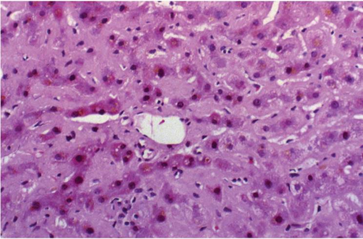
Amyloid is seen on routine hematoxylin-eosin-stained sections as pale eosinophilic or amphophilic, smudgy, amorphous, acellular material, usually deposited in the perisinusoidal space of Disse, in the vascular wall, or in the portal tract (Fig. 21.26, e-Figs. 21.51, 21.52). The characteristic apple-green birefringence after Congo red staining and polarization confirms the diagnosis (e-Fig. 21.53). With time, hepatocytes become compressed and atrophic.
Globular Amyloidosis
Pale eosinophilic amyloid globules deposit under the lining epithelium of large hepatic ducts, in the peribiliary glands, and on portal tract corrective tissue (Fig. 21.27, e-Figs. 21.54, 21.55) (42). Cholestasis and symptoms of large bile duct obstruction can ensue. Immunohistochemical stain for P component or immunofluorescent stain for thioflavine T can also be used.
Light-Chain Disease
Nonamyloid light-chain disease associated with plasma cell dyscrasias is seen in patients presenting with renal failure. Hepatomegaly with associated cholestasis can be seen. Light chain deposits are in the space of Disse and in portal tracts. These deposits generally do not stain with Congo red. Immunohistochemical studies show light chains, and sometimes heavy chains, as well as increased collagen type I and IV and fibronectin. Ultrastructurally, granular nonfibrillar material appears in the space of Disse.
|
|
|
FIGURE 21.26 Amyloidosis, with amyloid deposited in the space of Disse (hematoxylin-eosin, original magnification ×100). |
THYROID DISORDERS
The liver synthesizes thyroid-binding protein, metabolizes thyroid hormones, and is the major site for conversion of thyroxine to 3,5,38-triiodothyronine. Consequently, abnormalities of thyroid function are sometimes seen with various acute and chronic hepatic disorders.
|
|
|
FIGURE 21.27 Globular amyloidosis. showing pale eosinophilic globules in a portal tract (hematoxylin-eosin, original magnification ×100). |
Hyperthyroidism
Nonspecific changes can be seen, including mild macrovesicular steatosis, Kupffer cell hyperplasia, nuclear anisocytosis, and variable degrees, generally mild, of portal tract chronic inflammatory cell infiltration (6,68,74).
Hypothyroidism
Cholestasis has been described (4,5). Chronic autoimmune thyroiditis can be associated with primary biliary cirrhosis (see Chapter 17).
AUTOIMMUNE AND RELATED DISORDERS
Rheumatoid Arthritis
Generally only nonspecific changes are present, including steatosis, lipogranulomas, and fibrosis, perhaps related to treatment with corticosteroids and antimetabolites such as methotrexate (78). Sinusoidal dilatation and nodular hyperplasia are often seen in patients with Felty syndrome (rheumatoid arthritis, splenomegaly, and neutropenia) (12,20,34,80,107). Spontaneous rupture of the liver has been described with rheumatoid vasculitis, but this will not likely be an issue of liver biopsy interpretation (34,80,107). Rheumatoid nodules are exceedingly rare and virtually never found in liver biopsy. In juvenile rheumatoid arthritis (Still disease), the liver biopsy shows only mild, nonspecific changes, although there may be significant elevation of serum transaminase values.
Systemic Lupus Erythematosus
Liver involvement is uncommon in systemic lupus erythematosus (SLE), with fewer than 10% of patients demonstrating elevations of serum transaminase values. Histologic changes are exceedingly rare and not consistent. There may be mild portal tract nonspecific chronic inflammation, occasional lobular granulomas, sinusoidal congestion, peliosis hepatis, nodular regenerative hyperplasia, PSC, and local hepatic infarction (2,43,59,85). Macrovesicular steatosis can be seen after the treatment with corticosteroids. There is no association between antinuclear antibody-associated autoimmune hepatitis and SLE (see Chapter 10) (59).
Progressive Systemic Sclerosis
Chronic hepatitis has been described in some patients with progressive systemic sclerosis (scleroderma), as has nodular regenerative hyperplasia, PSC, PBC, and various changes secondary to vasculitis.
Polyarteritis Nodosa
The small branches of hepatic arteries can show typical vasculitis (e-Figs. 21.56, 21.57), with associated microscopic areas of infarction. Immune complexes containing HBV surface antigen have been demonstrated.
|
|
|
FIGURE 21.28 Cryoglobulinemia showing intravascular coagulation. Cryoglobulins were confirmed with immunofluorescence (hematoxylin-eosin, original magnification ×200). |
Cryoglobulinemia
Chronic hepatitis due to HCV can lead to the development of essential (type II) mixed cryoglobulinemia. Patients generally present with the manifestations of cryoglobulinemia before liver disease is recognized (1). Cryoglobulin is demonstrable in hepatic vascular spaces. Immune complexes containing HCV antigen are also demonstrable (Fig. 21.28) (1,65).
Calcinosis, Raynaud Phenomenon, Esophageal Motility Disorders, Sclerodactyly, and Telangiectasia (CREST) Syndrome
Changes of PBC can be seen in patients with CREST syndrome.
Mixed Connective Tissue Disease
Mixed connective tissue disease is a rare disorder characterized by features of systemic lupus erythematosus, progressive systemic sclerosis, and polymyositis. It is associated with high titers of antibodies to the ribonucleoprotein fraction of extractable nuclear antigens and may also be associated with chronic hepatitis. Budd-Chiari syndrome has also been reported (62).
Polymyalgia Rheumatica
Granulomas may be seen in polymyalgia rheumatica. However, the findings are usually nonspecific, including macrovesicular steatosis and nodular regenerative hyperplasia.
Polymyositis
PBC can occur in patients with polymyositis (64,89).
Weber-Christian Disease
Macrovesicular steatosis, both without inflammation as well as with steatohepatitis, is seen in patients with Weber-Christian disease (45). Mallory hyalin is observed in zone 1 (periportal) hepatocytes.
REFERENCES
1. Agnello V, Chung RT, Kaplan LM. Hepatitis C virus infection in type II cryoglobulinemia. N Engl J Med 1992;327:1490-1495.
2. Albert-Flor JJ, Jeffers L, Schiff ER. Primary sclerosing cholangitis occurring in a patient with systemic lupus erythematosus and diabetes mellitus. Am J Gastroenterol 1984;79: 889-891.
3. Al-Dalaan, Al-Balaa S, Ali MA, et al. Budd-Chiari syndrome in association with Behçet's disease. J Rheumatol 1991;18:622-626.
4. Ariza CR, Frate AC, Sierra I. Hypothyroidism-associated cholestasis. JAMA 1984; 252:2392.
5. Babb RR. Association between diseases of the thyroid and the liver. Am J Gastroenterol 1984;79:421-423.
6. Baca L, Gibbons RB. The HELLP syndrome: a serious complication of pregnancy with hemolysis, elevated levels of liver enzymes, and low platelet count. Am J Med 1988;85: 590-591.
7. Bach N, Theise ND, Schaffner F. Hepatic histopathology in the acquired immunodeficiency syndrome. Semin Liver Dis 1992;12:205-212.
8. Banks JG, Foulis AK, Ledingham IM, et al. Liver function in septic shock. J Clin Pathol 1982;35:1249-1252.
9. Barton JR, Riely CA, Adamec TA, et al. Hepatic histopathologic condition does not correlate with laboratory abnormalities in HELLP syndrome (hemolysis, elevated liver enzymes, and low platelet count). Am J Obstet Gynecol 1992;167:1538-1543.
10. Batman PA, Scheuer PJ. Diabetic hepatitis preceding the onset of glucose intolerance. Histopathology 1985;9:237-243.
11. Bernauau D, Guillot R, Durand AM, et al. Ultrastructural aspects of the liver perisinusoidal space in diabetic patients with and without cholangiopathy. Diabetes 1982;31: 1061-1067.
12. Blendis LM, Ansell ID, Lloyd-Jones K, et al. Liver in Felty's syndrome. Br Med J 1970;1: 131-135.
13. Bondini S, Kleiner DE, Goodman ZD, et al. Pathologic assessment of non-alcoholic fatty liver disease. Clin Liver Dis 2007;11:17-23.
14. Brown RE, Ishak KG. Hepatic zonal degeneration and necrosis in Reye's syndrome. Arch Pathol Lab Med 1976;100:123-126.
15. Bruguera M, Caballero T, Carreras E, et al. Hepatic sinusoidal dilatation in association with Hodgkin's disease. Liver 1987;7:76-80.
16. Brunt EM, Janney CG, Di Bisceglie AM, et al. Nonalcoholic steatohepatitis: a proposal for grading and staging the histological lesions. Am J Gastroenterol 1999;94:2467-2474.
17. Castillo I, Bartolome J, Medejon A, et al. Hepatitis delta virus RNA detection in chronic HBsAg carriers with and without HIV infection. Digestion 1991;48:149-156.
18. Chesner IM, Muller S, Newman J. Ulcerative colitis complicated by Budd-Chiari syndrome. Gut 1986;27:1096-1100.
19. Chopra S, Rubinow A, Koff RS, et al. Hepatic amyloidosis. A histopathologic analysis of primary (AL) and secondary (AA) forms. Am J Pathol 1994;115:186-193.
20. Cohen MD, Ginsburg WW, Allen GL. Nodular regenerative hyperplasia of the liver and bleeding esophageal varices in Felty's syndrome: a case report and literature review. J Rheumatol 1982;9:716-728.
21. Czapar CA, Weldo-Linne CM, Moore DM, et al. Peliosis hepatis in the acquired immunodeficiency syndrome. Arch Pathol Lab Med 1986;110:611-613.
22. De la Monte SM, Arcide JM, Moore GW, et al. Midzonal necrosis as a pattern of hepatocellular injury after shock. Gastroenterology 1984;86:627-631.
23. Devaney K, Goodman ZD, Epstein MS, et al. Hepatic sarcoidosis. Clinicopathologic features in 100 patients. Am J Surg Pathol 1993;17:1272-1280.
24. Dubois A, Dauzat M, Pignodel C, et al. Portal hypertension in lymphoproliferative and myeloproliferative disorders: hemodynamic and histologic correlation. Hepatology 1993;17:246-250.
25. Duffy LF, Daum F, Kahn E, et al. Hepatitis in children with acquired immunodeficiency syndrome. Histopathologic and immunocytologic features. Gastroenterology 1986;90: 173-181.
26. Falk S, Helm EB, Hubner K, et al. Disseminated visceral leishmaniasis (kala azar) in acquired immunodeficiency syndrome (AIDS). Pathol Res Pract 1988;183:253-255.
27. Fausa O, Nygaard K, Elgjo K. Amyloidosis and Crohn's disease. Scand J Gastroenterol 1977;12:657-662.
28. Favara BE. Hemophagocytic lymphohistiocytosis: a hemophagocytic syndrome. Semin Diagn Pathol 1992;9:63-74.
29. Foster JH, Donohue TA, Berman MM. Familial liver cell adenomas and diabetes mellitus. N Engl J Med 1978;299:239-241.
30. Garcia-Tsao G, Panzini L, Yoselevitz M, et al. Bacillary peliosis hepatis as a cause of acute anemia in a patient with the acquired immunodeficiency syndrome. Gastroenterology 1992;102:1065-1070.
31. Gitkind MJ, Wright SC. Amyloidosis complicating inflammatory bowel disease; a case report and review of the literature. Dig Dis Sci 1990;35:906-908.
32. Gordon SC, Reddy KR, Gould EE, et al. The spectrum of liver disease in the acquired immune deficiency syndrome (AIDS). Liver 1986;6:158-166.
33. Guarner J, del Rio C, Carr D, et al. Non-Hodgkin's lymphomas in patients with human immunodeficiency virus infection: presence of Epstein-Barr virus in situ hybridization, clinical presentation, and follow-up. Cancer 1991;68:2460-2465.
34. Harris M, Rash RM, Dymock IW. Nodular non-cirrhotic liver associated with portal hypertension in a patient with rheumatoid arthritis. J Clin Pathol 1974;27:963-966.
35. Hasan AF, Jeffers JL, Dickinson G, et al. Hepatobiliary cryptosporidiosis and cytomegalovirus infection mimicking metastatic cancer of the liver. Gastroenterology 1991;110:1743-1747.
36. Hay JE, Wiesner RH, Shorter RG, et al. Primary sclerosing cholangitis and celiac disease; a novel association. Gastroenterology 1988;94:A545.
37. Horsburgh CR Jr. Mycobacterium avium complex infection in the acquired immunodeficiency syndrome. N Engl J Med 1991;324:1332-1338.
38. Housset C, Lamas E, Courgnaud V, et al. Presence of HIV-1 in human parenchymal and non-parenchymal cells in vivo. J Hepatol 1993;19:252-258.
39. Hubscher SG, Lumley MA, Elias E. Vanishing bile duct syndrome: a possible mechanism for intrahepatic cholestasis in Hodgkin's lymphoma. Hepatology 1993;17:70-77.
40. Ishak KG, Rogers WA. Cryptogenic acute cholangitis: association with toxic shock syndrome. Am J Clin Pathol 1981;76:619-626.
41. Kahn E, Greco MA, Daum F, et al. Hepatic pathology in pediatric acquired immunodeficiency syndrome. Hum Pathol 1991;22:1111-1118.
42. Kanel GC, Uchida T, Peters RL. Globular hepatic amyloid: an unusual morphologic presentation. Hepatology 1981;1:647-652.
43. Khoury G, Tobi M, Oren M, et al. Massive hepatic infarction in systemic lupus erythematosus. Dig Dis Sci 1990;35:1557-1560.
44. Kilpatrick ZM. Structural and functional abnormalities of liver in infectious mononucleosis. Arch Intern Med 1966;117:47-53.
45. Kimura H, Kako M, Yo K, et al. Alcoholic hyalins (Mallory bodies) in a case of Weber-Christian disease: electron microscopic observation of liver involvement. Gastroenterology 1986;78:807-812.
46. Kirkpatrick CJ, Curry A, Galle J, et al. Systemic kappa light chain deposition and amyloidosis in multiple myeloma: novel morphological observations. Histopathology 1986;5:178-185.
47. Kleiner DE, Brunt EM, Van Natta M, et al. Design and validation of a histological scoring system for nonalcoholic fatty liver disease. Hepatology 2005;41:1313-1321.
48. Knowles DM, Chamulak GA, Subar M, et al. Lymphoid neoplasia associated with the acquired immunodeficiency syndrome. Ann Intern Med 1988;108:744-753.
49. Lai KK, Gang DL, Zawacki JK, et al. Fulminant hepatic failure with 28,38-dideoxyinosine (ddI). Ann Intern Med 1991;115:283-284.
50. Latry P, Bioulac-Sage P, Echinard E, et al. Perisinusoidal fibrosis and basement membrane-like material in the livers of diabetic patients. Hum Pathol 1987;18:775-780.
51. Leboit PE, Berger TC, Egbert BM, et al. Bacillary angiomatosis: the histopathology and differential diagnosis of a pseudoneoplastic infection in patients with human immunodeficiency virus disease. Am J Surg Pathol 1989;13:909-920.
52. Lefkowitch JH. Bile ductular cholestasis: an ominous histopathologic sign related to sepsis and “cholangitis lenta.” Hum Pathol 1982;13:19-24.
53. Lefkowitch JH, Fakow S, Whitlock RT. Hepatic Hodgkin's disease simulating cholestatic hepatitis with liver failure. Arch Pathol Lab Med 1985;109:424-426.
54. Lefkowitch JH, Mendez L. Morphological features of hepatic injury in cardiac disease and shock. J Hepatol 1986;2:313-327.
55. Leibowitz AI, Hartman RC. The Budd-Chiari syndrome and paroxysmal nocturnal hemoglobinuria. Br J Hematol 1981;48:1-6.
56. Lesens O, Deschenes M, Steben M, et al. Hepatitis C virus is related to progressive liver disease in HIV-positive hemophiliacs and should be treated as an opportunistic infection. J Infect Dis 1999;179:1254-1258.
57. Ludwig J, Braham SS, LaRusso NF, et al. Morphological features of chronic hepatitis associated with primary sclerosing cholangitis and chronic ulcerative colitis. Hepatology 1981;1:632-640.
58. Maccini DM, Berg JC, Bell GA. Budd-Chiari syndrome and Crohn's disease. An unreported association. Dig Dis Sci 1989;34:1933-1936.
59. Mackay IR. The hepatitis-lupus connection. Semin Liver Dis 1991;11:234-240.
60. Mahmond H, Gaber O, Wang W, et al. Successful orthotopic liver transplantation in a child with Langerhans cell histiocytosis. Transplantation 1991;51:278-280.
61. Margulis SJ, Honig CL, Siave R. Biliary tract obstruction in the acquired immunodeficiency syndrome. Ann Intern Med 1986;105:207-210.
62. Marshall JB, Ravendran N, Sharp GC. Liver disease in mixed connective tissue disease syndrome. Arch Intern Med 1983;143:1817-1818.
63. McMaster KR, Hennigar GR. Drug-induced granulomatous hepatitis. Lab Invest 1981; 44:61-73.
64. Milosevic M, Adams PC. Primary biliary cirrhosis and polymyositis. J Clin Gastroenterol 1990;12:332-335.
65. Misiani R, Bellavita P, Fenili D, et al. Hepatitis C virus infection in patients with essential mixed cryoglobulinemia. Ann Intern Med 1992;117:573-577.
66. Molina JM, Welker Y, Ferchal F, et al. Hepatitis associated with primary HIV infection. Correspondence. Gastroenterology 1992;102:739.
67. Nizalik E, Zayed E, Foyle A. Malignant lymphoma presenting as fulminant hepatic failure. Can J Gastroenterol 1989;3:111-114.
68. Ochmer SA, Brunt EM, Cohn S, et al. Fulminant hepatic failure caused by acute fatty liver of pregnancy treated by orthotopic liver transplantation. Hepatology 1990;11:59-64.
69. Pereira A, Bruguera M, Cervantes F, et al. Liver involvement at diagnosis of primary myelofibrosis: a clinicopathological study of twenty cases. Eur J Hematol 1988;40: 355-361.
70. Perkocha LA, Geaghan SM, Yen TSB, et al. Clinical and pathological features of bacillary peliosis hepatis in association with human immunodeficiency virus infection. N Engl J Med 1990;323:1581-1586.
71. Petrovic LM. HIV/HCV co-infection: histopathologic findings, natural history, fibrosis and impact of antiretroviral treatment. Liver Int 2007;27(5):598-606.
72. Piroth L, Duong M, Quantin C, et al. Does hepatitis C virus co-infection accelerate clinical and immunological evolution of HIV-infected patients? AIDS 1998;12:381-388.
73. Pol S, Romana C, Richard S, et al. Microsporidia infection in patients with the human immunodeficiency virus and unexplained cholangitis. N Engl J Med 1993;328:95-99.
74. Pollock DJ. The liver in celiac disease. Histopathology 1977;1:421-430.
75. Pottipati AR, Dave PB, Gumaste V, et al. Tuberculous abscess of the liver in acquired immunodeficiency syndrome. J Clin Gastroenterol 1991;13:549-553.
76. Pursell KJ, Telzak EE, Armstrong D. Aspergillus species colonization and invasive disease in patients with AIDS. Clin Infect Dis 1992;14:141-148.
77. Randhawa PS, Markin RS, Starzl TE, et al. Epstein-Barr virus-associated syndromes in immunosuppressed liver transplant patients. Am J Surg Pathol 1990;14:538-547.
78. Rau R, Karger T, Herborn G, et al. Liver biopsy findings in patients with rheumatoid arthritis undergoing long-term treatment with methotrexate. J Rheumatol 1989;16: 489-493.
79. Reye RDK, Morgan G, Baral J. Encephalopathy and fatty degeneration of the viscera, a disease entity in childhood. Lancet 1963;2:749-752.
80. Reynolds WJ, Wanless IR. Nodular regenerative hyperplasia of the liver in a patient with rheumatoid vasculitis: a morphometric study suggesting a role of hepatic arteritis in the pathogenesis. J Rheumatol 1984;11:838-842.
81. Riely CA. Acute fatty liver of pregnancy. Semin Liver Dis 1987;7:47-54.
82. Rolfes DB, Ishak KG. Liver disease in pregnancy. Histopathology 1986;10:555-570.
83. Rolfes DB, Ishak KG. Liver disease in toxemia of pregnancy. Am J Gastroenterol 1986;81:1138-1144.
84. Ross JS, Del Rosario A, Bui HX, et al. Primary hepatic leiomyosarcoma in a child with the acquired immunodeficiency syndrome. Hum Pathol 1992;23:69-72.
85. Runyon BA, LeBrecqui DR, Anuras S. The spectrum of liver disease in systemic lupus erythematosus: report of 33 histologically proved cases and review of the literature. Am J Med 1980;69:187-194.
86. Sabin CA, Telfer P, Phillips AN, et al. The association between hepatitis C virus genotype and HIV disease progression in a cohort of hemophiliac men. J Infect Dis 1997;175:164-168.
87. Sachs JR, Greenfield SM, Sohn M, et al. Disseminated Pneumocystis carinii infection with hepatic involvement in a patient with the acquired immune deficiency syndrome. Am J Gastroenterol 1991;86:82-85.
88. Saint-Marc, Girardin M-F, Zafrani ES, et al. Hepatic granulomas in Whipple's disease. Gastroenterology 1984;86:753-756.
89. Sattar MA, Guindi RT, Khan RA, et al. Polymyositis and hepatocellular carcinoma. Clin Rheumatol 1988;7:538-542.
90. Scoazec JY, Marche C, Girad PM, et al. Peliosis hepatis and sinusoidal dilatation during infection with the human immunodeficiency virus (HIV). Am J Pathol 1991;130:38-47.
91. Scully PA, Steinman HK, Kennedy C, et al. AIDS-related Kaposi's sarcoma displays differential expression of endothelial surface antigens. Am J Pathol 1988;130:244-251.
92. Shiramizu B, Herndier B, Meeker T, et al. Molecular and immunophenotypic characterization of AIDS-associated Epstein-Barr virus-negative, polyclonal lymphoma. J Clin Oncol 1992;10:383-389.
93. Slater LN, Welch DF, Min KW. Rochalimaea henselae causes bacillary angiomatosis and peliosis hepatis. Arch Intern Med 1992;152:602-606.
94. Soto B, Sanchez-Quijano A, Rodrigo L, et al. HIV infection modifies the natural history of chronic parenterally acquired hepatitis C with an unusual rapid progression to cirrhosis. A multicenter study on 547 patients. J Hepatol 1997;26:1-5.
95. Steffan A-M, Lafon M-E, Gendrault J-L, et al. Primary cultures of endothelial cells from the human liver sinusoid are permissive for human immunodeficiency virus type 1. Proc Natl Acad Sci U S A 1992;12:197-204.
96. Steeper TA, Rosenstein H, Weiser J, et al. Bacillary epithelioid angiomatosis involving the liver, spleen, and skin in an AIDS patient with concurrent Kaposi's sarcoma. Am J Clin Pathol 1992;97:713-718.
97. Sty JR. Hepatic vein thrombosis in sickle cell anemia. Am J Pediatr Hematol Oncol 1982;4:213-215.
98. Terada S, Reddy KR, Jeffers LJ, et al. Microsporidian hepatitis in the acquired immunodeficiency syndrome. Ann Intern Med 1987;107:61-62.
99. Thiruvengadam R, Penetranti R, Grolsky HJ, et al. Multiple myeloma presenting as space-occupying lesion of the liver. Cancer 1990;65:2784-2786.
100. Thung SN, Gerber MA, Bodenheimer HC Jr. Nodular regenerative hyperplasia of the liver in a patient with diabetes mellitus. Cancer 1982;49:543-546.
101. Vanjak D, Moreau R, Roche-Sciot J, et al. Intrahepatic cholestasis of pregnancy and acute fatty liver of pregnancy. An unusual but favorable association? Gastroenterology 1991;100:1123-1125.
102. Voinchet O, Degott C, Scoazec J-Y, et al. Peliosis hepatis, nodular regenerative hyperplasia of the liver, and light chain deposition in patient with Waldenström's macroglobulinemia. Gastroenterology 1988;95:482-486.
103. Waldman RJ, Hall WN, McGee H, et al. Aspirin as a risk factor in Reye's syndrome. JAMA 1990;150:456-459.
104. Welch DF, Pickett DA, Salter LN, et al. Rochalimaea henselae sp., a cause of septicemia, bacillary angiomatosis, and parenchymal peliosis. J Clin Microbiol 1992;30:275-280.
105. Witzleben CL, Marshall GS, Wenner W, et al. HIV as a cause of giant cell hepatitis. Hum Pathol 1988;19:603-605.
106. Woolf GM, Petrovic LM, Rojter SE, et al. Acute liver failure due to lymphoma: a diagnostic concern when considering liver transplantation. Dig Dis Sci 1994;39:1351-1358.
107. Young ID, Segura J, Ford PM, et al. The pathogenesis of nodular regenerative hyperplasia of the liver associated with rheumatoid vasculitis. J Clin Gastroenterol 1992;14:127-131.
108. Zafrani ES, Degos F, Guigui B, et al. The hepatic sinusoid in hairy cell leukemia: an ultrastructural study of 12 cases. Hum Pathol 1987;18:801-807.
109. Zafrani ES, Gaulard P. Primary lymphoma of the liver. Liver 1993;13:57-61.
110. Zimmerli W, Bianchi L, Gudat F, et al. Disseminated herpes simplex type 2 and systemic Candida infection in a patient with previous asymptomatic HIV infection. J Infect Dis 1988;157:597-598.