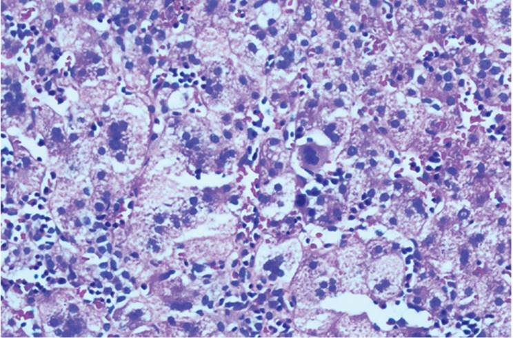
Embryologic issues arise relatively rarely in the interpretation of the liver biopsy. However, problems relating to prenatally acquired conditions, unrelated to disorders of embryogenesis, are not uncommon.
Anomalies of the liver manifest principally in terms of liver shape and position, as well as vascular variations (12,20). Although ectopia of the entire liver is exceedingly rare, liver tissue may be found in various places throughout the abdomen and even in the lung (25). The biliary tree is the principal site of abnormality in association with documented chromosomal disorders (12).
DEVELOPMENT OF THE LIVER AND INTRAHEPATIC BILIARY STRUCTURES
Parenchymal Development
Development of the human liver and biliary tract begins as a hepatic anlage at the 18th day of gestation, when the embryo is 2.5 mm in length (9), becoming more prominent in the third to fourth week as the hepatic diverticulum from the ventral distal foregut (12,26,29). The cephalic portion (pars hepatica) differentiates to become the liver, and the caudal portion (pars cystica) matures into the gallbladder and cystic duct.
The septum transversum mesenchymal cells form the connective tissue elements of the hepatic stroma, as well as the capsule (e-Figs. 6.1, 6.2). Near the end of the fourth week, the hepatic diverticulum acquires a T shape, creating the early right and left liver lobes (9). The mesodermal cells also form endothelial-lined spaces.
Initially, the liver plates are three to five cells thick. These decrease to two-cell-thick plates at the time of birth. After the fifth year of life, the normal plates are one cell thick. At 3 months, bile formation begins. The canaliculus appears in the 10-mm embryo as a differentiated zone of the intercellular surface of the liver and has its typical appearance by 7 to 8 weeks. Selected mitochondrial, microsomal, and lysosomal enzymes are demonstrable in liver cells by the eighth week.
|
|
|
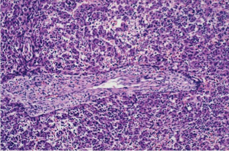
FIGURE 6.1 Image of developing liver showing ductal plate formation (hematoxylin-eosin, original magnification ×200). |
Primitive hepatocytes express cytokeratins 8, 18, and 19 (40,41). It has been suggested that these cells, precursors to the mature hepatocytes, give rise to the intrahepatic biliary system (11,33,34). Intrahepatic bile ducts develop progressively from the hilum, beginning at the eighth week. Portal vein extensions push into the primitive parenchyma, and the septum transversum mesenchyme induces the transformation of embryonic hepatocytes into ductal cells (12,15,23,35,36). The epithelial cells first transform into single- and then double-cell layered tubular structures, the so-called ductal plate (Fig. 6.1, e-Figs. 6.3-6.6) (8,10,35,36). At 5 to 6 weeks gestational age, a lumen forms between the two layers of the ductal plate, with the formation of true duct structures. These grow into the mesenchyme and further transform to become bile ducts. Cytokeratins, as well as the adhesion glycoprotein laminin, are thought to play a key role in this process (35,36,37,41). Progenitor cells have the potential for expressing multiple cytokeratins (37). The periportal mesenchymal sheath diminishes, and the newly formed ducts are incorporated into the portal tracts. Eventually, an arterial plexus envelops the ducts. True interlobular ducts are rare at 13 weeks but develop throughout gestation and even during the neonatal period (29). Ductal plate structures may persist, even at 40 weeks' gestation, and in some portal tracts there may not be an individual mature bile duct accompanying the most peripheral portal vein branches (9,10,41). Instead, the portal veins may be surrounded by a layer of cytokeratinrich cells in ductal plate arrangement.
Vascular Development
During the fifth week, the paired vitelline veins penetrate the primitive hepatic cords to form primitive sinusoids. The hepatic veins form from modifications of the vitelline veins. The portal vein results from multiple anastomoses between the caudal left and right vitelline veins. Some of the channels eventually atrophy, contributing to the tortuosity of the portal vein.
The ductus venosus forms as an enlargement of the sinusoids. The ductus is a potential conduit to the hepatic vein and inferior vena cava; since its diameter is less than 15% that of the umbilical vein, however, most of the blood flow is diverted through the liver parenchyma (14). A sinus intermedius connects the lobes, but the most highly oxygenated umbilical blood preferentially perfuses the left lobe.
Hepatic artery and portal vein development proceed from the hepatic hilum to the periphery. Portal vein extensions, with their accompanying cuff of mesenchyme within which bile ducts form, permeate the parenchyma. Arterial twigs become abundant by the last trimester (29).
Intrahepatic Hematopoiesis
Hematopoietic cells first appear in the fifth week, reaching a peak during the third month. The peak of hematopoietic activity is during the third month, diminishing progressively during the last trimester. In the normal infant, there is virtually no hematopoiesis by age 7 days (12).
Lymphocytes appear in the liver by 6 or 7 weeks' gestation and, after the 20th week, account for more than 20% of mononuclear cells (17).
Iron, demonstrable as hemosiderin, is seen in the fetal liver in both ductal plate cells and the developing limiting plate; the amount varies considerably throughout gestation, increasing as hematopoiesis progressively decreases and reaching peak concentrations at the end of gestation (9,28). Copper and zinc are also stored in these cells (13).
ABNORMALITIES OF THE BILE DUCT SYSTEM
Ductal Plate Malformation
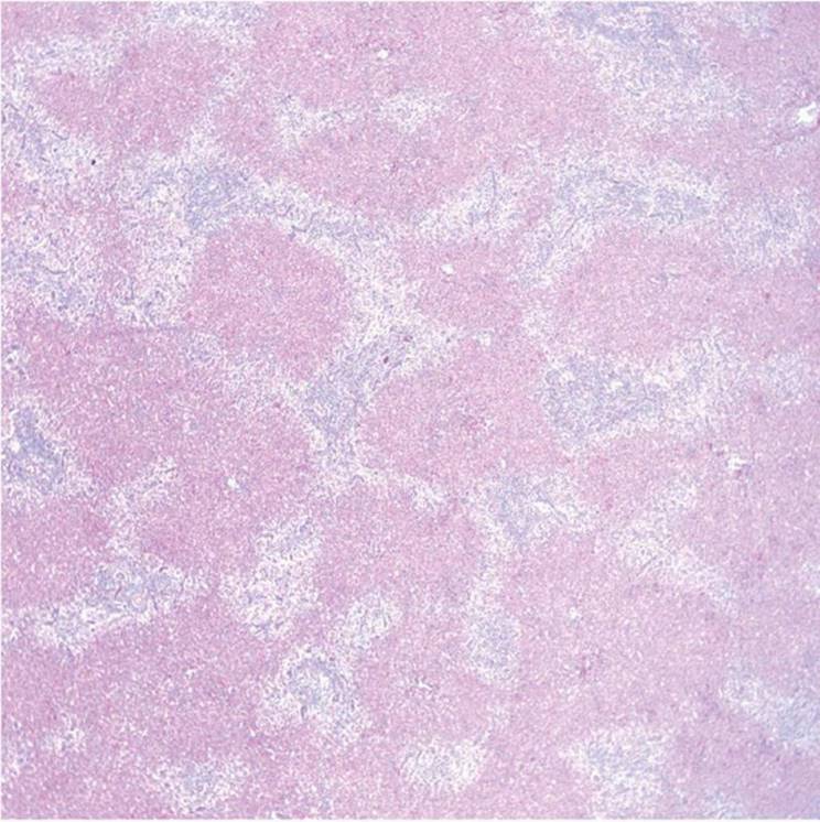
Ductal plate malformations are defects of the portal tract and its component structures, especially the bile duct system, arising during the formation and modification of the ductal plate. Failure of remodeling results in persistence of the embryologic ductal plate, the “ductal plate malformation.” This may appear in a biopsy as a ring of bile duct-like structures arrayed circumferentially at the periphery of the portal tract, generally with a central portal vein or groups of veins (Fig. 6.2, e-Fig. 6.7). The ring may be complete or incomplete (10). Typically, excess of mesenchymal tissue and ectasia of ducts is seen.
Conditions thought to be associated with ductal plate malformation are congenital hepatic fibrosis (Fig. 6.3, e-Figs. 6.8-6.13), bile duct hamartomas (von Meyenburg complexes) (e-Figs. 6.14-6.17), Caroli disease (e-Fig. 6.18), and Caroli syndrome, infantile polycystic disease, Meckel-Gruber syndrome, renal-hepatic-pancreatic dysplasia (Ivemark syndrome), and Beckwith-Wiedemann syndrome (7,10,12). Ductal plate and renal tubular differentiation share genetic determinants, and single-gene defects can give rise to such varied entities as congenital hepatic fibrosis, Caroli disease and syndrome, and autosomal recessive and autosomal dominant polycystic kidney disease (7).
|
|
|
FIGURE 6.2 Image of a liver biopsy from an adult with ductal plate malformation, showing peripherally arranged bile ducts and splayed portal vein segments (“pollard-tree” pattern). (hematoxylin-eosin, original magnification ×200) |
|
|
|
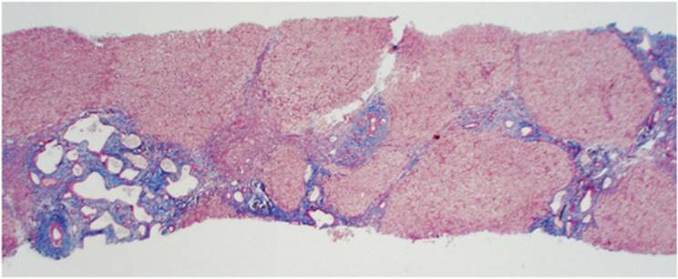
FIGURE 6.3 Image of congenital hepatic fibrosis, diffuse form, from a child with portal hypertension, showing portal tracts with increased numbers of abnormally developed bile ducts, with fibrosis within portal tracts and also linking portal tracts (hematoxylin-eosin, original magnification ×40). |
Recognition of these entities is important because their presence in liver biopsy specimens can be misinterpreted as metastatic, well-differentiated, desmoplastic adenocarcinoma.
Congenital Hepatic Fibrosis
Congenital hepatic fibrosis was originally described in association with autosomal recessive polycystic kidney disease but can occur as an isolated entity (Fig. 6.3, e-Figs. 6.8-6.13). Patients usually have hepatosplenomegaly and portal hypertension, but there are four patterns of expression: (a) portal hypertension, (b) cholangitis without portal hypertension, (c) cholangitis and portal hypertension, and (d) latent cholangitis, generally discovered incidentally either during an unrelated surgical procedure or at autopsy. Symptoms of cholangitis are due to associated Caroli disease.
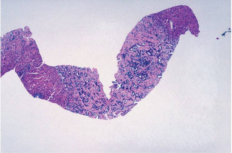
Microscopically, the picture can be variable (7,10). In the focal form, abnormally formed bile ducts are in irregularly shaped portal tracts with prominent fibrous tissue. The bile ducts can be multiple and appear branching or stellate. In the diffuse form the portal tracts are linked by fibrosis (Fig. 6.4). In both forms, ductal plate malformation is seen at the portal tract periphery. Inflammation is generally mild. Portal veins may appear hypoplastic, but arterioles are prominent. Degeneration of the biliary duct epithelium and mild cholestasis can also be seen. Congenital hepatic fibrosis can be limited to one lobe (19). The intervening hepatic parenchyma and the terminal hepatic venule are unremarkable.
|
|
|
FIGURE 6.4 Image of congenital hepatic fibrosis, incomplete form, from an adult, also with a bile duct hamartoma (von Meyenburg complex) (trichrome, original magnification ×100). |
|
|
|
FIGURE 6.5 Image of two bile duct hamartomas (von Meyenburg complex) found incidentally in a liver biopsy obtained from a patient with chronic hepatitis. There are irregularly dilated bile ducts surrounded by dense fibrous tissue (hematoxylin-eosin, original magnification ×200). |
The biopsy features suggesting the diagnosis are as follows: (a) fibrotic portal tracts containing few or no inflammatory cells; (b) increased numbers of prominent, often focally ectatic, bile ducts; (c) multiple small portal vein tributaries; (d) prominent arterioles; and (e) conspicuous, often irregularly ectatic, ductulelike structures encircling the portal tract. Other ductal plate malformation changes, such as bile duct hamartoma (Fig. 6.5) can also be seen.
Bile Duct Hamartoma (von Meyenburg Complex)
The small, benign lesions of bile duct hamartoma, often visible grossly as tiny white liver nodules, are composed of groups of dilated bile ducts embedded in connective tissue (Figs. 6.4, 6.5, e-Figs. 6.14-6.17). The ducts may contain polypoid epithelial projections. Bile duct hamartomas are most often seen in the otherwise unremarkable liver but may be associated with congenital hepatic fibrosis and Caroli disease. They may be single or multiple, and sometimes are immediately adjacent to a normal portal tract. The pattern of ductal plate malformation is seen only rarely. The ectatic ducts may contain bile or even small calculi (30). Recent evidence suggests that bile duct hamartomas communicate with the bile duct system (11). Cholangiocarcinoma may rarely arise (5).
Caroli Disease and Caroli Syndrome
The Caroli lesion is characterized by congenital dilatation, fusiform or saccular, of the larger intrahepatic bile ducts, generally at multiple sites. The ducts maintain continuity with the biliary tree. Caroli disease is not associated with other histologic abnormalities. In Caroli syndrome, the changes of congenital hepatic fibrosis are also seen. Caroli syndrome is significantly more common than Caroli disease.
Caroli disease is thought to be caused by ductal plate malformation affecting the large bile ducts (e-Fig. 6.18), whereas Caroli syndrome occurs when the entire bile duct system is affected. Patients with Caroli disease most often have repeated attacks of cholangitis, often complicated by the development of calculi and by abscess formation and sepsis. Cholangiocarcinoma may ensue (38). The liver biopsy may be normal or may show the changes of an ascending cholangitis.
In Caroli syndrome with congenital hepatic fibrosis, the cholangitic episodes are often accompanied by portal hypertension. The liver biopsy may appear normal, may show cholangitic changes, or may show the typical changes of congenital hepatic fibrosis.
Caroli disease and Caroli syndrome may be associated with autosomal recessive polycystic kidney disease and choledochal cysts.
Biliary Atresia
The biliary atresias have generally been considered to be related development disorders since they may occur together. It may be that isolated extrahepatic biliary atresia is an acquired disease, perhaps caused by intrauterine infection (27).
In contrast, isolated intrahepatic biliary atresia is most likely truly developmental, caused by a primary defect in the bile secretory mechanism at the level of hepatocyte with secondary loss of bile ducts (8,39). Indeed, in Zellweger (cerebrohepatorenal) syndrome, a peroxisomal disorder results in abnormal bile acid metabolism and may also be associated with the histologic picture of intrahepatic biliary atresia (8).
EXTRAHEPATIC BILIARY ATRESIA. Extrahepatic biliary atresia (EHBA) occurs in approximately 1 in 10,000 births and is more common in females than in males (9). Clinical manifestations include jaundice persisting beyond the physiologic period, acholic stools, and hepatomegaly. EHBA may occur with other disorders, such as galactosemia, α1-antitrypsin deficiency, and small bowel atresia, and with several malformations. The polysplenia syndrome occurs in 11% of patients with EHBA. Differentiation from other forms of neonatal cholestasis may be difficult (2,3). Imaging studies or direct examination of biliary structures establishes the diagnosis.
In type I EHBA, the common bile duct is affected while the cystic duct and hepatic ducts remain patent. In type IIa, the common hepatic duct is obliterated, but the cystic duct and common bile duct are unaltered. In type IIb, the cystic duct, common hepatic duct, and common bile duct are all affected, but the gallbladder is unremarkable and, typically, the hepatic ducts at the hilum are cystically dilated. In type III (uncorrectable) extrahepatic biliary atresia, there is absence or atresia of the common, hepatic and cystic ducts, and the hilar ducts are not cystically dilated, making anastomosis to the small intestine impossible. Liver biopsy can be helpful.
|
|
|
FIGURE 6.6 Image of early stage of extrahepatic biliary atresia, showing spherical enlargement of the portal tract, caused by edema and dilatation of lymphatic channels, with ductular proliferation at the periphery of the portal tract (hematoxylin-eosin, original magnification ×200). |
During the stage of ductular reaction, the portal tract shows typical changes of extrahepatic biliary obstruction, with edema and dilatation of lymphatic channels contributing to the characteristic rounding of the portal tracts (Fig. 6.6), and with ductular proliferation at the periphery of the portal tract. Inflammation involving polymorphonuclear leukocytes as well as lymphocytes follows. A stage of portal bile duct destruction follows, with epithelial cell vacuolization, necrosis, atrophy, and changes of repair, along with interruption of basement membrane and permeation by inflammatory cells. This characteristic stage generally occurs after 6 weeks, but its time of onset is highly variable.
Prior to the stage of ductular reaction, the biopsy shows hepatocytic and canalicular bile stasis, with little or no inflammation and only rare necrotic hepatocytes. Extramedullary hematopoiesis may persist. A few hydropic hepatocytes may be seen, and there may be scattered multinucleated giant cells.
During the stage of ductular reaction, the parenchymal changes may be dominant, with extensive hepatocyte necrosis and florid giant cell transformation of hepatocytes, suggesting the diagnosis of neonatal giant cell hepatitis (Fig. 6.7).
Portal and periportal fibrosis develops, with fibrous septa irregularly dissecting through the liver. Portal inflammatory changes diminish at this stage, but bile plugs become more prominent and there are more extensive changes secondary to the accumulation of bile, with feathery degeneration (cholestasis) of hepatocytes, frank necrosis, and small bile lakes. Dilated ductules contain bile concretions. Periductal fibrosis may be concentric, resembling that seen in primary sclerosing cholangitis. In the third month, fibrosis links portal tract to portal tract, and portal hypertension may ensue, although there are not yet regenerative changes of hepatocytes with formation of nodules and the full clinical picture of cirrhosis may not be seen. After 12 weeks, the fibrosis is accompanied by regeneration and typical secondary biliary cirrhosis is seen (Fig. 6.8). Bile ducts may be partially or completely obliterated. In as many as 25% of cases, the liver biopsy shows the pattern of ductal plate malformation (31).
|
|
|
FIGURE 6.7 Image of stage of ductular reaction of extrahepatic biliary atresia, with marked cholestasis and florid giant cell formation suggesting the diagnosis of giant cell hepatitis (hematoxylin-eosin, original magnification ×200). |
|
|
|
FIGURE 6.8 Image of the (secondary biliary) cirrhotic stage of extrahepatic biliary atresia, showing fibrous septa connecting portal areas, with changes of chronic cholestasis, including cholate stasis of zone 1 hepatocytes imparting the appearance of marked cell swelling |
|
TABLE 6.1 Histopathology of Extrahepatic Biliary Atresia |
|||||||||||||||||||||||||||||||||||||||||||||||||||||||||||||||||||||
|
|||||||||||||||||||||||||||||||||||||||||||||||||||||||||||||||||||||
The stages of evolution of fully developed secondary biliary cirrhosis as a sequel to extrahepatic biliary atresia are summarized in Table 6.1.
INTRAHEPATIC BILIARY ATRESIA (PAUCITY OF INTERLOBULAR BILE DUCTS). Paucity of interlobular bile ducts may be associated with extrahepatic anomalies (“syndromatic”) or may be isolated (“nonsyndromatic”).
Syndromatic paucity (e-Figs. 6.17-6.29) was first described by Alagille as a disorder morphologically distinct from other causes of cholestasis in infancy and childhood (1). Alagille syndrome, or arteriohepatic dysplasia, is the most common form of familial intrahepatic cholestasis. Cholestasis is severe in infancy and childhood but improves, and potentially even resolves, with age. Cirrhosis is uncommon. Extrahepatic anomalies can be seen, including pulmonary artery stenosis or hypoplasia, thickening of the junction of Descemet membrane with the endothelium of the anterior chamber of the eye (posterior embryotoxon), and abnormalities of vertebral bodies. Skeletal abnormalities can be seen. There may be a characteristic facies (1,3,4). A chromosomal anomaly, del(20p), has been demonstrated in some patients (6). There may be growth retardation and/or mental retardation, as well as impaired sexual development (3).
Nonsyndromatic paucity of interlobular bile ducts (e-Fig. 6.3) is less common than the syndromatic variant and is a heterogeneous group of disorders. There may be associated congenital rubella, α1-antitrypsin deficiency, Turner syndrome, Down syndrome, trisomy 17-18, congenital syphilis, and familial trihydroxycoprostanic acid excess. Cirrhosis develops in as many as 50% of patients. A variant in adults is termed idiopathic adulthood ductopenia (16).
Histologic diagnosis requires biopsy sufficiently large to contain at least five complete portal tracts (3,24). Immunohistochemical methods to stain high molecular weight keratins facilitate visualization of interlobular bile ducts (40) (Fig. 6.9). In normal newborns, the ratio of interlobular ducts to the number of portal tracts is between 0.9 and 1.8, although a ratio less than 0.9 may be seen in premature newborns when the liver biopsy is performed before the 38th week of gestation (22). The diagnosis of paucity is established when the ratio is less than 0.5 (3). Portal tracts generally appear somewhat hypoplastic, and the total number may be reduced (18). Cholestasis is hepatocellular rather than canalicular. Portal and perisinusoidal fibrosis may be apparent (21). Changes generally occur after infancy. Inflammatory destruction of bile ducts is observed more often in the nonsyndromatic variant. Extrahepatic bile ducts are patent but may be hypoplastic. Endoscopic retrograde cholangiography can demonstrate focal dilatation of segments of the biliary tree, resembling primary sclerosing cholangitis.
|
|
|
FIGURE 6.9 Image of a liver biopsy from a patient with paucity of interlobular bile ducts, showing adjacent portal tracts lacking bile ducts, with peripheral ductal reaction (proliferation). (hematoxylin-eosin, original magnification ×200). |
COMBINED EXTRAHEPATIC AND INTRAHEPATIC BILE DUCT ATRESIA. Cases demonstrating features of both extrahepatic biliary atresia and paucity of interlobular bile ducts have been described (32,41), rarely associated with ductal plate malformation (31). In most infants with extrahepatic biliary atresia, however, the intrahepatic changes may be secondary because of the obstruction of bile outflow.
REFERENCES
1. Alagille D, Odièvre M, Gautier M, et al. Hepatic ductular hypoplasia associated with characteristic facies, vertebral malformations, retarded physical, mental and sexual development and cardiac murmur. J Pediatr 1975;86:63-71.
2. Alagille D. Cholestasis in the first three months of life. In: Popper H, Schaffner F, eds. Progress in Liver Diseases, vol. 6. New York: Grune & Stratton, 1979:471-485.
3. Alagille D, Odievre M. Liver and Biliary Tract Disease in Children. New York: John Wiley and Sons, 1979:68-93.
4. Alagille D, Estrada A, Hadchouel M, et al. Syndromatic paucity of interlobular bile ducts (Alagille syndrome or arteriohepatic dysplasia): review of 80 cases. J Pediatr 1987;110: 195-200.
5. Burns C, Kuhns JG, Wieman TJ. Cholangiocarcinoma in association with multiple biliary microhamartomas. Arch Pathol Lab Med 1990;114:1287-1289.
6. Byrne JLB, Harrod MJE, Friedman JM, et al. Del (20p) with manifestation of arteriohepatic dysplasia. Am J Med Genet 1986;24:673-678.
7. D'Agata IDA, Jonas MM, Perez-Atayde AR, et al. Combined cystic disease of the liver and kidney. Semin Liver Dis 1994;14:215-228.
8. Desmet VJ. Cholangiopathies: past, present, and future. Semin Liver Dis 1987;7:67-76.
9. Desmet VJ. Embryology of the liver and intrahepatic biliary tract, and an overview of malformations of the bile duct. In: McIntyre N, Benhamou J-P, Bircher J, et al., eds. Oxford Textbook of Clinical Hepatology, vol. 1. New York: Oxford University Press,1991:498-519.
10. Desmet VJ. Congenital diseases of intrahepatic bile ducts: variations on the theme “ductal plate malformation.” Hepatology 1992;16:1069-1083.
11. Desmet VJ. What is congenital hepatic fibrosis? Histopathology 1992;20:465-477.
12. Dimmick JE. Hepatobiliary system. In: Dimmick JE, Kalousek DK, eds. Developmental Pathology of the Embryo and Fetus. Philadelphia: JB Lippincott Co, 1992:545-578.
13. Dubois AM. The embryonic liver. In: Rouiller C, ed. The Liver. New York: Academic Press, 1963:1-39.
14. Emery JL. The distribution of hematopoietic foci in the infantile human liver. J Anat 1956;90:293-311.
15. Enzan H, Ohkita T, Fujita H, et al. Light and electron microscopic studies on the development of the periportal bile ducts of the embryo. Acta Pathol Jpn 1974;24:427-447.
16. Faa G, Van Eycken P, Demelia L, et al. Idiopathic adulthood ductopenia presenting with chronic recurrent cholestasis. J Hepatol 1991;12:44-20.
17. Gale RP. Immune development in human fetal liver. In: Gale RP, Touraine J-L, Lucarelli G, eds. Fetal Liver Transplantation. New York: Alan R. Liss, 1985:73-88.
18. Hadchouel M, Hugon RN, Gautier M. Reduced ratio of portal tracts to paucity of intrahepatic bile ducts. Arch Pathol Lab Med 1978;102:402-403.
19. Hausner RJ, Alexander RW. Localized congenital hepatic fibrosis presenting as an abdominal mass. Hum Pathol 1978;9:473-476.
20. Hutchins GM, Moore DW. Growth and asymmetry of the human liver during the embryonic period. Pediatr Pathol 1988;8:17-24.
21. Kahn E, Daum F, Markowitz J, et al. Nonsyndromatic paucity of interlobular bile ducts: light and electron microscopic evaluation of sequential liver biopsies in early childhood. Hepatology 1986;6:890-901.
22. Kahn E, Markowitz J, Aiges H, et al. Human ontogeny of the bile duct to portal space ratio. Hepatology 1989;10:21-23.
23. Koga A. Morphogenesis of intrahepatic bile ducts of the human fetus. Light and electron microscopic study. Z Anat Entwicklungsgesch 1971;135:156-184.
24. Labrecque DR, Mitros FA, Nathan RJ, et al. Four generations of arteriohepatic dysplasia. Hepatology 1982;2:467-474.
25. Mendoza A, Voland J, Wolf P, et al. Supradiaphragmatic liver in the lung. Arch Pathol Lab Med 1986;110:1085-1086.
26. Moore KL. The developing human. 4th Ed. Philadelphia: WB Saunders, 1988.
27. Morecki R, Glaser JH, Johnson AB, et al. Detection of reovirus type 3 in the porta hepatis of an infant with extrahepatic biliary atresia: ultrastructural and immunocytochemical study. Hepatology 1984;4:1137-1142.
28. Oyer CE, Rogers BB, Singer DB. A histologic survey of iron positive cells in fetal and neonatal livers utilizing morphometry and a semiquantitative grading method. Pediatr Pathol 1989;9:798.
29. Peters RL. Early development of the liver: a review. In: Fisher MM, Roy CC, eds. Pediatric Liver Disease. New York: Plenum Publishing, 1983:1-15.
30. Popper H, Schaffner F. Liver: Structure and Function. New York: McGraw-Hill, 1957:587.
31. Raweily EA, Gibson AAM, Burt AD. Abnormalities of intrahepatic bile ducts in extrahepatic biliary atresia. Histopathology 1990;17:521-527.
32. Riely C. Familial intrahepatic cholestatic syndromes. Semin Liver Dis 1987;7:119-133.
33. Ruebner BH, Blankenberg TA, Burrows DA, et al. Development and transformation of the ductal plate in the developing human liver. Pediatr Pathol 1990;10:55-68.
34. Sell S. Is there a liver stem cell? Cancer Res 1990;50:3811-3815.
35. Shah KD, Gerber MA. Development of intrahepatic bile ducts in humans: immunohistochemical study using monoclonal antibodies to cytokeratin. Arch Pathol Lab Med 1989; 113:1135-1138.
36. Shah KD, Gerber MA. Development of intrahepatic bile ducts in humans. Arch Pathol Lab Med 1990;114:598-600.
37. Stosiek P, Kasper M, Karsten U. Expression of cytokeratin 19 during human liver organogenesis. Liver 1990;10:59-63.
38. Takei M, Yoda H, Kamijo N, et al. A case of Caroli's disease with hepatolithiasis, choledocholithiasis, and cholangiocarcinoma. Gastroenterol Jpn 1991;26:224-229.
39. Valencia-Mayoral P, Weber J, Cutz E, et al. Possible defect in the bile secretory apparatus in arteriohepatic dysplasia (Alagille's syndrome); a review with observations on the ultrastructure of the liver. Hepatology 1984;4:691-698.
40. Van Eyken P, Sciot R, Van Damme B, et al. Keratin immunohistochemistry in normal human liver. Cytokeratin pattern of hepatocytes, bile ducts and acinar gradient. Virchows Arch A Pathol Anat Histopathol 1987;412:63-72.
41. Van Eyken P, Sciot R, Callera F, et al. The development of the intrahepatic bile ducts in man: a keratin immunohistochemical study. Hepatology 1988;8:1586-1595.