Liver biopsy can demonstrate various alterations that are not specifically diagnostic (Table 7.1). Recognition and understanding of these phenomena is important so they are not misinterpreted as primary liver disease with subsequent unnecessary and potentially harmful therapies.
INCIDENTAL LIVER BIOPSY FINDINGS DURING SURGICAL PROCEDURES
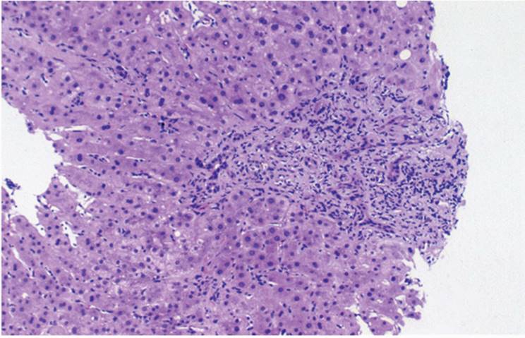
Not uncommonly, a liver biopsy may be obtained as part of a cholecystectomy, either open or laparoscopic (6) or, more recently, during a gastroplasty operation for morbid obesity. Most often biopsies show only features suggestive of a mild, chronic, nonspecific inflammation (hepatitis), including minimal or mild mononuclear inflammatory cell infiltrate in the portal tracts, sometimes even with mild portal fibrosis (Fig. 7.1). Occasionally, rare foci of parenchymal necrosis and a few scattered large fat droplets in liver cells are seen (10). In the morbidly obese, steatosis can be marked (e-Fig. 7.1), with changes of nonalcoholic fatty disease (NAFLD) or nonalcoholic steatohepatitis (NASH) (see Chapter 21). Biopsy obtained during surgical procedures can also show changes of primary liver disease that may not have been clinically recognized prior to surgery.
“Surgical Hepatitis”
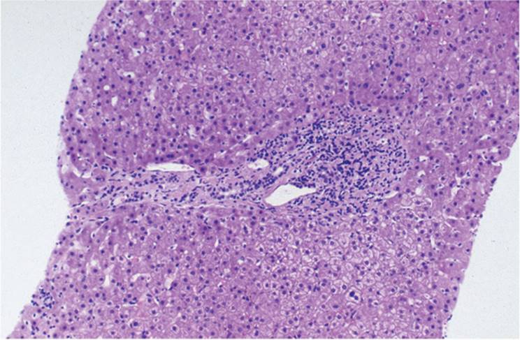
Biopsy obtained at the end of an abdominal surgical procedure, either as a core needle biopsy or, more often, as a wedge biopsy, can have clusters of polymorphonuclear leukocytes irregularly distributed in sinusoids (Fig. 7.2, e-Fig. 7.2) (1). These clusters, so-called surgical hepatitis, can be seen in any area but are most often seen in zone 3, near the terminal hepatic venule (central vein), or immediately beneath the liver capsule. It is thought that surgical hepatitis is caused by foci of anoxia within the liver or possibly by mechanical injury, perhaps from surgical retractors. Occasional true liver cell necrosis with rare acidophilic body formation can also be seen, but there is no true hepatitis.
|
TABLE 7.1 Nonspecific Histopathologic Changes Sometimes Seen in Liver Biopsy |
||||||||||||||||||||||||||
|
||||||||||||||||||||||||||
|
|
||||||||||||||||||||||||||
|
FIGURE 7.1 Photomicrograph of a liver biopsy obtained at the time of laparoscopic cholecystectomy. Mild nonspecific portal chronic inflammation and mild fibrosis are present. Occasional large fat droplets are present in the parenchyma (hematoxylin-eosin, original magnification ×100). |
|
|
|
FIGURE 7.2 “Surgical hepatitis.” Clusters of polymorphonuclear leukocytes are present in zone 3 of the acinus (hematoxylin-eosin, original magnification ×200). |
Nonspecific Reactive Change (Nonspecific Reactive Hepatitis)
Hepatic inflammation may be prominent with various extrahepatic, systemic, and febrile conditions (7). A mild portal tract inflammatory cell infiltrate, usually lymphocytes, without interface hepatitis (“piecemeal necrosis”) is seen (Fig. 7.3, e-Fig. 7.3) (10). Necrotic hepatocytes (acidophilic bodies) can uncommonly be seen, and Kupffer cells are prominent. The term “reactive change” is preferable to “reactive hepatitis” so the erroneous diagnosis “chronic hepatitis” is not applied. Distinguishing between this nonspecific, reactive change and residual, resolving acute viral hepatitis or mild, smoldering chronic hepatitis is clearly important, and clinical correlation is imperative. Some cases of nonspecific reactive change follow prior hepatitis, either viral or drug, or another etiologic factor.
|
|
|
FIGURE 7.3 Nonspecific reactive change of the liver, showing mild portal tract chronic inflammatory cell infiltration, with a few lymphocytes in sinusoids (hematoxylin-eosin, original magnification ×100). |
Febrile Illnesses
In protracted febrile illnesses, including fever of unknown origin, increased numbers of polymorphonuclear leukocytes circulate through the liver, prominently in sinusoids and, to a lesser degree, portal tracts. Zone 3 (centrilobular) sinusoidal dilatation is sometimes seen in the absence of other significant histopathologic findings.
Kupffer Cell Reactivity
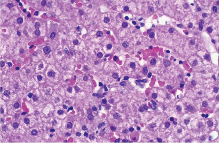
Kupffer cells are often indistinguishable from sinusoidal endothelial cells (see Chapter 4). Kupffer cells react to various stimuli, both intrahepatic and systemic, and are seen as prominent sinusoidal lining cells (Fig. 7.4, e-Figs. 7.4, 7.5). This feature alone is entirely nonspecific and must be evaluated in the context of more specific findings. For example, Kupffer cells may be prominent in primary hepatic diseases (e.g., primary biliary cirrhosis). Kupffer cells often show ceroidlike material representing degraded cell membrane material, which can be highlighted with periodic acid-Schiff (PAS) after diastase digestion (PAS/D) as well as with Victoria blue, which likely reflects binding of Victoria blue to disulfide bonds of intracellular degradation products. Indeed, PAS/D to demonstrate phagocytosed material in enlarged Kupffer cells can help to document a recent hepatitis when there is no longer significant lobular or parenchymal inflammation.
|
|
|
FIGURE 7.4 Prominent Kupffer cells in an otherwise normal liver (hematoxylin-eosin, original magnification ×400). |
|
|
|
FIGURE 7.5 Kupffer cell pseudogranuloma (microgranuloma) (hematoxylin-eosin, original magnification ×400). |
Kupffer Cell Pseudogranuloma
Sometimes Kupffer cells are distributed in small clusters and may have the appearance of microgranulomas, particularly concentrated around terminal hepatic venules (zone 3) (Fig. 7.5). They also often contain lipofuscin and ceroid pigments.
Extramedullary Hematopoiesis
Extramedullary hematopoiesis (EMH) as a nonspecific reactive phenomenon occurs more often in children but can be seen at any age. EMH can be seen in various primary liver tumors, including benign tumors such as adenoma and hepatocellular carcinoma and hepatoblastoma. EMH is entirely nonspecific and its significance unclear in the absence of an underlying hematologic disorder. EMH can follow transplantation, particularly combined liver-kidney transplantation, without any correlating hematologic factors (e.g., preoperative or postoperative hemoglobin values), rejection, or type of immunosuppression (Fig. 7.6) (2,9).
Circulating Megakaryocytes
The identification of isolated megakaryocytes in hepatic sinusoids is not necessarily EMH and does not indicate their origin in the liver. Rather, it may reflect the release, under conditions of stress, of megakaryocytes from the bone marrow into the peripheral blood and, consequently, throughout the circulatory system. Isolated circulating megakaryocytes in sinusoids are not infrequently seen in the early posttransplantation period (Fig. 7.7, e-Fig. 7.6) (8).
|
|
|
FIGURE 7.6 Extramedullary hematopoiesis in a patient following liver transplantation (hematoxylin-eosin, original magnification ×200). |
Granulomas
Noncaseating and nonnecrotizing epithelioid granulomas and microgranulomas are sometimes seen without evidence of other infectious or noninfectious granulomatous diseases (see Chapter 12) (3,8,11).
|
|
|
FIGURE 7.7 Sinusoidal megakaryocytes in a patient following liver transplantation (hematoxylin-eosin, original magnification ×400). |
|
|
|
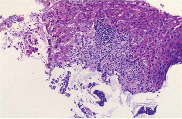
FIGURE 7.8 Vicinity of mass lesion. Focal sinusoidal dilatation and portal tract infiltration, including polymorphonuclear leukocytes, with mild portal tract edema (hematoxylin-eosin, original magnification ×200). |
Steatosis
Mild, often focal, macrovesicular steatosis is seen in some liver biopsies without obesity, diabetes mellitus, alcohol or drug toxicity, or other known specific association (10).
Vicinity of Mass Lesion
Changes that occur adjacent to liver masses, both neoplastic and nonneoplastic, include focal sinusoidal dilatation and variable degrees of bile ductular reaction (proliferation) with cholestasis accompanied by variable numbers of polymorphonuclear leukocytes (Fig. 7.8, e-Figs. 7.7, 7.8). Portal tracts can sometimes be edematous. These changes mimic large bile duct obstruction (5).
Bile Duct Epithelial Injury
Although the finding of bile duct epithelial injury is an important diagnostic feature of several hepatic disorders, including cholangiopathies (e.g., primary biliary cirrhosis, primary sclerosing cholangitis), acute allograft rejection, and graft versus host disease, bile duct injury, including frank necrosis, also occurs without a specific, identifiable etiologic factor. Mild and focal biliary epithelial irregularity, overlapping nuclei, and cytoplasmic vacuolization are seen. It is important, however, that these do not progress and do not cause bile duct destruction and loss. Mild bile duct epithelial injury is common in hepatitis C virus infection (see Chapters 8 and 9), and chronic hepatitis should always be considered. Bile duct injury also follows exposure to some drugs and toxins. Variable degrees of bile duct damage are common, both in simian monkeys with experimentally induced immune deficiency disease and in humans with acquired immune deficiency syndrome (4,6). The mechanism of injury remains obscure, although at least some degree of immunologically mediated response has been implicated.
|
|
|
FIGURE 7.9 Lipofuscin pigment in zone 3 and zone 2 hepatocytes (hematoxylin-eosin, original magnification ×400). |
Pigments
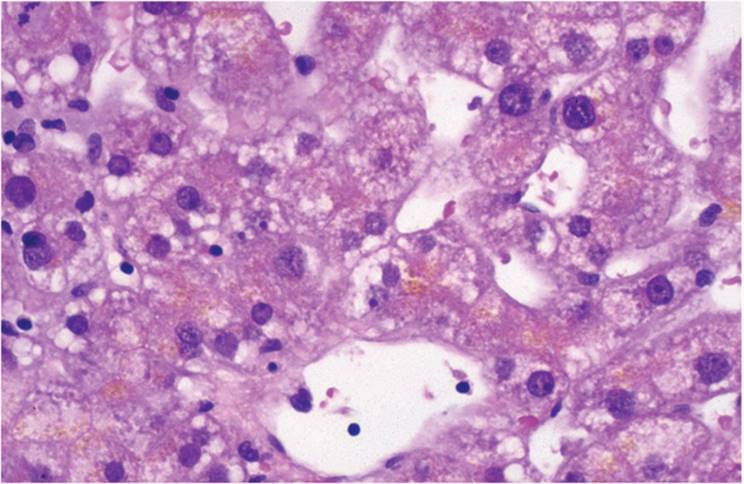
LIPOCHROME. Various pigments are seen in liver biopsy, the most common of which is lipochrome (lipofuscin). This “wear and tear” pigment is first in zone 3 hepatocytes adjacent to the terminal hepatic venule but with increasing age progresses from zone 3 to involve all hepatocytes (Fig. 7.9, e-Figs. 7.9, 7.10). It can also be seen in Kupffer cells. Individuals with chronic illnesses demonstrate lipofuscin pigment in excess of that expected for their age.
Hemosiderin
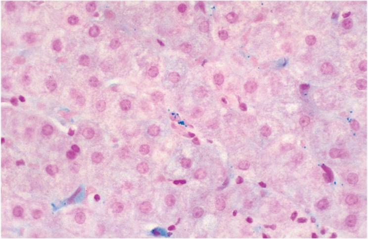
Hemosiderin is sometimes seen in Kupffer cells and sinusoidal endothelial cells with no clear explanation for its presence and in the absence of disorders usually associated with liver iron deposition (Fig. 7.10) (see Chapter 15). Patients who imbibe alcoholic beverages to excess have sinusoidal endothelial cells with hemosiderin. Hepatocytes may or may not show iron deposition in these cases. Bile duct epithelial cells can also show mild iron deposition, and the diagnosis of hemochromatosis should be considered even though the amount of hemosiderin is much less than in genetic hemochromatosis.
|
|
|
FIGURE 7.10 Hemosiderin granules are in sinusoidal cells (Perls stain, original magnification ×400). |
REFERENCES
1. Christoffersen P, Poulsen H, Skei E. Focal liver cell necrosis accompanied by infiltration of granulocytes arising during operation. Acta Hepatosplenol 1970;17:240-245.
2. Collins RH, Anastasi J, Terstappen LWMM, et al. Donor-derived long-term multilineage hematopoiesis in a liver-transplant recipient. N Engl J Med 1983;328:762-765.
3. Drebber U, Kasper HU, Ratering J, et al. Hepatic granulomas: histological and molecular pathological approach to differential diagnosis - a study of 442 cases. Liver Int 2008 Feb 26 [Epub ahead of print].
4. Gerber MA, Chen ML, Hu FS, et al. Liver disease in rhesus monkeys infected with simian immunodeficiency virus. Am J Pathol 1991;139:1081-1088.
5. Gerber MA, Thung SN, Bodenheimer HC Jr, et al. Characteristic histological triad in liver adjacent to metastatic neoplasm. Liver 1986;6:85-88.
6. Michel S, Lipsky R, Morgenstern L. “Routine” liver biopsy in upper abdominal surgery. Arch Surg 1977;112:959-961.
7. Popper H, Schaffner F. Liver: Structure and Function. New York: McGraw-Hill, 1957.
8. Sartin JS, Walker RC. Granulomatous hepatitis: a retrospective review of 88 cases at the Mayo Clinic. Mayo Clin Proc 1991;66:914-918.
9. Schlitt JH, Schafers S, Deiwick A, et al. Extramedullary erythropoiesis in human liver grafts. Hepatology 1995;21:689-697.
10. Tran TT, Changsri C, Shackleton CR, et al. Living donor liver transplantation: histological abnormalities found on liver biopsies of apparently healthy potential donors. J Gastroenterol Hepatol 2006; 21:381-383.
11. Zoutman DE, Ralph ED, Frei JV. Granulomatous hepatitis and fever of unknown origin. An 11-year experience of 23 cases with three years' follow-up. J Clin Gastroenterol 1991; 13:69-75.