Acute viral hepatitis (AVH) is the term commonly applied for the hepatitides caused by hepatotropic viruses: A (HAV), B (HBV), C (HCV), D (delta), E (HEV), and F (HFV) (Table 8.1) (6,7,8,69). The etiologic factors in some cases of community-acquired and posttransfusion hepatitis remains unclear. Hepatitis G virus (HGV), a flavivirus, may be the basis of at least some of those cases (4,7,35,38). Nonhepatotropic viruses can also cause clinical and histologic manifestations of acute hepatitis, particularly in the immunocompromised patient, including cytomegalovirus (CMV), Epstein-Barr virus (EBV), adenovirus (in children), and herpes simplex virus (HSV) (Table 8.2) (12,22,27,31,41,45,47,57,66). Both the clinical and the histologic pictures of viral hepatitis can be produced by various drugs and toxic agents (81). The list of those products is long and ever-expanding (discussed in more detail in Chapter 11). As one example of the nonspecificity of the histologic picture, drug toxicity can appear to be AVH with subsequent evolution to autoimmune hepatitis (2).
Patients usually present with icterus, although anicteric forms are common. Historically, liver biopsy findings were crucial in determining further management of a jaundiced patient. Advanced imaging methods and sophisticated laboratory tests, including serologic and molecular tests for almost all of the viruses that can cause hepatitis, generally allow for the establishment of the correct diagnosis in liver biopsy.
ACUTE HEPATITIS WITH FOCAL NECROSIS
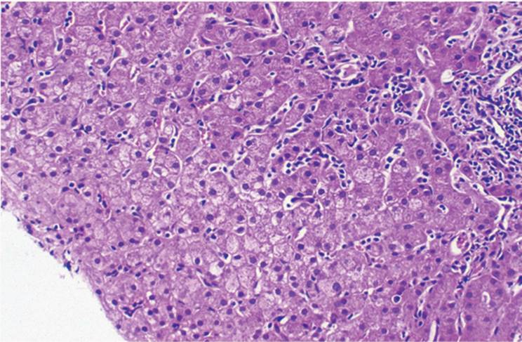
The parenchymal histopathologic changes seen in AVH are distinctive but not entirely specific. Portal tracts show varying degrees of chronic inflammatory cell infiltrate, but the dominant features are in the lobule/acinus. At low magnification, the biopsy is distinctive (Fig. 8.1). Typically the liver has a disordered or “dirty” appearance (Fig. 8.1, e-Figs. 8.1-8.6). In fully developed acute hepatitis, the changes are predominantly in zone 3 of the acinus (centrilobular, perivenular) and, to a lesser extent, in zone 1 (periportal).
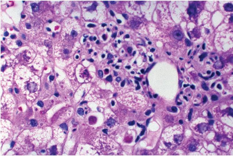
Zone 3 hepatocytes show ballooning degeneration apoptotic cell necrosis and acidophilic (Councilman-like) body formation (Fig. 8.2).
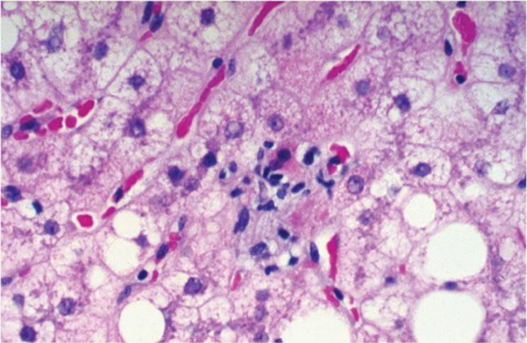
Ballooned hepatocytes have distended, pale eosinophilic, and finely granular cytoplasm. Nuclei may appear hyperchromatic and pleomorphic. Acidophilic bodies result from cell death either from cell injury or apoptosis, genetically programmed cell death at least partly controlled by the bcl-2 oncogene. Acidophilic bodies can be numerous and are recognized as refractile, deeply eosinophilic bodies, sometimes still with remnants of nuclear chromatin. Acidophilic bodies are often surrounded by mostly T lymphocytes, implicating a role for immunologically mediated cell injury and death (Fig. 8.3) (78). So-called naked acidophilic bodies may also be seen (Fig. 8.4). After cell death, there are transiently seen areas of dropout in which the liver plate structure is temporarily maintained despite the loss of one or more hepatocytes, best demonstrated with reticulin stain. The debris from dead cells is phagocytosed by Kupffer cells, which, in acute viral hepatitis, are often enlarged and contain periodic acid-Schiff (PAS)-positive material, best seen after diastase digestion (PAS/D) to remove glycogen (e-Fig. 8.7). The destruction and loss of many liver cells leads to extensive liver plate disarray and subsequent confluent, bridging, or submassive and massive necrosis.
|
TABLE 8.1 The Hepatotropic Viruses |
||||||||||||||||||||||||||||||||
|
||||||||||||||||||||||||||||||||
|
TABLE 8.2 Nonhepatotropic Viruses That Can Affect the Liver |
||||||||||||||||||||||||||||||||
|
|
|
|
FIGURE 8.1 Low-magnification photomicrograph of acute viral hepatitis, with extensive lobular inflammation and liver cell necrosis with liver cell disarray (hematoxylin-eosin, original magnification ×100). |
|
|
|
FIGURE 8.2 Acute viral hepatitis with moderately severe lobular inflammation and liver cell necrosis with numerous acidophilic (Councilman-like) bodies (hematoxylin-eosin, original magnification ×200). |
|
|
|
FIGURE 8.3 Acidophilic bodies surrounded by lymphocytes (hematoxylin-eosin, original magnification ×200). |
|
|
|
FIGURE 8.4 So-called naked acidophilic bodies, without surrounding lymphocytic infiltrate (hematoxylin-eosin, original magnification ×200). |
|
|
|
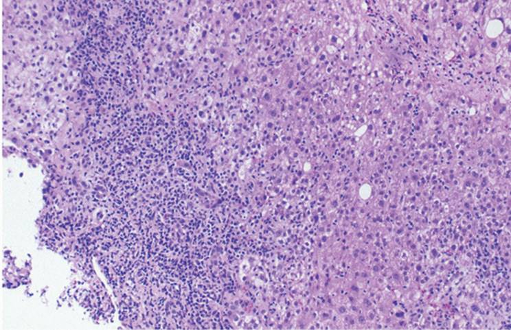
FIGURE 8.5 Acute viral hepatitis with necrosis that involves multiple acini (confluent necrosis) (hematoxylin-eosin, original magnification ×200). |
“AVH with confluent necrosis” is the term used when focal necroses become numerous, coalesce, and cause cell loss in large areas of liver parenchyma (Fig. 8.5). Confluent necrosis can involve multiple acini and take the form of acute hepatitis with multiacinar (panacinar, submassive, massive) necrosis (Fig. 8.6, e-Figs. 8.8-8.10).
Bridging necrosis typically involves all three zones of the acinus, forming necroinflammatory bridges between portal and perivenular areas (centroportal). However, bridging necrosis can also exist between portal tracts (portoportal). Although morphologically similar, the pathogenetic mechanisms of these two types of bridging necrosis seem to be different. Furthermore, because of the vascular shunting between the two vascular systems in centroportal bridging, this type of necrosis has more significant negative prognostic value in both acute and chronic hepatitis. AVH with bridging necrosis has a generally worse prognosis and is associated with high risk for progression to chronic hepatitis and ultimately cirrhosis. Patients with extensive bridging necrosis can die weeks to months after the onset of hepatitis with the clinical picture of subfulminant liver failure (e-Figs. 8.11-8.13). Complete recovery following acute hepatitis with bridging necrosis is not unusual, however.
|
|
|
FIGURE 8.6 Acute viral hepatitis with bridging necrosis (hematoxylin-eosin, original magnification ×100). |
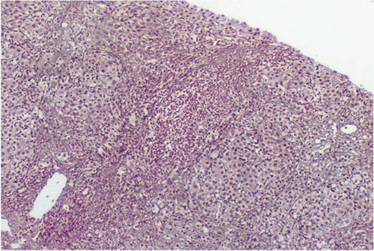
When large numbers of adjacent liver cells undergo necrosis, the underlying reticulin network undergoes collapse (Fig. 8.7). Collapse, with hematoxylin-eosin, is indistinguishable from newly formed fibrous septa but is appreciated with reticulin stain as compressed fibers (64). True septa include elastic fibers, demonstrable with elastic fiber stains as well as with Victoria blue or orcein. There are no elastic fibers in recently developed collapse (64).
In addition to degenerative changes and acidophilic body formation, AVH has variable degrees of lobular (parenchymal) inflammation with mature T lymphocytes, with some histiocytes and occasional plasma cells (Fig. 8.8) (78,79). The infiltrate in patients with acute hepatitis A may be particularly rich in plasma cells (1). Kupffer cells are also reactive and prominent in acute hepatitis, whether viral or drug induced.
|
|
|
FIGURE 8.7 Acute viral hepatitis with bridging necrosis and collapse of the underlying reticulin network (reticulin silver, original magnification ×100). |
|
|
|
FIGURE 8.8 Acute viral hepatitis with lobular inflammation. The inflammatory cell infiltrate is predominantly lymphocytic with some histiocytes and occasional plasma cells (hematoxylineosin, original magnification ×200). |
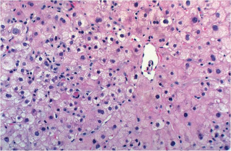
Multinuclear giant cells can be seen (Fig. 8.9). This occurs often in children (giant cell hepatitis), in whom it may be caused by various infectious and noninfectious factors, but may also be seen in AVH in adults, including hepatitis C (16,34). A clinically severe hepatitis with parenchymal giant cells can have paramyxoviruslike particles demonstrable with electron microscopy (59). Acute forms of autoimmune hepatitis may also show significant numbers of syncytial cells.
|
|
|
FIGURE 8.9 Acute hepatitis with giant cell formation (hematoxylin-eosin, original magnification ×400). |
|
|
|
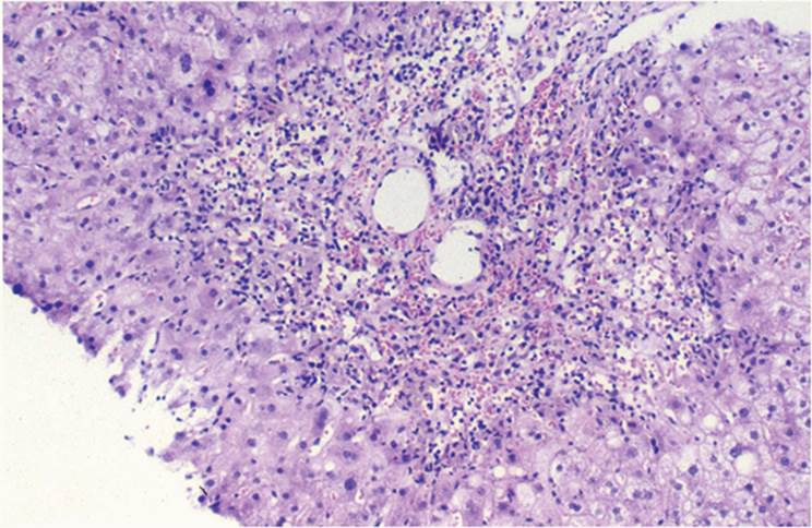
FIGURE 8.10 Acute hepatitis with relatively mixed inflammatory cell infiltrate, composed of mononuclear cells, but also including eosinophils and polymorphonuclear leukocytes (hematoxylin-eosin, original magnification ×200). |
Portal tracts in AVH are generally mildly expanded, with an inflammatory infiltrate of lymphocytes, histiocytes, some plasma cells, and, rarely, few polymorphonuclear leukocytes and eosinophils (Fig. 8.10). If polymorphonuclear leukocytes and/or eosinophils predominate, etiologic factors for hepatitis other than viral should be considered, especially drugs or toxins. Interface hepatitis (“piecemeal necrosis”) is characterized by the spilling of inflammatory cells from the portal tract into the limiting plate hepatocytes and is seen in various forms of acute viral hepatitis. Interface hepatitis has been thought to be associated with a greater potential for progressive disease and ultimate development of chronic hepatitis, but it is not as important in this regard as confluent necrosis.
The severity and progression of viral hepatitis is related more to cause than to the inflammatory pattern. Both AVH A and E can have severe liver inflammation, including interface hepatitis, but neither of these progresses to chronicity.
Mild cholestasis, generally confined to canaliculi, is seen in some forms of acute hepatitis (Fig. 8.11). Sometimes true cholestatic variants of hepatitis occur, however, and there may be clinically prolonged cholestasis.
Classically, three rather distinct stages of AVH are recognized: (a) early, (b) fully developed (weeks, rarely months), and (c) late, residual, or resolving hepatitis. In general, biopsy is now performed only in the late stage of acute viral hepatitis when there is concern that a relatively prolonged clinical course and/or persistence of biochemical abnormalities might be caused by another disorder.
|
|
|
FIGURE 8.11 Acute hepatitis with cholestatic component. Cholestasis is predominantly in zone 3 of the acinus and is intracanalicular (hematoxylin-eosin, original magnification ×100). |
Late, the usual parenchymal changes subside, with only rare focal necroses and/or clusters of PAS/D-positive macrophages indicating phagocytosis of cellular debris after necrosis (Fig. 8.12), most prominent in zone 3, where necrosis is most active. These macrophages contain pale brown, lipid-rich ceroid that can be stained with Perls method, resembling hemosiderin but generally paler. These mild, nonspecific changes can persist for months and may resolve without sequela.
|
|
|
FIGURE 8.12 Resolving acute hepatitis with numerous macrophages containing a mixture of ceroid and hemosiderin pigment (periodic acid-Schiff reaction, after diastase digestion, original magnification ×200). |
|
|
|
FIGURE 8.13 Hepatitis B surface antigen-containing hepatocytes (“ground-glass” cells) (hematoxylin-eosin, original magnification ×400). |
Complete resolution is likely in most AVH cases. Fewer than 1% of hepatitis A, B, or C patients have a fulminant course and, without liver transplantation, may die.
AVH B, C, and D can all progress to chronicity, particularly HCV. HBV patients can also become carriers, who generally are without significant clinical manifestations but harbor the virus. The liver may appear to be quiescent, without inflammation but with abundant hepatitis B surface antigen HBsAg-containing hepatocytes, “ground-glass” cells, recognizable with hematoxylin-eosin, and highlighted with Victoria blue, orcein, or anti-HBsAg immunostain (Fig. 8.13).
Differential diagnosis of AHV includes the various hepatotropic and other viruses that can cause hepatitis, acute autoimmune hepatitis, and drug-induced hepatitis.
Type A Hepatitis
HAV is a linear, single-stranded RNA hepatotropic virus, transmitted by fecal-oral contamination. Immunoglobulin M (IgM)-type anti-HAV antibody in the serum reflects recent infection. Viral genomic material (HAV RNA) may be demonstrated in the tissue with in situ hybridization (71). Serum IgG-type anti-HAV antibodies indicate previous exposure. There are no carrier or chronic forms of HAV infection, although relapsing hepatitis has been described.
Hepatitis A is usually mild, especially in younger people. In older patients, however, the course may be more severe and fulminant and can lead to death. At one time, as many as 85% of all people in the United States demonstrated IgG antibodies against HAV. The number of seropositive individuals has dramatically declined. Travelers from Western countries to endemic areas may have high susceptibility (Fig. 8.14) (1,3,30). Two distinct histologic patterns have been recognized, although overlapping features are commonly seen.
|
|
|
FIGURE 8.14 Acute viral hepatitis A, showing characteristic zone 3 (centrilobular) parenchymal necrosis (hematoxylin-eosin, original magnification ×400). |
The periportal variant of hepatitis A has portal and periportal inflammation (interface hepatitis). The inflammatory infiltrate is often plasma cell-rich, and differentiation from autoimmune hepatitis is difficult without data. Generally, mild zone 3 (perivenular) cholestasis is seen.
In the cholestatic variant, zone 3 cholestasis predominates with little inflammation, often misinterpreted as drug or toxic cholestasis caused by drug or toxic injury (e-Figs. 8.14-8.19) (1,65,72).
Microvesicular steatosis and fibrin ring granulomas, generally associated with Q fever, have also been described in association with HAV infection (55). In fulminant cases, the pattern is that of submassive and massive necrosis.
Type B Hepatitis
With HBV, a hepadna virus, transmission is parenteral, sexual, and vertical from infected mothers to newborns. Vertical transmission is particularly high in endemic areas.
The infectious virion, the Dane particle, is a 42-nm structure, contains a 27-nm core with circular, incompletely double-stranded DNA surrounded by an envelope of viral surface material. The complete virion is assembled in the endoplasmic reticulum of liver cells. Other organs and tissues in the body, including lymphocytes, also contain viral DNA material.
Histologically, acute hepatitis B is not significantly different from the other forms of AVH (1). There may be acute hepatitis with focal, confluent, or submassive and massive necrosis. HBsAg-containing (ground-glass) cells are usually not seen in acute forms of hepatitis B. Both HBsAg and hepatitis B core antigen (HBcAg) can be detected in the tissue in chronic forms of hepatitis B by applying immunohistochemical methods (Fig. 8.15A,B). HBcAg is mostly intranuclear, except when the concentration of virus is very high, in which case cytoplasmic staining will also be apparent (44). HBsAg, in the cytoplasm, is also demonstrated with histochemical methods, using Victoria blue (Fig. 8.15C), orcein (Shikata), or aldehyde fuchsin (Gomori) methods.
|
|
|
FIGURE 8.15 Chronic hepatitis B virus infection. A. Intracytoplasmic hepatitis B surface antigen (HBsAg) (immunoperoxidase with anti-HBsAg, original magnification ×200). B. Intra - nuclear distribution of hepatitis B core antigen (HBcAg) (immunoperoxidase with anti-HBcAg, original magnification ×200). |
|
|
|
FIGURE 8.15 (Continued) C. Intracytoplasmic hepatitis B surface antigen (Victoria blue, original magnification ×400). |
The chronic hepatitis B patient with acute exacerbation may have (a) superinfection with other viruses including delta, A, and C; (b) HBV reactivation; (c) superimposed drug-induced hepatitis; and (d), in patients being treated, interferon withdrawal or discontinuance.
The mechanism of cell injury is not entirely clear. The injury is immunologically mediated, including cytotoxic T cells, and natural killer (NK) cells. Although HBV does not have direct cytotoxic effect in the immunocompetent setting, recurrent hepatitis B after liver transplantation may reflect direct viral-induced cytotoxicity (19,84).
Type D (Delta) Hepatitis (HDV)
Infection with hepatitis D virus (HDV) occurs either as coinfection or as superinfection in a patient with existing HBV infection (9,11,62). In most cases the course is more severe and often fulminant. In some liver transplantation patients, HDV develops without HBV, suggesting that HDV can sometimes replicate in the absence of HBV, is not directly cytopathic, and does not necessarily require HBV to cause liver cell injury (14,36,51,60).
The viral antigen HDAg can be immunohistochemically demonstrated in the patients with superinfection or coinfection with HDV. Its expression is intranuclear, similar to that of HBcAg (33). The presence of viral RNA can be confirmed with in situ hybridization (40).
Severe acute delta virus hepatitis has been described in Venezuelan Indians (56). Significant portal inflammation is always seen, along with extensive microvesicular steatosis or spongiocytic change, in addition to focal lobular necrosis with acidophilic body formation (10,80). The liver cell nuclei containing HDAg are homogeneous and finely granular (“sanded”) (Fig. 8.16), resembling the ground-glass cell of hepatitis B (43).
|
|
|
FIGURE 8.16 So-called sanded nuclei in hepatitis D virus hepatitis (hematoxylin-eosin, original magnification ×400). |
Type C Hepatitis
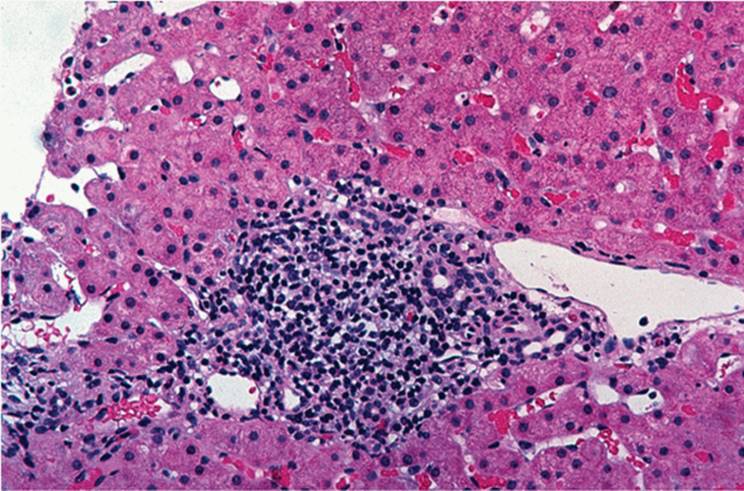
Acute forms of hepatitis C infection in the immunocompetent setting resemble other forms of virally induced acute hepatitis but are only rarely biopsied. The role of HCV in fulminant hepatitis is somewhat controversial (82,87). These patients have (a) portal tract enlargement and inflammation, often with lymphoid aggregates or true lymphoid follicles (Fig. 8.17); (b) generally mild lobular, sinusoidal inflammation, mostly lymphocytes, with only focal hepatocyte necrosis and spotty acidophilic body formation (Fig. 8.18); and (c) variable predominantly macrovesicular steatosis (Fig. 8.19) (8,18). There may also be (d) mild bile duct injury (Fig. 8.20), including vacuolation of the cytoplasm, overlapping nuclei, and nuclear pleomorphism, (e) prominent Kupffer cells, and (f) cholestasis. Despite this, HCV-associated antigens cannot be reliably demonstrated in tissue (23,30) and in situ hybridization methods do not show viral genome in bile duct epithelium or in liver cells. Reverse transcriptase polymerase chain reaction in situ has been used in a few selected cases to show viral genomic material in Kupffer cells, as well as in hepatocyte cytoplasm where the virus is localized to the area at the interface between the nucleus and cytosol (29,51,53). The most reliable method for detection of HCV in serum and in formalin-fixed, paraffin-embedded tissues, however, is the standard polymerase chain reaction (29,54,88).
|
|
|
FIGURE 8.17 Hepatitis C virus infection with typical lymphoid aggregate formation in portal tracts (hematoxylin-eosin, original magnification ×200). |
|
|
|
FIGURE 8.18 Lobular changes in hepatitis C virus infection with both lobular inflammation and focal liver cell necrosis, including acidophilic body formation (hematoxylin-eosin, original magnification ×200). |
|
|
|
FIGURE 8.19 Macrovesicular steatosis in hepatitis C virus infection (hematoxylin-eosin, original magnification ×400). |
|
|
|
FIGURE 8.20 Mild portal inflammation with mild bile duct injury (hematoxylin-eosin, original magnification ×200). |
In patients with recurrent HCV infection following liver transplantation, early histopathologic features of hepatitis may be very subtle. Recurrent forms of hepatitis in the posttransplantation setting are discussed in Chapter 25.
Type E Hepatitis
HEV infection is common in Asia, Africa, and Latin America, and only occasionally is seen in Western countries. Transmission is fecal-oral, similar to HAV (6,15,26,58,69,74,87). The RNA virus is nonenveloped single-stranded, 27 to 34 nm in diameter. Viral particles may be present in feces, bile ducts, and sinusoidal cells of patients with severe infection (8,70).
Histopathologic features of material obtained during several epidemics of HEV include portal and periportal inflammation and variable degrees of intracanalicular cholestasis, which can mimic large duct obstruction with liver cell rosette formation and extensive bile ductular proliferation (8,15).
Type F Hepatitis
Some patients with fulminant hepatitis have intranuclear 60- to 70-nm togalike virus particles seen ultrastructurally (3). After liver transplantation, some patients develop acute liver failure with similar viral particles in the graft in even greater abundance than in the native liver (18). The explanted livers showed extensive necrosis, collapse, cholestasis, and significant inflammatory infiltrate composed of lymphocytes and numerous plasma cells (18).
Hepatitis G Virus
HGV has been found in transfused patients who developed viral hepatitis, when no other agent was demonstrable. HGV, an RNA flavilike virus (4,7), is structurally closely related to HCV, with approximately 30% homology (29,35,38,67).
Most patients in whom HGV is demonstrable have an associated hepatitis. However, in some there is no elevation of transaminase values, and they could be healthy carriers or in a quiescent stage. The role of HGV infection in fulminant hepatitis and carcinogenesis is unclear (63,85) (e-Fig. 8.28). Some patients are also coinfected with HBV and HCV. In 6% to 10% of HGV patients other parenterally transmitted hepatotropic viruses can be identified, perhaps reflecting common risk factors such as intravenous drug use and multiple blood transfusions (37). Histopathologic features characteristic for HGV infection in humans have not been well delineated.
TT Virus Hepatitis
TT virus (TTV) is a recently discovered, nonenveloped, single-stranded DNA virus (48), discovered in the serum of patients with transfusiontransmitted hepatitis. TTV has been implicated in at least some cases of fulminant liver failure, cryptogenic chronic hepatitis, and cryptogenic cirrhosis. The highest carrier rate has been found in Japan, but the virus is found throughout the world. TTV is most frequently transmitted by blood transfusion, but other means of transmission exist. TTV DNA can be found in the bile and stool (50).
Herpes Simplex Virus
Hepatic HSV infection is almost exclusively seen in immunocompromised patients, including malnourished children, posttransplantation patients, and patients treated with corticosteroids and other forms of immunosuppressive medication (13,22,42,45). HSV hepatitis also occurs during pregnancy. In disseminated forms, transaminase and bilirubin values are markedly elevated, and there may be associated disseminated intravascular coagulopathy (DIC).
The liver has patchy coagulative necrosis, with no particular zonal distribution. Necrosis can be extensive with massive hepatic necrosis. Inflammation is usually not prominent (57,68).
Two types of viral intranuclear inclusions have been recognized in association with HSV infection, usually in hepatocytes at the periphery of the necrotic area: eosinophilic Cowdry type A and basophilic Cowdry type B. The intranuclear inclusions may be highlighted with Feulgen stain. A specific immunostain is available.
Cytomegalovirus
CMV hepatitis occurs mostly after renal and liver transplantation (53,73). Biopsy is often necessary to distinguish acute cellular rejection from CMV, since they can be clinically similar (53). Posttransplantation CMV hepatitis is discussed in Chapter 25.
In immunocompetent individuals, CMV produces an infectious mononucleosislike syndrome with mild hepatitis. Biopsy shows focal hepatocyte necrosis with sinusoidal lymphocytic infiltration. Mild bile duct injury is seen. Noncaseating epithelioid cell granulomas and ring granulomas of the type usually associated with Q fever occur (39). In contrast to posttransplant CMV, viral antigens are generally not demonstrable and viral inclusions are not seen.
CMV causes giant cell hepatitis in neonates with prominent cholestasis and inflammation, and easily demonstrable viral inclusions, mostly intranuclear, seen as amphophilic spherical masses surrounded by a clear halo. Intracytoplasmic inclusions, appearing as multiple, small, amphophilic, poorly defined inclusions, without halo, are also seen. There may be mild infiltration by polymorphonuclear leukocytes and lymphocytes around individual cells, but inflammation is generally not prominent (66). The intranuclear and intracytoplasmic inclusions represent aggregated virions, demonstrable immunohistochemically or with the electron microscope. Obliterative cholangitis with subsequent paucity of bile ducts has also been described (20).
Epstein-Barr Virus
EBV hepatitis occurs in immunocompetent and immunocompromised individuals, as well as after liver transplantation (e-Figs. 8.20-8.26). Posttransplantation EBV hepatitis is discussed in Chapter 25.
Biochemical hepatitis is relatively common in infectious mononucleosis, although clinical hepatitis is generally not seen. Jaundice is rare (41,80) (e-Fig. 8.27). Biopsy shows a diffuse sinusoidal lymphocytic infiltrate with varying degrees of portal inflammation. The lymphocytes are infected B cells, activated T lymphocytes, and NK cells. When inflammation is marked, apoptotic hepatocytes are seen focally (Fig. 8.21). When the infiltrate is prominent there can be many atypical lymphocytes, in parallel with the appearance of Downey cells in the peripheral blood, and the diagnosis of a malignant lymphoproliferative disorder can be erroneously made. Noncaseating epithelioid granulomas can be seen, and there may be varying degrees of macrovesicular steatosis (47).
|
|
|
FIGURE 8.21 Acute hepatitis caused by Epstein-Barr virus. Numerous lymphocytes are arranged in a linear pattern in the sinusoids (hematoxylin-eosin, original magnification ×200). |
Measles
Measles (rubeola) hepatitis is generally not a clinically important component in the patient infected with measles; particularly in adults, however, hepatitis can be prominent. Liver biopsy is virtually never obtained (3,10,21,43).
Rubella
Childhood rubella can be associated with a significant hepatitis, especially in neonates, in whom the histologic picture of giant cell hepatitis is seen. There may be massive necrosis, but generally involvement is milder with focal necrosis, cholestasis, and lymphocytic infiltration (86).
Varicella Zoster Virus
Hepatitis in association with varicella zoster is exceedingly rare (57,66).
Coxsackie Virus
Group B coxsackie virus usually causes a multisystem infection in neonates, leading to hemorrhagic necrosis of the liver. In adults, clinically demonstrable Coxsackie virus hepatitis is rare (23,24). There may be acinar zone 3 cholestasis, ballooning of hepatocytes, rare foci of hepatocyte necrosis, and mild infiltration of portal tracts and sinusoids by mononuclear cells and occasional polymorphonuclear leukocytes.
|
TABLE 8.3 Nonhepatotropic Viruses Causing Hemorrhagic Fevers and Involving the Liver |
||||||||||||
|
Other Viruses
Hepatitis has rarely been associated with adenovirus, echovirus, and parvovirus infections in both immunocompetent and immunocompromised children as well as in immunocompromised adults (5,12,27,31,32,50).
Exotic Hepatotropic Viruses
The exotic viruses are multisystem infections in which the liver serves as a primary target (Table 8.3). These infections tend to have similar clinical presentations and can be fatal. Biopsy findings include extensive coagulative necrosis and formation of acidophilic bodies. Although virtually never seen in Western society, they may rarely be encountered as more individuals travel to countries where these viruses are still active.
Lassa Fever
Lassa fever, caused by an RNA virus, manifests as a hemorrhagic fever in western and central Africa. It is transmitted by contact with rodent excrement (17). Patients present with fever, exudative pharyngitis, gastrointestinal symptoms, and coagulation disorders and develop hepatomegaly and right upper quadrant pain, with elevated transaminase, but normal bilirubin, values (39). Biopsy shows multiple foci of coagulative necrosis with numerous acidophilic bodies. Portal inflammatory infiltrates are relatively mild. Viral particles are seen with electron microscopy (17).
Yellow Fever
Yellow fever, a multisystem infection caused by an RNA arbovirus, is transmitted by the Aedes aegypti mosquito in parts of Africa, South America, and the Caribbean (76). Patients present with high fever, gastrointestinal hemorrhage, severe coagulopathy, and renal failure. Inflammatory cells are not prominent, although mononuclear cells may be seen as a mild portal infiltrate (Fig. 8.22, e-Figs. 8.29-8.32). The liver characteristically shows innumerable acidophilic (Councilman) bodies, mostly in zone 2 with extension to zone 3 (Fig. 8.22B). Nucleoli are enlarged, and there may be true nuclear inclusions (Torres bodies). There is mild microvesicular steatosis and Kupffer cell hyperplasia. An antibody for yellow fever viral antigen has been developed but is not commercially available.
|
|
|
FIGURE 8.22 A. Yellow fever showing many zone 2 and 3 acidophilic (Councilman) bodies with scanty inflammatory response (hematoxylin-eosin, original magnification ×100). B. High magnification of acidophilic (Councilman) bodies (original magnification ×400). |
Ebola Virus
Ebola virus disease has occurred in epidemic proportions in sub-Saharan Africa, particularly in Zaire, Sudan, and Uganda, and is associated with high mortality. Ebola virus has also been recovered from monkeys brought from the Philippines to the United States. The clinical course and biopsy findings are similar to that of Lassa fever (28).
Marburg Virus
Marburg virus disease was first reported in people in contact with African green monkeys and has been described in Uganda, Kenya, and South Africa. There is a 25% mortality rate. The clinical course and biopsy findings are similar to that of Lassa fever (28,59).
REFERENCES
1. Abe H, Beninger PR, Ikejiri N, et al. Light microscopic findings of liver biopsy specimens from patients with hepatitis type A and comparison with type B. Gastroenterology 1982;82:938-947.
2. Abraham C, Hart J, Locke SM, et al. A case of indomethacin-induced acute hepatitis developing into chronic autoimmune hepatitis. Nat Clin Pract Gastroenterol Hepatol 2008;5(3):172-176. [Epub 2008 Jan 29.]
3. Ackerman Z, Flugelman MY, Wax Y, et al. Hepatitis during measles in young adults: possible role of antipyretic drugs. Hepatology 1989;10:203-206.
4. Aikawa T, Sugai Y, Okamoto H. Hepatitis G infection in drug abusers with chronic hepatitis C. N Engl J Med 1996;334:195-196.
5. Anand A, Gray ES, Brown T, et al. Human parvovirus infection in pregnancy and hydrops fetalis. N Engl J Med 1987;316:183-186.
6. Anon. The A to F of viral hepatitis. Lancet 1980;336:1158-1160.
7. Anon. Hepatitis G? Lancet 1991;337:1070.
8. Asher LVS, Innis BL, Shrestha MP, et al. Virus-like particles in the liver of a patient with fulminant hepatitis and antibody to hepatitis E virus. J Med Virol 1990;31: 229-233.
9. Bonino F, Brunetto MR, Negro F, et al. Hepatitis delta virus, a model of liver cell pathology. J Hepatol 1991;13:260-266.
10. Britfeld V, Hasida Y, Sherman FE, et al. Fatal measles infection in children with leukemia. Lab Invest 1973;28:279-291.
11. Caredda F, Rossi E, d'Armino Monforte A, et al. Hepatitis B virus-associated coinfection and superinfection with D agent: indistinguishable disease with different outcome. J Infect Dis 1985;151:925-928.
12. Carmichael GP, Zahradnik JM, Moyer GH, et al. Adenovirus hepatitis in an immunosuppressed adult patient. Am J Clin Pathol 1979;71:352-355.
13. Chase RA, Pottage JC Jr, Haber MH, et al. Herpes simplex viral hepatitis: two case reports and review of the literature. Rev Infect Dis 1987;9:329-333.
14. Cole SM, Gowans EJ, Macnaughton TB, et al. Direct evidence for cytotoxicity associated with expression of hepatitis delta virus antigen. Hepatology 1991;13:845-851.
15. De Cock KM, Bradley DW, Sanford NL, et al. Epidemic non-A, non-B hepatitis in patients from Pakistan. Ann Intern Med 1987;106:227-230.
16. Devaney K, Goodman ZD, Ishak KG. Postinfantile giant-cell transformation in hepatitis. Hepatology 1992;16:327-333.
17. Edington GM, White HA. The pathology of Lassa fever. Trans R Soc Trop Med Hyg 1972;66:381-389.
18. Fagan EA, Ellis DS, Tovey GM, et al. Toga virus-like particles in acute liver failure attributed to sporadic non-A, non-B hepatitis and recurrence after liver transplantation. J Med Virol 1992;38:71-77.
19. Feray C, Zignego AL, Samuel D, et al. Persistent hepatitis B virus infection of mononuclear blood cells without concomitant liver infection. The liver transplantation model. Transplantation 1990;49: 1155-1158.
20. Finegold MJ, Carpenter RJ. Obliterative cholangitis due to cytomegalovirus: a possible precursor of paucity of intrahepatic bile ducts. Hum Pathol 1982;13:662-665.
21. Gavish D, Kleinman Y, Morag A, et al. Hepatitis and jaundice associated with measles in young adults. An analysis of 65 cases. Arch Intern Med 1983;143:674-677.
22. Goodman ZD, Ishak KG, Sesterhenn IA. Herpes simplex hepatitis in apparently immunocompetent adults. Am J Clin Pathol 1986;85:694-699.
23. Gregor GR, Geller SA, Walker G, et al. Coxsackie hepatitis in an adult with ultrastructural demonstration of the virus. Mt Sinai J Med 1975;43:575-580.
24. Hosier DM, Newton WA. Serious coxsackie infection in infants and children: myocarditis, meningoencephalitis and hepatitis. Am J Dis Child 1958;96:251-267.
25. Hu KQ, Yu CH, Vierling JM. One-step RNA polymerase chain reaction for detection of hepatitis C virus RNA. Hepatology 1994;18:27-275.
26. Jameel S, Durgapal H, Habibullah CM, et al. Enteric non-A, non-B hepatitis: epidemics, animal transmission, and hepatitis E virus detection by the polymerase chain reaction. J Med Virol 1992;37:263-270.
27. Janner D, Petru AM, Belchis D, et al. Fatal adenovirus infection in a child with acquired immunodeficiency syndrome. Pediatr Infect Dis J 1990;9:434-436.
28. Johnson KM. Marburg and Ebola viruses. In: Mandell GL, Douglas RG, Bennett JE, eds. Principles and Practice of Infectious Diseases. 3rd ed. New York: Churchill Livingstone, 1990:1303-1305.
29. Kanda T, Yokosuka O, Ehata T, et al. Detection of GBV-C RNA in patients with nonA-E fulminant hepatitis by reverse transcription polymerase chain reaction. Hepatology 1997;25:1261-1265.
30. Khuroo MS, Teli MR, Skidmore S, et al. Incidence and severity of viral hepatitis in pregnancy. Am J Med 1981;70:252-255.
31. Krilov LR, Rubin LG, Frogel M, et al. Disseminated adenovirus infection with hepatic necrosis in patients with human immunodeficiency virus infection and other immunode-ficiency states. Rev Infect Dis 1990;12:303-307.
32. Krous HF, Dietzman D, Ray CG. Fatal infections with echovirus types 6 and 11 in early infancy. Am J Dis Child 1973;126:842-846.
33. Lau JYN, Hansen LJ, Bain VG, et al. Expression of intrahepatic hepatitis D viral antigen in chronic hepatitis D virus infection. J Clin Pathol 1991;44:549-553.
34. Lau JYN, Koukoulis G, Mieli-Vergani G, et al. Syncytial giant-cell hepatitis—a specific disease entity? J Hepatol 1992;15:216-219.
35. Leary TP, Muerhoff AS, Simons JN, et al. Sequence and genomic organization of GBV-C: a novel member of the Flaviviridae associated with human non-A-E hepatitis. J Med Virol 1996;48:60-67.
36. Lefkowitch JH, Goldstein H, Yatto R, et al. Cytopathic liver injury in acute delta virus hepatitis. Gastroenterology 1987;92:1262-1266.
37. Lin HH, Kao JH, Chen PJ, et al. Mechanism of vertical transmission of hepatitis G. Lancet 1996;347:1116.
38. Linnen J, Wages J, Zhang-Keck Z-Y, et al. Molecular cloning and disease association of hepatitis G virus: a transfusion-transmissible agent. Science 1996;271:505-508.
39. Lobdell DH. ‘Ring’ granulomas in cytomegalovirus hepatitis. Arch Pathol Lab Med 1988;112:540-544.
40. Lopez-Talavera JC, Buti M, Casacuberta J, et al. Detection of hepatitis delta virus RNA in human liver tissue by non-radioactive in situ hybridization. J Hepatol 1993;17: 199-203.
41. Markin RS, Linder J, Zuerlein K, et al. Hepatitis in fatal infectious mononucleosis. Gastroenterology 1987;93:1210-1217.
42. Marrie TJ, McDonald ATJ, Conen PE, et al. Herpes simplex hepatitis: use of immunoperoxidase to demonstrate the viral antigen in hepatocytes. Gastroenterology 1982;82: 71-76.
43. McLellan RK, Gleiner JA. Acute hepatitis in an adult with rubeola. JAMA 1982;247: 2000-2001.
44. Moreno A, Ramon y Cajal S, Marazuela M, et al. Sanded nuclei in delta patients. Liver 1989;9:367-371.
45. Nahmias AJ, Roizman B. Infection with herpes-simplex viruses 1 and 2. N Engl J Med 1973;289:667-674.
46. Negro F, Pacchioni D, Shimizu Y, et al. Detection of intrahepatic replication of hepatitis C virus RNA by in situ hybridization and comparison histopathology. Proc Natl Acad Sci U S A 1992;89:2247-2251.
47. Nenert M, Mavier P, Dubuc N, et al. Epstein-Barr virus infection and hepatic fibrin-ring granulomas. Hum Pathol 1988;19:608-610.
48. Nishizawa T, Okamoto H, Konishi K, et al. A novel DNA virus (TTV) associated with elevated transaminase levels in posttransfusion hepatitis of unknown etiology. Biochem Biophys Res Commun 1997;241:92-97.
49. Nuovo GJ, Lidonnici K, MacConnell P, et al. Intracellular localization of polymerase chain reaction (PCR)-amplified hepatitis C cDNA. Am J Surg Pathol 1993;21:37-44.
50. Okamoto H, Nishizawa T, Kato N, et al. Molecular cloning and characterization of a novel DNA virus (TTV) associated with posttransfusion hepatitis of unknown etiology. Hepatology Res 1998;10:1-6.
51. Ottobrelli A, Marzano A, Smedile A, et al. Patterns of hepatitis delta virus reinfection and disease in liver transplantation. Gastroenterology 1991;101:1649-1655.
52. Patti ME, Selvaggi KJ, Kroboth FJ. Varicella hepatitis in the immunocompromised adult: case report and review of the literature. Am J Med 1990;88:77-80.
53. Paya CV, Hermans PE, Wiesner RH, et al. Cytomegalovirus hepatitis in liver transplantation: prospective analysis of 93 consecutive orthotopic liver transplantations. J Infect Dis 1989;160:752-758.
54. Phillips MJ, Blendis LM, Poucell S, et al. Syncytial giant-cell hepatitis. Sporadic hepatitis with distinctive pathological features, a severe clinical course, and paramyxoviral features. N Engl J Med 1991;324:455-460.
55. Ponz E, Garcia-Pagan JC, Bruguera M, et al. Hepatic fibrin-ring granuloma in patient with hepatitis A. Gastroenterology 1991;100:268-270.
56. Popper H, Thung SN, Gerber MA, et al. Histologic studies of severe delta agent infection in Venezuelan Indians. Hepatology 1983;3:906-912.
57. Raga J, Chrystal V, Coovadia HM. Usefulness of clinical features and liver biopsy in diagnosis of disseminated herpes simplex infection. Arch Dis Child 1984;59: 820-824.
58. Ramalingaswami V, Purcell RH. Waterborne non-A, non-B hepatitis. Lancet 1988;1: 571-573.
59. Rippey JJ, Schepers NJ, Gear JHS. The pathology of Marburg virus disease. S Afr Med J 1984;66:50-54.
60. Rizetto M, Chiaberge E, Negro F, et al. Liver transplantation in hepatitis delta virus disease. Lancet 1987;2:469-471.
61. Ross JS, Fanning WL, Beautyman W, et al. Fatal massive hepatic necrosis from varicellazoster hepatitis. Am J Gastroenterol 1980;74:423-427.
62. Sagnelli E, Felaco FM, Filippini P, et al. Influence of HDV infection on clinical, biochemical and histological presentation of HBsAg positive chronic hepatitis. Liver 1989;9:229-234.
63. Sallie R, Shaw J, Mutimer D. GBV-C virus in fulminant hepatic failure [letter]. Lancet 1996;347:121-122.
64. Scheuer PJ, Maggi G. Hepatic fibrosis and collapse: histological distinction by orcein staining. Histopathology 1980;4:487-490.
65. Sciot R, Van Damme B, Desmet VJ. Cholestatic features in hepatitis A. J Hepatol 1986;3: 172-181.
66. Shusterman NH, Frauenhoffer C, Kinsey MD. Fatal massive hepatic necrosis in cytomegalovirus infection. Ann Intern Med 1978;88:810-812.
67. Simmons JN, Leary TP, Dawson GJ, et al. Isolation of novel virus-like sequences associated with human hepatitis. Nat Med 1995;1:564-569.
68. Singer DB. Pathology of neonatal herpes simplex virus infection. Perspect Pediatr Pathol 1981;6:242-278.
69. Skidmore SJ, Yarbough PO, Gabor KA, et al. Imported hepatitis E in UK. Lancet 1991;337:1541.
70. Tam AW, Smith MM, Guerra ME, et al. Hepatitis E virus (HEV): molecular cloning and sequencing of the full length viral genome. Virology 1991;185:120-131.
71. Taylor M, Goldin RD, Ladva S, et al. In situ hybridization studies of hepatitis A viral RNA in patients with acute hepatitis A. J Hepatol 1974;20:380-387.
72. Texeira MR Jr, Weller IVD, Murray AM, et al. The pathology of hepatitis A in man. Liver 1982;2:53-60.
73. Vanstapel M-J, Desmet VJ. Cytomegalovirus hepatitis: a histological and immunohistochemical study. Appl Pathol 1983;1:41-49.
74. Velasquez O, Stetler HC, Avila C, et al. Epidemic transmission of enterically transmitted non-A, non-B hepatitis in Mexico, 1986-1987. JAMA 1990;263:3281-3285.
75. Verme G, Amoroso P, Lettieri G, et al. A histological study of hepatitis delta virus liver disease. Hepatology 1986;6:1303-1307.
76. Vieira W, Gayotto LC, De Lima CP, et al. Histopathology of the human liver in yellow fever with special emphasis on the diagnostic role of the Councilman body. Histopathology 1983;7:195-208.
77. Villamil FG, Hu KQ, Yu CH, et al. Detection of hepatitis C virus with RNA polymerase chain reaction in fulminant hepatic failure. Hepatology 1995;22:1379-1386.
78. Volpes R, van den Oord JJ, Desmet VJ. Memory T cells represent the predominant lymphocytic subset in acute and chronic liver inflammation. Hepatology 1991;13: 826-829.
79. Volpes R, van den Oord JJ, Desmet VJ. Vascular adhesion molecules in acute and chronic liver inflammation. Hepatology 1992;15:269-275.
80. White NJ, Juel-Jensen BE. Infectious mononucleosis hepatitis. Semin Liver Dis 1984;4: 301-306.
81. Woolf GM, Petrovic LM, Rojter SE, et al. Acute hepatitis associated with the Chinese herbal product jin bu huan. Ann Intern Med 1994;121:729-735.
82. Wright TL, Hsu H, Donegan E, et al. Hepatitis C virus not found in fulminant non-A, non-B hepatitis. Ann Intern Med 1991;115:111-112.
83. Yamada S, Koji T, Nozawa M, et al. Detection of hepatitis C virus (HCV) RNA in paraffin-embedded tissue sections of human liver of non-A, non-B hepatitis patients by in situ hybridization. J Clin Lab Anal 1992;6:40-46.
84. Yoffe B, Burns DK, Bhatt HS, et al. Extrahepatic hepatitis B virus DNA sequences in patients with acute hepatitis B infection. Hepatology 1990;12:187-192.
85. Yoshiba M, Okamoto H, Mishiro S. Detection of the GBV-C hepatitis virus genome in serum from patients with fulminant hepatitis of unknown etiology. Lancet 1995;346: 1131-1132.
86. Zeldis JB, Miller JG, Dienstag JL. Hepatitis in adults with rubella. Am J Med 1985;79: 515-516.
87. Zuckerman AJ. Hepatitis E virus. The main cause of enterically transmitted non-A, non-B hepatitis. Br Med J 1990;300:1475-1476.