
Shamai A. Grossman and Christopher M. Fischer
Syncope is a sudden and transient loss of consciousness that is associated with a loss of postural tone and that resolves spontaneously and completely without intervention. In the United States, syncope accounts for an estimated 740,000 emergency department (ED) visits annually. This represents approximately 0.8% of all ED visits nationally. Nationally, approximately one-third of all ED patients with syncope are admitted, representing approximately 2% of all admissions from the ED (1). Although the pathophysiology of syncope is related to a transient decrease in cerebral blood flow, the etiologies of syncope are myriad and range from the benign to the life threatening. This range of severity contributes to the problematic nature of the ED evaluation of the patient with syncope. In addition, the lack of accurate historical information and the fact that patients are usually asymptomatic when evaluated combine to fuel diagnostic uncertainty and often leads to extensive testing in the ED. Annual healthcare costs associated with syncope in the United States exceed $3.8 billion, an amount comparable to the total annual hospital costs of asthma, chronic obstructive pulmonary disease, and HIV (2). The emergency physician is faced with the challenge of identifying those patients with potentially life-threatening processes (e.g., dysrhythmias, pulmonary embolism, subarachnoid hemorrhage, acute coronary syndromes), other patients who may require hospitalization and intervention (e.g., patients with bradycardia or medication-induced orthostatic hypotension), and those patients who have a more benign cause of syncope. When the initial ED evaluation of a patient presenting with syncope does not reveal a clear etiology, the emergency physician must determine which patients require further diagnostic evaluation and monitoring and which patients can be safely discharged home.
CLINICAL PRESENTATION
A detailed history and thorough physical examination remain the cornerstone of accurate diagnosis of syncope in the ED. However, even with a thorough initial evaluation, a definitive etiology of syncope is often difficult to establish. Historical information should focus on the events and symptoms before, during, and after the episode, as well as on the past medical and medication histories. Certain elements of the history of the episode may direct the emergency physician toward possible causes of syncope. Tongue biting, the presence of a postictal period, or urinary incontinence may suggest that seizure rather than syncope caused the patient’s symptoms (3). Syncope occurring while the patient is seated or reclining is more likely to have a cardiac etiology (4), whereas syncope that occurs after standing may suggest orthostatic hypotension (5). Exertional syncope, especially in younger patients, raises concerns about structural defects producing fixed cardiac output, such as hypertensive cardiomyopathy. Absent or brief preceding symptoms, including palpitations and dizziness, may be associated with dysrhythmias, whereas longer prodromes, often including nausea or vomiting, more commonly accompany neurally mediated syncope, also known as vasovagal syncope or neurocardiogenic syncope. Obvious precipitating events or stresses may lead one to consider the diagnosis of neurally mediated syncope, but caution should be exercised, because these prodromal symptoms are often subjective and agreement on the presence of “vagal” symptoms and the eventual diagnosis is inconsistent among physicians (6).
Corroborating history from witnesses and prehospital care providers is a vital source of information and should be obtained whenever possible. These individuals are often able to provide information that the patient is unable to provide, including estimation of duration of loss of consciousness, evidence of seizure activity, and duration of confusion or lethargy after the episode.
A detailed medication history should also be obtained. Vasoactive medications, including antihypertensive agents, antianginal medications, and medications used to treat erectile dysfunction, may lead to syncope because of their vasodilatory effects. Medication interactions may prolong the QT interval and lead to potentially life-threatening dysrhythmias. Elderly patients in particular are more susceptible to medication effects that may cause syncope, and close attention should be directed to potential medication interactions in these individuals.
The role of patient age in the diagnostic evaluation of syncope is not clearly defined. Some studies have demonstrated the importance of age as a predictor of poorer outcome in patients with syncope of any cause, but other studies suggest that age is not an independent predictor of adverse outcome in syncope (7,8). Clearly, there is no single age that places a patient at higher risk for serious outcomes. It should be recognized that while syncope in younger patients is often caused by a single pathologic process, in older patients it is often multifactorial. Older patients are especially prone to syncope as a consequence of a combination of age-related alterations in the cardiovascular system, multiple comorbidities, and the use of numerous medications (9).
The medical history should focus on predisposing conditions that place patients with syncope at a higher risk for serious outcomes. Of particular note, attention should be paid to any history of cardiovascular disease. A history of coronary artery disease places patients at greater risk (10), and a history of poor left ventricular function or heart failure is consistently predictive of a greater risk of sudden death (6,11). Syncope in the patient with heart failure is a poor prognostic sign, and patients with impaired left ventricular function may be at higher risk for sudden death, even when they are diagnosed with a noncardiac etiology for their syncope (12).
Physical examination should focus on vital signs, a careful cardiovascular examination, and a detailed neurologic evaluation. Vital sign abnormalities, including persistent tachycardia or hypotension, are of concern and must prompt a search for an underlying cause. Orthostatic hypotension (usually defined as a decrease in systolic blood pressure with standing of 20 mm Hg or greater) may identify some patients with syncope related to volume depletion, autonomic insufficiency, or medications. However, it is a common finding in up to 40% of asymptomatic patients older than 70 years and 23% of patients younger than 60 (13). The diagnosis of orthostatic hypotension as a cause of syncope should be approached with caution and should probably represent a diagnosis of exclusion in low-risk patients, because many high-risk patients also have orthostasis.
Physical examination findings of congestive heart failure, including elevated jugular venous pressure, dependent edema, and rales are concerning predictors of sudden death after syncope. Murmurs that may be indicative of valvular heart disease or outflow obstruction should prompt further evaluation for structural heart disease, usually with echocardiography.
Abdominal pain or tenderness associated with syncope must be investigated, with close attention to possible intra-abdominal pathology or hemorrhage. Rectal examination, with testing of the stool for blood, is recommended if gastrointestinal hemorrhage is suspected.
A detailed neurologic examination should be performed. Because syncope is associated with complete and spontaneous return to normal neurologic function, focal neurologic deficits should raise concerns for other diagnoses including stroke and transient ischemic attack (TIA), and prompt further investigation, including brain imaging, Although objective evidence of seizure activity is difficult to ascertain, lateral tongue biting and signs of urinary incontinence suggest seizure, although the absence of either of these does not exclude the diagnosis. Some studies have suggested that anterior tongue biting is more commonly associated with syncope, whereas lateral tongue biting is more suggestive of seizure (14). Seizures classically present with a prodromal aura or “warning” symptoms and are typically followed by a postictal period in which the patient is often lethargic, agitated, or confused, with a gradual return to full consciousness. More than 5 minutes of loss of consciousness and rhythmic movements can be seen in both seizures and syncope but are far more common in patients who have had a seizure.
DIFFERENTIAL DIAGNOSIS
The transient reduction in cerebral blood flow that is responsible for most causes of syncope results in temporary underperfusion of both cerebral hemispheres simultaneously or sections of the brainstem thought to be responsible for the conscious state (the reticular activating system). Transient reversible underperfusion in the brain may also be caused by a sudden increase in intracranial pressure from trauma or other brain injuries that limit cerebral blood flow (Table 9.1).
TABLE 9.1
Differential Diagnosis of Syncope (From Higher to Lower Risk)

Dysrhythmias produce syncope by decreasing ventricular filling and stroke volume, resulting in hypotension and diminished cerebral blood flow. Loss of consciousness may also occur as a result of transient bradyarrhythmias. In pacemaker-dependent patients, pacemaker malfunction may produce syncope as a result of bradyarrhythmias or pacemaker-induced tachycardia.
Syncope can also be a manifestation of an obstruction to blood flow in the heart. Exertional syncope may be because of aortic stenosis or hypertrophic cardiomyopathy, both of which are also associated with tachyarrhythmias. Other valvular diseases, such as severe mitral stenosis, may also cause syncope. Mitral stenosis is most commonly associated with syncope during periods of atrial fibrillation.
Acute pulmonary hypertension can cause a functional obstruction to pulmonary flow, abruptly decreasing left ventricular preload and thus cardiac output. This is most likely the mechanism of syncope associated with massive pulmonary embolism. In patients with chronic pulmonary hypertension, activities that acutely raise intrathoracic pressure, such as coughing or Valsalva during defecation, can produce the same phenomenon.
Other causes of cardiac-related syncope include sudden left ventricular dysfunction or ventricular dysrhythmia associated with myocardial ischemia or infarction. Patients with pericardial tamponade, constrictive pericarditis, and constrictive myocardial disease may develop impairment of venous return, leading to decreased left ventricular output and resulting in syncope.
Reflex-mediated syncope is a common cause of syncope in all populations. Neurally mediated syncope (including vasovagal syncope) can be precipitated by painful or emotional situations. Other types of neurally mediated syncope include carotid sinus hypersensitivity and situational syncope (with micturition or defecation), which may share components of a common pathophysiologic mechanism. Most patients with vasovagal syncope have normal baseline vital signs and hemodynamics, and have immediate return to normal vital signs after their syncopal episode (15).
Syncope may be a manifestation of psychiatric illness. Studies have documented a high prevalence of psychiatric illness among patients with syncope, particularly patients with syncope of unknown origin (16). Psychiatric disorders include somatoform disorders, mood disorders (including major depression), anxiety disorders (including panic attacks), and substance abuse (17).
Seizures also can cause a transient loss of consciousness. Although syncope may be associated with generalized shaking movements (“convulsive syncope”), seizure is usually associated with postictal disorientation, may be preceded by sensory auras, and may be accompanied by lateral tongue biting and urinary incontinence.
TIAs and stroke rarely cause syncope. For cerebrovascular disease to cause a loss of consciousness, either both cerebral hemispheres or the brainstem must be deprived of blood flow. In a patient with a prior hemispheric infarction, marked ischemia or infarction on the contralateral side may cause a loss of consciousness, but it is unlikely that this alteration in consciousness would be transient and resolve spontaneously. Similarly, vertebrobasilar ischemia may cause syncope, but it is usually associated with other manifestations of brainstem dysfunction such as diplopia, vertigo, or nausea.
Subarachnoid hemorrhage can produce syncope, presumably as a result of a sudden increase in intracranial pressure. The complaint of headache or the appearance of focal neurologic findings in a patient with syncope should raise this possibility.
Medications can produce syncope by several mechanisms. Certain medications result in volume depletion, orthostasis, gastrointestinal hemorrhage, or depressed left ventricular function. Other medications have a proarrhythmic effect that may result in syncope.
Endocrine causes of syncope include hypoglycemia, which is easily recognized by blood glucose testing and is easily treated by administration of supplemental glucose.
Other causes of syncope include hypotension secondary to acute blood loss, dehydration, and disorders of autonomic function, including peripheral neuropathies and central nervous system disease.
DIAGNOSTIC APPROACH
The history and the physical examination may identify an underlying cause of syncope in nearly half of the patients (18). Although the electrocardiogram (ECG) provides diagnostic or clinically useful information in only approximately 5% of patients with syncope, a 12-lead ECG should be obtained to help identify myocardial infarction or life-threatening dysrhythmias. An ECG may also aid in the identification of a prolonged QT interval or other potentially life-threatening conditions, including Brugada syndrome and pre-excitation syndromes, as the cause of syncope. Although the definition of abnormal ECG varies among the many studies examining their role in the diagnosis of syncope, particular attention should be paid to any nonsinus rhythm, signs of ischemia, or conduction abnormalities.
If a significant dysrhythmia is suspected, cardiac monitoring for up to 72 hours may be warranted. However, many dysrhythmias discovered in the 24- to 72-hour window are asymptomatic. The diagnostic yield of longer-term monitoring is higher, and thus its utilization is justified if there is clinical suspicion of dysrhythmia. This is also true for patients with recurrent syncope of unknown etiology (19).
Brain imaging with noncontrast head computed tomography (CT) may be useful and appropriate in a patient population with syncope and concomitant signs of trauma above the clavicles, a history consistent with a seizure, symptoms of neurologic disease such as headache, or focal neurologic findings. Head CT should be routine in patients taking warfarin (20). However, the yield of head CT in other patient populations is low, and this test should be employed judiciously (21).
The utility of routine blood tests is limited. Blood tests are commonly ordered but often do not yield diagnostically useful information (22). Measurement of blood glucose is a simple test that may be of benefit, especially in patients with diabetes or in whom seizure, rather than syncope, is suspected. If blood loss is suspected, hemoglobin or hematocrit may be useful, although it should be recognized that they may be normal in the early stages of blood loss. Women of childbearing age who experience syncope should be evaluated with a pregnancy test. However, a physician should not automatically interpret pregnancy as the sole cause of a syncopal episode. Rather, a complete evaluation should be performed to search more closely for an etiology. Growing evidence suggests that cardiac enzymes may be of little value if drawn routinely on patients with syncope unless they have other signs or symptoms suggestive of myocardial ischemia (23,24). Psychiatric evaluation should be considered in patients with known psychiatric illness, no organic heart disease, and recurrent syncope.
The most recent clinical policy from the American College of Emergency Physicians recommends that patients with syncope should receive a 12-lead ECG (Level A recommendation—generally accepted principles for patient management that reflect a high degree of clinical certainty). The policy also recommends that laboratory testing and advanced investigative testing such as echocardiography or cranial CT scanning not be routinely performed unless guided by specific findings in the history or physical examination (Level C recommendation—based on preliminary, inconclusive, or conflicting evidence, or in the absence of any published literature, based on panel consensus) (25).
CRITICAL INTERVENTIONS
• Goal of initial evaluation is identification and treatment of cause of syncope, if possible.
• As with all patients, assessment of the adequacy of the patient’s airway, breathing, and oxygenation must be assessed immediately.
• Pulse oximetry and cardiac monitoring should be instituted in all ill-appearing patients and in patients in whom a cardiac etiology of syncope is considered.
• Bedside determination of the patient’s blood glucose level should be performed at presentation.
• Intravenous (IV) access should be established in patients with hypotension, tachycardia, evidence of hypovolemia, or significant blood loss.
• Immediate resuscitation with crystalloid solutions and packed red blood cells, as clinically indicated, in patients with hypotension, or evidence of hypovolemia.
• Emergent consultation with the appropriate services should be obtained when the clinical picture is consistent with a life-threatening emergency such as a ruptured abdominal aortic aneurysm, ruptured ectopic pregnancy, or pericardial tamponade.
DISPOSITION
It is often the case that despite a thorough history and physical examination, no obvious cause of syncope is identified. In these cases, the emergency physician must determine which patients require further diagnostic evaluation and monitoring and in what setting. As with the evaluation of chest pain, the role of the emergency physician in evaluating syncope has moved from the goal of determining a specific etiology of syncope to one of risk stratification, identifying those patients at highest risk of serious short-term outcomes.
A number of recent studies have attempted to identify predictors of short-term adverse outcomes in ED patients whose initial workup did not reveal an obvious cause of syncope (Tables 9.2 and 9.3) (6,10,26,27). However, there is no set of objective criteria that is 100% sensitive for determining which patients are at risk for serious outcomes and would benefit from admission to the hospital for further diagnostic evaluation. Moreover, the value of hospital admission in preventing a subsequent adverse outcome has not been demonstrated.
TABLE 9.2
The San Francisco Syncope Rule

TABLE 9.3
The Boston Syncope Criteria
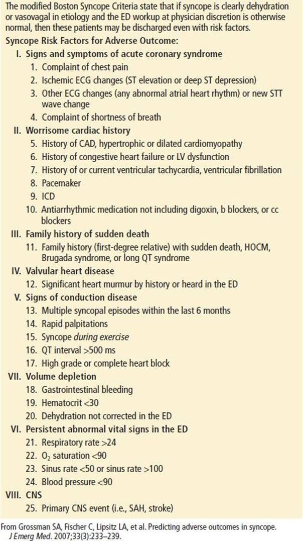
Patients who are judged to be at risk for dysrhythmia or sudden death should be admitted to an inpatient unit, observation unit, or other monitored area. Although the definition of high-risk patient is not consistent across the studies to date, certain clinical and diagnostic features appear to indicate a high risk of dysrhythmia; these include older age with associated comorbidities, an abnormal ECG, hematocrit <30% (if obtained), and history or presence of heart failure, coronary artery disease, or structural heart disease. Patients with near syncope (patients who feel like they are going to pass out but do not) have similar risks for adverse outcome when compared to patients who actually do have transient loss of consciousness and should be treated similarly (28,29). Patients presenting to the ED with syncope determined to be of benign etiology (clearly vasovagal or dehydration related) with a negative ED workup should be considered for discharge regardless of the presence of other risk factors (30).
Common Pitfalls
• Discharging a patient without performing a complete history and physical examination
• Failure to obtain an ECG and measure the blood sugar
• Failure to utilize outside resources such as family, ambulance crews, and bystanders in obtaining a complete history of events surrounding the syncopal episode
ACKNOWLEDGMENTS
The authors gratefully acknowledge the contributions of Thomas P. Martin to the content of this chapter.
REFERENCES
1. Sun BC. Quality-of-life, health service use, and costs associated with syncope. Prog Cardiovasc Dis. 2013;55(4):370–375.
2. Sun BC, Emond JA, Camargo CA. Direct medical costs of syncope-related hospitalizations in the United States. Am J Cardiol. 2005;95(5):668–671.
3. Brigo F, Nardone R, Bongiovanni LG. Value of tongue biting in the differential diagnosis between epileptic seizures and syncope. Seizure. 2012;21(8):568–572.
4. Calkins H, Shyr Y, Frumin H, et al. The value of the clinical history in the differentiation of syncope due to ventricular tachycardia, atrioventricular block, and neurocardiogenic syncope. Am J Med.1995;98(4):365–373.
5. Graham LA, Kenny RA. Clinical characteristics of patients with vasovagal reactions presenting as unexplained syncope. Europace. 2001;3(2):141–146.
6. Quinn JV, Stiell IG, McDermott DA, et al. Derivation of the San Francisco Syncope Rule to predict patients with short-term serious outcomes. Ann Emerg Med. 2004;43(2):224–232.
7. Sun BC, Hoffman JR, Mangione CM, et al. Older age predicts short-term, serious events after syncope. J Am Geriatr Soc. 2007;55(6):907–912.
8. Soteriades ES, Evans JC, Larson MG, et al. Incidence and prognosis of syncope. N Engl J Med. 2002;347(12):878–885.
9. Grubb BP, Karabin B. Syncope: Evaluation and management in the geriatric patient. Clin Geriatr Med. 2012;28(4):717–728.
10. Grossman SA, Fischer C, Lipsitz LA, et al. Predicting adverse outcomes in syncope. J Emerg Med. 2007;33(3):233–239.
11. Olshansky B, Poole JE, Johnson G, et al. Syncope predicts the outcome of cardiomyopathy patients: Analysis of the SCD-HeFT study. J Am Coll Cardiol. 2008; 51(13):1277–1282.
12. Middlekauff HR, Stevenson WG, Stevenson LW, et al. Syncope in advanced heart failure: High risk of sudden death regardless of origin of syncope. J Am Coll Cardiol. 1993;21(1):110–116.
13. Atkins D, Hanusa B, Sefcik T, et al. Syncope and orthostatic hypotension. Am J Med. 1991;91(2):179–185.
14. Benbadis SR, Wolgamuth BR, Goren H, et al. Value of tongue biting in the diagnosis of seizures. Arch Intern Med. 1995;155(21):2346–2349.
15. Jardine DL. Vasovagal syncope: New physiologic insights. Cardiol Clin. 2013; 31(1):75–87.
16. Weiner Z, Shapiro NI, Chiu D, et al. The prevalence of psychiatric disease in emergency department patients with unexplained syncope. Intern Emerg Med. 2013;8:427–430.
17. Oh JH, Kapoor WN. Psychiatric illness and syncope. Cardiol Clin. 1997; 15(2):269–275.
18. Linzer M, Yang EH, Estes NA, et al. Diagnosing syncope. Part 1: Value of history, physical examination, and electrocardiography. Clinical Efficacy Assessment Project of the American College of Physicians. Ann Intern Med.1997;126(12):989–996.
19. Kühne M, Schaer B, Moulay N, et al. Holter monitoring for syncope: Diagnostic yield in different patient groups and impact on device implantation. QJM. 2007;100(12):771–777.
20. Grossman SA, Fischer C, Bar JL, et al. The yield of head CT in syncope: A pilot study. Intern Emerg Med. 2007;2(1):46–49.
21. Giglio P, Bednarczyk EM, Weiss K, et al. Syncope and head CT scans in the emergency department. Emerg Radiol. 2005;12(1–2):44–46.
22. Martin TP, Hanusa BH, Kapoor WN. Risk stratification of patients with syncope. Ann Emerg Med. 1997;29(4):459–466.
23. Grossman SA, Van Epp S, Arnold R, et al. The value of cardiac enzymes in elderly patients presenting to the emergency department with syncope. J Gerontol A Biol Sci Med Sci. 2003;58(11):1055–1058.
24. Link MS, Lauer EP, Homoud MK, et al. Low yield of rule-out myocardial infarction protocol in patients presenting with syncope. Am J Cardiol. 2001;88(6): 706–707.
25. Huff JS, Decker WW, Quinn JV, et al. Clinical policy: Critical issues in the evaluation and management of adult patients presenting to the emergency department with syncope. Ann Emerg Med.2007;49(4):431–444.
26. Ammirati F, Colivicchi F, Santini M. Diagnosing syncope in clinical practice. Implementation of a simplified diagnostic algorithm in a multicentre prospective trial - the OESIL 2 study (Osservatorio Epidemiologico della Sincope nel Lazio). Eur Heart J. 2000;21(11):935–940.
27. Gabayan GZ, Derose SF, Asch SM, et al. Predictors of short-term (seven-day) cardiac outcomes after emergency department visit for syncope. Am J Cardiol. 2010;105(1):82–86.
28. Grossman SA, Babineau M, Burke L, et al. Do outcomes of near syncope parallel syncope? Am J Emerg Med. 2012;30(1):203–206.
29. Grossman SA, Babineau M, Burke L, et al. Applying the Boston syncope criteria to near syncope. J Emerg Med. 2012;43(6):958–963.
30. Grossman SA, Fischer C, Kancharla A, et al. Can benign etiologies predict benign outcomes in high-risk syncope patients? J Emerg Med. 2011;40(5):592–597.