
Sarah A. Sterling and Alan E. Jones
Fever is one of the oldest and most widely recognized signs of disease and accounts for numerous adult and pediatric visits to the emergency department (ED). Although the exact temperature that constitutes a fever is debatable, most consider a rectal temperature of 38°C in children and a temperature of 38.3°C in adults to represent a fever.
Body temperature is regulated by the preoptic area of the anterior hypothalamus. This area acts like a thermostat, continuously balancing heat production versus heat loss. Heat production is controlled by altering the basal metabolic rate by varying the level of circulating thyroxin, which in turn increases cellular metabolism. In addition, body heat is produced by increasing muscle activity (shivering when cold or the shaking chill of a fever), the fastest and most sensitive means of heat production. The principal method of adjusting body heat loss is to vary the volume of blood flowing to the skin’s surface through vasoconstriction and vasodilation, decreasing and increasing heat loss, respectively. The exocrine sweat glands also contribute to heat loss, cooling the body by vaporization.
The hypothalamic thermostat has an inherent set point of about 37°C but ranges from 36° to 37.8°C. This set point is affected by the normal daily circadian rhythm, with temperatures lowest around 4 AM and gradually increasing throughout the day to peak between 6 and 10 PM. This diurnal pattern is often apparent in clinical practice with most fevers lower in the morning and higher in the evening. The phenomenon of night sweats, most commonly related to tuberculosis or lymphoma, represents a nocturnal fluctuation in the hypothalamic set point, causing a slight rise in body temperature that leads to reactive perspiration.
Not all elevations of body temperature necessarily constitute a fever. A true fever is an elevation in body temperature caused by a change in the hypothalamic set point. This new higher set point causes the body to engage its normal heat-generating mechanisms, like shivering or shaking, to raise the body temperature until the new set point is reached. In hyperthermia, however, the body attempts to maintain a normal temperature but its homeostatic mechanisms (i.e., vasodilation, increasing sweat production) are insufficient to reduce body temperature and eventually become overwhelmed. Temperatures >41°C (106°F) almost always represent hyperthermia and not a true fever (1,2).
Elevation of the hypothalamic set point appears to be mediated by cytokines, which are released from lymphocytes, monocytes, and macrophages in response to exogenous and endogenous pyrogens. Exogenous pyrogens include microorganisms and their metabolic by-products, drugs, and other toxins, and endogenous ones include neoplasms and metabolic processes. Cytokines travel via the bloodstream to the hypothalamus, where they act to increase prostaglandin synthesis, primarily prostaglandin E2 (PGE2). This causes an elevated cyclic adenosine monophosphate (cAMP) level, which raises the hypothalamic set point by affecting peripheral vasoconstriction and internal heat production. Fever is maintained until pyrogen levels fall or until prostaglandin production is inhibited. Aspirin, ibuprofen, and acetaminophen exert their antipyretic effects by blocking central hypothalamic prostaglandin synthesis, but have no effect on circulating pyrogens. Corticosteroids can block the release of endogenous pyrogens as well as inhibit prostaglandins but are relatively weak antipyretics, nevertheless.
Other cytokines produced in response to pyrogens include interleukins (ILs), tumor necrosis factor (TNF), and interferons (IFNs). It is generally believed that bacteria and their products of metabolism typically provoke release of IL-1. In contrast, viral proteins appear to stimulate IFN. IL-1 induces the liver to produce acute phase reactants, including C-reactive protein (CRP), which are responsible for the elevated erythrocyte sedimentation rate (ESR) associated with some fevers. Although IFN is a potent pyrogen, it does not enhance production of acute phase reactants. Though not yet clearly supported by data, this is the rationale behind the expectation of an elevated ESR and CRP level with a bacterial fever but not with one of viral etiology. Procalcitonin, the precursor to calcitonin produced in the thyroid, rises from undetectable in well individuals to measurable levels within 2 to 3 hours (peaking in 24 hours) in patients with sepsis. Some studies have suggested this marker, rather than the later rising CRP, may be more specific for infection. Several studies have examined the use of CRP and procalcitonin as potential biomarkers of sepsis, which could be used in clinical practice to differentiate patients with bacterial versus nonbacterial infections; however meta-analyses suggest that procalcitonin is not yet ready to be deployed into routine clinical practice (3). While the use of these biomarkers appears promising, research is ongoing to determine their clinical utilization.
Many metabolic abnormalities are associated with the febrile state. Fever increases both the oxygen tension (PO2) and the carbon dioxide tension (PCO2) and shifts the oxyhemoglobin dissociation curve to the right, resulting in lower oxygen saturation. Fever also seems to lower the seizure threshold, which can result in seizures in children who do not have a predisposition to epilepsy (febrile seizures). Fever can also cause reactivation of latent herpes simplex infections and other chronic viral conditions.
Although higher metabolic demands are placed on the febrile patient, evidence suggests that fever has some physiologic benefits and protective effects as well. Neutrophils, macrophages, and lymphocytes are most active at elevated temperatures. In addition, higher temperatures decrease the level of serum iron, a substrate needed by many bacteria to replicate. Fever also seems to inhibit certain viruses, such as Coxsackievirus and poliovirus (1,4).
Despite this, many patients and parents, as well as practitioners, believe that fever is harmful or likely to cause harm. Many studies have shown that “fever phobia,” initially described in the 1980s, persists. A recent study in a pediatric ED showed that more than 50% of caregivers would define fever at <37.8°C (100°F) and a large majority would treat a fever <37.8°C (5). Despite these beliefs, no studies have shown true fevers to be harmful. In addition, research has shown that the response to antipyretics does not allow the differentiation between serious (e.g., bacterial) and nonserious (e.g., viral) infections (2). Fever anxiety can be greatly reduced when patients and caregivers are educated on the risks and benefits of fever and its proper management.
CLINICAL PRESENTATION
Though most febrile patients present to the ED with a chief complaint of fever, many present with more generalized complaints. Elderly patients, or their caregivers, may complain of generalized weakness, malaise, or a change in mental status. Parents may bring in infants who are eating poorly or appear lethargic. In these patients, fever may be an incidental finding, discovered only on review of systems or review of vital signs.
In children, a fever documented at home should be treated as real, even in the absence of a documented fever in the ED. One study found that while children referred to an ED by a general practitioner were more severely ill, approximately one in four children who self-reported with parents required extensive diagnostic testing, intravenous (IV) or aerosolized medications, or hospital admission (6). Studies have also found parents can correctly detect fever by palpation alone with sensitivities and specificities as high as 91% and 79%, respectively, reported in one study (7). Further, although bundling of infants can affect skin temperature, research has shown it has no effect on the rectal temperature of a healthy infant; so elevated rectal temperatures should not be attributed to bundling (8).
In the elderly, fever may be more difficult to detect and define. Previous studies have shown that while mean rectal temperature was similar to younger patients (37°C), elderly patients had less diurnal variation (9). Another study found that lowering the definition of fever to 37.8°C (100°F) increased the sensitivity of predicting an infection to 70% (vs. 40% if 38.3°C is used), while the specificity remained at 90% (10). Elderly patients may also have an impaired ability to mount a fever response altogether, with 20% to 30% of elderly patients who had a serious bacterial or viral infection having a decreased or absent fever response in some studies. To complicate the evaluation of this patient population more, the elderly may have a delayed fever response as well, which can be delayed more than 12 hours in 12% of patients despite known infection (9).
Similarly, immunocompromised patients with a low-grade fever may suffer from an acute, life-threatening condition, but may be unable to mount a febrile response. These findings highlight the importance of a high index of suspicion for infection in the setting of functional decline in the elderly or immunocompromised patient, and the need for reassessment after initial physical examination and evaluation.
History
Important points to address in the history include the following:
• Duration and magnitude of fever
• Associated symptoms
• Close contacts with similar illness
• Past medical history associated with immunocompromise
• For example, HIV, diabetes, chronic renal failure, blood dyscrasia, alcohol or drug abuse
• Recent hospitalizations
• Presence of medical hardware
• For example, prosthetic valves, ventriculoperitoneal shunts
• Current medications
• Recent antibiotics or chronic steroid use
• Serotonergic or neuroleptic drug
• Last time and dose of antipyretics
• Recent travel
• Areas with endemic malaria, parasitic, or fungal infections
• Prolonged immobility or symptoms concerning for venous thromboembolism
• Occupational exposures
• For example, medical personnel or postal worker/governmental official with possible biologic weapon exposure
Physical Examination
The most important observation during the physical examination is the patient’s overall appearance and mental status. Specifically, does the patient look well or toxic? Does the patient have an altered mental status, which might indicate infection or sepsis, especially in the elderly? The overall impression of the patient and their general well-being is vital in determining pretest probabilities which will guide and direct the patient’s workup.
The importance of reviewing vital signs cannot be overstated. Tachycardia and hypotension should not be ignored or attributed solely to a febrile response or faulty reading. One study found that hypotension, even if it is transient, significantly increases mortality in patients admitted to the hospital with sepsis (11). In the presence of a fever, the pulse can be expected to increase by approximately 18 beats/min for each 1°C increase in temperature. However, some diseases are associated with relative bradycardia for the degree of fever (pulse–temperature dissociation). This dissociation is classically seen with typhoid fever, but may also be seen in Legionnaires’ disease, mycoplasmal infections, drug fever, factitious fever, and some viral syndromes. In addition, patients taking β-blockers may be unable to increase their pulse in response to a fever.
Tachypnea may also be an important indicator of significant infection or acid–base disturbance. Tachypnea and dyspnea are often suggestive of pulmonary disease, whereas tachypnea without dyspnea may indicate sepsis or metabolic acidosis of any cause. In a study of nursing home patients, tachypnea >25 was the highest predictor of disease, with a sensitivity of 90% and a specificity of 95% for pneumonia (12).
The history will usually direct the physician to the most likely source of infection, though this may be challenging in patients with an altered mental status. In the absence of localizing symptoms, a thorough examination should be undertaken. The entire body should be examined for rashes, decubitus ulcers, cellulitis, track marks (needle marks), or lymphadenopathy. Rashes may suggest a viral exanthem, vasculitis, meningococcemia, tick-borne infection (e.g., Rocky Mountain spotted fever or Lyme disease), or toxic shock syndrome (staphylococcal or streptococcal). Lymphadenopathy associated with fever may suggest malignancy, autoimmune disorders, or infection. Tender, localized adenopathy is usually associated with an infection in the region drained by the involved nodes, whereas malignant nodes are usually painless, rubbery, and firm. The most common cause of acute generalized adenopathy is infectious mononucleosis.
The head and neck examination may also offer important clues to the source of infection. Nuchal rigidity or meningismus may indicate meningitis; however, these signs are often absent, particularly in the elderly or children. The eyes should be examined for scleral icterus, which could indicate hepatitis, cholecystitis, or ascending cholangitis. Examination in and around the ears may reveal otitis externa or media or may suggest mastoiditis. The oropharynx may show evidence of pharyngitis, peritonsillar or retropharyngeal abscess, or periodontal infection. Purulent rhinorrhea may suggest sinusitis or, in children, a nasal foreign body. Sore throat, drooling, and fever may suggest epiglottitis. It is important to remember that patients may not present classically, so airway edema, stridor, or hoarseness should be taken seriously as they could indicate significant infection and a potential airway emergency.
The chest should be examined for indications of a pulmonary or cardiac cause of fever. Localized rales or rhonchi suggest pneumonia. As x-ray findings can lag behind physical findings, an early pneumonia may not be visible on x-ray. A pericardial rub is indicative of pericarditis, and a new heart murmur suggests endocarditis, especially in IV drug users. However, functional murmurs may increase because of fever-induced tachycardia.
The abdomen should be examined for signs of peritoneal irritation or ascites. Localized pain may suggest cholecystitis, appendicitis, pancreatitis, or diverticulitis. Suprapubic pain suggests a urinary tract infection (UTI), and should prompt an evaluation for costovertebral angle (CVA) tenderness, which could indicate pyelonephritis. A rectal examination may demonstrate evidence of a perirectal abscess or prostatitis. In male patients with a urethral discharge, a sexually transmitted infection is likely and a swollen or painful testicle suggests epididymoorchitis. In females, a pelvic examination can be performed to evaluate for vaginal and cervical discharges and/or cervical motion tenderness suggestive of pelvic inflammatory disease, tuboovarian abscesses, or a retained vaginal foreign body. In neonates, examination of the periumbilical area is necessary to evaluate for omphalitis.
Examination of the musculoskeletal system is often overlooked in febrile patients, but examination of the spine may reveal tenderness suggestive of osteomyelitis, discitis, or epidural abscess (especially in IV drug users). A detailed examination of the extremities may demonstrate a septic or inflamed joint, or it may suggest osteomyelitis, myositis, or deep vein thrombosis (DVT). In addition, any retained catheters, such as peripherally inserted central catheter (PICC) lines or hemodialysis (HD) ports, should be evaluated for erythema, rubor, or discharge surrounding the catheter and regarded as a potential source of infection.
DIFFERENTIAL DIAGNOSIS
Infection
Infection is the most common cause of fever. Infection may originate in any portion of the body and lead to a febrile response, whether from bacterial or viral etiology. Fever may indicate localized infection, or may indicate a more significant systemic process, like sepsis. One of the more challenging aspects of practicing emergency medicine is determining the significance of infection and identifying patients early in the spectrum of sepsis.
Systemic Inflammatory Response Syndrome and Sepsis
Systemic inflammatory response syndrome (SIRS) indicates that the body has initiated a global inflammatory/immune response. SIRS is associated with a higher likelihood for organ dysfunction and organ failure. Although SIRS can result from noninfectious etiologies, in the setting of suspected or confirmed infection, if a patient has two or more SIRS criteria (Table 10.1), they meet the definition of sepsis (13,14).
TABLE 10.1
SIRS Criteria

Severe Sepsis and Septic Shock
According to consensus definition, severe sepsis is sepsis, or suspected infection with systemic manifestations, and evidence of organ dysfunction. Septic shock is sepsis-induced hypotension (systolic blood pressure [SBP] <90 mm Hg or mean arterial pressure [MAP] <70 mm Hg, or an SBP 40 mm Hg below the patient’s baseline) that does not respond to IV fluids. In addition, patients may have evidence of tissue hypoperfusion as indicated by an elevated blood lactate level, oliguria, renal failure, coagulopathy, or acute lung injury (15). Despite much attention to this area and aggressive treatment strategies, once septic shock occurs, the mortality rate remains high.
Drug-Induced Hyperthermia Syndromes
Drug-induced hyperthermia syndromes include neuroleptic malignant syndrome (NMS), malignant hyperthermia (MH), and serotonin syndrome (SS). Because all three syndromes have similar presentations, with severe muscle contraction and hyperpyrexia, they share common complications of rhabdomyolysis, myoglobinemia, and intravascular hemolysis. Temperatures >42°C cause central nervous system (CNS) damage, initially to the more sensitive cerebellum and eventually to the cerebral cortex, brainstem, and spinal cord. Patients may also develop pulmonary embolism, cardiovascular collapse, myocardial infarction, and acute respiratory failure.
NMS is a rare idiosyncratic reaction to neuroleptic medications. It has also been reported in patients taking lithium. NMS is thought to result from dopamine depletion in the brain, possibly in combination with increased anticholinergic activity. NMS presents with fever (temperature in excess of 41°C has been reported), muscle rigidity, and elevated creatine phosphokinase (CPK). Other findings may include altered mental status and autonomic instability. Symptoms develop rapidly over 1 to 3 days. Muscular rigidity of the thorax may impair breathing. Autonomic dysfunction may cause severe variations of both blood pressure and pulse, and some patients may develop seizures (eTable 10.1).
eTABLE 10.1
Diagnostic Criteria for NMS

MH is a rare, genetically determined reaction to inhaled anesthetic gases, depolarizing muscle relaxants (e.g., succinylcholine) and other agents. Succinylcholine is more likely to cause MH than inhaled anesthetic gases. The offending agent triggers the release of calcium by the sarcoplasmic reticulum resulting in increased muscle contraction and oxygen consumption. Masseter muscle spasm is generally noted initially, and then generalized muscle rigidity leads rapidly to hyperpyrexia. Symptoms typically develop as soon as 30 minutes following administration, though initial or recurrent symptoms may be delayed up to 48 hours later.
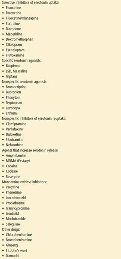
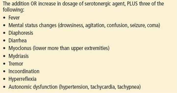
SS is a reaction to serotonin-active agents and most commonly occurs after a dosage increase of a serotonergic drug or with the addition of a potentiating drug to a serotonin-active agent. Common drugs that may cause SS are listed in eTable 10.2. Of particular note, the combination of triptans and serotonergic drugs has recently been shown to be a major cause of SS. SS involves an alteration in mental status, autonomic dysfunction, and increased neuromuscular activity, the latter causing a rise in temperature. Diagnostic criteria for SS are presented in eTable 10.3. Symptoms can be seen within 15 minutes of ingestion and can last for up to several hours. The duration of symptoms in other cases is dependent on drug half-life, but, in severe cases, symptoms can persist for weeks.
TABLE 10.2
Surviving Sepsis Campaign Guidelines

eTABLE 10.2
Drugs Associated with Serotonin Syndrome
eTABLE 10.3
Diagnostic Criteria for Serotonin Syndrome
TABLE 10.3
Treatment of Malignant Hyperthermia

Other Drug-Related Causes
Although any drug can cause a febrile reaction, penicillin and penicillin analogs are the most frequent causative agents. Many of the herbal and naturopathic supplements used by patients may interact with other medications causing fever as well. Drugs that are commonly associated with a drug fever are listed in eTable 10.4; however, drug fever should always be a diagnosis of exclusion.
eTABLE 10.4
Drugs Commonly Associated with Fever

Some drugs used recreationally or as toxic ingestions (overdoses) are associated with elevated temperature, including cocaine, amphetamines, belladonna alkaloids, tricyclic antidepressants, and monoamine oxidase inhibitors. Salicylates as acute overdoses or in chronic poisoning may also cause fever. Chronic salicylate intoxication may present similarly to sepsis, particularly in the elderly.
Venous Thromboembolism
Research using patient data from the Prospective Investigation of Pulmonary Embolism Diagnosis (PIOPED) study found, after eliminating other sources of fever, 24% of patients with a pulmonary embolus were febrile. Although the fever in the majority of these patients was low grade, 6% had a temperature of at least 38.3°C and 2% had a temperature of at least 38.9°C (16).
A recent study of more than 14,000 patients found that almost 5% of patients who had a DVT presented with a fever. They also found that in patients with a DVT, those who were febrile on presentation had a twofold higher mortality rate than those who were afebrile (17).
Central Nervous System Lesions
Structural lesions, head trauma, and strokes can cause elevated temperature either by direct destruction of the hypothalamic thermoregulatory center (hypothermia is more likely) or, more commonly, by blood irritating the central receptors in the preoptic area. Acute subdural hematomas may elevate body temperature, and therefore nonaccidental trauma should be considered in the differential diagnosis of an irritable infant with unexplained fever.
Rheumatologic and Connective Tissue Disorders
Fever may be the initial manifestation of many connective tissue and rheumatologic disorders. In systemic lupus erythematosus (SLE), fever is the initial symptom in 5% of cases. In older patients, temporal arteritis may present as recurrent fevers with no identifiable source.
Neoplastic Diseases
Although many different malignancies and tumors may produce fever, the most common are leukemia, Hodgkin disease and nonHodgkin lymphoma, hepatoma, and atrial myxoma. However, since the underlying disease and/or its treatment often leaves the patient immunosuppressed, it is important to eliminate occult infection as the etiology of the fever.
Inherited Diseases
Patients with familial Mediterranean fever have recurrent fevers of variable intervals, symptoms of polyarthralgias, and chest (pleural) and abdominal pain; however, this is a diagnosis of exclusion.
Endocrine Disorders
Thyroid disorders, including hyperthyroidism and subacute thyroiditis, and adrenal disorders, especially adrenal insufficiency (Addison disease), often include findings of fever.
DIAGNOSTIC APPROACH
An accurate temperature measurement is of utmost importance when approaching a patient with a report of fever. This is particularly true in the case of the very young, very old, or immunosuppressed patient, yet is often omitted for the sake of convenience. Recorded temperatures may vary widely depending on the method used to obtain them. Rectal temperatures have long been the gold standard, but because of the invasiveness and relative difficulty in obtaining them, easier methods are often employed. Newer generation urinary catheters allow for bladder temperature measurement, which is also an accurate method of core temperature measurement. Multiple studies have shown that none of these alternative methods consistently provides a reliable measurement of core temperature that is comparable to the rectal temperature. In addition, there appears to be no standard correction factor that may be used to improve their accuracy.
The rectal temperature tends to be 0.7°C higher than a simultaneously obtained oral temperature, although oral temperatures can vary more than 1.6°C, depending on where the probe is positioned in the mouth, the respiratory rate (an increase in respiratory rate results in decreased oral temperature), and the recent consumption of cold or warm foods or liquids.
Many studies have shown axillary, tympanic, and temporal artery temperatures are poorly sensitive for detecting fevers and that no reliable conversion factor to determine rectal temperature exists. While study results for pacifier thermometers have shown good correlation with rectal temperatures, poor sensitivity remains an issue. In addition, due to the need for patient cooperation (sucking the pacifier) and relatively long time periods for readings to results (several minutes), they have little practical use in the ED (7).
Laboratory Evaluation
While no test exists that easily identifies patients with serious infections, a thorough understanding of certain basic laboratory tests can greatly enhance patient evaluation. Although the WBC count is the oldest and best known screening test used to differentiate serious bacterial infections from other disorders, it is not specific or sensitive in either adults or children. Though still commonly used in clinical practice, a recent large systematic review found leukocytosis had a sensitivity and specificity of only 58% and 73%, respectively, for predicting serious bacterial infection in children (18). In a study of adults with unexplained fever after careful physical examination, only 15% had a WBC count ≥15,000/μL, and only 56% of those patients were subsequently shown to have a bacterial source (19). Several noninfectious processes including burns, fractures, steroids, diabetic ketoacidosis, and Addison disease can increase the WBC count. Patients with low WBC counts (<1,000/μL), however, are at significant risk for occult bacterial infections.
A “left shift” is only slightly more useful. Neutrophilia may occur secondary to emotional or physical stress, acute or chronic inflammation, benign or malignant tumors, myeloproliferative disorders, asthma, seizures, or medications such as steroids, lithium, or epinephrine. Neutrophil counts >30,000/μL have been reported secondary to strenuous exercise, seizures, and lithium use. In addition, a total neutrophil band count >1,500/μL was found in approximately one-third of adult patients with unexplained fever, of which only half proved to have bacterial infections (19).
The occurrence of thrombocytopenia in septic patients has been recognized for many years. Studies have shown longer stays in the intensive care unit and a higher mortality rate in patients with thrombocytopenia. Acute phase reactants, ESR and CRP, are often used to help recognize patients with serious occult infections, though both of these tests lack sensitivity and specificity. The ESR is often used as a screening test for temporal arteritis, bacterial endocarditis, tuberculosis, osteomyelitis, septic joints, and occult bacteremia in children.
CRP is more sensitive and specific and rises faster than the ESR, but is not particularly useful in the ED currently. Procalcitonin is another biomarker of interest for differentiating those patients with significant infections. A large systematic review looking at the diagnostic value of laboratory tests in identifying serious bacterial infections in children found that both CRP and procalcitonin had potential diagnostic value; however, there was no consensus on what cutoff values should be used for ruling in or out serious infections (18). Though both CRP and procalcitonin appear promising, further studies are needed to improve sensitivity and specificity before they can be used consistently in the ED.
Elevated lactate levels reflect tissue hypoxia and decreased global perfusion in the seriously ill patient and are associated with mortality in sepsis that is independent of organ dysfunction (20). In the most recent update to the Surviving Sepsis Guidelines, sepsis-induced hypoperfusion was defined as hypotension after fluid bolus or a lactate ≥4 mmol/L (15). In addition, research has shown that even moderate elevations in lactate (2 to 4 mmol/L) are associated with worse clinical outcomes (21). Further, a previous study has also shown that septic patients with an elevated lactate (≥4 mmol/L) who were normotensive, had a mortality rate similar to septic patients who were hypotensive (20% and 19%, respectively) (22). A point-of-care lactate can easily and quickly be obtained from a venous or arterial blood gas sample and rapidly help to risk stratify the patient and direct their care and disposition.
A urinalysis may also identify an infectious source of fever and indicate a UTI or pyelonephritis. In uncomplicated UTIs, urine cultures are probably not necessary at the time of initial diagnosis, though in elderly women, men, children, or patients who are pregnant or have comorbid diseases, and in cases resistant to therapy, urine cultures are recommended. In adults in whom there is a high clinical suspicion but a negative urinalysis, urine cultures should be obtained and treatment initiated. The formation of significant levels of nitrites or leukocytes in the urine occurs over time (up to 6 hours), and therefore, patients with urinary frequency may have a falsely negative urinalysis. Conversely, 10% to 50% of nursing home patients have chronic asymptomatic bacteriuria (essentially 100% in those with indwelling catheters), so the diagnosis of UTI as a cause of fever or mental status changes should be made with some caution (12).
Like many laboratory tests, there is no consensus on the value of routine blood cultures in febrile patients. The incidence of bacteremia in the ED has been reported to vary from 2.6% to as high as 10.7% of febrile patients. Mortality from bacteremia ranges from 20% to 40%; there are no data, however, on mortality for clinically unsuspected bacteremia.
Sklar and Rusnak (23) examined the value of outpatient blood cultures in febrile adults discharged from the ED. In this study, 5 of 86 patients subsequently proved to have positive blood cultures; 4 were subsequently admitted. No long-term morbidity was noted. Of these patients, 3 were diagnosed with endocarditis and 1 with pyelonephritis. The fifth patient, who refused to return for admission, had presumed pneumococcal endocarditis. Each of the patients with endocarditis had identifiable risk factors (2 with murmur and a history of rheumatic fever, 1 on dialysis and with a murmur, and 1 IV drug abuser). Other studies have shown that patients with underlying illnesses, most commonly diabetes, are at greater risk for bacteremia, and this study seems to support those findings. Therefore, it is prudent to limit outpatient blood cultures to febrile patients who have underlying diseases or risk factors (e.g., murmurs, IV drug use, immunosuppression, diabetes).
For inpatients, the current recommendations are to obtain cultures in patients who are septic, have an altered mental status, are ill appearing with unexplained leukocytosis, or are immunocompromised. In addition, some recommend blood cultures to identify the causative organism in pneumonia, osteomyelitis, meningitis, or septic arthritis, but in general it is preferable to obtain cultures directly from the infected site. Blood cultures are rarely helpful adjuncts in the management of immunocompetent patients with simple infections.
Laboratory studies, such as liver function tests or thyroid function tests, may identify a potential etiology for fever (such as hepatitis or thyroid storm) and should be directed based on historical clues and physical examination findings. Serologic testing may confirm the presence of other entities causing fever, such as amebiasis, mononucleosis, toxoplasmosis, chlamydial infections, cytomegalovirus (CMV), brucellosis, and HIV/AIDS. Specific studies on stool or cultures of the oropharynx could also be ordered when the history supports their consideration.
Cerebrospinal fluid (CSF) analysis can be obtained in patients with fever, headache, photophobia, and signs of meningeal irritation such as nuchal rigidity, Kernig sign (with the thigh flexed at the hip, there is pain or resistance with knee extension), or Brudzinski sign (passive flexion of the head onto the chest induces flexion of the lower limbs). The CSF should be routinely analyzed for protein, glucose, Gram stain, and cell count with differential. In selected cases, a venereal disease research laboratory (VDRL)/rapid plasma reagin (RPR) for syphilis, viral cultures, or tests for bacterial antigens should be added. Indwelling ventriculoperitoneal (VP) shunts may be accessed for obtaining CSF in the patient with symptoms of fever and meningitis with or without hydrocephalus.
Various antigenic tests are available; they have the advantage of identifying a specific organism when positive. Latex agglutination is rapid, economical, and noninvasive. Better results are obtained with urine than with serum or CSF, presumably because the antigens are more concentrated in urine. Unfortunately, these tests historically have had a high specificity (>99%) but a low sensitivity. Enzyme-linked immunosorbent assay (ELISA), an immunologic test, is much more sensitive in detecting pneumococci than latex agglutination. An ELISA assay is also used to detect West Nile virus IgM, but this test may be falsely negative if tested too early because IgM takes time to develop.
Quicker means of identifying organisms grown in blood cultures are also available. DNA probes have been developed that, when mixed with the growth from positive blood culture bottles, directly and rapidly identify Staphylococcus aureus, Streptococcus pneumoniae, Escherichia coli, Haemophilus influenzae, Enterococcus sp., and Streptococcus agalactiae. Polymerase chain reaction (PCR) is useful in diagnosing viral meningitis and has a high sensitivity and specificity for herpes simplex type I, Epstein–Barr virus, Enterovirus, and Cytomegalovirus (considered the gold standard for CMV). PCR can also detect tuberculosis and acute neurosyphilis.
For discussion on the workup and management of fever in infants and children, see Chapter 222.
Neutropenic Cancer Patients
The management of neutropenic cancer patients (absolute neutrophil count <500 cells/mm3 or a count <1,000 cells/mm3 with a predicted decrease to <500 cells/mm3) presenting with a fever is evolving. Whereas previous guidelines recommended admission and IV antibiotics for all of these patients, recent strategies focus on early oral therapy, outpatient management, and discontinuation of antibiotics in low-risk patients. Recommended evaluation of febrile neutropenic patients includes a thorough history and physical examination to localize a source of fever; CBC with differential, liver, and renal function tests (necessary for antibiotic choices); and blood cultures for bacteria and fungi (one set from any central venous access device and one peripheral set). Urinalysis should be performed, but urethral catheterization is discouraged in this patient population. Urine culture should be obtained if the urinalysis is abnormal, if there are signs or symptoms of a UTI, or if there is an indwelling catheter. Chest radiography should be obtained only if indicated by respiratory signs or symptoms or if there is consideration of outpatient therapy. Site-directed studies based on history and physical examination (e.g., lumbar puncture [LP], aspiration, and culture of abscesses) are also recommended. In addition, caution should be used in obtaining rectal temperatures in neutropenic patients due to the risk of causing occult bacteremia from rectal probe insertion.
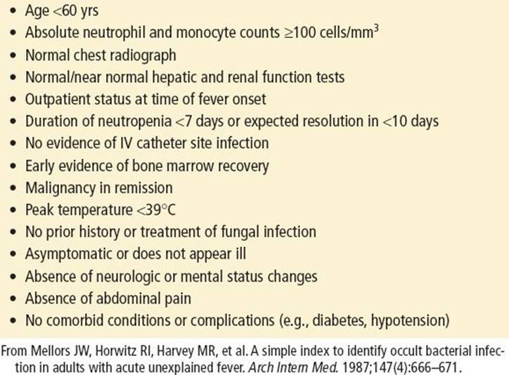
Because neutropenic patients with bacterial infections cannot reliably be distinguished from noninfected patients at presentation, empiric IV antibiotics should be administered promptly. After evaluation, patients who look well, have no focus of bacterial infection (including vomiting or diarrhea), and are felt to be at low risk for severe infection may be considered for outpatient antibiotic therapy if they can be carefully observed at home and have prompt access to health care 24 hours a day. Factors favoring a low risk of infection in adults are listed in eTable 10.5.
eTABLE 10.5
Factors Favoring Low Risk of Infection in Adults with Febrile Neutropenia
For discussion of the workup and management of infants and children, see Chapter 222.
Imaging Studies
A chest radiograph should be obtained in patients with lower respiratory complaints and examination abnormalities, or any patient who is at high risk for aspiration, such as an elderly patient with altered mental status or a patient with a history of alcohol abuse. Routine chest radiographs are commonly obtained in patients who present with fever without an obvious source as well. This practice is probably based on studies that show a lack of auscultatory findings in patients with infiltrates on chest radiographs. In one study, physical examinations performed by an experienced internist, pulmonologist, and infectious disease subspecialist were only 47% to 69% sensitive in detecting radiographically proven pneumonia. However, all of these patients had symptoms suggestive of pneumonia and did not represent patients with a fever and no source (24).
Signs or symptoms should guide the need for other imaging studies. Plain films may reveal osteomyelitis in patients with fever and bone pain, but bone scans and magnetic resonance imaging (MRI) are more sensitive. Abdominal radiographs may identify free air (bowel perforation), appendicoliths (appendicitis), or “thumbprinting” or gas in the bowel wall (mesenteric ischemia). Patients with fever and abdominal pain may benefit from an ultrasound to evaluate for the presence of cholecystitis or tuboovarian abscess. Ultrasound may also be of use in localizing soft tissue abscesses as fever source, especially in the IV drug user. Computed tomography (CT) scans should be used in selected patients to evaluate for appendicitis, abdominal abscess, or diverticulitis. MRI is the study of choice to rule out epidural abscess. Echocardiography (particularly transesophageal echocardiography [TEE]) should be used to evaluate suspected endocarditis patients for valvular disease. It may also be used in the patient with suspected pericardial effusion from inflammatory, malignant, or infectious processes.
CRITICAL INTERVENTIONS
• Initiation of antibiotics as appropriate
• Should be based on the type and site of infection if possible
• Send appropriate cultures prior to antibiotics if possible
• Do not delay antibiotics for LP in suspected meningitis
• Check lactate in patients with suspicion of infection and SIRS criteria
• A protocolized, quantitative resuscitation in severe sepsis and septic shock
• See Table 10.2 for treatment recommendations
• Antipyretics for patient discomfort or tachycardia
• Acetaminophen: 15 mg/kg in children, 650 to 1,000 mg in adults
• May repeat every 4 to 6 hours depending on dose
• Maximum dose: 90 mg/kg/day or 4 g/day
• Ibuprofen: 10 mg/kg in children, 400 to 600 mg in adults
• May repeat every 6 to 8 hours depending on dose
• Maximum dose: 40 mg/kg/day or 2.4 g/day
• Should not be used for more than a few days at max dose
• Not approved for infants <6 months old
• Repeat vital signs and reassess patient frequently
• Drug-related fever
• Stop offending agent
• MH: See Table 10.3 for treatment recommendations
• SS: Supportive care is the mainstay of treatment
• Benzodiazepines may help with hyperactivity
• NMS: Specific treatment not well established
• Dantrolene: Most useful in patients with prominent muscle contraction
• Bromocriptine mesylate: Use alone or in combination with dantrolene
• Most useful when dramatic muscle rigidity is not present
• Levodopa and carbidopa–levodopa: Useful in many patients
• Particularly useful in cases associated with medication withdrawal
• Aggressive cooling measures in hyperthermia
• If temperature >41°C, cool to 39°C within 30 minutes
• Use cool water spray with fans or cooling blankets
• Control shivering with benzodiazepines or chlorpromazine
• Avoid phenothiazines in NMS
• Monitor for rhabdomyolysis, electrolyte abnormalities, and disseminated intravascular coagulopathy (DIC)
• Remember: This is not a true fever, so antipyretics are typically ineffective
DISPOSITION
When considering the disposition of the febrile patient, the most important considerations are age and general health. Infants who are younger than 90 days old or incompletely immunized require a more extensive evaluation and may require admission or discharge with close follow-up.
For a discussion of the management of infants and children with a fever, see Chapter 222.
Patients who appear nontoxic and have no medical problems have a very slight chance of developing a serious infection (with the exception of IV drug abusers). As the age and presence of underlying medical problems increase, the chance of serious occult infection increases and makes admission advisable. Keating et al. (25) found that 95% of patients who were older than 60 years and had a temperature >38.3°C (101°F) on admission to the ED had a serious infection, and 92.5% required admission.
Most patients with unexplained fever and neutropenia or malignancy should be admitted for further workup. Patients with diabetes are at special risk, as are patients who are taking corticosteroids. Alcoholics are also considered to be immunosuppressed and at increased risk for complications. Also, pregnancy-related immunosuppression increases morbidity and mortality in pregnant females and may necessitate admission for infections that would otherwise be handled on an outpatient basis.
The postsplenectomy patient is at special risk for developing clinically unapparent bacteremia, usually from S. pneumoniae. These patients often have fever and a flu-like syndrome as their only complaints. Even when promptly diagnosed and started on antibiotics, the mortality rate remains high (50% to 75%). Although most postsplenectomy patients should be immunized against S. pneumonia, it is important to approach these patients cautiously and keep a high level of suspicion.
If good follow-up can be arranged, specimens for cultures can be obtained in the ED, and the nontoxic, low-risk patient can be discharged and rechecked in 24 hours when initial culture results become available. Continued follow-up or contact is necessary until cultures are confirmed to be negative.
Common Pitfalls
• Failure to obtain a rectal temperature, especially in the elderly or immunosuppressed
• Discounting parental reports of fever in infants who are afebrile in the ED
• Attributing fever in infants to overbundling
• Failure to check a lactate when clinically appropriate
• Relying on the WBC count or the response to antipyretics to determine the presence or seriousness of an infection
• Disregarding (even transient) hypotension
• Failure to perform a protocolized, quantitative resuscitation in septic patients
• Failing to reassess patient’s response to treatment and repeat vital signs
• Failure to immediately and aggressively cool patients with severe hyperthermia (>41°C)
REFERENCES
1. Knoebel EE. Fever: To treat or not to treat. Point. Clin Pediatr (Phila). 2002;41(1):9–11.
2. Plaisance KI, Mackowiak PA. Antipyretic therapy: Physiologic rationale, diagnostic implications, and clinical consequences. Arch Intern Med. 2000;160(4): 449–456.
3. Jones AE, Fiechtl JF, Brown MD, et al. Procalcitonin test in the diagnosis of bacteremia: A meta-analysis. Ann Emerg Med. 2007;50(1):34–41.
4. Saper CB. Neurobiological basis of fever. Ann N Y Acad Sci. 1998;856:90–94.
5. Poirier MP, Collins EP, McGuire E. Fever phobia: A survey of caregivers of children seen in a pediatric emergency department. Clin Pediatr (Phila). 2010;49(6):530–534.
6. van IY, Seiger N, van VM, et al. Self-referral and serious illness in children with fever. Pediatrics. 2012;129(3):e643–e651.
7. Callanan D. Detecting fever in young infants: Reliability of perceived, pacifier, and temporal artery temperatures in infants younger than 3 months of age. Pediatr Emerg Care. 2003;19(4):240–243.
8. Grover G, Berkowitz CD, Lewis RJ, et al. The effects of bundling on infant temperature. Pediatrics. 1994;94(5):669–673.
9. Norman DC. Fever in the elderly. Clin Infect Dis. 2000;31(1):148–151.
10. astle SC, Yeh M, Toledo S. Lowering temperature criterion improves detection of infection in nursing home residents. Aging Immunol Infect Dis. 2013;4: 67–76.
11. Marchick MR, Kline JA, Jones AE. The significance of non-sustained hypotension in emergency department patients with sepsis. Intensive Care Med. 2009;35(7):1261–1264.
12. Bentley DW, Bradley S, High K, et al. Practice guideline for evaluation of fever and infection in long-term care facilities. J Am Med Dir Assoc. 2001;2(5):246–258.
13. Bone RC, Balk RA, Cerra FB, et al. Definitions for sepsis and organ failure and guidelines for the use of innovative therapies in sepsis. The ACCP/SCCM Consensus Conference Committee. American College of Chest Physicians/Society of Critical Care Medicine. Chest. 1992;101(6):1644–1655.
14. Levy MM, Fink MP, Marshall JC, et al. 2001 SCCM/ESICM/ACCP/ATS/SIS International Sepsis Definitions Conference. Crit Care Med. 2003;31(4):1250–1256.
15. Dellinger RP, Levy MM, Rhodes A, et al. Surviving sepsis campaign: International guidelines for management of severe sepsis and septic shock: 2012. Crit Care Med. 2013;41(2):580–637.
16. Stein PD, Afzal A, Henry JW, et al. Fever in acute pulmonary embolism. Chest. 2000;117(1):39–42.
17. Barba R, Di MP, Blanco-Molina A, et al. Fever and deep venous thrombosis. Findings from the RIETE registry. J Thromb Thrombolysis. 2011;32(3):288–292.
18. Yo CH, Hsieh PS, Lee SH, et al. Comparison of the test characteristics of procalcitonin to C-reactive protein and leukocytosis for the detection of serious bacterial infections in children presenting with fever without source: A systematic review and meta-analysis. Ann Emerg Med. 2012;60(5):591–600.
19. Mellors JW, Horwitz RI, Harvey MR, et al. A simple index to identify occult bacterial infection in adults with acute unexplained fever. Arch Intern Med. 1987;147(4):666–671.
20. Mikkelsen ME, Miltiades AN, Gaieski DF, et al. Serum lactate is associated with mortality in severe sepsis independent of organ failure and shock. Crit Care Med. 2009;37(5):1670–1677.
21. Arnold RC, Sherwin R, Shapiro NI, et al. Multicenter observational study of the development of progressive organ dysfunction and therapeutic interventions in normotensive sepsis patients in the emergency department. Acad Emerg Med. 2013;20:433–440.
22. Puskarich MA, Trzeciak S, Shapiro NI, et al. Outcomes of patients undergoing early sepsis resuscitation for cryptic shock compared with overt shock. Resuscitation. 2011;82(10):1289–1293.
23. Sklar DP, Rusnak R. The value of outpatient blood cultures in the emergency department. Am J Emerg Med. 1987;5(2):95–100.
24. Wipf JE, Lipsky BA, Hirschmann JV, et al. Diagnosing pneumonia by physical examination: Relevant or relic? Arch Intern Med. 1999;159(10):1082–1087.
25. Keating HJ III, Klimek JJ, Levine DS, et al. Effect of aging on the clinical significance of fever in ambulatory adult patients. J Am Geriatr Soc. 1984;32(4):282–287.