
Boris Garber and Lance D. Wilson
Biliary disease is a common problem in emergency department patients, primarily because of the high prevalence of gallstones, which are present in as many as 25% of women and 15% of men over the age of 50. In the United States, over 120,000 cholecystectomies are performed yearly for acute cholecystitis, contributing to the $6.3 billion annual cost of gallbladder disease (1). Emergency physicians are confronted with a wide range of biliary pathology and must be familiar with the natural history of biliary disease to understand the variable presentations of acute disease and acute complications of chronic disease.
PATHOPHYSIOLOGY
The gallbladder is a reservoir off the common bile duct (CBD) that receives, concentrates, and secretes bile to assist in intestinal absorption of fat and fat-soluble vitamins. Gallstones form when cholesterol is supersaturated in the bile, leaves solution, and crystallizes on a matrix of calcium and other bile constituents. In Western countries, 75% to 80% of gallstones are cholesterol or mixed cholesterol stones (2). Once formed, gallstones may dissolve, remain unchanged, or grow larger.
Risk factors for gallstones are features that predispose to increased concentrations of cholesterol in the bile, including increased age, obesity, female sex, ethnicity, and hyperlipidemia. Family history is increasingly recognized as an important risk factor for biliary disease (3). Crohn disease, ileal resection, total parenteral nutrition, and rapid weight loss, particularly in the setting of bariatric surgery, also predispose patients to gallstone formation (3). Approximately 10% to 20% of gallstones are formed of calcium bilirubinate (pigment stones); these are associated with hemolytic anemias, cirrhosis, biliary tract anomalies, and biliary infections.
Although gallbladder pathology is secondary to gallstones, as much as 10% of acute cholecystitis is acalculous. Acalculous cholecystitis classically occurs in patients after major surgery, burns, trauma, or sepsis, where after a major insult, bile stasis and low arterial inflow lead to gallbladder inflammation, infection, and eventually necrosis (4); however, patients clearly present to the emergency department with acalculous cholecystitis. These patients are typically elderly men with significant atherosclerotic disease; however, other risk factors include diabetes, abdominal vasculitis, renal failure, congestive heart failure (CHF), and resuscitation from shock or arrest. Importantly, acalculous cholecystitis represents 50% to 70% of all acute cholecystitis in children (5). Acalculous cholecystitis must be considered in patients with acquired immunodeficiency syndrome (AIDS), who may present with cholecystitis and cholangitis secondary to cytomegalovirus and Cryptosporidium infection; however, the incidence of these infections is decreasing with highly active antiretroviral treatment (5). Gangrene or perforation of the gallbladder complicate a significant percentage of acalculous cholecystitis cases. Diagnosis is challenging as clinical and laboratory findings are nonspecific, so a high index of suspicion and imaging studies are necessary though the imaging criteria are complex. Definite therapy is cholecystectomy; rarely gallbladder drainage can be used (4). Acute acalculous cholecystitis is associated with a mortality rate of greater than 30% (5).
Gallbladder cancer also must be considered in the approach to patients with presumed biliary disease. It is the most common biliary tract tumor, is highly lethal, and early on lacks any specific symptoms or signs. When localized, surgery offers the hope for cure, so early diagnosis is crucial. Chronically inflamed gallbladders can become calcified, thereby leading to the radiographic finding of a porcelain gallbladder. These patients have as much as a 21% risk of developing gallbladder cancer, which normally occurs in less than 1% of patients with chronic cholelithiasis (6). Additional risk factors are asymptomatic gall stones 3 cm in diameter or larger (although surgery for asymptomatic gall stones is controversial) (6) and gallbladder polyps larger than 10 mm in diameter. Therefore, when these are discovered incidently during abdominal imaging, this mandates a discussion with the patient and a referral. Differentiating between benign and malignant gallbladder lesions based on imaging is difficult. Color Doppler study of polyps has been proposed to distinguish benign from malignant lesions (7).
CLINICAL PRESENTATION
It is helpful to view cholelithiasis in three categories: asymptomatic, symptomatic, and complicated. Clinical features of biliary emergencies are summarized in Table 103.1 (3,5,8–10).
TABLE 103.1
Diagnostic Features in Diseases of the Gallbladder

Asymptomatic Cholelithiasis
Most people with asymptomatic gallstones remain asymptomatic with only about 1% to 4% developing symptoms every year (1). When patients become symptomatic, nearly all initially develop pain, rather than cholecystitis or other acute complications, so they can be managed expectantly. The incidental discovery of gall stones when using ultrasound (US) or other imaging can be problematic and care should be taken not to attribute all the presenting symptoms to potentially incidental gallstones (6).
Symptomatic Cholelithiasis
Many people present to the emergency department with symptomatic gallstones. Pain is caused by distention of the gallbladder as it contracts against stones obstructing the gallbladder neck or cystic duct. Typically, biliary pain is a vague epigastric or right upper quadrant (RUQ) discomfort, usually associated with nausea. The term biliary colic is a misnomer, as biliary pain is constant, typically lasting longer than 30 minutes but less than 4 to 6 hours. Pain typically occurs in the evening and is usually postprandial (10). The vague nature and nonspecific location of the discomfort makes it easily mistaken for gastritis, reflux disease, or other benign abdominal pathologies. If the process continues, pain may become more severe, become localized to the epigastrium or the RUQ, radiates to the back or scapula, and may be associated with vomiting.
Complicated Cholelithiasis
Complicated cholelithiasis includes conditions such as acute cholecystitis and its complications, choledocholithiasis (stones in the CBD), cholangitis, and the acute complications of chronic gallstones. All are potentially life-threatening and require prompt recognition and treatment.
Acute Cholecystitis
In acute cholecystitis, the gallbladder neck or cystic duct is obstructed, and persistently increased intraluminal pressure along with irritation from bile and stones leads to mucosal damage, inflammation of the gallbladder wall, and eventually ischemia. Bacterial suprainfection (usually enteric gram-negative bacteria and occasionally anaerobes) plays a role in majority of the cases (1). In acute cholecystitis, pain is typically more severe and more often localized to the RUQ, with variable radiation to the epigastrium or back. Nausea and vomiting are common. Low-grade fever and tachycardia are typically present. Murphy’s sign (inspiratory arrest during palpation of the RUQ) or localized peritonitis may be elicited. In up to one-third of cases, a swollen, tender gallbladder is palpated. Typically, a leukocytosis with left shift is present, and mild increases in liver function tests, alkaline phosphatase, and bilirubin are seen in 30% to 40% of patients. Laboratory tests are frequently normal and fever can be absent, especially in the diabetic or chronically debilitated patient and must not be relied on to make the diagnosis. In acute cholecystitis, patients are rarely septic. If sepsis is present, consider more complicated conditions, particularly ascending cholangitis.
Complications of Acute Cholecystitis
Gallbladder perforation usually occurs in the setting of acute or chronic cholecystitis, typically in the elderly, debilitated, or diabetic patients. The gallbladder becomes gangrenous, perforates, and normally walls off to form an empyema, but occasionally a bile leak occurs, leading to bile peritonitis. Gallbladder wall gangrene may lead to diminishing pain and tenderness and absence of Murphy sign as nerve endings necrose (11). Rarely, acute cholecystitis is complicated by infection with a gas-forming organism, leading to emphysematous cholecystitis. This typically occurs in diabetics and elderly men with vascular disease. Emphysematous cholecystitis is a disease with mortality much higher than uncomplicated acute cholecystitis (11).
Choledocholithiasis
Gallstones in the CBD are termed choledocholithiasis. Stones usually form in the gallbladder and migrate from the cystic duct, but they can form primarily in the biliary tree. Presentation is usually similar to that of acute cholecystitis but can be variable and more subtle. Patients may have jaundice (3). Choledocholithiasis usually causes an elevation in alkaline phosphatase and conjugated bilirubin, consistent with extrahepatic obstruction. Increases in amylase and lipase are variable. A small proportion of patients with gallstones develop acute gallstone pancreatitis. Mirizzi syndrome is caused by extrinsic compression of the common hepatic duct by a gallstone impacted in the cystic duct or neck of gallbladder and presents in similar fashion to choledocholithiasis (3).
Ascending Cholangitis
Ascending cholangitis must be considered in any septic patient who presents with signs and symptoms of acute biliary tract disease, especially if the patient is diabetic, elderly, or debilitated. Ascending cholangitis is a bacterial infection of the biliary system, most frequently associated with CBD stones and obstruction from any cause. Various bacteria have been isolated from bile in patients with cholangitis; polymicrobial isolation is not unusual and blood cultures are positive in approximately 50% of patients. Endoscopic retrograde cholangiopancreatography (ERCP) in patients with biliary obstruction is the most common iatrogenic way of introducing bacteria into the biliary system, and cholangitis after biliary manipulation has a poor prognosis (12). Classically, it presents with Charcot triad of RUQ pain, jaundice, and fever; if mental status changes and shock occurs, this is considered Reynold pentad.Ascending cholangitis seldom presents classically, so it must be considered in any septic patient without a source. Mortality approaches 40%. To investigate the etiology of cholangitis, endoscopic ultrasonography is the most sensitive study to detect small stones, but ERCP is the only method which may also be therapeutic. Computed tomography (CT) and MRCP are also useful diagnostically. Tokyo criteria defines mild, moderate, and severe diseases. Treatment requires aggressive supportive measures, admission to ICU for patients with severe disease, intravenous antibiotics as soon as the diagnosis is suspected, followed by prompt biliary drainage. The more severe the presentation, the more urgently biliary decompression should be performed. A landmark study (13) showed that endoscopic therapy results in much lower mortality when compared to surgery. ERCP can be done at bedside, but it should be recognized that if contrast injection increases intrabiliary pressure, the patient may acutely decompensate from worsening septicemia (12).
Complications of Chronic Cholelithiasis
In chronic and occasionally in acute cholecystitis, gallbladder–enteric fistulas may form, causing pain, aerobilia, or infection. Rarely, a gallstone may pass through a fistula into the small bowel and lodge in the ileocecal valve, causing a small bowel obstruction (gallstone ileus) (3). Fistulas between the gallbladder and the duodenum, stomach, colon, pleura, and other organs have also been described. Presentation for gallstone ileus is typically abdominal distention, pain, vomiting, usually in an elderly patient with multiple medical problems. Patients will appear acutely ill and dehydrated, and have signs of intestinal obstruction, but clinical jaundice is unusual. Abdominal films are useful for showing evidence of mechanical bowel obstruction, pneumobilia, or a stone in the gastrointestinal tract.
DIFFERENTIAL DIAGNOSIS
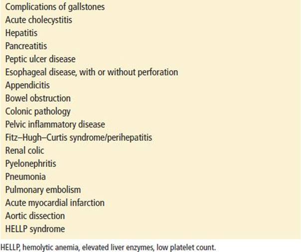
When patients present with complaints attributable to gallbladder disease, a wide range of both intra-abdominal and extra-abdominal processes must be considered (Table 103.2). Failing to consider biliary tract pathology in the differential diagnosis of nonspecific abdominal pain, fever, or sepsis in special populations can be problematic. The elderly can have very subtle presentations with only food intolerance or altered mental status (14). Elderly, diabetic, or debilitated patients are often difficult to assess and require ancillary studies to rule out life-threatening disease. Because pregnancy is associated with increased incidence of biliary sludge and cholesterol stone formation (14), biliary disease must be considered in the differential diagnosis of the pregnant patient with abdominal pain. Signs and symptoms of acute cholecystitis in pregnant and nonpregnant patients are similar. Although advancing age is a primary risk factor for gallstones, gallbladder disease and cholecystectomies are increasing among children, potentially related to increasing obesity (15). Acquired gall bladder pathology in infants is associated with prematurity, total parenteral nutrition status, and generalized severe illness, particularly in neonates, dehydration, and sepsis (16). Children with cholelithiasis typically have underlying risk factors for the development of stones and biliary disease, such as sickle cell disease or other hemolytic anemias. Hispanic race and female gender appear to be important risk factors (15).
TABLE 103.2
Differential Diagnosis of Acute Diseases of the Gallbladder
ED EVALUATION
Despite the diversity in acute gallbladder pathology, evaluation should follow a relatively straightforward sequence based on the patient’s clinical status (Table 103.1). The emergency physician’s goal is to rule out life-threatening problems and if gallbladder pathology is suspected, acute cholecystitis and other complicated acute biliary diseases should be ruled out. If the patient presents with symptoms attributable to gallstones without complications, referral for outpatient ultrasound and follow-up is appropriate. No further emergency department workup is necessary. In the patient with more severe symptoms or dehydration, further workup is indicated, including a complete blood count, electrolytes, liver function tests, bilirubin, amylase, lipase, urinalysis, and possibly diagnostic imaging. A summary of the clinical features and test characteristics in acute biliary diseases are shown in Table 103.2 (5,8,10,16). As no single clinical or laboratory finding is sufficient for diagnosing acute cholecystitis or acute biliary pathology, the physician’s overall impression of the likelihood of the disease determines whether the diagnosis is pursed and diagnostic imaging is initiated (10).
Diagnostic Imaging
Plain radiographs are helpful only for diagnosing associated bowel obstruction or ileus, although gallstones are visible on plain radiographs in approximately 10% of cases. US and cholescintigraphy (commonly referred to as HIDA scans) both have excellent positive and negative predictive values when done appropriately; HIDA is considered to have higher accuracy, but because of its accessibility and ease of use, US is typically the initial study of choice. Sonographic evaluation for gallstones and acute cholecystitis by emergency physicians is now an accepted part of routine practice, reduces length of stay, and has test characteristics similar to US performed in the radiology department (7,17). However, as with other radiographic studies, ED physicians must use caution when interpreting bed-side US and avoid premature diagnostic closure. Gallstones alone do not establish the diagnosis of acute cholecystitis. To establish US diagnosis of cholecystitis secondary signs such as pericholecystic fluid, sonographic Murphy sign, and thickened Gallbladder wall need to be present in addition to gall stones. Presence of stones together with either positive sonographic Murphy sign or Gallbladder wall thickening of 5 mm or greater has positive predictive value of greater than 90%, alternatively absence of the above results in negative predictive value of 95% (1). Gallbladder wall thickening is nonspecific and can occur in chronic cholecystitis, CHF, cirrhosis, hepatitis, and other conditions (9). Other relevant findings include sludge in the gallbladder, gallbladder distention, and pericholecystic fluid. US is also helpful in making the diagnosis of complications of acute cholecystitis where free fluid suggests gallbladder perforation.
CT scanning is reserved for patients with atypical presentations in whom other intra-abdominal pathology is a consideration and where a wide differential diagnosis persists (18). Ultrasound is superior to CT in ruling out acute cholecystitis and a CT diagnosis of cholecystitis should usually be confirmed with US. Only ∼2/3 of gall stones will be visible on CT. Normal CT examination of gallbladder makes acute cholecystitis unlikely but if high suspicion persists US should still be done. CT is most useful in evaluating complications such as gallbladder perforation, pericholecystic abscess, and gangrenous and emphysematous cholecystitis (9). Intraluminal gas, membranes, or irregular or absent wall on CT or US are indicative of gangrenous cholecystitis (11).
A dilated CBD or intraluminal stone is diagnostic of choledocholithiasis. However, these findings are not present in all cases, and if CBD stones are suspected, they may be evaluated by ERCP or MRI with cholangiography. Ultrasound can also assess other extrabiliary causes of RUQ pain involving liver, kidney, or pancreas. HIDA scans test the patency of the cystic duct. Failure to visualize the gallbladder is highly sensitive for acute cholecystitis. The main indication for HIDA is a high suspicion for acute cholecystitis, with an equivocal or negative ultrasound examination, particularly if acalculous cholecystitis is suspected (1).
KEY TESTING
• Although laboratory testing lacks sensitivity and specificity, liver function tests and lipase may be useful in identifying patients with biliary obstruction.
• Ultrasound is the most useful initial imaging study.
ED MANAGEMENT
Emergency department management of symptomatic cholelithiasis includes hydration, antiemetics, and analgesia. These patients can usually be sent home with appropriate follow-up. If acute cholecystitis or another complicated presentation of gallbladder disease is suspected, prompt consultation with a general surgeon is mandatory. Attention to the patient’s volume and electrolyte status, antiemetics, and analgesia, typically with nonsteroidal anti-inflammatory drugs or narcotics, is appropriate (3). For acute cholecystitis, antibiotics are indicated and should be discussed with the consultant. The patient who is septic or suspected of having ascending cholangitis, perforation, or emphysematous cholecystitis should be given antibiotics promptly. Typical biliary pathogens include enteric gram-negative bacteria and gram-positive species such as enterococcus or streptococci. Anaerobic species, particularly Clostridium and Bacteroides fragilis, occur in approximately 10% to 15% of cases, usually in the elderly or in patients with biliary obstruction. Appropriate coverage includes a parenteral antipseudomonal penicillin such as ticarcillin/clavulanate 3.1 g q6 h or piperacillin/tazobactam 3.375 g q6 h. Alternative regimens, especially if anaerobes are suspected, include a second- or third-generation cephalosporin or a quinolone plus clindamycin or metronidazole (1).
Treatment for symptomatic gallstones is surgical, almost exclusively with laparoscopic cholecystectomy (1,19). In acute cholecystitis, urgent cholecystectomy, optimally performed within 48 to 96 hours of presentation, is preferred over delayed surgery for mild disease as defined by Tokyo criteria (1,19,20). Patients with moderate disease can have either early or delayed surgical intervention while those with severe disease should be stabilized in the ICU first (1). Early cholecystectomy is safe and appropriate in elderly patients as well (21). In patients who present with pain, fever, or jaundice after laparoscopic cholecystectomy, one must consider the recognized complications of the procedure, including biliary injury and fistulas, wound infections, and intra-abdominal abscesses, which may present weeks to months after surgery (19). Newer techniques, such as endoscopic surgery through natural orifices, which do not require a skin incision and routine open cholecystectomies via minilaparotomies are currently being tested (3). Percutaneous urgent cholecystostomy followed by elective cholecystectomy is an option for patients with significant comorbidities who are at high operative risk (1,19). Oral dissolution therapy for gallstones, occasionally in conjunction with extracorporeal shock-wave lithotripsy, is used in patients who are substantial surgical risks or do not want surgery.
In patients with CBD stones, endoscopic sphincterotomy or dilatation to relieve obstruction followed by elective cholecystectomy is typically performed, rather than open cholecystectomy with CBD exploration. Almost all patients with ascending cholangitis require urgent biliary decompression. Typically this is performed by using endoscopic sphincterotomy rather than surgical approaches (12). All patients with gallbladder perforation, emphysematous cholecystitis, or gallbladder empyema need emergent surgery. Pregnant patients with biliary colic should be admitted and if symptoms persist more than 12 hours, antibiotics should be administered. Whether early surgical intervention is warranted in pregnancy is still a controversy (14). Children with biliary colic should also be admitted.
CRITICAL INTERVENTIONS
• Surgical consultation in all cases of acute cholecystitis or acute biliary disease
• Early surgical or gastroenterology consultation for ERCP when prompt biliary decompression is indicated
• Antibiotics in suspected acute cholecystitis or cholangitis
DISPOSITION
Most patients with symptomatic cholelithiasis may be managed as outpatients, with analgesics, antiemetics, and follow-up with a general surgeon. Those with more severe symptoms refractory to emergency department treatment should be admitted. Patients with acute cholecystitis, choledocholithiasis, gallstone pancreatitis, ascending cholangitis, or complications of chronic cholelithiasis such as gallstone ileus must be admitted to the hospital for antibiotics and/or surgical care.
Common Pitfalls
• Acute complications of gallstone disease must be considered and recognized, particularly in the elderly and debilitated who typically have subtle presentations.
• Remember to consider cholecystitis or cholangitis in the elderly, diabetic, or debilitated patient who presents with fever of unknown origin, change in mental status, or other nonspecific complaints.
• Symptomatic gallstones should be considered in patients with mild or atypical presentations, such as nausea, dyspepsia, or vague abdominal pain.
• Always consider acute gallbladder disease in pregnant women and children with abdominal pain.
• Always obtain timely surgical consultation when complications of gallstone disease are considered and arrange for urgent biliary decompression in cases of cholangitis.
REFERENCES
1. Strasberg SM. Clinical practice. Acute calculous cholecystitis. N Engl J med. 2008;358:2804–2811.
2. Portincasa P, Moschetta A, van Erpecum KJ, et al. Pathways of cholesterol crystallization in model bile and native bile. Dig Liver Dis. 2003;35:118–126.
3. Sanders G, Kingsnorth AN. Gallstones. BMJ. 2007;335:295–299.
4. Huffman JL, Schenker S. Acute acalculous cholecystitis: A review. Clin Gastroenterol Hepatol. 2010;8:15–22.
5. Barie PS, Eachempati SR. Acute acalculous cholecystitis. Gastroenterol Clin North Am. 2010;39:343–357, x.
6. Lazcano-Ponce EC, Miquel JF, Munoz N, et al. Epidemiology and molecular pathology of gallbladder cancer. CA Cancer J Clin. 2001;51:349–364.
7. Ross M, Brown M, McLaughlin K, et al. Emergency physician-performed ultrasound to diagnose cholelithiasis: A systematic review. Acad Emerg Med. 2011;18:227235.
8. Agrawal S, Jonnalagadda S. Gallstones, from gallbladder to gut. Management options for diverse complications. Postgrad Med. 2000;108:143–146, 149–153.
9. Gore RM, Yaghmai V, Newmark GM, et al. Imaging benign and malignant disease of the gallbladder. Radiol Clin North Am. 2002;40:1307–1323, vi.
10. Trowbridge RL, Rutkowski NK, Shojania KG. Does this patient have acute cholecystitis? JAMA. 2003;289:80–86.
11. Sen LC, Tublin M. Gangrenous cholecystitis. Ultrasound Q. 2012;28:213–214.
12. Lee JG. Diagnosis and management of acute cholangitis. Nat Rev Gastroenterol Hepatol. 2009;6:533–541.
13. Lai EC, Mok FP, Tan ES, et al. Endoscopic biliary drainage for severe acute cholangitis. N Engl J Med. 1992;326:1582–1586.
14. Gilo NB, Amini D, Landy HJ. Appendicitis and cholecystitis in pregnancy. Clin Obstet Gynecol. 2009;52:586–596.
15. Bogue CO, Murphy AJ, Gerstle JT, et al. Risk factors, complications, and outcomes of gallstones in children: A single-center review. J Pediatr Gastroenterol Nutr. 2010;50:303–308.
16. Lobe TE. Cholelithiasis and cholecystitis in children. Semin Pediatr Surg. 2000;9:170–176.
17. Summers SM, Scruggs W, Menchine MD, . A prospective evaluation of emergency department bedside ultrasonography for the detection of acute cholecystitis. Ann Emerg Med. 2010;56:114–122.
18. Shakespear JS, Shaaban AM, Rezvani M. CT findings of acute cholecystitis and its complications. AJR Am J Roentgenol. 2010;194:1523–1529.
19. Bhattacharya D, Ammori BJ. Contemporary minimally invasive approaches to the management of acute cholecystitis: A review and appraisal. Surg Laparosc Endosc Percutan Tech. 2005;15:1–8.
20. Gurusamy K, Samraj K, Gluud C, et al. Meta-analysis of randomized controlled trials on the safety and effectiveness of early versus delayed laparoscopic cholecystectomy for acute cholecystitis. Br J Surg. 2010;97:141–150.
21. Riall TS, Zhang D, Townsend CM Jr, et al. Failure to perform cholecystectomy for acute cholecystitis in elderly patients is associated with increased morbidity, mortality, and cost. J Am Coll Surg.2010;210:668–677, 677–669.