
Mark J. Ault and Joel M. Geiderman
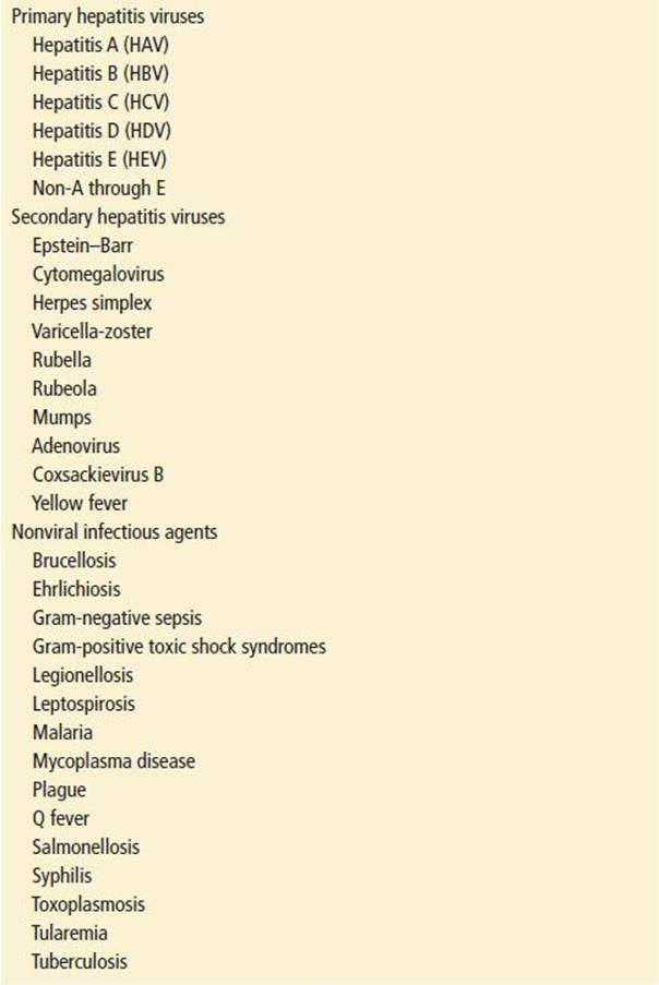
Viruses are responsible for the majority of cases of acute hepatitis. The primary hepatotropic viruses include hepatitis A virus (HAV), hepatitis B virus (HBV), hepatitis C virus (HCV), hepatitis D virus, also called the delta agent (HDV), hepatitis E virus (HEV), and the non-A through E hepatitis viruses. In addition, Epstein–Barr virus (EBV), cytomegalovirus (CMV), and other viruses are known to cause hepatitis as part of a systemic infection, as are a variety of bacteria, rickettsiae, and protozoal organisms (Table 104.1). A similar clinical and histopathologic picture can result from a variety of toxic and metabolic processes.
TABLE 104.1
Infectious Agents Known to Cause Hepatitis
CLINICAL PRESENTATION
The clinical manifestations of hepatitis, regardless of cause, are protean and vary from individual to individual with respect to the severity of symptoms, duration of illness, and outcome. The ultimate course of the illness is thought to depend on the nature of the etiologic agent, the extent of pre-existing or underlying liver disease, and, with infectious causes, the interaction between host factors, and the pathogen.
In its classic presentation, viral hepatitis has a highly characteristic clinical picture that is easily recognizable in its various stages. After a variable incubation period, a prodromal phase of illness occurs, with symptoms such as anorexia, low-grade fever, malaise, and lassitude. Often characterized as flu-like symptoms, they are nonspecific constitutional complaints typical of a variety of viral illnesses. Gastrointestinal symptoms may predominate, particularly in the adult patient, and abdominal discomfort representing hepatic, and occasionally splenic, enlargement may be reported. Within days to weeks after the onset of symptoms, the patient who enters the icteric phase of the illness first notes discoloration of the urine, representing the early rise of direct-reacting bilirubin and the spillage of bilirubin pigment into the urine. Subsequently, light (clay-colored) stools develop, scleral icterus may be noted, and clinical jaundice may become evident both to the patient and to the clinician as the serum bilirubin level exceeds 3 to 4 mg/dL. At this point, particularly in hepatitis A, symptoms may begin to resolve, heralding the convalescent phase of the illness.
Physical findings are highly variable in that the examination findings may be completely normal or may reveal frank jaundice and evidence of hepatic dysfunction. Liver enlargement and tenderness are common, and splenomegaly and lymphadenopathy may be present in 15% to 20% of cases.
Hepatitis may not be recognized when it does not present as the classic textbook picture because the history may be nonspecific or even misleading. This may occur, for example, when surreptitious toxin ingestion is involved or when the patient is reluctant to reveal specific risk factors for viral hepatitis.
If the patient is frankly jaundiced, hepatitis is usually readily considered. Recognition of more subtle findings such as scleral discoloration, sublingual yellowing, or excoriations, which may only be evident on careful examination, may also be informative.
When frank jaundice is absent, a higher index of suspicion is necessary. Constitutional symptoms alone are not at all specific for hepatitis but nonetheless are usually a sensitive trigger for initial laboratory screening which may reveal viral hepatitis.
Extrahepatic manifestations are particularly prevalent in hepatitis B and are generally considered to result from the deposition of immune complexes in various extrahepatic sites. They include urticaria and morbilliform rashes, arthralgias and frank arthritis, hematuria and proteinuria, and, less commonly, vasculitis of the mixed cryoglobulinemia or polyarteritis nodosa types (7). Other extrahepatic manifestations are not believed to be immunologically mediated. Pancreatitis, pneumonitis, and myocarditis are thought to be caused by direct viral involvement.
Occasionally, a patient presents with advanced hepatic failure and no history of hepatitis. This may occur in the context of progressive chronic hepatitis, a toxic insult, or acute-on-chronic hepatitis. Unexplained encephalopathy, personality change, coagulopathy, hypoglycemia, or respiratory alkalosis should prompt consideration of hepatic disease, including hepatitis. The following is a discussion of the myriad causes of hepatitis.
Viral Hepatitis
During the incubation period, there are no clinical manifestations of disease, but viral replication is occurring and serologic and chemical markers of this activity and the host response to it can be detected. Clinical manifestations appear only if and when enough hepatocytes are invaded. The etiologic agent causing hepatitis cannot be determined from the clinical presentation. Therefore, in evaluating the patient with presumed viral hepatitis, one must be familiar with the serologic markers associated with the various types.
HAV is spread primarily by the fecal–oral route, although the virus can be found in the blood as well as in the liver, bile, and stool during the latter part of the incubation period. The 2-week period before the onset of jaundice is the time of greatest infectivity; viremia, fecal shedding in the stool, and infectivity diminishes markedly by the time the icteric phase begins (Fig. 104.1). Source outbreaks are usually the result of fecal contamination of water supplies (e.g., with poor sanitation or during disasters) or food contamination, or occasionally by oral–anal contact. The incubation period is short: 2 to 7 weeks. The preicteric phase of hepatitis A usually lasts 4 to 14 days, presenting with nonspecific weakness, malaise, anorexia, nausea, and vomiting. There may be pain in the right upper quadrant and a characteristic loss of taste for cigarettes. Rash, urticaria, and joint pain occur infrequently, but the diagnosis of acute hepatitis should be considered in patients with these complaints. The term preicteric is presumptive (and often retrospective), because not all patients infected at this stage go on to have clinically evident jaundice. Children with hepatitis A typically manifest with gastroenteritis and commonly do not develop jaundice.
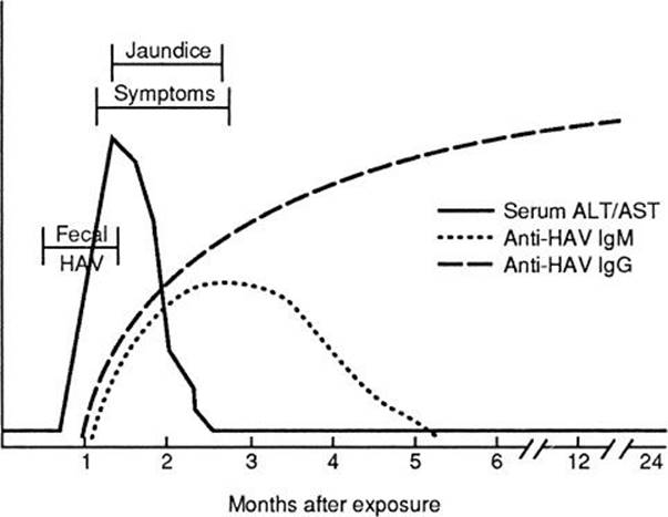
FIGURE 104.1 Serologic course of viral hepatitis A. Children usually have only mild symptoms with no jaundice. Adults more often develop jaundice. (Adapted from Hoofnagle JH. Serologic diagnosis of acute and chronic hepatitis. In: Hepatology update/portal hypertension: viral hepatitis [postgraduate course of the American Association for the Study of Liver Diseases]. Hosp Med. 1988;6:26.)
The presence of dark urine frequently heralds the onset of the icteric phase and is often what prompts the patient to seek medical attention. Most of the other symptoms, with the exception of malaise, resolve shortly after the appearance of jaundice, which may persist for up to a month. However, jaundice appears in the minority of patients infected with HAV. The convalescent period is variable; weakness and malaise may last for months. The onset of hepatitis A is usually more abrupt than that of the other types of viral hepatitis, but this observation is not useful in the individual case. Figure 104.1 depicts the usual serologic and biochemical course of hepatitis A. The alanine aminotransferase (ALT) level begins to rise late in the incubation phase, just before the onset of symptoms. Fecal shedding has peaked before this time and is abating. During the symptomatic period, about 4 to 5 weeks after exposure, immunoglobulin M (IgM) antibodies to HAV (anti-HAV IgM) begin to rise. Because this antibody usually becomes undetectable after 4 to 5 months, its presence implies recent infection. IgG antibodies rise later and remain detectable for life. The presence of such antibodies in the serum of at least 50% of adults reflects the high incidence of asymptomatic or unrecognized prior infection.
HBV is spread by exposure to blood, contaminated needles, infected secretions, sexual contact, and perinatal contact. Its prevalence is high among homosexual men, intravenous drug abusers, dialysis patients, patients and staff of institutions for the mentally retarded, and in certain endemic regions of the world. HBV has an incubation period of 45 to 160 days, during which the virus is actively replicating and is present not only in the hepatocyte but in virtually all body fluids. Patients are contagious during this asymptomatic period, and diagnostic serologic markers become positive. Immunoprophylaxis given before the onset of symptoms may prevent or attenuate the disease.
The acute clinical course of hepatitis B is similar to that of hepatitis A. Jaundice is more likely to occur in hepatitis B than A but still occurs in a minority of cases. About 85% of cases resolve completely, leaving the patient immune to hepatitis B; 1% to 3% of cases evolve into fulminant hepatitis, which has a mortality rate of 90%, and the remainder develop chronic hepatitis.
The serologic and biochemical courses of acute hepatitis B are depicted in Figure 104.2. The first marker to appear in the serum is the hepatitis B surface antigen (HBsAg), indicating infection with hepatitis B. Next, DNA polymerase, HBV DNA, and the hepatitis B e antigen (HBeAg) appear in the serum. These reflect active HBV replication and infectivity and appear well before the onset of biochemical damage to the liver. Of this group, only HBeAg can be measured easily; the presence of antibody to the HBeAg (anti-HBe) suggests low infectivity. In patients who become symptomatic, the ALT level rises and, in some, jaundice appears. It is important to understand the significance of the next immunologic event—the appearance of antibodies to the hepatitis B core antigen (anti-HBc), initially IgM, then IgG. This rise precedes the appearance of antibodies to the surface antigen (anti-HBs). In Figure 104.2, the gap between HBsAg and the rise of anti-HBs is called the “window,” when the sole specific marker of recent infection with HBV is the presence of anti-HBc IgM.
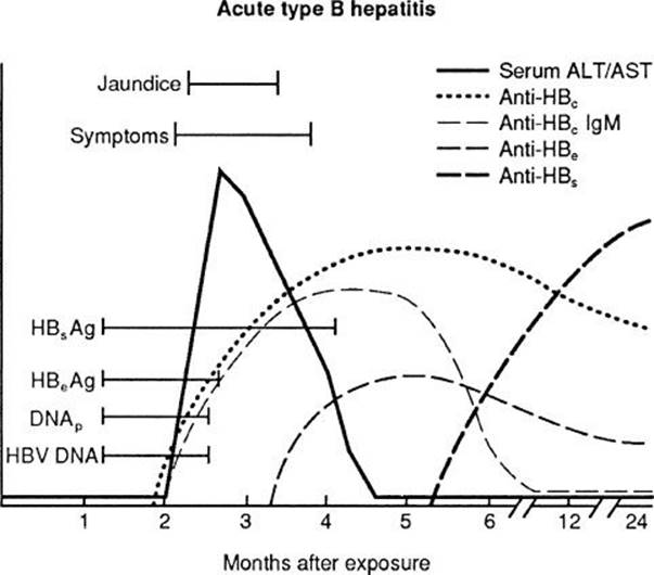
FIGURE 104.2 Serologic course of acute hepatitis B. This is more likely to cause jaundice than type A or non-A/non-B types. (Adapted from Hoofnagle JH. Serologic diagnosis of acute and chronic hepatitis. In: Hepatology update/portal hypertension: viral hepatitis [postgraduate course of the American Association for the Study of Liver Diseases]. Hosp Med. 1988;6:33.)
The rise of anti-HBsAg signifies immunity to HBV and corresponds to the abatement of symptoms and the return of ALT to normal levels. When hepatitis B fails to resolve, a chronic carrier state occurs. This is characterized by the persistence of HBsAg for a period exceeding 6 months and may be associated with the presence of either HBeAg or the antibodies to HBeAg. The carriage rate in United States is only 0.1% to 0.5% but may be as high as 5% to 20% in some demographic groups. Figure 104.3 depicts the typical serologic course of chronic HBV disease. Note that the HBsAg remains positive, anti-HBs is lacking, and the patient remains susceptible to further liver damage or to superinfection with hepatitis D.
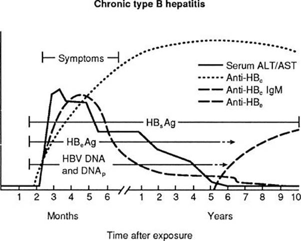
FIGURE 104.3 Serologic course of chronic hepatitis B virus (HBV) disease. (Adapted from Hoofnagle JH. Serologic diagnosis of acute and chronic hepatitis. In: Hepatology update/portal hypertension: viral hepatitis [postgraduate course of the American Association for the Study of Liver Diseases]. Hosp Med. 1988;6:33.)
HCV is an RNA virus of the Flaviviridae family. After the introduction of donor screening for HBV infection, posttransfusion hepatitis continued to be a significant problem. This led to the conclusion that most of the posttransfusion hepatitis was caused by another blood-borne pathogen, which was designated non-A, non-B (NANB) hepatitis. After the discovery of HCV in 1989 and the routine screening of the blood supply in the United States since 1990, it became clear that 80% to 90% of parenterally transmitted NANB hepatitis was caused by HCV. Currently, since the advent of genomic testing, the risk of transfusion-related hepatitis C is in the range of 1 in 1,000,000 units transfused. Aside from blood transfusion, other parenteral sources of HCV infection include intravenous drug use, hemodialysis, organ transplantation, and occupational exposure. Low levels of transmission occur in sexual and household contacts and perinatally (i.e., vertical transmission) (1). There is no evidence that breast feeding transmits HCV from mother to baby. Sporadic cases also occur.
Hepatitis C viral RNA can be detected in blood within 1 to 3 weeks of exposure. Anti-HCV can be detected in 50% to 70% of patients at the onset of symptoms and in approximately 90% of patients 3 months after the onset of infection. However, the majority of infected patients are asymptomatic and anicteric. The symptoms of HCV—malaise, weakness, and anorexia—are similar to those seen with other types of hepatitis. However, compared with hepatitis B, the acute disease is usually milder. Acute HCV infection rarely causes fulminant disease.
Chronic disease, with persistent although sometimes intermittent viremia, occurs in 85% of infected patients (10). Most patients with chronic disease are clinically well (12). Unfortunately, although the natural history of HCV infection is somewhat unclear, most researchers would agree that the disease ultimately causes progression to clinically significant liver disease in a large portion of infected patients. Important factors leading to more accelerated progression of hepatic fibrosis include coinfection with HIV, infection with other hepatotropic viruses, alcohol use, and young age of acquisition. Hepatitis C is the leading reason for liver transplantation in the United States, owing to the high prevalence rate, estimated to be approximately 4 million (3).
Chronic HCV infection also leads to hepatocellular carcinoma in up to 5% of patients after 20 years. Once cirrhosis develops, the rate of acquiring hepatocellular carcinoma rises to 1% to 4% per year.
There are several different tests for hepatitis C, including anti-HCV by enzyme immunoassay (EIA), recombinant immunoblot assay (RIBA), HCV-RNA, viral load, and genotyping. Anti-HCV is positive in approximately 90% of infected patients, usually within 5 to 6 weeks after the onset of clinical hepatitis. However, it does not distinguish among acute, chronic, and past infection, and positive tests are frequently falsely positive in populations with a low prevalence of infection. The HCV-RNA test identifies the presence of the virus in the blood. A reasonable testing strategy would be to order an anti-HCV and HCV-RNA. If both are negative, it is unlikely that the patient has acute hepatitis C. A positive anti-HCV and negative HCV-RNA generally represents remote infection. If both tests are positive, it may represent acute hepatitis C or an acute exacerbation of chronic hepatitis C. Finally, a positive HCV-RNA and negative anti-HCV is strongly suggestive of acute hepatitis C infection.
It appears that passive immunization with immune serum globulin derived from patients with previous HCV infection is unlikely to prevent disease, and no active vaccine is presently on the horizon. The Centre for Disease Control (CDC) recommends no postexposure prophylaxis. Until recently, treatment of chronic function has included combination therapy with recombinant α-interferon and ribavirin with the possible addition of boceprevir or telapravir, both protease inhibitors, for HCV genotype 1 infection (14). This resulted in a two- to threefold improvement in virologic response rates over the prior α-interferon monotherapy regimens. It is not known whether combination treatment can prevent the development of cirrhosis and hepatocellular carcinoma. Side effects of therapy are frequent and may prompt emergency department evaluation. Side effects of interferon include fever, nausea, irritability, and depression. Hemolytic anemia with subsequent decrease in the hematocrit is a common side effect of ribavirin and may require dose reduction.
In late 2013, the Food and Drug Administration approved 2 new direct acting antiviral agents, Sovaldi and Simeprivir, both oral agents, that can be effective in treating certain patients with chronic HCV infection. Treatment is tailored depending on viral genotype (14). A full discussion of the treatment of chronic HCV disease is beyond the scope of this text.
Hepatitis D, or delta hepatitis, is caused by a defective virus, the replication of which is dependent on the presence of HBsAg synthesis. The virus consists of the delta-antigen–bearing core encapsulated by an HBsAg coat. It should not be surprising that a virus that requires HBV for its replication follows a similar transmission pattern. The clinical course of HDV infection depends on whether the virus is acquired at the same time as HBV (coinfection) or whether HDV infects a patient with chronic HBV infection (superinfection).
In coinfection, the fate of HDV parallels that of HBV. The clinical course is similar to that described for hepatitis B, although it is often more severe. Because patients with HDV superinfection, by definition, have not been able to mount an immunologic response sufficient to clear their HBV infection, these individuals are prone to unchecked HDV replication and fulminant hepatitis.
Hepatitis E, formerly called enterically transmitted NANB hepatitis, is responsible for more than 50% of acute viral hepatitis occurring in some developing countries (5). Caused by an RNA virus of the genus Hepevirus, this organism was originally discovered during electron microscopy of feces contaminated with enteric NANB hepatitis. More recently, isolation and sequencing of the hepatitis E viral genome has revealed four genotypes that fall into two major groups. Genotypes 1 and 2 cause epidemic hepatitis. Genotypes 3 and 4 cause a sporadic (autochthonous) form of hepatitis. These two variants have distinct clinical and epidemiologic characteristics.
The epidemic form, as its name implies, can have a fairly widespread distribution particularly in developing countries. Large outbreaks have occurred in Sudan and Iraq. It is generally of little concern in the United States, although imported cases have occurred in patients who have traveled to endemic areas such as India, Nepal, Pakistan, and parts of the former Soviet Union and Mexico. Clinically, the presentation is very similar to hepatitis A. Transmission is largely by the fecal–oral route and can result from contamination of the water supply, with human feces a frequent source of epidemic outbreaks. Secondary spread is uncommon. The clinical course is very similar to that of hepatitis A, with an incubation period of 2 to 9 weeks and an anicteric phase lasting 3 to 8 weeks. As in hepatitis A, nausea, vomiting, and diarrhea are frequent and usually resolve with the onset of jaundice. Overall, the case fatality rate is approximately 5% but can be as high as 20% for pregnant women. No chronic hepatitis has been described for these genotypes.
While genotypically similar, autochthonous hepatitis E has a distinct clinical presentation from the more common epidemic form. Acquisition is generally attributed to food exposure, particularly undercooked pork and sausage. Autochthonous hepatitis E tends to present in an older age group. The disease is usually subclinical and mild, with icterus occurring in only 20% of cases. On the other hand, autochthonous hepatitis E is also associated with a striking spectrum of unique and serious extrahepatic complications which include acute-on-chronic liver failure, neurologic disorders, and chronic hepatitis. Patients with mild or unsuspected hepatitis from other etiologies may experience a fairly rapid appearance of ascites, encephalopathy, or overt liver failure, with secondary infection by hepatitis E. The extrahepatic manifestations may be striking and may be the predominant presentation. Neurologic symptomatology in particular, such as Guillain–Barré syndrome, Bell palsy, ataxia, confusion, or peripheral neuropathy, may be so striking as to obscure the primary diagnosis of hepatitis. Nonneurologic extrahepatic manifestations such as aplastic anemia, pancreatitis, or arthritis can present a similarly distracting clinical picture. While not common, autochthonous hepatitis E has been associated with chronic infection, predominantly in immunocompromised individuals. Liver transplantation patients, HIV-infected patients, and cancer patients receiving chemotherapy seem to be particularly susceptible. Although hepatitis E is not commonly diagnosed in the United States, exposure to the virus may be fairly common. Studies have found that up to 21% of adults in United States had anti-HEV antibodies, making it more common than hepatitis B (5.7%) or C (2%), and only slightly less common than hepatitis A (38.3%). Tests for anti-hepatitis E antibody are available commercially, but their sensitivity and specificity vary widely and at present do not have Food and Drug Administration approval. As with other forms of the viral hepatitis, treatment options are generally limited to supportive care. Limited reports describing the use of ribavirin and peginterferon suggest a potential for viral clearance and a sustained response with treatment of chronic hepatitis E. This combination therapy has also been used in the treatment of severe, acute hepatitis E, although neither agent has been approved for this use. Hepatitis E infection is potentially preventable by vaccination, though at present this is more applicable to endemic areas. The most effective form of prevention at this time is for travelers to endemic areas to use strict enteric precautions, avoid exposure to raw shellfish, and ensure that pork has been cooked at 260°F for at least 20 minutes.
Non-A through E hepatitis refers to the small population of patients with clinical hepatitis in whom all serologic studies on both the donor and the recipient patients are negative. With the discovery of new viruses and the development of new serologic tests, these patients will undoubtedly be further differentiated. One of these, independently discovered and named hepatitis G virus and hepatitis Gallbladder virus C, is an RNA virus of the Flaviviridae family (2). It appears to be blood-borne and has been found in as many as 10% of patients with non-A through E hepatitis. Its role has been questioned, and its definition as a true hepatotropic cause of acute or chronic hepatitis is yet to be determined (6). Similarly, hepatitis F virus has been isolated from the stool of a patient with hepatitis and transmitted to primates. As with hepatitis G, the role of this virus as a significant cause of non-A through E hepatitis is unclear.
Infectious, Nonviral Hepatitis
Although uncommon, hepatitis can accompany a wide variety of bacterial, rickettsial, and protozoal diseases. Included in this list are legionellosis, salmonellosis, tularemia, leptospirosis, brucellosis, plague, syphilis, gram-negative sepsis, gram-positive toxic shock syndromes, Q fever, mycoplasmal disease, tuberculosis, toxoplasmosis, ehrlichiosis, and malaria (see Table 104.1).
Autoimmune Hepatitis
Autoimmune hepatitis is a chronic inflammatory liver disorder of uncertain etiology. A standardized definition and diagnostic criteria have been developed (8). The diagnosis relies heavily on the exclusion of viral etiologies, lack of exposure to alcohol and other hepatotoxic agents, and hypergammaglobulinemia with gamma globulin levels >1.5 times the upper limits of normal. A scoring system for the quantitative diagnosis of autoimmune hepatitis has been proposed by the International Autoimmune Hepatitis Group. With a sensitivity of 97% to 100% and a specificity of 66% to 92%, it has utility in clinical practice especially for atypical or overlapping cases. Liver biopsy remains essential to the diagnosis and evaluation of disease severity in patients with autoimmune hepatitis. Compounding the diagnostic clarity of this illness is the recognition that the overlap syndrome, the simultaneous presence of autoimmune hepatitis and another liver disease, seems to occur more frequently than expected (9).
Clinically, the presentation may be extremely variable and difficult to differentiate from viral hepatitis. Two types have been described based on the presence of circulating autoantibodies. Type I (classic) autoimmune hepatitis has been associated with antinuclear and anti–smooth-muscle antibodies, and type II has been characterized by the presence of autoantibodies against liver–kidney microsome type I (anti-L K M-1). Both types are more prevalent in young females, and appropriate diagnosis is crucial, as they may be treated with prednisone and immunosuppressive therapy.
Toxic Hepatitis
Liver injury may result from direct hepatocellular toxicity or from idiosyncratic injury caused by a hypersensitivity-type reaction (10). Toxic exposures also may manifest as cholestasis, in which elevations of alkaline phosphatase and bilirubin levels predominate. Pharmacologic and chemical agents produce a diverse spectrum of hepatic insults, including both acute and chronic liver disease (Table 104.2). Drug-induced hepatic injury is the most frequent reason cited for withdrawal a drug approved by the U.S. Food and Drug Administration (FDA) from the market, and it accounts for more than 50% of cases of acute liver failure in the United States. Although any evidence of hepatic dysfunction should prompt a diligent search for both intentional and inadvertent exposure to any of a number of chemical agents, several agents deserve specific mention because of the inherent likelihood of hepatocellular injury, because of their widespread use, or because of their particular relevance to the practice of emergency medicine.
TABLE 104.2
Drug-Induced Hepatitis
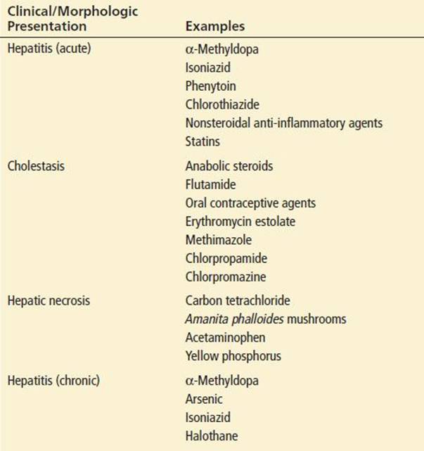
Acetaminophen is one of the most common agents encountered in the acute setting that is responsible for striking liver enzyme elevations (see Chapter 285). Toxicity is dose-dependent, but several factors have been shown to contribute in a synergistic way to toxicity at even relatively low doses. These include concomitant alcohol ingestion, chronic phenobarbital use, or chronic exposure to moderately large doses of acetaminophen.
Hepatitis caused by isoniazid clinically resembles acute viral hepatitis and may occur in as many as 20% of patients taking the drug, with the risk and severity of injury increasing with the age of the patient. Clinical toxicity generally occurs in the first few months of therapy, but transaminase elevations greater than two to three times normal at any time must be considered potentially serious, because progression to fulminant hepatitis may occur.
Statins are generally well tolerated but have been associated with severe hepatocellular injury. Moreover, there is concern that concomitant use of nicotinic acid for the treatment of hypercholesterolemia may promote additional toxicity.
Although overt hepatotoxicity to phenytoin is unusual, use of the drug is particularly common, and prevention of serious toxicity requires an awareness of the nature and mechanisms of injury. Hepatic injury is believed to be caused by toxic metabolites; current evidence has implicated a heritable metabolic defect that may predispose to a viral hepatitis-like hypersensitivity reaction within weeks of starting therapy at usual therapeutic doses. Like the other toxic exposures, early detection and interruption of therapy can result in complete resolution and the avoidance of serious hepatic injury.
Troglitazone (Rezulin) was the first of a new class of oral agents used for the treatment of type II diabetes. The agent was originally approved by the FDA in 1997, and the recognition of significant hepatotoxicity appeared to outweigh the benefits of therapy, thereby leading to the withdrawal of troglitazone from the market. Two new agents of the same class have been available since 1999, rosiglitazone (Avandia) and pioglitazone (Actos). They do not appear to have the same degree of hepatic toxicity, although liver injury has been reported. Recognition of potential severe liver injury, monitoring of liver enzymes, and early discontinuation of therapy are important to maintain the safety of these agents (11).
Flutamide is an antiandrogen drug used in the treatment of metastatic prostate cancer. Well-documented cases of serious hepatotoxicity have been reported. Although the toxicity appeared to be reversible, several patients have died of massive hepatic necrosis, emphasizing the need for prompt consideration and diagnosis.
Hepatitis has been reported to be associated with the use of several of the nonsteroidal anti-inflammatory drugs (NSAIDs). Because of the frequency with which NSAIDs are used, emergency physicians should be aware of their potential for causing hepatic toxicity. Sulindac has received special attention because it has been well established as a cause of hepatitis, including fatal hepatic necrosis. Similarly, bromfenac was introduced for short-term use for the treatment of orthopedic pain. Reports of severe liver injury led to withdrawal of this agent.
In the past, the inhalational anesthetics halothane and methoxyflurane were well-known causes of fulminant hepatitis. Halothane has been superseded by newer agents, and methoxyflurane is no longer marketed in the United States.
Recently, reports have implicated clopidogrel as a cause of mixed hepatocellular and cholestatic liver damage. The true incidence of this complication is unknown, but clopidogrel is virtually universally used for patients undergoing percutaneous coronary intervention and in those with acute coronary syndromes. Additionally, treatment courses are generally protracted, thus resulting in more prolonged exposure. Routine liver enzyme testing is not currently recommended; however, given the wide use of this drug, clinical suspicion is reasonable in appropriate clinical settings.
A careful medication history should include an inventory of complementary and alternative medications. Studies have demonstrated that use of these modalities is common but volunteer reporting of their use to the physician is limited. Often referred to as simply “Chinese herbs,” specific compounds of interest include germander, chaparral leaf, and weight loss preparations containing usnic acid.
It is worth mentioning that alcohol is probably the most common chemical to result in liver injury; this is covered in depth in Chapter 281 on Ethanol and its complications.
Metabolic Causes of Hepatitis
Although the metabolic causes of liver disease are numerous, several are notable for the potential for acute presentations. Wilson disease is a disorder of copper excretion. Patients generally present in adolescence when copper has had sufficient time to accumulate in excess. Liver involvement is common but, occasionally in younger patients, the illness will present as fulminant hepatitis. Prompt recognition and treatment are necessary to limit end-organ damage (13).
Acute fatty liver of pregnancy is a life-threatening condition that occurs late in pregnancy. Histologically, it is described as microvesicular fatty liver and is believed to be caused by disordered metabolism of fatty acids. Pregnant patients, usually in the 35th week or later, present with jaundice and hepatic failure frequently accompanied by disseminated vascular coagulation. The condition resolves with termination of pregnancy, although the mother must first be stabilized.
Reye’s syndrome presents almost exclusively in children younger than 15 years. Though an association with salicylate use has been noted, it can occur in the absence of salicylates. It is generally seen after upper-respiratory illness, especially influenza and chickenpox, and is characterized by hepatic injury, hypoglycemia, and encephalopathy. Care is supportive, and chronic liver disease has not been reported when recovery occurs.
Another cause of elevated transaminase levels (transaminitis) that should be considered in the differential diagnosis is hepatobiliary disease. Elevated ALT and aspartate amino transferase (AST) levels should not lead one on a fruitless search for infection or toxic causes of hepatitis when the chemistry patterns suggest hepatobiliary obstruction (i.e., elevated serum bilirubin, alkaline phosphatase, and γ-glutamyl transpeptidase levels [GGTP]).
DIFFERENTIAL DIAGNOSIS
The differential diagnosis for the patient with right upper quadrant abdominal pain is extensive and includes hepatitis, biliary colic, cholecystitis, peptic ulcer disease, pyelonephritis, renal colic, cardiac ischemia, and right lower lobe pneumonia. The differential diagnosis for jaundice includes many other possibilities, and is discussed further in Chapter 101. Once the differential has been narrowed to hepatitis, the next task is to determine the specific etiologic agent, whether infectious, toxic, metabolic, or autoimmune. Eliciting a history of risk factors, medications, exposures, and clinical course as described previously, combined with laboratory testing, will help in determining the specific cause.
ED EVALUATION
Evaluation of suspected hepatitis may be divided into several steps. The first step is to make the diagnosis, which, in light of the previous discussion, may not be entirely straightforward. Clearly, a reasonable index of suspicion is necessary in most cases, and the physician must consider the relative likelihood of illness in each patient compared with the risk of excessive testing in the many patients with nonspecific symptomatology.
If the likelihood of hepatitis is present but low, a reasonable approach initially is to order a serum AST level as a preliminary screen. Although nonspecific, this is a sensitive screen, and a normal AST value virtually excludes hepatocellular disease.
In patients in whom hepatic disease is believed to be more likely or who has an abnormal AST level on the preliminary screen, more extensive laboratory testing is necessary. This should consist of indices of necrosis (AST and ALT) as well as indices of cholestasis (alkaline phosphatase, total bilirubin, or GGTP). Although these values lack diagnostic specificity, certain patterns may be helpful in limiting the differential diagnosis and in directing further evaluation. For instance, a transaminase elevation >10 times normal strongly suggests acute viral or toxic injury and essentially excludes chronic hepatitis. Elevation of alkaline phosphatase and bilirubin values suggests intrahepatic or extrahepatic obstruction. Transaminase elevations of two to three times normal, with the AST higher than the ALT, suggest alcoholic injury.
In the second step of the evaluation, specific tests of hepatic synthetic function are necessary. Serum protein, albumin, and glucose levels should be used in conjunction with the serum bilirubin level to assess the extent of hepatocellular dysfunction. If there is indication of central nervous system involvement, with lethargy or confusion, a serum ammonia level may serve as a useful baseline for serial evaluations.
Further evaluation should focus on defining the specific cause of the illness. If hepatocellular necrosis is the dominant picture, detailed serologic studies (anti-HAV IgM, HBsAg, anti-HBs, anti-HBc, anti-HCV by enzyme immunoassay (EIA), HCV-RNA and Monospot test) are warranted, as well as a diligent historical search for foreign travel to HEV-endemic regions and for drug or toxic exposures. If cholestasis is the dominant picture, ultrasound examination should be performed to exclude mechanical obstruction.
KEY TESTING
• Consider the diagnosis of hepatitis and the risk profile. If risk is low, consider screening with an AST. If high, test more extensively for hepatic necrosis and obstructive etiologies.
• If hepatitis is confirmed, stage the severity of disease and seek evidence of clinically significant hepatic dysfunction: serum protein, albumin, glucose, and bilirubin, and ammonia levels if altered mental status is noted.
• Begin a search for a definitive diagnosis. Send acute hepatitis serologies and consider drug exposures (especially acetaminophen and alcohol) and travel history.
ED MANAGEMENT
Patients with acute viral hepatitis may present to the emergency department because of signs and symptoms typical of a viral syndrome that may include prostration, nausea, vomiting, and dehydration. Treatment for these patients is symptomatic and is usually limited to intravenous fluids, acetaminophen, and antiemetics. Metoclopramide (Reglan) or ondansetron (Zofran) are the antiemetic agents of choice, because phenothiazines can impair hepatic excretory function and may produce cholestasis. Patients who present without symptoms or present because they have noticed jaundice require no specific therapy.
For patients with diagnosed viral hepatitis, there is no specific therapy indicated beyond supportive care and symptomatic treatment. If the prothrombin time is prolonged, the patient should receive vitamin K. Immune globulin is of no use for the patient who has already contracted hepatitis. Corticosteroids are probably harmful rather than beneficial.
Patients who present because they have been exposed, or think they have been exposed, to hepatitis present a different management problem (see Chapter 194). Immunoprophylaxis is highly effective if given under the proper circumstances. Because this treatment should be guided in part by serologic studies, it is often not initiated in the emergency department.
If results of serologic testing are not available in a timely fashion, therapy must be empiric. The discussion that follows and Table 104.3 should help guide immunoprophylactic therapy.
TABLE 104.3
Immunoprophylaxis of Viral Hepatitis
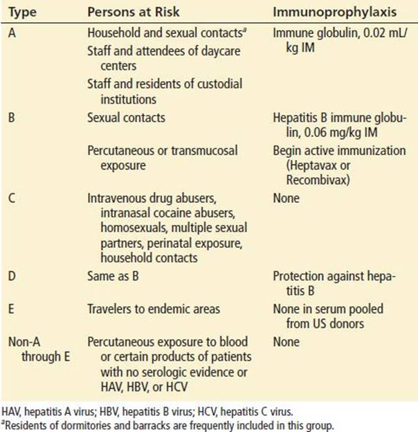
Patients considered at risk for contracting hepatitis A include household and sexual contacts of persons with hepatitis A, staff and attendees at daycare centers, and staff and residents in close contact at custodial care institutions. Immune globulin, which is derived from pooled human serum but treated so as not to be capable of transmitting HIV infection or other diseases, is given as a single intramuscular dose of 0.02 mL/kg as soon as possible within the first 2 weeks after exposure. Casual contacts need not be treated.
Postexposure prophylaxis for hepatitis B is indicated for sexual contacts of persons with hepatitis B or after percutaneous or transmucosal exposure to HBsAg-positive blood. For nonimmunized patients, treatment is with hepatitis B immune globulin, 0.06 mL/kg intramuscularly. Active immunization should be started at the same time. If information about the source patient is unknown but can be obtained within 5 to 7 days, it is reasonable to wait. If not, one must make a decision based on risk factors such as sexual contacts and intravenous drug use. See Chapter 191 for more information on postexposure prophylaxis.
Immunoprophylaxis has not proven useful in patients who have been exposed to hepatitis C or in those suspected of being exposed to non-A through E hepatitis.
CRITICAL INTERVENTIONS
• Hospitalize patients with evidence of fulminant hepatitis (e.g., significantly impaired synthetic function, encephalopathy).
DISPOSITION
Most patients with hepatitis do not require hospital admission, but it is recommended in circumstances such as intractable nausea and vomiting, dehydration, or severe electrolyte imbalance or for signs of hepatic deterioration, as evidenced by changes in sensorium or increased international normalized ratio. Patients with signs or symptoms suggesting a fulminant course of hepatic failure require admission. The absolute levels of serum transaminase values should not be a criterion for admission.
Patients with acute viral hepatitis who are discharged should be prescribed antiemetic agents and instructed to ensure adequate caloric intake. It is best to allow patients to eat small meals of their choice rather than insist they follow some of the high-calorie or high-protein dietary regimens that have been proposed. Prolonged bed rest, once a staple of care, has been shown to be of no value and has its own risks (6). Alcohol consumption should be avoided during the acute illness. After recovery, alcohol consumption has not been shown to be any more harmful for these patients than for the general population (4). Estrogen-based oral contraceptives may be continued during hepatitis.
Patients with acute hepatitis should be seen by an internist or gastroenterologist for follow-up within a few days, after the results of serologic tests. Patients with any prolongation of the prothrombin time are often admitted to the hospital, but others should probably be seen the next day. Instructions should include an admonition to return if food and fluids cannot be kept down or if there is any change in sensorium.
Finally, it is the duty of the emergency physician in many locales to notify public health authorities of cases of acute hepatitis. Patients and their close contacts should be counseled with regard to preventing the spread of hepatitis.
Common Pitfalls
• Missed toxic ingestion or exposure, with continued exposure.
• Failure to recognize hepatitis B or hepatitis C by performing serologic tests during “window periods” between infection and seroconversion.
• Missed hepatobiliary disease, with failure to diagnose surgically correctable lesions.
• Failure to recognize the distinction between liver tests and liver function (e.g., patients with cirrhosis may have nearly normal enzyme levels; patients with “sky high” transaminase levels may have reasonably intact synthetic capability).
• Inappropriate admission of patients with uncomplicated hepatitis and markedly elevated ALT or AST levels without consideration of synthetic factors or clinical status.
• Inappropriate discharge of patients with early evidence of synthetic failure.
• Failure to follow-up contacts for recommended prophylaxis.
• Inappropriate restrictions: Patients with hepatitis need not be excluded from school or work; education regarding precautions is indicated.
• Failure to consider the effects of hepatitis on drugs with hepatic clearance, resulting in high serum levels or long duration of action.
REFERENCES
1. Alter MJ. Epidemiology of hepatitis C. Hepatology. 1997;26:62S–65S.
2. Alter M, Gallagher M, Morris TT, et al. Acute non-A-E hepatitis in the United States and the role of hepatitis G virus infection. Sentinel Counties Viral Hepatitis Study Team. N Engl J Med. 1997;336:741–754.
3. Alter MJ, Kruszon-Moran D, Nainan OV, et al. The prevalence of hepatitis C virus infection in the United States, 1988 through 1994. N Engl J Med. 1999;341:556–562.
4. DeCock KM, Govindarajan S, Chin KP, et al. Delta hepatitis in the Los Angeles area: A report of 126 cases. Ann Intern Med. 1986;105:108–114.
5. Herrera JL. Hepatitis E as a cause of acute non-A, non-B hepatitis. Arch Intern Med. 1993;153:773–775.
6. Hoofnagel JH, Nelson KE, Purcell RH. Hepatitis E. N Engl J Med. 2012:367:1237–1244.
7. Schiff ER. Viral hepatitis. In: Schiff ER, Sorrell MF, Maddrey WC, eds. Diseases of the liver. 9th ed. Philadelphia, PA: Lippincott Williams & Wilkins; 2003:741.
8. Krawitt EL. Autoimmune hepatitis. N Engl J Med. 1996;334:897–903.
9. Durazzo M, Premoli A, Fagoonee S, et al. Overlap syndromes of autoimmune hepatitis: What is known so far. Dig Dis Sci. 2003;48:423–430.
10. Lee WM. Drug-induced hepatotoxicity. N Engl J Med. 2003;349;474–486.
11. Misbin RI. Troglitazone-associated hepatic failure. Ann Intern Med. 1999;130:330.
12. Herrine S. Approach to the patient with chronic hepatitis C infection. Ann Intern Med. 2002;136:747–757.
13. Podalsky DK. Infiltrative, genetic, and metabolic diseases of the liver. In: Kasper DL, Fauci AS, Longo DL, et al., eds. Harrison’s Principles of Internal Medicine. 16th ed. New York, NY: McGraw-Hill; 2002:1869.
14. Centers for Disease Control and Prevention. HYPERLINK “http://www.cdc.gov/hepatitis/HBV/index.htm” Hepatitis C Information for Health Professionals. http://www.cdc.gov/hepatitis/hcv/hcvfaq.htm. Accessed June 18, 2014.