
Stephen J. Wolf
Micturition is a process that relies on the nervous, musculoskeletal, and genitourinary systems. Disruption of any of these systems can potentially lead to urinary dysfunction, resulting in incontinence or retention. The key aspect for an emergency physician in treating patients with incontinence or retention is ruling out emergent causes or complications of the urinary dysfunction (e.g., spinal cord compression or acute postobstructive nephropathy). Often times in doing this, the emergency physician is able to identify key contributing pathology. Once emergent issues are ruled out, most patients can be referred for an outpatient workup of their urinary complaints.
The physiology of micturition involves both conscious (somatic) and unconscious (autonomic) regulation. A cholinergic parasympathetic sacral reflex arc is fundamental in this process and involves the bladder wall, the detrusor muscle, and the vesicular sphincter. At low bladder pressures and distention, parasympathetic tone is low, leading to high urethral sphincter tone and low detrusor activity via the pudendal nerve. β-Adrenergic sympathetic stimulation from levels T10 to T12 also contributes to detrusor and bladder relaxation, while α-adrenergic innervation of the vesicular sphincter results in its resting tone. All of these mechanisms work to promote urine storage and are, in part, modulated by pontine urinary centers.
When bladder distention increases and voiding is desired, micturition is achieved by increasing parasympathetic tone and decreasing sympathetic tone. This, combined with subcortical stimulation of the pontine urinary center and somatic stimulation of the bladder, results in bladder wall contraction and sphincter relaxation. Because sympathetic fibers utilize α- and β-adrenergic receptors and the parasympathetic fibers utilize cholinergic receptors, they are vulnerable to pharmacologic manipulation. Thus, physical or pharmacologic disruption of these systems can result in urinary dysfunction.
CLINICAL PRESENTATION
Urinary incontinence is the involuntary loss of urine, often associated with social embarrassment, isolation, depression, and hygienic complications (1). It can be attributed to a sudden increase in intra-abdominal pressure (stress incontinence), involuntary bladder contractions (urge incontinence), a mixture of the two (mixed incontinence), overflow in the setting of urinary retention (overflow incontinence), complete disruption of the continence apparatus (true incontinence), or nonpathologic causes (functional incontinence) (1–6).
Urinary retention is the inability to void voluntarily, resulting in bladder distention. With this distention, symptoms of prostatism often develop, including frequency, urgency, hesitancy, dribbling, decrease in voiding stream, and a sense of incomplete emptying (1). This can further progress to acute or chronic postobstructive nephropathy. These symptoms may be exacerbated with the application of suprapubic pressure (the Credé maneuver). In the past, this maneuver has been used to initiate voiding in patients with retention; however, it is not recommended because of high complication rates and poor efficacy (7,8).
Although urinary retention is often considered a disorder of elderly men with enlarged prostates, it can affect men and women of all ages and even is seen in children. Often the retention is insidious in its onset, resulting from chronic progressive obstructive or neurosensory causes (e.g., diabetes); however, acute presentations do occur, especially in the setting of urologic infections, certain medications, or neurologic disease.
DIFFERENTIAL DIAGNOSIS
Urinary Incontinence
Stress urinary incontinence (SUI) results when a rapid rise in intra-abdominal pressure is transmitted to the bladder, thereby causing a leakage of urine (3,4). It may result from sneezing, coughing, laughing, exercise, and so forth. Normally, the vesiculourethral anatomy is such that this transmitted abdominal pressure causes a narrowing of the posterior ureterovesical angle, making leakage of urine more difficult. However, this is not the case if there is hypermobility of the urethra or laxity in the intrinsic urethral sphincter. Nearly 85% of SUI occurs in women who have given birth, which can cause laxity of the anterior vaginal wall and pelvic floor, resulting in urethral hypermobility (5,9). Other risk factors include obesity and previous urinary incontinence surgery. Deficiency of the intrinsic urethral sphincter may also result from mechanical issues (trauma, surgery, and chronic bladder overdistention) or neurophysiologic issues (estrogen deficiency, diabetes, corticosteroid therapy, and radiation therapy). Finally, although the majority of SUI occurs in women, transurethral resection of the prostate (TURP) is a particular risk factor in men, whereas structural abnormalities should be considered as a cause for SUI in children.
Urge incontinence occurs when there is uninhibited involuntary contraction of the detrusor muscle and bladder wall (detrusor instability) (4). This is often called a “spastic bladder” or an “overactive bladder.” Detrusor instability is the most common cause of urinary incontinence among the elderly, with a prevalence of 40% to 70% (2). Urinary frequency and urgency are common symptoms. Up to 90% of these cases are considered to be idiopathic. The remaining 10% result from bladder wall irritation/inflammation, urethral syndrome, or neurologic causes (10). Bladder wall irritation and inflammation are usually caused by urinary infections. Treatment often results in resolution of the urge incontinence. Cystitis may also be caused by calculi, radiation, medications (cyclophosphamide, bacille Calmette–Guérin vaccine), or carcinoma in situ. Interstitial cystitis is a unique idiopathic, noninfectious cause of urge incontinence, with inflammation of the muscular layers of the bladder wall. It usually affects women in their third decade of life and results in progressive vesicular fibrosis, eventually leading to decreased bladder compliance and capacity.
Urethral syndrome may also lead to urge incontinence. It most commonly affects women and is a syndrome that consists of urgency, frequency, and dysuria. It is often clinically misdiagnosed as infectious cystitis, but urine cultures are negative and the urinary sediment is noninflammatory, showing squamous epithelial cells.
Neurologic causes of urge incontinence include, but are not limited to, stroke, multiple sclerosis, hydrocephalus, and suprasacral spinal cord lesions (acute cord compression or cauda equina syndrome). In these instances, the normal physiologic relations between the sacral detrusor-sphincter reflex arc and the pontine urinary centers can be disturbed, resulting in detrusor hyperreflexia as opposed to instability.
The differentiation between stress and urge incontinence is not always straightforward, and the clinical pictures overlap, yielding a mixed incontinence diagnosis. Fifty percent to 60% of patients presenting for evaluation of SUI also have an urgency component (5).
Overflow incontinence is a form of incontinence that occurs in the setting of obstructive urinary retention. Obstruction can result in constant dribbling, frequency, and an urge incontinence pattern with detrusor instability.
True incontinence resulting from complete disruption of the continence apparatus or fistula formation, functionally produces a “drain-spout urethra.” This can be due to trauma, surgeries (TURP or previous urethral surgeries), vaginal or cesarean deliveries, malignant erosion or ischemic disease from operative, chemical, or radiation factors. Congenital abnormalities are also a cause for true incontinence in children.
Finally, nonpathologic or functional incontinence should be considered in the differential diagnosis. Patients with functional incontinence have normal anatomy and function, but the incontinence is due to psychological factors or physical limitations in getting to the bathroom. Such constraints could include altered mental status, ethanol intoxication, isolated urinary tract infection, medication use, depression, excessive urine production, or restricted mobility. Other nonpathologic causes of incontinence include enuresis in the pediatric population and incontinence for other psychologic gains. The latter is associated with a sparing of nighttime incontinence while the patient sleeps.
Medications often contribute to urinary incontinence by causing detrusor instability, decreased sphincter tone, functional incontinence, or urinary retention resulting in overflow incontinence. Table 127.1 lists classes of medications that may contribute to urinary incontinence. A helpful mnemonic for transient causes of urinary incontinence is DIAPPERS (Table 127.2).
TABLE 127.1
Medications Associated with Voiding Dysfunctiona
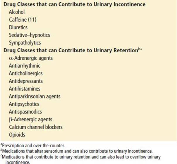
TABLE 127.2
DIAPPERS Mnemonic for Causes of Transient Urinary Incontinence

Urinary Retention
Urinary retention is most often the result of obstruction; however, it can also result from neuropathologic disease, pharmacologic side effects, or psychogenic causes (1). Special congenital considerations also arise when considering the etiology of urinary retention in the pediatric population (Table 127.3). Urinary retention needs to be delineated from anuria and oliguria, which is a lack or decrease of urine production from the kidneys and not an issue of voiding dysfunction.
TABLE 127.3
Obstructive Causes of Urinary Retention

Obstructive urinary retention can be a result of multiple etiologies and occurs much more frequently in men (see Table 127.3). Benign prostatic hypertrophy is the most common etiology, usually occurring in the sixth decade of life. Prostate cancer is a less common cause of obstruction and is associated with a marked elevation of prostate-specific antigen level (1). Fibromuscular contracture of the bladder outlet can occur in the younger male. At the level of the bladder outflow tract, calculi, blood clots, tumor, or polyps can all cause obstruction. At the level of the urethra, obstruction can be secondary to inflammation or stricture. Inflammatory causes include urethritis, prostatitis, tuberculosis, echinococcosis, and herpes simplex. Strictures can result from gonococcal urethritis, trauma, surgery, or radiation but tend not to be caused by nongonococcal urethritis (1). A higher incidence of psychogenic urinary retention tends to be found in women.
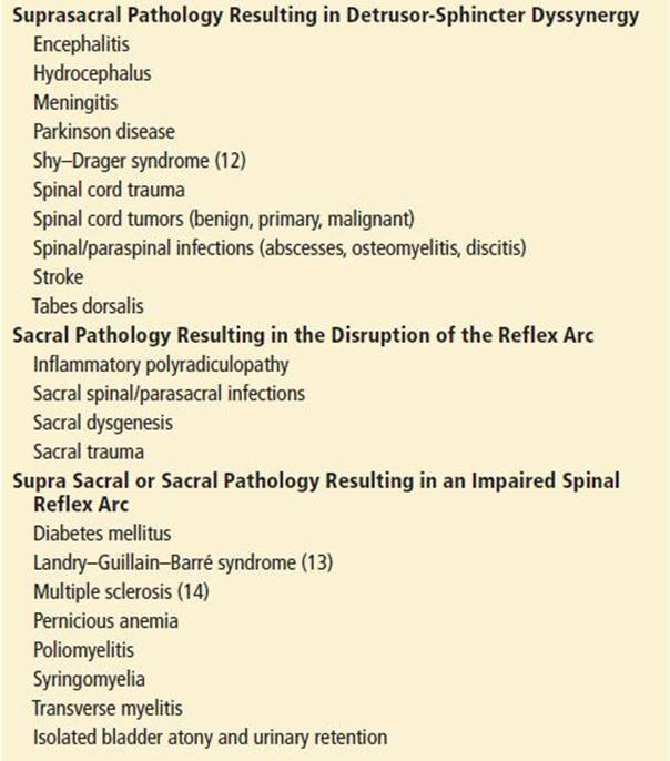
Neuropathologic causes of urinary retention deserve special attention (Table 127.4). Neurologic lesions (e.g., trauma, mass lesions, and infections) at the level of the sacral nuclei can disrupt the spinal reflex arc, thereby causing a flaccid paralysis of the bladder and an inability to void. Suprasacral lesions of the spinal cord or central nervous system may initially result in spinal shock–type physiology with urinary retention, but usually progress to overflow and urge incontinence with detrusor hyperreflexia, owing to the lack of upper-motor neuron control. However, a complication of suprasacral injuries can be detrusor-sphincter dyssynergy, in which there is external sphincter spasm during bladder contraction, resulting in urinary retention.
TABLE 127.4
Neuropathologic Causes of Urinary Retention
Finally, various medications, both prescription and over-the-counter, can precipitate urinary retention (see Table 127.1). Anticholinergic medications inhibit parasympathetic tone and promote urinary retention. Likewise, α- and β-adrenergic medications cause bladder wall relaxation and an increase in urethral sphincter resting tone. This risk is even greater in the setting of other risk factors, for example, a male patient with benign prostatic hypertrophy taking an over-the-counter cold medicine with diphenhydramine.
ED EVALUATION
Urinary Incontinence
The emergency department (ED) evaluation of urinary incontinence should be focused on identifying acute emergent or correctable causes. Most emergent causes are neurologic, resulting in detrusor hyperreflexia, whereas most correctable causes are infectious, overflow, or medication related. The evaluation should focus on a thorough history covering past medical, surgical, psychosocial, and medication histories. Any of these elements may yield clues to the etiology of incontinence. The physical examination should include a thorough neurologic examination, including mental status, gait, deep-tendon reflexes, saddle sensation of the perineum, rectal tone, and the bulbocavernosus reflex. An absent bulbocavernosus reflex in male patients is almost always associated with a neurologic lesion. In women, it can be absent up to 30% of the time (1).
SUI is often implied by the history and is accompanied by a normal physical examination. If needed, a provocation test for SUI can be performed by instilling 200 to 300 mL of sterile saline into the bladder after the measurement of a postvoid residual (PVR) (5). The patient can then be asked to sneeze, cough, or strain. Isolated leakage suggests SUI. Furthermore, if the Marshall test, digital urethral support during a provocation test, prevents the incontinence, the diagnosis is confirmed. Leakage with an inability to stop further urine flow during a provocation test suggests urge incontinence with detrusor instability. Urge incontinence may require investigation into associated signs and symptoms (e.g., back pain) for inflammatory etiologies and/or neurologic etiologies. Associated back pain or an abnormal neurologic examination warrants evaluation for overflow incontinence associated with an obstructive urinary retention.
Urinalysis and urine cultures should also be obtained. They may assist in the diagnosis of infection and noninfectious inflammatory causes.
Emergent urology consultation is not usually necessary, and extensive urologic evaluation is reserved for the outpatient setting. When acute neurologic causes of incontinence are suspected, imaging studies, neurologic, or neurosurgical consultation should be obtained as needed.
Urinary Retention
Evaluation of acute or chronic urinary retention in the ED should be directed at identifying the cause, when possible, and evaluating for the sequela of renal parenchymal damage. Neurologic causes must be carefully excluded. Special attention should be paid to the review of the patient’s medication history, the rectal examination, and a thorough neurologic examination. These can provide significant information as to the potential emergent etiologies of the urinary retention. It should be noted that a normal prostate on examination does not exclude prostatic cause from the differential diagnosis.
Two tests are particularly helpful. A PVR of more than 100 mL suggests retention. A bedside ultrasound can document bladder distention suggestive of outlet or urethral obstruction. This may be especially useful if symptoms persist in the setting of perceived adequate drainage.
Laboratory evaluation of patients found to have urinary retention should include a urinalysis with culture, blood urea nitrogen (BUN), and serum creatinine. The urinalysis can provide clues to infectious etiologies, calculi, or tumors, whereas the urine culture is necessary for detecting subclinical infections. Postobstructive nephropathy is often characterized by an elevated BUN/creatinine (Cr) ratio (i.e., >10). If new or previously undiagnosed renal insufficiency is present, a more complete workup with electrolytes and a renal ultrasound looking for hydronephrosis, hydroureter, or renal parenchymal damage is indicated.
KEY TESTING
Urinary Incontinence
• Blood Sugar, Urinalysis and culture
• Neurologic imaging (when indicated)
Urinary Retention
• Postvoid residual or postvoid volumetric bladder scan
• BUN and creatinine
• Urinalysis and culture
• Renal ultrasound (when indicated)
• Neurologic imaging (when indicated)
ED MANAGEMENT
Urinary Incontinence
Emergent neurologic causes should be addressed as dictated by the suspected etiology, such as ordering emergent magnetic resonance imaging (MRI) in cases of suspected spinal cord compression. Transient causes (see Table 127.2) should also be addressed (e.g., administering antibiotics for infectious cystitis or withholding offending medications when possible).
Further management of stress, urge, true, functional, and nonpathologic incontinence falls into the realm of outpatient urologic consultation. The management of SUI and true incontinence is often surgical, with repositioning of vesicourethral angle (e.g., bladder neck suspensions and pubovaginal slings). Alternatives for SUI include Kegel exercises, weight reduction, estrogen replacement, electrostimulation methods, and α-agonists (pseudoephedrine, phenylpropanolamine) for increasing bladder neck and urethral tone (2). Periurethral collagen injection therapy and urethral occlusion devices have also been used with moderate success for treating SUI (3). Anticholinergic medications (propantheline, hyoscyamine) increase the tolerable bladder volume and decrease involuntary bladder contractions (15). Tricyclic antidepressants (doxepin, imipramine) can also be used to increase bladder tone gives their central and peripheral anticholinergic effect. Oxybutynin and dicyclomine inhibit involuntary bladder contraction through anticholinergic effects and cause smooth muscle relaxation in the bladder wall. Tolterodine has similar effect as oxybutynin via antimuscarinic activity.
Urinary Retention
Initial management of urinary retention should focus on identifying serious neurologic causes and obtaining symptom relief through bladder decompression. Decompression can be accomplished with a well-lubricated 16 to 18Fr catheter and should be done in a timely manner. If this is unsuccessful, attempts can be made with a coudé catheter. Its angulated tip can facilitate catheter passage beyond the median lobe of the prostate. If a urethral catheter cannot be placed, temporary relief may be provided by placing a suprapubic catheter. This requires a palpable bladder and the absence of a history of prior suprapubic surgery. If there is doubt about bladder distention (as in the obese patient), ultrasound confirmation is advised. For percutaneous drainage techniques, a simple midline puncture is made one fingerbreadth above the symphysis pubis, with the trocar or needle directed slightly caudally and then advanced until urine is returned. The catheter or tubing is then advanced, the needle is withdrawn, and the drainage catheter is secured in position.
Traditionally, a single straight catheterization (in and out) of patients with urinary retention has not been recommended, as recurrence of symptoms is likely, and the risk of infection is high. More recently trials without catheter (TWOC) in men with prostatic hypertrophy using concomitant α-adrenergic blockade following initially catheter decompression have shown some success (48% to 62%), particularly in younger patients with low PVR volumes (10).
Bladder mucosal hemorrhage and postobstructive diuresis are two complications of bladder decompression, particularly in the patient with chronic urinary retention. Bladder mucosal hemorrhage is thought to be the result of sudden expansion of bladder wall veins after decompression (1). In the past, clamping of the catheter after a certain amount of drainage was recommended in an attempt to avoid this complication. However, it has been shown that the hydrostatic pressure in the bladder begins to fall immediately with catheter placement and falls markedly within the first 100 mL of urine drained. Therefore, clamping of the catheter does not affect the risk of mucosal hemorrhage and is not indicated (1,17).
Postobstructive diuresis may occur after the decompression of obstructive urinary retention at a rate up to or >1 L/hr. However, it is believed that this diuresis is an appropriate correction of volume overload. Therefore, milliliter-for-milliliter replacement of this diuresed fluid is not indicated. What is indicated is replacement of the concomitant electrolyte loss that accompanies the diuresis. This can often be done through administering a maintenance rate of one-half isotonic saline during the diuresis (18).
CRITICAL INTERVENTIONS
• Exclude serious neurologic disease (e.g., spinal cord compression) as the cause of acute urinary dysfunction.
• Place a bladder catheter immediately to relieve the discomfort of bladder distention in patients with acute urinary retention.
DISPOSITION
Patients with an obstructive uropathy usually require continuous urinary drainage. The patient in otherwise good health with no ancillary requirements for hospital care may be discharged from the emergency department with Foley catheter drainage collected into a leg bag. Follow-up should be arranged prior to discharge, and there should be a thorough discussion with the patient about when to seek further medical attention. Urinary retention related to drugs or medication requires referral to the prescribing physician for recommended adjustment or substitution therapy.
Patients with an emergent neurologic cause of urinary incontinence or retention should be admitted or transferred if appropriate specialty consultation is not available.
Common Pitfalls
• Failure to diagnose a true neurologic emergency such as cord compression or cauda equina syndrome in the patient with urinary retention or incontinence.
• Failure to inquire about over-the-counter medication use.
• Overly aggressive attempts at catheter insertion, resulting in urethral injury.
• Failure to detect associated infection or postobstructive nephropathy in the elderly patient with urinary retention.
• Failure to consider multiple sclerosis and diabetes in a woman with urinary retention.
REFERENCES
1. Curtis LA, Dolan TS, Cespedes RD. Acute urinary retention and urinary incontinence. Emerg Med Clin North Am. 2001;19:591–619.
2. Chutka DS, Fleming KC, Evans MP, et al. Urinary incontinence in the elderly population. Mayo Clin Proc. 1996;71:93–101.
3. Kobashi KC, Leach GE. Stress urinary incontinence. Curr Opin Urol. 1999;9:285–290.
4. Norton P, Brubaker L. Urinary incontinence in women. Lancet. 2006;367:57–67.
5. Rackley RR, Appell RA. Evaluation and medical management of female urinary incontinence. Cleve Clin J Med. 1997;64:83–92.
6. Resnick NM. Urinary incontinence. Lancet. 1995;346:94–99.
7. Barbalias GA, Klauber GT, Blaivas JG. Critical evaluation of the Crede maneuver: A urodynamic study of 207 patients. J Urol. 1983;130:720–723.
8. Chang SM, Hou CL, Dong DQ, et al. Urologic status of 74 spinal cord patients from the 1976 Tangshan earthquake, and managed for over 20 years using the Crede maneuver. Spinal Cord. 2000;38:552–554.
9. Rortveit G, Daltveit AK, Hannestad YS, et al. Urinary incontinence after vaginal delivery or cesarean section. N Engl J Med. 2003;348:900–907.
10. Jabs CF, Stanton SL. Urge incontinence and detrusor instability. Int Urogynecol J Pelvic Floor Dysfunct. 2001;12:58–68.
11. Arya LA, Myers DL, Jackson ND. Dietary caffeine intake and the risk for detrusor instability: A case-control study. Obstet Gynecol. 2000;96:85–89.
12. Lockhart JL, Webster GD, Sheremata W, et al. Neurogenic bladder dysfunction in the Shy-Drager syndrome. J Urol. 1981;126:119–121.
13. Kogan BA, Solomon MH, Diokno AC. Urinary retention secondary to Landry-Guillain-Barre syndrome. J Urol. 1981;126(5):643–644.
14. McGuire EJ, Savastano JA. Urodynamic findings and long-term outcome management of patients with multiple sclerosis-induced lower urinary tract dysfunction. J Urol. 1984;132:713–715.
15. Rai BP, Cody JD, Alhasso A, et al. Anticholinergic drugs versus non-drug active therapies for non-neurogenic overactive bladder syndrome in adults. Cochrane Database Syst Rev. 2012;12:CD003193.
16. Rendell S. Towards evidence-based emergency medicine: Best BETs from the Manchester Royal Infirmary. BET 1. Alpha-blockers increase the chances of a successful trial without catheter after acute urinary retention. Emerg Med J. 2011;28(2):161–165.
17. Foster MC, Upsdell SM, O’Reilly PH. Urological myths. BMJ. 1990;301:1421–1423.
18. Howards SS. Postobstructive diuresis: A misunderstood phenomenon. J Urol. 1973;110:537–540.