
Joseph C. Schmidt
CLINICAL PRESENTATION
Pelvic pain is a common emergency department (ED) presentation. It is often accompanied by various symptoms including fever, vomiting, dysuria, vaginal discharge, or bleeding. The practitioner must rapidly differentiate among etiologies that range from benign to life-threatening. This differentiation requires a complete but focused history and physical examination combined with a broad knowledge of potential disease processes to generate an appropriate differential diagnosis. This differential can then be tested by using appropriate diagnostic studies and generating an assessment and plan that will allow for the timely treatment and disposition of the patient.
DIFFERENTIAL DIAGNOSIS
The differential diagnosis of pelvic pain is broad and falls into two main categories. This discussion is focused on the presentation of acute pelvic pain, which by definition is pain of <6 months in duration. Chronic pelvic pain will be mentioned only in the context that a diligent evaluation of the patient with chronic pain must be accomplished to avoid missing a more worrisome complication or new diagnosis.
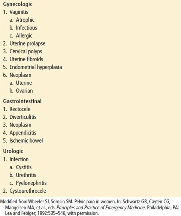
The differential diagnosis of acute pelvic pain encompasses multiple organ systems (Table 129.1). A useful organizational strategy is to divide the patients into functional categories according to reproductive status. This approach highlights the special consideration of each group. Children and prepubertal adolescents have a number of unique issues (Table 129.2). Likewise, postmenopausal women are at risk for conditions that are uncommon in other age groups (Table 129.3). The remainder of the section highlights the most common and important etiologies in each category. It is important to remember that significant overlap between these categories does occur.
TABLE 129.1
Differential Diagnosis of Acute Pelvic Pain
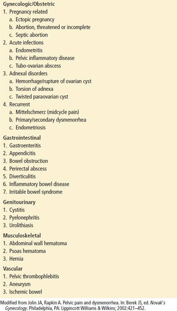
TABLE 129.2
Differential Diagnosis of Prepubertal Pelvic Pain
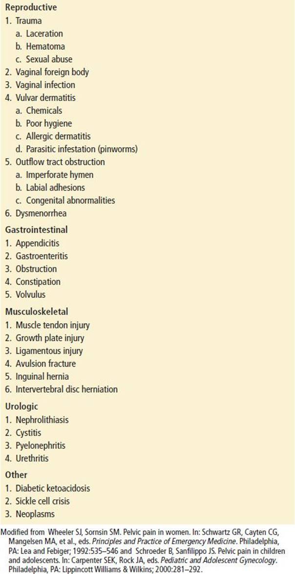
TABLE 129.3
Differential Diagnosis of Postmenopausal Pelvic Pain
Prepubertal
The approach to pelvic pain in the prepubertal patient requires attention to a broad spectrum of organ systems, though gastrointestinal and urologic causes are the most common. Significant gynecologic pathology is uncommon. Pelvic pain related to accidental perineal trauma is not uncommon, but a high index of suspicion should be maintained for possible sexual abuse. Any suspicion of abuse warrants a complete investigation. In the peripubertal age group, complications of sexual activity should be considered. In this age group, symptoms surrounding menarche are also common. In younger children, vulvovaginitis can arise from chemical irritation (i.e., bubble bath) or a vaginal foreign body.
Reproductive Age
Gynecologic and obstetric etiologies of pelvic pain increase in frequency as women enter reproductive age.
Ectopic pregnancy (EP) is more fully discussed in Chapter 137. The classic presentation of EP is catastrophic pain in association with missed menses and vaginal bleeding. Multiple variations of this presentation are common. Early stretch and growth of the ectopic gestation in the fallopian tube can result in mild pain caused by tubal distention and contraction. Intermittent leaking of blood, which often precedes rupture by several days, will cause mild, focal, and intermittent peritoneal irritation, which is often poorly characterized by the patient. Risk factors for EP included a history of infertility, use of an intrauterine device, prior tubal ligation, or other pelvic surgery (1). Because of the diversity of presentations, almost one-half of ectopic pregnancies are missed on initial physician contact (2).
Miscarriage in any of its forms can cause pelvic pain. Examination of the cervical os may differentiate a threatened abortion (closed os) from an inevitable or incomplete abortion. Septic abortion presents with a tender, boggy, palpable uterus and fever. Traction on the round ligament and other pelvic structures as the uterus grows during pregnancy can cause significant but transient pain.
Torsion of the ovary or other adnexal structures is a gynecologic emergency. The pain associated with torsion is generally intense, severe, and unilateral, with frequent radiation to the back or thigh. The pain may be intermittent and associated with nausea and vomiting. An adnexal mass may be palpable. Common predisposing factors include pregnancy, ovarian hyperstimulation syndrome, and ovarian or paraovarian cyst disease. Because the ovary and the kidney have the same innervation (T10 to T11), the clinical picture may mimic renal colic.
Pelvic inflammatory disease (PID) is discussed more fully in Chapter 130. A dull lower abdominal pain, most often bilateral, characterizes uncomplicated PID. Onset is more frequent within 1 week of menses, but the pain may be subacute and develop over several days. If infection extends beyond the endometrium and tubes, pain can become more severe and peritoneal irritation is seen. Clinical signs and laboratory parameters tend to be nonspecific and can include fever, vaginal discharge, cervical motion tenderness (CMT), uterine or adnexal tenderness, and, rarely, a mass. Risk factors include multiple sex partners, prior PID, young age, and either no contraception or the use of an intrauterine device (3,4).
Tubo-ovarian abscess (TOA) may occur with severe PID, particularly when caused by or associated with anaerobic organisms. TOA is the most common intra-abdominal abscess in premenopausal women. The mortality of PID has been reported to be as high as 8.6% when associated with rupture (5). Some TOAs are detected when PID fails to resolve with appropriate antibiotic treatment. In other cases, sterile hydrocele or pyosalpinx results from a remote acute infection. The most common signs and symptoms include abdominal pain, fever, and a palpable pelvic mass (6). Rupture of an abscess causes sudden, frank pain resulting from peritoneal contamination, and it is a gynecologic emergency.
Primary dysmenorrhea often occurs in adolescents, usually within 1 to 2 years of menarche. This diagnosis is very common, with 60% of menstruating women reportedly suffering some level of symptoms (7). Pain caused by myometrial contractions occurs just before or coincident with menstruation, and it is described as severe, crampy, and suprapubic. Pain tends to occur with every menstrual period and may be incapacitating, particularly for the first 1 to 2 days of menses. Secondary dysmenorrhea can occur at any age in a menstruating woman and is most often associated with endometriosis. The rate of dysmenorrhea decreases with increasing age. In a study of 165 university students aged 17 to 19 years, dysmenorrhea caused absenteeism or loss of activity at least once for 42% of the women studied. The most significant associated risk factors were early age at menarche, long menstrual periods, smoking, alcohol intake, and obesity (greater than the 90th percentile) (8).
Sexual abuse has also been strongly associated with dysmenorrhea and chronic pelvic pain. In a study of 581 nonpregnant women aged 18 to 45 years with complaints of dysmenorrhea and chronic pelvic pain, there was a childhood incidence of sexual abuse of 26% and an adult incidence of 28% (9). Other studies also support a significant association between a history of sexual abuse and chronic pelvic pain (10).
Uterine myomata, or fibroid tumors, of the uterus are common, particularly during later reproductive years. Myomata often cause heavy, irregular, and crampy menses. These tumors may undergo ischemia, necrosis, hemorrhage, or torsion. Torsion of the fibroid causes a sharp increase in severity of pain. Physical examination in a patient with fibroids reveals a tender, enlarged, and often irregular uterus. Myomata may also interfere with fertility, increasing the risk of spontaneous abortions (11).
Mittelschmerz is unilateral adnexal pain at the time of ovulation, resulting from leakage of blood or fluid from the Graafian follicle. Sudden, sharp, well-localized peritoneal irritation is usually found without fever, hypotension, or signs of inflammation or infection. An adnexal mass is infrequent. Occasionally, symptoms can be severe and generalized, but they classically decrescendo and resolve within 24 to 48 hours. In some patients, midcycle pain is recurrent and predictable.
Ovarian cyst disease may occur at any age. Simple functional cysts may contain follicular fluid or blood. Corpus luteum cysts are extremely vascular; ruptured corpus luteum cysts are common in pregnancy, presenting with sudden unilateral pain that is usually mild and localized. Less common are dermoid cysts, endometriomas, and serous and mucinous cysts. With cyst rupture, rapid onset of pain is characteristic, and local peritoneal irritation is common, but fever and systemic signs are rare. Pain may be diffuse if cyst fluid or blood contaminates the entire abdominal cavity. Rarely rupture with significant hemorrhage that may lead to hypovolemia and hemodynamic instability (12). Unruptured ovarian cysts may also cause pain if the cyst distends rapidly or if torsion occurs.
Appendicitis is the most common nonobstetric surgical emergency in pregnancy. It is particularly difficult to diagnose in women, reflected by the 25% to 50% negative laparotomy rate for women in reproductive years (13). Diagnosing appendicitis becomes more difficult as pregnancy progresses because the uterus obscures the examination and distorts the location. Delays in surgical management in pregnancy result in increased morbidity for both the mother and the fetus (2,13). CMT may occur in either PID or appendicitis, and pyuria occurs in 10% to 20% because of the proximity of the ureters to the adnexa and appendix. The presence of significant bilateral adnexal tenderness and longer symptom duration (>3 days) may suggest PID, but imaging or laparoscopy may be required to differentiate the two.
Urologic causes of pelvic pain include cystitis, pyelonephritis, and renal calculi. Pyelonephritis usually presents with suprapubic tenderness and costovertebral angle tenderness. Renal calculi typically cause severe unilateral, colicky pain. The patient may have a history of renal calculi, and hematuria is frequently present. A Bartholin gland cyst or abscess can cause incapacitating pelvic pain. The glands are located in the labia minora at approximately the 4 and 8 o’clock positions. Two potential ED treatment strategies are placement of a Word catheter or marsupialization of the cyst (14).
Postmenopausal
Pelvic pain in the postmenopausal patient adds a unique set of concerns to those already mentioned. In this age group, atrophic vaginitis is a common problem but should be a diagnosis of exclusion. Malignancies of both the reproductive and gastrointestinal systems increase with age. Urinary tract infections also increase in frequency; however, caution must be used in attributing symptoms to a questionable urinalysis. Diverticulitis can occur in younger women but is more common in this age group. Finally, ischemic bowel must be considered, especially when pain is out of proportion to the physical examination findings.
ED EVALUATION
An appropriate history includes a description of the pain—onset, progression, quality, character, intensity, and radiation—as well as the reason for the ED presentation. The menstrual history includes gravidity, parity, date, and duration of the last two menstrual periods; regularity; any recent menstrual abnormalities; as well as time of menarche and menopause, when applicable. A history of sexually transmitted diseases (STDs), use and type of contraceptives, recent vaginal discharge, breast symptoms, dyspareunia, and urinary complaints should be obtained. Gastrointestinal complaints must also be evaluated, including nausea, vomiting, diarrhea, or constipation. A history of fever and chills should be noted.
The physical examination should be direct but complete. General appearance may be helpful, although individual pain thresholds vary greatly. The patient who avoids movement may have peritoneal irritation, whereas the patient rocking in distress suggests a viscus under pressure. Alterations in vital signs may help identify systemic infection, sepsis, or significant hypovolemia. Abdominal, pelvic, and rectal examinations are mandatory in all women with pelvic pain.
The abdominal examination should focus on a search for masses or palpable organs, localizing areas of tenderness, and demonstrating peritonitis. Peritonitis is detected by performing maneuvers that move the peritoneum but do not put pressure on visceral organs (heel tap, cough, light percussion, pelvic jarring, and general patient movement). “Rebound” tenderness has been the classic procedure for demonstrating peritoneal irritation, yet it may be the least accurate for diagnosing peritonitis. Interobserver variability is substantial, and the test causes unnecessary pain in patients with potentially surgical disease (15).
The pelvic examination should include inspection of the external genitalia as well as the introitus and cervix. The bimanual examination assesses uterine size, palpable masses, and CMT. CMT is often considered pathognomonic of PID, but other diseases such as appendicitis, cyst rupture, and EP also produce pelvic–peritoneal irritation and CMT. A uterus that is fixed and poorly mobile often indicates adhesions secondary to endometriosis, tumor, or prior infection. Rectal examination involves palpation for focal pain, masses, induration, nodularity, or blood.
A sensitive qualitative pregnancy test (urine or serum) for the β-subunit of human chorionic gonadotropin (β-HCG) is essential for evaluation in all women of childbearing age. With a low threshold (<50 mIU/mL International Reference Preparation), false-negative pregnancy tests occur in <1% of patients, even with an abnormal pregnancy (ectopic or miscarriage). A quantitative serum β-HCG may also assist in the interpretation of ultrasound results in first-trimester pregnancies and allow tracking of pregnancy progression at follow-up. Significant pelvic pathology may exist despite a normal complete blood count. However, an increased white blood cell count may reflect systemic infection, and chronic blood loss may be suspected if a low hematocrit is obtained.
An uncontaminated urinalysis is needed to diagnose a urinary tract infection and may assist in differentiating renal colic from ovarian torsion. Vaginal wet mount can indicate various causes of vaginitis (rarely a cause of pelvic pain, but possibly a marker for STD exposure). Cervical cultures or tests to detect Neisseria gonorrhoeae and Chlamydia trachomatis may assist in the diagnosis of PID.
Pelvic ultrasound is the primary imaging modality if the suspected etiology involves the reproductive tract. Emergency physicians increasingly perform these tests, and several studies have suggested that this practice is safe, accurate, and efficient (16–18). Transvaginal ultrasound can reliably identify intrauterine pregnancy in the first trimester. A gestational sac and fetal pole should be seen by 5 weeks, and fetal heartbeat should be seen by approximately 8 weeks. Pelvic ultrasound is also adept at identifying adnexal masses and, with the use of Doppler flow technology, can confirm perfusion of the ovaries.
CT may be useful in the diagnosis of pelvic pain of otherwise unclear etiology. CT has become the primary imaging modality for renal colic, and it is an appropriate study when appendicitis or other bowel pathology leads the differential diagnosis.
Laparoscopy is the gold standard study in the diagnosis of pelvic pain. Laparoscopy remains the procedure of choice to confirm the diagnosis for the patient who is acutely ill or for whom surgical disease (e.g., appendicitis) cannot be excluded. In a study of laparoscopic examination of 316 women with acute pelvic pain, >76% had an abnormal laparoscopic examination, and 45% of those with abnormal laparoscopic examinations had EP (19).
KEY TESTING
• Urinalysis and pregnancy testing
• Pelvic ultrasound, CT scan, or laparoscopy in selected cases
ED MANAGEMENT
The ED treatment of the patient with pelvic pain can be broken down into several critical questions.
Is the Patient Hemodynamically Stable?
Large-bore intravenous access, monitoring, and crystalloid fluid resuscitation are the first steps in managing the unstable pelvic pain patient. Early operative intervention is indicated when patients are persistently unstable. When multiple etiologies are being considered, it is reasonable to simultaneously consult the appropriate services.
Is the Patient Pregnant?
Early establishment of pregnancy status is critical. It should occur as early as possible and occur concurrent with resuscitation when necessary. A positive pregnancy test requires effort to be focused first on confirming or excluding EP.
Does the Patient Have Severe Pain or Peritoneal Signs?
The presence of peritonitis requires a diligent search for surgically correctable etiologies. Early consultation for consideration of direct operative evaluation is prudent. Atypical presentations or severe pain without evidence of peritoneal irritation may require appropriate imaging studies to further focus the differential diagnosis. Pain control should be a primary goal, and evidence suggests analgesia may improve diagnostic accuracy (20).
Does the Pelvic Examination Reveal Evidence of Infection?
Pelvic examination findings suggestive of PID in the absence of severe pain or peritonitis can be treated by a number of approved regimens (see Chapter 130). Imaging in PID is limited to the evaluation of possible complications such as TOA.
Does the Patient Have an Alternative Diagnosis?
An alternative diagnosis should be considered in the stable, nonpregnant patient with mild-to-moderate symptoms and no evidence of PID. Urologic and gastrointestinal etiologies are most common. If other diagnoses are reasonably excluded, symptom control and outpatient referral are indicated.
CRITICAL INTERVENTIONS
• Provide aggressive resuscitation and monitoring of unstable pelvic-pain patients
DISPOSITION
Admission is necessary for potential surgical disease, including appendicitis, selected cases of PID, and EP associated with significant symptoms.
Observation may be necessary if a surgical etiology is possible, pain remains severe, or vomiting and other signs of systemic toxicity are worrisome to the clinician. Observation is appropriate for patients with focal peritonitis secondary to presumed cystic rupture or for suspected early appendicitis. Patients observed in the ED should have serial examinations and clear criteria for admission.
If a patient is discharged from the ED with pelvic pain in which the etiology is uncertain but the patient is stable, instructions for appropriate and early follow-up are imperative. Any woman with abdominal pain who is discharged from the ED must be instructed to obtain a repeat evaluation if her pain continues or worsens. Because many causes of pelvic pain, particularly when the pain is chronic, are not amenable to ED diagnosis, there will exist a population of patients for whom exclusion of surgical disease, short-term pain relief, and referral for definitive diagnosis are the appropriate actions in the ED.
Common Pitfalls
• Failing to recognize the severity of illness by relying inappropriately on vital signs.
• Failing to obtain pregnancy testing particularly around the time of menarche and menopause or in those who report birth control use or history of sterilization procedures.
• Attributing pelvic pain to urinary tract infection when the urinalysis is questionable and other causes have not been ruled out.
• Failing to recognize that CMT is neither specific nor sensitive for PID.
REFERENCES
1. Dart RG, Kaplan B, Veraklis K. Predictive value of history and physical examination in patients with suspected ectopic pregnancy. Ann Emerg Med. 1999;33:283–290.
2. Abbot J, Emmams LS, Lowenstein S. Ectopic pregnancy: Ten common pitfalls in diagnosis. Am J Emerg Med. 1990;8:515–522.
3. Baines PA, Allen GW. Pelvic pain and menstrual related illnesses. Emerg Med Clin North Am. 2001;19:763–781.
4. Zeger W, Holt K. Gynecologic infections. Emerg Med Clin North Am. 2003;21:631–665.
5. Krivak T, Propst A, Horowitz G. Tobo-ovarian abscess; principles of contemporary management. Female Patient 1997;22:27–44.
6. McNeeley SG, Hendrix SL, Mazzoni MM, et al. Medically sound, cost-effective treatment for pelvic inflammatory disease and tuboovarian abscess. Am J Obstet Gynecol. 1998;178:1272–1276.
7. Burnett MA, Antao V, Black A, et al. Prevalence of primary dysmenorrheal in Canada. J Obstet Gynaecol Can. 2005;27:765–770.
8. Harlow DS, Park M. Longitudinal study of risk factors for occurrence, duration and severity of menstrual cramps in a cohort of college women. Br J Obstet Gynaecol. 1996;103:1134–1142.
9. Jamieson DJ, Steege JF. The association of sexual abuse with pelvic pain complaints in a primary care population. Am J Obstet Gynecol. 1997;177:1408–1412.
10. Paras MI, Murad MH, Chen LP, et al. Sexual abuse and lifetime diagnosis of somatic disorders. JAMA. 2009;5:550–561.
11. Grabo T, Fahs PS, Nataupsky LG, et al. Uterine myomas: Treatment options. J Obstet Gynecol Neonatal Nurs. 1999;28:23–31.
12. McWilliams GD, Hill MJ, Dietrich CS 3rd. Gynecologic emergencies. Surg Clin North Am. 2008:88:265–283.
13. Brown JJ, Wilson C, Coleman S, et al. Appedicitis in pregnancy: An ongoing diagnostic dilemma. Colorectal Dis. 2009;11:116–122.
14. Hill DA, Lense JJ. Office management of Bartholin gland cysts and abscesses. Am Fam Physician. 1998;57:1611–1616.
15. Liddington MI, Thompson WH. Rebound tenderness test. Br J Surg. 1991;78:795–796.
16. Durham B, Lane B, Burbridge L, et al. Pelvic ultrasound performed by emergency physicians for the detection of ectopic pregnancy in complicated first-trimester pregnancies. Ann Emerg Med.1997;29:338–347.
17. Hahn SA, Lavonas EJ, Mace SE, et al. Clinical policy: Critical issues in the initial evaluation and management of patients presenting to the emergency department in early pregnancy. Ann Emerg Med.2012;60(3):381–390.
18. Shih CHY. Effect of emergency physician-performed pelvic sonography on length of stay in the emergency department. Ann Emerg Med. 1997;29:348–351.
19. Mikkelson A, Felding C. Laparoscopic and ultrasound examination in women with acute pelvic pain. Gynecol Obstet Invest. 1990;30:162–164.
20. Attard A, Corlett MJ, Kidner NJ, et al. Safety of early pain relief for acute abdominal pain. BMJ. 1992;305:554–556.