
Ericka L. Powell
Vaginal discharge and discomfort is the most frequent gynecologic complaint of women during their reproductive years. Vaginitis is the inflammation of the vaginal mucosa and vulva that often causes vaginal discharge, irritation, itching, and odor. Vaginal infection may also cause dyspareunia and dysuria. Vaginitis in women aged 14 to 49 is typically due to Candida, Trichomonas, and Gardnerella species (1). Noninfectious causes include allergies, immune dysfunction, and chemical irritation. Vaginitis after menopause is typically related to atrophy of the vaginal mucosa from decreased estrogen levels.
Evaluation in the emergency department (ED) includes the three “Ps”—pelvic examination, partners (sexual history), and pH combined with microscopic evaluation of the vaginal discharge on wet mount. This approach will yield a diagnosis in the majority of cases. Vaginal cultures have remained the gold standard for diagnosing the cause of vaginitis, although recent developments in point-of-care testing and polymerase chain reaction (PCR) tests may be more sensitive (2,3).
CLINICAL PRESENTATION
The etiology of vaginitis determines the patients’ predominant symptoms and clinical presentation. When taking the history, it is important to distinguish changes in the patient’s normal physiologic vaginal discharge, which vary with age, pregnancy, use of exogenous hormones, and menstrual cycle.
Candida vulvovaginitis, commonly called a yeast infection, is caused by the fungus Candida albicans. These filamentous organisms penetrate the mucosal surface, causing local tissue lysis and hyperemia. Fungi can be cultured from the vagina in up to 20% of asymptomatic women; they are commensals in the vaginal ecosystem until it is disturbed. Diabetes, immunosuppression (e.g., AIDS or chronic steroid use), and hormonal shifts as occur in pregnancy increase the likelihood of a yeast infection; low-dose oral contraceptives do not (4). Candida is typically not associated with other causes of vaginitis: Trichomoniasis or Gardnerella.
The hallmark complaint of patients with candida vulvovaginitis is vulvar itching and burning associated with dysuria and dyspareunia. Patients complain of a thick white “cottage cheese” discharge that is odorless. The quantity of discharge may vary.
Trichomoniasis is the most common nonviral mediated sexually transmitted disease (STD) in the United States. Non-Hispanic black women are ten times more likely to have this infection. Other risk factors include poverty, lower educational level, and greater number of sexual partners (5,6). Trichomonas vaginalis, a unicellular flagellated protozoan, is sexually transmitted and has been isolated in virtually all genitourinary structures of men and women. These protozoa cause urethritis and cervicitis. Trichomoniasis often presents concurrently with bacterial vaginosis (BV), gonorrhea, and other STDs. This infection has also been associated with preterm labor (5).
Patients with trichomonas vaginitis often complain of a thin, copious, malodorous discharge.
BV is the most common cause of vaginal discharge. This infection is diagnosed in 25% of pregnant women and up to 18% women deny recent sexual activity (1). The normal vaginal environment is a dynamic balance of endogenous bacterial flora, including Lactobacillus, which helps to maintain a relatively acidic pH of 3.8 to 4.2. Various events may cause the lactobacilli to be replaced by high concentrations of anaerobes, particularly Gardnerellaspecies, a gram-negative rod. BV is a synergistic polymicrobic infection in which the vaginal flora become primarily anaerobic. This does not produce inflammatory changes; hence the term bacterial vaginosis rather than vaginitis.
Women with BV complain of “fishy” vaginal odor and discharge. This odor tends to increase after sexual intercourse when alkaline sperm trigger the release of aromatic amines.
DIFFERENTIAL DIAGNOSIS
The differential diagnosis for patients presenting with symptoms of vaginal discharge, itching, or pain includes vaginitis and other infections such as gonorrhea, chlamydia, and herpes. A Bartholin cyst or abscess may present with vaginal pain or itching, and a retained foreign body may present with a malodorous discharge. Less commonly, a rectovaginal fistula or a cervical or uterine neoplasm may present with a chief complaint of vaginal discharge. Various forms of dermatitis may have similar presenting complaints.
ED EVALUATION
Patients with vaginitis can generally be diagnosed and treated based on the ED history, physical examination, wet mount findings, and pH testing. Occasionally, the diagnosis is revealed later on culture results or urinalysis.
In patients with candidiasis, the vulva may appear erythematous, excoriated, and edematous. There may be satellite pustules at the margins of the infection. The vagina may be sore on speculum examination, and a thick white “cottage cheese” discharge may be adherent to the walls of the vagina. The vaginal pH is typically <4.5. This diagnosis is often based on clinical criteria. Diagnostic testing typically requires examining vaginal discharge under a microscope. A small sample of vaginal discharge is placed on a slide with two drops of normal saline and one to two drops of 10% to 20% solution of KOH. This preparation must be scrutinized at multiple power fields to identify the mycelia or pseudohyphae of Candida, which have the appearance of tangled branching chains or oval spores (Fig 132.1). The sensitivity of this method varies based on experience, clinical setting, and training when compared to the gold standard yeast culture (3). Although rapid laboratory tests for yeast have been reported to be more sensitive, specific, and cost-effective, these studies are not widely used in the ED (7,8).
Trichomoniasis is characterized by a profuse malodorous “frothy” discharge on physical examination. The discharge is thin and copious and may be grey-yellow or green. Unlike candidiasis, speculum examination reveals inflammation limited to the vaginal walls and the labia minora. Colpitis macularis, “strawberry” cervix, occurs in a small percentage of women, and up to 50% of patients are asymptomatic (9).
Wet mount preparation should be done as soon as possible after collecting a specimen. Trichomonads can be easily distinguished from white blood cells (WBCs) on low-power microscopy (see Fig. 132.1). They are slightly larger than a WBC and show a “corkscrew” motility. It may be difficult to visualize the entire organism if the vaginal smear specimen is cold or undiluted or if the organism is covered by WBCs. In this situation, only the beating flagella are visualized among the WBCs. WBCs on wet mount are a common finding in Trichomonas infection, other STDs, and atrophic vaginitis but are not typical of candidiasis or BV. Wet mount examination has a specificity of up to 70% but a relatively low sensitivity. The pH of the discharge is typically elevated, between 5 and 7. Trichomonas infection is frequently diagnosed on Pap smear and can also be found on urine samples. PCR testing is not routinely done, and the reported sensitivity and specificity are variable (10,11).
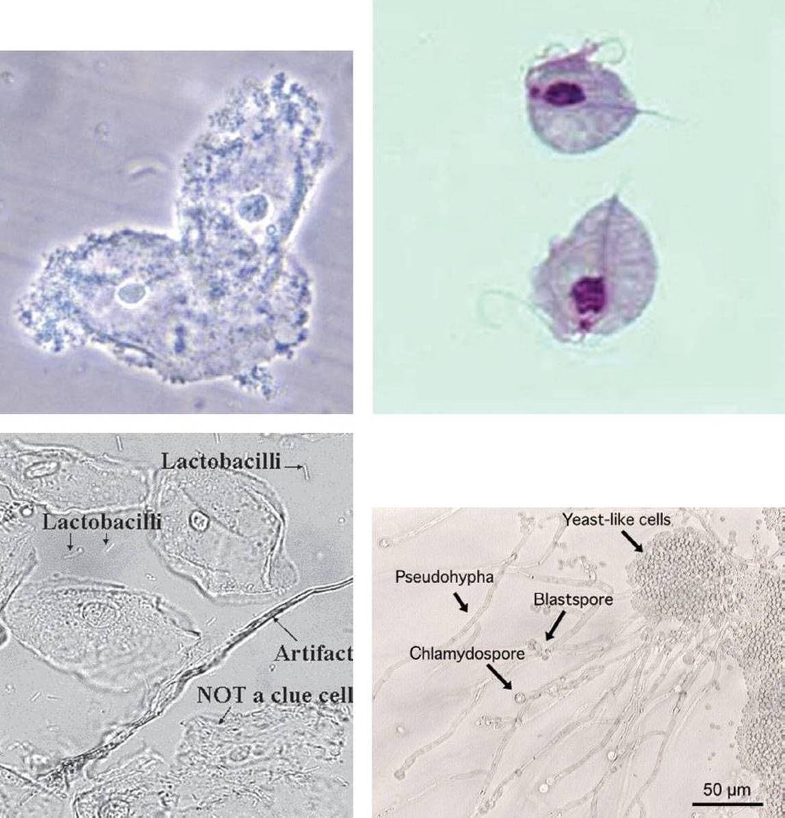
FIGURE 132.1 Wet mount findings in bacterial vaginosis, trichomonas vaginitis, and candida vaginitis.
Patients with BV often have a thin white or grey discharge that is noted on speculum examination to adhere to the vaginal walls. The examiner may notice bubbles in the vaginal discharge. If the cervix is friable or erythematous, the patient should be evaluated for concomitant infections with other STDs. The wet mount microscopic findings associated with BV have been historically based on Amsel criteria, which include the following:
1. Thin homogenous grey or white discharge
2. Clue cells on microscopy (see Fig. 132.1, bacterial studded vaginal epithelial cells)
3. vaginal fluid pH >4.5
4. positive whiff test (release of fishy amine odor with 10% KOH)
To make the diagnosis of BV, three of the four criteria must be present (12).
An alternative method of diagnosis is Gram stain of a vaginal smear to identify and quantify colonies of anaerobes. The accuracy of diagnosis increases when both tests are used in combination (13). A more rapid BV test with improved sensitivity (BVBlue, Gryphus Diagnostics, LLC, Birmingham, AL) is now available but rarely used. This is a point-of-care chromogenic diagnostic test based on the presence of elevated sialidase enzyme in vaginal fluid samples (14).
KEY TESTING
• Wet mount and pregnancy test
• Vaginal cultures in selected patients
ED MANAGEMENT
Treatment regimens for vaginitis have remained relatively the same over many years. Table 132.1 summarizes the treatment of vaginitis of various etiologies. Recurrence rates for vaginitis are particularly high for Candida but also relatively common with Trichomomas. Women with resistant infections may need to be referred for additional testing and evaluation for an underlying immunocompromised state. Vaginal culture should be done on all individuals with recurrent vaginitis. Resistant strains of T. vaginalis have been reported (15).
TABLE 132.1
Differential Diagnosis and Treatment of Vaginitis

Candida albicans is typically treated with fungicidal imidazole derivatives. These inexpensive intravaginal preparations are available over the counter and are tolerated by most women. A single oral dose of fluconazole (150 mg) has cure rates similar to those of topical therapies (16). Candidiasis is not sexually transmitted, and partner treatment is not recommended unless males have signs and symptoms of balanitis. Pregnant women should not receive oral fluconazole (5).
T. vaginalis can be cured with single-dose metronidazole (2 g). An alternative treatment regimen consists of metronidazole 500 mg twice daily for 7 days, but compliance is lower. Metronidazole gel is less effective against T. vaginalis (<50%) and is not recommended. Tinidazole has been approved by the Food and Drug Administration as an alternative treatment that is very useful for T. vaginalis resistant to metronidazole (17). Multiple studies have failed to show teratogenic effects of metronidazole, and symptomatic pregnant patients can be treated with a single oral 2-g dose (10). Patients taking metronidazole should avoid alcohol during treatment and for 24 hours after the last dose to avoid a “disulfiram-like” reaction (18). Patients with an allergy to metronidazole should be referred for desensitization. Other medications such as clotrimazole have higher treatment failure rates. It is imperative to recommend treatment to all sexual partners of women with diagnosed Trichomonas. Reinfection is twice as likely if all partners are not treated. Asymptomatic patients who have positive urine cultures or pap tests should also be treated, as a third of these women can be expected to become symptomatic within 3 months.
Symptomatic patients with BV can be treated with oral and vaginal preparations of metronidazole or clindamycin. All symptomatic pregnant patients should be treated with oral therapy. Intravaginal clindamycin cream has been reported to have adverse effects in pregnancy, including preterm delivery (18). An obstetrician should be consulted on asymptomatic pregnant patients who have diagnosed with BV.
CRITICAL INTERVENTIONS
• Treat infectious causes of vaginitis
DISPOSITION
Patients with simple vaginitis can be treated as outpatients, with follow up by their primary care physician or gynecologist. Those with trichomoniasis should undergo screening for other STDs, along with referral to community resources for counseling regarding safe sex practices and human immunodeficiency virus.
Common Pitfalls
• Failure to perform a thorough history and speculum examination.
• Failure to evaluate for Candida and Trichomonas in patients complaining of dysuria.
• Neglecting to refer patients for counseling regarding high-risk sexual behaviors and testing for HIV and other STDs.
• Failure to instruct patients to avoid alcohol during a course of metronidazole.
• Failure to recommend treatment for partners of patients with trichomoniasis.
REFERENCES
1. Koumans EH, Sternberg M, Bruce C, et al. The prevalence of bacterial vaginosis in the United States, 2001–2004; associations with symptoms, sexual behaviors, and reproductive health. Sex Transm Dis.2007;34(11):864–869.
2. Biggs WS, Williams RM. Common gynecologic infections. Prim Care. 2009;36(1):33–51, viii. [Medline].
3. Katz V. In: Gretchen ML, Roger AL, David MG, Vern LK, eds. Mosby. Katz:Comprehensive Gynecology. 5th ed. Philadelphia, PA: Elsevier Mosby; 2007:588–596.
4. Sobel JD. Vulvovaginal candidosis. Lancet. 2007;369:1961–1971.
5. Workowski KA, Berman S. Centers for Disease Control and Prevention (CDC). Sexually transmitted diseases treatment guidelines, 2010. MMWR Recomm Rep. 2010;59(RR-12):1–110. [www.cdc.gov.std].
6. Sutton M, Sternberg M, Koumans EH, et al. The prevalence of Trichomonas vaginalis infection among reproductive-age women in the United States. 2001–2004. Clin Infect Dis. 2007;45(10):1319–1326.
7. Chatwani AJ, Mehta R, Hassan S, et al. Rapid testing for vaginal yeast detection: A prospective study. Am J Obstet Gynecol. 2007;196(4):309.e1–e4. [Medline].
8. Foongladda S, Haouharn P, Sakulmaiwatana P, et al. Comparative evaluation of Candi Select test and conventional methods for identification of Candida albicans in routine clinical isolates. Mycoses.2002;45(3–4):75–78.
9. Wolner-Hanssen P, Krieger JN, Stevens CE. Clinical manifestations of vaginal trichomoniasis. JAMA. 1989;261(4):571–576.
10. Workowski KA, Berman SM. Sexually transmitted diseases treatment guidelines, 2006. MMWR Recomm Rep. 2006;55:1–94. [Medline].
11. Schwebke JR, Burgess D. Trichomoniasis. Clin Microbiol Rev. Oct 2004;17(4):794–803, table of contents. [Medline].
12. Amsel R, Totten PA, Spiegel CA, et al. Nonspecific vaginitis. Diagnostic criteria and microbial and epidemiologic associations. Am J Med. 1983;74:14–22.
13. Schwiertz A, Taras D, Rusch K, et al. Throwing the dice for the diagnosis of vaginal complaints? Ann Clin Microbiol Antimicrob. 2006;5:4.
14. Bradshaw CS, Morton AN, Garland SM, et al. Evaluation of a point-of-care test, BVBlue, and clinical and laboratory criteria for diagnosis of bacterial vaginosis. J Clin Microbiol. 2005;43(3):1304–1308.
15. Centers for Disease Control and Prevention. Sexually Transmitted Diseases Treatment Guidelines, 2010: Diseases Characterized by Vaginal Discharge. Available at http://www.cdc.gov/std/treatment/2010/vaginal-discharge.htm#a2. Accessed June 24, 2013.
16. Nurbhai M, Grimshaw J, Watson M, et al. Oral versus intra-vaginal imidazole and triazole anti-fungal treatment of uncomplicated vulvovaginal candidiasis (thrush). Cochrane Database Syst Rev. 2007;(4):CD002845. [Medline].
17. Nailor MD, Sobel JD. Tinidazole for the treatment of vaginal infections. Expert Opin Investig Drugs. 2007;16(5):743–751.
18. ACOG Practice Bulletin. Clinical management guidelines for obstetrician-gynecologists, Number 72, May 2006: Vaginitis. Obstet Gynecol. 2006;107(5):1195–1206..