
Alan C. Heffner
Acute joint pain is a common emergency condition that carries a broad differential diagnosis. Trauma and crystal disease are the most common causes, while bacterial infection carries the greatest potential for morbidity and mortality. Arthrocentesis and synovial fluid analysis are key steps in the evaluation of acute monoarticular arthritis.
CLINICAL PRESENTATION
Acute arthritis typically presents with joint pain and swelling. The onset and severity of inflammation varies greatly depending on etiology and host factors such as age, comorbid disease, and immunocompetence. This chapter focuses on the most important types of monoarticular arthritis including crystal and infective arthropathies. Septic arthritis is divided into gonococcal and nongonococcal disease, reflecting the varied pathogenesis and management of these diseases.
Gonococcal Arthritis
Neisseria gonorrhoeae remains a common cause of septic arthritis in young, healthy, sexually active adults in the United States. Disseminated gonococcal infection (DGI) should be suspected in any sexually active patient with joint complaints. Women are more frequently affected than men and are predisposed to systemic spread during pregnancy and the perimenstrual period. DGI presents in two distinct patterns. The first is an acute suppurative monoarthritis involving the knee, wrist, or ankle that occurs in 40% of DGI patients. Polyarthralgias may precede localization to a single joint. The second pattern is the gonococcal arthritis–dermatitis syndrome. Hematogenous spread of bacteria and immune complexes underlie the triad of dermatitis, tenosynovitis, and migratory polyarthritis. The classic skin lesions are scattered small painless erythematous macules or petechiae, which evolve into pustular to necrotic lesions on an erythematous base. Transient painful extensor tenosynovitis often involves the wrists, hands, and ankles but asymmetric migratory extremity polyarthralgias are the most common. Toxicity varies greatly in DGI. Patients can experience only mild constitutional symptoms and fever is often absent. Symptoms of primary genital infection may be absent.
Nongonococcal Septic Arthritis
Nongonococcal septic arthritis is a medical emergency due to the potential for rapid joint destruction and clinical sepsis. Permanent joint damage complicates 40% of septic joints and overall mortality is 15% but reaches as high as 50% in some subgroups (1,2). Large weight-bearing joints such as the knee and hip are most commonly involved, with the shoulder, ankle, elbow, and wrist occurring with decreasing frequency. Any articular surface may be affected including sternoclavicular, acromioclavicular, and sacroiliac joints.
Pathogenesis of nongonococcal septic arthritis involves one of four mechanisms: hematogenous seeding, contiguous spread, direct traumatic implantation, or postoperative invasion of prosthetic hardware. Hematogenous seeding of the vascular synovium is the most common source of septic arthritis. Bacteremia may stem from IV drug use, indwelling vascular access, recent invasive procedures, and distant active or recent infection. Contiguous spread into a joint occurs from adjacent cellulitis, tenosynovitis, osteomyelitis, or abscess. Examples include diabetic foot infections and pediatric metaphyseal osteomyelitis. Direct joint inoculation occurs with trauma or iatrogenically; the clenched fist “fight bite” injury and cat bites to the hand and wrist are commonly overlooked injuries.
Local factors such as antecedent arthritis, joint surgery or trauma, intra-articular steroid injections, and prosthetics predispose patients to septic arthritis. The risk of septic arthritis is severalfold higher in patients with underlying rheumatoid arthritis (1,3). Infections complicate up to 2% of prosthetic joint procedures, with revision arthroplasty carrying even greater risk. Pain localized to a prosthetic joint may signify mechanical loosening, prosthesis failure, or joint infection. Crystal-induced arthritis is rare in prosthetic joints.
Nongonococcal septic arthritis classically presents with rapidly progressive joint pain and swelling. Chronic illness and immunocompromise increase the risk of septic arthritis but also moderate the presenting symptoms. Insidious clinical progression in the absence of systemic toxicity is common in this group. Polyarticular involvement occurs in 10% to 20% of patients (4).
Gram-positive cocci remain the major nongonococcal pathogens in adults. Staphylococcus aureus is the most common causal organism, followed by streptococci including Streptococcus pneumoniae. Gram-negative bacilli including Pseudomonas aeruginosa cause 20% of infections and occur predominantly in immunocompromised patients and IV drug users. Anaerobes are isolated in a minority of cases but are more common with penetrating trauma. Inoculation of oral flora occurs in penetrating bite wounds. Mycobacterial and fungal arthritis are uncommon but important considerations in immunocompromised patients and those presenting with subacute progressive pain of the spine or weight-bearing joints.
Crystal-Induced Monoarthritis
Crystal-induced acute arthropathy is more common than septic arthritis and represents the most important mimic of infection. Gout and pseudogout may be clinically indistinguishable from septic arthritis. In both disorders, patients may demonstrate systemic symptoms, including fever.
Gout develops when monosodium urate (MSU) crystals precipitate within joints and trigger an acute inflammatory reaction. The typical presentation is a middle-aged man with severe joint pain, swelling, redness, and warmth that evolves rapidly over hours to days. The initial attack is monoarticular in 85% of cases. Although any joint may be affected, the first metatarsophalangeal (MTP) joint (podagra) is involved in 60% of cases, followed by ankle, midfoot, and knee. Left untreated, gout follows a course of increasingly frequent attacks separated by shorter intercritical periods. Recurrent exacerbations cause chronic arthritic changes with bony erosions resembling rheumatoid arthritis or degenerative joint disease. Noninflammatory soft tissue tophaceous deposits may develop in the Achilles tendon or extensor surface of the fingers, forearms, or elbows. Common precipitants of acute attacks include increased purine intake, enhanced cellular breakdown (i.e., chemotherapy), alcohol, and joint trauma. In contrast to the classic presentation, older men and postmenopausal women may present with subacute to chronic polyarticular synovitis involving the small joints of the hands and feet. This presentation, resembling rheumatoid arthritis, is strongly associated with diuretic use and renal insufficiency.
Pseudogout is the result of calcium pyrophosphate dihydrate (CPPD) crystal deposition and manifests three common clinical courses: (1) asymptomatic radiographic stippling of articular cartilage (chondrocalcinosis), (2) acute attacks of mono- or oligoarticular inflammatory arthritis (pseudogout), and (3) progressive degenerative joint changes similar to osteoarthritis. Pseudogout usually develops after age 50 and may involve single or multiple joints, especially the knees, wrists, ankles, and elbows. Acute attacks may exhibit abrupt onset and marked discomfort, clinically indistinguishable from gout or septic arthritis. More commonly, the exacerbations evolve over days. Systemic illnesses including surgery or trauma are common triggers. Most cases are idiopathic, but CPPD deposition is associated with hyperparathyroidism and hemochromatosis.
DIFFERENTIAL DIAGNOSIS
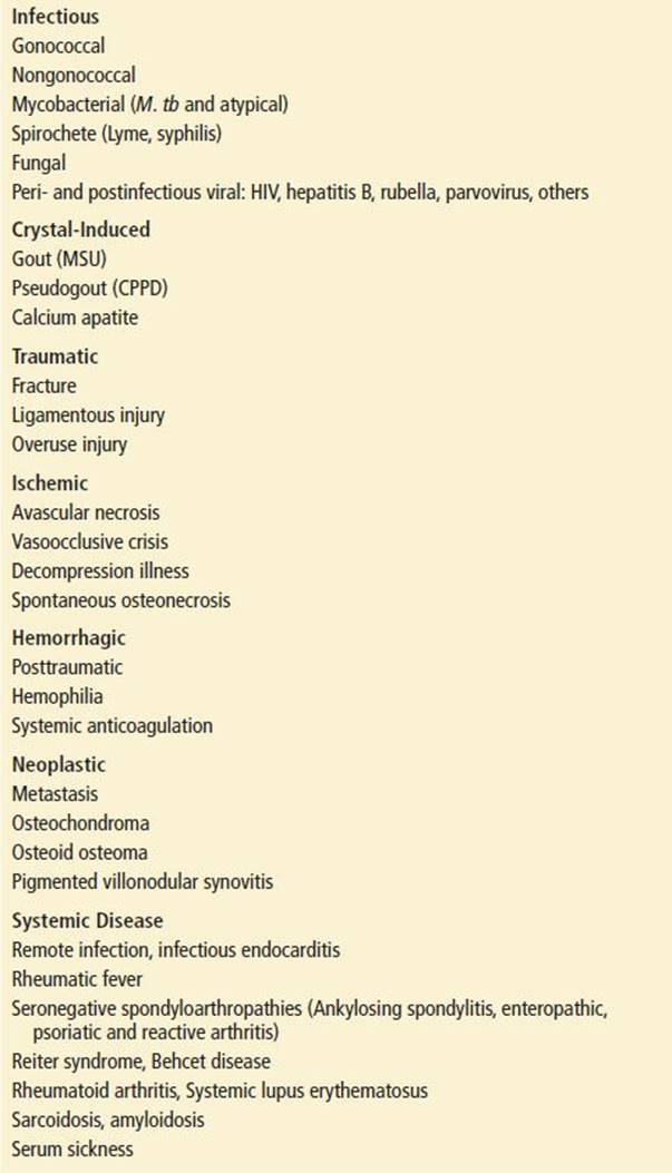
Acute monoarthritis occurs with inflammatory, infectious, ischemic, neoplastic, traumatic, mechanical, or hemorrhagic processes (Table 150.1). The major diseases to consider include septic arthritis, crystal arthropathy, and trauma, but many disorders may produce pain, swelling, and erythema in or around a joint.
TABLE 150.1
Differential Diagnosis of Acute Monoarticular Arthritis
Periarticular conditions such as cellulitis, tendonitis, and bursitis must be distinguished from intra-articular pathology. Enthesitis is localized to tendinous insertions to bone. Bursitis results in pain and swelling overlying bony prominences. Most cases are noninfectious but septic bursitis may occur in the setting of local trauma; the olecranon and prepatellar bursas are the most commonly infected.
A variety of classically polyarticular inflammatory conditions including rheumatoid arthritis, seronegative spondyloarthropathy, systemic lupus erythematosus, and postinfectious arthritis may present with acute monoarticular symptoms. The seronegative arthritides, characterized by the absence of rheumatoid factor, include ankylosing spondylitis, psoriatic arthritis, enteropathic arthritis (associated with inflammatory bowel disease), and reactive arthritis (such as Reiter syndrome). Sterile postinfectious arthritis may be triggered by preceding or coincident infection with a variety of agents including Group A streptococci, HIV, parvovirus, hepatitis B, and rubella. Serum sickness and endocarditis are additional considerations.
Lyme disease manifests musculoskeletal features ranging from acute episodic arthralgia to chronic mono- or oligoarticular arthritis. Knee involvement is common. A history of tick exposure, travel to an endemic region, or erythema migrans rash is helpful but not always present.
Vascular compromise may lead to bone infarction and osteonecrosis. The weight-bearing surface of the femoral head and medial condyle of the knee are common locations. Patients complain of severe pain in the absence of trauma. Precipitants include sickle cell vasoocclusive disease, vasculitis, decompression illness, posttraumatic injury (i.e., hip dislocation), and corticosteroid-induced avascular necrosis.
Traumatic joint injuries are common. Development of an immediate joint effusion after injury suggests hemarthrosis due to ligamentous disruption or osteochondral fracture. Hemarthrosis may occur spontaneously or after minor trauma in the setting of coagulopathy due to anticoagulation or hemophilia. Osteoarthritis is a chronic, slowly progressive disease affecting weight-bearing joints. Acute exacerbations may coincide with new or worsening effusion following overuse or minor trauma. Neoplastic lesions present with pain or pathologic fracture.
Children who present with a limp or refusal to walk may have septic arthritis or acute transient synovitis. Transient synovitis is the most common cause of hip pain in children 3 to 10 years of age. This self-limited inflammatory condition typically presents 1 to 3 weeks after a viral illness. There is considerable overlap in the clinical presentation of septic arthritis and transient synovitis. Consider slipped capital femoral epiphysis (SCFE) in a portly pubescent child and Legg–Calve–Perthes disease (aseptic necrosis of the femoral head) in young school-age children (see Chapter 242).
ED EVALUATION
Septic arthritis poses risk of joint destruction and even death if treatment is delayed. For this reason, the emergency evaluation of any joint complaint focuses on excluding infection. Assessment centers on history, physical examination, and arthrocentesis when indicated. Serum inflammatory markers are rarely informative and may mislead the physician if used as a substitute for arthrocentesis.
The temporal course, duration of symptoms, and history of similar joint symptoms should be clarified; acute and acute-on-chronic presentations deserve special attention. A history of recurrent monoarthritis or typical podagra suggests crystal-induced or other noninfectious arthritis. However, acute symptoms in patients with pre-existing arthritis deserve special attention, as antecedent joint pathology predisposes to infection (3,4). Atypical or marked exacerbations of chronic arthritis also warrant a high suspicion for superimposed infection. Acute worsening should not be attributed to progression of disease until infection is excluded. Rheumatoid arthritis carries a 10-fold higher incidence of septic arthritis and greater mortality compared to the general population; delayed diagnosis contributes to this poor outcome.
The degree and distribution of joint involvement separates arthritis into mono-, oligo- (less than three joints), and polyarthritis patterns. Unless prompted, patients may complain only of a single painful joint, to the exclusion of other symptoms. Constitutional symptoms and morning stiffness are classically associated with rheumatic diseases.
A focused history should include comorbid diseases, especially immunosuppression. Immunomodulating drugs and antibiotics blunt the clinical presentation of acute infection. Potential septic sources include recent infection, invasive procedures, IV drug use, sexually transmitted diseases (STDs), and tick exposure. Genital ulcerations or discharge occur with gonococcal disease or Reiter syndrome. Rash, oral ulcers, uveitis, urethritis, and diarrhea suggest reactive arthritis. Medications should be reviewed with attention to precipitants of crystal arthropathy, such as diuretics. Infection following intra-articular injection is uncommon (2,5). Recent trauma suggests a fracture or musculotendinous injury but even minor trauma can precipitate gout. Penetrating injuries may introduce infection.
Examination begins with general assessment of toxicity and hemodynamic stability. Sepsis and shock may complicate infectious arthritis. Systemic findings including fever are not reliable to confirm or exclude septic arthritis. Fever is absent in up to 50% of patients with septic arthritis (6). Extra-articular sources of infection including cellulitis, pneumonia, genitourinary infection, and indwelling vascular access should be investigated as potential sources of bacteremic joint seeding. Cellulitis overlying a joint prosthesis increases the risk for joint involvement (7). Skin examination for disseminated gonococcal lesions is important. Oral ulcers may be present in Behcet syndrome, Reiter syndrome, and systemic lupus erythematosus. Oral thrush suggests immunosuppression. Infectious endocarditis commonly manifests with musculoskeletal symptoms and should be considered in any patient with a murmur and arthritis. Acute rheumatic fever and systemic lupus may also present this clinical picture.
Examine all joints to classify the number, degree, and symmetry of joint involvement. Resting position provides important clues to intra-articular pathology. The inflamed hip is often flexed and externally rotated. An irritable knee is held in mild flexion. Testing active and passive range of motion distinguishes intra-articular from periarticular conditions such as bursitis, tendonitis, strain/sprain, and cellulitis. Pain on gentle motion, whether active or passive, is the hallmark of joint inflammation. Patients with intra-articular pathology typically experience pain with any range of motion. Refusal to move is common in severely inflamed joints. In contrast, periarticular conditions produce focal inflammation and localized tenderness without the same limited passive movement. Deep axial joints are difficult to evaluate. Pain localized to the sacroiliac joint may be exacerbated with the FABER maneuver (placing the hip into forced flexion, abduction, and external rotation).
The etiology of acute monoarthritis is difficult to diagnose by history and examination alone (6). Synovial fluid analysis is the diagnostic cornerstone for acute arthritis discrimination. Arthrocentesis should be performed in all cases of acute monoarticular arthritis, with the possible exception of classic recurrent gout. The emergency physician can safely aspirate most peripheral joints, but deep joints such as the hip and sacroiliac may require specialist consultation and fluoroscopic or ultrasound guidance. Orthopedic consultation is advisable before arthrocentesis of prosthetic joints.
Synovial fluid samples should be analyzed for cell count with differential, Gram stain, culture, and light microscopy for crystals. These tests require a minimum of 2 mL of fluid. Culture and light microscopy for crystals are the most informative tests if fluid is limited (8). Synovial fluid chemistries including lactate dehydrogenase (LDH), glucose, protein, and lactate are rarely helpful and may be misleading.
Synovial fluid leukocyte count distinguishes inflammatory versus noninflammatory effusions. Normal synovial fluid is clear, straw-colored, and highly viscous, with <200 WBC/mm3. Leukocyte counts <2,000 WBC/mm3 are considered noninflammatory and occur in osteoarthritis and chronic or inactive inflammatory diseases. More than 2,000 WBC/mm3 indicates an inflammatory process. The synovial fluid of septic arthritis is classically opaque to purulent with leukocyte counts >50,000 WBC/mm3. However, the synovial leukocyte count must be interpreted with caution. The likelihood of infection increases with progressively greater synovial WBC count, but 40% of patients with nongonococcal infection have <50,000 WBC/mm3 (9,10). Early infection, prior antibiotics, and immunocompromise (including immunosuppression) contribute to low leukocyte counts in the setting of infection. Predominance of polymorphonuclear cells (especially >90%) also suggests infection (6). Elevated leukocyte count is not specific for infection; rheumatoid and crystal arthropathy may induce vigorous inflammatory responses.
Gram stain may confirm infection but a negative stain does not exclude infection as it is diagnostic in less than 50% of cases. Culture should be performed on all diagnostic synovial aspirates. Synovial fluid culture is also imperfect. Direct inoculation of joint fluid into blood culture bottles and onto chocolate agar medium enhances organism yield. Potential sources of primary infection (i.e., blood and urine) should be cultured. Suspected DGI cases also warrant genital tract, oropharynx, rectum, and skin lesion cultures. Infection in these locations may be asymptomatic. Fungal and mycobacterial cultures are warranted in patients at risk.
Polarized light microscopy is the gold standard for crystal identification. MSU appears as needle-shaped or elongated crystals exhibiting brilliant negative birefringence. The rhomboid or rod-shaped crystals of CPPD are more difficult to visualize under white light and display weak positive birefringence. Differentiating intracellular and extracellular crystal formations is not diagnostically helpful. Because of interobserver variability, repeat examination is warranted when initial microscopy is inconclusive and clinical suspicion of crystal disease remains high (8). Septic arthritis is generally excluded by confirmation of an alternative diagnosis such as crystal arthropathy. Although rare, concomitant septic and crystal arthritis is described in 1.5% of cases (11–13).
Blood tests rarely aid in the diagnosis of acute monoarthritis (6). Inflammatory markers including WBC count, erythrocyte sedimentation rate (ESR), C-reactive protein (CRP), and procalcitonin do not discriminate infective arthritis (7). Peripheral leukocytosis is present in half of patients with septic arthritis, and patients with acute gout can have significant leukocytosis (9). Acute-phase reactants such as ESR and CRP are common in bacterial arthritis but also rise in noninfectious disease. Serial acute-phase reactants are sometimes used to follow therapeutic response in established infection. Serum uric acid levels are likewise discouraged, as they do not correlate with acute gout symptoms (5). In short, blood tests are often unhelpful or misleading in attempts to exclude septic arthritis.
Radiography yields little in the evaluation of acute nontraumatic monoarthritis. Plain films are commonly normal or reveal nonspecific soft tissue swelling or joint effusion in the setting of inflammation. Joint space narrowing, periarticular osteoporosis, and erosive osteomyelitis caused by infection occur over days to weeks. Neonates rarely manifest joint dislocation or subluxation in the setting of infection. Chronic or recurrent gout may induce joint space narrowing and juxta-articular bony erosions with overhanging edges. Linear stippling and punctate densities of articular cartilage such as the menisci or triangular ligament of the radioulnar joint identifies chondrocalcinosis of CPPD but does not exclude other disease. Prosthetic loosening may be evident on plain films but does not differentiate mechanical failure from infection. Radiographs in patients with subacute to chronic symptoms should be scrutinized for fracture, foreign body, avascular necrosis, loose joint bodies, chronic joint changes, osteomyelitis, tumor, Paget disease, and osteochondritis desiccans among other processes.
Advanced imaging with ultrasound, computed tomography (CT), or magnetic resonance imaging (MRI) can differentiate articular from periarticular soft tissue processes and is sensitive for joint effusions. This is particularly helpful in difficult-to-examine joints such as the hip, sternoclavicular, and sacroiliac joints. Unfortunately, these techniques do not distinguish infected from sterile joint fluid.
KEY TESTING
• Arthrocentesis, with synovial fluid analysis for:
• Cell count with differential, Gram stain, culture, light microscopy for crystals
• If the synovial fluid sample is limited, culture and light microscopy for crystals are the most helpful tests.
ED MANAGEMENT
Gonococcal Arthritis
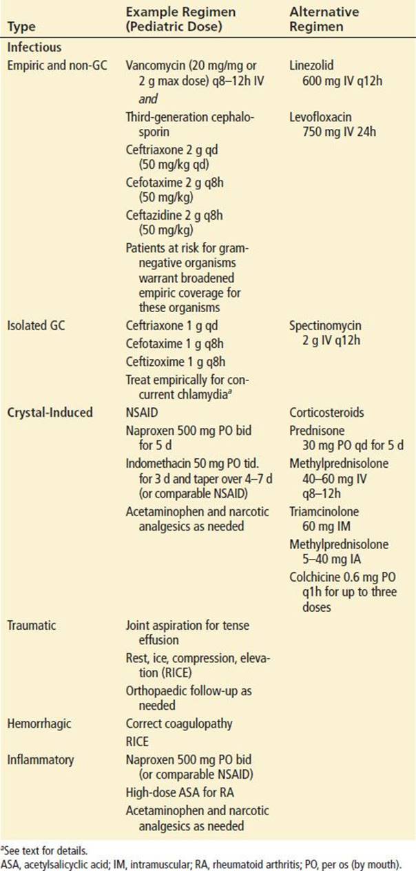
Initial therapy for disseminated gonococcal disease is a parenteral third-generation cephalosporin; alternative regimens include spectinomycin. Empiric therapy for concurrent chlamydia infection is also required.
Septic Arthritis
Rapid diagnosis and therapy is important to prevent joint and systemic morbidity of septic arthritis. Treatment requires appropriate antibiotics and surgical source control in most cases. As with any infectious process, begin antibiotics immediately after appropriate culture collection if the diagnosis is highly suspected. Antibiotic selection is based on the clinical probability of a specific pathogen as determined by patient presentation, age, comorbid risks, and Gram stain. Empiric broad-spectrum parenteral antibiotics are indicated in most situations (Table 150.2). Although primary pathogens are somewhat dissimilar, combination therapy with vancomycin and a third-generation cephalosporin are appropriate for most adults and children. The growing incidence of methicillin- resistant S. aureus (MRSA) warrants empiric coverage for this agent (14). Patients with gram-negative bacilli identified on stain and those at high risk for these pathogens warrant broadened empiric coverage. Intra-articular antibiotics provide no additional value for septic arthritis and carry the risk of chemical synovitis. Removal of infection and inflammatory debris is an essential component of treatment. Closed needle aspiration versus open surgical drainage of septic joints is controversial and should be left to the discretion of the orthopedic consultant. Adjunctive systemic steroids to reduce the inflammatory response following infection control remains under investigation.
TABLE 150.2
Treatment of Monoarticular Arthritisa
Crystal-Induced Arthritis
Although acute crystal-induced arthritis is generally self-limited, treatment relieves discomfort and prevents progressive joint destruction (Table 150.2). Nonsteroidal anti-inflammatory drugs (NSAIDs) are the historic treatment of choice for most patients. High initial dosing typically provides marked relief within 24 hours and may be tapered as symptoms improve to reduce potential adverse effects. The usefulness of NSAIDs is limited by their side effects including gastropathy, renal dysfunction, and interaction with warfarin.
Systemic corticosteroids are an important alternative. Comparable efficacy and safety in acute gout supports first-line use of glucocorticoid therapy, especially for patients with contraindication or suboptimal response to NSAID therapy (15–17). Oral, IV, and intramuscular options are available. Intra-articular steroids are very effective for crystal disease but their use requires the confident exclusion of infection. The appropriate intra-articular steroid dose is dependent on joint size; 20 to 40 mg of methylprednisolone acetate is adequate for larger joints such as the knee, whereas 5 to 10 mg is appropriate for smaller joints.
The leukocyte inhibitor colchicine provides dramatic relief of gout if utilized within 24 hours of onset but use has declined due to its narrow therapeutic window and higher risk of adverse effects. While traditional dosing was higher, a newer, more conservative recommendation is to administer 0.6 mg/hr up to three doses (1.8 mg total). Patients should not take additional colchicine for 7 days following an oral load. IV colchicine carries increased risk of systemic toxicity and is not recommended. Elderly patients and those with renal and hepatic dysfunction are not candidates for colchicine.
Serum urate-lowering agents such as allopurinol and probenecid are contraindicated in the setting of acute or resolving gout. The resultant change in serum urate may exacerbate or prolong an attack. Joint rest and cold applications provide symptomatic relief. Warm compresses exacerbate joint inflammation and should be avoided. Acetaminophen and narcotic analgesics provide additional relief if needed.
CRITICAL INTERVENTIONS
• Initiate early parenteral antibiotic therapy for patients with septic arthritis.
DISPOSITION
Gonococcal arthritis is typically responsive to antibiotics and may not require surgical drainage. Reliable nontoxic patients with gonococcal disease of nonweight-bearing joints may be treated as outpatients with daily parenteral therapy and close monitoring. Clinical improvement should occur within 48 hours at which time patients may be transitioned to oral therapy. Counseling includes discussion of STDs, birth control, partner counseling, and referral for HIV and syphilis testing. Patients with nongonococcal or undifferentiated septic arthritis require hospitalization. Most patients with acute gout can be successfully treated as outpatients. Precipitating factors such as alcohol use, purine-rich foods, and diuretics should be addressed.
Common Pitfalls
• Failure to consider infection in patients with acute worsening of pre-existing arthritis.
• Failure to obtain synovial fluid for analysis.
• Excluding septic arthritis based on absence of fever or normal serum inflammatory markers.
• Ordering protein and glucose on synovial fluid.
• Failure to perform culture on all diagnostic synovial fluid specimens.
• Inadequate empiric antibiotic selection in cases of suspected infection.
• Failure to recognize the migratory polyarthritis and tenosynovitis of disseminated gonorrhea.
• Diagnosing or excluding gout based on the serum urate level.
• Prescribing serum urate–lowering agents during an acute gout attack.
REFERENCES
1. Gardner GC, Weisman MH. Pyarthrosis in patients with rheumatoid arthritis: A report of 13 cases and a review of the literature from the past 40 years. Am J Med. 1990;88(5):503–511.
2. Gupta MN, Sturrock RD, Field M. A prospective 2-year study of 75 patients with adult-onset septic arthritis. Rheumatology (Oxford). 2001;40(1):24–30.
3. Kherani RB, Shojania K. Septic arthritis in patients with pre-existing inflammatory arthritis. CMAJ. 2007;176(11):1605–1608.
4. Mathews CJ, Kingsley G, Field M et al. Management of septic arthritis: A systematic review. Ann Rheum Dis. 2007;66(4):440–445.
5. Schlesinger N, Baker DG, Schumacher HR Jr. Serum urate during bouts of acute gouty arthritis. J Rheumatol. 1997;24(11):2265–2266.
6. Margaretten ME, Kohlwes J, Moore D, et al. Does this adult patient have septic arthritis? JAMA. 2007;297(13):1478–1488.
7. Carpenter CR, Schuur JD, Everett WW, et al. Evidence-based diagnostics: adult septic arthritis. Acad Emerg Med. 2011;18(8):781–796.
8. Brannan SR, Jerrard DA. Synovial fluid analysis. J Emerg Med. 2006;30(3):331–339.
9. Li SF, Cassidy C, Chang C, et al. Diagnostic utility of laboratory tests in septic arthritis. Emerg Med J. 2007;24(2):75–77.
10. McGillicuddy DC, Shah KH, Friedberg RP, et al. How sensitive is the synovial fluid white blood cell count in diagnosing septic arthritis? Am J Emerg Med. 2007;25(7):749–752.
11. Ilahi OA, Swarna U, Hamill RJ, et al. Concomitant crystal and septic arthritis. Orthopedics. 1996;19(7):613–617.
12. Shah K, Spear J, Nathanson LA, et al. Does the presence of crystal arthritis rule out septic arthritis? J Emerg Med. 2007;32(1):23–26.
13. Yu KH, Luo SF, Liou LB, et al. Concomitant septic and gouty arthritis–an analysis of 30 cases. Rheumatology (Oxford). 2003;42(9):1062–1066.
14. Frazee BW, Fee C, Lambert L. How common is MRSA in adult septic arthritis? Ann Emerg Med. 2009;54(5):695–700.
15. Cattermole GN, Man CY, Cheng CH, et al. Oral prednisolone is more cost-effective than oral indomethacin for treating patients with acute gout-like arthritis. Eur J Emerg Med. 2009;16(5):261–266.
16. Janssens HJ, Janssen M, van de Lisdonk EH, et al. Use of oral prednisolone or naproxen for the treatment of gout arthritis: A double-blind, randomised equivalence trial. Lancet. 2008;371(9627):1854–1860.
17. Man CY, Cheung IT, Cameron PA, et al. Comparison of oral prednisolone/paracetamol and oral indomethacin/paracetamol combination therapy in the treatment of acute goutlike arthritis: A double-blind, randomized, controlled trial. Ann Emerg Med. 2007;49(5):670–677.