
Richard S. Hartoch
Polyarticular arthritis presents as an acute or chronic inflammatory synovitis (synovial warmth, swelling, and tenderness) developing at a minimum of two joints. The emergency physician encounters polyarthritis in two contexts: the patient without a known rheumatologic disease who presents with multiple painful joints, and the patient with complications of a previously diagnosed condition, most commonly rheumatoid arthritis (RA).
CLINICAL PRESENTATION
Many polyarthritis syndromes have a gradual onset and may defy precise diagnosis despite extensive initial workup. Symptoms of early hepatitis C, systemic lupus erythematosus (SLE), RA, and other collagen–vascular diseases overlap and laboratory test results may be equivocal. A definitive diagnosis may not readily be made on a single emergency department (ED) visit. Although the syndromes share arthritis as a common feature, all have associated symptoms that are helpful in evaluation (Table 151.1). A thorough history and physical examination may be the most important tools in establishing a diagnosis.
TABLE 151.1
Polyarticular Arthritis: Clinical Features

Rheumatoid Arthritis
The most commonly encountered etiology of polyarthritis is RA, characterized by symmetric joint inflammation, constitutional symptoms, and in some cases, extra-articular organ involvement. The disorder ranges in severity from a mild nonprogressive arthritis to a fulminant, sometimes fatal, illness with destruction of joints and serious systemic involvement. Early recognition and initiation of appropriate therapy generally results in an improved outcome. A majority of patients who have been newly diagnosed with RA will experience remission if aggressively treated by a rheumatologist.
The onset of RA may be acute or insidious. Inflammation in multiple joints in almost any combination may be present, although symmetric inflammation of the metacarpophalangeal (MCP) and proximal interphalangeal (PIP) joints and wrists is most common. The vast majority of RA patients will complain of pain when their four MCP joints are gently squeezed together. RA virtually never affects the thoracic or lumbar spine. Systemic manifestations include malaise, anorexia, fever, myalgias, and weight loss. In some patients, especially those with high titers of rheumatoid factor and more severe joint disease, there is systemic organ involvement, most commonly lungs and heart.
Acute Rheumatic Fever
Although the incidence of acute rheumatic fever (ARF) has dramatically declined over the past 40 years, recent reports have described episodic outbreaks. The diagnosis of ARF mandates fulfillment of the modified Jones criteria (Table 151.2). Arthritis, the most common finding in ARF, is seen in more than 75% of first attacks. It is typically migratory and most often first affects the large joints of the lower extremities, knees, and ankles, followed by wrists and elbows. Each joint is inflamed for up to 1 week. Carditis, clinically seen in 40% to 50% of acute cases, is the most serious finding. Recent echocardiographic studies have revealed that the carditis is often subclinical and present to varying degrees of severity in virtually all patients with ARF. The patient may present with pleuritic chest discomfort, a new murmur (usually mitral regurgitation), cardiomegaly, and possibly congestive heart failure. Chorea occurs in 5% of cases. It develops several months after streptococcal infection and is characterized by emotional disturbances, weakness, and sudden involuntary movements that cease with sleep. Erythema marginatum (5% of cases) begins as a macule that extends outward with central clearing. It may resemble erythema chronicum migrans, the rash of Lyme disease (LD). It is not painful or pruritic. It usually fades within hours but may recur. Subcutaneous nodules, seen in <10% of patients, are firm, painless, and of variable size (2 to 10 mm). They are most commonly seen over the extensor joint surfaces or bony prominences.
TABLE 151.2
Modified Jones Criteria for Diagnosis of Acute Rheumatic Fever

Poststreptococcal reactive arthritis (PSRA) is a clinical syndrome with insufficient Jones criteria to be called ARF. Consider obtaining an antistreptolysin O (ASO) titer. Treat strep pharyngitis with antibiotic and arthritis with nonsteroidal anti-inflammatory drugs (NSAIDs). Prednisone may be indicated in severe cases.
Gonococcal Arthritis
Gonococcal arthritis (GA), although infrequently seen, is still the most common bacterial arthritis among healthy young people. It develops in 1% to 3% of patients with untreated gonococcal infection. Disseminated gonococcal infection (DGI) is characterized by a clinical triad: dermatitis, tenosynovitis, and migratory polyarthritis. Joint involvement is predominantly asymmetric, often affecting the knee, ankle, elbow, and wrist. A painless, nonpruritic skin rash consisting of scattered papules or vesicles with a hemorrhagic or necrotic center is seen in roughly 50% of patients. HIV patients are infected in unusual joints (hip and sternoclavicular) and may develop a more aggressive course.
Lyme Disease
LD is caused by the spirochete Borrelia burgdorferi, transmitted by the bite of the deer tick, Ixodes scapularis (formerly I. dammini). A characteristic rash, erythema chronicum migrans, develops in 60% of patients. It starts as a single red macule that expands to form a large annular lesion with a red outer border and central clearing. Two weeks to 2 years after the initial skin lesion, 60% of patients develop an acute arthritis, typically asymmetric oligo- or monoarticular, involving large joints, most commonly the knee. About 10% of patients develop cardiac abnormalities, most often atrioventricular (AV) block. Although cardiac involvement is transient, some patients require temporary pacemakers. Approximately 10% of patients develop significant neurologic abnormalities, including meningitis, encephalitis, and cranial neuropathies, particularly facial nerve paralysis.
Reactive Arthritis
Reactive arthritis (formerly Reiter syndrome) may occur as a polyarthritis in young, often human leukocyte antigen (HLA) B-27–positive men. It classically presents as a triad of nongonococcal urethritis, asymmetric polyarthritis, and conjunctivitis. The arthritis is usually acute in onset, involving the large joints of the lower extremities, particularly the knees and ankles. Urethritis usually precedes the development of arthritis, although it may be asymptomatic and suggested only by the finding of white blood cells on microscopic examination of first-voided, morning urine. Conjunctivitis occurs in 30% of patients and iritis in 10%. Two distinctive skin lesions are occasionally seen. Keratoderma blennorrhagica (15% of patients), scaling lesions on the palms and soles that begin as erythematous macules, may progress to lesions indistinguishable from those of psoriasis. Circinate balanitis (25% of patients) produce painless superficial ulcers of the glans penis. Reactive arthritis may be triggered by intestinal infections (Salmonella, Shigella, or Campylobacter) or sexually transmitted diseases (Gonorrhea or Chlamydia).
Viral Arthritis/Postinfectious Arthritis
Transient nondestructive arthritis has been associated with viral infection, the best documented being hepatitis A, B, and C and rubella. The arthritis of hepatitis is believed to be due to the deposition of circulating immune complexes in affected joints. It typically occurs during the prodrome of the illness before the appearance of jaundice. Liver enzymes are usually elevated, and HBsAg or anti-HBc may be positive. The most common presentation is that of a symmetric polyarthritis potentially involving the hand, wrist, elbow, and ankle. It can be painful with prominent morning stiffness but seldom lasts more than 3 weeks. The presentation of hepatitis C may easily be mistaken for RA.
Parvovirus has been implicated as the etiologic agent in an acutely painful polyarthritis involving hands, wrists, elbows, knees, and shoulders. Parvovirus, perhaps better known for its causative role in erythema infectiosum (fifth disease in children), may be detected by elevated IgM titers. The arthritis, often seen in patients who work with children, persists for several weeks and is generally responsive to NSAID therapy. Polyarthritis syndromes have also been described in association with enteric infections (Yersinia, Shigella, Salmonella, Clostridium difficile), respiratory infection (mycoplasma), and α-viruses outside US.
Serum Sickness
Serum sickness, typically occurring 6 to 10 days after an antigenic stimulus, presents with the abrupt onset of fever, lymphadenopathy, and arthritis especially involving knees, ankles, shoulders, and wrists. Urticaria or angioedema may also develop. A more severe reaction tends to result after subsequent exposure to the same antigen. Penicillin is the most commonly implicated causal agent, but many other drugs including other α-lactams, ciprofloxacin, levoquin, angiotensin-converting enzyme inhibitors, and allopurinol have been implicated as well.
HIV Arthritis
Several polyarthritis syndromes have been described in association with HIV infection. A painful, asymmetric, oligoarticular arthritis affecting the knees and ankles has been widely reported. Also seen are seronegative spondyloarthritis and lupus-like syndromes. These polyarthritides may occur at any stage of HIV infection and have been reported to develop months or years before the actual diagnosis of HIV. Because of impaired immunity, HIV patients are at risk for pyogenic joint infections. Streptococcus and Staphylococcus sp predominate, but opportunistic pathogens may be involved.
DIFFERENTIAL DIAGNOSIS
There are numerous disorders that produce polyarticular arthritis, including RA, acute rheumatic fever, GA, LD, gout, postviral arthritis, septic arthritis, serum sickness, SLE, osteoarthritis, ankylosing spondylitis, ulcerative colitis, regional enteritis (Crohn disease), and psoriatic arthritis. Clinical features that may be helpful in distinguishing the disorders are listed in Table 151.1. Inflammation in the periarticular soft tissues (tendons, bursae, etc.) can cause swelling and limitation of motion, simulating joint disease. Arthritis generally affects both active and passive motion equally, whereas acute tendinitis or bursitis limits active motion significantly more than passive ranging.
ED EVALUATION
Sudden onset of joint pain (developing over several hours, rather than days) strongly suggests gout or infection. The most critical joint disorder to consider and diagnose immediately is septic arthritis, which can cause rapid destruction and systemic illness. Although the classic presentation is single joint involvement in a febrile patient, 15% of cases of septic arthritis are polyarticular and 20% are afebrile. Patients with underlying joint disease, diabetes, alcoholism, and malignancy are at particular risk for polyarticular infection. These patients are often bacteremic and have inpatient mortality rates exceeding that of myocardial infarction.
The persistence of symptoms for over 4 to 6 weeks suggests a systemic rheumatologic disease. Symmetric joint involvement is seen in RA, hepatitis C, and SLE. Asymmetric joint involvement is typical of the seronegative arthritides (e.g., reactive arthritis and psoriatic arthritis). Migratory involvement (leaving one joint before involving another) is characteristic of acute rheumatic fever. A family history of arthritis supports a diagnosis of RA or one of the seronegative spondyloarthropathies. Thiazide and loop diuretics, alcohol, high protein intake, low-dairy diets, and obesity are linked with the development of gout, which may be polyarticular in presentation. Hydralazine, procainamide, isoniazid, phenytoin, and several tumor necrosis factor (TNF) inhibitors can induce a lupus-like reaction.
To assess patterns of involvement and chronicity, all joints should be examined, not just those that are symptomatic. A relatively rapid assessment can survey up to 60 joints. Several acute polyarthritides are associated with a characteristic rash. Subcutaneous nodules are seen in 35% of patients with RA and <10% of those with ARF. Psoriatic arthritis may be associated with pitting of the nails and the typical papulosquamous eruption. A general physical examination may note such important findings as a pleural effusion in SLE and RA or clinical signs of liver dysfunction in arthritis associated with hepatitis. A pelvic examination should be performed on all women with unexplained arthritis to rule out Neisseria gonorrhoeae. Generalized lymphadenopathy or thrush may suggest HIV.
The most important laboratory test is examination of the joint fluid. Usually obtained studies include white cell count with differential, Gram stain and culture, and examination for crystals. Specific diagnostic tests (e.g., antinuclear antibody, rheumatoid factor, Lyme titer, HIV, hepatitis screen) are seldom available on an emergency basis. They should be ordered in appropriate patients for use in subsequent evaluation. The erythrocyte sedimentation rate (ESR) and C-reactive protein (CRP) can be elevated in any inflammatory condition. Such tests are generally of only marginal diagnostic assistance but may be useful in monitoring disease activity in patients with chronic arthritis. The diagnosis of ARF, which should be especially considered in children and young adults, requires laboratory confirmation of recent group A streptococcal infection. In patients with GA, arthrocentesis cultures are positive in only half of affected joints. Higher yield for positive culture may be obtained from the mucosal surface of initial infectious contact (throat, cervix, urethra, or rectum). Polymerase chain reaction (PCR) may be useful in identifying gonococcal infection from a joint that is culture-negative.
Radiographs are of limited utility in the evaluation of the patient with nontraumatic joint pain. Radiographically visible inflammatory changes (such as symmetric erosions of the wrist and MCP joints in RA) occur late in the course of disease. A normal radiograph does not rule out a diagnosis of arthritis.
Recent RA literature repeatedly stresses the importance of early diagnosis and prompt referral of suspected cases to a rheumatologist. Proper treatment, which involves the very early usage of disease-modifying antirheumatic drugs (DMARDs), yields a markedly improved long-term outcome. Delay of as little as 3 months in initiating therapy has been shown to result in significantly increased joint damage at 5 years. The diagnosis of RA is based largely on a high index of suspicion, a careful history, and directed physical examination. Rheumatoid factor, although not a specific marker for RA, is present in the serum of 75% of patients. Clinical criteria established by the American Rheumatism Association include prolonged morning stiffness, symmetric joint involvement, chronicity, rheumatoid nodules, and radiographs revealing erosions. Recent studies reveal that the characteristic bone erosions may be visualized by ultrasound and magnetic resonance imaging (MRI) significantly earlier than by conventional radiography.
KEY TESTING
• Arthrocentesis with joint fluid analysis to exclude septic joint
ED MANAGEMENT
The management of polyarthritis depends on the correct diagnosis. Septic arthritis must be managed as an inpatient with IV antibiotics, orthopedic consultation, and discovery of the source of possible bacteremia. Suspected RA mandates prompt rheumatologic consultation for rapid initiation of DMARD therapy, usually methotrexate, biologic TNF inhibitors, biologic non-TNF inhibitors, or other agents. NSAIDs are not effective in limiting the destructive consequences of RA.
The mainstay of treatment for general, undiagnosed polyarthritis is NSAIDs. These agents control both pain and inflammation. If one NSAID fails to control symptoms, another in a different class may be more effective. All NSAIDs should be prescribed with caution, and patients should be monitored for adverse gastrointestinal effects, renal dysfunction, edema, and cardiac complications. NSAIDs should generally be taken with food.
TNF inhibitors are newer agents used in the treatment of RA. They are generally well tolerated, but the practitioner must be alert to clinically significant, potentially life-threatening side effects. TNF has antitumor and antiviral effects and has particular activity against intracellular pathogens, notably Mycobacterium tuberculosis. TNF stimulates inflammatory cells to areas of infection and may be involved in the formation of granulomas which contain TB. It plays an important role in modulating the systemic inflammatory response of sepsis.
Patients receiving TNF inhibition are subject to a variety of serious infections and may present to the ED appearing “less sick” than other patients with similar conditions. Particular issues include unrecognized sepsis with various bacteria, notably Streptococcus pneumoniae and Klebsiella. One should also consider reactivation of TB, opportunistic infections including Listeria, Legionella, and disseminated fungal infections. These patients with their blunted immune response may not show expected signs and symptoms of severe infection. They may have a negative PPD test for TB due to anergy.
Several disorders require more specific treatment. Patients with a first attack of ARF should be hospitalized and closely monitored for worsening carditis, arrhythmias, and valve damage. High-dose salicylates and prednisone may be useful. The patient must be monitored for the development of congestive heart failure. A course of penicillin is recommended to eradicate any remaining streptococci, and the patient may be kept on low-dose penicillin prophylaxis indefinitely thereafter.
Most patients with GA should be hospitalized. Therapy is initiated with a parenteral third-generation cephalosporin. Because of recently described increased rates of GC resistance, fluoroquinolones may not be effective. Repeated joint aspirations, arthroscopy, or surgery may be required. In most cases, dramatic clinical improvement is noted in several days, and the patient may transition to oral antibiotics. Patients should be treated concurrently for chlamydia.
The arthritis associated with LD is generally treated for at least a month with doxycycline or amoxicillin. Resistant cases are treated with IV ceftriaxone. Reactive arthritis is not curable, but treatment of the initial urethritis may prevent development of the full-blown disease. Slit-lamp examination of the anterior chamber is essential to rule out associated iritis. Arthritic inflammation is treated symptomatically with NSAIDs.
Complications of RA
RA is the most common and most serious of the chronic polyarthritides and may be associated with a number of emergent complications (Table 151.3). The emergency physician should be aware of classic acute issues arising in patients with well-established RA. RA patients are also at risk for toxic and immunosuppressive effects of treatments.
TABLE 151.3
Complications of Rheumatoid Arthritis
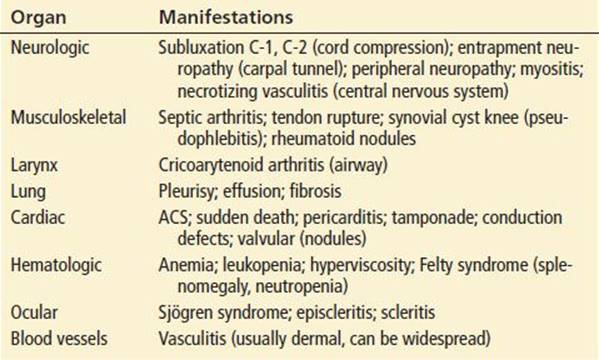
Cardiovascular disease (CVD) is one of the most common causes of death in patients with RA and occurs at a younger age than in the general population. Chronic inflammation as seen in RA, PA, and SLE results in accelerated atherosclerosis. RA patients with an acute coronary syndrome (ACS) are more likely to present to the ED with atypical symptoms. RA may be considered an independent risk factor for coronary artery disease, and in fact several studies reveal it to be as significant as diabetes or hypertension. The increased CVD risk may be present even before clinical evidence of RA develops. Aggressive efforts to control inflammation and modify traditional cardiac risk factors are particularly vital in these patients.
Radiographic abnormalities of the cervical spine are common, particularly subluxation of C-1 on C-2. Usually these abnormalities are asymptomatic, but the appearance of neurologic symptoms or signs necessitates neurosurgical consultation. Patients may note a peculiar slipping sensation with neck movements that is associated with either exacerbation or relief of symptoms. A patient with RA who sustains even minor cervical trauma is at risk for serious injury. The potential for cervical spine instability should be considered when intubating a patient with chronic RA, and cervical spine precautions should be observed.
Various entrapment neuropathies occur in RA, of which carpal tunnel syndrome is the most common. Patients initially complain of sensory changes over the distribution of the median nerve and a “toothache-like” pain at the wrist. The diagnosis is confirmed with electromyography and nerve conduction studies. Treatment consists of splinting the wrist and arranging consultation with a rheumatologist, orthopedist, or neurologist. Local corticosteroid injection is occasionally useful. Surgical release of median nerve entrapment is sometimes necessary. Generalized muscle wasting, weakness, and peripheral neuropathy in RA patients may also be manifestations of systemic vasculitis.
Rupture of the extensor tendons of the hand is not uncommon in advanced RA. Once one tendon ruptures, the increased strain on the remaining tendons may lead to rupture of others. Patients cannot extend the fingers actively at the MCP joints, but passive extension is still possible. Treatment is elective surgical repair.
The development of a synovial cyst behind the knee is common in RA. Synovial inflammation may result in large knee effusions, and high pressure generated during flexion of the knee may cause cyst formation in the popliteal fossa. The diagnosis is confirmed by an arthrogram or ultrasound of the knee joint. Curative treatment is difficult. Joint aspiration and local corticosteroid injection may be temporarily helpful. These cysts may rupture, dissecting into the calf. The resulting calf pain, swelling, and redness may mimic the presentation of deep vein thrombosis. Duplex sonography is helpful in distinguishing these clinical entities.
The cricoarytenoid joint is a synovial articulation and can be involved in RA. Patients may present with stridor and significant airway obstruction, dyspnea, dysphagia, painful speech, foreign-body sensation, or hoarseness. There may be tenderness over the thyroid cartilage. Laryngoscopy reveals redness and edema over the arytenoids and abnormal bowing of the vocal cords during inspiration. Intubation may be difficult. Cricothyrotomy or tracheostomy may be necessary to secure the airway. Corticosteroids and cool humidification are useful in milder cases.
The most common type of vasculitis associated with RA involves small dermal vessels, causing relatively benign infarctions in the periungual areas of the fingers. Occasionally patients develop a necrotizing vasculitis, which can follow a fulminant course. The presenting manifestations may be large ulcerations, digital gangrene, neuropathies, mesenteric ischemia, or coronary arteritis. Patients with systemic vasculitis usually have fever, leukocytosis, high titers of rheumatoid factor, an elevated ESR, and antinuclear antibodies. They should be admitted to the hospital for initiation of high-dose corticosteroids. At times cytotoxic agents or apheresis are useful. The prognosis in patients with extensive vasculitis is poor.
Sjögren syndrome (keratoconjunctivitis sicca), sometimes associated with RA, is characterized by dry eyes and dry mouth and is caused by decreased lacrimal and salivary gland function. Patients complain of grittiness in the eyes and thick mucus beneath the eyelids. The diagnosis is made by the Schirmer test which measures tear production. Treatment consists of artificial tears, humidified air, and eye goggles to minimize tear evaporation.
Episcleritis, also associated with RA, is usually benign but should be followed by an ophthalmologist because it can lead to scleritis. A brief course of topical corticosteroids as prescribed by an ophthalmologist is usually beneficial. Scleritis is a more diffuse process and can cause uveitis, glaucoma, and rupture of the globe. Patients complain of a deep aching sensation and tenderness on gentle palpation of the globe. Slit-lamp examination reveals cellular exudate in the anterior chamber. Prompt ophthalmologic consultation is indicated.
Hematologic problems, including anemia, leukopenia, and hyperviscosity, occur in RA. Felty syndrome (triad of RA, splenomegaly, and neutropenia) is associated with severe disease. These patients are prone to recurrent infections and have hepatic dysfunction and recurring cutaneous ulcers. Splenectomy is occasionally beneficial.
Pleurisy and pleural effusions are common in RA but are not usually life-threatening. Pericarditis is also common, but only rarely does pericardial tamponade or constrictive pericarditis occur.
CRITICAL INTERVENTIONS
• Consider ACS in patients with RA, PA, and SLE.
• Consult a rheumatologist as soon as possible when RA is suspected.
DISPOSITION
Septic arthritis requires emergent intervention and inpatient management. Patients with a first attack of ARF should be hospitalized. Most patients with GA should also be hospitalized for initial therapy. LD in early stages is generally treated with doxycycline on an outpatient basis. Advanced cases may require hospitalization.
Patients with RA or SLE may present with complications requiring admission, but isolated nonseptic exacerbations of joint pain are often managed successfully on an outpatient basis. Admission decisions for other causes of polyarticular arthritis are often dependent upon the certainty of diagnosis, the adequacy of pain control, and the presence or absence of other disease complications.
Common Pitfalls
• Failure to analyze synovial fluid in a patient with new joint tenderness, warmth, and swelling.
• Failure to diagnose ACS in a patient with RA who presents with atypical symptoms.
• Failure to identify sepsis or a serious opportunistic infection in a patient on TNF inhibitor therapy.
• Failure to recognize an acute septic joint in a patient with underlying chronic polyarthritis.
• Failure to consider a diagnosis of RA, resulting in delayed diagnosis and treatment.
• Failure to consider acute rheumatic fever in a child or young adult.
• Failure to consider underlying HIV infection in a person with a newly diagnosed, seronegative spondyloarthropathy or lupus-like syndrome.
SUGGESTED READINGS
1. Alexandroff AB, Pauriah M, Camp RD, et al. More than skin deep: Atherosclerosis as a systemic manifestation of psoriasis. Br J Dermatol. 2009;161(1):1–7.
2. Barash J. Rheumatic fever and post-group a streptococcal arthritis in children. Curr Infect Dis Rep. 2013;15(3):263–268.
3. Bleich AT, Sheffield JS, Wendel GD Jr, et al. Disseminated gonococcal infection in women. Obstet Gynecol. 2012;119(3):597–602.
4. Christodoulou C, Gordon P, Coakely G. Polyarticular septic arthritis. BMJ. 2006; 333:1107–1108.
5. Connor V. Anti-TNF therapies: A comprehensive analysis of adverse effects associated with immunosuppression. Rheumatol Int. 2011;31:327–337.
6. Emery P. Treatment of rheumatoid arthritis. BMJ. 2006;332:152–155.
7. Dinser R, Jendro MC, Schnarr S, et al. Antibiotic treatment of Lyme borreliosis: What is the evidence? Annals Rheum Dis. 2005;64:519–523.
8. Felson D. Osteoarthritis of the knee. N Engl J Med. 2006;354:841–848.
9. Gabriel SE. Cardiovascular morbidity and mortality in rheumatoid arthritis. Am J Med. 2008;121(10 suppl 1):S9–S14.
10. Jansen LM, van Schaardenburg D, van der Horst-Bruinsma IE, et al. One year outcome of undifferentiated polyarthritis. Annals Rheum Dis. 2002;61:700–703.
11. John H, Toms TE, Kitas GD. Rheumatoid arthritis: Is it a coronary heart disease equivalent? Curr Opin Cardiol. 2011;26(4):327–333.
12. Kerola AM, Kauppi MJ, Kerola T, et al. How early in the course of rheumatoid arthritis does the excess cardiovascular risk appear? Ann Rheum Dis. 2012; 71(10):1606–1615.
13. Lawson E, Walker-Bone K. The changing spectrum of rheumatic disease in HIV infection.Br Med Bull. 2012;103(1):203–221.
14. Levine M, Siegel LB. A swollen joint: Why all the fuss? Am J Ther. 2003;10:219–224.
15. Liao KP, Solomon DH. Traditional cardiovascular risk factors, inflammation and cardiovascular risk in rheumatoid arthritis. Rheumatology (Oxford). 2013; 52(1):45–52.
16. Lopez-Ben R, Bernreuter WK, Moreland LW, et al. Ultrasound detection of bone erosions in rheumatoid arthritis: A comparison to routine radiographs of the hands and feet. Skeletal Radiol.2004;33(2):80–84.
17. Mackie SL, Keat A. Poststreptococcal reactive arthritis: What is it and how do we know? Rheumatology (Oxford). 2004;43(8):949–954.
18. Marijon E, Phalla O, Celermajer D, et al. Prevalence of rheumatic heart disease detected by echocardiographic screening. N Engl J Med. 2007;357(5):470–476.
19. Margaretten ME, Kohlwes J, Moore D, et al. Does this adult patient have septic arthritis? JAMA. 2007;297:1478–1488.
20. Mody GM, Parke FA, Reveille JD. Articular manifestations of HIV infection. Best Pract Res Clin Rheumatol. 2003;17:265–287.
21. Moore TL. Parvovirus-associated arthritis. Curr Opin Rheumatol. 2000;12:289–294.
22. Moreland LW, Bridges SL Jr. Early rheumatoid arthritis: A medical emergency? Am J Med. 2001;111:498–500.
23. Murdaca G, Colombo BM, Cagnati P, et al. Update upon efficacy and safety of TNF-inhibitors. Expert Opin Drug Saf. 2012;11(1):1–5.
24. Nell VP, Machold KP, Eberl G, et al. Benefit of very early referral and very early therapy with DMARDs in patients with early rheumatoid arthritis. Rheumatology (Oxford). 2004;43:906–914.
25. O’Dell JR. Therapeutic strategies for rheumatoid arthritis. N Engl J Med. 2004; 350(25):2591–2602.
26. Ostergaard M, Hansen M, Stolenberg M, et al. New radiographic bone erosions in the wrists of RA, MRI results. Arthritis Rheum. 2003;48(8):2128–2131.
27. Ramos-Casals M, Trejo M, Garcia-Carrasco M, et al. Therapeutic management of extrahepatic manifestations in patients with chronic hepatitis C virus infection. Rheumatology (Oxford). 2003;42:818–828.
28. Scott DL, Kingsley MB. Tumor necrosis factor inhibitors for rheumatoid arthritis. N Engl J Med. 2006;355:704–712.
29. Terkeltaub RA. Gout. N Engl J Med. 2003;349:1647–1655.
30. Turiel M, Sitia S, Atzeni F, et al. The heart in rheumatoid arthritis. Autoimmun Rev. 2010;9(6):414–418.
31. Tutar E, Atalay S, Yilmaz E, et al. Poststreptococcal reactive arthritis in children: Is it really a different entity from rheumatic fever? Rheumatol Int. 2002; 22(2):80–83.
32. van Halm VP, Peters MJ, Voskuyl AE, et al. Rheumatoid arthritis versus diabetes as a risk factor for cardiovascular disease: A cross-sectional study, the CARRE Investigation. Ann Rheum Dis.2009;68(9):1395–1400.
33. Vassilopoulos D, Calabrese LH. Rheumatologic manifestations of hepatitis C infection. Curr Rheumatol Rep. 2003;5(3):200–204.
34. Wallis RS. Biologics and infections: Lessons from tumor necrosis factor blocking agents. Infect Dis Clin North Am. 2011;25(4):895–910.
35. Weinblatt ME. Rheumatoid arthritis: More aggressive approach improves outlook. Cleve Clin J Med. 2004;71(5):409–413.
36. Williamson L, Bwness P, Mowat A, et al. Difficulties in diagnosing acute rheumatic fever, arthritis may be short lived and carditis silent. BMJ. 2000;320:362–365.
37. Wilson PW. Evidence of systemic inflammation and estimation of coronary artery disease risk: A population perspective. Am J Med. 2008;121(10 suppl 1): S15–S20.