
Andy Jagoda
Seizures are both a primary problem and a result of an underlying disorder. They account for an estimated 1% to 2% of emergency department (ED) visits. Two to 4 million Americans have epilepsy, a condition of recurrent unprovoked seizures.
Seizures result from excessive and disorderly neuronal discharge in the cerebral cortex. Epilepsy is a condition of unprovoked seizures, whereas an underlying pathologic process provokes other seizures. The clinical manifestations of a seizure reflect the area of the brain in which neurons are discharging abnormally. Seizure discharges and symptoms may be focal or may be generalized to both hemispheres. The clinical spectrum of seizures includes focal or generalized motor activity, altered mental status, sensory or psychic experiences, or autonomic disturbances.
Ictus refers to the period during which a seizure occurs. An aura, or “warning,” represents the beginning of a partial seizure, which can remain focal or can spread into a generalized event; a corollary is that patients with a primary generalized seizure (affecting both hemispheres without a focus) disorder do not have an aura. The period after a seizure but before the patient returns to baseline mental status is called the postictal period.
An acute symptomatic seizure, also referred to as a provoked seizure, is a seizure that occurs within 7 days of an acute CNS or systemic event, for example, stroke, hypoglycemia, head trauma. A remote symptomatic seizure is a seizure that occurs more than 7 days from an inciting event.
Status epilepticus (SE) is broadly defined as prolonged seizure activity with a high morbidity and mortality. Time definitions are changing for SE: It had been defined as seizures that last >30 minutes or in which there are recurrent seizures without a return to baseline mental status between events. Currently some researchers define SE as a seizure that lasts 5 to 10 minutes or two seizures without full recovery between them (1,2). The longer a seizure lasts the more neuron injury occurs, and the more difficult it is to stop the seizure activity. There are a projected 125,000 to 200,000 cases of SE each year in the United States, with an overall mortality of 22%. There is a bimodal distribution of cases, with the highest incidence occurring in the first year of life and after the age of 60 years. More than one-half of patients presenting to the ED in SE have no prior seizure history.
CLINICAL PRESENTATION
The classification of seizures (Table 156.1) is based on the behavioral, electrophysiologic, and clinical features of the event rather than on pathophysiologic mechanisms or anatomic localization. Seizures can be broadly divided into primary generalized seizures and partial seizures (also called focal seizures). Primary generalized seizures do not have an inciting focus but begin bilaterally in both hemispheres of the cerebral cortex. Partial seizures begin in a localized area of the brain; they may remain focal but can also spread to incorporate both hemispheres, in which case, the seizure is termed secondary generalized.Among the types of new onset seizures in adults, partial seizures are more common than primary generalized seizures (3).
TABLE 156.1
Classification of Seizures

There are six types of generalized seizures (Table 156.1); five have convulsive activity, and one has nonconvulsive activity (absence seizure). All primary generalized seizures, except myoclonic seizures, are associated with an altered level of consciousness. Generalized convulsive seizures classically begin as myoclonic jerks followed by loss of consciousness and sustained generalized skeletal muscle contractions. The seizure activity lasts 90 to 120 seconds and may be associated with urinary incontinence and tongue biting. The convulsive seizure is followed by the postictal state characterized by a brief period of confusion and somnolence. It is not possible to have a bilateral tonic–clonic event with preservation of mental status. This fact can be helpful at times in distinguishing neurogenic seizures from psychogenic seizures.
Absence seizures are primary generalized events causing an alteration in mental status without significant motor activity. Patients have short episodes of sudden immobility and a blank stare. Absence seizures are more frequent between the ages of 5 and 10 years and are infrequent after the midteens. The average duration is 10 seconds, and there is rarely a postictal period.
Partial seizures are subdivided according to associated impairment of consciousness. Simple partial seizures have brief sensory or motor manifestations with no impairment of consciousness and are categorized according to clinical presentation (Table 156.1). Partial seizures that cause an impairment of consciousness are termed complex partial seizures. These seizures have been known as temporal lobe or psychomotor seizures and are characterized by mental and psychological symptoms including changes in effect, confusion, or hallucinations. Partial seizures that evolve to generalized seizures are associated with altered consciousness just as other generalized seizures.
When a patient arrives to the ED with a generalized seizure or postictal, it can be difficult to differentiate between a primary generalized seizure and a secondary generalized partial seizure. A careful history can elicit a preceding symptom such as an aura, focal movement abnormality, or even staring behavior leading to a diagnosis of partial seizure that has generalized. A focal finding on computed tomography (CT) scan suggests a partial seizure that has secondarily generalized.
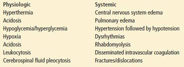
Seizures may have harmful secondary traumatic or metabolic consequences. Seizures may result in a fall or a motor vehicle accident. The convulsive movements of a seizure may result in injury from direct trauma to head, trunk, or limbs. There is typically a period of transient apnea and hypoxia, the duration of which varies with the duration of seizure activity. There is an increase in blood pressure, serum lactate and serum glucose levels, and white blood cell (WBC) count without a left shift. Body temperature is frequently elevated. Lactic acidosis occurs within 60 seconds of a convulsive event and normalizes within 1 hour. A transient cerebrospinal fluid pleocytosis of up to 20 WBC/μL has been reported to occur in 2% to 23% of seizure patients (Table 156.2).
TABLE 156.2
Physiologic and Systemic Consequences of Convulsive Seizures
DIFFERENTIAL DIAGNOSIS
Seizure Mimics
Some nonseizure conditions can mimic seizures. Such conditions include syncope, dysrhythmia, migraine, vertigo, sleep disorders, decerebrate posturing, drug reactions, tetanus, strychnine poisoning, and psychogenic events. Other, less common, seizure mimics include narcolepsy, hyperventilation syndrome, and dystonic reactions. A detailed history and physical can usually differentiate among these conditions.
Syncope can be associated with occasional twitching movements, which can be misdiagnosed as a seizure and can be called convulsive syncope. Full tonic–clonic movements, tongue biting, and postictal amnesia have not been commonly described in convulsive syncope. When put in the context of when and where the event occurred, the length of event, and the type of movements, convulsive syncope can usually be reliably differentiated from seizure.
Migraine with an aura can be confused with nonconvulsive seizures. This is compounded by the finding that many migraine patients have abnormal electroencephalograms (EEGs). Basilar migraine can result in loss of consciousness, making the differentiation even more difficult.
Psychogenic seizures, referred to as pseudoseizures, are functional events with a clinical presentation mimicking neurogenic seizures, with no corresponding alteration in EEG activity. These events are often conversion reactions and not under the patient’s conscious control. Up to 30% of patients referred to epilepsy clinics are diagnosed with psychogenic seizures with a mean delay from onset of events to diagnosis of 7.2 years (4). Psychogenic seizures often last longer than neurogenic events, and there usually is not a postictal period. Patients can often recall events during the seizure, have not been incontinent, and do not incur physical injury. Psychogenic seizures are classically manifested by forward-thrusting pelvic movements and head turning from side to side (4). Several maneuvers and tests are useful in diagnosing psychogenic seizures. These patients often avoid or resist noxious stimuli. These patients may display gaze aversion and look away from an examiner, regardless of position. On laboratory testing, psychogenic seizure patients do not have a metabolic acidosis, which is nearly universal with generalized convulsive seizures. Though not helpful in the ED setting, there is not a postictal increase in serum prolactin levels.
Seizure Etiologies
Emergency physicians encounter a number of clinical scenarios involving both acute and remote symptomatic seizures. The medical causes of new seizures may be the result of acute or progressive neurologic insults or systemic stressors, and the evaluation of the patient with seizures must always look for those potentially reversible or treatable causes (Table 156.3). The two most commonly identified etiologic antecedents to seizures are neurologic injury from birth (8%) and cerebrovascular disease (11%) (3). Other identified etiologies of secondary seizures include trauma (5.5%), neoplasm (4%), degenerative disease (3.5%), and infection (2.5%). In the elderly, the cause of seizure is more likely to be vascular (stroke), degenerative, or neoplastic than the causes in the young (5).
TABLE 156.3
Etiology of Seizures
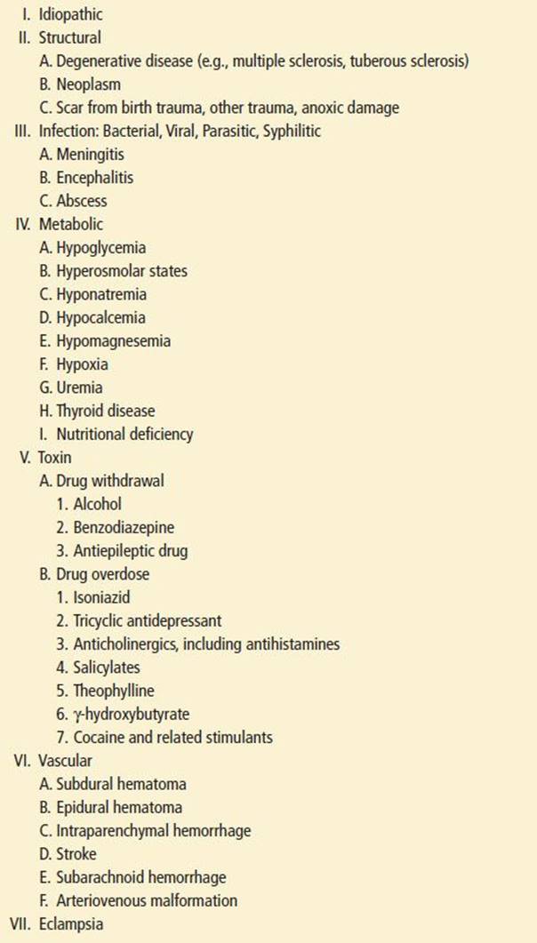
Metabolic and toxin-related etiologies must always be considered in the differential diagnosis and are often the most reversible. Hypoglycemia is the most common metabolic cause of seizures; hyponatremia, hypocalcemia, and hypomagnesemia are much less common (6). Interestingly, the metabolic encephalopathies, such as nonketotic hyperglycemia and uremia, can cause both focal and generalized seizures. Alcohol is the toxin most commonly associated with seizures, followed by tricyclic antidepressants, anticholinergics, cocaine, amphetamines, antihistamines, theophylline, and isoniazid. Drug withdrawal, including noncompliance with anticonvulsant medications in patients with a known seizure disorder, is a leading cause of recurrent seizures.
Acute central nervous system (CNS) infections are responsible for 4% to 12% of acute isolated seizures and up to 28% of cases of refractory SE.
A number of physiologic and psychological stressors can activate seizure disorders. These stressors include fatigue, sleep deprivation, hyperventilation, photic stimulation, emotional stress, and menstruation. In rare cases, seizures can be triggered by colors, objects, music, and voices. Pregnancy can also result in the onset of a seizure disorder, which is separate from eclamptic seizures.
ED EVALUATION
History and Physical Examination
The history must include a description of the event and an indication of the frequency, pattern, and duration of recent or previous seizures. Every attempt should be made to interview observers and emergency medical services (EMS) to obtain a clear description of the seizure to avoid misdiagnosing nonseizure events. One should ask the patient or EMS personnel about incontinence, loss of consciousness, and injury and determine whether there was an aura or a postictal period. Inciting factors such as medication noncompliance, infection, pregnancy, sleep deprivation, alcohol use, or other drug or medication use should be identified. A history of head trauma, headaches, diabetes, cancer, cerebrovascular disease, electrolyte imbalances, or infections can prove vital to making a diagnosis.
The physical examination begins with the vital signs, which can provide important red flags of underlying pathology. Signs of trauma should be sought, and a careful neurologic examination performed. Both unilateral and bilateral pupillary dilation as a result of seizure has been reported. Postictal patients often have hyperreflexia, upgoing toes, evidence of incontinence, or a tongue laceration. A focal deficit can represent a metabolic derangement such as hypoglycemia, an old or a new CNS lesion, or a Todd paralysis secondary to the seizure. Todd paralysis can be manifested as either a motor or a sensory deficit. Although it typically resolves within hours of seizure, neuroimaging may be necessary to differentiate a Todd paralysis from other causes of acute focal neurologic dysfunction.
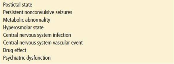
Postictal confusion is common but almost always resolves in a few hours. Other causes of altered mental status must be strongly considered in patients whose mental status remains impaired or when family or friends note an abnormally longer than normal postictal phase or unusual behavior manifestations. It is estimated that approximately 15% of patients treated for convulsive SE continue to be in nonconvulsive status after their motor activity is controlled; this unrecognized status may be a factor in the high mortality associated with SE and suggests the need for EEG monitoring if there is a prolonged “postictal” period (7). Causes of altered mental status in patients who have had a seizure are listed in Table 156.4.
TABLE 156.4
Causes of Altered Mental Status in Patients Who Have Had a Seizure
Laboratory Testing
Laboratory testing in postictal patients often demonstrates a metabolic acidosis or an elevated serum–creatine phosphokinase level. These findings can be helpful and should be sought, but, unfortunately, they are time and patient dependent and their absence does not rule out the possibility that a seizure occurred.
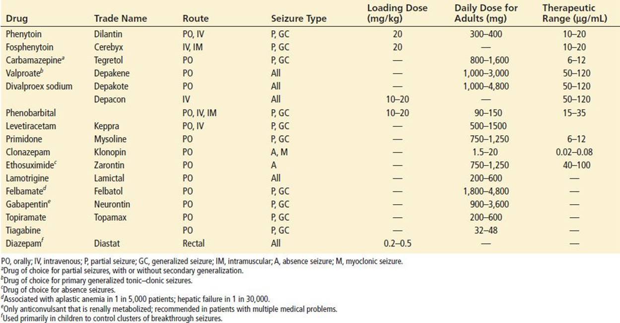
Of all laboratory tests, serum glucose determination followed by a serum sodium determination has been found to be the most valuable in diagnosing an unsuspected etiology of seizure (8). The decision to measure other electrolytes, creatinine, blood urea nitrogen, calcium, and magnesium is based on the individual patient assessment; generally, these tests are of limited value in patients with no underlying medical problems who have returned to baseline. An arterial blood gas analysis is indicated when hypoxia or an underlying acidosis is suspected. A pregnancy test should be obtained in all women of childbearing age (6). Serum antiepileptic drug (AED) levels should be checked in patients taking such medications and in those who have had a seizure or when a history cannot be obtained (Table 156.5). Such therapeutic ranges are available for phenytoin, carbamazepine, valproate, and phenobarbital. Serum levels of most of the newer AEDs cannot be measured rapidly in most hospitals.
TABLE 156.5
Drugs Used in the Treatment of Epilepsy
Alcohol is by far the most common drug of abuse associated with seizures, followed by cocaine. Serum alcohol, salicylate, lithium, and theophylline levels and urine drug–screening tests should be ordered based on clinical suspicion. Since patients with epilepsy can have other medical problems (e.g., depression), other possibilities (e.g., tricyclic overdose) should not be overlooked and an electrocardiogram (ECG) considered.
Patients who have had a seizure but who are not immunocompromised and who do not have an abnormal mental status, fever, or meningeal signs do not require a lumbar puncture as part of their ED evaluation. However, seizures, especially SE, are associated with hyperthermia, leukocytosis, and altered mental status, thus often forcing consideration of encephalitis. It is also important to note that, as mentioned previously, seizures can cause cerebrospinal fluid pleocytosis, further confusing the issue occasionally. It is recommended that antibiotics and/or antivirals, once started, be continued until cerebrospinal fluid culture results are available.
Neuroimaging and Electroencephalography
CT scanning of the head is indicated in the ED when an acute intracranial event, such as subdural or subarachnoid hemorrhage, is suspected. Patients with acute trauma, a past history of malignancy, or with an abnormal neurologic examination are most likely to have an abnormal imaging study. Special populations, including immunocompromised hosts, the elderly, and immigrants from countries where cysticercosis is endemic are candidates for neuroimaging.
All patients who have had a first-time seizure will need an imaging study but not necessarily an emergent CT scan obtained during the ED stay. If the patient has a normal mental status and neurologic examination and is judged to be reliable for follow-up, it is reasonable, after consultation with a neurologist, to arrange for outpatient magnetic resonance imaging (MRI). MRI is superior to CT scanning in identifying structural epileptogenic abnormalities (9).
EEG monitoring is not a standard practice in the ED, yet there are clear indications for its use on an emergent basis (6). Any patient with altered mental status in whom nonconvulsive SE is suspected requires an emergent EEG. An emergent EEG is also indicated in patients with refractory SE who have been managed with paralysis or intravenous (IV) agents.
Additional laboratory and radiographic testing may be indicated to identify underlying complicating factors, such as cardiac dysrhythmia, urinary tract infection, sepsis, or pulmonary disease.
KEY TESTING
• Serum glucose
• Serum AED levels
• Electrolytes, creatinine, blood urea nitrogen, calcium, and magnesium (limited value in patients who have returned to baseline state)
• Urine/serum drug screen if suspected overdose
• CT or MRI of brain if suspected intracranial event. First-time seizure may have imaging in ED or as outpatient.
ED MANAGEMENT
Prehospital Concerns
In most cases, prehospital personnel will arrive at least 5 minutes after the onset of seizure activity, so patients who are still seizing on EMS arrival are best managed as presumed SE. If a patient is found convulsing or remains confused or unresponsive, the patient’s blood sugar should be measured, or, if this test is not available, dextrose (D50 or D25 in children) should be administered. A randomized, double-blind out-of-hospital trial comparing IV lorazepam, diazepam, and placebo demonstrated benefit and safety of paramedic administered benzodiazepines in the management of SE in the field (10).
When IV access is not immediately available, rectal diazepam is an option, especially in children. Many parents of children with epilepsy have rectal diazepam on hand to be given, should their child have a seizure. Intramuscular (IM) injection of midazolam is also an option when IV access has not been established. A randomized double-blind noninferiority trial demonstrated that IM midazolam is as least as safe and effective as IV lorazepam for children and adults (11).
Patients with a known seizure disorder who experience a typical event and are asymptomatic afterward do not necessarily require transport to the hospital, if they have the capacity to refuse transport. Prehospital personnel should advise these patients to contact their primary care provider as soon as possible.
First-Time Seizures
Patients who have had a single, unprovoked seizure have about a 35% risk of recurrence within the next 5 years; the risk of recurrences increases to approximately 75% after two or three seizures (12). When a cause of the seizure is identified, management is based on the underlying pathology. When no cause can be identified, a risk–benefit analysis regarding initiation of antiepileptic therapy must be performed. There are significant psychological and social consequences of a diagnosis of epilepsy (i.e., individual state limitations on driving cars), as well as potential dangers from the chronic use of antiepileptic medications. Thus, one must be cautious when labeling patients as epileptic and instituting treatment from the ED.
For most first-time seizures, if a head CT was not indicated or if one is done that is negative, the patient can be discharged with very close follow-up by a primary care physician or neurologist (6). If such follow-up cannot be assured, a neurologist should ideally see the patient prior to discharge.
If there is a structural cause noted on CT scan that does not need emergent intervention—such as calcification suggestive of cysticercosis or a remote ischemic lesion—initiation of antiepileptic medications (AED) should be considered in consultation with a neurologist. Similarly, in the case of a patient who has had prior seizures but is not on any medications, an AED should be considered.
The choice of drug is based upon the type of seizure and the ease of achieving therapeutic serum levels. First-line medications that are effective for both primary generalized seizures and partial seizures with or without secondary generalization are carbamazepine, phenytoin, and valproate. Phenytoins and valproate are available for IV loading while oral carbamazepine may take days to reach therapeutic levels.
IV phenytoin loading, 15 to 20 mg/kg, can be accomplished quickly and leads to therapeutic blood levels within 1 hour of the infusion’s completion. IV phenytoin has a pH ∼2, and loading can be associated with local site irritation and even necrosis if extravasation occurs. Confusion, ataxia, and hypotension—caused primarily by the diluent propylene glycol—are dose- and rate-related effects. Infusions should not be run faster than 50 mg/min in adults. Fosphenytoin is a water-soluble disodium phosphate ester of phenytoin, which does not need a propylene glycol vehicle and has a more physiologic pH than does phenytoin. Fosphenytoin has fewer side effects than phenytoin, though too rapid an infusion can still cause hypotension, ataxia, and confusion. Fosphenytoin is measured in phenytoin equivalents (PE); its dosing is the same 20 mg (PE) as phenytoin though it can be given at up to 150 mg (PE)/min. Fosphenytoin can also be given safely by the IM route, with 100% bioavailability and therapeutic serum levels within 1 hour of a loading dose, although this requires a large volume IM injection 10 mL/500 PE. Oral phenytoin loading is another option, although it takes up to 6 hours for serum therapeutic levels to be reached (6). A daily maintenance dose of 300 mg can be given at bedtime, although, in some patients, divided dosing can provide better seizure control by minimizing variations in the blood level.
Recurrence of Seizures in Patients with Known Seizure Disorder
Noncompliance with anticonvulsant medication is, by far, the most common cause of seizure recurrence in a patient with a known seizure disorder. A patient who has been on phenytoin, but has a subtherapeutic level, should be given supplemental IV phenytoin or fosphenytoin to raise serum levels into the therapeutic range. IM fosphenytoin is another option that eliminates the need for infusion pumps and cardiac monitoring. The patient can then be discharged with instructions to resume oral dosing within 12 hours. Likewise, patients with subtherapeutic valproic acid levels can be supplemented with IV valproic acid administered at 20 mg/min and discharged on their oral regimen. Levetiracetam is also available in an IV formulation; however, because serum drug levels are not readily available, dosing augmentation must be done empirically.
Patients who have seizures despite therapeutic anticonvulsant levels must be evaluated carefully for precipitating factors such as infection, new anatomic lesions, or new medications. Decisions to increase the drug dose should be made only after communication with the patient’s primary care physician. A second anticonvulsant is generally only added once a patient has failed monotherapy taken to maximum doses.
The evaluation of pregnant patients with new onset seizures should follow the same principle as in other patients. However, if no etiology is identified, anticonvulsants should be withheld, pending a neurology consult. Eclampsia is an additional consideration in patients at more than 20 weeks’ gestation (see Chapter 140, Hypertensive Disorders of Pregnancy).
Noncompliance and sleep deprivation are the two most common causes of seizure recurrence in pregnant patients with epilepsy (13). Active seizures should be treated pharmacologically, just as in the nonpregnant patient, because the risks to the fetus of seizure-related hypoxia and acidosis are greater than the potential teratogenicity of anticonvulsant medications. Phenytoin and phenobarbital have similar teratogenetic profiles; carbamazepine is probably the safest of the anticonvulsants to use in pregnancy. Beyond 24 weeks’ gestation, fetal monitoring should be provided during and after a seizure.
Alcohol-Related Seizures
Alcohol-related seizures deserve special mention because alcohol use is frequently involved in patients presenting to EDs with seizures. Alcohol-withdrawal seizures classically occur 6 to 48 hours after a significant reduction in the serum alcohol level, but they have also been associated with rising blood ethanol levels in some patients. Alcohol-related seizures are temporally associated with alcohol use but are not necessarily solely the result of the alcohol. Alcoholics are susceptible to cerebrovascular insults, trauma, metabolic disorders, and infections and are thus at increased risk for seizures. Management of the alcoholic who has had a seizure focuses on determining whether the event was a withdrawal seizure or because of another etiology. Patients with a first-time alcohol-related seizure should be evaluated fully in the same manner as any other patient.
The diagnosis of alcohol-withdrawal seizures is based on the history, and the clinician must always consider the possibility that another etiology is responsible. There is no good evidence to support the use of phenytoin either acutely or chronically in the management of alcohol-withdrawal seizures. Benzodiazepines alone are sufficient to prevent successive withdrawal seizures in the acute setting. Patients who have had one alcohol-withdrawal seizure should be treated with lorazepam 2 mg IV. Following this, if the patients have been observed to remain seizure-free and at baseline mental status for 6 hours with no evidence of withdrawal, they can be discharged if a suitable environment is available (14).
Psychogenic Seizures
Psychogenic seizures should be suspected in patients who are refractory to anticonvulsants and whose clinical presentation is atypical. The diagnosis is suspected particularly when a convulsing patient does not have a metabolic acidosis, and it can be confirmed, when possible, by demonstrating that an abnormal EEG does not accompany the clinical convulsion. Management of psychogenic seizures is dependent on making the correct diagnosis; although this sounds obvious, the anxiety associated with evaluating a convulsing patient often leads to the initiation of pharmacologic interventions before an adequate assessment is performed (15). The key to management is avoiding iatrogenic harm by aggressive pharmacologic interventions or supportive measures such as intubation. Long-term management usually involves a comprehensive evaluation in an epilepsy monitoring unit, with a coordinated approach involving both an epileptologist and a psychiatrist.
Status Epilepticus
The management of convulsive SE involves expedited and simultaneous maintenance of the patient’s airway, breathing, and circulation; diagnostic testing; and pharmacologic intervention (Fig. 156.1). The definition of SE has changed in recent years to reflect the importance of rapid therapeutic intervention. Many researchers consider 5 minutes of continuous seizure activity to be SE. The term “impending status” has been suggested for any seizure lasting >5 minutes because videotape studies show that the mean duration of seizures is 1 minute ± 12 seconds. This average would make a seizure of 5 minutes duration “18 to 20 standard deviations away from the norm of a single seizure, indicating that something distinctly unusual and severe is happening” (16).
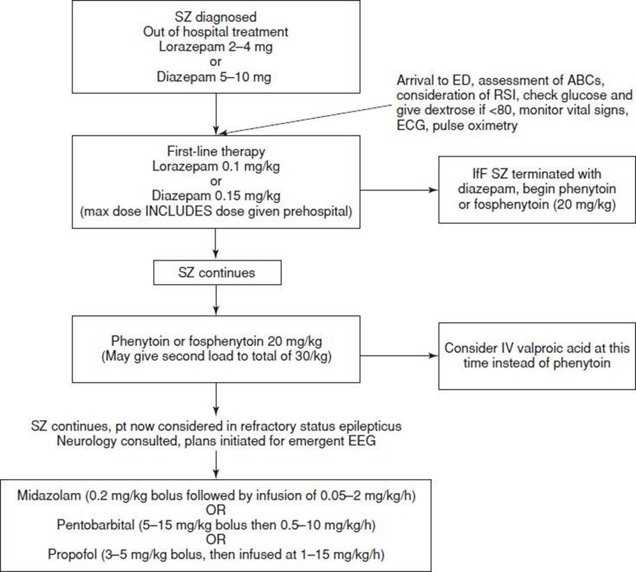
FIGURE 156.1 Approach to patient in status epilepticus. (Modified from Shearer P, Park D. Seizures and status epilepticus. Ann Emerg Med. 2002;39:190–192.)
SE occurs more frequently in patients with an underlying neurologic insult, either old or new, and most patients do not have a prior history of epilepsy. A population-based study reported the causes of SE as low AED levels in patients with known seizures (34%), remote symptomatic causes (24%), cerebrovascular accidents (22%), anoxia or hypoxia (∼0%), metabolic causes (∼0%), and alcohol and drug withdrawal (∼0%) (17). Morbidity and mortality in SE is related to the etiology, duration, and systemic consequences of the inciting event and the seizure. First-line agents for SE are the benzodiazepines (midazolam, lorazepam, or diazepam). IV phenytoin, fosphenytoin, valproic acid, and possibly levetiracetam are second-line agents. Patients who continue to seize despite aggressive use of these agents are considered to be in refractory SE and require treatment with IV anesthetic dose of midazolam, propofol, or pentobarbital. Refractory SE almost always indicates the presence of an underlying CNS disorder (18).
Airway support needs to be aggressive with continuous pulse oximetry. Patients should be intubated at any sign of hypoxia or loss of protective gag reflex. IV access should be secured using a nondextrose-containing solution because phenytoin precipitates if it is inadvertently administered with dextrose (dextrose will not precipitate fosphenytoin). Fluids should be run at keep-open rates unless the patient is hypotensive, because SE can be associated with both cerebral and pulmonary edema. When patients are intubated, short-acting paralytic agents should be used as continued motor activity, will be masked by long-acting agents lulling a clinician into a false sense of security. If prolonged paralysis is needed to carry out a procedure, a bedside EEG should be arranged.
The blood glucose level should be checked immediately by bedside testing. Blood should be tested for electrolytes, magnesium, phosphate, calcium, liver and renal function, hematocrit, WBC count, platelet count, AED levels, toxic drug screen (particularly salicylates), and alcohol. The urine should be checked for evidence of rhabdomyolysis, and a pregnancy test should be done on all women of childbearing age. The family and EMS should be questioned about the possibility of isoniazid or other drug overdose.
Patients in SE should be placed on a cardiac monitor, and an ECG should be done. A nasogastric tube should be inserted to prevent gastric distention, and a Foley catheter should be placed to help monitor volume status. A head CT scan should be done at some point in all patients. Consideration should be given to performing a lumbar puncture, if no source of the seizure activity is found.
The pharmacologic management of SE begins while the patient is being stabilized and diagnostic testing is being performed. Hypoglycemia is treated immediately with thiamine and dextrose. Antibiotics should be given very early in the management of any patient in whom meningitis, encephalitis, or sepsis is suspected.
The benzodiazepines diazepam and lorazepam are considered the first-line drugs in the management of SE. Lorazepam (0.1 mg/kg IV, up to 10 mg) or diazepam (0.15 mg/kg IV, up to 20 mg) will stop seizures in the majority of cases. Both drugs are efficacious, but lorazepam is preferred because its anticonvulsant action lasts up to 12 hours. Diazepam has a greater volume of distribution, and, although it rapidly enters the CNS, it quickly redistributes and the CNS effect may only last 20 minutes. Consequently, if lorazepam terminates the seizure activity, no additional anticonvulsant needs to be immediately given. However, a long-lasting anticonvulsant such as phenytoin must be added when diazepam is used, or the patient will be at higher risk of seizure recurrence. The Veterans Affairs (VA) Cooperative Study compared four IV treatment regimens for convulsive SE: Diazepam followed by phenytoin, lorazepam alone, phenytoin alone, and phenobarbital alone. This well-designed, randomized, double-blind study found no outcome difference between the four treatments, although lorazepam was more effective when compared directly with phenytoin alone. The authors recommended lorazepam as the first-line agent in generalized convulsive status (2).
When seizures persist despite a full loading dose of a benzodiazepine, either valproic acid (20 mg/kg over 5 to 20 minutes), or a phenytoin is recommended (20 mg/kg of phenytoin or fosphenytoin equivalents at a rate of 50 mg/min or 150 mg/min, respectively). There is a growing body of literature supporting the early use of valproic acid instead of a phenytoin due to its efficacy and its favorable side effect profile (19,20). There is some, but limited, evidence to support IV levetiracetam as a second-line agent in treating SE. Unlike most of the agents used in treating status, levetiracetam has no hepatic metabolism, is renally excreted, and can be rapidly loaded making it an attractive agent in select cases (21).
SE that does not terminate after a benzodiazepine and a second-line agent is usually the result of a significant underlying CNS lesion. The patient is considered to be in refractory SE and should be treated with anesthetic doses of midazolam, pentobarbital, or propofol. Studies have not shown one drug to be better than the others (22). The choice of drug for refractory SE is made on the basis of accessibility, familiarity with use, the potential role of side effects of each drug, and consideration of the preference of the admitting neurocritical care service. Underdosing and delay in starting any drug are the main pitfalls.
Midazolam is given as a loading dose of 0.2 mg/kg and is followed by an infusion of 0.05 to 2 mg/kg/h (23). Propofol provides almost immediate suppression of seizure activity after a bolus infusion (24). It is rapidly metabolized and can be titrated easily. Propofol is given as a bolus of 3 to 5 mg/kg followed by an infusion at 1 to 15 mg/kg/h. The propofol infusion syndrome of hypotension, lipidemia, rhabdomyolysis, and metabolic acidosis limits the prolonged infusion of propofol in both adults and children (25). Pentobarbital is given as 5 to 15 mg/kg at 25 mg/min, followed by 0.5 to 10 mg/kg/h as an infusion (26). Besides requiring ventilatory support, these patients are at significant risk for hypotension and therefore require hemodynamic monitoring. Hypotension should be treated with fluids and vasopressors as needed. EEG monitoring should be instituted to evaluate for continued seizure activity (27). Pentobarbital has the longest duration of action of the three agents, making it more difficult to remove when evaluating seizure suppression (28).
Many other IV and inhalational drugs have been tried in the management of refractory SE, but no controlled trials support their use (29). These drugs include inhalational anesthetics, continuous infusions of ketamine, IV lidocaine, and rectal chloral hydrate. Current recommendations would support use of these drugs only if all other measures fail.
Subtle Status Epilepticus
Subtle status occurs when the clinical manifestations of convulsive SE diminish and become less overt but the condition persists; mortality is very high in this group especially if it goes unrecognized. Continuous EEG monitoring should thus be considered in all SE patients (2).
Nonconvulsive Status Epilepticus
Nonconvulsive SE presents as altered mental status ranging from a mild behavioral alteration noticeable only to family or friends to frank coma (30–32). Other manifestations may include speech arrest, cognitive deficits, delusions, paranoia, hallucinations, or psychosis. Because, by definition, there is rarely a significant motor component, the diagnosis is often missed, and there are many case reports of these patients initially being diagnosed with a psychiatric illness. Nonconvulsive SE has been reported in every age group, can be the initial presentation of a seizure disorder, and has been reported to last as long as 8 weeks.
The diagnosis should be suspected in patients with a seizure history who present with a prolonged postictal period or an unusual behavior pattern. Nonconvulsive SE should also be considered in patients without a seizure history who present with altered mental status of undetermined etiology. The diagnosis is made by EEG. Treatment is the same as for convulsive SE, beginning with lorazepam, 2 mg/min, until the EEG normalizes or a total of 10 mg is administered. This treatment should be followed by phenytoin loading, although it is questionable whether phenytoin is effective in absence status.
CRITICAL INTERVENTIONS
• Check a rapid glucose in patients with seizures or refractory SE.
• Protect actively seizing patients from injury.
• Administer rapid, aggressive benzodiazepine therapy for SE.
• Provide aggressive airway support for patients in SE, and intubate if there are signs of hypoxia or a loss of protective gag reflex.
• Establish a departmental protocol for management of SE.
DISPOSITION
The disposition of patients who have had a seizure depends on the etiology of the event and, often, on the socioeconomic environment to which the patient will return (33). Of prime importance is communication with the primary care provider who will be responsible for following up on the patient’s diagnostic tests and medications.
Hospital admission should be considered when the cause of a seizure is unclear, when seizure recurrence has not been controlled, when the seizure is secondary to a treatable underlying illness, or when there is concern that the patient will not receive timely outpatient evaluation. All patients who have been in SE should be admitted, usually to an intensive care setting.
Common Pitfalls
• Improper infusion of phenytoin
• Assumption that altered mental status is because of a postictal state and failure to consider other possible etiologies such as subtle status or nonconvulsive status
• Failure to have and to activate an SE protocol
REFERENCES
1. Brophy G, Bell R, Claassen J, et al. Guidelines for the evaluation and management of status epilepticus. Neurocrit Care. 2012;1:3–23.
2. Treiman D, Meyers P, Walton N, et al. A comparison of four treatments for generalized convulsive status epilepticus. N Engl J Med. 1998;339:792–798.
3. Hauser W, Anneger J, Kurland L. Incidence of epilepsy and unprovoked seizures in Rochester, Minnesota: 1935–1984. Epilepsia. 1993;34:453–468.
4. Friedman J, LaFrance W. Psychogenic disorders. J Neurol. 2010;67:753–755.
5. Martindale J, Goldstein J, Pallin D. Emergency department seizure epidemiology. Emerg Med Clin North Am. 2011;29:15–27.
6. ACEP Clinical Policies Committee; Clinical Policies Subcommittee on Seizures. Clinical policy: Critical issues in the evaluation and management of adult patients presenting to the emergency department with seizures. Ann Emerg Med. 2004;43:605–625.
7. DeLorenzo RJ, Hauser WA, Towne AR, et al. A prospective, population-based epidemiologic study of status epilepticus in Richmond, Virginia. Neurology. 1996;46(4):1029–1035.
8. American Academy of Neurology. Practice Parameter: Evaluating an apparent unprovoked first seizure in adults (an evidence based review). Neurology. 2007;69:1996–2007.
9. American Academy of Neurology. Reassessment: Neuroimaging in the emergency patient presenting with seizure (an evidence based review). Neurology. 2007;69:1772–1780.
10. Alldredge BK, Gelb AM, Isaacs SM, et al. A comparison of lorazepam, diazepam, and placebo for the treatment of out-of-hospital status epilepticus. N Engl J Med. 2001;345:631–637.
11. Silbergleit R, Durkalski V, Lowenstein D, et al. Intramuscular versus intravenous therapy for prehospital status epilepticus. N Engl J Med. 2012;366:591–600.
12. Lowenstein DH, Aldredge BK. Status epilepticus. New Engl J Med. 1998;338:970–976.
13. Delgado-Escueta A, Janz D. Consensus guidelines: Preconception counseling, management, and care of the pregnant woman with epilepsy. Neurology. 1992;42(suppl 5):149–161.
14. D’Onofrio G, Rathlev NK, Ulrich AS, et al. Lorazepam for the prevention of recurrent seizures related to alcohol. N Engl J Med. 1999;340:915–919.
15. Howell S, Owen L, Chadwick D. Pseudo status epilepticus. Q J Med. 1989;71:507–519.
16. Chen JWY, Wasterlain CG. Status epilepticus: Pathophysiology and management in adults. Lancet Neurol. 2006;5(3):246–256.
17. DeLorenzo RJ, Waterhouse EJ, Towne EJ, et al. Persistent nonconvulsive status epilepticus after the control of convulsive status epilepticus. Epilepsia. 1998;39:833–840.
18. Brophy G, Meierkord H, Boon P, et al. EFNS guideline for the evaluation and management of status epilepticus in adults. Eur J Neurol. 2010;17;346–355.
19. Agarwal P, Kumar N, Chandra R, et al. Randomized study of intravenous valproate and phenytoin in status epelepticus. Seizure. 2007;26:527–532.
20. Misra U, Kalita J, Patel R. Sodium valproate vs. phenytoin in status epilepticus: A pilot study. Neurology. 2006;67:340–342.
21. Fattouch J, Di Bonaventura C, Casciato S, et al. Intravenous levetiracetam as first line treatment of status epilepticus in the elderly. Acta Neurol Scand. 2010;121:418–421.
22. Claassen J, Hirsch LJ, Emerson RG, et al. Treatment of refractory status epilepticus with pentobarbital, propofol, or midazolam: A systematic review. Epilepsia. 2002;43:146–153.
23. Kumar A, Bleck TP. Intravenous midazolam for the treatment of refractory status epilepticus. Crit Care Med. 1992;20:483–488.
24. Brown LA, Levin GM. Role of propofol in refractory status epilepticus. Ann Pharmacother. 1998;32:1053–1059.
25. Vasile B, Rasulo F, Candiani A, et al. The pathophysiology of the propofol infusion syndrome: A simple name for a complex problem. Intensive Care Med. 2003;29:1417–1425.
26. Manno EM. New management strategies in the treatment of status epilepticus. Mayo Clin Proc. 2003;78:508–518.
27. Osorio I, Reed R. Treatment of refractory generalized tonic clonic status epilepticus with pentobarbital anesthesia after high dose phenytoin. Epilepsia. 1989;30:464–471.
28. Rossetti A, Milligan T, Vulliemoz S, et al. A randomized trial for the treatment of refractory status epilepticus. Neurocrit Care. 2011;14:4–10.
29. Jagoda A, Riggio S. Refractory status epilepticus in adults. Ann Emerg Med. 1993;22:1337–1348.
30. Chang A, Shinnar S. Nonconvulsive status epilepticus. Emerg Med Clin North Am. 2011;29:65–72.
31. Lee S. Nonconvulsive status epilepticus: Ictal confusion in later life. Arch Neurol. 1985;42:778–781.
******
32. Tomson T, Lindbom U, Nilsson B. Nonconvulsive status epilepticus in adults: Thirty-two consecutive patients from a general hospital population. Epilepsia. 1992;33:829–835.
33. Cloquet C, Depret-Vassal J, Doumenc B, et al. Predictors of early seizure recurrence in patients admitted for seizures in the emergency department. Eur J Emerg Med. 2008;15:261–267.