
Phillip A. Scott
The term stroke refers to the sudden onset of a neurologic deficit caused by an acute vascular lesion of the brain that is hemorrhagic, embolic, thrombotic, or aneurysmal in nature. Cerebrovascular disease is a broader term, encompassing stroke and other vascular causes of central nervous system dysfunction.
ISCHEMIC CEREBROVASCULAR DISEASE
Ischemic neurologic deficits are commonly categorized by their time course. A transient deficit, not accompanied by tissue infarction, is considered a transient ischemic attack (TIA), with the majority resolving within 60 minutes of onset (1). By extension, the presence of cellular loss on advanced imaging, typically accompanied by persistent clinical signs, defines infarction—that is, stroke. Other classification systems define stroke syndromes by probable cause or anatomic location.
Risk Factors
Not surprisingly, risk factors for ischemic cerebrovascular disease parallel those for atherosclerotic heart disease. Hypertension, diabetes, and coronary artery disease substantially increase the risk of stroke. Left ventricular hypertrophy, atrial fibrillation, and acute myocardial infarction (AMI) are also independent risk factors for ischemic stroke.
There are significant associations of ischemic cerebrovascular disease with age, gender, and race. The incidence of all strokes increases with age, doubling with each decade of life. There is a slightly higher risk in men as compared with women, and black men and women have an overall age- and sex-adjusted stroke incidence rate 1.5 times higher than whites.
Medical conditions predisposing to stroke include a history of migraine headaches, elevated fibrinogen levels or hematocrit (causing increased blood viscosity), and advanced age with elevated total serum cholesterol levels.
Environmental risk factors for ischemic cerebrovascular disease include smoking and heavy alcohol consumption. Women taking oral contraceptives, particularly high-estrogen formulations, are also at increased risk for stroke. This risk is more pronounced in female smokers older than 35 years of age and in those with other cardiovascular risk factors.
Finally, patients with a history of ischemic stroke represent a high-risk population, with 1- and 5-year recurrence rates among survivors of 10% and 20%, respectively.
TRANSIENT ISCHEMIC ATTACK
There is a higher risk of early stroke following TIA than previously thought. Ten percent to 15% of patients with TIA have a stroke within 3 months, with half experiencing a stroke within 48 hours of the initial TIA (1).
TIAs may result from plaque ulceration or embolism of platelet aggregates from extracranial or intracranial atherosclerosis. After impeding local blood flow, these emboli break up, accounting for the transient nature of the episode. Other sources of emboli include prosthetic heart valves and infective or marantic vegetations on valve leaflets. Small vessel disease in penetrating cerebral arteries may actually be the most common cause of TIAs and that these “lacunar” TIAs may represent a subgroup with a better prognosis than TIAs with cortical symptoms. Hypotension and mechanical kinking of the extracranial vessels are less common causes of TIAs. Cardiac dysrhythmias are an important potential etiology for the emergency physician to consider.
CLINICAL PRESENTATION
Carotid artery syndrome results from transient disruption of the anterior circulation of one cerebral hemisphere or of the retina. Hemispheric ischemia results in an abrupt onset of contralateral monoparesis or hemiparesis, numbness, tingling, aphasia, or hemianopia. Occlusion of the ophthalmic branch of the internal carotid artery results in retinal ischemia, causing temporary monocular blindness (amaurosis fugax). The patient complains of a “fog,” “mist,” “cloud,” or “shade being drawn” over the visual field of one eye. Funduscopic examination of the involved eye may reveal cholesterol crystals (Hollenhorst plaques) in the retinal vessels.
Transient ischemia involving the posterior circulation (the brainstem or occipital lobes) is termed vertebrobasilar artery syndrome. Interruption of brainstem circulation may result in symptoms of vertigo, dizziness, ataxia, nausea, or vomiting secondary to involvement of the vestibular nuclei. Dysarthria, dysphagia, and perioral numbness may also occur. Occipital lobe ischemia results in blurred or dim vision or total cortical blindness. Diplopia may result from loss of conjugate gaze.
Basilar artery TIAs may cause drop attacks—brief paretic spells resulting in the patient’s dropping to his or her knees. There is no loss of consciousness, distinguishing it from true syncope, and the patient attempts to rise immediately after falling.
Transient global amnesia (see Chapter 157, “Transient Global Amnesia”) is thought to be caused by ischemia of the temporal lobes or thalamic area supplied by the posterior cerebral arteries. These episodes affect men more frequently than women and are characterized by a rapid development of retrograde memory loss and confusion, with preservation of self-identity. The length of retrograde amnesia gradually shortens until only the period during the attack remains lost.
DIFFERENTIAL DIAGNOSIS
The differential diagnosis of TIAs includes transient deficits secondary to mass lesions from either tumor or hemorrhage (these patients often have a residual neurologic deficit detectable on examination); infection (as with focal abscess, septic emboli, meningitis, or encephalitis); seizure with a subsequent Todd paralysis; complicated migraine; exacerbation of multiple sclerosis; hypoglycemia; and syncope from any cause, particularly cardiac dysrhythmias.
ED EVALUATION
Clinical investigation of a TIA is directed toward identifying whether the deficit involves the anterior or posterior circulation, establishing the source of the TIA, and ruling out other causes of focal deficits. A detailed history often provides the most significant information. The patient may not be aware of any deficits, and the family should be questioned, if possible, along with the patient. The onset and course of symptoms should be carefully elicited. Were there any prior episodes? If so, how many? Were the events in the same or differing vascular distributions?
The neurologic examination in patients with TIAs is often unremarkable, but a thorough assessment is essential. Orthostatic blood pressures should be measured in patients with vertebrobasilar TIAs, because symptoms are often related to postural changes. Auscultation for cervical and subclavian bruits should be performed, as should evaluation for cardiac dysrhythmias, murmurs, and left ventricular dysfunction.
Determination of the ABCD2 score may assist in predicting short-term risk of stroke in patients presenting with TIA. Patients are assessed on points based on age ≤60 years (1 point); blood pressure ≤140/90 mm Hg on initial evaluation (1 point); clinical symptoms of weakness (2 points) or speech impairment without weakness (1 point); duration 10 to 59 minutes (1 point) or ≤60 minutes (2 points); and diabetes (1 point). The 48-hour risk of stroke was 0% for scores of 0–1, 1.3% for 2–3, 4.1% for 4–5, and 8.1% for 6–7 (2).
KEY TESTING

ED MANAGEMENT
Medical therapy is indicated for patients with posterior circulation TIAs, patients with anterior circulation TIAs who have <50% stenosis on carotid duplex scanning, and patients who are not candidates for carotid endarterectomy. Medical treatment options are limited to antiplatelet therapy and anticoagulation.
Acceptable options for initial antiplatelet therapy in patients with TIA include the following:
• Aspirin (50 to 325 mg/d) monotherapy
• Extended-release dipyridamole combined with aspirin (Aggrenox)
• Clopidogrel (Plavix) monotherapy (75 mg/d)
• Clopidogrel (300-mg loading dose) combined with aspirin (75 mg)
Antiplatelet therapy with aspirin (50 to 325 mg/d) reduces the incidence of nonfatal stroke, nonfatal myocardial infarction, and death from all vascular causes in patients with TIAs. Because the lower dosages require several days to achieve maximal platelet inhibition, therapy in the early setting should be initiated with 300 to 325 mg/d. The combination of dipyridamole plus aspirin (Aggrenox) was modestly better than aspirin alone; however, a number of patients discontinue combination therapy owing to medication intolerance (3,4).
Clopidogrel is an ADP-inhibitor and also acceptable as monotherapy for patients with TIA. Its role as a primary or secondary agent in the treatment of TIA is debated, given its cost and the small (0.5%) absolute improvement in reducing risk for the combined end point of stroke, AMI, and vascular death in patients with atherosclerotic vascular disease compared to aspirin alone (5). The combination of clopidogrel with aspirin was superior to aspirin alone in preventing recurrent stroke in patients with TIA or minor stroke (8.2% vs. 11.7%) in a large Chinese study (6). A companion trial is currently underway in the United States (7).
The use of anticoagulation in patients experiencing noncardioembolic TIA lacks a rigorous scientific basis. Though heparin has traditionally been used in select patients with TIA, no prospective randomized study of significant size has demonstrated a clear benefit and its use cannot be recommended on the basis of existing data. If used at all, it should be reserved for “high-risk” patients such as those with crescendo TIAs, vertebrobasilar TIA, recurrent TIA while receiving maximum antiplatelet therapy, high-grade carotid stenosis, or a suspected cardiac source for emboli. Neurologic consultation is recommended prior to initiation.
Adjusted-dose oral anticoagulation with warfarin has been the predominant treatment for stroke prevention in patients with a cardioembolic TIA due to atrial fibrillation. A target international normalized ratio (INR) of 2 to 3 is recommended. The efficacy of aspirin is considerably less than warfarin in this population and is recommended as an alternative for those with contraindications to oral anticoagulation. Newer oral anticoagulants inhibiting thrombin or factor Xa have demonstrated improved stroke prevention with comparable safety to warfarin. The lack of an available antidote has raised concern over their use. Anticoagulant therapy may be appropriate for patients with other sources of cardiogenic emboli who have a TIA; however, randomized clinical trials have not been performed in these specific patient populations.
A subset of patients with TIA has surgically correctable carotid stenosis and represent an important group for the emergency physician to identify. Those with ≥50% carotid stenosis are potential candidates for endarterectomy and should receive medical therapy in the ED pending a decision on vascular intervention.
CRITICAL INTERVENTIONS
• Initiate antiplatelet therapy for patients with TIA.
• Identify and refer patients with surgical or endovascular correctable carotid stenosis.
DISPOSITION
Patients who have a new anterior circulation TIA and are potential surgical candidates should not be discharged without arranging expeditious evaluation of the carotid arteries. If duplex scanning is not immediately available, the patient should be started on antiplatelet therapy and carotid duplex scanning performed as soon as possible, with subsequent vascular consultation for patients determined to have stenosis of 50% or more.
Because of the increased short-term risk of stroke, it may be optimal to admit patients with TIA to an inpatient or observation bed. Current guidelines state it is reasonable to hospitalize patients with TIA if they present within 72 hours of the event and have either an ABCD2 score ≤3; ABCD2 score of 0–2 and inability to complete diagnostic workup as outpatient within 2 days; or an ABCD2 score of 0–2 and other evidence indicating the event was caused by focal ischemia (2). The clinical utility of the ABCD2 score in the emergency department setting, however, has been questioned owing to its low sensitivity and specificity (8). Admission and neurologic consultation should be considered for high-risk patients. Standard protocols for patients with TIAs should be jointly developed with neurologic specialists in each institution. Active collaboration can ensure the provision of efficient and consistent patient care and facilitate future modifications as new data become available.
Common Pitfalls
• Discharging patients with anterior circulation TIAs before determining whether there is a potentially surgically correctable carotid lesion. This carries a high medicolegal risk.
• Failure to obtain consultation in high-risk patients: Those with crescendo TIAs, vertebrobasilar TIA, recurrent TIA on maximal antiplatelet therapy, high-grade carotid artery stenosis, or presumed cardioembolic source.
• Failing to discuss signs and symptoms of stroke with the patient and emphasizing the need to immediately return for recurrent symptoms.
ISCHEMIC STROKE
Ischemic strokes have traditionally been classified by clinical presentation, time course of onset, progression of deficits, accompanying symptoms, and risk factor profiles. The clinical description of the deficit identifies the extent and location of the stroke and allows accurate communication regarding patient status. The time course and progression of symptoms suggest certain etiologies; for example, prolonged, “stuttering” deficits indicate a thrombotic phenomenon, whereas an abrupt onset of the maximal deficit suggests an embolic event. Associated symptoms may suggest other causes for neurologic deficits (e.g., a seizure preceding a Todd paralysis or chest pain preceding an ischemic stroke of cardioembolic origin). Consideration of cerebrovascular risk factors helps to classify the stroke as lacunar, embolic, thrombotic, extracranial, or intracranial.
CLINICAL PRESENTATION
Anterior Cerebral Artery Syndrome
Patients with infarction in the territory of the anterior cerebral artery present with hemiparesis involving the leg more than the face or arm. Urinary incontinence and primitive reflexes (grasp and suck) are often present. The patient may perseverate with speech or motor movements and respond slowly to questions. If both anterior cerebral arteries originate from a common trunk, a bilateral parasagittal infarct may occur, with paraplegia and speechlessness (anarthria).
Middle Cerebral Artery Syndrome
Infarction in the distribution of the middle cerebral artery is the most common of the stroke syndromes. Patients with infarcts of the middle cerebral artery present with weakness of the face and arm greater than that of the leg, often with corresponding cortical sensory loss. The presence of aphasia localizes the lesion to the dominant hemisphere. Inattention, extinction on double simultaneous sensory or visual testing, neglect, or constructional or dressing apraxia indicates nondominant hemisphere involvement. Homonymous hemianopia and conjugate eye deviation toward the side of the infarct may also be found. All of these findings are present in the patient with a proximal trunk occlusion and variably present when branches of the middle cerebral artery are involved.
Posterior Cerebral Artery Syndrome
Infarcts in the posterior cerebral artery territory present with homonymous hemianopia secondary to visual cortex involvement. The patient is often unaware of the deficit until formally tested. Motor involvement is minimal, but sensory loss, including both light touch and pinprick, may be severe. There is no aphasia or disruption of nondominant hemisphere functions. Small branches of the posterior cerebral artery anastomose with branches of the anterior and middle cerebral arteries in border zones between the arterial distributions. These areas are at risk for watershed infarcts during periods of decreased blood flow.
Vertebrobasilar Artery Syndrome
The classic sign of brainstem stroke is the presence of crossed symptoms: Ipsilateral cranial nerve palsy and contralateral hemiplegia. One of the most common strokes in this region is due to occlusion of the vertebral or posterior-inferior cerebellar artery, resulting in the lateral medullary (Wallenberg) syndrome. Patients may present with ipsilateral facial pain (described as a sharp or stabbing sensation to the face or eye), vertigo, headache, limb ataxia, Horner syndrome, and weakness of the soft palate, with dysphagia and dysphonia. Contralateral symptoms include loss of pinprick and temperature sensation in the arm and leg. Other brainstem syndromes may result in deafness, nausea, vomiting, vertigo, and diplopia.
Cerebellar Infarction
A subset of posterior circulation strokes involves the cerebellum. The most common initial symptom is the sudden inability to walk or stand. This may be followed by complaints of headache, nausea, vomiting, central vertigo, and, possibly, a stiff neck. Cranial nerve findings may also be present. Although the initial CT scan is often unremarkable, one-third of patients develop significant posterior fossa edema. In this confined space, edema produces pressure on the brainstem, causing a decrease in the level of consciousness, typically after a stable period of 6 to 12 hours. Pathologic respiratory patterns and pupillary dilation are late findings, indicating impending herniation. Recognition of cerebellar infarction is critical, and frequent neurologic assessment is imperative. Repeat CT scanning or magnetic resonance imaging (MRI) is indicated for patients with a declining level of consciousness. Surgical decompression and/or medical treatment of elevated intracranial pressure (ICP) may be lifesaving.
Lacunar Syndromes
Lacunar infarcts cause several distinct stroke syndromes. Lacunes are found almost exclusively in the basal ganglia, internal capsule, thalamus, and brainstem and are associated with hypertensive arteriopathy. Approximately 20% of patients with lacunar infarctions have had a prior TIA, and the onset of symptoms is often surprisingly gradual (>36 hours in up to 30% of patients). Deficits often improve with time, and treatment is aimed at control of hypertension after the acute episode resolves. An evaluation for embolic sources should also be undertaken. Lacunar strokes are classified by clinical presentation.
The most frequent lacunar syndrome, pure motor stroke, causes paresis/paralysis of the face, arm, and leg. It is easily diagnosed when the deficit involves all three areas equally but may also present as a partial deficit. The location of the lacune may be in the internal capsule or pons. Cortical sensory deficits are notably absent.
Pure sensory stroke is the result of infarction of the ventral posterior nuclei of the thalamus and results in sensory loss in the face, arm, and leg. The patient may also complain of paresthesias involving the affected side. There is no hemiplegia or other cortical sign.
Patients with clumsy hand–dysarthria syndrome present with slurred speech and weakness and ataxia of the upper limb. Facial weakness may also be present. The syndrome is most commonly associated with lesions of the anterior limb and genu of the internal capsule and typically has a good functional outcome.
DIFFERENTIAL DIAGNOSIS
Table 155.1 lists conditions that may mimic ischemic stroke and is divided into structural and nonstructural diseases. Neuroimaging is used initially to evaluate these possibilities and to rule out structural causes of neurologic deficits.
TABLE 155.1
Differential Diagnosis of Ischemic Stroke
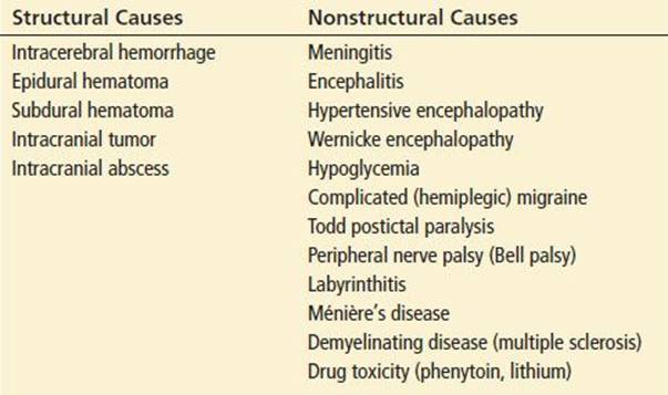
Diseases presenting with features similar to posterior circulation strokes can be difficult to distinguish from stroke. Wernicke encephalopathy presents as the triad of ophthalmoplegia, ataxia, and confusion and is found typically in chronic alcoholics. Ménière’s disease, with its characteristic triad of vertigo, tinnitus, and deafness, occurs in patients between the ages of 30 and 60 and is paroxysmal in nature. Gross ataxia and other cranial nerve deficits should be absent. Multiple sclerosis typically presents in the third to fourth decades of life and has variable symptoms, depending on the location of demyelination. Symptoms of phenytoin and carbamazepine toxicity include sedation, nystagmus, vertigo, ataxia, and nausea. Central nervous system symptoms of acute lithium toxicity include confusion, hyperreflexia, tremors, and cranial nerve deficits; dysarthria also may be a prominent feature.
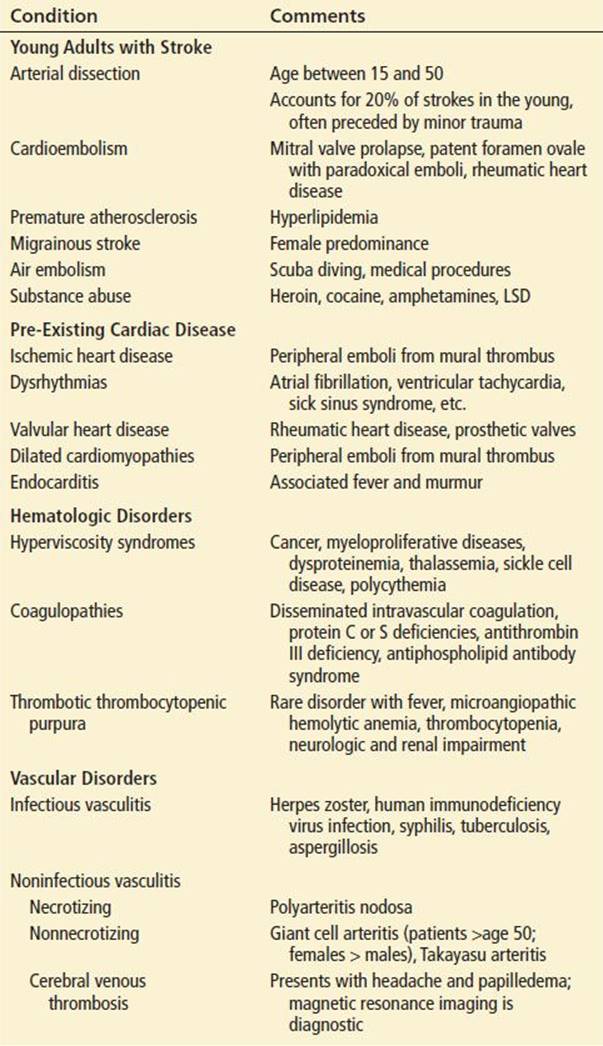
Special consideration should be given to stroke patients younger than 50 years of age or those who have pre-existing heart disease, hematologic disorders, or vasculitis. These patients typically fall into the cardioembolic, other, or unknown subtype classifications and may have reversible etiologies or other contributing disease states that should be identified as rapidly as possible. Conditions associated with each of these groups are listed in Table 155.2.
TABLE 155.2
Differential Diagnosis of Special Patient Groups with Ischemic Stroke
ED EVALUATION
Like myocardial infarction, stroke is an acute disease necessitating early identification and treatment. Prehospital personnel should obtain information surrounding the onset of symptoms, and particularly noting the time deficits started (“last seen normal” time). Patients found lying immobile for prolonged periods are at risk for decubitus ulcers, dehydration, hypothermia or hyperthermia, and rhabdomyolysis. Intravenous access should be obtained and a fingerstick glucose level determined. In the prehospital phase, it is unusual for patients with ischemic stroke to have an altered level of consciousness; an altered sensorium suggests other etiologies. Glucose-containing solutions and routine administration of dextrose (D50) should be avoided unless hypoglycemia is documented or strongly suspected. Narcan is generally not required unless there are historic features or pinpoint pupils, suggesting opiate ingestion.
On arrival in the ED, stroke patients require a monitored bed and rapid assessment of airway, breathing, and circulatory status. Complete vital signs should be obtained, including a core temperature. A focused physical examination should be performed, including a search for evidence of head trauma. The neck should be examined for meningeal signs present with either infection or subarachnoid hemorrhage (SAH). Auscultation for carotid bruits, particularly on the side opposite a clinical deficit, should be performed. The heart is examined for rate, regularity, and murmurs, searching for conditions predisposing to stroke, such as atrial fibrillation and mitral valvular disease. The extremities should be assessed for evidence of peripheral vascular disease or emboli and the skin examined for needle tracks or evidence of petechiae or ecchymosis, suggesting endocarditis, sepsis, or coagulopathy.
The neurologic examination should include a brief assessment of mental status, including level of consciousness, language, memory, and interpretation. A careful eye examination should follow, with testing of both visual acuity and visual fields. Pupillary size, reactivity, and extraocular movements provide information on brainstem function and cranial nerves III, IV, and VI. Anisocoria suggests a third nerve palsy, potentially from elevated ICP, and should be investigated with a “swinging flashlight” test. Conjugate eye deviation toward the normal arm and leg suggests a hemispheric lesion, whereas deviation toward the paretic arm or leg suggests a brainstem lesion. Funduscopic examination of the retina may reveal papilledema, hypertensive changes, Hollenhorst plaques, diabetic changes, or Roth spots suggestive of specific stroke etiologies. Cranial nerves V and VII through XII may quickly be assessed by examining facial and corneal sensation; smiling symmetry, eyebrow raising, and forehead wrinkling; gross auditory acuity; gag reflex; shoulder shrug (trapezius strength); head rotation against resistance (sternocleidomastoid strength); and tongue protrusion, respectively.
Motor strength and tone should be assessed in both proximal and distal muscle groups. Pronator drift is a sensitive indicator of upper extremity weakness.
The sensory examination is the most subjective part of the neurologic examination and should be interpreted in view of the patient’s ability to cooperate and the overall examination. Double simultaneous extinction is tested at this time, particularly if visual field deficits were identified. The ability of a patient to identify, by touch, common objects such as keys or pens (stereognosis) and to identify a number drawn on the skin surface (graphesthesia) is an easy test of sensory function done in the ED.
Cerebellar function can be assessed by finger-to-nose and heel-to-shin tests; these are particularly useful with the bedridden patient. Important reflex arcs to test include the biceps (C5, C6), triceps (C6, C7, C8), patellar (L2, L3, L4), and Achilles (L5, S1), and pathologic reflexes. Asymmetry of deep tendon reflexes or a unilateral Babinski sign may indicate corticospinal tract lesions.
Assessment of station and gait is one of the most revealing parts of the neurologic examination. Subtle weakness, ataxia, rigidity, and sensory defects can be identified by observation of the gait, toe walking, heel walking, tandem walking, and Romberg tests.
KEY TESTING
All patients:
• Noncontrast brain CT or brain MRI
• Blood glucose
• Oxygen saturation
• Serum electrolytes/renal function tests
• Complete blood count, including platelet count
• Cardiac enzymes
• Coagulation studies (prothrombin time/INR/activated partial thromboplastin time)
• EKG
Selected patients:
• Thrombin time and/or ecarin clotting time if patient suspected of taking direct thrombin inhibitors or factor Xa inhibitors
• Liver function tests
• Toxicology screens
• Blood–alcohol level
• Blood cultures (if febrile, or infectious etiology suspected)
• Serum creatine phosphokinase and/or urine myoglobin (if rhabdomyolysis suspected)
• Pregnancy test
• Arterial blood gas
• Chest x-ray (if pulmonary disease suspected)
• Lumbar puncture (if SAH is suspected and CT scan is negative)
• Electroencephalogram (if seizures suspected)
Noncontrast CT imaging will provide the information necessary for most emergent treatment decisions. An ischemic stroke does not become hypodense for 6 to 72 hours after vessel occlusion and is often not visible on initial CT evaluation. If CT scanning is unavailable, the patient should be stabilized and transferred as rapidly as possible to a facility with CT capability.
Multimodal CT (combining noncontrast brain CT with CT angiography and/or CT perfusion mapping) provides further vascular and tissue information and can enhance both diagnostic and management decisions. Disadvantages include an increased time to image acquisition and interpretation, the use of iodinated contrast, and additional radiation exposure. Multimodal MRI also provides enhanced information but typically plays a more limited role in the acute evaluation of most patients with stroke. Limitations include availability, patient cooperation, and contraindications to the procedure because of metallic foreign bodies or devices. Though their exact role in acute stroke care has yet to be defined, computed tomography angiography and magnetic resonance arteriography (MRA) may be of particular benefit in evaluating the patient with suspected carotid or vertebrobasilar artery dissection.
ED MANAGEMENT
The primary goal of acute stroke management is to reverse the clinical deficit by reestablishing cerebral blood flow, if possible. Patients who present within 3 hours of symptom onset are the most likely to benefit from intravenous recombinant tissue plasminogen activator (r-TPA, alteplase) and eligible patients should be treated (9,10). Data also support the use of intravenous r-TPA in selected patients who can be treated within 4.5 hours of symptom onset, though this indication does not have FDA approval (11).
Intravenous Thrombolytic Therapy
The American College of Emergency Physicians, American Academy of Neurology, and American Heart Association support the use of r-TPA (alteplase) in eligible patients. The NIH/NINDS Stroke Study found that r-TPA-treated stroke patients were at least 30% more likely to have minimal or no disability 3 months after the event, compared with those who received placebo (absolute increase in favorable outcome, 11% to 13%). Although treated patients had a symptomatic intracranial hemorrhage rate of 6.4% (vs. 0.6% for placebo), there was no significant difference in mortality at 3 months (17% r-TPA, 21% placebo) (12). Post hoc analyses do not support withholding therapy from any patient on the basis of suspected stroke subtype, age, clinical deficit, or CT findings of early ischemic changes.
Data also demonstrate the safety of community delivery of r-TPA in stroke. In a study of Michigan community hospitals, 557 stroke patients received intravenous r-TPA in the emergency department. The symptomatic intracerebral hemorrhage (ICH) rate was equivalent to the NINDS study (12).
The development of treatment guidelines for stroke may improve patient selection and speed the delivery of thrombolytics to appropriate patients. The National Institutes of Health recommends a target “door-to-needle” time for eligible r-TPA patients of 60 minutes.
Table 155.3 outlines inclusion and exclusion criteria for the use of intravenous r-TPA in acute stroke. Eligible patients should be treated with 0.9 mg/kg of r-TPA (maximum dose, 90 mg), with 10% given as a bolus over 1 to 2 minutes and the remainder given over 60 minutes via infusion pump. Administration of r-TPA between 3 and 4.5 hours after stroke onset may be considered for patients meeting the additional criteria listed in Table 155.3 (11). Table 155.4 presents posttreatment blood pressure control guidelines. An algorithm for the management of suspected intracranial hemorrhage secondary to thrombolytic therapy is shown in Figure 155.1.
TABLE 155.3
Intravenous r-TPA in Ischemic Stroke: Inclusion and Exclusion Criteria


TABLE 155.4
Post r-TPA Blood Pressure Management in Acute Stroke

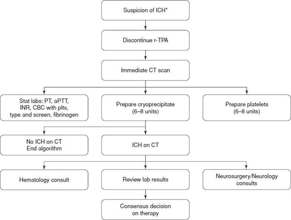
FIGURE 155.1 Management of suspected intracranial hemorrhage after r-TPA use. *Symptoms include neurologic deterioration, headache, acute hypertension, nausea, vomiting.
The development of institution-specific evaluation and treatment guidelines is recommended to identify local responsibilities concerning thrombolytic delivery and posttreatment care and disposition. A multidisciplinary approach will deliver the highest level of care in a time-critical setting.
Endovascular Therapy
Intra-arterial thrombolysis or mechanical reperfusion are potential considerations for patients who are ineligible for intravenous thrombolytic therapy (due to late presentation >4.5 hours from symptom onset or other reasons) presenting within 6 to 8 hours of symptom onset (13,14). Their exact role, however, is unclear. Two recent trials found no clinical improvement using endovascular therapy compared to standard intravenous r-TPA alone (15,16). The use of advance neuroimaging to select stroke patients with favorable penumbral patterns for mechanical embolectomy using the Merci Retriever or Penumbra System also failed to demonstrate superiority of those devices over standard treatment (17). Technology in this area is rapidly advancing and decisions regarding endovascular treatment should be made with qualified specialists, preferably within the context of a clinical trial.
In patients who are ineligible for reperfusion strategies, no acute intervention has proven effective in improving functional outcome. The objective of treatment is then to limit infarct extension and recurrence. Current approaches to preventing or minimizing infarct extension include maintenance of adequate cardiac output to preserve cerebral blood flow, regulation of core temperature, and control of the blood glucose level. Strategies to prevent recurrent infarction focus on antiplatelet and anticoagulation therapy.
Hypertension Management
Acute elevations of blood pressure typically decline, without treatment, within a few days to prestroke levels. Because the benefit of blood pressure control is unproved, it is reasonable to avoid treating hypertension in the absence of specific indications such as thrombolytic therapy for acute stroke, AMI, hypertensive encephalopathy, arterial dissection, and hemorrhagic transformation. Although no data exist to define the point of treatment for severe isolated hypertension in stroke, there is consensus that antihypertensive agents should be withheld unless the blood pressure exceeds 220/120 mm Hg.
In stroke patients who are not eligible for r-TPA and who require antihypertensive therapy, the goal is a gradual reduction of blood pressure by about 15%. No data are available to guide selection of medications for the lowering of blood pressure in this setting. Reasonable initial agents include labetalol, 10 to 20 mg IV over 1 to 2 minutes, repeated or doubled every 10 minutes (maximum dose, 300 mg) or nicardipine, 5 mg/h IV infusion as initial dose; titrated to desired effect by increasing 2.5 mg/h every 5 minutes to maximum of 15 mg/h. For patients with DBP >140 mm Hg, consider a sodium nitroprusside infusion, 0.5 μg/kg/min IV as initial dose with continuous blood pressure monitoring and titration. The use of antihypertensive agents requires frequent neurologic checks so that treatment may be discontinued if there is neurologic deterioration (10).
Volume Management
Maintenance of intravascular volume is important in optimizing cardiac output in stroke patients. These patients are often unable to maintain adequate oral intake, and up to one-third may be significantly hypovolemic. Volume deficits should be corrected as rapidly as possible and followed by maintenance fluids. Avoidance of hypoosmolar solution is recommended to prevent exacerbation of cerebral edema. Aggressive hypervolemic therapy to increase cerebral blood flow should be avoided because it does not improve outcome and may contribute to cerebral edema. Hemodilution has also not been proven to alter stroke outcome and cannot be recommended.
Temperature Regulation
Temperature regulation of the ischemic stroke patient is often neglected in the ED. Experimentally, hypothermia has a protective effect on ischemic neurons, whereas hyperthermia may worsen damage by increasing cellular metabolic demand. Fever is not uncommon in stroke patients and should be promptly treated.
Glucose Regulation
Because the optimal glucose level after stroke is unknown, it is reasonable to treat excessive levels (possibly beginning at levels of 140 to 180 mg/dL) with insulin. Close monitoring of glucose levels with adjustment of therapy is recommended (10). Hypoglycemia should be promptly corrected.
Anticoagulation
In patients with ischemic stroke, early anticoagulation has no proven benefit and is associated with an increased risk of serious bleeding complications and hemorrhagic transformation of the infarct. No well controlled, randomized, clinical trial has demonstrated a benefit from anticoagulation even in potentially high-risk groups: Patients with intracardiac or intra-arterial thrombi, vertebrobasilar disease, or arterial dissection. Therefore, emergent anticoagulation to improve neurologic outcome or prevent early recurrent stroke is not recommended (10). Given the lack of evidence and the potential risks, consultation is recommended prior to anticoagulation initiation in stroke. All anticoagulants should be withheld for 24 hours after the use of r-TPA in acute stroke.
Antiplatelet Therapy
Antiplatelet therapy is a cornerstone of stroke management and reflects the central role of platelet activation in clot formation. The use of aspirin in patients with a previous mild stroke or TIA results in a 20% reduction in stroke risk. In patients treated within 48 hours of stroke onset with 300 mg of aspirin daily, there was a statistically significant reduction in recurrent ischemic stroke at 14 days, with no increase in risk of hemorrhagic stroke (18). Initiation of aspirin therapy (initial dose 325 mg) is recommended in patients who are not treated with thrombolytic therapy and do not have aspirin sensitivities or swallowing difficulty. The combination of clopidogrel (Plavix) and aspirin proved superior to aspirin alone in reducing the 90-day incidence of stroke (8.2% vs. 11.7%) in a Chinese study of patients with acute TIA or minor stroke (NIH stroke scale <3) (6) (Fig. 155.2). Data on non-Chinese populations is not yet available.
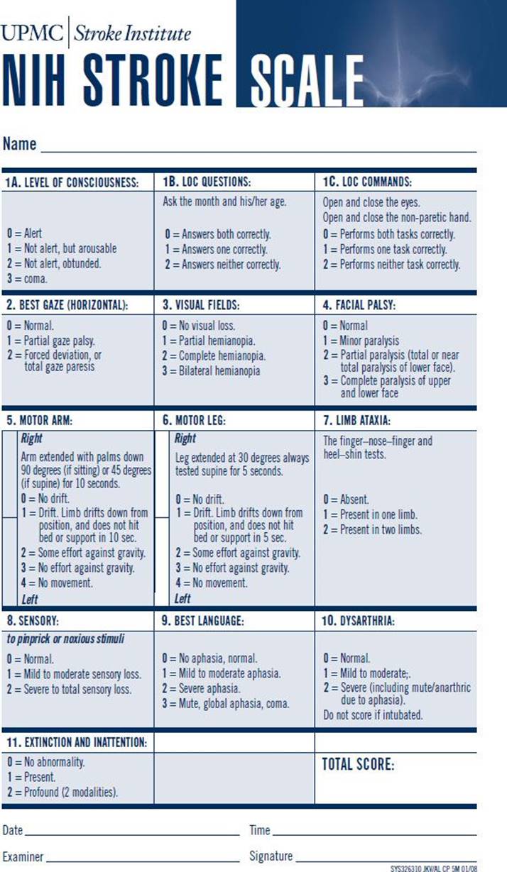
FIGURE 155.2 NIH Stroke Scale. (Courtesy of UPMC Stroke Institute with permission.)
CRITICAL INTERVENTIONS
• Obtain an emergent CT scan.
• Initiate aspirin therapy for nonhemorrhagic ischemic strokes that are not eligible for TPA and do not have swallowing difficulties.
• Identify patients eligible for thrombolytic therapy and expedite their diagnostic evaluation and treatment.
• Correct hyperthermia.
DISPOSITION
Factors influencing disposition in the patient with ischemic stroke include infarct location, time of symptom onset, infarct size, stroke etiology, concurrent medical conditions, and social resources available to the patient. The vast majority of stroke patients are admitted to the hospital, often to a specialized unit or floor skilled in the care of patients with neurologic deficits. Cardiac monitoring is recommended to assess for atrial fibrillation and other arrhythmias.
Patients with minor, completed strokes are potential candidates for evaluation in an ED clinical decision unit. Predefined protocols for neurologic consultation, subsequent neurologic and vascular imaging, and initiation of therapy to prevent recurrent stroke in this setting speed patient care and reduce length of hospital stay.
Common Pitfalls
• Failure to perform a complete history and physical examination, particularly in uncooperative elderly patients. The history and physical examination are the basis for determining the extent of a stroke and its etiology.
• Failure to consider unusual etiologies of stroke, such as arterial dissection and emboli, in younger patients with new neurologic deficits.
• Misdiagnosis of cerebellar stroke as alcohol or drug intoxication, labyrinthitis, benign positional vertigo, or severe gastroenteritis, and failure to obtain neurosurgical consultation.
• Failure to optimize cardiac output and to treat temperature elevations in the ED.
• Overly aggressive blood pressure management. Reduction of blood pressure may reduce cerebral blood flow and lead to extension of an ischemic stroke.
• Initiation of anticoagulation in patients with acute stroke prior to consultation.
Patients treated with r-TPA and those with impaired gag or swallowing reflexes or mental status changes should be admitted to the intensive care unit or a specialized stroke unit. Similar admission is also indicated for patients with cerebellar strokes and should be considered for patients with large hemispheric strokes who are at risk for the development of cerebral edema.
Stroke patients are often cared for in the inpatient setting without neurologic consultation. Neurologic consultation in the ED is indicated when the diagnosis is unclear or when further evaluation is required (e.g., arterial dissection, migrainous infarct, unusual hematologic conditions). Patients with cerebellar infarction should have neurosurgical consultation, because decompressive craniectomy may be lifesaving in selected patients.
HEMORRHAGIC CEREBROVASCULAR DISEASE
ICH, defined as hemorrhage directly into the brain parenchyma, affects 10% to 15% of all stroke patients. Major risk factors for intracranial hemorrhage include advanced age, hypertension, and race. Blacks have a higher incidence of ICH than do whites; other risk factors include cigarette smoking and alcohol use.
The most common site for ICH is the putamen, which accounts for 35% to 50% of all hemorrhages. Next in frequency are the subcortical white matter (30%), the cerebellum (16%), the thalamus (10% to 15%), and the pons (5% to 12%) (11). Pathologically, these are areas of the brain supplied by small arteries that are 50 to 200 μm in size.
CLINICAL PRESENTATION
The onset of ICH is usually heralded by a sudden headache, vomiting, and neurologic deficit. Many patients exhibit a decreased level of consciousness owing to ICP. ICH often occurs during activity and only rarely has its onset during sleep. In one-third of cases, clinical deficits are maximal at onset. In the remaining two-thirds, the symptoms follow a steadily progressive course over the first 30 minutes. Coma is a poor prognostic sign and is associated with larger hemorrhages and ventricular extension. Headache and vomiting are typically absent in ischemic strokes and thus are of help in identifying patients with ICH. Early seizures are uncommon in ICH involving the putamen, thalamus, and pons but may be present in up to one-third of patients with subcortical white matter (“lobar”) hemorrhages. Hypertension is noted in more than 90% of patients with ICH.
The clinical deficits are related to the location and size of the hemorrhage. Putaminal hemorrhages cause a dense contralateral hemiplegia with sensory loss and homonymous hemianopia. The eyes show conjugate deviation away from the paralyzed side, which may overcome with caloric testing or doll’s-eye reflex testing. Thalamic hemorrhages cause similar motor and sensory findings but may also cause conjugate downward gaze with pinpoint, unresponsive pupils. Deficits from a lobar hemorrhage depend on the size and location of the hemorrhage. Pontine hemorrhage typically causes a severe occipital headache, followed rapidly by a decreased level of consciousness; coma is present in 80% of patients. Patients may become hyperthermic and may have severe neurologic deficits. These include quadriplegia, loss of corneal reflexes, and pinpoint pupils with absent horizontal eye movements on caloric or doll’s-eye reflex testing. Cerebellar hemorrhage causes extremity and truncal ataxia and an inability to walk. These symptoms are commonly accompanied by vomiting, disequilibrium, and headache. An ipsilateral gaze palsy may also be present. Patients with cerebellar hemorrhage may initially appear stable but can deteriorate rapidly to coma and death.
DIFFERENTIAL DIAGNOSIS
The presence of intracerebral blood on head CT should not automatically be ascribed to a hypertensive hemorrhage. Various nonhypertensive causes of ICH include arteriovenous malformation, berry aneurysm, sympathomimetic drug use, intracranial tumor, use of anticoagulant and thrombolytic agents, hematologic disorders (e.g., leukemia, thrombotic thrombocytopenic purpura, hemophilia), cerebral vasculitis, and mycotic aneurysm.
ED EVALUATION
The evaluation of patients with suspected ICH is similar to that for ischemic stroke. However, patients with ICH may exhibit rapid neurologic deterioration and a decreased level of consciousness, necessitating acute airway intervention. Vital signs should be monitored closely, and frequent neurologic assessments are required. A general physical examination should be completed in addition to a rapid and thorough neurologic examination. Use of structured examination tools including the NIH Stroke Scale (Fig. 155.2) can provide easy quantification of deficits to allow serial comparisons over time and enhance communication between medical providers. The Glasgow Coma Score is a good predictor of long-term outcome.
KEY TESTING
• Additional investigations in the patient with suspected or confirmed ICH are identical to the Key Testing recommended for Ischemic Stroke.
• Laboratory data should be obtained to help identify possible contributing causes, such as coagulopathy, thrombocytopenia, or platelet dysfunction.
• Toxicologic screening is indicated for patients with atypical presentations and for younger patients without a history of hypertension.
ED MANAGEMENT
The goals of emergency care for ICH are rapid diagnosis, management of the airway, correction of coagulation deficiencies, control of ICP, and early neurosurgical consultation. Oxygen should be administered to maintain oxygen saturation as needed. Intubation is indicated for airway protection or hyperventilation to reduce ICP. Rapid sequence intubation with paralytic agents may be necessary to obtain adequate CT images in a combative patient.
Patients on warfarin should have their warfarin held, undergo replacement of vitamin K–dependent factors to correct the INR, and receive intravenous vitamin K. Prothrombin complex concentrates (PCCs) have not demonstrated improved clinical outcomes compared to traditional fresh frozen plasma (FFP) but have advantages of speed of administration, smaller volume, and reduces risk of infectious disease transmission, and are increasingly recommended for warfarin reversal. Recombinant factor VIIa can limit the extent of hematoma expansion in noncoagulopathic ICH patients but increases the risk of thromboembolic events and has no clear clinical benefit (19). It does not replace all clotting factors and is not recommended as the sole reversal agent for warfarin-associated hemorrhage (20).
Recommendations for the optimum reversal strategy of newer oral direct thrombin inhibitors and factor Xa inhibitors are problematic, as no direct antidote is available and only limited data exists on effective reversal strategies. The best data at this time suggest that PCCs might be useful. Emergent consultation with local coagulation specialists is reasonable in this rapidly changing area.
Hyperglycemia should be avoided, and maintenance of normoglycemia is recommended.
Prophylactic anticonvulsant medication should not be used, though patients with clinical seizures or evidence of seizure on EEG should be treated with antiepileptic drugs (20).
Management of hypertension in the patient with ICH is also rapidly evolving. Data from the international INTERACT2 trial demonstrated safety in targeting acute blood pressure reduction to 140 mm Hg—with a trend toward reducing death or severe disability—in patients with spontaneous ICH with elevated systolic blood pressures (21). A North American trial (ATACH II) is currently underway. Useful antihypertensive agents in the setting of ICH include nitroprusside, esmolol, labetalol, nicardipine, enalapril, hydralazine, and nitroglycerine.
Data regarding ICP control in patients with ICH is very limited. ICH patients with a GCS score of ≤8, clinical evidence of herniation, or those with significant intraventricular hemorrhage or hydrocephalus might be considered for ICP monitoring and treatment. Elevations in ICP, defined as readings ≤20 mm Hg for 5 minutes, may present as a decreasing level of consciousness and/or neurologic deterioration. Initial management options include elevation of the head (30 degrees), osmotherapy, and hyperventilation. Osmotherapy options include mannitol (20% solution; 1 g/kg bolus) and hypertonic saline (250 cc of 7.5% saline). Target serum osmolality has been recommended to be 300 to 320 mOsm/kg, although definitive data are lacking (20).
Reduction of the pCO2 to 30 to 35 mm Hg lowers ICP by 25% to 30% in most patients, though peak ICP reduction may be delayed up to 30 minutes after the pCO2 is changed. Because hypocarbia causes cerebral vasoconstriction, overaggressive reduction in pCO2 may reduce cerebral blood flow below critical values and should thus be avoided.
In general, ICP monitoring is indicated in patients whose condition is thought to be deteriorating owing to increases in ICP. Other patients may warrant ICP monitors, however, particularly those with intraventricular hemorrhage. Placement of an intraventricular catheter offers the potential for direct removal of cerebrospinal fluid and reduction of ICP.
Neurosurgical consultation should be obtained early and, if unavailable, the patient should be stabilized and transferred. The indications for hematoma evacuation depend on many factors, including patient age, hemorrhage location and size, and surgical accessibility. Patients who present initially with coma or loss of brainstem function generally do not benefit from surgical evacuation, whereas those who experience progressive deterioration after a lobar hemorrhage are often operated on. In patients with cerebellar hemorrhage, surgical evacuation as soon as possible is recommended for patients who are deteriorating neurologically or have brainstem compression and/or hydrocephalus.
CRITICAL INTERVENTIONS
• Protect the airway.
• Correction of abnormal coagulation.
• Manage elevated ICP.
• Obtain neurosurgical consultation.
DISPOSITION
All patients with ICH should be admitted for close observation and monitoring. Typically, intensive care unit monitoring is required. If neurosurgical treatment capabilities are not available locally, arrangements should be made to transfer the patient.
Common Pitfalls
• Failure to diagnose cerebellar hemorrhage. Patients may be misdiagnosed with acute viral syndrome, labyrinthitis, or intoxication. Surgical intervention may be lifesaving.
• Delay in transport. Patients with ICH require early neurosurgical consultation and should be transported to an appropriate facility if immediate neurosurgical attention is not otherwise available.
REFERENCES
1. Easton JD, Saver JL, Albers GW, et al. Definition and evaluation of transient ischemic attack: A scientific statement for healthcare professionals from the American Heart Association/American Stroke Association Stroke Council; Council on Cardiovascular Surgery and Anesthesia; Council on Cardiovascular Radiology and Intervention; Council on Cardiovascular Nursing; and the Interdisciplinary Council on Peripheral Vascular Disease. The American Academy of Neurology affirms the value of this statement as an educational tool for neurologists. Stroke. 2009;40(6):2276–2293.
2. Johnston SC, Rothwell PM, Nguyen-Huynh MN, et al. Validation and refinement of scores to predict very early stroke risk after transient ischaemic attack. Lancet. 2007;369(9558):283–292.
3. Dengler R, Diener HC, Schwartz A, et al. Early treatment with aspirin plus extended-release dipyridamole for transient ischaemic attack or ischaemic stroke within 24 h of symptom onset (EARLY trial): A randomised, open-label, blinded-endpoint trial. Lancet Neurol. 2010;9(2):159–166.
4. Halkes PH, van Gijn J, Kappelle LJ, et al. Aspirin plus dipyridamole versus aspirin alone after cerebral ischaemia of arterial origin (ESPRIT): Randomised controlled trial. Lancet. 2006;367(9523):1665–1673.
5. CAPRIE Steering Committee. A randomised, blinded, trial of clopidogrel versus aspirin in patients at risk of ischaemic events (CAPRIE). CAPRIE Steering Committee. Lancet. 1996;348(9038):1329–1339.
6. Wang Y, Wang Y, Zhao X, et al. Clopidogrel with aspirin in acute minor stroke or transient ischemic attack. N Engl J Med. 2013;369(1):11–19.
7. Johnston SC, Easton JD, Farrant M, et al. Platelet-oriented inhibition in new TIA and minor ischemic stroke (POINT) trial: Rationale and design. Int J Stroke. 2013;8(6):479–483.
8. Perry JJ, Sharma M, Sivilotti ML, et al. Prospective validation of the ABCD2 score for patients in the emergency department with transient ischemic attack. CMAJ. 2011;183(10):1137–4115. PMCID: 3134721.
9. Tissue plasminogen activator for acute ischemic stroke. The National Institute of Neurological Disorders and Stroke r-TPA Stroke Study Group. N Engl J Med. 1995;333(24):1581–1587.
10. Jauch EC, Saver JL, Adams HP Jr, et al. Guidelines for the early management of patients with acute ischemic stroke: A guideline for healthcare professionals from the American Heart Association/American Stroke Association. Stroke. 2013;44(3):870–947.
11. Hacke W, Kaste M, Bluhmki E, et al. Thrombolysis with alteplase 3 to 4.5 hours after acute ischemic stroke. N Engl J Med. 2008;359(13):1317–1329.
12. Scott PA, Meurer WJ, Frederiksen SM, et al. A multilevel intervention to increase community hospital use of alteplase for acute stroke (INSTINCT): A cluster-randomised controlled trial. Lancet Neurol.2013;12(2):139–148.
13. Furlan A, Higashida R, Wechsler L, et al. Intra-arterial prourokinase for acute ischemic stroke. The PROACT II study: A randomized controlled trial. Prolyse in Acute Cerebral Thromboembolism. JAMA.1999;282(21):2003–2011.
14. Smith WS, Sung G, Saver J, et al. Mechanical thrombectomy for acute ischemic stroke: Final results of the Multi MERCI trial. Stroke. 2008;39(4):1205–1212.
15. Broderick JP, Palesch YY, Demchuk AM, et al. Endovascular therapy after intravenous t-PA versus t-PA alone for stroke. N Engl J Med. 2013;368(10):893–903. PMCID: 3651875.
16. Ciccone A, Valvassori L. Endovascular treatment for acute ischemic stroke. N Engl J Med. 2013;368(25):2433–2434.
17. Kidwell CS, Jahan R, Gornbein J, et al. A trial of imaging selection and endovascular treatment for ischemic stroke. N Engl J Med. 2013;368(10):914–923. PMCID: 3690785.
18. The International Stroke Trial (IST): A randomised trial of aspirin, subcutaneous heparin, both, or neither among 19435 patients with acute ischaemic stroke. International Stroke Trial Collaborative Group. Lancet.1997;349(9065):1569–1581.
19. Mayer SA, Brun NC, Begtrup K, et al. Efficacy and safety of recombinant activated factor VII for acute intracerebral hemorrhage. N Engl J Med. 2008;358(20):2127–2137.
20. Morgenstern LB, Hemphill JC 3rd, Anderson C, et al. Guidelines for the management of spontaneous intracerebral hemorrhage: A guideline for healthcare professionals from the American Heart Association/American Stroke Association. Stroke. 2010;41(9):2108–2129.
21. Anderson CS, Heeley E, Huang Y, et al. Rapid blood-pressure lowering in patients with acute intracerebral hemorrhage. N Engl J Med. 2013;368(25):2355–2365.