
Lisa R. Palivos
Vesicular lesions are commonly seen in the emergency department. Like other dermatologic lesions, vesicles can present a challenge in determining their etiology, contagiousness, and proper treatment (1–4).
Vesicles are circumscribed epidermal elevations 1 to 4 mm in diameter that usually contain clear fluid. Several characteristics help distinguish the lesion from other vesicular disorders, including the nature of the contained fluid (seropurulent, serosanguineous), the shape of the lesion’s apex (rounded, acuminate, umbilicated), the pattern of the eruption (discrete, irregularly scattered, grouped, linear), and the distribution over the body and associated mucous membrane involvement.
In this discussion, there are two types of vesicular disorders: those appearing in groups and those that are individual or disseminated.
GROUPED LESIONS
Herpes Simplex
Herpes simplex infections are caused by two different DNA virus types: HSV-1 and HSV-2. Both viruses can infect oral or genital mucosa and are spread by direct contact. HSV-1 usually affects the lips and is often referred to as a fever blister or cold sore. HSV-2 is the virus most frequently associated with genital herpes. The hallmark of herpes simplex virus (HSV) infections is the ability to infect epithelial cells and then travel up the peripheral nerve to the dorsal root ganglia where it may remain latent for years followed by reactivation.
CLINICAL PRESENTATION
Herpes simplex is characterized by recurrent, painful, vesicular lesions of the lips, genitalia, hands, rectum, or eyes. The lesions of herpes simplex are typically tightly clustered vesicles on an erythematous base and evolve into pustules and erosions over 5 to 7 days. Grouped, crusted erosions are the typical presentation. Associated symptoms may include fever, local pain, regional lymphadenopathy, fatigue, and malaise. Urinary retention occasionally occurs with genital lesions and generally requires hospital admission.
Primary lesions are usually more widespread with a bilateral distribution. Recurrent lesions typically are tightly clustered and unilateral and occur in the same location as previous episodes of infection. Recurrences are usually milder, involve fewer lesions, and heal faster. Approximately 50% of patients with recurrent lesions have a 24-hour prodrome of local itching, burning, tingling, and stinging. Precipitating factors for recurrence include stress, fever, infections, sunburn, menstruation, cosmetic facial procedures, dental interventions, or trauma.
Primary lesions usually resolve in 2 to 3 weeks, but the duration may be longer in immunocompromised patients. Typical sites of involvement are the oral, genital, and periorbital areas and the fingertips (herpetic whitlow), but any body area may be involved. Oral infections can cause gingivostomatitis, or pharyngitis. Herpetic whitlow is an infection of the pulp and lateral aspect of the finger. This may occur from autoinoculation with a primary oral or genital infection. With periorbital lesions, fluorescein staining of the cornea is necessary to identify dendritic lesions. Herpetic keratitis is the most common cause of corneal blindness and presents with pain, photophobia, and blurry vision. Patients with herpetic keratitis should be referred to an ophthalmologist. Herpes gladiatorum is contracted by athletes, especially wrestlers. It is transmitted by direct skin-to-skin contact and usually appears in the head and neck region. With the increasing incidence of the acquired immunodeficiency syndrome and the growing use of bone marrow and solid organ transplantation, more severe manifestations of herpes infections are being seen (5). Neonatal herpes occurs when the virus is transmitted from an infected mother to the neonate, with the highest risk occurring when the mother has primary genital herpes during delivery. Along with the cutaneous lesions, the infected neonate may develop multiorgan involvement with high mortality rate. HSV vaccines are currently in early clinical trials.
DIFFERENTIAL DIAGNOSIS
The differential diagnosis of herpes simplex includes aphthous stomatitis; tinea cruris; hand, foot, and mouth disease; herpangina; and primary syphilis. Aphthous stomatitis consists of one or two painful lesions in the mouth, but they are not in a circumscribed area. Although early tinea, which is vesicular, can be difficult to distinguish from herpes, later tinea has central healing. Lesions in hand, foot, and mouth disease are asymptomatic. Herpangina lesions are found in the posterior oropharynx and not outside the mouth. The chancre in primary syphilis is classically painless and the borders are indurated.
ED EVALUATION
Diagnosis of herpes simplex is usually made clinically. Viral cultures of vesicular fluid or direct fluorescent antibody stain of scraped lesions may be helpful for definitive diagnosis. The presence of intranuclear inclusions and multinucleated giant cells on a Tzanck preparation supports a diagnosis of herpes infection but does not distinguish between HSV and varicella zoster virus. Polymerase chain reaction and serology testing are not widely available and are expensive. Point-of-care HSV-2 and HSV-2-LFIA rapid test kits are available. The 10-minute tests only require a few drops of blood from a finger stick to detect the presence of HSV-2 antibodies to confirm genital herpes diagnosis and also identify asymptomatic carriers. They do not detect HSV-1.
ED MANAGEMENT AND DISPOSITION
Acyclovir is the most widely used antiviral agent in clinical use. Treatment with 200 mg of oral acyclovir five times per day or 400 mg three times a day for 10 days is indicated for primary genital herpes. However, the duration of symptoms is decreased only minimally with this therapy and only if started within 24 hours after onset of the rash. Time for crusting decreases from 2.7 to 2.2 days. Other medications that are effective and available for treating herpetic infections are famciclovir and valacyclovir (Table 172.1). All three oral agents have been shown to reduce the frequency of symptomatic recurrences of genital herpes. Over-the-counter tetracaine cream (Viractin) may reduce healing time of recurrent herpes labialis by about 2 days. Topical penciclovir 1% cream (Denavir), which is more effective than acyclovir cream, can reduce average time to healing of recurrent orolabial HSV infections by 0.7 days and the duration of pain by 0.6 days (6).
TABLE 172.1
Treatment of Genital Herpes Simplex

In children with oralfacial or gingivostomatitis, generally, only supportive treatment with hydration and topical anesthetics (viscous lidocaine, diphenhydramine elixir) is needed. Importantly, patients receiving suppressive antiviral medication should be counseled that they may still transmit HSV to their sexual partners. Intravenous therapy (5 mg/kg every 8 hours) should be considered for patients with urinary retention, compromised immunity, or life-threatening disseminated infection.
Common Pitfalls
• Failure to instruct patients about measures to prevent transmission to others.
• Failure to admit patients with genital herpes and urinary retention.
• Failure to admit patients with disseminated infection.
• Failure to recognize herpetic whitlow and making an incision, which may lead to spread of infection.
HERPES ZOSTER
Herpes zoster (HZ) (shingles) results from the reactivation of a dormant varicella zoster virus that entered the dorsal root ganglia during an earlier episode of chicken pox. HZ can occur in patients of any age who have had a previous infection of varicella zoster virus. The incidence of HZ increases with age, peaking in the seventh decade. Reactivation factors include fever, wind, UV light, illness, or stress. An attack of HZ does not confer immunity, and it is not unusual to have two or three episodes in a lifetime.
CLINICAL PRESENTATION
HZ is characterized by unilateral vesicular eruption along one or two adjacent dermatomes that is associated with severe pain. Dermatomes T3–L3 are most frequently involved (Fig. 172.1). Occasionally, the lesions may extend beyond the midline. There is usually a prodrome of pain, itching, burning, and hyperesthesia in the same dermatome 4 to 7 days before the lesions appear. Fever, headache, and malaise along with lymphadenopathy may also precede the eruption. The pain may imitate myocardial infarction, appendicitis, renal colic, or migraine headache and may present a diagnostic dilemma until the characteristic rash develops. The eruption begins as an erythematous maculopapular rash that gives rise to vesicles containing purulent fluid. These pustules either umbilicate or rupture before forming crusts. The crusts fall off in 2 to 3 weeks.
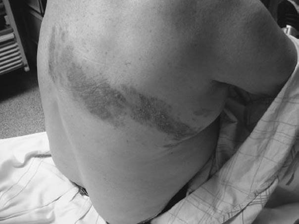
FIGURE 172.1 Herpes zoster. Lesions can involve more than one dermatome. (Courtesy of Scott Sherman, MD.)
When the ophthalmic division of the trigeminal nerve is involved, HZ ophthalmicus results. Lesions can appear on the forehead, eyelid, and nose. Symptoms include headache, fever, malaise, and eye pain. Ocular involvement is common and each individual should have a thorough eye examination. Hutchinson sign (lesion at the tip of the nose) usually heralds ocular involvement. Urgent (next-day) ophthalmology referral is indicated. When the sensory branch of the facial nerve is involved, HZ oticus results. Lesions appear in the ear, mouth, face, neck, or scalp. Patients can complain of severe otalgia, vertigo, hearing loss, or loss of taste in the anterior two-thirds of the tongue. When associated with facial paralysis, the disease is called Ramsay Hunt syndrome. Treatment consists of acyclovir, corticosteroids, and diazepam for vertigo. Facial paralysis and hearing loss may be permanent. Patients should be referred to an ear, nose, and throat specialist urgently. Hospitalization should be considered in severe cases.
The severe pain associated with HZ is the principal reason that patients seek medical care. Traditionally, pain that is present while the rash persists or in the first 30 days from onset has been termed acute pain.Pain that is present after this period is termed postherpetic neuralgia (PHN) and may persist for months to years after lesions have disappeared. As age increases, the risk and duration of PHN also rise. Nearly half of the patients older than 60 years have this complication. Other complications of HZ include secondary bacterial infections, pneumonitis, and meningitis. HZ virus vaccine (Zostavax) is available for the prevention of shingles and PHN in persons 60 years or older.
DIFFERENTIAL DIAGNOSIS
The differential diagnosis includes chemical burns, Bell’s palsy, conjunctivitis, and trigeminal neuralgia. The blisters in poison ivy often occur in a linear “dermatomal,” but they are painless.
ED EVALUATION
The diagnosis of HZ is based on history of pain and appearance of the dermatomal rash. Laboratory tests are not necessary. Scraping for a Tzanck smear or viral culture is usually negative, and a biopsy is usually required for definitive diagnosis. HZ ophthalmicus is diagnosed by slit lamp detection of corneal dendritic lesions.
ED MANAGEMENT AND DISPOSITION
A meta-analysis found that acyclovir reduces pain duration and prevalence, especially in patients 50 years of age or older (7). The recommended therapy is acyclovir 800 mg five times a day for 7 days. Famciclovir 500 mg or valacyclovir 1,000 mg tid for 7 days has also been shown to decrease the median duration of PHN (8,9). Treatment should be started within 72 hours to be effective. Symptomatic treatment with oral analgesics and Burow solution–soaked compresses three times a day is often helpful as well.
It is unclear whether adding prednisolone is beneficial (10). Corticosteroids do not change the incidence or duration of PHN, but they may improve the quality of life (time to return to usual activity, cessation of use of analgesic agents, and time to return to uninterrupted sleep) in patients older than 50 years of age with acute HZ (11,12). A tapering dose of 60 mg/day for 7 days, then 30 mg/day for 7 days, then 15 mg/day for 7 days can be used. Relative contraindications to the use of corticosteroids include hypertension, renal insufficiency, diabetes, osteoporosis, and peptic ulcer disease. The incidence of adverse effects is higher in patients treated with corticosteroids.
The treatment of PHN includes analgesics (oxycodone), lidocaine patches, tricyclic antidepressants, (nortriptyline), and gabapentin. Lidocaine patches are very effective and should be applied to the skin rash along the affected dermatome. A referral to a pain specialist may be necessary.
Ophthalmologic consultation is indicated if the ophthalmic division of the trigeminal nerve is involved. Dissemination beyond the original dermatomes should raise the suspicion of immunodeficiency. Immunocompromised patients with severe disease should receive acyclovir 10 to 12 mg/kg IV infused over 1 hour every 8 hours. If lesions persist or there is concern for acyclovir resistance, consider foscarnet 40 mg/kg IV every 8 hours.
Hospital employees who are exposed to patients with HZ should be tested for antibody to the varicella zoster virus. Isolation for 8 to 21 days after exposure is recommended when antibody is not detected. If antibody is present, isolation is unnecessary (13). Varicella zoster immune globulin (VZIG) is recommended within 72 hours for exposed immunocompromised contacts.
Common Pitfalls
• Failure to instruct patients about measures to prevent transmission. Patients with HZ can transmit the virus as chickenpox to persons who have not already been infected.
• Failure to recognize a central cause of facial paralysis.
• Failure to recognize and treat ocular involvement.
• Failure to recognize or admit immunocompromised individuals.
ALLERGIC CONTACT DERMATITIS
Because allergic contact dermatitis is a result of delayed hypersensitivity, skin lesions may appear hours to days after exposure to the offending antigen, and it is often difficult to identify the antigen. Commonly identified etiologic agents include poison ivy, oak, and sumac; soaps; detergents; and perfumes. Pruritus is the initial symptom, which is quickly followed by erythema and vesiculation. Areas with a thin stratum corneum (such as eyelids and genitalia) may swell severely. Without treatment, symptoms may persist for 2 to 3 weeks.
Mild contact dermatitis may be treated with topical corticosteroids. For severe cases, oral prednisone (40 to 60 mg/day for 7 days) accelerates resolution of symptoms (usually within 24 to 48 hours) and poses minimal risks of adverse effects.
FIXED DRUG ERUPTION
Like HSV, fixed drug eruption causes recurrent episodes of skin lesions in one location. Unlike herpes simplex, however, fixed drug eruption generally leaves persistent slate gray to brown hyperpigmentation after the attack resolves. The most commonly implicated drugs include phenolphthalein (e.g., Ex-Lax, Feen-A-Mint), tetracycline, sulfa, and barbiturates. Symptoms usually arise 1 to 2 days after ingestion. Pruritus generally occurs first, followed by the appearance of clusters of vesicles or bullae in a hyperpigmented macule. The process resolves in 2 to 3 weeks, after discontinuation of the offending agents.
OTHER CAUSES OF VESICULAR ERUPTIONS
Dyshidrotic eczema (pompholyx) usually occurs on the medial and lateral aspects of the fingers and sometimes on the palms and soles. It is intensely pruritic and responds to topical corticosteroids. If severe, oral steroids can be used.
The vesicular eruption of tinea pedis may usually be diagnosed clinically by potassium hydroxide staining of vesicle fluid. Treatment is with a topical antifungal agent such as clotrimazole. Accompanying involvement of the hands is often an eczematous reaction.
Atopic dermatitis is generally accompanied by a personal or family history of eczema, allergic rhinitis, asthma, or urticaria. Nonetheless, the eruption is usually precipitated by physical irritation (wool gloves, soap, and water) or emotional stress. Treatment with topical steroids is effective.
Dermatitis herpetiformis (unrelated to the herpes viruses) may be confused with scabies or neurotic excoriations. It generally presents with broken, excoriated vesicles on an erythematous base. It usually begins on extensor surfaces and is pruritic. Treatment is with dapsone and oral antihistamines.
INDIVIDUAL OR DISSEMINATED VESICLES
Varicella
Varicella (chickenpox) is primarily a disease of childhood, but it is not uncommon in adults. It is acquired by direct contact with infected lesions or inhalation of airborne droplets. Varicella begins with replication of the virus at the sites of contact and is followed by the development of a primary viremia, which establishes replication in the reticuloendothelial system. A secondary viremia occurs after 1 week, which disseminates the infection to the skin. The virus then establishes latency in the sensory ganglion. Reactivation of the latent infection causes HZ.
CLINICAL PRESENTATION
Varicella presents with a rash, low-grade fever, and malaise. The lesions start as erythematous macules, progress to vesiculation with the classic “dew drop on a rose petal” appearance, become pustules, and finally crust over (Fig. 172.2). The hallmark of varicella rash is the simultaneous presence of lesions in all stages of development. The lesions usually appear on the trunk and face and then spread to other areas of the body, including scalp, mouth, ears, and genitalia. They are intensely pruritic. There may be a history of exposure 10 to 14 days before the rash. Varicella is highly contagious. Second attack rates for this virus may reach 90% for susceptible household contacts. Patients are considered infectious 2 days before the appearance of the rash until all vesicles have formed crusts, which is usually 5 to 6 days after the first appearance of the eruption. The severity of the illness varies from individual to individual and is inversely related to age. An attack of chickenpox usually confers lifelong immunity, although secondary attacks can happen. Varicella vaccine is now available in the United States and is approved for children and adults.
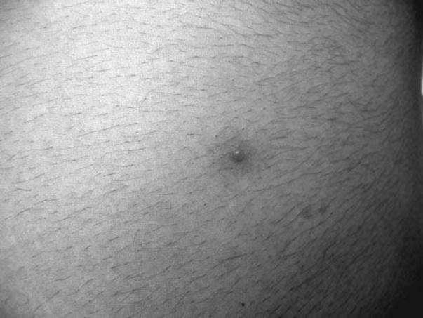
FIGURE 172.2 Varicella. The lesion starts as a 2- to 4-mm red papule, then develops into an irregular outline (rose petal) as a thin clear vesicle appears on the surface (dewdrop).
Varicella is usually a benign illness in immunocompetent patients. Immunocompromised individuals or those using immunosuppressive drugs, especially systemic corticosteroids, are at a greater risk for complications (14). The most common complication in children is secondary bacterial skin infections after scratching, leading to a cellulitis. Varicella pneumonitis is rare in children but is the most common serious complication in adults. It can present with tachypnea, cough, fever, and pleuritic chest pain leading to acute respiratory distress syndrome. Other complications include hepatitis, nephritis, orchitis, and encephalitis. Encephalitis in children is usually self-limited and includes cerebellar ataxia, nystagmus, and headache. Adult encephalitis has a mortality rate of 35% (15). Reye syndrome is an encephalopathy with fatty liver and is associated with salicylate use. Twenty to 30% of Reye syndrome cases are preceded by varicella (16).
Varicella during pregnancy poses a risk to both mother and child. The highest risk for fetal varicella syndrome is between 13 to 20 weeks of gestation. It is characterized by small infant size, cicatricial skin lesions, limb hypoplasia, and other anomalies. For newborns, the risk of infection and complications is greatest when the mother develops chickenpox 5 days before to 2 days after delivery. These infants have an increased chance of dissemination as they lack both maternal antibodies and a mature immune system. They should be given VZIG. VZIG is effective in preventing chickenpox in exposed susceptible, especially immunocompromised individuals (Table 172.2).
TABLE 172.2
Varicella Zoster Immune Globulin Indications

DIFFERENTIAL DIAGNOSIS
The differential diagnosis includes disseminated zoster, disseminated herpes simplex, eczema herpeticum, and folliculitis.
ED EVALUATION
Diagnosis is usually made clinically. Laboratory tests are not necessary. A Tzanck smear and viral culture may aid in making the diagnosis. A chest x-ray should be obtained if respiratory symptoms develop. A lumbar puncture is needed if encephalitis is suspected.
ED MANAGEMENT AND DISPOSITION
Treatment of varicella in healthy children is optional because the disease is self-limited. Symptoms can be managed with supportive care consisting of daily baths and soaks to destroy the virus. Close cropping of the fingernails, calamine lotion, and antihistamines to avoid excessive excoriations can provide relief. Isolate the patient, especially from immunocompromised hosts. Aspirin and Caladryl lotion, which can lead to an anticholinergic response from excessive skin absorption, should be avoided. Acyclovir should be considered in children up to age 12 who are being treated with corticosteroids or long-term salicylate therapy, or those with chronic cutaneous or pulmonary diseases (17). Acyclovir, if started within 24 hours of rash development, has been shown to reduce the number of lesions, the duration of fever, and pruritus. Treatment after the first day of illness is of no value in uncomplicated cases of varicella (18). All immunocompromised individuals with varicella should be hospitalized for intravenous acyclovir (Table 172.3). Hospitalization is also required for skin sepsis, pneumonitis, dense or hemorrhagic rash, and bleeding.
TABLE 172.3
Treatment of Varicella

COXSACKIEVIRUS AND ECHOVIRUS INFECTIONS
Coxsackievirus and echovirus infections may cause papulovesicular lesions that are evenly spread across the face and torso. Associated symptoms may include fatigue, malaise, fever, vomiting, and diarrhea. Resolution occurs spontaneously in 10 to 14 days, and treatment is symptomatic.
Hand, foot, and mouth disease caused by coxsackieviruses presents with tender, flat-topped, whitish vesicles 3 to 4 mm in diameter on the palms and soles and in the mouth. It is commonly associated with mild systemic complaints. The duration is generally 4 to 5 days and no treatment is necessary.
INSECT BITES
Vesiculobullous skin lesions may occur as hypersensitivity reactions to insect bites, commonly those of fleas, bedbugs, and mites. A clue to the diagnosis is a linear or grouped pattern of lesions; some may have central puncta. Treatment is with topical corticosteroids and oral antihistamines, and avoidance of further contact with the insect.
CRITICAL INTERVENTIONS
• Initiate acyclovir within 24 hours of the onset of rash in adult varicella and within 72 hours of onset of HZ.
• Obtain ophthalmology consult for patients with herpetic keratitis due to herpes simplex or with involvement of the ophthalmic division of the trigeminal nerve in HZ.
Common Pitfalls
• Failure to instruct patients about measures to prevent transmission of varicella.
• Failure to admit immunocompromised patients with varicella.
REFERENCES
1. Dahl M. The blistering diseases. In: Dahl M, ed. Common Office Dermatology. New York: Grune & Stratton; 1983.
2. Domonkos AN, Arnold HL, Odom RB. Andrews Disease of the Skin. Philadelphia, PA: WB Saunders; 1982.
3. Flowers FP, Krusinski PA. Dermatology in Ambulatory and Emergency Medicine. Chicago, IL: Year Book Medical; 1984.
4. Furey N. Blistering disorders. In: Roenigk H, ed. Office Dermatology. Baltimore, MD: Williams & Wilkins; 1981.
5. Sasadeusz JJ, Sacks SL. Systemic antivirals in herpes virus infections. Dermatol Clin. 1993;11:171–185.
6. Spruance SL, Rea TL, Thoming C, et al. Penciclovir cream for the treatment of herpes simplex labialis. A randomized, multicenter, double-bind, placebo-controlled trial. JAMA. 1997;277:1374–1379.
7. Wood MJ, Kay R, Dworkin RH, et al. Oral acyclovir therapy accelerates pain resolution in patients with herpes zoster: A meta-analysis of placebo-controlled trials. Clin Infect Dis. 1996;22:341–347.
8. Beutner KR, Friedman DJ, Forszpaniak C, et al. Valacyclovir compared with acyclovir for improved therapy for herpes zoster in immunocompetent adults. Antimicrob Agents Chemother. 1995;39:1546–1553.
9. Tyring S, Barbarash RA, Nahlik JE, et al. Famciclovir for the treatment of acute herpes zoster: Effects on acute disease and postherpetic neuralgia—a randomized, double-blind, placebo-controlled trial. Ann Intern Med. 1995;123:89–95.
10. Wood MJ, Johnson RW, McKendrick MW, et al. A randomized trial of acyclovir for 7 days or 21 days with and without prednisolone for treatment of acute herpes zoster. N Engl J Med. 1994;330:896–900.
11. Mcfarlane LL, Simmons MM, Hunter MH. The use of corticosteroids in the management of Herpes Zoster. J Am Board Fam Pract. 1998;11:224–228.
12. Whitley RJ, Weiss H, Gnann JW, et al. Acyclovir with and without prednisolone for the treatment of herpes zoster—a randomized, placebo-controlled trial. Ann Intern Med. 1996;125:376–383.
13. Sayre MR, Lucid EJ. Management of varicella-zoster-virus exposed hospital employees. Ann Emerg Med. 1987;16:421–424.
14. Dowell SF, Bresee JS. Severe varicella associated with steroid use. Pediatrics. 1993;92:223–228.
15. Preblud SR. Varicella: Complications and costs. Pediatrics. 1986;78:728–735.
16. Hurwitz ES, Nelson DB, Davis C, et al. National surveillance for Reye syndrome: A five-year review. Pediatrics. 1982;70:895–900.
17. Arvin AM. Antiviral therapy for varicella and herpes zoster. Semin Pediatr Infect Dis. 2002;13(1):12–21.
18. Wallace MR, Bowler WA, Murray NB, et al. Treatment of adult varicella with oral acyclovir. A randomized, placebo-controlled trial. Ann Intern Med. 1992;117:358–363.