
Carol A. Freire, Dana E. Robinson, and Theodore A. Christopher
Urticaria, or hives, is a commonly occurring vascular reaction of the superficial dermis of the skin resulting in pruritic wheals that form in response to a triggering stimulus. Acute urticaria is characterized by symptomatic attacks lasting less than 6 weeks and chronic urticaria is characterized by attacks that last 6 weeks or longer, with episodes of the latter tending to be less frequent and less severe than the former. One of the most common skin lesions seen in emergency medicine, urticaria can occur in any age group, but children and young adults are more commonly affected. In a majority of cases, a trigger cannot be determined (1).
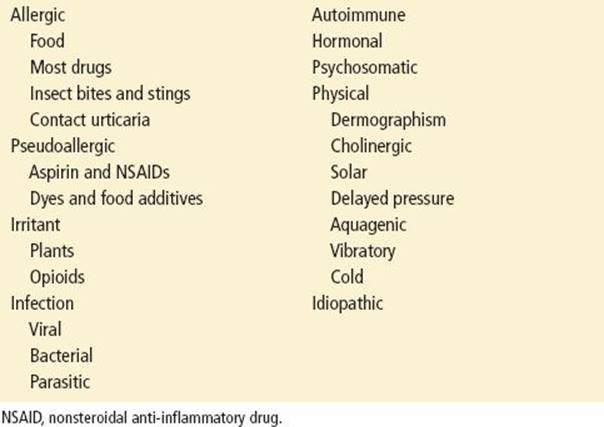
The etiologies of urticaria are extensive and complex in nature (2). Generally, they may be broken down into several broad categories based on their underlying pathophysiology (Table 174.1). In most cases, however, the cause of urticaria is primarily mast cell degranulation, which releases histamine as well as a wide array of vasoactive peptides. The released histamine binds to receptors (predominantly H1) in the microvasculature, especially the postcapillary venules, causing superficial local edema (wheals) and vasodilation (flare). Histamine also binds to H1 receptors in the histamine-selective (itch-specific) C nerve fibers, making them more irritable and causing pruritus. In many cases, this reaction is allergic in nature and mediated by IgE, following prior sensitization (type I hypersensitivity), but degranulation can also occur by other immunologic and nonimmunologic routes (3,4). Histologically, urticaria involves only the superficial dermis, distinguishing it from angioedema, which involves the deeper dermis. Perivascular lymphocytic and mast cell infiltration are also features.
TABLE 174.1
Classification of Urticaria
It should be noted that type I hypersensitivity occurs in two phases—the immediate reaction, which occurs within the first hour after antigen challenge, and the late phase, which occurs several hours later, usually peaking at 3 to 9 hours. The late phase is not always noticeable, and the delay time, severity, and duration are variable and depend on the allergen dose. This late phase is due to continued synthesis and release of vasoactive substances by mast cells and can produce much more involved and long-lasting edema than the immediate wheals.
High-quality estimates of the prevalence of either acute or chronic urticaria are lacking. A recent survey conducted in Germany showed an 8.8% (95% CI 7.9% to 9.7%) lifetime prevalence for urticaria of any type and a 1.8% (95% CI 0.6% to 1.1%) lifetime prevalence for chronic urticaria (5). Chronic urticaria in this population afflicted women (70.3%) more than men. Chronic urticaria is also notable for being diagnosed later in life, most often in the third or fourth decade.
CLINICAL PRESENTATION
Urticaria is characterized by slightly raised, sharply defined, erythematous, blanching wheals. They are typically pruritic, though usually nontender. The wheals may appear anywhere on the body and are often localized to the site of irritation but can cover large areas of the body with systemic triggers such as food allergies. The color of the wheals ranges from pink to red and central clearing may be present. The shape can be round, ovoid, or ring like, commonly with serpiginous borders, and multiple wheals may be aggregated into large plaques, giving an irregular appearance. Wheal size can range from a few millimeters to plaques that cover significant regions of the body. One of the hallmarks of urticaria is that the affected region can change size and new areas may become affected over time. Individual wheals typically last less than 8 to 12 hours; a longer persistence suggests urticarial vasculitis or delayed pressure urticaria (see below).
Table 174.2 summarizes the types and triggers of urticaria. Foods and drugs are the most common causes of acute urticaria. They act through both immunologic and nonimmunologic mechanisms (6). Among foods, nuts, berries, shellfish, chocolate, fish, tomatoes, and eggs are frequent causes of acute urticaria. In urticaria of chronic duration, milk, cereals, beef, pork, cheese, garlic, onions, and spices may be responsible. Food additives, both natural and synthetic, are also important etiologic factors. Reaction timing can be anywhere from within minutes of ingestion to many hours later. Worthy of particular mention is scombroid poisoning (7), which is a type of food poisoning that is classically associated with the scombridae family of fish such as mackerel and tuna, but can be caused by many other species. These fish contain large quantities of the amino acid histidine in their muscle tissue, which is converted to histamine by bacterial histamine decarboxylase in improperly stored fish.
TABLE 174.2
Agents Known to Cause Urticaria

Many medications have the potential to cause an allergic response and should always be considered as a possible cause of urticaria. Antibiotics, particularly the β-lactams, and aspirin/NSAIDs are the most common drugs that cause urticaria, with aspirin and NSAID intolerance having been identified in 20% to 40% of patients with chronic urticaria. Urticaria may occur from 15 minutes to 24 hours (average 1 to 4 hours) after aspirin ingestion. The condition usually resolves within a day or two but may last up to 2 weeks. The pathogenic mechanism of aspirin intolerance is unclear, but a likely hypothesis is that mast cell degranulation is due to rapid PGE2 depletion by drugs capable of inhibiting COX-1 and is not an allergic reaction. It usually appears in the third decade of life and is often accompanied by wheezing, increased bronchial secretions, rhinorrhea, and flushing (8).
With physical urticaria, wheals and itching develop at sites of a physical stimulus (9). Urticaria factitia or dermatographism is defined as a whealing reaction of the skin within 10 minutes of stroking or scratching the skin. Clinically, dermatographism occurs within seconds after stimulation and fades within 30 minutes. It is often a response to contact with seatbelts or brassieres. In contrast to dermatographism, delayed pressure urticaria is a response to more sustained physical pressure applied to the skin (10). It produces itchy, often painful wheals. These lesions can be delayed up to 12 hours and may last several days. Common causes are tight-fitting garments, prolonged walking (feet), or sitting (buttocks).
Cholinergic urticaria is characterized by pruritic, follicular-bound, 1- to 4-mm wheals surrounded by a red flare and produced within 15 minutes of a stimulus. Exercise, hot baths, spicy food, and emotional stress can precipitate episodes of cholinergic urticaria, which usually begins on the upper thorax and neck before spreading to the entire body, often sparing the palms and soles. The exact pathophysiology is unclear but may involve an autoimmune reaction to a sweat antigen. Clinically, patients can present with varied symptoms, including lacrimation, salivation, diarrhea, abdominal pain, weakness, flushing, and headache. Rarely, severe, life-threatening symptoms (e.g., angioedema, bronchospasm, vascular collapse, and hypotension) can occur.
Solar urticaria presents as a whealing response within 10 minutes in skin exposed to ultraviolet or visible irradiation from any source, though UVA is most commonly implicated. In addition to hives, symptoms may include wheezing, dizziness, and (rarely) hypotension; all resolve within 1 to 3 hours after the light stimulus is removed (11).
Cold urticaria occurs on exposed areas, such as the face and hands, and is commonly induced by swimming. It occurs within minutes of exposure, usually during rewarming, and can be elicited by placing an ice cube on the skin, holding cold objects in the hand, or drinking cold fluids (leading to lip swelling). It also may be associated with other symptoms, including syncope and shock.
Other physical urticarias include generalized aquagenic urticaria, which is induced by contact with water of any temperature; vibratory urticaria, in which localized swelling occurs within minutes of local vibration (e.g., lawn mowing, power tool use); and adrenergic urticaria, an extremely rare variant of stress-related urticaria, characterized by blanched, vasoconstricted skin surrounding very small erythematous wheals. The inhalation of allergens may also induce hives, but respiratory symptoms typically predominate. Common aeroallergens include household dust, pet dander, pollens, and mold.
Insect bites can cause widespread urticarial lesions that are distinct from the common local erythematous and vesicular reaction. Wasps, hornets, fleas, gnats, mites, mosquitoes, and bedbugs are common offenders.
Urticaria can also occur during a blood transfusion. This reaction is known to be an antibody-mediated response to a donor’s plasma proteins and is not a signal of transfusion-related hemolysis. This occurrence is not an indication to stop the transfusion, and treatment with an antihistamine will suffice.
Urticaria can also be caused by an infection, particularly in children. Bacterial, fungal, viral (especially the prodrome of hepatitis A, B, and C), and parasitic agents have all been implicated, some of which are thought to be a likely cause for urticarias of chronic duration. In most cases, urticaria does not appear to be related to hypersensitivity to the infectious agent. The exception is children in whom viral illnesses commonly cause a rash that resembles urticaria. These lesions typically resolve with resolution of the viral infection.
Contact urticaria is whealing of the skin due to exposure to certain substances in susceptible patients. A diverse range of agents have been implicated, including food, metals (nickel, platinum, rhodium), and hair-care products. Of particular concern to health care workers is the emergence of latex allergy, which presents as a local or generalized urticaria but can rapidly progress to angioedema and anaphylaxis. For this reason, many hospitals are now moving toward a completely latex-free environment.
Pruritic urticarial papules and plaques of pregnancy (PUPPP) is the most common skin condition associated with pregnancy (12). The disorders are often seen in the third trimester or (rarely) in the early postpartum period. These small (1- to 2-mm) lesions appear suddenly on the abdomen and can spread symmetrically to include the trunk and extremities, often coalescing into large plaques. The face, palms, and soles of the feet are usually spared. The etiology is unknown, but may involve connective tissue damage in the striae distensae. They are not associated with increased fetal or maternal mortality or morbidity and usually clear within 1 week of delivery. The wheals and pruritus of PUPPP are not relieved with antihistamines and are instead treated with topical corticosteroids.
Serum sickness is a type III hypersensitivity reaction that can cause urticaria and is of particular concern as more monoclonal antibody therapies have been developed. Symptoms normally appear 7 to 10 days after serum administration. Patients commonly present with fever, chills, rash, and urticaria. Glomerulonephritis is common.
Finally, several underlying systemic diseases may be responsible for unexplained chronic urticaria. Systemic lupus erythematosus, acute febrile neutrophilic dermatosis (Sweet syndrome), bullous pemphigoid, reticular erythematous mucinosis, Schnitzler syndrome, cutaneous mastocytosis (urticaria pigementosum), urticarial vasculitis, and cryopyrin-mediated autoinflammatory disorders are all recognized causes (13).
DIFFERENTIAL DIAGNOSIS
Uncomplicated urticaria can be most easily differentiated from other conditions by the characteristic painless, pruritic, slightly raised wheals. If the history and physical examination reveals findings that are suspicious for an underlying cause (e.g., thyroid disease or infection), additional tests and consultation may be warranted. Although useful for avoiding the trigger in the future, differentiating between the various causes of urticaria is normally not necessary as the treatment in the emergency department (ED) is identical for all forms of urticaria.
Some differential diagnoses worth mentioning are as follows.
Angioedema involves the deeper dermis and mucous membranes and typically presents as a swelling of the face (especially the lips and eyes), extremities, and/or genitalia. It can occur concurrently with urticaria. It is often associated with abdominal symptoms and is more often life threatening. The two most common causes of angioedema are angiotensin-converting enzyme inhibitors and hereditary angioedema. Chapter 176 of this text covers angioedema.
Urticarial vasculitis causes lesions that may be clinically indistinguishable from urticaria, but the individual wheals last from 24 to 72 hours, are less likely to be pruritic, and may be painful or associated with a burning sensation. Secondary pigmentation and scaling are common, as are systemic symptoms such as fever and arthralgia.
Urticaria pigmentosa is a cutaneous mastocytosis that presents as disseminated tan or brown papules that form wheals within 10 minutes of vigorous rubbing with a blunt instrument (Darier sign).
KEY TESTING
• No routine laboratory testing is recommended for uncomplicated urticaria that responds to antihistamines.
• Additional tests and consultation may be warranted, depending on the findings of the history and physical examination.
ED EVALUATION
Patients must be carefully evaluated and observed closely due to the risk of urticaria escalating to anaphylaxis, with particular attention paid to the patient’s airway, breathing, and circulation (ABCs). Once a patient is deemed hemodynamically stable, a more detailed history and physical examination should be performed.
The history of present illness is a key part of diagnosing urticaria. Patients should be queried about the timing and onset of symptoms; any previous episodes of urticaria, asthma, allergies, or any other atopic conditions; foods eaten; insect stings; and physical or environmental exposures. Urticaria may be delayed after exposure, so the history should cover the past 12 hours before the onset of symptoms. The history should also include a complete inventory of all medications taken by the patient, paying particular attention to antibiotics and aspirin or other NSAIDs. Many patients will not consider OTC medications or supplements (vitamins, botanicals, etc.) to be significant and will neglect to mention them. Since many of these substances can elicit urticaria, it is very important to specifically inquire about them. It is also important to inquire about recent travel, as this may provide clues about potential infections and parasites.
The physical examination should include a thorough skin examination with the patient undressed, noting any other skin conditions. All urticarial lesions should be described in detail as to size, shape, pattern, color, and temperature. The distribution of wheals may provide clues to the etiology of the condition since physical urticaria may be localized to skin sites exposed to vibrations, pressure, latex, cold, sun, or water. Particular importance should also be placed on looking for signs of insect bites or stings. The face and extremities should be inspected for signs of angioedema, which can present concurrently with urticaria. A thorough pulmonary examination should also be performed as respiratory complications such as wheezing, shortness of breath, and airway compromise may occur with urticaria. Particularly, if deep cutaneous edema is noted, the physician must ensure that the airway is patent and uninvolved. A thorough physical examination should also look for signs of infection or other systemic conditions as potential causes.
In a majority of cases, a precise underlying cause of urticaria will not be identified (1). Beyond a good history and physical examination, extensive evaluation in the ED is not indicated. However, simple maneuvers, such as placing an ice cube on the forearm (cold urticaria), immersing a hand in warm water (cholinergic urticaria), or “writing” on the skin (dermatographism), may be performed in an attempt to elicit symptoms if the clinical history is suggestive.
ED MANAGEMENT
In the emergency room, the primary therapeutic intervention to treat urticaria has been directed at relief of pruritus. The most immediate concern is that all potentially offending agents and exposures should be eliminated to reduce progression of the rash and clinical symptoms. The mainstay of treatment for urticaria (Table 174.3) is antihistamines, though steroids may be considered in severe cases, especially when angioedema is present. Urticaria that progresses to anaphylaxis is treated with epinephrine (see Chapter 175).
TABLE 174.3
Treatment of Urticaria

H1 Receptor Antagonists (Antihistamines)
Antihistamines reduce symptoms by binding to H1 receptors in the blood vessels and histamine-selective (itch-specific) C nerve fibers, reducing permeability and irritability, respectively. They do not prevent mast cell degranulation. The antihistamines are divided into two generations. First-generation drugs (e.g., diphenhydramine, chlorpheniramine) distribute readily into the CNS and are relatively nonspecific for H1 receptors, having antimuscarinic, anti-serotonin, and anti–α-adrenergic activity. This leads to a wide variety of adverse effects, most notably sedation. Second-generation drugs (e.g., loratadine, cetirizine, fexofenadine) are much more highly specific for H1 receptors and are less lipid soluble, making them less capable of traversing the blood–brain barrier and dramatically reducing sedative and other CNS effects. Note that relief with any antihistamine may be incomplete as many additional vasoactive peptides can be released by mast cells.
Second-generation, nonsedating antihistamines are considered first-line treatment for urticaria (14–16). In controlled studies, these drugs have been shown to be more effective than first-generation antihistamines at reducing wheals and erythema (17), and they have few adverse side effects, making them safer and raising patient compliance. Dosing should begin at the usual therapeutic dose and, if ineffective, can be increased until four times the normal therapeutic dose has been administered. Evidence of the effectiveness of higher doses is mixed, but appears safe and is worth consideration. First-generation drugs can also be used, but have a markedly increased adverse effect profile, including tachycardia, constipation, urinary retention, dizziness, and orthostatic hypotension. The first-generation drug diphenhydramine does have the advantage of potential IV administration for faster relief of symptoms (oral administration of all antihistamines can take 1 to 2 hours to reach peak blood concentration).
Solar urticaria and delayed pressure urticaria have both been shown to be refractory to antihistamine treatment (18). In both cases, antihistamines should be attempted, however. Solar urticaria that is resistant to antihistamines is usually treated with phototherapy and/or plasmapheresis, neither of which are useful in the ED (11). Fortunately, the condition clears in a few hours after removal from the triggering light source. Few effective options exist for delayed pressure urticaria that does not respond to antihistamines, aside from high-dose corticosteroids (10). PUPPP is not relieved with antihistamines and is instead treated with topical corticosteroids.
Steroids
Corticosteroids are not normally used to treat mild, uncomplicated urticaria (14–16), but may be useful in severe cases, especially when angioedema or respiratory complications exist. When used, a short, 3- to 5-day course with no taper is all that is required.
Epinephrine
Epinephrine is the first-line treatment in the setting of urticaria accompanied by anaphylaxis. Epinephrine may be administered intravenously (1 mL of 1:10,000 solution over several minutes, repeated every 5 to 15 minutes as necessary or followed by a drip of 1 to 4 μg/min). Epinephrine can be also given intramuscularly (0.3 to 0.5 mL of 1:1,000 solution every 3 to 5 minutes). After first-line therapy has been started, antihistamines, corticosteroids, and intravenous fluids should be administered. Bronchodilators may also be given as deemed necessary.
Other
Several studies have investigated the use of H2 receptor antagonists (e.g., famotidine, cimetidine), both as monotherapy and in combination with H1 receptor antagonists. However, a recent Cochrane review concluded that the evidence was insufficient to make a recommendation as to their use (19). Recent literature also shows little evidence for the effectiveness of leukotriene receptor antagonists (e.g., montelukast) in treating most forms of urticaria (20). Although potentially useful in the management of some forms of chronic urticaria, they have little place in the ED. Many other drugs have been investigated for efficacy in treating urticaria, with mixed results, though there is not enough evidence to recommend their routine use in the ED at this time (14–16).
One final interesting development is that soaps with a higher pH can significantly irritate and weaken the skin, which could either provide an entry route for allergens or exacerbate existing urticaria (21). It may be useful to advise patients with chronic idiopathic urticaria to test the pH of their soap (pH paper can be purchased online) and to switch brands if it is higher than pH 6.5, which is the normal upper boundary of the skin’s pH.
Special Considerations
Solid data are lacking concerning the use of antihistamines in pregnancy, though most are recognized as safe. The majority of antihistamines are listed as pregnancy category B, though a few (e.g., fexofenadine, desloratadine) are category C and should be used with caution or avoided in pregnancy. All antihistamines cross into the placenta and can be found in very small amounts in breast milk (<0.1% of maternal dose). First-generation drugs should be avoided immediately before parturition as this can cause respiratory depression and seizures in the neonate. Systemic corticosteroids are pregnancy category D and should only be used in life-threatening situations. Low-dose topical corticosteroids are pregnancy category C and can be used with caution in PUPPP.
Second-generation antihistamines are recommended for treatment of urticaria in pediatric populations due to their increased safety profile (22). In infants and young children, first-generation antihistamines can additionally cause a paradoxical central nervous system stimulation. Care must also be taken to ensure that pruritus is well controlled so as to prevent excessive scratching and subsequent infection. In addition, it is important to review underlying etiologies and exposure risks that are more specific to young children and infants.
Elderly patients are more vulnerable to adverse effects of first-generation antihistamines such as cognitive decline, delirium, inattention, and altered consciousness due to penetration of the blood–brain barrier. This is particularly problematic in patients with underlying dementia. Special consideration should also be given to elderly diabetic patients who may need to have their insulin dose altered to compensate for the predictable increase in blood glucose during steroid therapy.
CRITICAL INTERVENTIONS
• Administer epinephrine, antihistamines, and steroids for life-threatening urticaria accompanied by cardiovascular collapse, laryngeal edema, or bronchospasm.
• Second-generation antihistamines are the first-line therapy in the treatment of most uncomplicated urticaria without respiratory involvement.
DISPOSITION
Most acute urticaria begins to respond to treatment within 2 hours. Patients should generally be observed for at least this period of time or until definitive improvement is documented. Only patients with anaphylaxis, signs of airway compromise, or hemodynamic instability (hypotension) require admission. Persistent urticaria that fails to improve with antihistamine therapy should prompt further investigation. Urticaria with respiratory complications warrants prescribing injectable epinephrine. All patients should be advised that alcohol, aspirin, and NSAIDs can worsen symptoms. Antihistamines and steroids, if necessary, should be continued on an outpatient basis. Patients should be given instructions to return to the ED if a significant delayed reaction develops. Follow-up with an allergist may be advisable with severe or recurrent symptoms.
Common Pitfalls
• Patients should be carefully monitored. Urticaria may be the first sign of serious angioedema or anaphylaxis.
• First-generation H1 receptor antagonists can cause side effects that include dry mouth, diplopia, blurred vision, tachycardia, constipation, and urinary retention. Sedation may also be prominent and may hinder performance at school or at work.
• Urticarial skin lesions may be the first manifestation of infection, parasitic infestation, or systemic diseases such as systemic lupus erythematous or urticarial vasculitis.
• Extensive laboratory workups in the ED are usually unnecessary and should be discouraged. A thorough history and physical examination are typically all that is needed.
REFERENCES
1. Kozel MA, Mekkes JR, Bossuyt MM, et al. The effectiveness of a history-based diagnostic approach in chronic urticaria. Arch Dermatol. 1998;134:1575–1580.
2. Ring J, Grosber M. Urticaria: Attempts at classification. Curr Allergy Asthma Rep. 2012;12:263–266.
3. Murphy K. Janeway’s Immunobiology, 8th ed. New York, NY: Garland Science; 2012.
4. Adkinson NF, Bochner BS, Burks AW, et al, eds. Middleton’s Allergy: Principles and Practice, 8th ed. Philadelphia, PA: Elsevier; 2014.
5. Zuberbier T, Balke M, Worm M, et al. Epidemiology of urticaria: A representative cross-sectional population survey. Clin Exp Dermatol. 2010;35:869–873.
6. Sicherer SH, Sampson MD. Food allergy. J Allergy Clin Immunol. 2010;125:S116–S125.
7. Hungerford JM. Scombroid poisoning: A review. Toxicon. 2010;56:231–243.
8. Szczeklik A, Sanak M. The broken balance in aspirin hypersensitivity. Eur J Pharmacol. 2006;533:145–155.
9. Abajian M, Młynek A, Maurer M. Physical urticaria. Curr Allergy Asthma Rep. 2012;12:281–287.
10. Cassano N, Mastrandrea V, Vestita M, et al. An overview of delayed pressure urticaria with special emphasis on pathogenesis and treatment. Dermatol Ther. 2009;22:S22–S26.
11. Botto NC, Warshaw EM. Solar urticaria. J Am Acad Dermatol. 2008;59:909–920.
12. Matz H, Orion E, Wolf R. Pruritic urticarial papules and plaques of pregnancy: Polymorphic eruption of pregnancy (PUPPP). Clin Dermatol. 2006;24:105–108.
13. Brodell LA, Beck LA. Differential diagnosis of chronic urticaria. Ann Allergy Asthma Immunol. 2008;100:181–188.
14. Zuberbier T, Asero R, Bindslev-Jensen C, et al. EAACI/GA2LEN/EDF/WAO guideline: Management of urticaria. Allergy. 2009;64:1427–1443.
15. Morgan M, Khan D. Therapeutic alternatives for chronic urticaria: An evidence-based review, part 1. Ann Allergy Asthma Immunol. 2008;100:403–412.
16. Morgan M, Khan D. Therapeutic alternatives for chronic urticaria: An evidence-based review, part 2. Ann Allergy Asthma Immunol. 2008;100:517–526.
17. Simons FE, Silver NA, Gu X, et al. Skin concentrations of H1-receptor antagonists. J Allergy Clin Immunol. 2001;107:526–530.
18. Jáuregui I, Ferrer M, Montoro J, et al. Antihistamines in the treatment of chronic urticaria. J Investig Allergol Clin Immunol. 2007;17:41–52.
19. Fedorowicz Z, van Zuuren EJ, Hu N. Histamine H2-receptor antagonists for urticaria (Review). Cochrane Database Syst Rev. 2012;3:1–36.
20. Di Lorenzo G, D’Alcamo A, Rizzo M, et al. Leukotriene receptor antagonists in monotherapy or in combination with antihistamines in the treatment of chronic urticaria: A systematic review. J Asthma Allergy. 2009;2:9–16.
21. Ali SM, Yosipovitch G. Skin pH: From basic science to basic skin care. Acta Derm Venereol. 2013;93:261–267.
22. Church MK, Weller K, Stock P, et al. Chronic spontaneous urticaria in children: Itching for insight. Pediatr Allergy Immunol. 2011;22:1–8.